
CD66 identifies a neutrophil-specific epitope within the hematopoietic
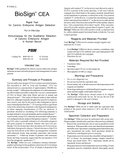
From www.bloodjournal.org by guest on October 21, 2014. For personal use only.
1991 78: 63-74
CD66 identifies a neutrophil-specific epitope within the hematopoietic
system that is expressed by members of the carcinoembryonic
antigen family of adhesion molecules
SM Watt, G Sala-Newby, T Hoang, DJ Gilmore, F Grunert, G Nagel, SJ Murdoch, E Tchilian, ES
Lennox and H Waldmann
Updated information and services can be found at:
http://www.bloodjournal.org/content/78/1/63.full.html
Articles on similar topics can be found in the following Blood collections
Information about reproducing this article in parts or in its entirety may be found online at:
http://www.bloodjournal.org/site/misc/rights.xhtml#repub_requests
Information about ordering reprints may be found online at:
http://www.bloodjournal.org/site/misc/rights.xhtml#reprints
Information about subscriptions and ASH membership may be found online at:
http://www.bloodjournal.org/site/subscriptions/index.xhtml
Blood (print ISSN 0006-4971, online ISSN 1528-0020), is published weekly by the American
Society of Hematology, 2021 L St, NW, Suite 900, Washington DC 20036.
Copyright 2011 by The American Society of Hematology; all rights reserved.
From www.bloodjournal.org by guest on October 21, 2014. For personal use only.
CD66 Identifies a Neutrophil-Specific Epitope Within the Hematopoietic System
That Is Expressed by Members of the Carcinoembryonic Antigen Family of
Adhesion Molecules
By Suzanne M. Watt, Graciela Sala-Newby, Trang Hoang, David J. Gilmore, Fritz Grunert, Gerhard Nagel,
Sarah J. Murdoch, Elma Tchilian, E.S. Lennox, and Herman Waldmann
Preliminary results from the lVth Leucocyte Culture Conference have classified the monoclonal antibody (MoAb), YTH
71.3.2, as CD66. Two other MoAbs, YPC 2112.1 and CE61
2D3.1, share a common cellular specificity, reacting with cells
of the neutrophil series and colonic epithelium. The YTH
71.3.2 and CE6/2D3.1 MoAbs both recognize a similar CD66
defined epitope that is distinct from that identified by YPC
2/12.1. By Western blotting, these antibodies react with
different molecular species from cells of different lineages.
The antibodies identify 50- to 55-Kd, 80- to 100-Kd. and 130to 200-Kd components present in a semi-purified carcinoembryonic antigen (CEA) preparationfrom colonic adenocarcinomas and a 90- to 130-Kd molecule from HL-60 cells. With the
colonic cell line, LS174T, YPC2112.1 stains diffuse bands of
160 to 200 Kd and 90 to 130 Kd with equal intensity, whereas
the binding of CE612D3.1 and YTH 71.3.2 is biased toward the
lower molecular weight set of molecules. Remarkably, all
three antibodies recognize CEA-related molecules. Defined
analyses using HeLa cells transfected with CEA, NCA(NCAQO/
go), and CGM6(NCA-95) cDNAs show that the three MoAbs
identify CEA to varying degrees. While YTH 71.3.2 and
CE612D3.1 also bind to NCA-50/90, YPC 2/12.1 recognizes an
epitope expressed by both the NCA-50/90 and NCA-95
molecular species.
o 1991by The American Society of Hematology.
C
(NCA-50), and 26 Kd have been de~cribed.'~.'~
In addition
to these NCA-like components, the PSG 5 glycoprotein
(another member of the CEA gene family but belonging to
the PSG subgroup) is also expressed in the granulocyte
lineage as shown by cDNA
It is still not clear if
the NCA-like components (other than NCA-50/90 and
NCA-95) represent distinct molecules or differentially glycosylated forms of one or a limited number of peptide
cores. Recent cloning experiments have shown that the
CGM6(NCA-95) cDNA sequence is highly homologous to
In contrast
but distinct from NCA(NCA-50/90) CDNA.~,'~,''
to NCA-50/90, NCA-95 appears to be restricted to cells of
the myeloid lineage in the later differentiation stages and is
not expressed in other tissues tested thus far." Its expression, at the mRNA level, is high in chronic myeloid
leukemic leukocytes and bone marrow but is barely detectable in mature neutrophils." Both NCA-50190 and NCA-95
are thought to appear within the neutrophilic series on
promyel~cytes.~'-~~
The monoclonal antibody (MoAb) YTH 71.3.2 has recently been clustered as CD66.24x25
We have characterized
ARCINOEMBRYONIC antigen (CEA) was initially
described by Gold and Freedman as a 180-Kd tumorassociated antigen in colon carcinomas.' Subsequent studies have identified a family of glycoproteins that share
common epitopes with CEA. The CEA family comprises all
the CEA-related antigens described thus far,14 and these
include a set of nonspecific cross-reacting antigens (NCA)
from epithelial and hematopoietic cells and in serum; the
NFA-1 (20 to 30 Kd) and NFA-2 (160 Kd) fecal antigens;
BGP-1 biliary glycoprotein, and the pregnancy-specific p
glycoproteins (PSGs). Four CEA-like antigens with apparent molecular weights of 200, 180,114, and 85 Kd have also
been found in human serum.5Recently, at least 17 different
CEA-related genes or cDNAs have been cloned: allowing
division of the CEA family into two subfamilies: the CEA
and PSG subgroups. However, the relationship between
cloned DNA sequences and the multiple forms of CEArelated proteins is still not entirely clear.
NCA was first identified in normal lung and spleen and
shown to exist as two glycosylation variants with apparent
molecular weights of 50 to 55 Kd (NCA-50) and 90 Kd
(NCA-90).6 Subsequently, these forms were shown to be
present in normal colonic mucosa, colonic adenocarcinomas, and granulocytes.6 Comparative cDNA analyses have
shown close structural and sequence homology between
CEA and NCA(NCA-50/90) cDNAs with 90% homology at
the nucleotide level and 85% at the amino acid leveL7 Both
proteins are members of the Ig superfamily and contain a
leader peptide, an IgV-like NH,-terminal domain, a variable number of IgC-like domains, and a short hydrophobic
carboxy-terminal region that is posttranslationally modified
to allow membrane anchoring via PI-linkage.6-"The differences between CEA and NCA(NCA-50/90) cDNAs reside
mainly in the 3' untranslated sequences of their mRNAs.
These CEA-NCA genes encode three mRNA species: two
of 3.0 and 3.5 kb for CEA and one of 2.5 kb for NCA in
colonic epithelium." l 4
More recently a variety of NCA-like or CEA-related
antigens has been found in human granulocytes. NCA-like
molecules with molecular weights'of 160 Kd (NCA-160), 95
Kd (NCA-95), 90 Kd (NCA-90), 80 Kd, 50 to 60 Kd
Blood, Vol78, No 1 (July 1). 1991: pp 63-74
From the Imperial CancerResearch Fund, London; the Department
of Medical Biochemistry, University of Wales College of Medicine,
Cardiff;Celltech Ltd, Slough, London; MRC Laboratory of Molecular
Biology, Cambridge; the Department of Pathology, University of
Cambridge, UK; the Clinical Research Institute of Montreal, Quebec,
Canada; and Institut fur Immunobiologie,Albert-Ludwigs Universitat,
Freibutg, Germany.
Submitted September 4, 1990; accepted February 15, 1991.
Supported by the Imperial Cancer Research Fund, the Medical
Research Council of Great Britain, and an EC concerted action
program grant.
Address reprint requests to S.M. Watt, PhD, Medical Oncology Unit,
Imperial Cancer Research Fund, St Bartholomew's Hospital, London
EClA 7BE, UK
The publication costs of this article were defrayed in part by page
charge payment. This article must therefore be hereby marked
"advertisement" in accordance with 18 U.S.C.section 1734 solely to
indicate this fact.
0 1991 by The American Society of Hematology.
0006-4971I9117801-0027$3.00/0
63
From www.bloodjournal.org by guest on October 21, 2014. For personal use only.
64
WATT ET AL
three CD66-like MoAbs both with respect to their cellular
reactivity and antigenic binding. The molecules defined by
CD66 and CD66-like MoAbs, in this report, belong to the
CEA family of adhesion molecules and are neutrophilspecific within the hematopoietic lineage. The possible
functional significance of such antigens within the neutrophilic compartment is discussed.
MATERIALS AND METHODS
Cell Preparation
Granulocytes, monocytes, and lymphocytes from heparinized
human peripheral blood were depleted of erythrocytes by centrifugation over Ficoll-Hypaque (density = 1.114 g/mL)!6 Normal human bone marrow obtained with ethical consent from patients
undergoing elective hip replacement or cardiac surgely was collected into bicarbonate-free Iscove’s medium (IMDM; pH 7.4)
buffered with 20 mmol1L N-2-hydroxyethy1-piperazine-Nf-2ethane sulphonic acid (HEPES) containing 10 UimL of presemativefree heparin and the total leukocyte fractions isolated on FicollHypaque as described above. Cells at the Ficoll-Hypaque interface
were collected and washed three times in 0.02 moVL phosphate
buffer containing 0.127 m o m sodium chloride (PBS; pH 7.4) and
1% (wtivol) bovine serum albumin (BSA) before flow cytometric
and clonogenic cell analyses. For pure neutrophil preparations,
peripheral blood was separated on a discontinuous gradient of
Ficoll-Hypaque with densities of 1.077 gimL and 1.114 gimL. The
neutrophils were collected from the 1.114 g/mL interface and any
residual erythrocytes were lysed at room temperature for 15
minutes with isotonic ammonium chloride. Monocytes were preenriched from light density ( < 1.077 g h L ) peripheral blood
mononuclear cells by a 1-hour adhesion to Petri dishes coated with
extracellular matrix (ECM) derived from baby hamster kidney
fibroblasts and followed by their removal with 2 mmol/L EDTA in
PBS (pH 7.4).
Cell Lines
The cell lines HL-60, U937, LS174T, and HT29 were maintained
in IMDM with 10% (wtivol) fetal calf serum (FCS) and fed with
fresh medium 24 hours before analysis. The colon carcinoma cell
lines LS174T and HT29 were trypsinized before use. HL-60 cells
(2 x 10s/mL) were induced to mature neutrophils by the addition
of 1pmol/L retinoic acid for 7 days.
MoAbs
DA rats were immunized with either human peripheral blood T
lymphocytes (the YTH MoAbs) or with blast cells (the YAML
antibodies) from the peripheral blood of a patient with acute
myeloblastic leukemia (M1 in the French-American-British classification) as detailed
The YPC2/12.1a and CE612D3.1
antibodies were derived by immunizing Lou rats with preparations
of human colon carcinomas and partially purified CEA.29MoAbs
were produced by fusion of the Y31Ag 1.2.3 rat myeloma cell line3’
with the hyperimmunized spleen cells. Clones were derived from
the original fusion by cloning and recloning in agar and the culture
supernatants containing various antibody activities were selected
on the basis of indirect binding assays on fixed normal and
neoplastic cells. YTH71.3.2 and YPC2112.1 are of the IgG2a
subclass, whereas CE612D3.1 is an IgGl antibody.
YAML 537.2% reacts mainly with intracellular components in
neutrophils and was used as a negative control that was not
CEA-related for cell sorting experiments. The YTH 76.9.111’’ rat
MoAb reacts with human HLA-Class I molecules and was used as a
positive control. The MoAb 47 (a kind gift from Dr F. Buchegger,
Epalinges, Switzerland) and N1 mouse monoclonals bind to CEA
and NCA-95 and to NCA-50190, re~pectively.’~~’~
The 3.9 mouse
MoAb (CDllc) was used as a monocyte marker and was supplied
by Dr Nancy Hogg (Imperial Cancer Research Fund, London,
UK). The mouse MoAb to CEA(H58) was kindly provided by Dr
G.T. Rogers (Charing Cross Hospital, London, UK) and prepared
as previously des~ribed?’,~’
The antibodies were used as culture supernatants or ascites and
purified on protein G (Pharmacia, Uppsala, Sweden) or by
ammonium sulphate precipitation and ion exchange chromatography on DE52 cellulose according to the manufacturer’s instructions.
Cell Labeling and Cytometric Analysis
The low-density peripheral blood and bone marrow nucleated
cells were suspended at a concentration of 2 x lo7 cells per
milliliter in resuspension medium containing PBS with 1% (wtivol)
BSA and 0.02% (wtivol) sodium azide (NaN,). Fc receptor binding
was inhibited by adding 1%(wtivol) normal rabbit or human serum
(heat inactivated at 56°C for 30 minutes) to the cell suspension for
30 minutes at 4°C. Cells were washed once in PBS with BSA and
NaN, and suspended to 2 X lo7 cells per milliliter in the same
medium. Saturating levels of each hybridoma supernatant or of the
purified antibody were added to the cells for 30 minutes at 4°C.
Cells were washed twice and made to 5 x lo7 cells1mL in
resuspension medium containing 100 pL of fluorescein isothiocyanate (F1TC)-labeled rabbit F(ab)’ antirat Ig (2 ~ng/mL)’~
or
biotinylated sheep antirat Ig (Amersham International, Amersham, UK) followed by FITC-streptavidin (Amersham). Cells were
incubated at 4°C for 30 minutes, washed, and resuspended to 2 x
lo6 cells/mL for cell sorting. In some cases, a fluoresceinconjugated MoAb, FITC-mouse antirat Ig (MAR 18.5), was used
as the second reagent?’,33 For the negative control, cells were
labeled with or without an irrelevant first-layer MoAb followed by
FITC-rabbit F(ab)’ antirat Ig, with FITC-MAR 18.5 or with
biotinylated sheep antirat Ig followed by FITC-streptavidin.
Cells were analyzed on the FACScan (Becton Dickinson, Sunnyvale, CA) and sorted on a FACS I1 fluorescence-activated cell
sorter (Becton Dickinson, CA) on the basis of forward light scatter
and fluorescein fluorescence using a single tunable Argon ion laser
(Spectra Physics, Heme1 Hempstead, UK; model 164-05) set at an
output power 0.3 W and an emission wavelength of 488 nm.34The
forward light scatter was determined with an amplifier gain of 2.
For fluorescein fluorescence, a photomultiplier voltage of 600 V
and an amplifier gain of 4 were used. The green fluorescence
emitted was filtered with a 530-nm long pass interference filter and
a 530-nm long pass glass filter (both from Ditric Optics, Marlboro,
MA). Cells were sorted at a rate of 2,000 cells per second at 4°C
into HEPES-buffered IMDM containing 50% (volivol) FCS in
siliconized earthed glass tubes at 4°C. The cells were centrifuged at
800 rpm for 15 minutes, resuspended in HEPES-buffered IMDM
in 10% (vol/vol) FCS, and either cytocentrifuged onto glass slides
and stained with May Griinwald-Giemsa stain for morphologic
analysis or analyzed for colony formation in methyl~ellulose.’~
ClonogenicAssays
Bone marrow cells were plated at 5 x lo4 cells/mL in IMDM
containing 0.9% (wthol) methylcellulose, 50 pmol/L 2-mercaptoethanol, 10 p g h L delipidated and deionized BSA, 360 p g h L
transferrin, 10% (vol/vol) prescreened FCS, 1 UimL erythropoietin, and 2% (vol/vol) human placental conditioned medium or 5%
to 10% (volivol) phytohemagglutin leukocyte-conditioned med i ~ m . ~ ~Colonies
,)’
containing more than 50 cells were scored on
From www.bloodjournal.org by guest on October 21, 2014. For personal use only.
65
CD66 IDENTIFIES THE CENNCA FAMILY
days 7 and 14 as macrophage (M-CFC), granulocyte (G-CFC),
mixed granulocyte-macrophage (GM-CFC), erythroid (BFU-E),
and mixed erythroid (CFU-Mix). The mixed erythroid colonies
contained at least three cell types, one of which was erythroid. The
myeloid colonies have been grouped together in the text and are
subsequently referred to a G/M-CFC. CFU-E were scored at day 7
and contained one or two erythroid clusters of at least eight cells.
Nonerythroid clusters containing 3 to 49 cells were also scored at
day 7.
ion-exchange chromatography on DE52 cellulose and coupled to
Atfigel-10 (Biorad, Heme1 Hempstead, UK) in 0.1 mol/L HEPES,
pH 7.5, according to the manufacturer’s instructions. The H58
MoAb was coupled to AlTigel-10 via rabbit antimouse Ig as detailed
earlier.38Solubilized, crude membranes or R41/R43 CEA samples
were applied to the columns in PBS pH 7.4 and the antigens were
eluted in 50 mmol/L diethylamine HCI pH 11.5 and 0.5% NP-40.
The eluates were neutralized with glycine and gel electrophoresed
or used for binding to other affinity columns.
Histochemistry
Binding Assays
Cryostat sections (5 pm) of frozen tissue were mounted on glass
slides, fixed, and incubated with saturating concentrations of the
rat MoAbs, followed by a second-layer detection reagent essentially as described by Rogers et al.”
Cell lines and neutrophils were suspended in Earles BSS
(GIBCO Biocult, Paisley, UK) containing 10 mmol/L HEPES pH
7.4, 0.8% (wtivol) BSA, and 0.1% (wthol) NaN,. Red blood cells
(RBCs) were resuspended in PBS with 10 mmoVL HEPES pH 7.4.
Nucleated cells 2 x lo’, and 4 X lo6 RBCs were dispersed in soft
microtiter plates, spun, and resuspended in the appropriate dilution of MoAb. The cells were incubated for 1 hour at PC, spun to
remove the supernatant, washed twice, and the binding monitored
by the addition of 2 x lo5cpm of iodinated sheep antirat Ig in 100
pL of medium. After washing, the cells were suspended in 50 p L of
2N NaOH and counted. Partially purified CEA, diluted in PBS, 10
mmoliL HEPES pH 7.4, and 0.1% (wtivol) NaN, was bound to soft
microtiter plates (20 pglwell) overnight at 4°C. Wells were blocked
with 5% (wthol) BSA overnight. The binding assay was as
described for cells, except that incubations were conducted at room
temperature. Counts less than 200 cpm were considered negative.
In positive samples, counts were in the range of 3,000 (+) to 28,000
(+ + +) cpm.”
CEA Preparation
The CEA preparations (CEA R41 and R43) used in the present
study were kindly donated by Dr G.T. Rogers. These preparations
were isolated from a pool of liver metastases of colon carcinomas as
detailed” by 2 mol/L perchloric acid extraction and fractionation
on Sepharose 6B. The void volume from this column was separated
on Con A sepharose. The bound fraction eluted with 2% (wt/vol)
a-methyl-D-mannoside was used as the crude CEA R41/43 fraction.
Cell Membrane Preparations
Cell lines growing exponentially were washed twice with saline
and incubated for 10 minutes on ice in 20 mmol/L Tris-HC1 pH 7.4,
0.5 mmol/L phenylmethyl sulfonyl fluoride (PMSF), 1 mmol/L
EDTA, 1 mmol/L EGTA, and 1% aprotinin. Adherent cells were
then scraped off the flasks with a rubber policeman. Cells were
homogenized for 15 seconds using a dounce homogenizer and spun
at 4°C for 30 minutes at 10“. The supernatant was collected and
centrifuged at 4°C for 30 minutes at lO’g. The pellet was collected
as the crude membrane fraction, For affinity and lectin chromatography, the membranes were solubilized in PBS containing 0.5%
(wtivol) NP-40, 0.1% (wthol) NaN,, 0.5 mmol/L PMSF for 20
minutes in ice and spun for 10 minutes in a microfuge at 4°C. The
pellet was discarded.
HeLa Transfectants
cDNAs encoding CEA, NCA-50/90,7,’0and NCA-95*’ were
cloned into the expression vector ~ B E H - p u c 9 . 3HeLa
~
cells were
transfected with these constructs together with the plasmid carrying the neomycin resistance gene, pSV2ne0, to give stable HeLaCEA, HeLa-NCA, and HeLa-CGM6 cell lines expressing the
respective proteins. HeLa cells transfected with the plasmid
pSV2neo (HeLa-Neo) were used as a negative control. Transfection was performed as previously described.m
RESULTS
Immunoblots
Crude membrane preparations were solubilized by boiling for 2
minutes in 12.5 mmol/L Tris-HC1 pH 6.5, 2% (wthol) sodium
dodecyl sulfate (SDS) 10% (volhol) glycerol, and 0.001% bromophenol blue (BPB). For reducing conditions, 50 mmol/L dithiothreitol (DTT), was also included. These fractions were electrophoresed in 7.5% (wthol) polyacrylamide gels (SDS-PAGE).36The
separated proteins were transferred electrophoretically to nitrocellulose sheets as detailed by Towbin et a].)’ The filters were blocked
with 3% (wt/vol) BSA in TBS (50 mmol/L Tris-HC1 pH 7.4, 150
mmol/L NaCl) and the strips were cut for overnight incubation at
4°C with each MoAb in TBS with 1% (wthol) BSA. The unbound
antibody was removed with four washes of TBS, 1 mmol/L EDTA,
1%(wtivol) BSA, and 0.05% (vol/vol) NP-40 and the nitrocellulose
strips incubated with 2 x to5 cpmimL of lZSI-sheepantirat Ig or
antimouse Ig for 3 hours at 4°C. The excess second antibody was
removed as described above and the strips were air dried and
autoradiographed.
Afinity Chromatography
The YPC2112.1, YTH71.3.2, and CE6/2D3.1 antibodies were
purified from ascitic fluid by ammonium sulfate precipitation and
Characterizationof the CD66-like MoAbs
CD66 binding to cells and semipurified CEA. The specificity of CD66 (YTH71.3.2) and of two CD66-like MoAbs,
YPC2/12.1 and CE6/2D3.1, was analyzed on a series of cells
and cell lines (Table 1). YTH71.3.2, YPC2/12.1, and CE6/
2D3.1 all bound to the LS174T and HT29 colon carcinoma
cell lines, to neutrophils and HL-60 cells, but not to K562
cells or isolated erythrocytes of various blood groups.
Slightly weaker binding of YPC 2112.1 to HT29 cells
occurred when compared with the other MoAbs (Table 1).
Because the colonic cell lines express CEA-related products, binding of lZI-MoAbs to a partially purified CEA
preparation was examined. All three antibodies showed
positive binding (Table 1).
Competitive binding assays. The studies presented above
suggested that YTH 71.3.2, CE6/2D3.1, and YPC 2/12.1
might react with similar epitopes. Therefore, competitive
binding experiments were set up to test the epitope specificity of these MoAbs. YPC 2/12.1 and YTH 71.3.2 were
iodinated and assayed for their binding to a crude CEA
From www.bloodjournal.org by guest on October 21, 2014. For personal use only.
66
WATT ET AL
Table 1. Binding of '251-MoAbsto Cells and Semipurified CEA
MoAbs
Cells
YTH 71.3.2
CE612D3.1
YPC 2/12.1
+
++
+
++
+
++
HL-60
Peripheral blood neutrophils*
K562
Erythrocytes'
Group 0
Group A
Group B
LS174T
HT29
Semipurified CEA
-
-
-
-
-
~
-
-
-
-
-
+++
+++
++
+++
+++
+++
++
++
++
~~
Binding assays described in Materials and Methods. Binding levels
are (-J, <200 cpm; (+), 3,000 to 5,000 cpm; (++), >5,000 and
< 20,000 cpm; (++ +), > 20,000 cpm.
'Freshly isolated cells.
degrees with both monocytes and neutrophils. In all cases,
the majority of lymphocytes occurred in the negative
fraction (Fig 2, D through F) as did erythrocytes (data not
shown).
CD66 binding increases with neutrophilic maturation. A
comparison of the levels of antibody binding by FACScan
analysis of the promyelocytic cell line HL-60, neutrophils
derived from retinoic acid-induced HL-60 cells, purified
peripheral blood neutrophils, the macrophage cell line
U937, and enriched normal peripheral blood monocytes is
presented in Fig 3. A much higher mean level of surface
.-_ ,
100 80 -
40 60
preparation coated onto microtiter plates. Displacement of
binding of these radiolabeled antibodies was determined by
the addition of unlabeled YPC 2112.1, YTH 71.3.2, and
CE6i2D3.1. The binding of labeled YTH 71.3.2 was displaced by cold CE612D3.1 or YTH71.3.2, but not with YPC
2112.1 and vice versa (Fig 1). This indicates that YPC 2112.1
recognizes a different epitope to that detected with YTH
71.3.2 and CE612D3.1 and that YTH 71.3.2 and CE6/2D3.1
share common epitopes or epitopes that are sufficiently
close to cause steric hindrance in binding.
Histochemical analysis of colonic sections. Because all
MoAbs reacted with partially purified CEA from colonic
carcinomas and because YTH 71.3.2 and CE612D3.1 (but
not YPC 2112.1) recognize similar epitopes, YTH 71.3.2
and YPC 2112.1 were tested by immunohistochemistry on
sections from normal colon and on colonic adenocarcinomas. Both MoAbs stained similar cells, reacting with the
brush border of epithelium from normal colon and staining
the whole cell membrane of colonic adenocarcinomas as
detailed in Table 2.
Flow cytometn'c analyses of CD66 MoAbs with human
peripheral blood leukocytes. MoAbs were analyzed by flow
cytometry for their reactivity with different viable leukocytes present in human peripheral blood. The cells were
labeled as described in Materials and Methods and were
sorted into two subsets according to whether their fluorescence was above (positive) or below (negative) a threshold
defined using cells stained with an irrelevant first-layer
antibody and the fluoresceinated second-layer reagent. The
dot plots showing both fractions are presented in Fig 2, A
through C. The sorted cells were cytocentrifuged and
analyzed for their morphology. The percentage of neutrophils, monocytes, and lymphocytes recovered in each fraction are shown in Fig 2, D through F, respectively. The
YTH 71.3.2 and YPC 2112.1 antibodies reacted specifically
with neutrophils when peripheral blood cells were analyzed
(Fig 2, A, B, D, and E). YAML 537.2 was used as a control
antibody that does not recognize CEA-related molecules
and that occurs mainly as an intracellular component in
cells of the neutrophilic series and on monocytes.26As
shown in Fig 2, C and F, YAML 537.2 reacted to varying
20
I
'i' d
' J b '4; .
;
ANTIBODY DILUTION
120
E
100
-
m-
*,*
YPCZ/I~.~
40 60
YTH71.3.2
26
28
2 0
--L
22
J
0
ANTIBODY DILUTION
120
,
4
c;
80 60 40 -
100
20
-
Fig 1. Inhibition of '%YPC2/12.1 or '"I-YTH71.3.2 binding to
semipurified CEA. Inhibition of binding of '=I.YPC2/12.1 (A) or '=IYTH71.3.2 (Band C) to the CEA R41/43 preparationwith unlabeled (A)
YPC2/12.1 (Wn],YTH71.3.2 (e),or CE6/2D3.1 ( 0 )ascites at antibody dilutions in the range F to 2'*; (B) YTH71.3.2 (-13) or
YPC2/12.1 (+) ascites at dilutions ranging from P to 213; and (C) YTH
71.3.2 (8--8) or CE6/2D3.1 (e)purified Ig (0 to 16 pgl. The 0 point on
the axis represents binding of '=Mabeled antibodiesin the absence of
unlabeled antibody. The maximum levels of binding for each set of
curves was normalized to 100% and were in the range of 10' to 5 x 10'
cpm per iodinatedantibody added per well.
From www.bloodjournal.org by guest on October 21, 2014. For personal use only.
CD66 IDENTIFIES THE CENNCA FAMILY
67
Table 2. Reactivity of MoAbs With Colonic Tissue
Positive Staining of Colonic Sections'
Normal
MoAbs
Immunofluorescence
YAML 537.2
Intense intracellular staining in globet cells with
weak staining of the cellular membrane
The brush border of epithelial cells
The brush border of epithelial cells
YTH 71.3.2
YPC U12.1
Malignantt
Immunofluorescence
lmmunoperoxidase
Minimal staining distribution
Patchy intracellular staining
Apical staining. No brush
border staining
The brush border of epithelial cells
The brush border of epitheIiaI cells
Cell membrane
Cell membrane
Cell membrane
Cell membrane
lmmunoperoxidase
'CD66 (YTH 71.3.2) and YPC U12.1 staining was compared with a contr.01 MoAb (YAML 537.2) that does bind to CENNCA molecules but reacts
with LS174T and HT29 colonic cell lines.
tAdenocarcinomas.
expression i s evident on the mature neutrophils whether
these are induced from HL-60 or obtained from normal
peripheral blood. Twenty-five percent to 30% of uninduced
HL-60 and 73% to 89% of retinoic acid-induced HL-60
cells stained above background with the CD66 and CD66like MoAbs. Staining of U937 or with an enriched population of normal peripheral blood monocytes was negliglible
(Fig 3 , 7 through 12). This result suggests that the epitopes
detected by CD66 and CD66-like antibodies increase in
their level of expression as cells mature from the promyelocyte to the mature neutrophilic stages, and are absent or
very low on monocytic cells.
In human bone marrow, antigens defined by CD66 and
CD66-likeMoAbs are limited to cells of the neutrophilic series.
YTH71.3.2 and YPC 2/12.1 were examined in detail for
their reactivity with clonogenic cells in normal human bone
marrow. Total bone marrow leukocytes were isolated on
Ficoll-Hypaque (density < 1.114 g/cm3) and labeled with
Y'I'l!71.3.2
A
120
100
80
60
+
40
20
-
0
C
Fig 2. Flow sorting of peripheral blood leukocytes. FACS cell sorter analysis of MoAbs YTH 71.3.2
(A and D),YPC 2/12.1 (B and E), and YAML 537.2(C
and F) on total human peripheral blood leukocytes
showing the positive (+) and negative (-) fluorescence fractions used for sorting. The sorted cells
were cytocentrifuged and analyzed by morphology
(D through F). The recovery of each cell type in the
positive (+) and negative I-) sorted fractions is
shown for neutrophils (PMN), for monocytes (Mo),
and for lymphocytes (Ly) after sorting cells labeled
with YTH 71.3.2(D),YPC U12.1 (E), and YAML 537.2
(F).
Y A A i I. 5 3 7.Z
FORWARD SCATTER
+.
-m
From www.bloodjournal.org by guest on October 21, 2014. For personal use only.
68
WATT ET AL
I
I
NEUTROPHILS
1
21
I
HL-60
4 1
L
FLUORESCENCE
Fig 3. Binding of CD66 and CD66-like MoAbsto myeloid cells. ComparativeFACScananalyses of CE6/2D3.1(1B, 41, Al, 7B.10H). anti-HLAclass
I YTH 76.9.111 (1C). YTH 71.3.2 (2E, 5L. 5M. 80, 11K). and YPC2/12.1 (36. 60,6P, 9F. 12M) binding to purified peripheral blood neutrophils (1
through 3); to uninduced HL-60cells (41,5L. 60); to retinoic acid-induced HL-60 cells (4J.5M. 6P); to the macrophage cell line U937 (7 through 9);
and to isolated human peripheralblood monocytes (10 through 12). Monocytes were identified by staining with (CDllc) MoAb 3.9 (101). Binding
was detected using FITC-rabbitFlab), antirat Ig or FITC-rabbitFlab), antimouse Ig. Negative controls for each sample are 1A. 20.3F. 4H. 5K. 6N.
7A,8C,9E,10G,11J,and 12L.
each antibody and a fluorescein-tagged detection reagent.
The cells were sorted on the basis of fluorescence intensity
into positive and negative components as described in the
previous section. Each sample was analyzed for the presence of clonogenic cells and for the distribution of more
mature cell types. Tables 3 and 4 show the recovery of
hematopoietic progenitors and more mature cells after
sorting human bone marrow samples with YTH 71.3.2 and
YPC 2/12.1 antibodies, respectively. In three separate
experiments using these MoAbs, cells of the neutrophil
series were consistently present in the positive fraction.
These included cells from the late-myeloblast or earlypromyelocyte stages to the mature neutrophils. Again
monocytes were generally negative. Most of the recovered
cells initiating day 7 erythroid clusters and day 14 colonies
(Tables 3 and 4), whether erythroid, myeloid or multipotential, occurred in the negative fraction. However, a substantial proportion of cells initiating day 7 nonerythroid colonies and clusters copurified with the neutrophilic cells and
were presumably restricted to this lineage (Tables 3 and 4).
Recoveries of the different progenitors varied among experiments. With the YPC 2/12.1 antibody, recoveries of sorted
progenitors were in excess of 100% (Table 4), while those
with the YTH 71.3.2 antibody were lower (Table 3). In
experiment 2 in Table 1, the recovery of cells initiating day
14 erythroid-containing colonies was high (loo%), while in
experiment 1 the recovery of those cells forming day 7 and
14 myeloid colonies and clusters was high ( > 94%).
Antigen Identification
Zmmunoblotting. LS174T and HL-60 cells and partially
purified preparations of CEA were solubilized in SDS,
electrophoresed in polyacrylamide gels, and the proteins
transferred electrophoretically to nitrocellulose. Binding of
antibodies to their antigens was detected using radioiodinated anti-Ig.
When the membrane fraction from HL-60 cells was
analyzed, a 90- to 130-Kd component was detected with
YPC2112.1 (Fig 4, track B), CE6/2D3.1 (C), and YTH71.3.2
(D), but not with the anti-CEA MoAb H58 (track A). With
the LS174T cell extract, the YPC 2/12.1 antibody recognized antigens with apparent molecular weights of 160 to
200 Kd and 90 to 130 Kd both under reducing (data not
shown) and nonreducing conditions (Fig 4, track H). YTH
71.3.2 bound only to the smaller (90 to 130 Kd) molecular
weight molecule (Fig 4, track I). CE6/2D3.1 bound preferentially to the lower molecular weight component (Fig 4,
track J), while its binding to the 160- to 200-Kd component
varied from experiment to experiment. In parallel experiments on LS174T cells, the anti-CEA MoAb H58 labeled
the 160- to 200-Kd band only (Fig 4, track G). Pronase
digestion of the membrane fraction from LS174T cells
From www.bloodjournal.org by guest on October 21, 2014. For personal use only.
69
CD66 IDENTIFIES THE CENNCA FAMILY
Table 3. Recovery of Clonogenic Cells After Sorting Human Bone Marrow With CD66 (YTH 71.3.2)
Experiment 1
Cell Fraction
CFC per lo5 cells sorted (%)*
Day 7
CFU-E
G/M-CFC and clusters
Day 14
G/M-CFC, BFU-E, and CFU-Mix
% of Nucleated cells
% Morphology
Blast cells
Neutrophilic cellst
Others*
Starting Bone Marrow
Experiment 2
Negative
Positive
374
(100)
461
(100)
287
(77)
383
(83)
(0)
194
(42)
282
(100)
436
(100)
143
(51)
112
(51)
0
(0)
90
(21)
208
(100)
155
(75)
7
(3)
242
(100)
156
(65)
13
(6)
100
40
58
100
41
59
0
4
(100)
54
(100)
42
4
(100)
1
(2)
36
(86)
1
(25)
56
(104)
2
(5)
0
3
3
(100)
52
(100)
45
(100)
Starting Bone Marrow
( 100)
(0)
54
(104)
4
(9)
2
(4)
35
(78)
Negative
(100)
Positive
*(%), The percentate of cells or CFC recovered in the positive or negative sample as a fraction of those found in the starting bone marrow. CFC
recovery is the number of CFC per lo5cells plated in each sorted fraction x % of cells in eachfraction.
tNeutrophilic cells include all cells in this lineage from the promyelocyte to the mature neutrophil stage.
$Others include lymphocytes, monocytes, eosinophils, and nucleated erythroid cells.
before electrophoresis resulted in the elimination of high
molecular weight bands from the blots (Fig 4, tracks M
through R).
With the partially purified CEA preparation, bands of
equivalent intensity within the 160- to 200-Kd and 50- to
55-Kd regions and a diffuse band in the 90- to 100-Kd area
were detected by immunoblotting with YPC 2112.1 (Fig Sa;
track A). CE6/2D3.1 bound to the 160-200kD component
weakly, but preferentially detected the lower molecular
weight components (Fig Sa, track B). YTH 71.3.2 bound the
90- to 100-Kd and 50- to 55-Kd molecules, but did not bind
the 160- to 200-Kd glycoprotein (Fig 5a, track C). In
contrast, H58 reacted strongly with the 160- to 200-Kd
component (Fig Sa, track D).
Table 4. Separation of Human Bone Marrow Cells With YPC 2/12.1
Starting
Bone Marrow
Cell Fraction
Day 7
CFU-E
G/M-CFC and clusters
Day 14
G/M CFC, BFU-E, and CFU-Mix
% Nucleated cells
Negative
Positive
Negative
Positive
Colonies per io5Cells
Sorted (recovery)
Colonies per 10' Cells Plated
(frequency)
0
496
(100%)
632
(100%)
1,062
499
(101%)'
491
(78%)
1,045
600
101
(100%)
365
100
4
(1 00%)
54
(100%)
42
( 100%)
0
282
(45%)
5
172
(170%)
3
(3%)
47
53
47
53
8
0
9
95
83
5
4
(100%)
4
(7%)
39
(93%)
0
(2%)
50
(93%)
3
(7%)
(0%)
% Morphology
Blasts
Neutrophilic cellst
Others*
*(%), The percentage of cells or CFC recovered in the positive or negative sample as a fraction of those found in the starting bone marrow. CFC
recovery is the number of CFC per IO5cells plated from each sorted fraction x % of cells in each fraction.
tNeutrophilic cells include all cells in this lineage from the promyelocyte to the mature neutrophil stage.
*Others include lymphocytes, monocytes, eosinophils, and nucleated erythroid cells.
From www.bloodjournal.org by guest on October 21, 2014. For personal use only.
70
W A l l ET AL
A
B
CDFF
G
H I J K L M
---
N O P Q R
-
Mr
kD
-210
- I30
-94
-80
-68
-43
-4.
Westun bkd
d mdo and LSl74TuJng antl CEA-NCA, COW, orCD6Mko Mdbr.Th. HLaO (A through F) md LS174T (0
WON rdUbi1h.d. tun on 7.5% SDSpo~.ay(anIdogeh,W
lo" f e w a d to nbocdluk..
und.r nondudng condltlons, Momdwl(t, ch.antlbodlr IndMod k l o w . and dwdoped wtth kdirutod rabbit atlmouw lg 01 sheep antlr.1 b
M o m autoradiography. Samples M through R m r e wbjeeted to pronne digeation befor. eI.chophoresh. The molecular mlo)ltmarkets were
throuoh R) d
l nwmkrcw pfspa"
myorin. 210 Kd; Bgaladoa1d.w. 130 Kd; phosphorylase b. 94 Kd; human transferrin, Bo Kd; BSA. 68 Kd; ovalbumin, 43 Kd. The antibodies uaed
for lmmunoblottlngare In the following trocka: (A. G, M), anti-CEA H58; (6,H,N).YPC U12.1;(C, 1.0).COW, VTH 71.3.2; (0.J, P), CE6/2D3.1; (E. K.
0).rabbtt antlmouae 10; (F. L. R),sheep antirat 19.
Afiinity chmmutogmphy. To confim that thc antibodies
b u n d to thc samc componcnts. the partially purificd CEA
prcparation was iodinatcd (I':'-CEA containing preparation) and applicd to affinity columns couplcd with the
various antibodies indicatcd in Fig Sb, clutcd, and subjcctcd
to gcl electrophoresis and autoradiography. Undcr thcsc
conditions. B 1x0- to 200-Kd protcin bound prcfcrcntially to
the anti-CEA (H58) column (Fig 5b, track €3). ldcntical
electrophoretic pattcms wcrc obtaincd whcn thc crudc
I':'-CEA preparation was fractionatcd on affinity columns
couplcd with CD66 (YTH 71.3.2) (Fig Sb. track C) or with
CE6nD3.1 and YPcU12.1 (data not shown). Broad bands
in the 100- to 200-Kd. 80-Kd. and 50- to 55-Kd rcgions wcrc
obscrvcd in all caws.
Thc crudc I':'-CEA samplc was applicd to YPC 312.1 or
CE6nD3.1 affinity columns. Bound protcins wcrc clutcd
with 50 mmol/L dicthylaminc. pH 11.5, rc-applicd to
columns couplcd with various MoAbs. and analyzcd by
SDS-PAGE. The material clutcd from thc YPC 312.1
column was rccognizcd to varying cxtcnts by all other
antibodies tcstcd (Fig Sb. tracks G through J). In a similar
manncr, thc crudc I'"-CEA samplc. clutcd from the CE6/
2D3.1 affinity column, bound to both CEW2D3.I and
YPcUI2.I columns (Fig Sb. tracks D and E, rcspcctivcly).
The run-through fraction that did not bind to thc YPC
U12.1 mlumn containcd matcrial that was recognized by
YTH 71.3.2 (Fig Sb, track L) and CE6DD3.1 (Fig Sb. track
M). but not by thc anti-CEA antibody. H58 (Fig 5b. track
K). Thcsc studics confirm that, in addition to binding thc
low molccular wcight spccics. YTH 71.3.2 binds to the high
molccular wcight spccics cvcn though this binding was not
dctcctcd by immunoblotting. Variations in thc molcculcs
idcntificd by affinity chromatography and Wcstcm blotting
indicatc that protcin folding is important for the dctcction
of wmc antigcnic sitcs.
Thcsc findings indicatc that the CD66 master antibody.
YTH 71.3.2. and two others like it (YPC 312.1 and
CE6DD3.I) idcntify cpitopcs prcscnt on a set of molecules
of diffcrcnt sizcs in thc scmipurificd CEA preparation, one
of thcsc bcing CEA. Howcvcr. within this colonic carcinoma cxtract. thcrc arc components that arc reactive with
YTH 713.2 and CE6/2D3. I that do not bind to Y PC U12.1.
Analysis of HeLa tmmfectants. To show that CD66
binds to CEA and NCA componcnts dircctly, HcLa cells
stably cxprcssing CEA (HcLa-CEA), NCA-S0/90 (HcLaNCA). and NCA-95 (HcLa-CGM6) molecules were labclcd with YPC 312.1, YTH 71.3.2. and CE6DD3.1 and
analpcd by FACScan analysis. The rat monoclonal. YTH
From www.bloodjournal.org by guest on October 21, 2014. For personal use only.
CD66 IDENTIFIES
A
0 C
71
THE C E m C A FAMILY
D
E F
A
0 c
D E F
G W
I
J
K
L M
" M r
i*
no
5. IO) wnt.m bbl O M w of a wmlpUrHkd colonk CEA pnp.r.tlon d n g mntlCEA -,or
CD68-like MoAb..TIN CEA pF.p.rOth w n
extroetad from 12 lhrw motastaw of colonk ed.nourclnomn with 2 m d l L perchlork nid, fractionated on Sophamu 68, and bound t o and
eluted from Con A Sopharose aa dotailed in tho Materiala and Moth&. (a) The extract was e I M r o p h o r d on 7.5% SDS-PAGE under reducing
condltiom, blotted with the antibodlw indkated, and developed with iodinated rabbk antimouu Ig or s h w p antirat Ig M o r e autoradiography.
The molecular weight marken are described in Fig 4. Tracks: (A), VPCW12.1; (E), CE6/2D3.1; (C). Y"ti 71.3.2; (D). H58; (E), rabblt antimouw Ig; IF).
8heep antirat Ig. Them were the antibodies u u d for blotting. (b) Affinlty chromatography and SDS-PAGE anawws of semipurified colonic CEA
preparations using anti-CEA. CD66. or CD6B.Iike MoAba. The remipurified CEA preparation waa iodinated and bound t o and eluted from affinity
columns made with the various antibodies i n d k a t d M o r e SDS-PAGE and autoradiography. Track (A) h the total iadlnated extract. Affinity
columns used are for tracks (E) H 5 8 and (C) YTH 71.3.2. The crude "'MEA sample waa bound to and eluted from CEW2D3.1 coupled h o d s and
mapplied t o CE6/203.1(0) or to VPC 2/12.1 (E) affinity columns before elution and SDS-PAGE. Tho fraction that did not blnd t o the first CE6l2D3.1
column waa alao reapplied t o a second CE6/2D3.1 column (F) and the bound material eluted and aho analyzed by SDS-PAGE. In similar
experiments, the YPC 2/12.1 bound (G t o J) and unbound (K t o M) wmipurified '"I-CEA fraction, wera reapplied t o affinity columna. The
molecules thst thon bound t o and were eluted from beads coupled with (G, K) H58; (H. L) Y l M 71.3.2; (I, M) CE6/203.1; and (J) VPC W12.1 were
analyzed by SDS-PAGE as described in Materiala and Methods.
76.9.1 1 I. was uscd as an ovcrall positivc control for class I
cxprcssion on HcLa cclls. Thc N1 antibody. which rcacts
with NCA-S0/90. and thc MoAb 47. which rcacts with both
CEA and NCA-95. wcrc uscd as positivc spccificity controls. The rcsults prcscntcd in Fig 6 show that thc positivc
control MoAb. YTH 76.9. I 1 I . rcacts with all transfcctants
including thc HcLa Nco ncgativc control cclls. CE6/2D3.1
and YTH 71.3.2 rccognizc thc HcLa-NCA transfcctant
strongly (which cxprcsscs NCA-50/90) and HcLa-CEA
wcakly. CE6QD3.1 appcars to bind CEA morc avidly than
docs YTH 71 3.2. confirming thc immunoblotting rcsults.
YPC Ul2.l idcntifics a dctcrminant prcscnt on all thrcc
CEA, NCA-S0/90, and NCA-95 spccics cxprcsscd by thc
trsnsfcctants.This rcsult is also consistcnt with thc immunoblotting data. Thcrcforc, at lcast somc of thc molcculcs
rccognizcd by YTH71.3.2. YPC2/12.1. and CE6QD3.1 arc
mcmbcrs of thc CEA family.
DISCUSSION
In this rcport wc havc shown that CDM and two othcr
CDM-likc rat MoAbs rcact with CEA-rclatcd molcculcs.
Cross-blocking cxpcrimcnts havc shown that thc mastcr
MoAb. YTH 71.3.2 (clustcrcd as CDW"."). and CE6QD3.1
rccognizc thc samc or clmcly linkcd cpitopcs that arc
distinct from thosc rccognizcd by YPC 212.1. Analysis of
YTH 71.3.2 and thc YPC 212.1 MoAbs on pcriphcral
h l d and bonc marrow cclls by FACS ccll sorting has
shown that thc cpitopcs rccognizcd by thcsc MoAbs arc
rcstrictcd in thcir ccll surfacc cxprcssion to ncutrophilic
cclls. Exprcssion of thcsc cpitopcs is first obscrvcd on
promyclcmcs or latc mycloblasts and on a proportion of
progcnitor cclls capablc of forming latc (day 7) mycloid
clustcrs and colonics in vitro. Prcliminary studics suggest
that cxprcssion is low on promyclocytcs, incrcascs t o a
maximum on myclocytcs and mctamyclocytcs. and shows
similar or slightly lowcr cxprcssion on ncutrophils. Thc
componcnts wcrc not dctcctcd on crythroid precursors nor
on carly myclomonocytic or multipotcntial progcnitor cclls
from human bonc marrow.
Antigcnic analysis of the moleculcs recognized hy Y T H
71.3.2, CE6QD3.1. and YPC 212.1 shows that all thrcc
antibodics idcntify a sct of componcnts (with similar
molccular wcights) in cxtracts of mctastasizcd human
colonic carcinomas. Thc antibodics rcact with componcnts
in thc 50- to 55-Kd. 80- to IO-Kd. and 130- to 200-Kd
molccular wcight rcgions in a scmipurificd CEA prcparation and two componcnts in LS174T cclls with molccular
wcights of 90 to 130 Kd and I 6 0 to 200 Kd to varying
dcgrccs. In immunohlotting cxperimcnts. YPC U12.1 bound
high and low molccular wcight forms to an cquivalcnt
dcgrcc. whcrcas YTH 71.3.2 and CEhQD3.1 prcfcrcntially
rccognizcd the lowcr molccular wcight componcnts. That
CE6QD3.1 and YTH 71.3.2 rcact with slightly diffcrcnt
cpitopcs is suggcstcd by thc ability of CE6QD3.1. but not
From www.bloodjournal.org by guest on October 21, 2014. For personal use only.
72
WATT ET AL
z
a
W
z.
W
0
z
W
0
v,
W
a
2
-I
LL
W
2
c
5
W
a
N,
'Mab47'y p c l
- Y T .H 7 ,
CE6 ' Y T H 7 6 . ,
MAb
MONOCLONAL ANTIBODIES
Fig 6. Binding of CD66 and CD664ike antibodies to HeLa transfectants. HeLa cells were transfected with cDNAs encoding CEA (HeLaCEA), NCA-50/90 (HeLa-NCA), and NCA-95 (HeLa-CGM6). HeLa cells
transfected with the pSV2 neo plasmid were used as a negative
control. Cells were stained without specific first antibody ( - 1. MoAb);
with the CD66 and CD66-like antibodies (YTH 71, CE6, YPC 2); with
anti-major histocompatibility complex class I, YTH 76; with the
NCA-50/90 specific antibody, N1; or with the CEA and NCA-95
reactive MoAb 47 antibody. The reaction was developed with fluorescent second antibody reagents and analyzed on the FACScan as
described in Materials and Methods. The mean fluorescence is plotted
for each analysis. (m), HeLa-neo;
HeLa-CEA;
HeLa-NCA;
HeLa-CGM6.
a),
m,
a),
YTH 71.3.2, to recognize the 160- to 200-Kd molecule on
blots of LS174T cells and of the CEA preparation. The
epitope also identified by YPC2112.1 is present on several
molecular species. In this report we have shown that YPC
2/12.1 recognizes at least three CEA-related molecules
(CEA, NCA-50/90, and NCA-95), while the YTH 71.3.2
and CE612D3.1 antibodies identify NCA-50/90 strongly and
CEA weakly. Previous studies'6,20*2S
have shown that while
NCA-50/90 and NCA-95 (but not CEA) are both expressed
on granulocytes, only the NCA-95 molecule is myeloid-cell
specific?' Because the CEA family members are highly
homologous at the amino acid level, the variability in the
molecular weights detected between different cell types
probably indicates variation in the type of CEA-related
gene products expressed by those different tissues. Complete identity of the set of molecules detected with CD66
and CD66-like antibodies requires more detailed biochemical analyses. However, recent expression cloning studies
confirm that these antibodies also identify at least one other
member of the CEA gene family (S. Watt and D. Simmons,
unpublished data, March 1991) that occurs in HT29 cells.
The expression of CEA-related molecules on cells of the
neutrophil series is of interest with respect to their possible
functional significance. In earlier studies, the CD66-defined
molecule was identified on mature peripheral blood neutrophils using the YTH 71.3.2 antibody as a 150- to 180-Kd
glycoprotein specie^.'^,^ This component may be equivalent
to the NCA-160 molecule previously des~ribed,'~?'~
although
in our experiments the CD66-defined epitope is not detected on the surface membrane of monocytes. On the
promyelocytic leukemic cell line HL-60, epitopes defined by
CD66 and CD66-like MoAbs are expressed to a lesser
degree than on mature neutrophils and components in the
90- to 130-Kd molecular weight region are consistently
identified. Therefore, the possibility arises that either
different NCA-related molecules46occur on mature neutrophils and their precursors, or that during myeloid differentiation the same component undergoes an apparent increase in molecular size. This possibility requires further
investigation but could be explained by an increased level of
glycosylation of a common peptide core occurring as
neutrophil maturation proceeds.
The function of CEA-related molecules on cells of the
neutrophil series is unknown, although recent research on
CEA may provide some insight into their role in myeloid
development. It has been proposed that these molecules
may function as ecto-ATPases."" In addition, the CEA and
NCA components analyzed to date are members of the Ig
s~perfamily.4'>~~
More recent studies using rodent cells
transfected with functional CEA and NCA cDNAs and
producing the 200-Kd CEA molecule and the 50- to 130-Kd
NCA components, respectively, have shown that both CEA
and NCA are involved in homotypic and heterotypic adhe~ion:~," It has recently been suggested that CEA may
function as an accessory adhesion molecule by controlling
the binding activity of type I collagen receptors to ECM
component^^^ in colonic epithelial cells. Uncontrolled growth
or loss of ordered tissue architecture in colonic carcinomas
may result from loss of CEA-associated ECM receptors
causing disruption of cell matrix interaction^,^^ as well as
from over-e~pression,~~
altered glycosylation, or redistribution43,46 of CEA molecules altering cell surface charge and
further disrupting the adhesion process. The adhesive
properties of CEA may also promote metastases by allowing circulation of tumor cells. CEA and NCA have been
detected in serum,2*3'5332
and systemic injections of CEA into
mice have been shown to enhance the metastatic potential
of a colorectal carcinoma cell
Studies such as these provide a basis for determining the
function of CEA-related molecules on myeloid cells. It has
been well established that myeloblasts and all cells of the
neutrophil series adhere to the ECM molecules, stromal
cells, and endotheli~m.~'
It is likely that adhesion molecules
mediating myeloblast-stromal cell-matrix interactions in
bone marrow bring the hematopoietic cells into contact
with growth factors concentrated by heparin-binding components of the stromal cell ECM!9,50 Subsequent development within the neutrophil lineage results in dissociation of
these cells from stromal elements in bone marrow and is
followed by their entry into the periphery. It would be of
interest to know if CD66-defined or the different CEArelated components can regulate such binding in the
neutrophilic lineage. Alternatively, because it is still unclear if all CEA-related gene products have adhesion
properties, it is possible that the different molecular forms
may have different functions during myeloid differentia-
From www.bloodjournal.org by guest on October 21, 2014. For personal use only.
73
CD66 IDENTIFIES THE CENNCA FAMILY
tion. Thus, it could be envisaged that the introduction of
increasing levels of specific types of NCA-like molecules or
of increasing degrees of glycosylation of these components
from the promyelocyte to the mature neutrophil stages
alters the Surface charge on these cells. This effect, together
with a diminished number of other adhesion receptors, may
weaken stromal-hematopoietic cell adhesion in a manner
reminiscent of the loss of adherence of colonic epithelial
cells during tumor development. More detailed studies are
necessary to test such possibilities.
ACKNOWLEDGMENT
The authors thankDr C. Milstein for kindly supplying the Y3 rat
myeloma cell line, Dr D. Bentley for advice, Dr A. Edwards for
advice on the FACScan analyses, Dr H. Durbin for reading the
manuscript, and M. Cotter for her expert typing of the manuscript.
REFERENCES
1. Gold P, Freedman SO: Demonstration of tumor-specific
antigens in human colonic carcinoma by immunological tolerance
and absorption techniques. J Exp Med 121:439,1965
2. Rogers G T Carcinoembryonic antigens and related glycoproteins molecular aspects and specificity. Biochim Biophys Acta
695:227,1983
3. Thompson J, Zimmermann W: The carcinoembryonic antigen gene family: Structure, expression and evolution. Tumor Biol
9:63,1988
4. Barnett T, Zimmermann W: Workshop Report: Proposed
nomenclature for the carcinoembryonic antigen (CEA) gene family. Tumor Biol 1159,1990
5. Neumaier M, Fenger C, Wagener U Delineation of four
carcinoembryonic antigen (CEA) related antigens in normal plasma
by transblot studies using monoclonal anti-CEA antibodies with
different epitope specificities. Mol Immunol22:1273,1985
6. Thompson J, Barnert S, Berling B, von Kleist S, Kodelja V,
Lucas K, Mauch E-M, Rudert F, Schrewe H, Weiss M, Zimmermann W Structure, expression and evolution of the human and rat
carcino-embryonic antigen (CEA) gene families, in Yachi A,
Shively J (eds): The Carcinoembryonic Gene Family. New York,
NY, Elsevier, 1989, p 65
7. Neumaier M, Zimmermann W, Shively L, Hinoda Y, Riggs
AD, Shively JE: Characterization of a cDNA clone for the
non-specific cross-reacting antigen (NCA) and a comparison of
NCA and carcinoembryonic antigen. J Biol Chem 263:3202,1988
8. Thompson JA, Pande H, Paxton RJ, Shively L, Padma A,
Simmer RL, Todd CW, Riggs AD, Shively J E Molecular cloning of
a gene belonging to the carcinoembryonic antigen gene family and
discussion of a domain model. Proc Natl Acad Sci USA 84:2965,
1987
9. Tawaragi Y, Oikawa S, Matsuoka Y, Kosaki G, Nakazato H:
Primary structure of nonspecific crossreacting antigen (NCA), a
member of carcinoembryonic antigen (CEA) gene family, deduced
from cDNAsequence. Biochem Biophys Res Commun 150:89,1988
10. Kolbinger F, Schwarz K, Brombacher F, von Kleist S,
Grunert F: Expression of an NCA cDNA in NlW3T3 cells yields a
llOK glycoprotein, which is anchored into the membrane via
glycosyl-phosphatidyl inositol. Biochem Biophys Res Commun
161:1126,1989
11. Hefta SA, Hefta UF, Lee TD, Paxton RJ, Shively J E
Carcinoembryonic antigen is anchored to membranes by covalent
attachment to a glycosylphosphatidylinositol moiety: Identification
of the ethanolamine linkage site. Proc Natl Acad Sci USA 85:4648,
1988
12. Zimmermann W, Weber B, Ortlieb B, Rudert F, Schempp
W, Fiebig H-H, Shively JE, von Kleist S, Thompson JA: Chromosomal localization of the carcinoembryonic antigeh gene family and
different expression in various tumors. Cancer Res 48:3153,1988
13. Cournoyer D, Beauchemin N, Boucher D, Benchimol S,
Fuks A, Stanners CP: Transcription of genes of the carcinoembryonic antigen family in malignant and nonmalignant human tissues.
Cancer Res 48:3153,1988
14. Boucher D, Cournoyer D, Stanners CP, Fuks A Studies on
the control of gene expression of the carcinoembryonic antigen
family in human tissue, Cancer Res 49:847, 1989
15. Buchegger F, Schreyer M, Carrel S, Mach J-P: Monoclonal
antibodies identify a CEA crossreacting antigen of 95 kD (NCA95) distinct in antigenicity and tissue distribution from the previously described NCA of 55 kD. Int J Cancer 33:643,1984
16. Audette M, Buchegger F, Schreyer M, Mach J-P: Monoclonal antibody against carcinoembryonic antigen (CEA) identifies
two forms of crossreacting antigens of molecular weight 90,000 and
160,000 in normal granulocytes. Mol Immunol24:1177,1987
17. Kuroki M, Matsuo Y, Kuroki M, Matsuoka Y: Nonspecific
cross-reacting antigen (NCA) expressed by human granulocytes:
Six species with different peptide sizes and membrane anchoring
forms. Biochem Biophys Res Commun 166:701,1990
18. Oikawa S, Inuzuka C, Kuroki M, Matsuoka Y, Kosaki G,
Nakazato H: Pregnancy-specific-pl-glycoprotein,
a CEA gene
family member, expressed in a human promyelocytic leukemia cell
line, HL60: Structures of protein, mRNA and gene. Biochem
Biophys Res Commun 163:1021,1989
19. Arakawa F, Kuroki M, Misumi Y, Oikawa S, Nakazato H,
Matsuoka Y Characterization of a cDNA clone encoding a new
species of the nonspecific cross-reacting antigen (NCA), a member
of the CEA gene family. Biochem Biophys Res Commun 166:1063,
1990
20. Berling B, Kolbinger F, Grunert F, Thompson JA, Brombacher F, Buchegger F, von Kleist S, Zimmermann W: Cloning of a
carcino-embryonic antigen gene family member expressed in leukocytes of chronic myeloid leukemia patients and bone marrow.
Cancer Res 50:6534,1990
21. Wahren B, Gahrton G, Hammarstrom S: Non-specific crossreacting antigen in normal and leukemic myeloid cells and serum
of leukemic patients. Cancer Res 40:2039, 1980
22. Noworoksla A, Harlozinska A, Richter R, Brodelo W:
Non-specific cross-reacting antigen (NCA) in individual maturation stages of myelocytic cell series. Int J Cancer 51:371,1985
23. Knapp W, Dorken B, Gelks WR, Rieber EP, Schmidt RE,
Stein H, von dem Borne AEG K r White Cell Differentiation
Antigens-Leucocyte Typing IV. Oxford, UK, Oxford University,
1989
24. Van der Schoot CE, Daams M, von dem Borne AEG Kr,
Skubitz KM, Skubitz APN, van Agthoven A, Braully H, Romagne
F, Lanini S, Kniep B, Civin CI, Fackler MJ, Chorvath B, Duraj J,
Bazil V, Horejsi V, Hildreth J, Hyman J, Tetteroo PAT: Biochemical analysis of the myeloid panel, in Knapp W, Dorken B, Gelks
WR, Rieber EP, Schmidt RE, Stein H, von dem Borne AEG Kr
(eds): White Cell Differentiation Antigens-Leucocyte Typing IV.
Oxford, UK, Oxford University, 1989, p 887
25. Noworolska A, Harlozinska A, Buchegger F, Lawinska B,
Richter R: Expression of non-specific cross-reacting antigen species in myeloid leukemic patients and healthy subjects. Blut 58:69,
1989
26. Swirsky DM, Watt SM, Gilmore DJ, Hayhoe FGJ, Waldman
From www.bloodjournal.org by guest on October 21, 2014. For personal use only.
74
H: The characterisation of monoclonal antibodies against haemopoietic cells. Comparison of an immuno peroxidase method with
fluorescence activated cell sorting. J Immunol Methods 61:171,
1983
27. Hale G, Hoang TK, Prosper0 T, Watt SM, Waldman H:
Removal of T cells for bone marrow transplantation. Comparison
of rat monoclonal antibodies of different isotypes. Mol Biol Med
1:305,1983
28. Finan PJ, Grant RM, De Mattos C, Takei F, Berry PJ,
Lennox ES, Bleehan NM: Immunohistological techniques in the
early screening of monoclonal antibodies to human colonic epithelium. Br J Cancer 46:9,1982
29. Rogers GT, Searle F, Bagshawe KD: Carcinoembryonic
antigen: Isolation of a subfraction with high specific activity. Br J
Cancer 33:357,1976
30. Galfre G, Milstein C Preparation of monoclonal antibodies:
Strategies and procedures. Methods Enzymol73:3,1981
31. Rogers GT, Rawlins GA, Kardana A, Gibbons AR: Binding
studies on two different monoclonal antibodies raised against
CEA. Eur J Cancer Clin Oncol19:629,1983
32. Rogers GT, Rawlins GA, Keep PA, Cooper EH, Bagshawe
KD: Application of monoclonal antibodies to purified CEA in
clinical radioimmunoassay of human serum. Br J Cancer 44:371,
1981
33. Watt SM, Gilmore DJ, Davis JM, Clark MR, Waldman H:
Cell surface markers on mouse haemopoietic cells. Reagents for
the isolation of progenitor cell populations. Cell Mol Probes 1:297,
1987
34. Watt SM, Katz FE, Davis L, Capellaro D, Gordon MY,
Tindle RW, Greaves MF: Expression of HPCA-1 progenitor
antigen and HLA-DR on growth factor and stroma dependent
colony forming cells. Br J Haematol66:153,1987
35. Zhou Y-Q, Stanley ER, Clark SC, Hatzfeld JA, Levesque
J-P, Federici C, Watt SM, Hatzfeld A Interleukin-3 and interleukin-la allow earlier bone marrow progenitors to respond to human
colony stimulating factor 1. Blood 72:1870,1988
36. Harlow E, Lane D: Antibodies. A Laboratory Manual. Cold
Spring Harbor, NY,Cold Spring Harbor Laboratory, 1988
37. Towbin H, Staehelin T, Gordon J: Electrophoretic transfer
of proteins from polyacrylamide gels to nitrocellulose sheets:
Procedure and some applications. Proc Natl Acad Sci USA
76:4350,1979
38. Schneider C, Newman RA, Sutherland DR, Asser U,
Greaves MF: A one-step purification of membrane proteins using a
high efficiency immunomatrix. J Biol Chem 257:10766,1982
WATT ET AL
39. Artelt P, Morelle C, Ausmeier M, Fritzek M, Hauser H:
Vector of efficient expression in mammalian fibroblastoid, myeloid
and lymphoid cells via transfection or infection. Gene 68:213,1988
40. Lin S-W, Guidotti G: Cloning and expression of a cDNA
coding for a rat liver plasma membrane ecto-ATPase. J Biol Chem
264:14408,1989
41. Paxton RJ, Mooser G, Pande H, Lee TD, Shively JE:
Sequence analysis of carcinoembryonic antigen: Identification of
glycosylation sites and homology with the immunoglobulin supergene family. Proc Natl Acad Sci USA 84:920,1987
42. Oikawa S, Imajo S, Noguchi T, Kosaki G, Nakazato H: The
carcinoembryonic antigen (CEA) contains multiple immunoglobulin-like domains. Biochem Biophys Res Commun 144:634,1987
43. Benchimol S, Fuks A, Jothy S, Beauchemin, Shirota K,
Stanners CP: Carcinoembryonic antigen, a human tumor marker,
functions as an intercellular adhesion molecule. Cell 57:327, 1989
44. Oikawa S, Imuzuka C, Kuroki M, Matsuoka Y, Kosaki G,
Nakazato H: Cell adhesion activity of non-specific cross-reacting
antigen (NCA) and carcinoembryonic antigen (CEA) expressed on
CHO cell surface: Homophilic and heterophilic adhesion. Biochem
Biophys Res Commun 164:39,1989
45. Pignatelli M, Durbin H, Bodmer WF: Carcinoembryonic
antigen functions as an accessory adhesion molecule mediating
colon epithelial cell-collagen interactions. Proc Natl Acad Sci USA
87:1541,1990
46. Fantini J, Rogoni J-B, Culouscou J-M, Pommier G, Marvaldi J, Tirard A: Induction of polarized apical expression and
vectorial release of carcinoembryonic antigen (CEA) during the
process of differentiation of HT29-D4 cells. J Cell Physiol 141:126,
1989
47. Hostetter RB, Campbell DE, Chi KF, Kerchoff S, Cleary
KR, Ullrich S , Thomas P, Jessup JM: Carcinoembryonic antigen
enhances metastastic protential of human colorectal carcinoma.
Arch Surg 125:300,1990
48. Campbell AD, Long MW, Wicha MS: Haemonectin, a bone
marrow adhesion protein specific for cells of granulocyte lineage.
Nature 329:744,1987
49. Gordon MY, Riley GP, Watt SM, Greaves M F Compartmentalisation of a human growth factor, GM-CSF, by glycosaminoglycans in the bone marrow microenvironment. Nature 326:403, 1987
50. poberts R, Gallagher J, Spooncer E, Allen TD, Bloomfield
F, Dexter TM: Heparan sulphate bound growth factors: A mechanism for stromal cell mediated haemopoiesis. Nature 332:376,1988


© Copyright 2026