
Retinoids (all-trans and 9-cis retinoic acid) stimulate production of
From www.bloodjournal.org by guest on October 28, 2014. For personal use only.
1994 84: 4107-4115
Retinoids (all-trans and 9-cis retinoic acid) stimulate production of
macrophage colony-stimulating factor and granulocyte-macrophage
colony- stimulating factor by human bone marrow stromal cells
H Nakajima, M Kizaki, A Sonoda, S Mori, K Harigaya and Y Ikeda
Updated information and services can be found at:
http://www.bloodjournal.org/content/84/12/4107.full.html
Articles on similar topics can be found in the following Blood collections
Information about reproducing this article in parts or in its entirety may be found online at:
http://www.bloodjournal.org/site/misc/rights.xhtml#repub_requests
Information about ordering reprints may be found online at:
http://www.bloodjournal.org/site/misc/rights.xhtml#reprints
Information about subscriptions and ASH membership may be found online at:
http://www.bloodjournal.org/site/subscriptions/index.xhtml
Blood (print ISSN 0006-4971, online ISSN 1528-0020), is published weekly by the American
Society of Hematology, 2021 L St, NW, Suite 900, Washington DC 20036.
Copyright 2011 by The American Society of Hematology; all rights reserved.
From www.bloodjournal.org by guest on October 28, 2014. For personal use only.
Retinoids (All-trans and 9 4 s Retinoic Acid) Stimulate Production of
Macrophage Colony-Stimulating Factor and Granulocyte-Macrophage
Colony-Stimulating Factor by Human Bone Marrow Stromal Cells
By Hideaki Nakajima, Masahiro Kizaki, Akira Sonoda, Shigehisa Mori, Kenichi Harigaya, and Yasuo lkeda
Retinoic acids (RAs) exert pleiotropic effects on cellular
growth and differentiation. All-trans retinoic acid (ATRA)
and 9-cis retinoic acid (9-cis RA), a stereoisomer of ATRA,
induce differentiation of leukemic cell lines and cells from
patients with acute myelogenous leukemia (AML) in vitro.
Despite information on the
effects of RAs on hematopoietic
cells, little is known about howRAs act on the hematopoietic microenvironment, especially on bone marrow stromal
cells. Based on recent observations that various cytokines
produced mainly bybone marrow stromalcells regulate hematopoiesis, we analyzed the effects of RAs on cytokine
production by these cells. ATRA or 9-cis RA treatment of
human bone marrow stromal cell line KM101, which produces macrophage colony-stimulating factor (M-CSF) and
granulocyte-macrophage colony-stimulating factor(GM-
B
ONE MARROW stromal cells play important roles in
hematopoiesis by regulating proliferation and differentiation of hematopoietic stem cells.’**Membrane-bound form
of cytokines, such as stem cell factor (SCF) and macrophage
colony stimulating factor (M-CSF), are expressed on the
surface of stromal cells, and their receptors on stem cells
can act as adhesion rnole~ules.’~~
Proliferation and differentiation of stem cells are regulated by stromal cells through
signals mediated by adhesion molecules or cytokines. Bone
marrow stromal cells produce a variety of cytokines, including granulocyte colony-stimulating factor (G-CSF), granulocyte-macrophage colony-stimulating factor (GM-CSF),
M-CSF, interleukin-6 (IL-6), SCF, and leukemia inhibitory
factor (LIF), some of which are produced constitutively, and
others are induced on stimulation with tumor necrosis factora (TNF-a), IL-1P or lipopolysaccharide (LPS).5-7Monocytes
produce several of these stimulatory factors in response to
bacterial or viral infections or other inflammatory states.
Retinoic acids (RAs) exert pleiotropic effects in embryonic morphogenesis, epidermal cellular growth, and hematopoiesis.8-”All-trans retinoic acid (ATRA) induces differentiation of some leukemic cell lines and teratocarcinoma cell
lines inand
complete remission in a high proportion
of patients with acute promyelocytic leukemia (APL).I4”*
RAs exert their effects through two different sets of receptors; retinoic acid receptors (RAR-a, -P, - 7 ) and retinoid X
receptors (RXR-a, -0, -Y).”-’~ 9-cis retinoic acid ( 9 4 s RA),
a stereoisomer of ATRA, was recently found to be a highaffinity ligand for RXR.26,27We have shown that 9 4 s RA
is more potent than ATRA in inducing differentiation and
inhibiting proliferation of acute myelogenous leukemia
(AML) cell lines and fresh leukemic cells from patients,
including those with APL and AML M2 French-AmericanBritish [FAB] classification.”
Despite the wide clinical application of ATRA for the
treatment of APL, its effects on bone marrow stroma are
totally unknown. We report here the effects of ATRA and
9 4 s RA on cytokine production by human bone marrow
stromal cells and stromal cell lines and on the expression
and regulation of RAR-a and RXR-a mRNAs by stromal
cells.
Blood, Vol 84, No 12 (December 15). 1994: pp 4107-4115
CSF) constitutively, enhanced mRNA levels of both cytokines in a dose-dependent manner. Both RAs also stimulated
M-CSF production from primary cultures of human bone
marrow stromal cells. Both retinoic acid receptor (FIAR)-a
and retinoid X receptor (RXRI-a were expressed constitutively in KMlOl cells. ATRA did not affect the expression of
either receptor, whereas 9-cis RA increased RXR-a mRNA
expression in a dose-dependent manner, but did not affect
levels of RAR-a mRNA. These findings may have important
biologic implications for both the role
RAsof
in hernatopoiesis and the therapeutic effects of ATRA on thehematopoietic
microenvironment in patients with
acute promyelocytic leukemia (APL).
0 1994 b y The American Society of Hematology.
MATERIALS AND METHODS
Cells. K M l O l is a well-characterized functional bone marrow
stromal cell line generated by transfecting 6-week-old human bone
marrow stromal cell primary cultures with recombinant plasmid
pSV3gpt DNA containing the coding sequence of the early region
of simian virus 40 (SV-40).z9K M l O l cells are fibroblastic in appearance and produce several cytokines constituti~ely.~~
The cells were
maintained in a-minimal essential medium (a-MEM; GIBCO, Santa
Clara, CA) with 10% fetal bovine serum (Cytosystems, New South
Wales, Australia) and 1 % penicillin and streptomycin (GIBCO),
and passaged with 0.25% trypsin (GIBCO). All experiments were
performed using cells in logarithmic growth phase.
Long-term bone marrow cultures (LTBMC) were established as
described3’ from normal human bone marrow cells obtained from
bone marrow transplant donors with informed consent. Briefly, lightdensity bone marrow cells were isolated by density gradient centrifugation on Ficoll-Hypaque (Pharmacia Fine Chemicals, Piscataway,
NJ), and suspended in 75-cmz tissue culture flasks (Coming Glass
Works, Coming, NY) at a final concentration of 1 X lo6 cells/mL.
Cultures were maintained at 37°C in a humidified 5% CO, atmosphere with complete replacement of medium weekly and the removal, at each medium change, of all nonadherent cells. Cells were
passaged twice with 0.25% trypsin (GIBCO) before use.
Chemicals. ATRA (Sigma Chemical CO, St Louis, MO) was
dissolved in 100%ethanol at concentrations of lo-* m o m and stored
at -20°C in the dark. The 9-cis RA was kindly provided by H.P.
From the Division of Hematology and Laboratory Medicine, Keio
University School of Medicine, Tokyo, Japan; and the First Department of Pathology, Chiba University School of Medicine, Chiba,
Japan.
Submitted April 14, 1994; accepted August 18, 1994.
Supported by a grant from the Ministry of Education, Science and
Culture in Japan.
Address reprint requests to Hideaki Nakajima, MD, Division of
Hematology, Keio University School of Medicine, 35 Shinano-machi,
Shinjuku-ku, Tokyo 160, Japan.
The publication costs of this article were defrayed in part by page
charge payment. This article must therefore be hereby marked
“advertisement” in accordance with 18 U.S.C. section 1734 solely to
indicate this fact.
0 1994 by The American Society of Hematology.
0006-4971/94/8412-0007$3.00/0
4107
From www.bloodjournal.org by guest on October 28, 2014. For personal use only.
NAKAJIMA ET AL
4108
B
A
(Ulml)
420
-
1
400.
380.
360
m
-
340.
h
600
320.
ooo.
280.
260.
240.
200
220.
m-.
1
n
2
c c
4.0 kb
0.7 kb
-
3
4
v
l
2 3
4 5
6
7 8 91011121
10-1
GM-CSF
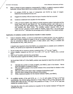
Fig 1. Cytokine production by KMlOl
cells treated with ATRA or 9 4 s RA. (A andB) ELISA SubconfluentKMlOl cells were incubatedwith
various concentrations ofATRA or 9 4 s RA added t o t h eculture medium after complete mediumchange. Cells were cultured for2 days under
shaded conditions before harvesting thesupernatant. Concentrations of M-CSF (A) and GM-CSF (B) were measured by ELISA (see Materials
and Methods). Ethanol was
added t o control cultures t o a final concentration of0.1%. A Bar 1, control; bars 2 t o 4, ATRA at lo-'' mollL,
mol/L,
mol/L, respectively. B Bar 1, control; bars 2 t o 7, ATRA
mol/L.
mol/L, respectively; bars 5 t o 7, 9 4 s RA at lo-'' mollL,
molll, lo-' mol/L, 10" mol/L, 10" mollL, respectively; bars 8 t o 13, 9 4 s RA at lo-" mollL, 10"' mollL,
at 10"' mollL, 10"' mollL,
lo-' mol/L, lo-' mollL, mollL,
lo-' mollL, respectively. Error bars indicate standard deviation. The results are the mean of triplicate
experiments. (C and Dl Northern analysis: Cells were exposed t o various concentrationsof ATRA (C) or 9 4 s RA (D) for 24 hours. Total RNA
was extracted, run on gels (20 pgllanel, and analyzed by Northern blotting as described in Materials and Methods. Blots were hybridized
with M-CSF, GM-CSF probes, and with a /3-actin probe as a control for the amountof RNA.
Koeffler (UCLA,Los Angeles, CA) and stored under the
same condisured by cellcountand'H-thymidineincorporationinto
cellular
DNA.KM101 cells were incubated in 24-well plates with various
tions as ATRA. TNF-a was kindly providedby Mochida Pharmaceuconcentrations of RAs for 48 hours and viable cells were counted
ticals CO (Tokyo, Japan). Cyclohexamide and actinomycin D were
purchased fromSigma Chemical Co. None of the cultures contained
based on trypan blue dyeexclusion. For 'H-thymidine incorporation
studies, KMlOl cells wereincubatedwithRAsfor24hoursand
more. than 0.1% ethanol.
Assayfor cellular proliferation. Cellular proliferationwasmea-"-thymidine
1 pL/well, (6.7 Cimmol",NewEnglandNuclear,
From www.bloodjournal.org by guest on October 28, 2014. For personal use only.
RETlNOlCACID AND BONE MARROW STROMAL CELLS
4109
ATRA
A
1
2
3
4
5
1
4.0 kb-,
0.7 k
c
-
B
9 cis RA
2 3 4 5
- W ”
-
M CSF
-
b
GM CSF
-
M CSF
GM-CSF
12,
ATRA
0
S e i s RA
3
o!
0
. . . .
,
.
.
10
time ( h )
,
,
,
20
,
,l
Fig 2. Time course analysis of cytokine mRNA induction by RAs. (A and B] KM101 cells were exposed to ATRA (lo” mol/L) (AI or 9-cis RA
(10” mollL) (B1 for various times and subjected to Northern analysis. Lane 1, control; lanes 2 to 5,3 hours, 6 hours, 12 hours, and 24 hours,
respectively. (C, D) The graphs show kinetics of relative mRNA level of M-CSF (C) and GM-CSF (D) compared with pactinmRNA.
Boston, MA) was added
for the last4 hours of incubation.Cells were
washed, treated with phosphate-bufferedsaline (PBS) containing 1%
EDTA, harvested, precipitated in S% trichloroacetic acid (TCA), (30
mmol/L Na2HP04)at 4°C for I hour, filtered onto glass microfiber
membranes (Whatman. Hillsboro, OR), and washedin 3% TCA (30
mmol/L Na2HP04). Samples wereassayed by liquidscintillation
counting.
RNA exrracrion and Northern andysis. Confluentstromal cells
were harvested and totalRNA was extracted by the methodof Chomczynski and Sacchi.” RNA samples (S to 20 pg) were electropho1.0% agarose gels (GIBCO BRL,
resedandsize-fractionatedon
Gaithersberg, MD) containing 17% formaldehyde and transferred to
nylon membranes (Hybond N’, Amersham, Arlington Heights, IL).
cDNAprobes for Northern analysis werelabeledwith[”PIdCTP
by using a random primerDNA labeling kit (Takara Shuzo CO,LTD.
Tokyo, Japan). Hybridization with the labeled probe was carried out
for 16 to 48 hours at 42°C in SO% formamide, 2 X SSC (1 X SSC
= I .S mmol/L sodium citrate, pH 7.0).S X Denhardt’s, 0.1 % SDS,
10% dextran sulfate (Pharmacia), and 1 0 0 mg/mLsalmonsperm
DNA (Sigma). Filters were washed to stringency of 0.1 X SSC at
65°C and exposed to Kodak XAR film (Eastman Kodak, Rochester,
NY). Autoradiograms were exposed from 24 hours
to 7 days and
hybridization signals werequantitated by scanningdensitometer
(Advantec DM-303, Tokyo, Japan).
DNA probes. cDNA probes for human M-CSF and human GMCSF were the Xhol-EcoRI fragment (2.0 kb) from plasmid pXMZ2
and the EcoRI-EcoRI fragment (0.8 kb) from plasmid p91023(B),1’
respectively. A human RAR-a cDNA(Kpnl-EcoRI; 1.9 kb) was
purified from plasmid pSG12” and
a human RXR-a cDNA probe
(EcoRI-EcoRI; 1.9 kb) was purified from pSMR3-I .24 The 8-actin
probewastheEcoRI-BarnHIfragment
(0.7 kb)fromplasmid
pHFbA-3’.”
Assays f i r cyrokineconcenrrarion. Concentrations of GM-CSF
in culture supernatants were assayed by enzyme-linked immunosorbent assay (ELISA) (BIOTRAK, Amersham) accordingto the manufacturer’s protocol. M-CSF concentrations were also measuredby
ELISA as follows: samples were spotted
onto anti-M-CSFantibodycoated 96-well plates andafter I hour, wells were washed with PBS,
reacted with rabbit anti-M-CSF antibody (IgG), washed again and
reacted with peroxidase-labeled goat antirabbit IgG antibody. Absorbance at 492 nmwasmeasuredby
spectrophotometer, andMCSF concentrations were calculated according to the standard concentration curve.
Flow cyrornerry. KM101 cells weretreatedwithATRA
(IO”
mol/L) and 9 4 s RA (IO” mol/L) for 24 hours, harvested by PBS
containing 1% EDTA, incubated with horse anti-M-CSF antibody
(generous gift from Green Cross CO, Osaka, Japan) at 4°C for 30
minutes, followed by fluorescein isothiocyanate (FITC)-conjugated
From www.bloodjournal.org by guest on October 28, 2014. For personal use only.
NAKAJIMA ET AL
4110
B
A
-
GM CSF
M = CSF
1
2
3
2
1
4
control
control
ATRA
ATRA
4
3
5
”
-
9
9 cis RA
Q
>
3
a
z
1.04
0.8 -
a 0.6 -
Plx
> 0.4 -
.->
E
E
Q
-
Q
-m
L
m
Q
a
- C ~ SRA
L
0.2 -
o
Q
a
~
0
,
2
l
,
,
,
4
6
time ( h )
l
,
8
l
I
10
0
l
0
1
’
l
‘
l
2
3
time ( h )
‘
l
4
*
5
Fig 3. Effects of actinomycin D on half-lives of RA-induced cytokine mRNA. K M l O l cells were exposed t o ATRA (lo” mollL) or 9 4 s RA
mol/L) for 24 hours before addition of actinomyeinD. Cells were treatedwith actinomycin D (5 pglmL) forvarious times (0 t o 8 hours)
and total RNA was extracted for Northern blotting
analysis. Untreated K M l O l cells were similarly analyzed as a control. A M-CSF, lanes 1 t o
4,O hour, 2 hours, 4 hours, 8 hours, respectively. B GM-CSF, lanes 1 t o 5 , O hour, 0.5 hours, 1 hour, 2 hours, 4 hours, respectively. Hybridization
mRNA of each cytokine are plotted onvertical axis of each graph.
signals were quantitated by
scanning densitometer, and relative amounts of
(U), control; (0).ATRA; ( 1, 9-cis RA.
(lo”
antihorse IgG antibody for 30 minutes. Cell surface M-CSF expression was analyzed by FACScan (Becton-Dickinson, Mountain View,
CA).
RESULTS
Effects of retinoids on cytokineproduction by KM101
cells. KMlOl cells produce M-CSF and GM-CSF constitutively as detected by ELISA. ATRA and 9-cis RA enhanced
M-CSF and GM-CSF production in a dose-dependent manner, although 9-cis RA was morepotent in inducing the
enhancement (Fig IA and B). To exclude the possibility that
enhanced M-CSF and GM-CSF levels reflect increased cell
proliferation stimulated by the RAs, ”-thymidine incorporation and cell counts were measured in cells incubated with
RAs for 2 days. ‘H-thymidine incorporation increased
slightly (1.4-fold maximum) atRA concentrations of IO-‘
mol& but cell counts were unchanged at any concentration
of RAs tested (data not shown).
Previous studies have reported three different mRNAs of
M-CSF that arise by alternative splicing.3’.’s.3‘ However,
KMlOl cells expressed only 4.0-kb mRNA by Northern blotting analysis, and both ATRA and 9-cis RA enhanced MCSF and GM-CSF mRNA expression in a dose-dependent
manner (Fig IC and D).
Time course analysis of M-CSF and GM-CSF mRNA
expression after addition of ATRA or 9-cis RA showed induction of mRNA of both
cytokines at 3 to 6 hours after
addition of RAs, with a plateau or a slight decrease between
12 and 24 hours (Fig 2).
Erects of retinoids on cytokine mRNA half-life. KM 101
cells were incubated with or without RAs for 24 hours before
the addition of actinomycin D to block RNA synthesis at
various time points. Neither ATRA nor 9-cis RA extended
From www.bloodjournal.org by guest on October 28, 2014. For personal use only.
4111
RETlNOlC ACID AND BONE MARROWSTROMAL CELLS
B
A
Log Fluorescence Intensity
12007
2
1000-
S!
800-
E
600-
8
4oo
e
Fig 4. Fbw cytometric analysis ofmembranebound form M-CSF. Upper panel: KMlOl cells were
treated with TNF- (10 nglmL1 (A), ATRA
moll
L) (B), or 9-cis RA
mollL1(C),M-CSF (l@UlrnL)
Q)
(D) for
24 hours, stained with anti-M-CSF
antibody
andanalyzedbyFACScan(Becton-Dickinson)asdescdbed in Materiels and
Methods.
Rough
dotted
0
lines
represent
negative
control.
Fine
dotted
lines
3
represent untmatd cells. Solid lines represent TNFa- (A), A T ” fa),S-cMA- {C), or M-CSF-(D) treated
cells.Lowerpanel: Mean fluorescenceintensity is
presented
graphs. M bar
g
200-
0
i
‘
untreated
control
the half-lives of these mRNAs (Fig 3). The calculated halflives of M-CSF and GM-CSF mRNA were about 3 hours
and 0.6 hours, respectively.
Flow cytometric analysis of membrane-bound form MCSF. M-CSF is primarily synthesized as a transmembrane
protein. After it is expressed on the cell surface, M-CSF is
proteolytically cleaved near the C-terminal of the extracellular domain and released in mature form into the culture
medium.32.35.36
To analyze cell surface expression of M-CSF,
K M l O l cells were incubated withATRA or 9-cis R A ,
stained with anti-M-CSF antibody, and assayed byflow
cytometry. Untreated KMlOl cells expressed M-CSF on the
cell surface, and both ATRA ( l o ” m o m ) and 9-cis RA
(lo” m o m ) enhanced the expression (Fig 4). The 9-cis
RA was more potent than ATRA in this enhancement. No
enhancement in fluorescence intensity was detected in
KMlOl cells cultured with M-CSF ( lo3U/mL) for 24 hours,
A
B
C
D
excluding the possibility of entrapment of M-CSF in the
culture medium by the extracellular matrix (Fig 4D).
Effects of R A s on normal human bone marrow stromal
cells. Unlike KMlOl cells, normal human bonemarrow
stroma is a heterogeneous mix that includes fibroblasts, endothelial cells, macrophages, adipocytes, and preadipo~ y t e s Therefore,
.~~
we examined the effects of RAs on primary cultures of normal human bone marrow stromal cells,
LTBMC. Both ATRA and 9-cis RA induced M-CSF production by LTBMC in a dose-dependent manner (Fig 5A).
Again, 9-cis RA was slightly more potent thanATRA.
Northern blot analysis gave consistent results at the mRNA
level (Fig 5B). The enhancement of M-CSF production was
comparable to that of TNF-a ( l 0 ng/mL) (Fig 5A and B).
We could not detect GM-CSF production either by ELISA
or Northern blotting (data not shown).
Effects of RAs on RAR-a and RXR-(Y mRNA expression.
From www.bloodjournal.org by guest on October 28, 2014. For personal use only.
NAKAJIMA ET AL
4112
T
1
4
10
B
1
2
3
4.0 kb +
To analyze the signalling pathway of both retinoids, the
effects of ATRAand 9-cis RA on RAR-a and RXR-a
mRNA expression by KM IO1 cells were examined by Northem blotting. RAR-a and RXR-a were expressed constitutively in untreated KMlOl cells; RAR-a mRNA expression
was not affected by either retinoid at various concentrations
(Fig 6A and B), whereas RXR-a mRNA was induced by 9cis RA (Fig 6D) butnotbyATRA
(Fig 6C) in a dosedependent manner.
DISCUSSION
The present study demonstrates that ATRA and 9-cis RA
stimulate M-CSF and GM-CSF production by human bone
marrow stromal cells. These stimulatory effects on cytokine
production were comparable to that of TNF-a (Fig 5A and
B). Thus RAs, as well as TNF-a, can act as stimuli for
cytokine production by stromal cells.
Although RA stimulated both M-CSF and GM-CSF production in KMlOl cells, human bone marrow stromal cell
primary culture produced only M-CSF in response to RAs.
The differences are probably due to the cellular heterogeneityofbonemarrow
stroma. Possibly, particular cell types
that produce only M-CSF in response to RAs predominate
in the population during culture in the flask. Alternatively,
cytokine gene expression and responses to mitogenic stimuli
Fig 5. M-CSF production and mRNA expression by LTBMC treated
with ATRA or 9-cis RA. (A) Subconfluent LTBMCs were treated with
various concentrations of ATRA or 9-cis RA after complete medium
change. Cells were cultured for5 days under shaded conditions and
supernatants wereanalyzed by ELISA for M-CSF. Bar1, control (ethanol 0.1%); bar 2, TNF-a, 10 ng/mL; bars 3 t o 6, ATRA 10”’ mol/L,
lo-’ mol/L, lo-’ mol/L,
mol/L, respectively; bars 7 t o 10, 9-cis
RA 10”’ mol/L, ’10.’ mol/L, lo-’ mol/L, 10” mol/L, respectively.
Error bars indicate standarddeviation. The results are the mean of
triplicate experiments. (B) Subconfluent LTBMCs were treated with
various concentrationsof ATRA, 9-cis RA, or TNF-a (10 ng/mL) for24
hours. Total RNA was extractedand subjected t o Northern analysis (5
pgllane). Lane 1, control (ethanol 0.1%); lane 2, TNF-a, 10 ng/mL;
lanes 3 t o 5, ATRA 10”’ mol/L, lo-’ mol/L,
mol/L, respectively;
lanes6 t o 8,g-cisRA 10”’ mol/L, lo-’ mol/L, 10” mol/L, respectively.
5
6
7
8
M - CSF
may be aberrant in the virally transformed KMlOl cells, as
compared with that of normal bone marrow stromal cells.
In fact, KMlOl cells produce relativelyhigh levels ofMCSF, G-CSF, and GM-CSF constitutively, whereas
LTBMCs produce no measurable levels of these cytokines
without stimulation.
The 9 4 s RA was a more potent stimulator than ATRA
in all experiments, consistent with previous studies describing the greater relative potency of 9-cis RA compared with
ATRA in inducing differentiation of HL-60 cells and leukemic cells from AMLpatients.” The9-cis RA bindsand
activates bothRARandRXR
efficiently, whereas ATRA
binds and activates only RAR. The greater potency of 9-cis
RA might rest in its ability to act, not only through RAR,
but also by forming heterodimers of RAR and RXR’“” or
RXR-RXR homodimers.‘“
We previously demonstrated that 9-cis RA downregulates
RXR-a expression in a dose-dependent manner during 9 4 s
RA-induced differentiation of HL-60cells,4’ suggesting a
role for RXR genes in cellular differentiation. In the present
study, we find a dose-dependent upregulation of RXR-a expression by 9-cis RA in conjunction with enhancement of
M-CSF and GM-CSF production. It remains unclear whether
RXR-a induction is the cause or the result of the enhanced
cytokine production. However, if the induction of RXR-a
From www.bloodjournal.org by guest on October 28, 2014. For personal use only.
RETlNOlC ACID ANDSTROMAL
BONE MARROW
A
4.5 kb
3.4 kb
1
2
CELLS
3
4
5
41 13
6
7 B
1
2
3
4
5
6
7
+
RAR CY
+
0 - actin
2.1 kb +
C
4.8 kb-
1
2
3
4
5
D 1
2
3
4
5
RXR CY
Fig 6. Expression of R A R a and R X R a mRNA in KM101 cells treated with RAs. Cells were exposed to various concentrations of ATRA (A,
C) or 9-cis RA (B, D) for 24 hours. Total RNA l20 pg/lane) was extracted and analyzed by Northern blotting with probes for RAR-a (A, B) and
R X R a (C, D). (A and B): lane 1, ethanol 0.1% as a control; lanes 2 to 7, lo-” mollL, 10”’ mol/L, lo-’ mollL, 10.’ molll, 10” mol/L,
moll
L, respectively. (C and D): lane 1, ethanol 0.1% as a control; lanes 2 to 5,
mol/L, 10”’ mol/L,
lo-’ mol/L, and lo” mol/L, respectively.
has some role in enhancement of cytokine production, RXRtion. Clarification of the precise mechanism of hyperleukocytosis awaits further study.
dependent pathways through RAR-RXR or RXR-RXR must
be critical in the action of 9-cis RA.
Our demonstration here that RAs (ATRA and 9 4 s RA)
ATRA is currently the first therapeutic choice in the treatcan stimulate M-CSF and GM-CSF production in human
bone marrow stromal cells may have important implications
ment of APL patients, in whom it is used as a differentiationinducing agent. Under ATRA therapy, serum ATRA concenin proliferation and differentiation of leukemic cells in patrationscanreadilyreach
mol/L;’which
issufficient to
tients with APL undergoing ATRA therapy.
stimulate cytokine production in stromal cells, as shown in
REFERENCES
this study and may modulate proliferation and differentiation
1.
Dexter
TM,
Allen
TD,
Lajtha LG: Conditions controlling the
of leukemic cells. Indeed, the hyperleukocytosis that occurs
proliferation of haematopoietic stem cells in vitro. J CellPhysiol
from 1 to 3 weeksafterinitiation of ATRA therapyl4lXis
91:335, 1977
thought to reflecttheelongation of life span or therapid
2. Reimann J, Burger H:In vitro proliferation of hematopoietic
proliferation of APL cells along with differentiation. Although
cells in the presence of adherent cell layers. 1. Culture conditions
the factors that affect the life span and proliferation of APL
and strain dependence. Exp Hematol 7:45, 1979
cells have not been identified, our study raises the possibility
3. Long MW, Briddell R, Walter AW, Bruno E, Hoffman R:
thatthestimulatory effects on cytokine production by RA
Human hematopoietic stem cell adherence to cytokines and matrix
might be involved. For example, RA might stimulate stromal
molecules. J Clin Invest 90251, 1992
cells in patients with APL to produce M-CSF, which in turn,
4. Uemura N,Ozawa K, Takahashi K, Tojo A, Tani K, Harigaya
might directly modulate
proliferative state of APL cells underK, Suzu S,Motoyoshi K, Matsuda H, Yagita H, Okumura K, Asano
S: Binding of membrane-anchored macrophage colony stimulating
going differentiation, or stimulate monocytes to induce production of cytokines thatenhance proliferation and differentia- factor (M-CSF) to its receptor mediates specific adhesion between
stromal cells and M-CSF receptor-bearing hematopoietic cells.
tion of APL cells. Although M-CSF does not often promote
growth of AML cells in vitro as compared with G-CSF,h2.4s Blood 82:2634, 1993
5. Migliaccio AR, Migliaccio G, Adarnson J W , Torok-Stobb B:
it does stimulate G-CSF production by monocytes in vitro.&
Production of granulocyte-colony stimulating-factor and granuloWe detected no GM-CSF production in the LTBMC system,
cyte/macrophage-colony-stimulatingfactor after interleukin-l stimubut undetectableamounts of GM-CSF produced by bone marlation of marrow stromal cell cultures from normal or aplastic anemia
row stromal cells might be concentrated in the surrounding
donors. J Cell Physiol 152: 199. 1992
mi~roenvironment~~
and might act preferentially onAPL cells
6. Wang SY, Su CY, HsuML, Chen LY, Tzeng CH, Ho CK:
attached to the stroma, promoting their growth and differentia-Effect of lipopolysaccharide on the production of colony-stimulating
From www.bloodjournal.org by guest on October 28, 2014. For personal use only.
4114
factors by the stromal cells in long-term bone marrow culture. Exp
Hematol 19:122, 1991
7. Wetzler M, Talpaz M, Lowe DG, Baiocchi G, Gutterman JU,
Kurzrock R: Constitutive expression of leukemia inhibitory factor
RNA by human bone marrow stromal cells and modulation by ILI, TNFa, and TGF-p. E X Hematol
~
19:347, 1991
8. Lotan R: Effects of vitamin A and its analogs (retinoids) on
normal and neoplastic cells. Biochem Biophys Acta 605:23, 1980
9. Maden M: Vitamin A and pattern formation in the regenerating
limb. Nature 295:672, 1982
10. Dower D, Koeffler H P Retinoic acid enhances colony-stimulating factor-induced clonal growth of normal human myeloid progenitor cells in vitro. Exp Cell Res 138:193, 1982
11. Dower D, Koeffler HP: Retinoic acid enhances growth of
human early progenitor cells in vitro. J Clin Invest 69:1039, 1982
12. Breitman TR, Selonick SE, Collins SJ: Induction of differentiation of the human promyelocytic leukemia cell line (HL-60) by
retinoic acid. Proc Natl Acad Sci USA 77:2936, 1980
13. Strickland S, Mandavi V: The induction of differentiation in
teratocarcinoma stem cells by retinoic acid. Cell 15:393, 1978
14. Huang M, YeY, Chen S, Chai J, Lu J, Xhoa L, Gu L,
WangZ:Useof
all-trans-retinoic acid in the treatment of acute
promyelocytic leukemia. Blood 72567, 1988
15. Castaigne S, Chomienne C, Daniel MT, Ballerini P, Berger
R, Fenaux P, Degos L: All-trans retinoic acid as a differentiation
therapy for acute promyelocytic leukemia: 1. Clinical results. Blood
76:1704, 1990
16. Chomienne C, Ballerini P, Balitrand N, Amar M, Bernard JF,
Boivin P, Daniel MT, Berger R, Castaigne S, Degos L: Retinoic
acid therapy for promyelocytic leukemia. Lancet 2:746, 1989
17. Warrell RP Jr, Frankel SR, Miller WH Jr, Scheinberg DA,
Itri LM, Hittelman WN, Vyas R, Andreeff M,Tafuri A, Jakubowski
A: Differentiation therapy of acute promyelocytic leukemia with
tretinoin (all-trans-retinoic acid). N Engl J Med 324:1385, 1991
18. Chen ZX, Xue YQ, Zhang R, Tao W, Xia XM, Li C, Wang
W, Zu WY, Yao XZ, Ling BJ: A clinical and experimental study on
all-trans retinoic acid-treated acute promyelocytic leukemia patients.
Blood 78:1413, 1991
19. Giguere V, Ong ES, Segni P, Evans RM: Identification of a
receptor for the morphogen retinoic acid. Nature 330:624, 1987
20. Petkovich M, Brand NJ, Krust A, Chambon P: A human
retinoic acid receptor which belongs to the family of nuclear receptors. Nature 330:444, 1987
21. Evans RM: The steroid and thyroid hormone receptor superfamily. Science 240“
1988
22. Brand N, Petkovich M, Krust A, de The H, Machino A,
Tiollais P, Dejean A: Identification of a second human retinoic acid
receptor. Nature 332:850, 1988
23. Krust A, Kastner F, Petkovich M, Zelent A, Chambon P A
third human retinoic acid receptor, hRAR-y. Proc NatlAcad Sci
USA 865310, 1989
24. Mangelsdorf DJ, Ong ES, Dyck JA, Evans RM: A nuclear
receptor that identifies a novel retinoic acid response pathway. Nature 345:224, 1990
25. Mangelsdorf DJ, Borgmeyer U, Heyman RA, Zhou JY, Ong
ES, Or0 ARE, Kakizuka A, Evans RM: Characterization of three
RXR genes that mediate the action of 9 4 s retinoic acid. Genes Dev
6:329, 1992
26. Heyman RA, Mangelsdorf DF, Dyck JA, Stein RB, Eichele
G, Evans RM, Thaller C : 9 4 s retinoic acid is a high affinity ligand
for the retinoid X receptor. Cell 68:397, 1992
27. Levin AA, Sturzenbecker LJ, Kazmer S, Bosakowski T, Huselton C, Allenby G, Speck J, Kratzeisen C, Rosenberger M, Lovey
A, Grippo JF: 9 4 s retinoic acid stereoisomer binds and activates
the nuclear receptor RXR-a. Nature 355:359, 1992
NAKAJIMA ET AL
28. Sakashita A, Kizaki M, Pakkala S, Schiller G, Tsuruoka N,
Tanosaki R, Cameron JF, Dawson MI, Koeffler HP: 9-cis-retinoic
acid: Effects on normal and leukemic hematopoiesis in vitro. Blood
81:1009, 1993
29. Harigaya K, Handa H: Generation of functional clonal cell
lines from human bone marrow stroma. Proc NatlAcad Sci USA
82:3477, 1985
30. Ma DDF, Da W-M, Purvis-Smith S, Biggs IC: Chromosomal
analysis of bone marrow stromal fibroblasts in allogeneic HLA compatible sibling bone manow transplantations. Leuk Res 11:661, 1987
31. Chomczynski P, Sacchi N: Single-step method of RNA isolation by acid guanidium thiocyanate-phenol-chloroform extraction.
Anal Biochem 162:156, 1987
32. Wong GG, Temple PA, Leary AC, Witek-Giannotti JS, Yang
YC, Ciarletta AB, Chung M, Murtha P, Kriz R, Kaufman RJ, Ferenz
CR, Sibley BS, Turner KJ, Hewick RM, Clark SC, Yanai N, Yokota
H, Yamada M, Saito M, Motoyoshi K, Takaku F Human CSF-l:
Molecular cloning and expression of 4-kb cDNA encoding the human urinary protein. Science 235:1504, 1987
33. Wong GG, Witek JS, Temple PA, Wilkens KM, Leary AC,
Luxenberg DP, Jones SS, Brown EL, Kay R M , Om EC, Shoemaker
C, Golde DW, Kaufman RJ, Hewick R M , Wang EA, Clark SC:
Human GM-CSF: Molecular cloning of complementary DNA and
purification of the natural and recombinant proteins. Science
228:810, 1985
34. Ponte P, Gunning P, Blau H, Kedes L: Human actin genes
are single copy for a-skeletal and a-cardiac actin but multicopy for
p- and y-cytoskeletal genes: 3’ untranslated regions are isotype
specific but are conserved in evolution. Mol Cell Biol 3:1783, 1983
35. Cerretti DP, Wignall J, Anderson D, Tushinski RJ, Gallis
BM, Stya M, Gillis S, Urdal DL, Cosman D: Human macrophagecolony stimulating factor: Alternative RNA and protein processing
from a single gene. Mol Immunol 25:761, 1988
36. Kawasaki ES, Ladner MB, Wang AM, Arsdell JV, Warren
MK, Coyne MY, Schweickart VL, Lee KT, Wilson KJ, Boosman
A, Stanley ER, Ralph P, Mark D F Molecular cloning of a complementary DNA encoding human macrophage-specific colony-stimulating factor (CSF-I). Science 230:291, 1985
37. Weiss L:An electron microscopic study of the vascular sinuses of the bone marrow of the rabbit. Bull Johns Hopkins Hosp
108:171, 1961
38. Zang X-K, Hoffmann B, Tran PB-V, Graupner G, Pfahl M:
Retinoid X receptor is an auxiliary protein for thyroid hormone and
retinoic acid receptors. Nature 355:441, 1992
39. Kliewer SA, Umesono K, Mangelsdorf DJ, Evans RM: Retinoid X receptor interacts with nuclear receptors in retinoic acid,
thyroid hormone and vitamin D signalling. Nature 355:446, 1992
40. Bugge TH, Pohl J, Lonnoy 0, Stunnenberg HG: RXR-a, a
promiscuous partner of retinoic acid and thyroid hormone receptors.
EMBO J 11:1409, 1992
41. Zhang XK, Lehmann J, Hoffmann B, Dawson MI, Cameron
J, Graupner G, Hermann T, Tran P, Pfahl M: Homodimer formation
of retinoid X receptor induced by 9 4 s retinoic acid. Nature 358:587,
1992
42. Kizaki M, keda Y, Tanosaki R, Nakajima H, Morikawa M.
Sakashita A, Koeffler HP: Effects of novel retinoic acid compound,
9 4 s retinoic acid, on proliferation, differentiation, and expression
of retinoic acid receptor-a and retinoid X receptor-a RNA by HL60 cells. Blood 82:3592, 1993
43. Muindi J, Frankel SR, Miller Jr WH, Jakubowski A, Scheinberg DA, Young CW, Dmitrovsky E, Warrell RPJr: Continuous
treatment with all-trans retinoic acid causes a progressive reduction
in plasma drug concentrations: implications for relapse and retinoid
“resistance” in patients with acute promyelocytic leukemia. Blood
79:299, 1992
From www.bloodjournal.org by guest on October 28, 2014. For personal use only.
RETlNOlCACID AND BONEMARROWSTROMALCELLS
44. Suzuki T, Nagata K, Murohashi I, Nara N: Effect of recombinant human M-CSF on the proliferation of leukemic blast progenitors
in AML patients. Leukemia 2358, 1988
45. Miyauchi J, Wang C, Kelleher CA, Wong GG, Clark SC,
Minden MD, McCulluch EA. The effects of recombinant CSF-1 on
the blast cells of acute myeloblastic leukemia in suspension culture.
J Cell Physiol 13555, 1988
41 15
46. Ishizaka Y, Motoyoshi K, Hatake K, Saito M, Takaku F,
Miura Y:Mode of action of human urinary colony-stimulating factor.
Exp Hematol 14:1, 1986
47. Gordon MY,Riley GP, Watt SM, Greaves MF: Compartmentalization of a haematopoietic growth factor (GM-CSF) by glycosaminoglycans in the bone marrow microenvironment. Nature
326:403, 1987


© Copyright 2026