
Respiratory System A&P
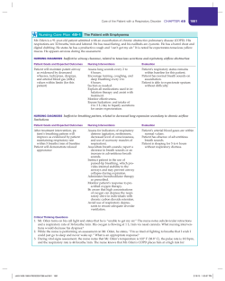
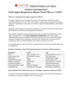
Respiratory System A&P Adapted from Dr. Marian Diamond and Essentials of Human Anatomy and Physiology Pearson Education 7th Edition Elaine N. Marieb Function of the Respiratory System Oversees gas exchanges (oxygen and carbon dioxide) between the blood and external environment Exchange of gasses takes place within the lungs in the alveoli(only site of gas exchange, other structures passageways Passageways to the lungs purify, warm, and humidify the incoming air Shares responsibility with cardiovascular system to distribute blood Copyright © 2003 Pearson Education, Inc. publishing as Benjamin Cummings Slide 13.2 Structures of the Respiratory System Science of OtorhinolaryngologyEars, nose, and throat • 3 types of tonsils (full of lymphocytes) 1. Palatine-posterior of oral cavity 2. Lingual-base of tongue 3. Pharyngeal-nasopharynx Which one of these tonsils do we typically remove due to frequent infection? Lingual Tonsils Tonsils with Mono Tonsillectomy CAUTION: Maybe very graphic if you have a weak stomach • https://www.youtube.com/watch?v=Pvj3CAof3k8 • Tonsilloliths aka Tonsil Stones- calcified minerals like calcium, phosphorus, magnesium, ammonia and carbonate found in crevices of the palatine or lingual tonsils • Removal• https://www.youtube.com/watch?v=rO0Cj3iFZBM 2 types of respiration • External respiration occurs when oxygen gas passes from the alveoli of the lungs and enters the blood stream through the capillaries • Internal respiration occurs when the oxygen gas in the blood of a capillary diffuses to the body’s tissues • • • • Structures of the Upper Respiratory System Nose- made of hyaline cartilage Attached to nasal bone External Nares- aka nostrils Functions of the nose: – Cleans air with cilia on pseudostratisfied columnar epithelium – Moistens air with goblet cells that produce mucus – Warms air with venous sinus beneath the epithelium that lines the nasal cavity – Resonates voice – Houses olfactory nerves Types of Pharynx (Throat) • Nasopharynx-nose • Oropharynx-mouth • Laryngopharynx“Food tube” before esophagus Nasopharynx • Eustachian tube originates here to the middle ear to equalize pressure • Home of pharyngeal tonsils • Pseudostratisfied columnar ciliated epithelium with goblet cells Oropharynx • Posterior to oral cavity • Home to palatine tonsils • Stratisfied squamous epithelial tissue Laryngopharynx • Posterior to larynx (windpipe) • Home of lingual tonsils • Stratisfied Squamous epithelial tissue Hard and Soft Palate • Hard palate is the bone of the maxilla – “roof of your mouth” – attached to teeth • Soft palate is the muscle tissue with the uvula - Uvula means “grape” it prevents water/food from getting into your nose from your mouth • Using your tongue you can feel the soft tissue behind the roof of your mouth Cleft Palate • 1 out of 900 children are born with a cleft palate • Exposes mouth to the nasal cavity Sinuses • Hollowed out areas within a bone • Clear air passage ways and help humidify the air as it enters the body • Ethmoid sinus (Ethmoid bone) home of cribiform plate where olfactory nerves send signals to the brain • Sphenoid sinus (Sphenoid bone) Tongue/Tear ducts • Tongue muscle at the base of the mouth attached to the mandible • Nasolacrimal duct is the inner tube that releases tears from the lacrimal glands Larynx • 4-5 inches in length • Anterior of neck • Surrounded by cartilage to keep the lumen (tube) open – Thyroid cartilage: • Largest • Protective shield • Increases in size for males to deepen the voice – Cricoid cartilage: inferior to thyroid complete ring that supports the thyroid and trachea – Voice Box and vocal cords found here http://www.youtube.com/watch?v=E4UG-dkVjrk Larynx: Epiglottis and Glottis • The glottis is the area in between the vocal cords • It is the opening of the larynx • The epiglottis (epi-upon) is what covers the glottis and is controlled by the tongue Top view of larynx http://www.youtube.com/watch?v=zQjJ5Alx40g The Structures of the Lower Respiratory Tract • Trachea (windpipe) – 4-5 inches in length – Connects larynx to bronchi – Rings of cartilage – (ave. 16) supports the structure http://www.youtube.com/watch?v=_Aw7EkIsYK0 Primary Bronchi (Bronchus) • First branches from trachea • Right bronchus is straighter and obstructed more easily • Left bronchus is bent more to make room for the heart Secondary Bronchi • Connected to primary bronchi • Smaller tubes to decrease air volume Tertiary and Terminal Bronchioles • Even smaller tubes made of cartilage to decrease air volume as it travels to the terminal bronchioles • Terminal bronchioles are not made of cartilage – This is where asthma attacks take place due to constriction – Epinephrine is used to dialate the bronchioles Alveoli • Alveoli Duct- is the small tube that extends from the terminal bronchiole to the Alveoli • Alveoli sac- individual hollow open area made of epithelial tissue where gas exchange takes place • Secretes surfactant-liquid that prevents alveoli from sticking to one another • Made also of elastic fibers that expands air when you inhale and recoils when you exhale Lungs • Held in pleural cavity surrounded by parietal and visceral pleura with serous fluid in between the pleura • Right lung has 3 lobes and the Left lung has 2 lobes WHY?? • 2 surfaces: Mediastinal (Mediastinum) and Costal (Intercostal muscles of the ribs) • Diaphragm pushes air out as you exhale • Lung is attached to the body via: – Bronchi – Pulmonary artery and pulmonary vein – Nerves http://www.youtube.com/watch?v=nJuFmyXeHkA Diseases of the Respiratory System • Laryngitis-inflammation of larynx • COPD- Chronic Obstructive Pulmonary Disease – Emphysema-inhales but can’t exhale no elasticity in lungs due to damaged elastic fibers from too much carbon – Bronchitis-long term cough with mucus • Lung Cancer- swollen alveoli, moves rapidly • Tuberculosis (TB)- bacterial infection destroys alveoli by releasing toxins and replaces tissue with scars looses elasticity • Pneumonia- pathogen bacterial infection that causes fluid to build up in the lungs Respiratory Failure Sudden Infant Death syndrome (SIDS) Apparently healthy infant stops breathing and dies during sleep Some cases are thought to be a problem of the neural respiratory control center One third of cases appear to be due to heart rhythm abnormalities Copyright © 2003 Pearson Education, Inc. publishing as Benjamin Cummings Slide Neural Control of Breathing • Neural Innervation- controlling rhythm of breathing from pons and medulla of the brain * Phrenic nerve and vagus nerve • Travels through the spinal cord to C3-C4 • Causes the diaphragm to contract • In an emergency if damage occurs above C4 you will need to immediately get oxygen for the individual Respiratory Volumes and Capacities Normal breathing moves about 500 ml of air with each breath (tidal volume [TV]) Many factors that affect respiratory capacity A person’s size Sex Age Physical condition Residual volume of air – after exhalation, about 1200 ml of air remains in the lungs Copyright © 2003 Pearson Education, Inc. publishing as Benjamin Cummings Slide Respiratory Volumes and Capacities Inspiratory reserve volume (IRV) Amount of air that can be taken in forcibly over the tidal volume Usually between 2100 and 3200 ml Expiratory reserve volume (ERV) Amount of air that can be forcibly exhaled Approximately 1200 ml Copyright © 2003 Pearson Education, Inc. publishing as Benjamin Cummings Slide Respiratory Volumes and Capacities Residual volume Air remaining in lung after expiration About 1200 ml Copyright © 2003 Pearson Education, Inc. publishing as Benjamin Cummings Slide Respiratory Volumes and Capacities Vital capacity The total amount of exchangeable air Vital capacity = TV + IRV + ERV Dead space volume Air that remains in conducting zone and never reaches alveoli About 150 ml Copyright © 2003 Pearson Education, Inc. publishing as Benjamin Cummings Slide Respiratory Volumes and Capacities Functional volume Air that actually reaches the respiratory zone Usually about 350 ml Respiratory capacities are measured with a spirometer Copyright © 2003 Pearson Education, Inc. publishing as Benjamin Cummings Slide Respiratory Capacities Figure 13.9 Copyright © 2003 Pearson Education, Inc. publishing as Benjamin Cummings Slide Air Volume and Lungs Capacity TV


© Copyright 2026