
Tuesday November 23, 2010
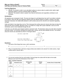
Tuesday November 23, 2010
•
•
•
•
You need your notebooks today.
We are beginning our chapter on the cell membrane.
Turn a sheet of paper sideways.
In the center of the sheet, draw a membrane like the one
on the board.
• On the left, make a chart titled: The 4 Roles of a Cell
Membrane.
• On the right, make a chart titled: The 4 Membrane
Bound Proteins.
Progress Towards Objectives
• Review your Cells Learning Objectives.
• How are you progressing?
Homeostasis
• Discovery
What are some conditions
in the human body that
need to be maintained so
that we maintain our
health?
What are some of the
reactions our body has
when these internal
conditions fall outside of
optimal?
sleep, energy, immune
system, water, food
hunger pains, yawning,
feeling tired and sick
Objectives: Cell Transport/Homeostasis
Unit
• To understand how particles such as water,
nutrients, macromolecules and bacteria
transport across cell membranes making them
semi permeable and be able to evaluate passive
and active methods of cell transport.
Objectives Cell Membrane
• Understand how the cell membrane is built and helps a
cell maintain homeostasis.
• Identify the ways the cell membrane restricts the
exchange of substances.
• Identify 4 membrane proteins.
• List the functions of membrane proteins.
• Vocabulary
• Phospholipid
• Lipid Bilayer
Cause & Affect
•
•
•
•
•
•
When it’s cold outside, we shiver.
Why?
When it’s hot outside, what do we do?
We sweat.
Why?
Sweating & shivering are just two examples of our
bodies trying to maintain a regular body temperature.
• Who remembers what this process is, one of the
properties of life, that causes us to maintain balance in a
changing environment?
Inside our Bodies
• The process of any organism trying to maintain
balanced, stable internal conditions in a changing
environment is called homeostasis.
• It’s natures balancing act it does because the world
always changes.
• What about inside our bodies?
• Does our internal environment change at all?
• How?
• What about the concept of dehydration?
• What do you do to fix this?
• How do you know to do this?
• Do you not feel thirsty?
Homeostasis
• Being thirsty is a chemical solution to satisfying
our body’s need for water.
• As our water levels change in our bodies we
have to adjust these to be as healthy as
possible.
• This is just one reaction that takes place in our
bodies but all living things react to their
environments.
• These reactions help organisms maintain
homeostasis.
Homeostasis, continued
• Individual cells, as well as organisms, must
maintain homeostasis in order to live too.
• One way that a cell maintains homeostasis is by
controlling the movement of substances across
the cell membrane.
• In our continued study of cells, we are going to
focus on how cells maintain homeostasis by the
properties of the cell membrane.
Homeostasis
What are the four functions that the cell membrane
performs to maintain homeostasis?
1. Regulates what goes in and out of the cell.
• Acting as a gate keeper
2. To provide structural support.
• Provides pressure against the internal environment
3. Recognizes foreign material.
• Uses ‘feelers’ called receptors to identify good and bad stuff in
the immediate area
4. Communicates and organizes with other cells.
• Uses the ‘feelers’ and glycoproteins to organize with other cells
into tissues forming the Extra Cellular Matrix (ECM)
Cell Membrane Construction
• The cell membrane is made of phospholipids.
• A phospholipid is a specialized lipid made
of a phosphate “head” and two fatty acid
“tails.”
– It is a lipid that contains phosphorus and that is a
structural component in cell membranes.
• The phosphate head is polar and is
attracted to water.
– The head is water ‘loving’ = HYDRO - PHILIC
• The fatty acid tails are nonpolar and are
repelled by water.
– The tail is water ‘hating’ = HYDRO - PHOBIC
WATER
Phospholipid
REGION
• Polar Heads are HYDROPHILIC
– They love water
– Made of phosphates
Hydrophilic
Heads
Hydrophobic
Tails
• Tails are HYDROPHOBIC
– They hate and are afraid of water.
– Made of fatty acids
NON- WATER REGION
Visual Concept: Cell Membrane
Outside of Cell
Inside of Cell
Cell Membranes, a.k.a. Lipid Bilayer
Structure
• Because there is water inside and outside the cell, the
phospholipids will naturally form a double layer called the
lipid bilayer.
– The basic structure of a biological membrane, composed of two
layers of phospholipids
• The nonpolar tails, repelled by water, make up the
interior of the lipid bilayer.
• The polar heads are attracted to the water, so they point
toward the surfaces of the lipid bilayer.
– One layer of polar heads faces the cytoplasm
– The other layer is in contact with the cell’s immediate
surroundings outside.
Lipid Bilayer
Visual Concept: Lipid Bilayer
Lipid Bilayer: Selectively Permeable
Barrier: Selectively Permeable
• Only certain substances can pass through the lipid bilayer.
• The phospholipids form a barrier through which only small,
nonpolar substances can pass.
• This feature makes the membrane SELECTIVELY
PERMIABLE, or allowing only SELECTED substances to
cross into the cell.
– This is also called “semi=permeable”
• Ions and most polar molecules are repelled by the
nonpolar interior of the lipid bilayer and therefore have to
be ushered in other ways.
Polar Molecules, like water, are repelled by the polar
heads of the phospholipid bilayer.
Other non-polar molecules, like carbon dioxide, are
not repelled and allowed to flow through the bilayer.
Membrane Proteins
• Homeostasis is also helped out by various proteins that
can be found in the cell membrane.
• Some proteins face inside the cell, and some face
outside. Other proteins may stretch across the lipid
bilayer and face both inside and outside.
• Proteins are made of amino acids. Some amino acids
are polar, and others are nonpolar.
• The attraction and repulsion of polar and non-polar parts
of the protein to water help hold the protein in the
membrane.
Membrane Proteins
What are the 3 major types of membrane proteins?
1. Peripheral Proteins: these are buoyed to the surface of
the membrane.
1.
Usually are associated with integral type proteins.
2. Integral Proteins: these penetrate into the hydrophobic
regions of the membrane
1.
2.
3.
Cell surface markers: glycoproteins are proteins with attached
sugar chains. These chains of sugars (remember
polysaccharides and carbohydrates) act as markers to help
identify themselves to other cells
Receptor proteins: these are the ‘feelers’ that identify good and
bad substances in the environment
Enzymes: catalyze reactions that happen on the inside of the
cell
3. Transmembrane Proteins: these span from outside to
inside the cell.
1.
Transport & Channel Proteins: allow large and/or polar
substances to pass through the membrane
Membrane Proteins: Examples. Can you
describe the type of protein?
G
H
Hydrophilic
Heads
F
A
B
C
Transmembrane
protein
Based upon what you learned, identify each of the
labeled structures in the illustration. You have 3
minutes.
E
Hydrophobic
Tails
D
Fluid Mosaic Model
• The cell membrane isn’t rigid.
• It has the ability to be squeezed and move around.
• And the same proteins are embedded throughout this
squishy membrane.
• This is called the Fluid Mosaic Model of the membrane.
Questions?…
• Finish the notes handout & the directed reading
from Friday.
• Complete the directed reading (book questions)
from Friday.
– When done, check in with me.
– Due tomorrow.
• Once completed, construct your own model of
the membrane.
Build a Membrane Activity
• Go to your groups and
gather
your
supplies.
Coloring Key
• For the remainder of the
Membrane = Yellow
Transporter Protein = Blue period you will build a cell
Channel Protein = Red
membrane!
Tethered Protein = Purple
• Check in with me when
Anchored Protein = Green
Receptor Protein = Orange finished = 25pts lab
Build a Membrane Directions
1. Color the parts (neatly) described on the board.
2. Cut out the phospholipid bilayer (page S2) along the
solid lines. Cut all the way to the edges of the paper in
the direction of the arrows.
3. Fold the phospholipid bilayer along the dotted lines
and tape the edges together to form a fully enclosed
rectangular box.
4. Cut out each protein (pages S3 and S4) along the solid
black lines and fold along the dotted lines.
5. Form a 3-D shape by joining the protein sides and tops
together and tape them in to place. Use the tabs to
help you.
6. Tape the 3-D proteins into place along the edges of the
phospholipid bilayer.
7. By staggering the membrane proteins back and forth
along both long sides of the bilayer “box”, the whole
model will stand up by itself on a table.
Summary
• One way that a cell maintains homeostasis is by
controlling the movement of substances across the cell
membrane.
• The lipid bilayer is selectively permeable to small,
nonpolar substances.
• Proteins in the cell membrane include cell-surface
markers, receptor proteins, enzymes, and transport
proteins.
Group Discussion/Reflection
Talk in your groups to answer the following questions.
1. Identify the 4 functions of the cell membrane that
allows it to maintain homeostasis
2. If I said the cell membrane is like a “gatekeeper”
what does that mean?
3. How does the membrane regulate things going in
or out of the cell? What is that property called?
4. What are the 4 types of proteins within the cell
membrane and describe the structure/function of
each of them.


© Copyright 2026