
Atopic Dermatitis, Eczema, and Noninfectious Immunodeficiency Disorders Rick Lin, DO MPH
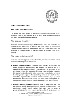
Atopic Dermatitis, Eczema, and
Noninfectious Immunodeficiency
Disorders
Rick Lin, DO MPH
July 15, 2003
Atopic Dermatitis
•
•
•
•
•
Aka atopic eczema
Aka infantile eczema
Aka flexural eczema
Aka disseminated neurodermatitis
Aka prurigo diathsique
Atopic Dermatitis
• High level of IgE antibodies to House dust
mites
• IgE bound to Langerhans cells in atopic
skin
• Food exacerbates symptoms in some
patients: eggs, peanuts, cow’s milk
represent up to 75% of positive test.
Atopic Dermatitis
• Pruritis is the hallmark of AD
• Eczematous eruption leads to lichenified
dermatitis
• Itching precedes the appearance of lesions
Infantile Atopic Dermatitis
• 60% of case AD present in the first year of
life, after 2 months of age
• Begin as itchy erythema of the cheeks
• Distribution include scalp, neck, forehead,
wrist, and extensors
• May become desquamate leading to
erythroderma.
Infantile Atopic Dermatitis
• Most cases the symptoms will disappear
toward the end of the second year.
• The role of food allergy in infantile and
childhood atopic dermatitis has been
clarified
• Egg, peanut, milk, wheat, fish, soy, and
chicken may exacerbate infantile AD
Involvement of the cheeks is characteristic of the infantile pattern of AD.
Childhood Atopic Dermatitis
• Characterized by less acute lesions
• Distribution: antecubital and popliteal
fossae, flexor wrist, eyelids, and face.
• Severe atopic dermatitis involving more
than 50% of body surface area is associated
with growth retardation.
Adult Atopic Dermatitis
• Distribution: antecubital and popliteal
fossae, the front side of the neck, the
forehead, and area around the eyes.
• Atopic individuals are at greater risk of
developing hand dermatitis than are the rest
of the population
• 70% develop hand dermatitis some times in
their lives
Cutaneous stigmata
•
•
•
•
Dennie-Morgan fold
Pityriasis alba
Keratosis pilaris
Hertoghe’s sign – thinning of the lateral
eyebrows
• Keratosis punctata palmaris et plantaris
Vascular Stigmata
• Headlight sign – perinasal and periorbital
pallor
• White dermographism – blanching of the
skin at the site of stroking with a blunt
instrument – cause edema and obscure color
of underlying vessels.
Infection
• Staph aureus – 90% of chronic lesions
• Eczema herpeticum – generalized herpes
simplex infection. Young children usually.
• Vaccination against smallpox is
contraindicated in person with atopic
dermatitis. Even when condition is in
remission, widespread and even fatal
vaccinia can occur.
Immunology
• T helper cell type 2 (Th2) dominance
• Th2 produces IL-4, 5, and 10
• IL-4 and IL-5 produce elevated IgE and
eosinophilia
• IL-10 inhibits delayed type hypersensitivity
• Th2 maybe sensitive to house mites or grass
pollen
Immunology
• Monocytes produces elevated amount of
prostaglandin E2 (PGE2)
• PGE2 reduces gamma-interferon
production, but not IL-4 from helper cells
thereby enhancing the Th2 dominance
• PGE2 also directly enhances IgE production
from B cells
Immunology
• Langerhans cells of AD patient stimulate
helper T cells into Th2 phenotype without
the presence of antigen
• Langerhans cells have IgE bound to their
suface receptors. These IgE are associated
with atopic antigens, such as house dust
mites
Differential Diagnosis
•
•
•
•
•
Seb Derm
Contact dermatitis
Nummular eczema
Scabies
Psoriasis
Histology
• Spongiotic dermatitis
• Lichen simplex chronicus
• Eosinophiles may be seen
Management
• Protect from scratching
• Adequate cleansing but not over bathing or
rubbing
• Gentle cleanser
• Anti-histamines, especially at night
• Bathing protocol
• Food allergies concerns and dietary restrictions.
• Hydrate skin daily with moisturizers
Management
• Topical steroid
• Wet compress of Burow’s solution such as
Domeboro.
• Crude coal tar/liquor corbonis detergens
(LCD)
Management
• “Topical FK506 (Tacrolimus) is
dramatically beneficial in SEVERE atopic
dermatitis”
• 95% showed good improvement in Alaiti
and Rusicka study in JAAD 1998, Archive
1999
Regional Eczema
•
•
•
•
•
•
•
Ear eczema
Eyelid dermatitis
Nipple eczema
Hand eczema
Diaper dermatitis
Infectious eczematoid dermatitis
Juvenile plantar dermatosis
Ear Eczema
• Most frequently caused by seborrheic or
atopic dermatitis
• Staph, Strep, or Psoeudomonas
• Earlobe is pathognomonic of nickel allergy
Eyelid dermatitis
• When on one eye only, it is most frequently
caused by nail polish
• When both eyelids are involved, consider
mascara, eye shadow, eyelash cement,
eyeline, etc
Nipple eczema
• Painful fissuring, seen especially in nursing
mothers
• Maybe an isolated manifestation of atopic
dermatitis
• If persist more than 3 month, and/or
unilateral, biopsy is mandatory to rule out
Paget’s
Hand eczema
• Spongiosis histologically
• Irritant hand dermatitis- seen in
homemakers, nurses. Resulting from
excessive exposure to soaps
• Pompholyx- tapioca vesicles, on sides of
fingers, palms, and soles
• Differentials – Bullous Tinea, id, allergic
contact dermatitis
Treatment
•
•
•
•
Barrier
Moisturizer
Systemic Corticosteroids
Phototherapy – UVA, PUVA, Radiotherapy
(Grenz Ray)
Diaper dermatitis
•
•
•
•
Jacquet’s erosive diaper dermatitis
Pseudoverrucous papule and nodules
Graduloma gluteal infantum
Irritation caused by bacteria, change in the
environment (wet, lower PH, feces)
• Candida albicans are secondary infection.
Infectious eczematoid dermatitis
• Vesicular, pustular, or cursted
• Ulceration and superficial infection may be
present
• Treatment involve the removal of irritant
and antibiotic treatment.
Juvenile plantar dermatosis
• Begins as a patchy symmetrical, smooth, red,
glazed macules on the base of the great toes
• Affect age 3 to puberty.
• Symmetrical lesions on weight bearing area
• “toxic sock syndrome” – caused by repeated
maceration of the feet by occlusive shoes and
nonabsorbent synthetic socks
• Virtually always resolve after puberty
Xerotic Eczema
• Aka winter itch, nummular eczema, eczema
craquele, and asteototic eczema.
• Anterior shins, extensor arms, and flank
• Elderly person predisposed.
• Use of bath oils in bath water is
recommended to prevent water loss
• Moisturizers – urea or lactic acid.
Nutritional Deficiency Eczema
• Localized, thickened pattern with scaling
patches.
• Exacerbated by nutritional deficiency
Hormone Induced Dermatoses
• Autoimmune progesterone dermatitis – urticaria,
urticarial paplues, papulovesicular lesion, or
eythema multiforme. Appear 5-10 days before
menses
• Autoimmune estrogen dermatitis – a cyclic skin
disorder with variable morphologies. Exacerbate
premenstrually or occur only immediately before
the menses. Treatment with tamoxifen maybe
effective.
Immunodeficiency Syndromes
•
•
•
•
•
•
•
X-Linked Agammaglobulinemia
Isolated IgA Deficiency
Common Variable Immunodificiency
Isolated Primary IgM Deficiency
Immunodificiency with Hyper-IgM
Thymic Hypoplasia
Thymic Dysplasia with Normal Immunoglobulins
(Nezelof Syndrome)
Immunodeficiency Syndromes
• Purine Nucleoside Phosphorylase
Deficiency
• Miscellaneous T-Cell Deficiencies
• Severe Combined Immunodeficiency
Disease (SCID)
• Thymoma with Immunodeficiency
• Ataxia-Telangiectasia (Louis-Bar’s S.)
• Wiskott-Aldrich Syndrome
Immunodeficiency Syndromes
•
•
•
•
•
•
•
•
X-Linked Lymphoproliferative Syndrome
Chronic Granulomatous Disease
Myeloperoxidase Deficiency
Leukocyte Adhesion Molecule Deficiency
Chediak-Higashi Syndrome
Hyperimmunoglobulinemia E Syndrome
Complement Deficiency
Graft-Versus-Host Disease
X-Linked Agammaglobulinemia
• Aka Bruton’s syndrome, sex-linked
agammaglobulinemia.
• Appear after 3-6 month of life
• Frequent Strep and staph infection. Viral
resistance intact.
• IgA, IgM, IgD, and IgE are absent in the serum.
IgG present with small amount
• Cell-mediated immunity intact. T lymphocytes
are normal, B cells are completely lacking
X-Linked Agammaglobulinemia
• Defect lies in the maturation block in pre-Bcell to B-cell differentiation
• Protein tyrosine kinase (PTK) gene deletion
and point mutation
• May develop leukemia, fatal encephalitis,
resporatory
Isolated IgA Deficiency
• Absence or marked reduction of serum IgA
• 1:600 in white population, most are entirely
well.
• Malignancy is increased in adult with IgA
deficiency.
Common Variable
Immunodificiency
•
•
•
•
Aka acquired hypogammaglobulinemia
HLA marker B8 and DR3 are affected
Recurrent sinopulmonary infections
B cells present but not terminally
differentiated
• T cells dysfunction evident
Isolated Primary IgM Deficiency
• Eczematous dermatitis presents in 1/5 of
patient with this condition
• Predisposition to bacterial infection
• Defect in maturation of IgM producing
plasma cell.
Immunodificiency with HyperIgM
• Low or absent IgG, IgE, and IgA level. Normal or
elevated IgM and IgD
• IVGG, and allogenic bone marrow transplant
• X-linked form caused by mutation or deletion of
Xq26.3-27.1 region, which encodes a ligand of
CD40, gp39
• Gp39-CD40 interaction signals for Ig isotype
switching.
Thymic Hypoplasia
• DiGeorge anomaly, aka III and IV pharyngeal
pouch syndrome
• Facies: notched, low-set ears, micrognathia,
shorten philtrum, hypertelorism
• Congenital absence of the parathyroid, thymus,
and abnormal aorta
• Hpocalcemia is the first sign
• Aorteic and cardiac defects are the cause of death
• Deletions within proximal long arm of
chromosone 22
Thymic Dysplasia with Normal
Immunoglobulins
(Nezelof Syndrome)
• Faulty development of thymus gland
• Autosomal recessive
• Thymus is present but under developed, no
cardiac abnormalities.
• Contrast to DiGeorge syndrome.
Purine Nucleoside Phosphorylase
Deficiency
• Greatly reduced T-Cell counts, depressed
cell mediated immunity
• B cells and antibody formation intact
• Mutation on 14q13
• Usually died of overwhelming viral
infection
Miscellaneous T-Cell
Deficiencies
• Cartilage-hair hypoplasia syndrome, AR, patient
with short-limbed dwarfism, fine sparse,
hypopigmented hair, defective cell mediated
immunity.
• Omenn’s syndrome, AR, mimic GVHD,
exfoliative erythroderma, eosinophilia, recurrent
infection, hypogammaglobulinema, diarrhea,
hepatosplenomegly, early death by 6 month.
Inefficient and abnormal generation of T-Cell
receptor.
SCID: Severe Combined
Immunodeficiency Disease
• Severe impairment of humoral and cellular
immunity
• Triad of Moniliasis of the oropharynx and
skin, intractable diarrhea, and pneumonia.
• Overwhelming viral infection is the cause
of death.
• Deficiency or total absence of circulating
lymphocytes
Thymoma with
Immunodeficiency
• Good’s syndrome
• Deficient in cell mediated immunity and
benign thymoma occurring simultaneously
• Thymectomy does not affect the
immunodeficiency
Ataxia-Telangiectasia
(Louis-Bar’s S.)
• Distinctive telangiectasia in bulbar conjuctiva and
flexural suraces of the arm developing during the
5th year of age
• Telangiectasia occurs on butterfly are of the face,
palate, ear, and exposed skin. Café au lait patches,
and Graying hair also present.
• Cerebellar ataxia is the first sign of this syndrome,
beginning in the second year of life.
• Choreic and athetoid movement present.
• Persistent granulomatous
plaques on the leg of child
with ataxia–telangiectasia.
Wiskott-Aldrich Syndrome
• Triad: chronic eczematous dermatitis resemble
AD, increase suseptibility to infections (OM), and
thrombocytopenia purpura/hepatoslpenomegly
• Death by age 6
• Accelerated IgA, IgM and IgE synthesis
• T-cell decline in numbers and activity
• Xp11 gene mutation. Codes for WASP protein
which reorganize cytoskeleton
X-Linked Lymphoproliferative
Syndrome
• Aka Duncan’s disease
• Inability to control Epstein-Barr virus
infection.
• Pt normal until develop infectious Mono.
• Xq26 abnormailty
• B-cell lymphoproliferative disease with
acquired hypoglobulinemia.
Chronic Granulomatous Disease
• Recurring purulent and granulomatous
infections involving long bones, lymphatic
tissue, liver, skin, and lung.
• Deficient in one of the component of
NADPH-oxidase complex, which generates
superoxide.
• Leads to inability to destroy bacteria per
radical mechanism
Chronic Granulomatous Disease
• 65% of cases are the X-linked form, lacks
the subunit of cytochrom b 558(gp91-phox)
• Female carrier has mixed and normal and
abnormal cells thus shows an intermediate
phenotype.
Leukocyte Adhesion Molecule
Deficiency
• Autosomal recessive
• Recurrent bacteria and fungal infections and
pus formations as a result of a block of
leukocyte migration
• Faulty complexing of the CD11 and CD18
integrins
• Death occurs in first 4 years of life unless
bone marrow transplant is undertaken.
Chediak-Higashi Syndrome
• Progressively degenerative, fatal, familial disease
of young children
• Partial oculocutaneous albinism, cutaneous and
intestinal infections early in childhood
• Ocular albinism is accompanied by nystagmus and
photophobia.
• Defect in the gene LYST, resulting in defective
vascular transport to and from the lysosome
Hyperimmunoglobulinemia E
Syndrome
• Consists of atopic-like eczematous
dermatitis, recurrent pyogenic infection,
high lever of IgE, elevated IgD, IgE
antistaph antibodies, and eosinophilia.
• Face is consistently involved. Begin early
in life (2 month to 2 years)
• Lesions resemble prurigo
• Keratoderma of the palms and soles
Job’s syndrome
• Subset of HIE.
• Mainly affect girls with red hair, freckles,
blue eyes, and hyperextensible joints. Cold
abscesses occur.
Graft-Versus-Host Disease
• Immunocompetent cells are intor duced as
graft or blood transfusion to host who is
unable to reject the graft cell.
• Most commonly after bone marrow
transplant.
• Begins between 4-5th weeks after transplant.
• Result in exfoliative erythroderma.
Early, chronic graft-versus-host reaction with widespread, almost
confluent hyperpigmented lichenoid papules and toxic epidermal
necrosis-like appearance on knee
Late, chronic graft-versus -host reaction
with hyperpigmented sclerotic plaques
on the back
Acute graft-versus-host reaction with vivid palmar erythema
Graft-versus-host reaction with early, chronic, diffuse,
widespread lichenoid changes of lips
Acute erosions of the buccal mucosa in graft-versushost reaction
Graft-versus-host reaction; acute basal cell hydropic
degeneration with interepidermal necrotic keratinocytes
Graft-versus-host reaction; early chronic hyperkeratosis
and hypergranulosis, irregular acanthosis, cytoid body
and basal cell hydropic degeneration reminiscent of
lichen planus
End of Lecture…


© Copyright 2026