
Document 393779
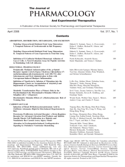
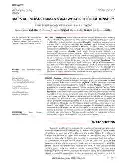
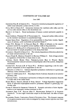
Journal of Cancer Research and Treatment, 2014, Vol. 2, No. 3, 55-63
Available online at http://pubs.sciepub.com/jcrt/2/3/3
© Science and Education Publishing
DOI:10.12691/jcrt-2-3-3
The Effect of L-carnitine on Amethopterin-induced
Toxicity in Rat Large Intestine
Ehab Tousson*, Mona Hegazy, Ezar Hafez, Alaa Abdullah Ahmed
Zoology Department, Faculty of Science, Tanta University, Tanta, Egypt
*Corresponding author: [email protected]
Received October 15, 2014; Revised October 31, 2014; Accepted November 02, 2014
Abstract Background: Amethopterin is a folic acid antagonist which used as a chemotherapeutic agent and its
anti-oxidant activity is used to treat many cancer types. This study aimed to determine the possible protective effects
of L-carnitine against Amethopterin induced large intestine toxicity. Methodology: A total 60 male albino rats were
equally divided into six groups; the first and second groups were the control and L-carnitine groups respectively
while the 3rd group was Amethopterin rat group; the 4th and 5th groups were co- and post- treated Amethopterin rat
with L-carnitine respectively and the 6th group was self treated Amethopterin rat group. Results: Glutathione,
catalase and total protein levels in Amethopterin and self-treated groups showed a significant decrease when
compared with control group, while MDA levels in Amethopterin and self-treated groups showed a significant
increase when compared with control group. Many of abnormalities as colonic epithelial cell damage in the form of
epithelial separation, cellular loss, congestion of blood vessels, crypt hyperplasia and focal depletion of goblet cells
were detected in large intestine tissues in Amethopterin rat group (Amethopterin and self-treated groups). A
significant increase of the apoptotic protein p53 and a significant decrease in the antiapoptotic Bc1-2 proteins after
Amethopterin injection when compared with control group was observed. Conclusions: Treatment (Co- and post-)
with L-carnitine were improved the biochemical, histopathological and immunohistochemical alterations in large
intestine that treated with Amethopterin.
Keywords: Amethopterin, large intestine, L-carnitine, oxidative stress, apoptotic markers, P53, Bcl2
Cite This Article: Ehab Tousson, Mona Hegazy, Ezar Hafez, and Alaa Abdullah Ahmed, “The Effect of Lcarnitine on Amethopterin-induced Toxicity in Rat Large Intestine.” Journal of Cancer Research and Treatment,
vol. 2, no. 3 (2014): 55-63. doi: 10.12691/jcrt-2-3-3.
1. Introduction
Amethopterin (methotrexate, MTX) is a folic acid
antagonist which used as a chemotherapeutic agent and its
anti-oxidant activity is used to treat many cancer types
[1,2,3,4]. Amethopterin inhibits dihydrofolate reductase,
which is an essential enzyme for DNA and RNA
synthesis. One of the most common limiting factors
preventing further dose escalation of Amethopterin is
gastrointestinal toxicity [5,6,7]. Amethopterin enters the
cell via active transport across the reduced folate carrier
and is effluxes from the cell by several of the ATP-binding
cassette transporters [8]. There are other synonyms for
Amethopterin such as Metatrexate, Metatraxan, Mexate,
Rheumatrex, Trexall, Abitrexate, Antifolan, Folex, Tremetex.
Oxidative stress is known to appear because of an
imbalance between the production and degradation of
ROS in tissues. ROS-induced injury is well characterized
and includes DNA base oxidation, lipid peroxidation, and
protein oxidation [9]. Levels of both enzymatic and nonenzymatic anti-oxidants are inhibited and the levels of
oxidants increase in the heart, testes, liver, kidney, lung
and gut tissues of laboratory animals given Amethopterin
[4,6,10-16]. Since the cytotoxic effect of Amethopterin is
not selective for cancer cells, it also affects the normal
tissues that have a high rate of proliferation, including the
hematopoietic cells of the bone marrow and the actively
dividing cells of the gut mucosa. Thus, one of the major
toxic effects of Amethopterin is intestinal injury and
enterocolitis [6].
L-carnitine (4-N-trimethylammonium-3-hydroxybutyric
acid) is a natural nutrient that transports long-chain fatty
acids into the mitochondria, where they are oxidized to
produce adenosine triphosphate and prevent the toxic
accumulation of long-chain fatty acids [17-22]. Recent
studies suggest that L-carnitine may play an important role
in oxidative/antioxidative balance and has an antiperoxidative
effect on several tissues [4,21,22,23,24]. Therefore, the
present study aimed to study the protection and
ameliorating role of L-carnitine in the physiological,
histopathological and immunohistochemical altrations
in Amethopterin induced large intestine toxicity in male rat.
2. Materials and Methods
2.1. Animals
The experiments were performed on 60 male albino rats
weighing 140 -150 gm and of 9-10 week’s age. They were
Journal of Cancer Research and Treatment
obtained from our laboratory farms, Zoology Department,
Faculty of Science, Tanta University, Egypt. The rats were
kept in the laboratory for one week before the
experimental work and maintained on a standard rodent
diet (20% casein, 15% corn oil, 55% corn starch, 5% salt
mixture and 5% vitaminzed starch; Egyptian Company of
Oils and Soap Kafr-Elzayat Egypt) and water available ad
labium The temperature in the animal room was
maintained at 23±2°C with a relative humidity of 55±5%.
Light was on a 12:12 hr light -dark cycle. The
experimental protocol was approved by Local Ethics
Committee and Animals Research.
2.2. Animal Grouping
Sixty rats were equally divided into six groups (10
animals each).
G1: Control group in which animals did not received any
treatment.
G2: L-carnitine or positive control group in which animals
received L-carnitine (Mepaco Co., Egypt; 500 mg/Kg
body weight/day) orally by stomach tube for four weeks
according to Sener et al. and Salama et al [6,22].
G3: Amethopterin group in which rats were injected
intraperitoneally with Amethopterin (Ebewe Co., Egypt;
0.5 mg/kg body weight/twice a week) for four weeks
according to AL-Motabagani and Tousson et al [4,25].
G4: Co-treated group in which animals were injected
intraperitoneally with Amethopterin (0.5 mg/kg body
weight/twice a week) plus received orally L-carnitine
(500mg/Kg body weight/day) for four weeks.
G5: Post-treated group in which animals were injected
intraperitoneally with Amethopterin (0.5 mg/kg body
weight/twice a week) for four weeks and then treated
orally with l-carnitine (500 mg/Kg body weight/day) for
another four weeks.
G6: Self-treated rat group in which rats were injected
intraperitoneally with Amethopterin (0.5 mg/kg body
weight/twice a week) for four weeks and left for another
four weeks without receiving any treatment.
2.3. Preparation of Tissue Homogenates
At the end of the experimental period, rats were fasted
overnight and for clinical chemistry. Rats were and
weighed and euthanized with intravenous injection with
sodium pentobarbital and subjected to a complete
necropsy. After scarification of rats large intestine was
quickly removed and weighed after removed any debris
from tissues carefully. Specimens were separated into two
parts. Each piece was weighed and homogenized
separately with a 3 Potter Elvenhjem tissue homogenizer.
One part was homogenized in phosphate buffer (pH 7.0)
for estimation of protein content and catalase enzymes
activities levels; the second was 10% w/v large intestine
homogenate in ice-cold saline for estimation of MDA and
GSH levels. The crude tissue homogenate was centrifuged
at 11,739 g, for 15 minutes in a cold centrifuge, and the
resultant supernatant was used for different estimations.
2.4.
Enzymatic
Antioxidant Assays
and
Non-enzymatic
Catalase: The catalase (CAT) activity was measured
by monitoring H2O2 (The substrate of the enzyme)
56
decomposition at 240 nm according to the method
described by Aebi [26].
MDA assay: Malondialdehyde (MDA), a noxious
product of lipid peroxidation, was detected by TBARS
analysis and measured as reported by Saggu et al. [27].
The MDA results were expressed as the nmol/mg protein.
Reduced GSH: GSH content was determined with
dithionitrobenzoic acid using the method described by
Beutler et al. [28] and was expressed in μmol GSH/mg
protein. The method is based on the reduction of DTNB to
produce a yellow compound. The reduced chromogen is
directly proportional to GSH concentration and its
absorbance can be measured at 412 nm.
Total protein: Total protein content in tissue
homogenate was measured according to the method of
Lowry et al [29].
2.5. Histopathological Investigation
Immediately after decapitation animals were dissected,
large intestine from different groups were quickly
removed, washed in 0.9 saline solutions and simples of
large intestine were fixed in 10 % neutral buffered
formalin. After fixation, specimens were dehydrated in an
ascending series of alcohol, cleared in two changes of
xylene and embedded in molten paraffin (mp. 50–58°C).
Sections of 7 microns thickness were cut using rotary
microtome and mounted on clean slides. Sections were
stained with Ehrlich's haematoxylin and counterstained
with eosin as a routine method after Bancroft and Stevens
[30].
2.6. Immunohistochemical Detection of p53
and Bc1-2
Expression of p53 and Bc1-2 immunoreactivities (p53ir and Bc12-ir) was detected using avidin Biotin Complex
(ABC) method [4,31,32]. Paraffin sections (5μm thick) of
fixed rat large intestine that mounted on gelatin
chromalum–coated glass slides were dewaxed and
rehydrated sections were washed in distilled water for 5
min, rinsed in PBST for 10 min and incubated with 10%
normal goat serum for 15 min to reduce non-specific
background staining. Then, the sections were incubated
with anti-rabbit p53 or anti-rabbit Bc1-2 (monoclonal
antibody (Dako, 1:80 and 1:2000 respectively) for 1-2
hours at room temperature. The sections after 5 baths in
PBST were incubated with biotinylated goat anti-rabbit
immuoglobulin (Nichirei, Tokyo, Japan). The sections
after 5 baths in PBST were further incubated with Avidin
Biotin Complex (ABC: Nichirei, Tokyo, Japan) for 1 hour
at RT. The reaction was developed by using 20 mg 3-3´diaminobenzidine tetrahydrochloride (DAB, Wako pure
chemical industries, Ltd) in 40 ml PBST, pH 7.2
containing 10 ml of hydrogen peroxide (H2O2) for 7-9 min
at a dark room followed by distilled water then dehydrated
and mounted. The criterion for a positive reaction
confirming the presence of p53 and Bcl-2 is a dark,
brownish, intra cytoplasmic precipitate. For the negative
control, the primary antibody was omitted to guard against
any false positive results which might develop from a nonspecific reaction. Brightness, contrast were adjusted using
Adobe Photoshop software. Image analysis was adjusted
using PAX-it image analysis software.
57
Journal of Cancer Research and Treatment
2.7. Statistical Analysis
Data were expressed as mean values ± SEM and
statistical analyses were performed using SPSS statistical
version 16 software package (SPSS® Inc., USA). The
criterion for statistical significance was set at p<0.01.
3. Results
3.1. Toxicity
The animals under practice appeared healthy and show
no clinical signs of disease and mortality was recorded
during the exposure to L-carnitine. Various side effects
were observed in animals treated with Amethopterin such
as 4% mortality, loosing of body weight, loss of activity,
weakness, yellowish body hair, diarrhea and bleeding
around eyes.
3.2. Biochemical Investigations
Figure 1 shows that; MDA levels in Amethopterin
group (G3) showed significant increase when compared
with control (G1) and L-carnitine (G2) groups. On the
other hand, glutathione (GSH), total protein and catalase
levels in Amethopterin group (G3) showed significant
decrease when compared with control (G1) and Lcarnitine (G2) groups. In contrast, MDA levels in treated
Amethopterin group with L-carnitine group (G4&G5)
were significantly decreased when compared with
Amethopterin group (G3) while GSH, total protein and
catalase levels in treated Amethopterin group with Lcarnitine group (G4&G5) were significantly increased
when compared with Amethopterin group (G3). MDA
levels in co-treated Amethopterin group with L-carnitine
group (G4) were significantly decreased when compared
with post treated Amethopterin group with L-carnitine
group up (G5) while GSH, total protein and catalase levels
in co-treated Amethopterin group with L-carnitine group
(G4) were significantly increased when compared with
post treated Amethopterin group with L-carnitine group
up (G5). On the other hand, Glutathione, total protein and
catalase levels in self-treated group (G6) showed
significant decrease when compared with Amethopterin
group (G3) while, MDA levels in self-treated group (G6)
showed significant increase when compared with
Amethopterin group (Figure 1).
Figure 1. Changes in MDA, GSH, total protein and catalase levels in large intestine in different groups under study. Data are expressed as mean ±
S.E.M of 10 observations. Significant difference from the control group (G1) at *p<0.05. Significant difference from Amethopterin group (G3) at
#
p<0.05. Where G1, Control group; G2, L-carnitine group; G3, Amethopterin group; G4, Co-treated Amethopterin group with L-carnitine; G5, Posttreated Amethopterin group with L-carnitine; G6, Self-treated Amethopterin group
3.3. Effect of Amethopterin on Histopathology
On Large Intestine
Examination of sections stained with H & E showed no
differences in colonic mucosal structure between control
(G1) and L-carnitine (G2) groups, all of which showed
normal histological appearance (Figure 2A & Figure 2B).
The colonic mucosa showed normal intestinal crypts lined
by epithelial columnar cells and goblet cells. The lamina
propria contained few mucosal mast cells full of
cytoplasmic granules (Figure 2A & Figure 2B). Light
microscopy of the rat large intestine sections in
Amethopterin group (G3) raveled colonic epithelial cell
damage in the form of epithelial separation, cellular loss,
congestion of blood vessels, crypt hyperplasia and focal
depletion of goblet cells as well as mononuclear cellular
infiltration in lamina propria. These inflammatory cells
included macrophages, fibroblasts, and plasma cells
Journal of Cancer Research and Treatment
(Figure 2C). Co-treatment of Amethopterin rats with Lcarnitine for four weeks revealed more or less normal
mucosal structure with a few inflammatory cells and mild
congestion of blood vessels (Figure 2D). Large intestine
sections on post-treatment of Amethopterin rats with L-
58
carnitine revealed mild to moderate colonic epithelial cell
damage, atrophy and mild crypt hyperplasia (Figure 2E).
Large intestine sections of self treated rats revealed severe
tissue injury as marked colonic epithelial cell damage and
strong congested blood capillaries (Figure 2F).
Figures 2(A-F). Photomicrographs of the rat large intestine sections stained by HE. A&B: Micrographs from control and L-carnitine groups revealed
normal structure. C: Micrograph of the large intestine in Amethopterin rat group revealed many of abnormalities as colonic epithelial cell damage in the
form of epithelial separation, cellular loss (arrow heads), congestion of blood vessels, crypt hyperplasia and leukocyte infiltration (arrows). D:
Micrograph of the large intestine in Co-treatment of Amethopterin rats with L-carnitine revealed more or less normal mucosal structure with a few
cellular loss (arrow heads), a few inflammatory cells and mild congestion of blood vessels. E: Micrograph of the large intestine in post treatment of
Amethopterin rats with L-carnitine revealed mild to moderate colonic epithelial cell damage, atrophy and mild crypt hyperplasia. F: Micrograph of the
large intestine in self treatment revealed severe tissue injury as marked colonic epithelial cell damage and strong congested blood capillaries
3.4. Immunohistochemical Examination of
Large Intestine Tissue
3.4.1. P53 Immunoreactivity in Rat Large Intestine
Figure 3A - Figure 3F) and Table 1 showed the
detection and distribution of p53 immunoreactivity (P53-ir)
in the large intestine in the different groups under study.
P53 is a nuclear phosphoprotein which acts as a tumor
suppressor. Large intestine in control and L-carnitine
groups showed finite reaction for p53-ir (grade 1). Severe
positive reaFigure ctions for p53-ir (grade 4) were
detected in Amethopterin and self- Amethopterin treated
rat groups (Figure 3A - Figure 3C, Figure 3F). Moderate
(grade 3) to mild (grade 2) positive reactions for p53-ir
were observed in the large intestine in Co-treated and
post-treated rats with L-carnitine (Figure 3D & Figure 3E)
respectively.
59
Journal of Cancer Research and Treatment
Figures 3(A-F). A-F High power micrographs of the rat large intestine stained by P53-ir. A&B: Negative or faint p53-ir reaction in control and Lcarnitine respectively. C: Severe positive reactions for p53-ir in Amethopterin group. D&E: Moderate to mild positive reactions for p53-ir in Co-treated
and post treated rats with L-carnitine respectively. F: Moderate positive reaction for p53-ir in self treated rats
3.4.2. Bcl2 Immunoreactivity in Rat Large Intestine
Figure 4A - Figure 4F) and Table 1 show the detection
and distribution of Bcl2-ir immunoreactivity (Bcl2-ir) in
the large intestine tissues in the different groups under
study. large intestine in control and L-carnitine groups
showed marked positive reaction for Bcl2-ir (grade 4) as
in Figure 4A & Figure 4B).
Mild positive reactions for Bcl2-ir (grade 1) were
detected in the large intestine sections in Amethopterin rat
group (Figure 4C). The intensity of Bcl2-ir in the rat large
intestine sections in Amethopterin group was significantly
decreased when compared with control rat. Moderate
(grade 3) to mild (grade 2) positive reactions for Bcl2-ir
were observed in the large intestine in co-treated and posttreated rats with L-carnitine (Figure 4D & Figure 4E)
respectively. On the other hand; the intensity Bcl2-ir were
stay mild to moderate positive reaction (grade 2) in selftreated large intestine sections when compared with treated
Amethopterin rats with L-carnitine groups (Figure 4F).
Table 1. Changes in P53 and Bcl2 expressions in large intestine
sections in different groups under study
P53
Bcl2
G1
1
4
G2
1
4
G3
4
1
G4
3
3
G5
2
2
G6
3
2
Data are expressed as mean ± S.E.M of 5 observations. G1, Control
group; G2, L-carnitine group; G3, Amethopterin group; G4, Co-treated
Amethopterin group with L-carnitine; G5, Post-treated Amethopterin
group with L-carnitine; G6, Self-treated Amethopterin group. where 0,
Negative reaction; 1, light reaction; 2, mild reaction; 3 moderate reaction;
4, strong reaction.
Journal of Cancer Research and Treatment
60
Figures 4(A-F). A-F High power micrographs of the rat large intestine stained by Bcl2-ir. A&B: Severe positive Bcl2-ir reaction in control and Lcarnitine respectively. C: Faint to mild positive reactions for Bcl2-ir in Amethopterin group. D&E: Moderate to mild positive reactions for Bcl2-ir in
Co-treated and post treated rats with L-carnitine respectively. F: Mild positive reaction for Bcl2-ir in self treated rats
4. Discussion
Chemotherapy commonly produces structural and
functional damage to the intestinal mucosa in cancer
patients [33] and in patients with rheumatoid arthritis [34].
Amethopterin (MTX), is an antineoplastic agent used to
fight a number of different cancers, such as acute
lymphoblastic leukemia and solid cancers, due to its
inhibitory effect on de novo purine and pyrimidine
synthesis through dihydrofolate reductase inhibition [35].
On the other hand, substantial evidence supports the
concept that Amethopterin was mutagenic and
carcinogenic in animals. And the efficacy of this
compound is often limited by its severe gastrointestinal
toxicity [6,7,36,37,38]. Amethopterin is recognized as the
most effective of the traditional disease modifying
antirheumatic drugs but it has several toxicities.
This study conducts a biochemical, histopathological
and immunohistochemical investigation into whether Lcarnitine has a protective and ameliorated effect on
Amethopterin -induced large intestine damage in male rats.
The results of the studies indicate that Amethopterin
causes oxidative tissue damage by increasing lipid
peroxidation in the large intestine tissues and decreasing
the level of antioxidant enzymes. Moreover, the
histopathological and p53 and Bcl2 alterations supported
this conclusion. It has also been shown that L-carnitine
given for 28 days with or after the Amethopterin
application provided significant protection from the large
intestine damage of Amethopterin. In current study, there
was a significant.
Many aspects of the pathophysiology are mediated by
oxidative stress [20,39,40]. In the present study,
Amethopterin significantly altered the oxidant/antioxidant
balance. In the current study, MDA levels in
Amethopterin group were significantly increased unlike
61
Journal of Cancer Research and Treatment
glutathione, catalase and total protein levels which were
significantly decreased when compared with control group.
So, Amethopterin increased MDA level accompanied with
decreased GSH content and CAT activities. Similar results
were previously reported by other investigators [10,41].
Oxidative stress or oxidative cellular damage with its dual
of free radical generation and profound lipid peroxidation
are hallmarks of Amethopterin toxicity [42]. Amethopterin
induces oxidative stress in tissues as demonstrated by
increasing MDA levels [10,11].
MDA, as an end product of lipid peroxidation, usually
used to estimate the extent of lipid peroxidation. The high
level of MDA in the Amethopterin and self-treated groups
in large intestine tissues indicates that Amethopterin gives
rise to oxidative stress in colonic tissue. Our current
results agree with Vardi et al. [43] who reported that,
oxygen radicals and hydrogen peroxides have been
associated with the many side effects of Amethopterin and
these free radicals trigger cell damage through binding to
cellular macromolecules. It has been shown that many
pathological conditions that resulted in elevation of MDA
due to lipid peroxidation were prevented by L-carnitine
[44,45]. The increase lipid peroxidation responsible for
the formation of lipid hydroperoxides.
GSH is considered to be one of the most very important
components of the antioxidant defense of living cells. The
reduced tri-peptide GSH is a hydroxyl radical and singlet
oxygen scavenger, and participates in a wide range of
cellular functions [15]. Actually, the nadir in colonic GSH
content promoted by Amethopterin represents an
alteration in the cellular redox state, suggesting that the
cells could be more sensitive to reactive oxygen
metabolites and leads to a reduction in the effectiveness of
the antioxidant enzyme defense system [46,47]. Also in
the same way and supported for earlier results; GSH, total
protein and catalase levels in self treated group showed
significant decrease when compared with Amethopterin
group while, MDA levels in self-treated group showed
significant increase when compared with Amethopterin
group. Our results not agreed with Ciralik et al. [48] who
observed an increase in reduced glutathione levels, also
observed increased activities of catalase of the tissue
induced by Amethopterin. Catalase acts as a preventative
antioxidant whereas it catalyses the reduction of H2O2 and
plays an important role in protection against the
deleterious effects of lipid peroxidation, ROS and
hydroxyl radicals caused by Amethopterin administration
[13].
In the present study, increase in the lipid peroxidation
activity due to toxic effects of Amethopterin was
accompanied by significant reduction in reduced GSH
level of the large intestine tissue, indicating the presence
of oxidative tissue damage. lipid peroxidation, mediated
by oxygen free radicals is believed to be an important
cause of destruction and damage to cell membranes [6,49].
Our results showed that L-carnitine exhibited anti-oxidant
effects not only on the non enzymatic defense system
(GSH), but also on the enzymatic one such as catalase.
Treatment with L-carnitine following Amethopterin
apparently increased GSH content and increased catalase
activities compared to Amethopetrin treated animals. Data
so far obtained from this study would suggest that
administration of L-carnitine after Amethopterin challenge
may have beneficial effects that could possibly be ascribed,
in part, to its regulation of the oxidant/anti-oxidant balance.
These results were supported by our histopathological
findings. Due to its high turnover rate, the epithelium of
gastrointestinal tract is susceptible to chemotherapyinduced damage, leading to the destruction of intestinal
mucosa barrier and subsequent clinical manifestations
[38,50].
In the current study, many abnormalities were reported
in the large intestine in Amethopterin group. This
alterations as colonic epithelial cell damage in the form of
epithelial separation, cellular loss, congestion of blood
vessels, crypt hyperplasia and focal depletion of goblet
cells. The mechanism by which Amethopterin causes large
intestine damage results from binding to the enzyme
dihydrofolic reductase, thus preventing conversion of folic
acid to its active form, folinic acid. This in turn blocks the
synthesis of nucleic acids, certain amino acids and indirectly
proteins. This might lead to damage of organelles and
plasma membranes with their function and allowing
leakage of enzymes. Protective mechanisms of L-carnitine
include the inhibition of mitochondrial membrane
permeability, as transition, the decrease of oxidative stress,
and the prevention of pro-apoptotic protein expression
[20]. It was also reported that antioxidant properties of Lcarnitine may be related to the transport of fatty acids into
mitochondria for β-oxidation and thus to the decrease of
lipid usage and protection of the cell membrane against
toxic reactive oxygen species (ROS) and other free
radicals [51]. Our results presented in this study show
similarity with the findings of previous studies on the
antioxidant effects of L-carnitine [52,53].
Apoptosis, or programmed cell death, is a crucial
cellular activity in the behavior of mammalian cells in a
wide range of pathophysiological conditions. There are
numerous external signals that are involved in the
regulation of the apoptosis; p53 is the one of most extensively
investigated pathways [4,54,55,56]. Immunohistochemical
observations of the large intestine tissues showed a
significant increase of the apoptotic protein p53 and a
significant decrease in the antiapoptotic Bc1-2 proteins
after Amethopterin injection.
An inverse correlation was found between the
expression of Bcl-2 and p53 proteins in our results.
Similar bidirectional changes of protein expression
patterns of bcl-2 and p53 were also observed by Tousson
et al. and Diebold et al. [4,32,57] although Diebold et al.
[57] failed to reveal any correlation between the
expressions of these two proteins. So, the increasing of
p53 apoptotic cells and the decreasing Bcl-2 antiapoptotic
cells in the present study reveal the possibility of the
apoptosis occurrence after Amethopterin administration.
Our results showed that, the expression of bcl-2 increased,
cancer cells would resist the apoptosis induced by
chemical drugs or radiation during therapy. Amethopterin
acts as a dihydrofolic acid analogue that binds to the
dihydrofolic acid reductase enzyme by inhibiting the
synthesis of tetrahydrofolate, which is required for DNA
synthesis [46].
L-carnitine can carry long chain fatty acyl groups into
mitochondria for beta-oxidation [58]. We can conclude
that co-treatment prevented oxidative damage by inhibiting
ROS production and improving antioxidant enzymes.
While, post-treatment regulated the oxidant/antioxidant
balance and neutralized the toxic side effects caused by
Journal of Cancer Research and Treatment
Amethopterin induced oxidative stress. Concurrently, cotreatment acted with more effectiveness than posttreatment. Thus, our study provides important evidences
for experimental and clinical investigations about the role
of the co- and post- ways of treatment against the toxic
side effects of Amethopterin therapeutics. Also, we
recommend to propose directions for further studies in the
near future.
[9]
[10]
[11]
[12]
Conflicts of Interest
[13]
The authors had no conflicts of interest to declare in
relation to this article.
[14]
Acknowledgments
I thank Prof. Dr. Ahmed Masoud (Zoology dep., Fac. of
Science, Tanta Univ., Egypt) for her critical and helpful
comments on a draft of this manuscript.
List of Abbreviations
ABC, Avidin Biotin Complex; FAICAR, formyl AICAR;
H&E, Hematoxylin Eosin staining; H2O2, hydrogen peroxide;
MDA, Malondialdhyde; RA, Rheumatoid arthritis; S, Serum;
S.E.M, Standard Error Mean; TCA, trichloronacetic acid;
WBCs, white blood cells; ATP, Adenosine triphosphate;
DNA, Deoxyribonucleic acid; RNA, Ribose nucleic acid;
ROS, Reactive oxygen species; CAT, catalase; GSH,
Glutathione; MTX, Methotrexate; Bcl2-ir, Bcl2
immunoreactivity.
References
[1]
[2]
[3]
[4]
[5]
[6]
[7]
[8]
Cronstein BN. Molecular therapeutics. Methotrexate and its
mechanism of action. Arthritis Rheum 2004; 39: 1951-60.
Kimura E, Nishimura K, Sakata K, Oga S, Kashiwagi K, Igarashi
K. Methotrexate differentially affects growth of suspension and
adherent cells. Int J Biochem Cell Biol 2004; 36: 814-25.
Salzer WL, Winick NJ, Wacker P, Lu X, Devidas M, Shuster JJ.
Plasma methotrexate, red blood cell methotrexate, and red blood
cell folate values and outcome in children with precursor n Bacute lymphoblastic leukemia:a report from the children’s
oncology group. J Pediatr Hematol Oncol 2012; 34: 1-7.
Tousson E, Hafez E, Zaki S, Gad A. P53, Bcl-2 and CD68
expression in response to amethopterin-induced lung injury and
ameliorating role of l-carnitine. Biomed and Pharmacotherapy
2014; 68: 631-9.
Carneiro-Filho BA, Lima IP, Araujo DH, Cavalcante MC,
Carvalho GH, Brito GA, Lima V, Monteiro SM, Santos FN,
Ribeiro RA, Lima AA. Intestinal barrier function and secretion in
methotrexate-induced rat intestinal mucositis. Dig Dis Sci 2004;
49: 65-72.
Sener G, Demiralp EE, Etiner M, Ercan F, Yegen BC. Glucan
ameliorates methotrexate-induced oxidative organ injury via its
antioxidant and immunomodulatory effects. European Journal of
Pharmacology 2006; 542: 170-8.
Soares PM, Lopes LO, Mota JM, Belarmino-Filho JN, Ribeiro RA,
de Souza MH. Methotrexate-induced intestinal mucositis delays
gastric emptying and gastrointestinal transit of liquids in awake
rats. Arq Gastroenterol 2011; 48: 80-85.
Wielinga P, Hooijberg JH, Gunnarsdottir S, Kathmann I, Reid G,
Zelcer N, van der Born K, de Haas M, van der Heijden I, Kaspers
G, Wijnholds J, Jansen G, Peters G, Borst P. The human multidrug
resistance protein MRP5 transports folates and can mediate
cellular resistance against antifolates. Cancer Res 2005; 65: 442530.
[15]
[16]
[17]
[18]
[19]
[20]
[21]
[22]
[23]
[24]
[25]
[26]
[27]
[28]
[29]
[30]
[31]
[32]
[33]
62
Laskin JD, Black AT, Jan YH, Sinko PJ, Heindel ND, Sunil V,
Heck DE, Laskin DL. Oxidants and antioxidants in sulfur
mustard-induced injury. Ann N Y Acad Sci 2010; 1203: 92-100.
Jahovic N, Cevik H, Sehirli AO, Yˇegen BC, Sener G. Melatonin
prevents methotraxate- induced hepatorenal oxidative injury in
rats. J Pineal Res 2003; 34: 282-7.
Cetin A, Kaynar L, Kocyigit I, Hacioglu SK, Saraymen R, Ozturk
A, Sari I, Sagdic O. Role of grape seed extract on methotrexate
induced oxidative stress in rat liver. American Journal of Chinese
Medicine 2008; 36: 1-12.
Hemeida AR, Omar MM. Curcumin Attenuates MethotraxateInduced Hepatic Oxidative Damage in Rats. Journal of the
Egyptian Nat. Cancer Inst 2008; 20(2): 141-8.
Vardi N, Parlakpinar H, Ozturk F, Ates B, Gul M, Cetin A,
Erdogan A, Otlu A. Potent protective effect of apricot and βcarotene on methotrexate induced intestinal oxidative damage in
rats. Food and Chemical Toxicology 2008; 46: 3015-3022.
Vardi N, Parlakpinar H, Ates B, Cetin A, Otlu A. Antiapoptotic
and
antioxidant
effects
of
beta-carotene
against
methotrexateinduced testicular injury. Fertil Steri 2009; 92: 202833.
Iyyaswamy A, Rathinasamy S. Effect of chronic exposure to
aspartame on oxidative stress in the brain of albino rats. Biosci
2012; 37: 679-88.
Ozogula B, Kisaoglua A, Turanb MI, Altunerc D, Senerd E,
Cetine N, Ozturk C. The effect of mirtazapine on methotrexateinduced toxicity in rat liver. Science Asia 2013; 39: 356-36.
Agarwal A, Said TM. Carnitines and male infertility. Reprod
Biomed Online 2004; 8: 376-84.
Sinclair C, Gilchrist JM, Hennessey JV, Kandula M. Muscle
carnitine in hypo- and hyperthyroidism. Muscle Nerve 2005; 32:
357-9.
Ulvi H, Aygul R, Demir R. Effect of L-carnitine on diabetic
neuropathy and ventricular dispersion in patients with diabetes
mellitus. Turk J Med Sci 2010; 40: 169-75.
Chao HH, Liu JC, Hong HJ, Lin JW, Chen CH, Cheng TH. Lcarnitine reduces doxorubicin-induced apoptosis through a
prostacyclin-mediated pathway in neonatal rat cardiomyocytes. Int
J Cardiol 2011; 146: 145-52.
Salama AF, Kasem SM, Tousson E, Elsisy MK. Protective role of
L-carnitine and vitamin E on the kidney of atherosclerotic rats.
Biomedicine & Aging Pathology 2012; 2: 212-5.
Salama A, Kasem S, Tousson E, Elsisy MK. L-carnitine and
vitamin E alleviate reproductive toxicity caused by triton WR
1339 in male albino rats. Toxicology and Industrial Health; 2013.
Cayir K, Karadeniz A, Yildirim A, Kalkan Y, Karakoc A, Keles
M. Protective effect of L-carnitine against cisplatin-induced liver
and kidney oxidant injury in rats. Cent Eur J Med 2009; 4: 184-91.
Pehlivan M, Coşkun A, Zengin A, Aslaner A, Yavuz T. Does Lcarnitine increase serum TNF -α and IGF-1 during liver
regeneration in the rat?. Turk J Med Sci 2009; 39: 875-80.
Al-Motabagani MA. Estudios histológico e histoquímico del
efecto del metotrexato en el hígado de rata macho albina adulta.
Int. J. Morphol 2006; 24(3): 417-22.
Aebi H. Catalase. In: Bergmeyer HU, (ed.) Methods of Enzymatic
Analysis. New York: Academic Press 1984; pp: 673-84.
Saggu S, Kumar R. Modulatory effect of seabuckthorn leaf extract
on oxidative stress parameters in rats during exposure to cold,
hypoxia and restraint (C-H-R) stress and post stress recovery. J
Pharm Pharmacol 2007; 59(12): 1739-45.
Beutler E, Duron O, Kelly BM. Improved method for the
determination of blood glutathione. J.Lab. Clin. Med 1963; 61:
882-8.
Lowry OH, Rosebrough NJ, Farr AL, Randall RJ. Protein
measurement with Folin phenol reagent. J Biol Chem 1951; 193:
265-75.
Bancroft JD, Stevens GA. Theory and Practice of Histological
Techniques. 2nd Ed. Churchill Livingstone, London 1990.
Sternberger LA. The unlabelled antibody peroxidaseantiperoxidase (PAP) method. In: “immunocytochemistry”, 2nd
edn., (Cohen S, and McClusky RT.ed.). John Wiley and sons, New
York 1979; pp: 104-69.
Tousson E, Alm-Eldeen A, El-Moghazy M. p53 and Bcl2expression in response to boldenone induced liver cells injury.
Toxicology and Industrial Health 2011; 27(8): 711-8.
Cunningham D, Morgan RJ, Mills PR. Functional and structural
changes of the human proximal small intestine after cytotoxic
therapy. J Clin Pathol 1985; 38: 265-70.
63
Journal of Cancer Research and Treatment
[34] Phelen MJ, Taylor W, van Heyningen C, Williams E, Thompson
[47] Fiocchi C. Inflammatory bowel disease: New insights into
RN.Intestinal absorption in patients with rheumatoid arthritis
treated with methotrexate. Clin Rheumatol 1993; 12: 223-25.
Zhang P, Li J, Liu Y, Chen X, Kang Q, Zhao J, Li W. Human
neural stem cell transplantation attenuates apoptosis and improves
neurological functions after cerebral ischemia in rats. Acta
Anaesthesiol Scand 2009;53(9):1184-91.
Nagakubo J, Tomimatsu T, Kitajima M, Takayama H, Aimi N,
Horie T. Characteristics of transport of fluoresceinated
methotrexate in rat small intestine. Life Sci 2001; 69: 739-47.
Kolli RP, Seidman DN. Comparison of compositional and
morphological atom-probe tomography analyses for a
multicomponent Fe-Cu steel. Microsc Microanal 2007; 13: 272-84.
Wardill HR, Bowen JM, Gibson RJ. Chemotherapy-induced gut
toxicity: are alterations to intestinal tight junctions pivotal?.
Cancer Chemother Pharmacol 2012; 70: 627-35.
Rachmilewitz EA, Schrier S. Pathophysiology of β thalassemia. In:
Steinberg MH, Forget BG, Higgs DR, Nagel RL, editors.
Disorders of Hemoglobin: Genetics, Pathophysiology, and
Clinical Management. Cambridge, UK: Cambridge University
Press 2001; PP: 233-51.
Rund D, Rachmilewitz E. Beta-thalassemia. N Engl J Med 2005;
353: 1135-46.
ALL C, Ertan B, Ergul BK, Bulent K. N-acetylcysteine
ameliorates methotrexate-induced oxidative liver damage in rats.
Med Sci Monit 2006; 12(8): 247-8.
Jahovic N, Sener G, Cevic H, Ersoy Y, Arbak S, Yegen BC.
Amelioration of methotrexate-induced enteritis by melatonin in
rats. Cell Biochem Funct 2004; 22: 169-78.
Vardi A, Bosviel R, Rabiau N, Adjakly M, Satih S, Dechelotte P.
Soy phytoestrogens modify DNA methylation of GSTP1,
RASSF1A, EPH2 and BRCA1 promoter in prostate cancer cells.
In Vivo 2010; 24: 393-400.
Atila K, Coker A, Sagol O, Coker I, Topalak O, Astarcioglu H,
Karademir S, Astarcioglu I. Protective effects of carnitine in an
experimental ischemia reperfusion injury. Clin Nutr 2002; 21:
309-13.
Derin N, Agac A, Bayram Z, Asar M, Izgut-Uysal VN. Effects of
L-carnitine on neutrophil-mediated ischemia-reperfusion injury in
rat stomach. Cell Biochem Funct 2006; 24: 437-42.
Babiak RM, Campello AP, Carnieri EG, Oliveira MB.
Methotrexate: pentose cycle and oxidative stress. Cell Biochem
Funct 1998; 16: 283-93.
mechanisms of inflammation and increasingly customized
approaches to diagnosis and therapy. Curr.Opin. Gastroentrol
2004; 20: 309-10.
Ciralik H, Bulbuloglu E, Cetinkaya A, Kurutas EB, Celik M, Polat
A. Effects of N acetylcysteine on methotrexate induced small
intestinal damage in rats. The Mount Sinai Journal of Medicine
2006; 73: 1086-92.
Tarasub N, Junseecha T, Tarasub C, Na Ayutthaya WD. Protective
Effects of Curcumin, Vitamin C, or their Combination on
Cadmium-Induced Hepatotoxicity. J Basic Clin Pharm 2012; 3:
273-81.
Stringer AM, Gibson RJ, Bowen JM, Logan RM, Yeoh AS, Keefe
DM. Chemotherapy-induced mucositis: the role of gastrointestinal
microflora and mucins in the luminal environment. J Support
Oncol 2007; 5: 259-67.
Kalaiselvi, T, Panneersalvam C. Effect of L-carnitine on the status
of lipid peroxidation and antioxidants in aging rats. J. Nutr.
Biochem 1998; 9: 575-81.
Atroshi F, Rizzo A, Biese L, Veijalainen P, Saloniemi H, Sankari
S, Andersson K. Fumonisin B1-induced DNA damage in rat liver
and spleen: Effect of pretreatment with coenzyme Q10, Lcarnitine, α-tocopherol and selenium. Pharmacol. Res 1999; 40:
459-67.
Arockia Rani PJ, Panneerselvam C. L-carnitine as a free radical
scavenger in aging. Exp. Gerontol 2001; 36: 1713-26.
Vaux DL, Strasser A.The molecular biology of apoptosis. Proc
Natl Acad Sci USA 1996; 93: 2239-44.
Nagatu S. Apoptosis by death factor. Cell 1997; 88: 355-65.
Tousson E, Beltagy DM, Abo Gazia M, Al-Behbehani B.
Expressions of P53 and CD68 in mouse liver with Schistosoma
mansoni infection and the protective role of silymarin. Toxicology
and Industrial Health; 2012.
Diebold J, Baretton G, Felchner M, Meier W, Dopper K, Schmidt
M, Lohrs U. Bcl-2 expression, p53 accumulation, and apoptosis in
ovarian carcinomas. Am J Clin Pathol 1996; 105: 341-34.
Augustyniak A, Skrzydlewska E. L-carnitine in the lipid and
protein protection against ethanol-induced oxidative stress.
Alcohol 2009; 43(3): 217-23.
[35]
[36]
[37]
[38]
[39]
[40]
[41]
[42]
[43]
[44]
[45]
[46]
[48]
[49]
[50]
[51]
[52]
[53]
[54]
[55]
[56]
[57]
[58]


© Copyright 2026