
Regulated expression and function of CD122 (interleukin-2/interleukin- 15R-beta) during lymphoid development
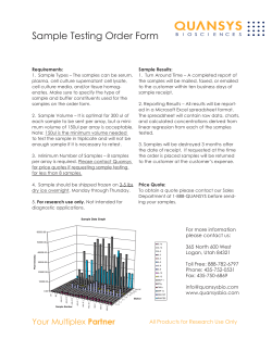
From www.bloodjournal.org by guest on November 13, 2014. For personal use only.
1996 87: 190-201
Regulated expression and function of CD122
(interleukin-2/interleukin- 15R-beta) during lymphoid development
T Reya, JA Yang-Snyder, EV Rothenberg and SR Carding
Updated information and services can be found at:
http://www.bloodjournal.org/content/87/1/190.full.html
Articles on similar topics can be found in the following Blood collections
Information about reproducing this article in parts or in its entirety may be found online at:
http://www.bloodjournal.org/site/misc/rights.xhtml#repub_requests
Information about ordering reprints may be found online at:
http://www.bloodjournal.org/site/misc/rights.xhtml#reprints
Information about subscriptions and ASH membership may be found online at:
http://www.bloodjournal.org/site/subscriptions/index.xhtml
Blood (print ISSN 0006-4971, online ISSN 1528-0020), is published weekly by the American
Society of Hematology, 2021 L St, NW, Suite 900, Washington DC 20036.
Copyright 2011 by The American Society of Hematology; all rights reserved.
From www.bloodjournal.org by guest on November 13, 2014. For personal use only.
Regulated Expression and Function of CD122
(Interleukin-2/Interleukin-l5R-~)During Lymphoid Development
By Tannishtha Reya, Julia A. Yang-Snyder, Ellen V. Rothenberg, and Simon R. Carding
TO determine whether signaling via CD122 (interleukin-2 [IL21/IL-15 receptor p-chain) plays a role in regulating the
expansion and differentiation of lymphocyte precursors, we
have characterized its expression and evaluated its ability
to influence the activity of developing lymphoid cells. A significant fraction of Scal'Lin- hematopoietic stem cells in
day 12 fetal liver were found to be CD122+. CD122-mRNA+
and IL-2-mRNA' cells were also localized in embryo sections within pharyngeal blood vessels adjacent to and surrounding the thymic analgen. This distribution is consistent
with the migration of CD122+ progenitor cells from the liver
to the developing thymus where a majority of Scal' intra-
thymic T-cell progenitors were CD122+. Analysis of CD122
expression in theday 12 fetal liver revealed that themajority
of B220' cells were CD122+. Furthermore, CD122 expression
was restricted to the earliest B220+ cells (CD43'CD24-; prepro B cells; fraction A) that proliferate vigorously to IL-2 in
the absence ofany stromal cells, but .not to IL-15. Consistent
with a role for the IL-2/lL-2R pathway in lymphocyte development is the progressive loss of B cells seen in IL-2-deficient mice. Together, these observationssuggest that CD122
plays a role in regulating normal lymphocyte development
in vivo.
0 1996 by The American Societyof Hematology.
S
studies of IL-2 and lymphopoiesis is made difficult in view
oftherecentfindingsthatthe
y-chain is a component of
other cytokine receptors'*." and, that both the P-(CD122)
and the y-chain can interact with the newly described cytokine, IL-15.14 Thus, ligands other than IL-2 may be able to
influence the development of lymphoid cell progenitors that
express individual chains of the IL-2R complex. Although
only limited studies of IL- 15 have been reported to date, the
production of this cytokine by bone marrow (BM) stromal
cells andepithelial cell lines" implies thatit may be available
for utilization by immatureB-andT-cell populationsthat
develop in the BM and thymus, respectively.
To betteraddress the role cytokines play during lymphoid
development,wehave analyzedhematopoietic stemcells
and B- and T-cell precursors for expression of CD122, the
signaling component of the IL-2 and IL-15 receptor. Moreover,we have also determinedwhethersignalingthrough
this receptor influences hematopoietic precursorcell activity.
Finally, we have analyzed IL-2-deficient mice to determine
if they display any hematopoietic defects in the absence of
one of the known ligands for IL-2RP.
TEM CELLS undergo a program ofproliferation and
differentiation in anatomically distinct sites to give rise
to functionally competent B and T cells. The stages through
which developing B and T cells pass, as distinguished by
thedifferential expression of a variety of genesandcell
surface glycoprotein antigens,have beenwell characterized.l-3 However, we still know very little about the nature
of the mechanisms that promote or regulatea cell's transition
between these stages. Recently, several linesof experimental
evidence have suggested that in addition to stromal cell contact the ability to respond to certain cytokines may be required formaintaining the viability and promoting the expansionand/ordifferentiationof
B- and T-cellprogenitor
p0pu1ations.l.~ In
particular, a number of studies suggestthat
the interleukin-2 (IL-2)/IL-2R signaling pathway may play
a role in promoting lymphopoiesis.
The expression of individual chains of the IL-2 receptor
complex immature B5."and T cells4.'.* in hematopoietic tissues of both humans and mice is consistent with the requirement for afunctionalIL-2R
duringlymphopoiesis.The
disruption of IL-2R function by the administration of antiIL-2R antibodies to intactanimals,' by creatingnonfunctional IL-2Rs in transgenic mice'" or, as a result of the mutations inthe y-chain of the IL-2R seenin humansevere
combined immunodeficiency disorder (X-SCID) patients' I
also suggest that an intact and functional IL-2R may play a
role in thedevelopment of T-cell-precursorpopulations.
However, interpretation of the resultsofthese
andother
From the Department of Microbiology, University of Pennsylvania, Philadelphia; and the Division of Biology, California Institute
of Technology, Pasadena.
Submitted February 7, 1995; accepted August 17, 1995.
Supported in part by grants from
the W.W . Smith Charitable
Trust, the American Cancer Society (JFRA-399), and the National
Institutes of Health (A131972).
Address reprint requests to Simon R. Carding, PhD, Department
of Microbiology, 209 Johnson Pavilion, University of Pennsylvania,
Philadelphia, PA 19104-6076.
The publication costs of this article were defrayedin part by page
chargepayment.Thisarticle
must thereforebeherebymarked
"advertisement" in accordance with 18 U.S.C. section 1734 solely to
indicate this fact.
0 1996 by The American Society of Hematology.
0006-4971/96/8701-0024$3.00/0
190
MATERIALSANDMETHODS
Animals. Adult (6 to 8 weeksof age) C57BL.6mice (Jackson
Laboratories, Bar Harbor, ME) werebredand maintained by the
University of Pennsylvania animal facilities. Fetal tissues were obtained from time-mated mice with the day of detection of a vaginal
plug designated day 0. Adult C13-SCID mice (6 to 8 weeks of age)
were providedby Dr John Cebra (Department of Biology, University
of Pennsylvania).
Cell preparation. Liver and thymi obtained from fetuses of the
appropriate age (day 12 through day 16 of gestation) were disaggregated in phosphate-buffered saline, pH 7.3 (PBS) using fine forceps
and a single cell suspension obtained by repeated aspiration through
a I-mL syringe barrel. BM mononuclear cells were obtained from
the femurs and tibias of adult SCID mice by flushing the bones
with PBS and repeated aspiration through a l-mL syringe barrel.
Erythrocytes were removed by sedimentation centrifugation using a
Ficoll Hypaque solution (FicoKite-LM, density 1.086g/mL; Atlanta
Biologicals, Norcross, GA). Viable mononuclear cells were obtained
from the interface, washed, and resuspended in PBS containing 2%
fetal calf serum (FCS; Atlanta Biologicals). Human peripheral blood
mononuclear cells (PBMC) were obtained from whole blood donated
by normal healthy adults after sedimentation centrifugation through
Ficoll Hypaque solution (LSM, density 1.080 g/mL; Organon Teknika, Durham, NC).
Blood, Vol 87,No 1 (January l), 1996:pp 190-201
From www.bloodjournal.org by guest on November 13, 2014. For personal use only.
191
CD122 AND LYMPHOIDDEVELOPMENT
Monoclonal antibodies (MoAbs). The following mouse and rat
MoAbs were used to stain and analyze populations of cells isolated
from the fetal liver, thymus, and adult BM by flow cytometry: antimouse CD3UCD16IFcRyIYm (2.462, ATCC); CD12UIL2RP
(TM-P1; gift from Dr Masayuki Miyasaka, Tokyo Metropolitan Institute of Medicine, Japan); CD45RIB220 (FL43-6B2; Pharmingen,
San Diego, CA); Sca-l/LydA/E (E13-161.7; Pharmingen); CD901
Thy 1.2 (30H.12; GIBCO-BRL, Gaithersburg, MD); CD24/HSA
(M1/69; Pharmingen); Mac-l (Mlf70.15; Caltag Laboratories, San
Francisco, CA); Gr-l (RE36-8C5; Pharmingen); CD43Ly-48 (S7;
Pharmingen); IgM (Fab2 fragments goat-antimouse; Jackson Immunoresearch, Westgrove, PA). Streptavidin-phycoeythrinand streptavidin-red 670 (SA-PE SA-RED670; GIBCO-BRL) were also used
to detect reactivity of biotinylated primary antibodies.
Flow cytometric analysis and cell sorting. All steps of the twoand three-color staining procedure were performed using 96-well,
"V"-bottomed microtiter plates. Approximately 1 X lo5 cells in 50
p L of staining buffer (PBS, 2% FCS) were incubated with 5 pg of
antimouse FcRy antibody for 1 hour at 4°C to block nonspecific
binding of mouse and rat antibodies. The cells were then incubated
with the appropriate biotin-conjugated antibodies for 45 minutes at
4"C, washed twice, incubated for 30 minutes with SA-PE or SARED670, and flourochrome (FITC, Red 613, or PE)-conjugated antibodies and washed twice before flow cytometric analysis using a
FACScan (Becton Dickinson, San Jose, CA). Antibody-positive populations of cells were distinguished according to the level of staining
obtained with flourochrome-conjugated,mouse and/or rat Ig isotypematched, control antibodies of irrelevant specificity. Ten thousand
events were collected and analyzed using Lysis II software (Becton
Dickinson). Because preliminary characterization of CD122+ cells
in the day 14 to 15 fetal liver indicated that a majority of CD122'
cells coexpressed FcRy IUIIII, they were in some instances first
enriched for by positive immunomagnetic selection using the 2.4G2
antibody before flow cytometric analysis and cell sorting.
For cell sorting, 15 to 30 X lo6 fetal liver or SCID BM cells
(BMC) were stained with anti-B220 and anti-CD24 antibodies as
described above except the staining buffer contained antibiotics
(PenicillidStreptomycin and Gentamycin; GIBCO-BRL). Stained
cells were run on a FACStar Plus cell sorter (Becton Dickinson)
and the B220', and B220- fractions from fetal liver or B220fCD24and B220+ CD24+ fractions from adult BM were collected in 2 to
3 mL of media (DMEM with 10% FCS).
Cellular proliferation assays. One to 5 X lo4 nonfractionated
or sorted populations of fetal liver or adult SCID mouse BMC were
cultured in 100 pL of DMEM/lO% FCS alone or in media containing
100 ng/mL of recombinant murine IL-2 (Boehringer Mannheim,
Indianapolis, IN) or recombinant human L 1 5 (Pepro Tech Inc.
Rocky Hill, NJ) at 37°C for 48 hours. At this time, cultures were
pulsed with 0.5 pCi of [3H] thymidine and cells were procured 12
hours later. As a positive control for the effects of L - 2 on cellular
proliferation, l@ freshly isolated human PBMC were cultured in
RPM1 1640/5% FCS with 4 pg/mL PHA (Wellcome, Research Triangle Park, NC) for 48 hours, after which IL-2 or E-15 was added
for a further 48 hours before pulsing with['HI-thymidine as described for murine BMC cultures.
Hybridization probes. All probes used for in situ hybridization
were cDNA fragments consisting of sequences complementary to
coding sequence only inserted into the polylinker site of the @EM3 or -4 plasmids (Promega Corp, Madison, WI) using standard procedures. The L - 2 and IL-2Ra (CD25) probes have been described
Two nonoverlapping L - 2 w probes corresponding
to either the 176-729 or 9851,744-bp region of the gene" were
used. The former cDNA clone was obtained from a day 15 mouse
fetal thymus mRNA sample using a reverse transcriptase-polymerase
chain reaction (RT-PCR) assay incorporating primers derived from
the published sequence of the murine L-2RP gene sequence. The
latter cDNA probe was kindly provided by Dr Glen Gaulton (University of Pennsylvania). The identity of the cloned L-2RP cDNAs
were confirmed by DNA sequencing. pGEM/cytokine/cytokine receptor cDNA constructs were linearized and sense and antisense 35Slabeled RNA transcripts were synthesized using SP6 or T7 RNA
polymerase according to the supplier's recommendations (Promega
Corp).
In situ hybridization. Cytocentrifuge preparations of fetal liver
mononuclear cells were processed for hybridization in situ as described previo~sly.'~
Coded slides were hybridized with sense and
antisense probes in triplicate and the number of silver grains overlaying samples hybridized with sense probes was used to determine the
level of nonspecific hybridization (routinely between 1 and 5 grains
per cell) and to identify mRNA+ cells in samples hybridized with
antisense probes. From each coded slide, between 500 and 1,000
positive cells from at least 10 different fields were examined to
provide the frequency of positive cells per slide. For in situ hybridization analysis of whole embryo sections, day 12 and day 13 mouse
embryos were fixed in 4% (wt/vol) paraformaldehyde in PBS for 16
to 20 hours at 20°C and stored in 70% ethanol until processed for
paraffin-embedding and sectioning (American Histolabs, Gaithersburg, MD). Six-micron sections were dewaxed in xylene, rehydrated
in decreasing concentrations of ethanol, and then processed for hybridization in situ as described for cytocentrifuge samples. After
hybridization (18 hours at 50°C) and posthybridization washes all
of the samples were dipped in autoradiographic emulsion (NTB-2;
Kodak, Rochester, NY) and developed after 1 to 4 days autoradiography at 4°C. Samples were counterstained with hematoxylideosin
before mounting and photomicroscopy.
Immunohistochemistry. Immunohistochemical staining of frozen
section^'^ of day 15 fetal liver and omentum was performed as
described previo~sly.'~
The rat-antimouse IL-2 MoAB, S4B6, (Pharmingen), 1 mg/mL, was used to detect IL-2 protein. Normal rat IgG
(Miles Inc. Kankakee, L)was used as a control. As an additional
specificity control the S4B6 antibody was preincubated with recombinant IL2 (1,OOO U/mL) for 60 minutes at 20°C before staining.
RESULTS
Distribution of CD122 expression amongmurine fetal
liver hematopoietic cells. Blood cell lineage-restricted
stem cell populations are derived from a multipotential hematopoietic stem cell (HSC) in the fetal liver and can be
distinguished by the expression of lineage-specific surface
antigens. HSC activity has been shown to be contained
within a population of cells that expresses low levels of Thy1, high levels of the stem cell-associated antigen, (Sca-1)
and undetectable levels of blood cell lineage-specific differentiation markers (Lin)." To determine the stage in hematopoiesis at which CD122 is first expressed, we analyzed cells
from the day 12 fetal liver by three color flow cytometry
using antibodies able to distinguish HSC and lineage-specific
progenitor populations. The results shown in Fig 1 are representative of four independent experiments. The Sca-l+LinHSC population, represented approximately 2% of the mononuclear cell fraction of day 12 C57BL.6 fetal liver cells
(Fig 1C). Although CD122+ cells represented only 6% of
the mononuclear fraction of the day 12 fetal liver (Fig lB),
the Sca-l+Lin- population was enriched for cells expressing
this cytokine receptor chain; approximately 20% of the Scal+Lin- HSCs were CD122+ (Fig 1D). Interestingly, in
subsequent analyses of CDl22-eMched fetal liver cell prep-
From www.bloodjournal.org by guest on November 13, 2014. For personal use only.
192
REYA ET AL
A
B
I s 0 &l
CD122
C
DSca-l+Lin-
E
l!!!iiL
8.
arations (see Materials and Methods), expression of c-kit, a
cell-surface antigen that distinguishes populations of pluripotential stem cells:' could not be detected (data not shown).
Staining with a combination of F'ITC-labeled antibodies reactive with macrophages (Mac-l), granulocytes (Gr-l), B
cells (B220) and erythroid cells (Ter 119) showed that between 30% and 40% of lineage-committed cell populations
(Scal', Lin+ and Sca-l-, Lin+) also expressed CD122 (Fig
1E; discussed later).
IL-2IUIL-2-mRNA and IL-2 protein expression in fetal
hematopoietic tissues. Expression of CD122 in the day 12
fetal liver was verified using in situ hybridization. Cytocentrifuged fetal liver mononuclear cells were probed with antisense cDNA probes specific for CD122, developed, and the
number of positive cells on each slide scored as described
in Materials and Methods. As shown in Table 1, the proportion of CD122+ cells detected by in situ hybridization was
similar to that observed byflow cytometry (4% to 6%).
In addition, cells expressing mRNA encoding the a-chain
(CD25) of the L-2-receptor were present at a similar frequency. Although we have been unable to detect surface
expression of CD25 on day 12 fetal liver cells, approximately
25%
of
CD122+ cells in day 14 fetal liver coexpress IL2R
a (data not shown). These cytocentrifuge preparations were
also analyzed for IL-2-mRNA expression. As shown in Table
a significant number Of L-2-mRNA+
were detected
in the liver at this time in gestation.
Immunohistochemical staining of fetal tissues for the presence of IL-2 protein yielded results consistent with mRNA
expression. L - 2 protein was abundant in the day 15 fetal
Lin+
,L
e
B
Lin
Fig 1. Expression of CD122 in
the fetal liver. Day 12 fetal liver
mononuclear cells were stained
with either the anti-CD122 antibody, TMpl (B) or an isotypematched
antibody
of
irrelevant specificity (A). To identify
CD122+cella among populations
of hematopoietic cells, samples
of day 12 fetal liver cells were
also stained with T W 1 , Sca-1
and lineages-specific (Lin) antibodies (Mac-l, Or-1, and 8220)
then
anahzed
by
three-color
flow cytometry (C t o E). The
boxed regionsin (C) identify the
Sca-l+, Lin-(4% of totalcells analyzed) and Lin' (6%of totalcells
analyzed) populations analyzed
for expression of CD122 (D and
E, raspectivaly). The relative frequency of CDl22+ cells among
dMerent fetal liver cell popula.
tions is indicated oneach histogram.
CD122
liver (Fig 2, upper right panel). The ability to abolish reactivity of the anti-IL-2 antibody by preincubating with an excess
of recombinant IL-2 (upper left panel) shows the specificity
of the staining. In addition, we also found abundant expression of IL-2 in the day 15 fetal omentum (lower right panel,
control shown on lower left), which is an active site of
hematopoiesis during fetal development?' In both of these
tissues it is interesting to note that L - 2 is not localized to
specific cells or compartmentalized to defined areas of the
tissue but has instead a rather diffuse and even distribution.
Localization of IL-2 and IL-2RP-chain mRNA' cells in
the day 12 embryo. To determine if CD122+ fetal liver
cells were destined to colonize the thymus, we examined
frozen sections of 5, day 12, C57BL.6 mouse embryos for
expression of CD122 mRNA within areas of the developing
neck and pharyngeal region adjacent to the developing thy-
Table 1. % mRNA+ Cells in the Fetal Liver at Geatational Age
dl2
dl4
"
Gene
IL-2Ra
IL-PRfl
IL-2
as.
4,2(,20)+
6.1 (10 2.7)
4.5
(8.2 2 1.3)
<l
3.0
S
<It
<l
as
S
3.1 ( 2 2 0 )
4.4 (11 rt 4.1)
(>20)
<l
<l
<l
Cytocentrifuge preparations of cells probedwith 35S-labeledRNA
probes synthesized in either the antisense (a.s) or sense (S) Orientstion.
* Average grain count overlaying positive (>5grains/cell) cells.
t Frequency of positive cells less than 10 in 1.000.
From www.bloodjournal.org by guest on November 13, 2014. For personal use only.
193
CD122 AND LYMPHOID DEVELOPMENT
_. .-.
Fig 2. Immunohistochemical localization of 1 1 2
production infetal hematopoietic tissues. Frozen
sections of day 15 C57BL.6 fetal liver (upper panels)
and omentum (lower panels) were stained with the
rat-antimouse IL-2 antibody, S4B6 (upper and lower
right panels) or with antibody previously incubated
with 1,000 UlmL of recombinant IL-2 (upper and
lower left mnels). Bound antibodv was visualized
with a secondary biotinylated-antirat lg antibody
and an avidin-biotin-peroxidase conjugate (ABC reagent; Vector Labs). (Original magnification (OM)
x40.)
mus. Longitudinal sections from one of these embryos
probed with "S-labeled RNA probes complementary to either the coding or noncoding sequences of the &chain of
the IL-2R and IL-2showed an interesting pattern of mRNA'
cells (Fig 3). Individual cells expressing high levels of
CD 122-mRNA' were detected in close proximity to the vascular endothelium of a branchial blood vessel directly above
the developing pharynx (Fig 3A and upper panel A') and
within the connective tissue immediately belowit (Fig 3,
lower panel A'). On adjacent sections of the same embryo,
distinct clusters of cells expressing CD122-mRNA were also
identified in association with the connective tissue below the
pharynx (Fig 3B) and adjacent to the developing pharyngeal
pouches (Fig 3, upper and lower panels of B'). In addition,
CD122-mRNA' cells were also localized to the base of the
stem of the thymic analgen in the region of the third pharyngeal pouch (data not shown). A similar distribution of
CD122-mRNA' cells was seen using additional embryo sections processed in parallel or separately (Fig 3C). In contrast,
no hybridization signal could be detected in any areas of
adjacent embryo sections probed with either a control, sense,
IL-2RO (Fig 3D) or IL-2 (Fig 4B and B') probe. Interestingly, on sections of day 12 embryos probed with an antisense IL-2-specific probe, IL-2-mRNA' cells could be detected in close proximity to the developing thymus and
within connective tissue surrounding the branchial arch (Fig
4A and A'). Consistent with findings from in situ hybridization analysis of cytocentrifuge preparations (Table l), cells
expressing the genes encoding the &chain of the IL-2R and
IL-2 were identified within the liver of day 12 fetal embryo
sections.
The pattern of distribution of IL-2RP- and IL-2-positive
cells within the pharyngeal region of day12 embryos is
consistent with their migration from the pharyngeal blood
vessels after whichthey traverse the surrounding mesenchyme before entering the
This suggested that
at
least some of the CD122' progenitor cells in the pharyngeal
region of the embryo sections may be destined to enter the
thymus and ultimately give rise to T cells. To directly address this possibility we analyzed thymocyte populations
presentwithin the day13 fetal thymus for expression of
CD122 using multicolor flow cytometry.
CD122 is expressedh? pre-T cell populations and is regulated during T-cell development. Although lymphoid cells
are first detected in the mouse thymic rudiment at day 1 1 to
12 of gestation, it is not until day 13 of mouse gestation that
sufficient numbers of thymocytes can be isolated for flow
cytometric analysis. In the day I3 thymus a small population
of Sca-l+ cells (15%) can be detected (Fig 5A). Analysis of
CD122 expression revealedthatmorethan
95% of these
expressed CD122 (Fig 5B). In addition, this population of
cells also expressed low levels of HSA (M169 antibody
staining) andhigh levels of CD44 (data not shown). As
thymocyte development progressed, the frequency of Scal '
cells (Fig 5C) and in particular Scal ' CD122' cells (Fig
5D) declined, consistent with these cells being one of, if not
the earliest thymic immigrant and T-cell precursor population.'
From www.bloodjournal.org by guest on November 13, 2014. For personal use only.
194
i
Expression of CD122 is regulated during B-celldevelopment. In view of our finding that a subset of Lin' progenitor cells in the day 12 fetal liver express CD122 (Fig 1) we
investigated the possibility that B-cellprogenitor populations
(pro-B cells) may express this cytokine receptor chain. Using
an antibody to B220, which is thought to represent the earliest cell surface marker expressed by committed B cell pro-
REYA ET AL
Fig 3. Localization of CD122mRNA expressing cells in the
neck region of a day 12 embryo.
Six-micron paraffin-embedded
sections of day 12 C57BL.6
mouseembryoswereprobed
with 3sS-labeled RNA probes
complementary t o mRNA encoding CD122 (A, B, and C) as described in Materials and Methods. As a control, a CD122 probe
transcribed in thesense orientation was also used (D).CD122mRNA' cells present in (A) and
(B) (arrowheads) are shownat
higher magnification in (A') and
(B'), respectively. BV, branchial
blood vessel; P, pharynx; PP,
pharyngeal pouches, T, thymus.
Magnification: (A), x200; (B)
x250; IC and D) x320; A' and B',
x 500.
genitors,'.*' to analyze day 12 fetal liver cells it was determined that (Fig 6C) the majority (>60%) of B220' cells
were CD122'. The proportion ofB220'
cells that were
CD122+ decreased during fetal life; whereas the proportion
of B220' cells expressing CD122 was greater than 60% at
day 12 of gestation, only 10% of B220' cells were CD122'
in the day 15 fetal liver. Compared to B220+ cells, only a
PMENT
From www.bloodjournal.org by guest on November 13, 2014. For personal use only.
CD122 AND LYMPHOID
Fig 4. Localization of IL-2-mRNA expressing cells in the neck region of a day 12 embryo. Six-micron paraffin-embedded sections of day 12
C57BL.6 mouse embryos were probed
with either an"S-labeled RNA probe complementaryt o mRNA encoding IL-2 (A and A') or as a control,
an IL-2 probe transcribedin the sense orientation (B and B') as described in Materials andMethods. IL-2-mRNA' cells present in (A) are shown
at higher magnification in (A'). B, branchial arch; PP, pharyngeal pouches; T, thymus. Magnification: (A and B') x320; (B) ~ 2 5 0 ;(A') x500.
d l 3 Fetal Thvmus
A
B
CD45'
CD45'Sca-1'
Sca-l
CD122
dl4 Fetal Thymus
c
Fig 5. Expression of CD122 in the fetal thymus.
Mononuclear cells isolated from the day 13 (A and
B) and day 14 (C and Dl fetal thymus were stained
with anti-CD122, -Sca-l, and -CD45 antibodies and
analyzed by three-color flow cytometry. CD45'. Sca1b T-cell progenitor cells (A andCl were analyzed for
expression of CD122 (B and D). The percentage of
Sca-l' or CD122' cells are indicated on each histogram. The results are representative of three independent experiments.
D
CD4S
CD45'Sca-l'
I
I
Sca- 1
62%.
<'l1122
From www.bloodjournal.org by guest on November 13, 2014. For personal use only.
REYA ET AL
196
,112 Fetal Liver
R22O'
CD122
B-d
B220*
lL.c!J!L
I *
Is0 ctrl
CD122
d l 6 Fetal I.lver
B2211
CD122
Fig 6. Expression of CD122 during B-cell development in the fetal liver. CD122 expression by developing B cells in the day 12 (A through
C) and day 16 (D through F) fetal liver was analyzed by three-color flow cytometry using antibodies specific for CD122, 8220. and CD24/HSA.
The results are representative of those obtained from five independent experiments. In A through C, day 12 fetal liver mononuclear cellswere
stained with either an isotype control antibody (B) or the anti-CD122 antibody, T M p l (C). The boxed region in (A) identifies the 8220' CD122+
(2.4% of total cells analyzed) population shown as a histogram plot of CD122' cells in (C). Day 16 fetal liver cells (D through F) were used to
analyze CD122 expression by pro-B cell populations, as defined by the B220 and HSA antibody staining profile (D). The boxed regions in (D)
identify the HSA' 8220' (upper box, representing 7% of cells analyzed) and the HSA- 8220' (lower box, representing 1% of cells analyzed)
populations analyzed for expression of CD122 (E and F, respectively).The frequency of CD122' cells among B220+ HSA- (fraction A; E) and
8220' HSA' (fraction B; F) pro-B cell populations is shown on each histogram.
small proportion (between 10% and 20%) of developing
erthyroid (TER 119') myeloid (Mac-l') and granulocytic
(Gr-l') cells at this stage expressed CD122 (data not shown).
To determine whether the expression of CD122 on B220'
day 12 fetal liver cells was restricted to a particular subset
of B cells, we analyzed its expression on populations of day
16 fetal liver cells using antibodies that have previously been
used to resolve pro-B cells in the BM and fetal liver into
three sequential stages (A through C) of development.'.*'
Based on the differential expression of the surface markers
B220, CD24 (HSA), and BP-l, pro-B cells can be resolved
into pre-pro-B (fraction A;B220'S7'HSA-),
early pro-B
(fraction B; B220+S7+HSAi"')and late pro-B (fraction C;
B220'S7+HSAhi) populations. Since fraction A can be distinguished from fractions B and C by the differential expression of HSA, a three-color flow cytometric analysis using
HSA- B220- and CD 122-specific antibodies was performed.
As can be seen in Fig 6E, by day 16 of gestation the proportion of B220' pre-pro B cells (HSA-) that express CD122
decreased to about 30% and of the newly emerging pro/
late pro-B cells (HSA') only 2% were CD122' (Fig 6F),
consistent with its expression being restricted to an early Bcell precursor. These results were confirmed usingBMC
from SCID mice that serve as an enriched source of pro-B
cells (Fig 7A). As in the fetal liver, the B220' cells in SCID
BM were fractionated into pre-pro B cells (B220'HSA-)
and late pro B cells (B220'HSA') (Fig 7B). The pattern of
expression of CD122 on these subpopulations was strikingly
similar to that in fetal liver; approximately 80% of the
B220'HSA- cells were CD122' (Fig 7C) whereas only 10%
of B220'HSA' cells were CD122' (Fig 7D).
Lymphoid progenitor cells proliferate preferentially to
IL-2. To determine the functional significance of CD122
expression by pro-B cell populations, cell fractions enriched
for CD122 andor B220 expression were isolated from either
day 15 fetal liver or BMof adult SCID mice using the
fluorescence activated cell sorter and cultured in the presence
of either IL-2 or IL-15. As can be seen in Fig 8 (which is
representative of three different experiments), the B220'
pro-B cell populations from both sources proliferated vigorously to IL-2. The analysis of SCID BMC response to IL-2
shows that the responding cells within the HSA- fraction
was further enriched for in the B220' fraction. This suggests
that the ability to respond to IL-2is primarily contained
within this population of cells in the BM and fetal liver.
Interestingly, although a small subset of B220'HSA' pro-B
cells in SCID BM are CD122', virtually no response to IL2 could be detected. The difference in the response of the
B220'HSA- cells obtained from the adult BM (stimulation
index 1 0 0 to 125) and the fetal liver B220' (S.I. 3 to 4) cells
probably reflects the difference in frequency of CD122' cells
in each sample. Among the B220' cells isolated from day
15 fetal liver the CD122' population represented less than
10% whereas in the adult BM samples 80% of the B220'
HSA- expressed CD122'. This increase in the size of the
B220TD122' cell pool in the SCID BM may simply reflect
the expansion of these cells in the absence of more mature
B-cell populations. It is also possible that it may be a conse-
From www.bloodjournal.org by guest on November 13, 2014. For personal use only.
CD122 AND LYMPHOIDDEVELOPMENT
197
A
3
B220
B220
c
Fig 7. Expression of CD122 on B220+ cells in the
BM of adult SClD mice.Populationsof developing B
cells were identified using combinationsof antibodies specific for8220, CD24lHSA. and CD43/S7 (A and
B ) and analyzed forexpression of CD122 by twoand three-colorflow cytometry (C and D). The boxed
areas shown in (B) were used to identify and analyze
pre-pro B (fraction B; lower box) and pro/late pro B
(fraction C; upper box) cells for expression ofCD122
(C and D, respectively). The relative proportion of
each pro-B cell population that is CD122' is shown
on each of the histograms.
9
=
f
3
D
B220+HSA(Pre-h B cells)
LX"
quence of differences in the hematopoietic microenvironments present within the fetal liver and BM. These microenvironments may also change over time in such a way that
they are only able to support the expansion and development
of different progenitor populations during distinct periods of
an animal's development. The decline in the proportion of
B220' fetal liver cells that are CD122' between day 12 and
15 of gestation may bea consequence of such a phenomenon.
The specificity of the B220'CD122' cells response to cytokines was shown by the absence of any proliferative response
to IL-15. The optimal concentration of IL-2 (100 ng/mL)
and IL-15 (100 ng/mL) for proliferation was determined by
incubating PHA-stimulated human peripheral blood lymphocytes with varying concentrations of IL-2 or IL-15 (data not
shown). In addition, we have also consistently failed to show
any proliferative response to IL-15 over a wide range of
concentrations in total or fractionated day 12 to 16 fetal liver
cell populations (data not shown).
A loss of B220' cells in the bonemarrow of IL-2-dejcient
mice. The observation that large amounts of IL-2 protein
are present in the fetal liver (Fig 2) and that B220'CD122'
cells in the fetal liver and adult BM proliferate in response
to IL-2, suggests that they may utilize this cytokine in some
way during their development in vivo. Consequently, we
examined IL-2-deficient mice for evidence of anydisruption
of B-cell generation. As previously reported, up to 2 weeks
of age the B-cell composition of the IL-2-/- mice was very
similar to that of wild type (IL-2"') mice?"
However, beyond 2 weeks of age a progressive loss B220+ cells was
seen in IL-2"- until by approximately 6 weeks of age the
B220' cells represented only 6% of the mononuclear cell
BZZO+HSA+(h/latePro B cells)
'L
14%
CD122
fraction in the BM (Fig 9). Populations of immature (pro-,
pre-) and mature (IgM') B cells (B220+) were equally affected (T.R. and S.R.C., manuscript submitted). Considering
the number of cells recovered from the femurs of IL-2"- (6
X 10") and IL-2+/+ (11 X 10") shown in Fig 9 this reduction
in frequency of B220' cells represents a more than sixfold
reduction in the absolute number of B cells in the IL-2"mice. Similar results have been obtained from the analysis
of an additional 20 IL-2"- mice.
DISCUSSION
The expression of CD122 (the IL-2/IL-l5R @-chain)by
developing hematopoietic cell populations during the fetal
and adult phases of hematopoiesis in C57BL.6 mice has been
characterized. We have shown that CD122 is expressed by
multiple hematopoietic cell populations and in particular by
T- and B-cell progenitors. In embryo sections, cells expressing this receptor were identified in and around the thymus
during progenitor cell colonization and the majority of cells
of the earliest pro-T cell population (expressing Scal) also
were found to express CD122. During B-cell development,
expression of this cytokine receptor is also tightly regulated,
being almost entirely restricted to the earliest known B-cell
progenitor population, the pre-pro or fraction A' B cells.
Moreover, we have shown that during lymphoid cell development in the fetal liver, large amounts of IL-2 are produced
in situ at the time CD122' progenitor populations are present. The significance of this observation was reinforced by
our finding that highly purified adult BM and fetal liver
CD122' fraction A pro-B cells proliferate in response to IL2 but not to IL-15. Interestingly, the ability of these cells to
From www.bloodjournal.org by guest on November 13, 2014. For personal use only.
REYAET AL
198
Fetal Liver
Total
"1
BZO+
2
0
6
4
Stimulation Index
AduIt SCID Bone Marrow
n
Total
Total
B220+HSA+
HSA+
HSA-
~~
I
0
2
4
6
8
10 1 2
Stimulation Index
0
30
96 0
120
Stimulation Index
respond to L - 2 was independent of any stromal cell contact.
Finally, we have shown that in L-2-deficient mice there is
a progressive lossof B220' cells in the BM, consistent with
a requirement for IL-2 for the continued generation andlor
viability of developing B cells.
The frequency of CD122+ cells(6% to 7%) in the mouse
fetallivercontrastswiththemuchhigherfrequencyof
CD122 mRNA' cells (approximately75%) identified in the
human fetal liver.27 Although the authors of this study
inferred the presence
of P-chain-containing L-2R on the basis
of showing the expression of intermediate-affinity L 2 R ,
using'251-labeled L-2, it was notpossibletoenumerate
the cells expressing surfaceL-2R-p chains. Therefore, it is
possible that CD122-mRNA detected might not
be translated
and expressed on the surface of the mRNA+ cells. Alternatively, the difference in frequency
of CD 122+ cellsreported
in the regulation
in these studies could reflect differences
and/or cellular distribution of this cytokine receptor chain
during hematopoiesis in humans and mice.
From our detailed analysis of the distribution of CD122
on various hematopoietic subpopulationsin the day 12 fetal
liver we have identified CD122 expression on a subset of
1
Fig 9. B220 expression in the BM of IL-2-deficient mice. Mononuclear BMC from &week-old IL-2-deficient (-/-l
and age-matched
wild-type littermate control (+/+l mice werestained with B22O-FlTC
and analyzed by flow cytometry. The percentageof cells that were
positive for B220 is indicated on thehistograms.
Fig 8. 112-inducedexpansion of W O + cells from
fetal liver and adult BM. Populations ofM A + , HSA-,
B220* and/or 8220- cells wereisolated from the day
12 fetal liver or BM ofaduk SCID mice by FACS and
cultured in media alone or in media containing 100
ng/mL of 11-2 or 11-15 for 48 hours. Relative amounts
of 3H-thymidineincorporation for each of the cell
populations analyzed is shown in theinsets of each
figure. The ratio of cpmobtained from cells cultured
in the absence and presence of IL-2
or IL-15 was used
to determine the stimulation index.
+/+
I
B220
-/L
From www.bloodjournal.org by guest on November 13, 2014. For personal use only.
CD122 ANDLYMPHOID DEVELOPMENT
199
and adult BM insights into the role CD122 mayplay in
Sca-l+Lin- hematopoietic stem cells. However, because this
lymphopoiesis have been obtained. In both the day 12 fetal
fetal liver population does not express c-kit (data not shown),
liver and adult SCID BM CD122 expression on B-lineage
an antigen that has previously been shown to be expressed
cells was developmentally regulated, being restricted to the
at high levels by pluripotential stem cells,2‘ it is possible
earliest defined B-cell progenitor population, the pre-pro B
that CD122 expression is restricted to lineage-committed
cells that are B220’HSA- (fraction A). Our finding that preprogenitor populations. The expression of CD122 by cells
pro B cells can be divided into CD122+ and CD122- subsets
committed to either the myeloid (Mac-l+), granulocytic (Grimplies, as originally proposed byHardy et al? that this
l+), or erythroid (Ter 119’; data not shown) progenitors is
population is phenotypically heterogeneous. The observation
consistent with this interpretation. The lack of c-kit expresthat large numbers (lo5) of fraction A cells are required to
sion by B220+CD122+fetal liver cells would appear, on the
reconstitute B cells in irradiated SCID recipients also implies
basis of previous phenotypic characterization of lymphocyte
that these cells are functionally heterogeneous. However,
progenitor populations which have shown them to be cthis result together with the finding that highly purified prepto exclude it from being a pro-B population. Howarations of fraction A cells can give rise to pro-B cells (fracever, there are several possible explanations for this inconsistion B and C) after short term culture in vitro with stromal
tency. A trivial explanation is that since we have used the
cells shows that this population of cells contains B-cell proanti-FcRyIVIII antibody, 2.4G2, to enrich for CD122+ cells
genitors. At this time we do not know if this progenitor
from the fetal liver it is possible that a B220+FcRy II/III-cactivity is contained within the CD122+ or CD122- subset.
kit+CD122+ progenitor population may also be present but
Because several studies have shown that the B220-isoform
was excluded from our analyses. Alternatively, it is possible
of CD45 can also be expressed by mitogen- and cytokinethat CD122 expression during B-cell development in the
activated T cells:’ activated-natural killer (NK) cells3’ and
fetal liver is primarily restricted to a pre pro-B cell populaother lymphokine-activated major histocompatibility comtion that precedes the c-kit+ pro-B cell stage; the population
plex-nonrestricted killer (LAK) cells” it is possible that the
we have described may acquire c-kit expression as they deCD122+B220+HSA- cells we have detected in the fetal liver
velop. Although we have not characterized c-kit expression
may contain cells not only of the B lineage but of other
on CD122-mRNA+ cells that are found in close association
lineages as well. In view of the observation that CD122 is
with the day 12 thymus, if they are (Fig 3 and below) cconstitutively expressed by developing murine splenic NK
kit-, it may indicate that this antigen is acquired during
cells3’ it is possible that the B220+CD122+ fetal liver and
their development upon entry into the thymus. Furthermore,
BM population may be multipotential. However, because
during colonization, the thymus may be seeded by both cour studies have been performed at a time in gestation before
kit- and c-kit+ progenitor cell populations. Finally, in view
of the demonstration that fetal liver- and adult BM-derived
the generation of functionally mature T cells and because
CD122+ NK1.1+ cells can only be detected after birth3’ it is
T-cell progenitor populations can be distinguished by their
likely that the CD122+B220+cells we have identified in the
maturational potential,” it is possible that the phenotype of
progenitor cells that colonize the thymus during fetal and
day 12 fetal liver are primarily B-cell progenitors.
adult life may also be different in that adult progenitors may
As cells progressed through subsequent stages of B-cell
express c-kit whereas fetal progenitors may not.
development CD122 expression was downmodulated with
Previous studies have shown that during colonization,
pre-B cells (fraction C) being essentially devoid of any
blood-borne pro-T cells leave adjacent vessels and traverse
CD122 expression. This developmentally regulated expresthe surrounding mesenchyme before seeding the t h y m ~ s . ~ ~sion
, ~ ~of CD122 is particularly interesting in light of recent
The presence of CD122 mRNA+ cells within the pharyngeal
results showing that among developing B-cell populations
blood vessels and in close proximity to the developing thyin the adult BM the IL-2Ra-chain (CD25) is restricted to
mus is consistent with them being in the process of migrating
pre-B cells.536Although we have identified a population of
to the thymus and suggests that T-cell precursors migrating
CD122+ cells that express CD25 in the day 14 fetal liver
from the liver to the thymus express CD122. In agreement
(data not shown) it is possible that these cells are B220-.
with this idea is our observation that the majority of Seal+
Although the ability of C D 2 9 pre-B cells to respond to ILintrathymic T-cell precursors within the day 13 thymus are
2 was not directly tested, the results from previous studies
CD122+ and the demonstration by Falk et a18 that this popushowing that in the absence of the p- and y-chains the alation of cells isolated from day 14 fetal thymi can give rise
chain is incapable of mediating signals in response to IL-2
to functionally mature T cells. Although the nature of the
suggest that CD122+ pre-pro B cells can be distinguished
mechanism@)involved in promoting or regulating stem cell
from CD25+ pre-B cells by their ability to respond to IL-2
migration and thymic colonization are not known, some studin vitro.
ies have implied the involvement of specific homing molePerhaps the most interesting feature of the proliferative
cules” and thymic-chemotactic factor^.'^ The co-localization
response of the B220+HSA-CD122+ cells to IL-2 is that it
of IL2-mRNA+ cells in close proximity to the developing
occurred in the absence of any stromal cell contact. Although
thymus in adjacent day 12 embryo sections suggests that ILthis contrasts with the inability of other known B-cell growth
2 (or another cytokine able to interact with CD122) may
factors (eg, IL-7 and steel locus factor) to promote the expanalso facilitate stem cell migration perhaps by maintaining
sion of pro-B cells in the absence of stromal
it
their viability during transit.
is similar to the IL-7-induced, stromal cell independent,
From our analysis of B-cell development in the fetal liver
proliferation of BM-derived pre-B
Together these
From www.bloodjournal.org by guest on November 13, 2014. For personal use only.
200
results suggest that different cytokines function in conjunction with, or independently of, stromal cells in a stage-specific manner to regulate the growth and differentiation of
developing B cells.
The presence of IL-2 protein in the fetal liver at the time
when CD122+ pro-B cells can be detected suggests that these
cells may normally use IL-2 in vivo. A possible source of
this IL-2 are 76 T cells that we have previously identified38
in the day 12 fetal liver of C57BL.6 mice. However, the
production of IL-2 from a non T-cell source is also possible.
Indeed, morphologic analysis of the IL-2-mRNA+ cells suggest that some maybeof the myelo-granulocytic lineage.
Further experimentation is required to definitively identify
the cellular source of this IL-2-mRNA. Our finding thatIL2 was produced by cells in the fetal omentum is interesting
because besides being an embryonic source of T and B lymphocytes in the mousez2it is also the principal tissue from
which CD5'" B cells arise." These B cells represent a separate lineage that can be distinguished from conventional B
cells with regard to several characteristic^.^^-" Fetal omentum has also been shown to give rise to IgA producing cells
the
in
Therefore, we cannot exclude the possibility
that some B-cell populations may express and/or use IL-2
during their development.
Although it was originally reported that lymphocyte development was normal in IL-2-deficient mice26our finding that
these animals have dramatically reduced numbers of B cells
by 6 weeks of age clearly shows that B-cell generation,
expansion and/or viability is severely compromised. Although wedo not yethave an explanation for why this defect
is not apparent earlier in these mice, it may reflectdifferences
in the requirement for IL-2 by lymphocyte progenitor populations that arise at different times during the lifespan of the
animal (eg, fetal, neonatal, and adult stages). Alternatively,
IL-2 through its mitotic activity may serve to maintain the
viability and the size of CD122'B220+ cells in vivo as suggested by our observation that this population proliferates in
response to this cytokine (Fig 7). Therefore, in the absence
of an endogenous or exogenous supply of IL-2 this progenitor pool may eventually become exhausted as a consequence
of the successive waves of B-cell development that occur
before and after birth, eventually producing the phenotype
we observe in the IL-2-deficient mice after 6 weeks of age.
Interestingly, we have also identified a similar late-onset
defect in other hematopoietic cell populations in these mice
(T.R. and S.R.C., manuscript submitted). Very recently a
similar late-onset defect in B-cell generation and/or viability
has been observed in CD122-deficient mice!'
Although the
authors of this study suggest that this may be caused by the
presence of large numbers of activated CD4' T cells, the
possibility that B-cell progenitor activity was also affected
wasnot addressed. However, the observation that B-cell
colony formation wassignificantly reduced in these mice
evenat 1 week of age, at which time the spleen contains
very few T cells, implies that there may be an intrinsic Bcell defect in these mice. These mice also displayed abnormal levels of granulocyte and erythrocyte production, which
in view of our finding that CD122 is expressed by immature
granulocytic and erythroid cells in the fetal liver may reflect
a requirement for IL-2 during their development in vivo.
REYA ET AL
In summary, all our results are consistent with a role for
the IL-2-IL-2R signaling pathway in lymphocyte development perhaps by regulating the size of the progenitor pool
or by promoting their progression through different developmental stages.
ACKNOWLEDGMENT
We thank Dr Randy Hardy for reviewing the manuscript, Drs M.
Tanaka andM. Miyasaka for providing the TMPl antibody, Dr
Ronald Schwartz for providing the IL-2-deficient mice, and Hank
Pletcher for cell sorting.
REFERENCES
1. Rolink A, Melchers F: Molecular and cellular origins of B
lymphocyte diversity. Cell 66: 1081, 1991
2. Hardy RR, Carmack CE, Shinton SA, Kemp ID, Hayakawa
K: Resolution and characterization of pro-B and pre-pro-B cell
stages in normal mouse bone marrow. J Exp Med 173:1213, 1991
3. Godfrey DI, Zlotnik A: Control points in early T cell development. Immunol Today 14:547, 1993
4. Carding SR, Tannishtha: Cytokines in the fetal thymus, in
Zugic JN (ed): Intrathymic T-cell Development. Austin, TX Landes,
1994, p 46
5. Rolink A, Grawunder U, Winkler TH, Karasuyama H, Melchers F:IL-2 receptor a chain (CD25, TAC) expression defines a
crucial stage in pre-B cell development. Int Immunol 6:1257, 1994
6. Jianzhu C, MaA, Young F, Alt F: IL-2 receptor (Y chain
expression during early B-lymphocyte differentiation. Int Immunol
6:1265, 1994
7. Tanaka T, Takeuchi Y, Shiohara T, Kitamura F, Nagasaka
Y, Hamamura K, Yagita H, Miyasaka M: In ufero treatment with
monoclonal antibody to IL-2 receptor P-chain completely abrogates
development of Thy-l' dendritic epidermal cells. Int Immunol
4:487, 1992
8. Falk I, Levelt CN, Eichmann K: Lineage relationships of the
fetal thymocyte subset that expresses the P chain of the interleukin2 receptor. Eur J Immunol 23:3373, 1993
9. Zuniga-Pflucker JC, Kruisbeek AM: Intrathymic radioresistant
stem cells follow an IL-2/IL-2R pathway during thymic regeneration
after sublethal irradiation. J Immunol 144:3736, 1990
10. Gutierrez-Ramos JC, Martinez AC, Kohler G: Analysis of T
cell subpopulation in human IL-2R-alpha transgenic mice: Expansion of Thy. 1.2- thymocytes and depletion of double-positive T cell
precursors. Immunol Res 140:661, 1990
I I. Noguchi M, Yi H, Rosenblatt HM, Modi WS: Interleukin
2 receptor y-chain mutation results in X-linked severe combined
immunodeficiency in humans. Cell 73:147, 1993
12. Noguchi M, Nakamura Y, Russell SM, Ziegler SF, Tsang M,
Cao X, Leonard WJ: Interleukin-2 receptor y chain: A functional
component of the interleukin-7 pathway. Science 262:1877, 1993
13. Kondo M, Takeshita T, Naoto I, Masataka N, Watanabe S,
Arai K, Sugamura K: Sharing of the interleukin-2 (IL-2) receptor y
chain between receptors for IL-2 and IL-4. Science 262: 1874, 1993
14. Giri JG, Ahdieh M, Eisenman J, Shanebeck K, Grabstein K,
Kumaki S, Namen A, Park LS, Cosman D, Anderson D: Utilization
of the P and y chains of the IL-2 receptor by the novel cytokine
IL-15. EMBO J 13:2822, 1994
15. Grabstein KH, Eisenman J, Shanebeck K, Rauch C, Srinivasan S, Fung V, Beers C, Richardson J, Schoenborn MA, Ahdieh M,
Johnson L, Alderson MR, Watson JD, Anderson DM, Gin JG: Cloning of a T cell growth factor that interacts with the P chain of the
interleukin-2 receptor. Science 264:965, 1994
16. Carding SR. Jenkinson EI, Kingston R: Developmental con-
From www.bloodjournal.org by guest on November 13, 2014. For personal use only.
CD122 AND LYMPHOID DEVELOPMENT
trol of lymphokine gene expression in fetal thymocytes during T
cell ontogeny. Proc Natl Acad Sci USA 86:3342, 1989
17. Bottomly K, Luqman M, Greenbaum L, Carding S , West J,
Pasqualini T, Murphy DB: A monoclonal antibody to murine CD45R
distinguishes CD4 T cell populations that produce different cytokines. Eur J Immunol 19:617, 1989
18. Kono T, Doi T, Yamada G, Hatekayama M, Minamoto S ,
Tsudo M, Miyasaka M, Miyata T, Taniguchi T: Murine interleukin
2 receptor p chain: Dysregulated gene expression in lymphoma line
EL-4 caused by a promoter insertion. Proc Natl Acad Sci USA
87:1806, 1990
19. Yang-Snyder JA, Rothenberg EV: Developmental and anatomical patterns of IL-2 gene expression in vivo in the murine thymus. Dev Immunol 3:85, 1993
20. Ikuta K, Uchida N, Friedman J, Weissman IL: Lymphocyte
development from stem cells. Annu Rev Immunol 10759, 1992
21. Okada S, Nakauchi H, Nagayoshi K, Nishikawa S , Nishikawa
S , Miura Y, Suda T: Enrichment and characterization of murine
hematopoietic stem cells and express c-kit molecule. Blood 78:1706,
1991
22. Kubai AI, Auerbach A: A new source of embryonic lymphocytes in the mouse. Nature 301:154, 1983
23. Savagner P, Imhof BA, Yamada KM, Thiery J: Homing of
hemopoietic precursor cells to the embryonic thymus: Characterization of an invasive mechanism induced by chemotactic peptides. J
Cell Biol 103:2715, 1986
24. Kyewski BA: Seeding of thymic microenvironments defined
by distinct thymocyte-stromal cell interactions is developmentally
controlled. J Exp Med 166:520, 1987
25. Hardy RR, Hayakawa K: A developmental switch in B lymphopoiesis. Proc Natl Acad Sci USA 88:11550, 1991
26. Schorle H, Holtschke T, Hunig T, Schimpl A, Horak I: Development and function of T cells in mice rendered interleukin-2 deficient by gene targeting. Nature 352:621, 1991
27. Sanchez MJ, Gutierrez-Ramos JC, Femandez E, Leonard0 E,
Lozano J, Martinez-AC, Toribio ML: Putative Prethymic T cell
precursors within the early human embryonic liver: A molecular and
functional analysis. J Exp Med 177:19, 1993
28. Wu L, Kincade PW, Shortman K The CD44 expressed on
the earliest intrathymic precursor population functions as a thymus
homing molecule but does not bind to hyaluronate. Immunol Letters
38:69, 1993
29. Dunon D, Imhof BA: Mechanisms of thymus homing. Blood
81:1, 1993
30. Wantanabe Y, Akaike T: Activation signal induces the expression of B cell-specific CD45R epitope (6B2) on murine T cells.
Scand J Immunol 39:419, 1994
31. Ballas ZK, Rasmussen W: Lymphokine-activated killer cells.
J Immunol 150:17, 1993
201
32. Tanaka T, Kitamura F, Nagasaka Y, Kuida K, Suwa H, Miyasaka M: Selective long-term elimination of natural killer cells in
vivo by an interleukin 2 receptor p chain monoclonal antibody in
mice. J Exp Med 178:1103, 1993
33. Hayashi S-I, Kunisada M, Ogawa M, Sudo T, Kodaman H,
Suda T, Nishikawa S , Nishikawa S-I: Stepwise progression of B
lineage differentiation supported by interleukin 7 and other stromal
cell molecules. J Exp Med 171:1683, 1990
34. Faust EA, Saffran DC, Toksoz D, Williams DA, Witte 0:
Distinctive growth requirements and gene expression patterns distinguish progenitor B cells from pre-B cells. J Exp Med 177:915, 1993
35. Namen AE, Lupton S , Hjemld K, Wignall J, Mochizuki DY,
Schmeirer A, Mosley B, March CJ, Urdal D, Gillis S : Stimulation
of B cell progenitors by cloned murine interleukin-7. Nature
333:571, 1988
36. Lee G , Namen AE, Gillis S , Ellingsworth LR, Kinkade PW:
Normal B cell precursors responsive to recombinant murine IL-7
and inhibition of IL-7 activity by transforming growth factor-p. J
Immunol 142:3875, 1989
37. Sudo T, Ito M, Ogawa M, Iizuka M, Kodama H, Kunisada
S-I, Hayashi M, Ogawa M, Sakai K, Nishikawa S , Nishikawa S-I:
Interleukin 7 production and function in stromal cell-dependent B
cell development. J Exp Med 170:333, 1989
38. Carding SR, Kyes S , Jenkinson El, Kingston R, Bottomly K,
Owen JJT, Hayday AC: Developmentally regulated fetal thymic and
extrathymic T-cell receptor y5 gene expression. Genes and Dev
4:1304, 1990
39. Solvason N, Lehuen A, Keamey JF: An embryonic source of
Lyl but not conventional B cells. Int Immunol 6:543, 1991
40. Hayakawa K, Hardy RR, Parks DR, Herzenberg LA: The
‘Lyl B’ cell subpopulation in normal, immunodeficient and autoimmune mice. J Exp Med 157:202, 1983
41. Kehry MR, Hudak SA: Characterization of B cell populations
bearing FCCreceptors 11. Cell Immunol 118:504, 1989
42. Wetzel GD: IL-5 regulation of peritoneal Lyl B-lymphocyte
proliferation, differentiation and autoantibody secretion. Eur J Immunol 19:1701, 1989
43. Waldschmidt TJ, Kroese FGM, Tygnett LT, Conrad DH,
Lynch RG: The expression of B cells surface receptors. 111. The
murine low-affinity IgE FceR is not expressed on Lyl or ‘Lyl-like’
B cells. Int Immunol 3:305, 1991
44. Carmack CE, Shinton SA, Hayakawa K,Hardy RR: Rearrangement and selection of Vhll in the Lyl B-cell lineage. J Exp
Med 172:371, 1990
45. Suzuki H, Kundig TM, Furlonger C, Wakeham A, Timms
E, Matsuyama T, Schmits R, Simard JJL, Ohashi PS, Griesser H,
Taniguchi T, Paige CJ, Mak T W : Deregulated T cell activation
and autoimmunity in mice lacking interleukin-2 receptor 8. Science
268:1472, 1995


© Copyright 2026