
Occupational exposure to radon in ... platinum mine. Mr. M. Schoonhoven
Occupational exposure to radon in a South African
platinum mine.
Mr. M. Schoonhoven
BSc; BSc Hons.
Mini-dissertation submitted in partial fulfillment of the requirements for the degree
Master of Science in Occupational Hygiene at the Potchefstroom Campus of the NorthWest University
Supervisor:
Mr. P.J. Laubscher
Co-Supervisor:
Mr. M.N. van Aarde
Assistant Supervisor:
Prof R. Vermeulen
November 2012
i
ACKNOWLEDGMENTS
The writing of this mini-dissertation has been the most challenging and rewarding academic
challenge I have faced since starting on my journey within the field of Occupational Hygiene.
Without the support, patience and guidance of the following people, this study would not have been
completed. It is to them, I owe my heartfelt gratitude.
Mr. Petrus Laubscher, who undertook to act as my Supervisor, and guided me through the
process with kindness and patience, even in the most demanding of times.
Mr. Willie Deysel, who guided me through the unfamiliar mining world and help with all the
technical arrangements and necessary approvals and discussions needed for my research to
be done at Bafokeng Rasimone platinum mine.
All the employees at Bafokeng Rasimone platinum mine who took part in my research with
enthusiasm.
Prof Cas Badenhorst, who gave encouragement and stern words when needed, and who has
directly and indirectly guided my professional development in the field of Occupational
Hygiene.
Coenie and Susan Schoonhoven, my parents, who have unselfishly supported me in every
goal I have ever set myself, and without who, I would not be the person I am today.
ii
AUTHOR`S CONTRIBUTION
The contribution of each of the role-players in this study is given in the following table:
Mr. M Schoonhoven
Mr. P.J Laubscher
Mr. M.N Van Aarde
Prof R. Vermeulen
Prof C.J Badenhorst
Mr. W Deysel
Table 1.1: Authors` contributions
Responsible for literature searches, statistical analysis, collection of
data, design and planning of manuscript, sampling, interpretation of
results and writing all of the mini-dissertation.
Supervisor. Supervised the writing of the mini-dissertation, initial
planning and design of the mini-dissertation.
Co-Supervisor. Supervised the initial planning and sampling design.
Assistant Supervisor. Supervised the technical and statistical
planning of the mini-dissertation.
Supervised the writing of the mini-dissertation, initial planning and
design of the mini-dissertation.
Provided technical assistance on site at Bafokeng Rasimone
platinum mine
The following is a statement from the co-authors confirming their individual roles in the study and
giving permission that the data may form part of this mini-dissertation.
I declare that I have approved the above-mentioned manuscript, that my role in the study, as
indicated above, is representative of my actual contribution and that I hereby give my consent that
they may be published as part of the M.Sc. mini-dissertation of Martin Schoonhoven.
Mr. P.J Laubscher
Mr. M.N Van
Aarde
Prof R Vermeulen
________________
________________
________________
iii
LIST OF ABBREVIATIONS
Elements
General
As-77
Arsenic-77
Pb-206
Lead-206
amu
Atomic mass unit
Au-198
Gold-198
Pb-210
Lead-210
BRT
Brachytherapy
Ba-133
Barium-133
Pb-214
Lead-214
cAMP
Cyclic Adenosine Monophosphate
Bi-210
Bismuth-210
Pd
Palladium
cGMP
Cyclic Guanosine Monophosphate
Bi-214
Bismuth-214
Pm-147
Promethium-147
DNA
Deoxyribonucleic Acid
Br-80
Bromine-80
Po-210
Polonium-210
EBRT
External Beam Radiation Therapy
Br-82
Bromine-82
Po-214
Polonium-214
EGFα
Epidermal Growth Factor Alpha
C-12
Carbon-12
Po-218
Polonium-218
EHF
Extra High Frequency
Ca-47
Calcium-47
Pt
Platinum
ELF
Extremely Low Frequency
Cd-109
Cadmium-109
Ra-226
Radium-226
HEG
Homogeneous Exposure Group
Ce-144
Cerium-144
Rh
Rhodium
HF
High Frequency
Cf-252
Californium-252
Rn-222
Radon-222
LF
Low Frequency
Co
Cobalt
Ru
Ruthenium
MF
Medium Frequency
Co2
Carbon Dioxide
Sb-124
Antimony-124
mRNA
Messenger Ribonucleic acid
Cr
Chromium
Si-31
Silicon-31
OEL
Occupational Exposure Limit
Cs-134
Cesium-134
Sm-151
Samarium-151
PGM
Platinum Group Metals
Cs-137
Cesium-137
Sm-153
Samarium-153
RDO
Rock Drill Operator
Cu
Copper
Sn
Tin
Cu-67
Copper-67
Th-230
Thorium-230
RLS
TGFα
Rustenburg Layered Suite
Transforming Growth Factor Alpha
Fe
Iron
Th-234
Thorium-234
TGFβ
Transforming Growth Factor Beta
Ge-75
Germanium-75
Ti
Titanium
TNFα
Tumor Necrosis Factor Alpha
Ge-77
Germanium-77
Tm-170
Thulium-170
UHF
Ultra High Frequency
Hf-181
Hafnium-181
U-234
Uranium-234
VF
Voice Frequency
I-128
Iodine-128
U-238
Uranium-238
VLF
Very Low Frequency
I-131
Iodine-131
V
Vanadium
Ir
Iridium
V-52
Vanadium-52
La-140
Lanthanum-140
ZnS
Zinc Sulfide
Mn-56
Manganese-56
Units
Mo-99
Molybdenum-99
µm
Micrometer
Nb-92
Niobium-92
Bq
Becquerel
Nb-94
Niobium-94
eV
Electron Volt
Ni
Nickel
Gy
Gray
Ni-65
Nickel-65
MeV
Milli Electron Volt
O-19
Oxygen-19
mm
Millimetre
O2
Os
Oxygen
Osmium
mSv
PAEC
P-33
Phosphorus-33
ppm
Millisievert
Potential Alpha Energy
Concentration
Parts per million
Pa-234
Protactinium-234
Sv
Sievert
iv
LIST OF FIGURES
Page Nr
Fig 2.1
-
Atom Structure
5
Fig 2.2
-
Radiation Spectrum
10
Fig 2.3
-
Alpha particle formation
17
Fig 2.4
-
Beta particle formation
18
Fig 2.5
-
Gamma particle formation
19
Fig 2.6
-
Uranium-238 decay chain
22
Fig 2.7
-
Location of BRPM
37
Fig 2.8
-
Shaft layout and area sampling locations
40
Fig 3.1
-
Personal sampling results per shaft (mSv/a)
57
Fig 3.2
-
Area sampling results per shaft (mSv/a)
58
Fig 3.3
-
North shaft area and personal sampling results (mSv/a)
59
Fig 3.4
-
South shaft area and personal sampling results (mSv/a)
60
Fig 3.5
-
Exposure results per occupation sampled (mSv/a)
61
Fig 3.6
-
Safety representative exposure results (mSv/a)
62
Fig 3.7
-
Team leader exposure results (mSv/a)
63
Fig 3.8
-
Rock Drill Operator exposure results (mSv/a)
64
v
LIST OF TABLES
Page Nr
Table 2.1 -
Summary of radiation exposure dosage and effects
11
Table 2.2 -
Radionuclides half-lives
11
Table 2.3 -
Passive sampling methods
30
Table 2.4 -
Active sampling methods
31
Table 2.5 -
Characteristics of Radon-222
34
Table 2.6 -
Sampling information – Area sampling locations
39
Table 2.7 -
Sampling information – Personal sample distribution
41
Table 3.1 -
Basic statistical interpretation for personal sampling results (mSv/a)
58
Table 3.2 -
Basic statistical interpretation for area sampling results (mSv/a)
59
vi
TABLE OF CONTENTS
Page Nr
ABSTRACT
Page Nr
viii
OPSOMMING
ix
CHAPTER 1: GENERAL INTRODUCTION
2.7.1.2 ACTIVE SAMPLING METHODS
31
2.7.1.3 AREA SAMPLING METHODS
31
2.7.1.4 PERSONAL SAMPLING
33
2.8 RADON-222
34
2.9 BAFOKENG RASIMONE PLATINUM MINE
35
2.10 EXPERIMENTAL PROCEDURE
38
1.1 INTRODUCTION
1
2.10.1 INTRODUCTION
38
1.2 PROBLEM STATEMENT
2
2.10.2 INSTRUMENTATION
38
1.3 RESEARCH OBJECTIVES
3
2.10.3 SAMPLING
38
1.4 HYPOTHESIS
3
1.5 REFERENCES
3
CHAPTER 2: LITERATURE STUDY
2.11 REFERENCES
42
GUIDELINES FOR AUTHORS
49
CHAPTER 3: ARTICLE
2.1 OVERVIEW
5
2.2 RADIATION AND MINING
10
2.3 PROTECTION AGAINST RADIATION
12
2.4 RADIATION IN THE ENVIRONMENT
13
2.5 TYPES OF RADIATION
OCCUPATIONAL EXPOSURE TO RADON-222 IN A SOUTH
AFRICAN PLATINUM MINE
3.1 ABSTRACT
52
13
3.2 INTRODUCTION
53
2.5.1 NON-IONIZING RADIATION
14
3.3 METHOD
55
2.5.2 IONIZING RADIATION
15
3.3.1 STRATEGIC AREA SAMPLING
55
3.3.2 PERSONAL SAMPLING OF POTENTIALLY
HIGHEST EXPOSED EMPLOYEES
55
3.3.3 DATA AND STATISTICAL ANALYSIS
56
3.4 RESULTS AND DISCUSSIONS
57
3.5 CONCLUSIONS AND RECOMMENDATIONS
66
3.6 REFERENCES
66
2.5.2.1 ALPHA-PARTICLES OR RADIATION
16
2.5.2.2 BETA-PARTICLES OR RADIATION
17
2.5.2.3 X-RADIATION
18
2.5.2.4 GAMMA RADIATION
19
2.5.2.5 NEUTRON RADIATION
20
2.5.3 RADIATION CHARACTERISTICS
20
2.5.4 URANIUM (U) AND RADIATION
21
2.6 PHYSIOLOGICAL EFFECTS OF RADIATION
23
2.6.1 OVERVIEW
23
2.6.2 PATHOLOGY
27
2.6.2.1 LUNGS
28
2.6.2.2 RADIATION SICKNESS
29
2.6.3 EXPOSURE LIMITS AND REGULATIONS
2.7 RADIATION MEASUREMENT
2.7.1 AVAILABLE METHODS
2.7.1.1 PASSIVE SAMPLING METHODS
CHAPTER 4: CONCLUDING CHAPTER
29
29
30
30
vii
4.1 CONCLUSIONS
68
4.2 RECOMMENDATIONS
68
ABSTRACT
Background: The Platinum mining operations in South Africa mining platinum containing ore
from areas where variable amounts of uranium are found, leading to the possibility of
occupational exposure to the radioactive disintegration products of Uranium-238 and in
particular the gas Radon-222. No scientific data is available for occupational exposure to
Radon-222 in South African platinum mining operations. Objective: To determine the risk of
occupational exposure to the radioactive disintegration products of naturally occurring
Radon-222 gas in a South African platinum mine. Design: Quantitative sampling (personal
and static) to establish baseline data on exposure to radioactive disintegration products of
naturally occurring Radon-222 gas in a underground South African platinum mine. Setting:
The Bafokeng Rasimone platinum mine located 30 km North West of Rustenburg in the
Bushveld complex in the North West Province of South Africa. Study subjects: One hundred
and seventy four potentially highest exposed underground employees and one hundred and
twelve static underground samples were sampled. Method: Personal and area samples
were taken on selected employees and in locations using RGM samplers using CR-39 plastic
as a detection medium. Employees were selected to sample the highest exposed
occupations and static samples were located to sample returning air from levels underneath
the sampling point before it is exhausted to the above ground atmosphere. After analysis by
an accredited laboratory, the results were converted to exposure following the National
Council on Radiation Protection-78 methodology. Main outcome measures: Quantify the
relative risks of potentially highest exposed employee`s exposure to the radioactive
disintegration products of naturally occurring Radon-222 gas in underground working areas
in milliSievert per year. Results: The mean reference background exposure averaged 0.6168
mSv/a with underground personal exposure averaging 0.6808 mSv/a, and underground
static exposure averaging 0.8726 mSv/a. These values are substantially below the 50 mSv/a
Occupational Exposure Limit, and only pose a slightly elevated risk for the development of
lung cancer above the normal back-ground exposure. Mining Team leaders and rock drill
operators were identified as the potentially highest exposed employees due to the close
proximity to the working face, large amounts of time spent close to the working face and
the lower ventilation volumes at the working face, with Team leaders having the highest
exposure of the sampled occupations with a average of 1.16 mSv/a. Conclusions:
Occupational exposure to radioactive disintegration products of naturally occurring Radon222 gas in the underground air of a South African platinum mine does not pose a significant
risk to the health of employees working in the platinum mine.
Key words: Alpha Radiation, Occupational Exposure, Occupational exposure limit, Platinum
Mine, Radon-222, RGM, South Africa, Uranium.
viii
OPSOMMING
Agtergrond: Die Platinum mynbedryf in Suid Afrika ontgin platinum bevattende materiaal
van areas waar daar variasies in die hoeveelheid uraan binne die natuurlike grondstowwe
gevind word en lei tot die moontlikheid van blootstelling aan die radioaktiewe disintegrasie
produkte van Uraan-238 en meer spesifiek Radon-222 gas. Doelstelling: Om die risiko van
blootstelling aan radioaktiewe disintegrasie produkte van Radon-222 gas in `n Suid
Afrikaanse platinum myn te bepaal. Ontwerp: Kwantitatiewe monsterneming (beide
persoonlik en staties) om `n basislyn te ontwikkel ten opsigte van die blootstelling aan
radioaktiewe disintegrasie produkte van Radon-222 gas in `n ondergrondse platinum myn in
Suid Afrika. Ligging: Die Bafokeng Rasimone platinum myn is geleë 30 km noordwes van
Rustenburg in die Bosveldkompleks van die Noordwes Provinsie van Suid Afrika. Studie
proefpersone: Honderd vier en sewentig moontlike hoogs blootgestelde ondergrondse
werknemers en honderd en twaalf statiese ondergrond monsters is geneem tydens die
monsternemings periode. Metode: Persoonlike en area monster is geneem met radon gas
monitor monsternemers wat gebruik maak van CR-39 plastiek as `n deteksie medium. Drie
beroepe is geïdentifiseer om die potensiële hoogste blootgestelde werknemers te moniteer
terwyl statiese monsters so geposisioneer is om blootgestel te word aan lug wat van
onderliggende vlakke in die myn af terugbeweeg oppervlak toe. Na analise deur `n
geakkrediteerde laboratorium, is die resultate omgeskakel na blootstelling deur van die
NCRP-78 metodologie gebruik te maak. Hoof uitkoms: Die kwantifisering van die relatiewe
risiko van potentieël hoogs blootgestelde werknemers se blootstelling aan die radioaktiewe
disintegrasie produkte van Radon-222 gas in `n ondergrondse areas van die platinum myn in
Millisievert per jaar. Resultate: Die gemiddelde agtergrond blootstelling was 0.6168 mSv/j
met ondergrond persoonlike blootstelling wat gemiddeld 0.6808 mSv/j was en gevolg deur
ondergrond statiese monsters met `n gemiddelde blootstelling van 0.8726 mSv/j. Hierdie
waardes is noemenswaardig minder as die beroeps blootstellings drempel van 50 mSv/j, en
hou slegs `n effense verhoogde risiko, bo die agtergrond blootstelling in vir werknemers ten
opsigte van die ontwikkeling van longkanker. Span leiers en rotsboor operateurs is
geïdentifiseer as die beroepe wat die hoogste potentiële blootstelling het as gevolg van die
nabyheid aan die rots area, die groot hoeveelheid tyd wat hulle naby die rots area spandeer
en die laer ventilasie volumes teenwoordig in daardie areas. Span leiers het die hoogste
blootstelling gehad met `n gemiddeld van 1.16 mSv/j. Samevatting: Beroeps blootstelling
aan radioaktiewe disintegrasie produkte van Radon-222 gas in `n ondergrondse platinum
myn in Suid Afrika openbaar nie `n beduidende risiko vir die gesondheid van werknemers nie.
ix
Kern woorde: Alpha Radiasie, Beroepsblootstelling, Beroepsblootstellings limit, Platinum
Myn, Radon-222, RGM, Suid Afrika, Uraan.
x
CHAPTER 1: INTRODUCTION
1.1
INTRODUCTION
Mining is an ancient, multi-disciplinary industry, long recognized as being arduous and liable to injury
and disease.
1,4
The industry employs a labor force of several hundred thousand miners, both in
South Africa and in the rest of the world. 1,3 Studies of underground miners have consistently shown
an increased risk of lung cancer with cumulative exposure to Radon-222 and its decay products.
1
Working with natural raw radioactive materials increases exposure to radiation. 1,4 All rock and soils
contain uranium and thorium, which are both radioactive (Uranium isotopes Uranium-238 and
Uranium-237, and Thorium isotope, Thorium-232), with uranium concentrations in the Bushveld
Complex ranging between 11 and 66 parts per billion. 4,7
Radon-222 is formed by the decay of Uranium-238 and ultimately Radium-226.
7
Uranium ores
contain high concentrations of radioactive elements in relation to other ores with the two main
uranium isotopes being Uranium-238 and Uranium-235. 7
Exposure to radiation in the mining industry varies greatly, depending for instance, on the uranium
concentration in the rock, as well as the presence of radon.
7,15
Radon (in this case mainly Radon-
222) is a decay product of uranium with radon gas concentrations higher in soil with high
concentrations of uranium. 7
Radon-222 is a radioactive gaseous element usually found in areas with high concentrations of
uranium.
6,17
Radon-222 is formed through the decay of Uranium-238 and its daughter elements
over thousands of years. 6,17 The decay of Uranium-238 over thousands of years results in situations
where there is a mixture of radioactive substances existing in a single environment.
17
Radon-222 is
normally found in mines, but Radon-220, formed from the decay of Thorium-232, may sometimes
also be found. 6,17 Because of its gaseous nature, Radon-222 concentrations are highly dependent on
the amount of ventilating air in which it is dispersed within the underground environment. 6,17
Radon-222 and its progeny form 54.8% of the effective dose of natural radiation received by the U.S
population.
2
Exposure occurs through the inhalation of the radioactive Radon-222 gas and the
inhalation of radioactive particles produced by mining and milling.
8
The inhalation of high
cumulative levels of Radon-222 and its α-particle emitting decay products has been linked to an
increased risk of lung cancer among underground miners. 8 Short lived radon progenies have been
established as causative agents of lung cancer.
8
The main carcinogens formed by the decay of
Radon-222 are the short-lived progeny Polonium-218 and Polonium-214, both alpha-particle
emitting elements. 19
xi
1
The decay products of Radon-222 are all solids and are readily deposited on the bronchial airways
and inside the alveoli of the lungs during inhalation and exhalation. 12
The exposure to Radon-222 and Radon-222 progeny only exhibits effects on the lung and bronchial
epithelium, because of the weak penetrating power of alpha-particles and the proximity of the
Radon-222 gas and progeny with the epithelium cells in the lungs and bronchi, and thus Radon-222
does not affect any other organ or system.
10,17,18
The radiation dose from Radon-222 gas itself is
very low in comparison with its decay daughters, as the Radon-222 decay daughters deposit and
accumulate on the airway surfaces, increasing the received dose through increased retention time.
17,19
Bafokeng Rasimone Platinum Mine lies on the Western Limb of the Bushveld Igneous Complex
(known as the Bushveld Complex), which hosts approximately 80% of the world’s known platinum
resources. 1,3 The Bushveld Complex is estimated to have formed approximately 2,060 million years
ago and its mafic rock sequence, the Rustenburg Layered Suite (RLS), is the world’s largest known
mafic igneous layered intrusion containing approximately 90% of the world's known Platinum Group
Metals (PGM - Platinum, Palladium, Rhodium, Iridium, Osmium & Ruthenium) reserves.
3,4
In
addition to the Platinum Group Metals (PGM’s), extensive deposits of Iron, Tin, Chrome, Tin,
Vanadium, copper, Nickel and Cobalt also occur. 4 The Bushveld Complex extends approximately 450
km east to west and approximately 250 km north to south.
4
It underlies an area of some 65,000
km², spanning parts of the Limpopo, North West, Gauteng and Mpumalanga Provinces of South
Africa. 4
Occupational exposure to Radon-222 gas in the South African Platinum Mining Industry has never
been measured and quantified and no information is available on the possible exposure of
underground employees
1.2
PROBLEM STATEMENT
The geological composition of the Bushveld complex contains uranium in variable amounts, giving
rise to the possibility of occupational exposure to Radon-222 and its decay products in an
underground mining environment. No data is available to quantify underground occupational
exposure to Radon-222.
xii
2
1.3
RESEARCH OBJECTIVES
To measure the occupational exposure to Radon-222 in a underground platinum mine in South
Africa to establish baseline data for exposure in the South African Platinum mining industry
To evaluate factors influencing occupational exposure to Radon-222 in a underground platinum
mine in South Africa
1.4
HYPOTHESIS
The exposure to Radon-222 gas in an underground South African platinum mine is below the
occupational exposure limit level of 50 mSv per year.
1.5
REFERENCES
1.
Anon. (1995) An Insidious hazard, Western
2.
5.
Donaghue AM. (2004) Occupational health
Deep Levels – a clean bill of health. SA Mining,
hazards in mining: an overview. Occupational
coal, gold & base minerals, June p15-16
Medicine, 54:283-289.
Anon. (2000) American Lung Association. Lung
Cancer.
6.
Evans RD, Harley JH, Jacobi W, Mclean MS,
[WEB:]
Mills WA & Stewart CG. (1981) Estimate of risk
http://www.alahv.org/bookfiles4/lung_cancer.
from environmental exposure to radon-222
html. [Date of access: 12 June 2009]
and its decay products. Nature, 290(5802):98100.
3.
Anon. (2010) Royal Bafokeng Platinum. [WEB:]
http://www.bafokengplatinum.co.za/b/o_i.ph
7.
p. [Date of access: 14 April 2011].
Gijbels RH, Millard HT, Desborough GA &
Bartel AJ. (1973) Osmium, ruthenium, iridium
and uranium in silicates and chromite from the
4.
Barnes SJ & Maier WD. (2002) Platinum-group
estern
Bushveld
Complex,
South
Africa.
element distributions in the Rustenburg
Geochimica et Cosmochimica Acta, 38(2):319-
Layered Suite of the Bushveld Complex, South
337.
Africa. The Geology, geochemistry, mineralogy
and mineral beneficiation of platinum-group
8.
Gulson BL, Mizon KJ, Dickson BL & Korsch MJ.
elements. Canadian Institute of Mining and
(2005) The effect of exposure to employees
Metallurgy Special Volume, 54:431-458.
form mining and milling operations in a
uranium mine on lead isotopes – a pilot study.
Science of the Total Environment, 339:267272.
xiii
3
equilibrium equivalent thoron concentration
9.
Hnizdo E, Murray J & Klempman S. (1997) Lung
(EETC) in indoor environment. Radiation
cancer in relation to exposure to silica dust,
Measurements, 43: 1408-1416.
silicosis and uranium production in South
African gold miners. Thorax, 52:271-275.
15.
Mudd GM. (2008) Radon releases form
Australian
10.
uranium
mining
and
milling
Klaassen & Watkins CD. (2001) Casarett &
projects: assessing the UNSCEAR approach.
Doull`s Toxicology: The basic science of
Journal
poisons. 6
th
Ed. McGraw-Hill: New York.
of
Environmental
Radioactivity,
99:288-315.
p1236.
16.
11.
12.
Papp Z & Dezsó Z. (2006) Measuring radon
Klaassen & Watkins CD & Watkins III JB. (2003)
progeny and thoron progeny in air by absolute
Casarett & Doull`s essentials of Toxicology.
beta counting subsequent to grab sampling.
McGraw-Hill: New York. p533.
Radiation Measurements, 41:617-626.
Mahur AK, Kumar R, Sonkawade RG, Sengupta
17.
Perrier F, Richon P, Crouzeix C, Morat P & Le
D & Prasad R. (2008) Measurement of natural
Mouël J. (2004) Radon-222 signatures of
radioactivity and radon exhalation rate from
natural ventilation regimes in an underground
rock samples of Jaduguda uranium mines and
quarry. Journal of Environmental Radioactivity,
its
71(1): 17-32.
radiological
implications.
Nuclear
Instruments and Methods in Physics Research
B 266:1591-1597.
18.
Stanton DW, Kielblock J, Schoeman JJ &
Johnston JR. (2007) Handbook on Mine
13.
Marieb EN. (2004) Human Anatomy &
Physiology.
6
th
Ed.
Pearson
Occupational
Benjamin
Johannesburg:
Cummings: New York. p 1242.
14.
sensor
(DTPS)
Mine
Measurements.
Health
and
Safety
Council. p391.
Mishra R & Mayya YS. (2008) Study of a
deposition-based
Hygiene
direct
thoron
technique
for
19.
progeny
Yamada Y. (2003) Radon exposure and its
health effects. Journal of Health Science,
estimating
49(6):417-422.
4
14
CHAPTER 2: LITERATURE STUDY
2.1
OVERVIEW
Atoms are the extremely small particles of which all matter is made.
16
There are 92 naturally
occurring elements and scientists have up to now made another 17, bringing the total number of
elements known to man to 109.
16
Atoms are the smallest unit of an element that behaves
chemically the same way the element does.
16
When any two chemicals react with each other, the
reaction takes place between electrons of the individual atoms of the respective chemicals. 11,16 The
instability that causes materials to be radioactive and to emit particles and energy also occurs at the
atomic level. 11,16
In the early 20th century, a New Zealand scientist working in England, Ernest Rutherford, and a
Danish scientist, Niels Bohr, developed a system that described the structure of an atom as looking
very much like the solar system, seen in figure 2.1. 16,24 At the centre of every atom was a nucleus,
which is comparable to the sun. Electrons moved around the nucleus in "orbits" similar to the way
planets move around the sun. 24
Figure 2.1: Atom structure
(http://www.physics.isu.edu/radinf/images/atom.gif)
15
5
The nucleus contains protons and neutrons. 24 Neutrons have no electrical charge, and like protons,
are about 1800 times as heavy as an electron. 24 Protons are positively charged particles. 24 All atoms
of an element (radioactive and non-radioactive) have the same number of protons.
24
The protons
and neutrons in the nucleus, and the forces among them, affect an atom's radioactive properties. 25
The particles that orbit the nucleus as a cloud are called electrons.
16
They are negatively charged
and balance the positive electrical charge of the protons in the nucleus.
16
The interactions with
electrons in the outer orbits affect an atom's chemical properties. 16,24 Opposite electrical charges of
the protons and electrons do the work of holding the electrons in orbit around the nucleus.
16
Electrons closer to the nucleus are bound more tightly than the outer electrons because of their
distance from the protons in the nucleus. 16 The electrons in the outer orbits are more loosely bound
and affect an atom's chemical properties. 16
The nucleus is held together by the attractive strong nuclear force between protons and neutrons.
24
This force is extremely powerful, but extends only a very short distance, about the diameter of a
proton or neutron.
24
There are also electromagnetic forces, which tend to shove the positively-
charged protons apart. 24 In contrast to the strong nuclear force, the electric field of a proton falls off
slowly over distance extending way beyond the nucleus, binding electrons to it.
16
The balance
between the strong nuclear force pulling the nucleus together and the positive charges of the
protons pushing it apart is largely responsible for the properties of a particular kind of atom or
nuclide. 25
The delicate balance of forces among nuclear particles keeps the nucleus stable. 25 Any change in the
number, the arrangement, or energy of the nucleons can upset this balance and cause the nucleus
to become unstable or radioactive. 25 An atom that has an unbalanced ratio of neutrons to protons
in the nucleus seeks to become more stable.
24
The unbalanced or unstable atom tries to become
more stable by changing the number of neutrons and/or protons in the nucleus. 25 This can happen
in several ways:
Converting neutrons to protons
Converting protons to neutrons
Ejecting an alpha particle (two neutrons and two protons) from the nucleus.
Whatever the mechanism, the atom is seeking a stable neutron to proton ratio.
16
In changing the
number of protons and neutrons, the nucleus gives off energy in the form of ionizing radiation.
25
The radiation can be in the form of alpha particles (2 protons and 2 neutrons), beta particles (either
16
6
positive or negative), x-rays, or gamma rays. 25 When there is a change in the number of protons, the
atom becomes a different element with different chemical properties.
16
If there is a change in the
number of neutrons, the atom is the same element, but becomes a different isotope of that
element. 25 All isotopes of one element have the same number of protons but different numbers of
neutrons.
24
All isotopes of a certain element also have the same chemical properties but have
varying radiological properties such as half-life and type of radiation emitted. 25
Nuclide is a term used to categorize different forms of atoms very specifically. 16 Each nuclide has a
unique set of characteristics 16
Number of protons
Number of neutrons
Energy state.
If the number of protons, neutrons or the energy state changes, then the atom becomes a different
nuclide.
24
Approximately 3,700 nuclides have been identified, with most of them being
radionuclides, meaning that they are unstable and undergo radioactive decay. 25 Isotopes are sets of
nuclides having the same number of protons, but different number of neutrons, thus having the
same atomic number but a different atomic mass.
radioactive decay are called radioisotopes.
the charge of the atom.
16
25
25
Isotopes that are unstable and undergo
A change in the number of neutrons does not affect
Every known element has isotopic forms (natural or man-made) and
heavier elements tend to have more isotopes than lighter elements. 25 A naturally-occurring element
has one isotope that is more prevalent than any other.
25
In some cases, the dominant isotope
accounts for all, or nearly all, of that specific element found in nature.
25
In other cases, the
proportion may be nearly equal among two or more isotopes. 25
The atomic mass assigned to the element in the periodic table usually represents an average of the
masses of its isotopes. 24 The average has been adjusted (weighted) to reflect the relative abundance
of the different isotopes found in nature.
24
Sometimes the mass of the most stable (longest-lived)
isotope is listed. 24 Therefore, even though the carbon isotope Carbon-12 is the basis for the Atomic
Mass Unit, the atomic mass of carbon is usually listed as 12.011, because of its isotopes.
24
Nuclear
isomers are two nuclides that have different energy states, but have the same number of protons
and the same number of neutrons. 25 As a result, they undergo radioactive decay differently. 25 One
of these nuclides is generally less stable and will decay very quickly, although both or neither may be
unstable.
25
The nuclear isomer that decays very quickly (has a very short half-life) is sometimes
referred to as being metastable. 25 Sometimes the metastable isomer decays to the longer-lived (or
17
7
more stable) isomer. 25 This type of decay is called isomeric transition.
25
Because both isomers are
identical (except for their energies), the existence of a metastable isomer may only be suspected
because the energy it gives off is different from the energy given off by the more stable isomer. 25
Most naturally occurring radioactive materials and many fission products undergo radioactive decay
through a series of transformations rather than in a single step.
25
Until the last step, these
radionuclides emit energy or particles with each transformation and become another radionuclide.
25
25
Man-made elements, which are all heavier than uranium and unstable, undergo decay in this way.
This decay chain, or decay series, ends in a stable nuclide.
24
Radionuclide decay chains are
important in planning for the management of occupational exposure.
19
As radioactive decay
progresses, the concentration of the original radionuclides decreases, while the concentration of
their decay products increases and then decreases as they undergo transformation. 16
The importance of understanding decay chains is illustrated by Radon-222 during the decay of
Uranium-238. 4,66 Uranium was distributed widely in the earth's crust as it formed. 17 Given the age
of the earth, uranium's slowly progressing decay chain now commonly produces Radon-222.
108
Radon-222 is radioactive and has several characteristics that magnify its health effects:
Radon is a gas. It can seep through soil and cracks in rock into the air. It can seep through
foundations into homes (particularly basements), and accumulate into fairly high
concentrations. 82
Radon decay emits alpha particles, the radiation that presents the greatest hazard to lung
tissue. 82
Radon's very short half-life (3.8 days) means that it emits alpha particles at a high rate. 82
Higher than expected levels of lung disease found in uranium miners helped call attention to the
effects of Radon-222. 75 The miners worked long hours in enclosed spaces, surrounded by uranium
ore and radon that seeped out of the rock. 75 Health workers expected to see health problems in the
miners that would reflect direct exposure to radiation. 75 Instead, the predominant health problems
were lung cancer and other lung diseases. 65
Radiation is a form of energy that occurs naturally in the environment and has always been present
on earth. 109 Radiation travels through space and air, and exposure to naturally occurring radiation is
referred to as background radiation. 109 Background radiation can be explained as the radiation one
is constantly exposed to as a result of natural sources. 44 It is similar to background noise, where the
818
ongoing noise in one’s immediate surroundings are taken into account, but does not have a direct
influence on one’s activities. 44
The knowledge or radiation levels and radionuclide distribution in the environment is important for
assessing the effects of radiation exposure on workers due to both terrestrial and extra terrestrial
sources.
43
Although large amounts of ionizing radiation are artificially produced, the greater
proportion of the general public`s exposure is to naturally occurring radiations from radioactive
materials in the earth`s crust, radioactive gases in the atmosphere and cosmic radiation from outer
space.
66
Terrestrial radiation is due to the radionuclides present in rocks, soils, building materials,
water and the atmosphere, with some radionuclides ending up in the food chain or being inhaled,
and the ionizing radiation produced artificially by man.
66
The majority of human exposure to
ionizing radiation is attributed to naturally occurring radioactive elements in solids and the ground,
cosmic rays entering the earth`s atmosphere and internal exposure from radioactive elements
ingested with food, water and by breathing. 72
Exposure to radiation can and must be controlled, since over exposure, depending on the type of
radiation, can be hazardous to health, leading to damage or structural and functional changes in
various cells involved in genetic storage, immune response, oxygen and carbon dioxide transport
and possible leading to cancerous formation.
26,31,62,73
The most common form of thermal radiation
known to man is ultraviolet radiation. 31,39 Without it, no life on earth would be sustainable.
much sunshine, on the other hand, has adverse health effect on all life forms on earth.
39
39
Too
Sunshine
consists of radiation in a range of wavelengths starting at 10-1 µm, which is in the ultraviolet region,
up to 102µm, also known as infrared, as indicated in Figure 2.2.
the most hazardous.
39
39
Of these, ultraviolet radiation is
Sources of chronic low-dose radiation have become almost omnipresent in
our environment as a result of nuclear testing, radiation accidents, and diagnostic-, therapeutic- and
occupational exposures. 44
Exposure to radiation is a great concern in goldmines, due to the high uranium content of the ore,
constant employee exposure to radiation, as well as the lack of knowledge concerning the influence
of irradiation on the long-term health of employees.
29
This inefficiency will have major financial
implications on the mines, as compensation will have to be paid should employees develop
pathology as a result of exposure during their employment.
19
9
29
Underground exposure to ionizing
radiation is higher than surface exposure due to the close proximity of radiation sources coupled
with the limited air circulation. 52
Figure 2.2 Radiation Spectrums
(http://www.umanitoba.ca/faculties/medicine/radiology/stafflist/staffitems/RADPRO%20Course
/spectrum.htm)
Figure 2.2 shows the complete radiation spectrum with thermal radiation as indicated. 58 Visible light
can also be found within the thermal radiation region, starting at a wavelength of 0.4 µm and ending
at 0.7 µm. 58 it consists of violet, blue, green, yellow and red waves. 58
2.2
RADIATION AND MINING
Mining is an multi-disciplinary industry, long recognized as being arduous and liable to injury and
disease. 34,50 The industry employs a labour force of several hundred thousand miners, both in South
Africa and in the rest of the world.
34,52
Working with natural raw materials will always increase
exposure to radiation. 108 All rock and soils contain uranium and thorium, which are both radioactive
(Uranium isotopes, Uranium-238 and Uranium-237, and thorium isotope, Thorium-232), with
uranium concentrations in the Bushveld Complex ranging between 11 and 66 parts per billion. 17,45
Most of these radio-nuclides have extremely long half lives, as can be seen in Table 2.2. These half
lives have a great impact on determining exposure time, since the half-life of the nuclide influences
the risk to become exposed and develop pathology. 75,82 Exposure to radiation in the mining industry
varies greatly, depending for instance, on the uranium concentration in the rock, as well as the
20
10
presence of radon.
45,75
Radon (in this case mainly Radon-222) is a decay product of uranium with
radon gas concentrations higher in soil with high concentrations of uranium. 45
Table 2.1 Summary of radiation exposure dosage and effects 53
Dose per annum
0-50 mSv
Effect
Typical artificial exposure
± 0.005 mSv: Design target for perimeter fences at nuclear electric generating stations.
± 0.6 mSv: Most medical exposure doses.
± 3-5 mSv: Mining Background exposure (North-America, Australia, Canada).
± 20 mSv: Lowest dose that may cause cancer and the highest allowable dose over 5 years
consecutively.
± 50 mSv: Highest allowable annual dose.
>100 mSv
Possibility of cancer
>1 Sv
Short term dose: Threshold for immediate radiation sickness
>10 Sv
Short term and whole body dose: Immediate illness and subsequent death.
The above table briefly summarizes the doses of radiation used in certain instances, as well as some
presumed pathology as a result of exposure. 53 The maximum allowable annual dose is 50 mSv, but
for the continuous exposure the limit of 20 mSv is used, which is referred to as the “threshold” or
occupational exposure limit in this study. 77
Table 2.2: Radionuclide half-lives. 89
Radio-nuclide
Half-live
Antimony-124
(Sb-124)
60.2 Days
Arsenic-77
(As-77)
111 Seconds
Barium-133
(Ba-133)
10.54 Years
Bromine-80
(Br-80)
4.4 Hours
Bromine-82
(Br-82)
35.3 Hours
Cadmium-109
(Cd-109)
464 Days
Calcium-47
(Ca-47)
4.53Days
Cerium-144
(Ce-144)
285 Days
Cesium-134
(Cs-134)
2.056 Years
Cesium-137
(Cs-137)
30 Years
Copper-67
(Cu-67)
2.58 Days
Germanium-75
(Ge-75)
82 Minutes
Germanium-77
(Ge-77)
52 Seconds
21
11
Radio-nuclide
2.3
Half-live
Gold-198
(Au-198)
2.69 Days
Hafnium-181
(Hf-181)
46 Days
Iodine-128
(I-128)
25 Minutes
Iodine-131
(I-131)
8.02 Days
Lanthanum-140
(La-140)
40.3 Hours
Manganese-56
(Mn-56)
2.57 Hours
Molybdenum-99
(Mo-99)
66 Hours
Nickel-65
(Ni-65)
2.6 Hours
Niobium-92
(Nb-92)
34.7 Million Years
Niobium-94
(Nb-94)
20300 Years
Oxygen-19
(O-19)
26 Seconds
Phosphorus-33
(P-33)
25.3 Days
Promethium-147
(Pm-147)
2.62 Years
Samarium-151
(Sm-151)
90 Years
Samarium-153
(Sm-153)
46.3 Hours
Silicon-31
(Si-31)
2.62 Hours
Thulium-170
(Tm-170)
128 Days
Vanadium-52
(V-52)
3.67 Minutes
PROTECTION OF MINE WORKERS AGAINST RADIATION
Protection against radiation aims to lower or limit the possible long-term effects of radiation.
There are four ways of protecting people against radiation.
through various barriers.
22
22
The first method entails shielding
Lead barriers are most commonly used for this purpose.
measure is typically applied at nuclear installations and radio-therapy institutions.
time of exposure to sources of radiation is the second method.
22
18,22
22
22
This control
Limiting the
In the mining industry workers
are continually exposed to rock with variable uranium concentrations. Many mine workers work
shifts that are longer than eight hours a day due to production pressures, bonuses for overtime
worked and a lack of self-discipline.
2,22
Limiting shifts to eight hours a day will have a significant
impact on the reduction of exposure levels. 22,26
The third method entails the distance between the source and the exposed employee.
22,93
This
distance can be enlarged to minimize the effect of radiation when the half-life of radon is taken into
account.
2,22
Unfortunately this solution is not viable in the mining industry because mine work
demands immediate contact with the rock face. The fourth method entails containing the source.
22,93
Containing the source of emission in the mining industry is an impossible task, since the labour
required involves direct contact with the rock face. 2 The Fifth method is dilution ventilation.
22
12
22,40
Ventilation plays an important role in pro-actively reducing exposure to radiation by Radon-222 in
the mine environment. 2,22 It prevents build-up of radon gas and contaminants through dilution, in
areas that are no longer worked in, as well as areas that are actively mined. 2,40 The lower levels of
radon gas and contaminants reduce the formation of agglomerated particles that cause internal
exposure. 2,96
Factors that could help to improve the situation are personal hygiene, personal protective
equipment and job rotation, depending on the type and source of radiation exposure. 26,28,94 Wearing
personal protective equipment (PPE) is impractical in the mine, because the workforce already
wears a number of compulsory protective equipment (including hard hats, overalls, gumboots, ear
protection, eye protection and sometimes respirators). 26,94 Adding the weight of yet another set of
protective equipment, especially the kind of equipment used for radiation exposure, cannot be
justified. 41,94 This type of equipment is heavy, expensive and does not allow free movement. 41,94 The
physical workload upon these employees, as well as the environment conditions underground, also
rules out any possibility of this type of PPE. 94
2.4
RADIATION IN THE ENVIRONMENT
Radiation can be divided into two types on the basis of the formation, namely Natural and manmade. 98 Natural sources of non-ionizing radiation include cosmic radiation, magnetic fields, sunlight
and lightning discharges.
98
Man-made sources of non-ionizing include wireless communications,
industrial, scientific, medical and household instruments and appliances. Ionizing radiation sources
include natural sources such as radioactive elements found in soil, water, air and food items, cosmic
radiation, exposure to Radon-222 gas and gamma rays, Man-made sources of ionizing radiation
include medical procedures and diagnostic instrumentation, nuclear waste, nuclear weapons,
industrial gamma ray use and certain consumer products.
98,103
Ionizing radiation can easily cause
damage to matter, and in particular to living tissue, hence the need to control excessive exposure.
103
2.5
TYPES OF RADIATION
Radiation can be divided into 2 groups on the basis of their molecular interaction with matter
namely ionizing and non-ionizing radiation. 11 Non-ionizing radiation is a term used to describe part
of the electromagnetic spectrum that does not induce ionization in living cells and includes two main
23
13
groups, namely optical radiation and electromagnetic fields.
ultraviolet, infrared and visible light.
frequencies and microwaves.
21,63
21,63
21,63
Optical radiation includes
Electromagnetic fields include power frequencies, radio
Non-Ionizing radiation consists of a stream of photons, each
moving at the speed of light, possessing a certain amount of energy and traveling in a wave-like
pattern.
21,63
Radio waves have a long wavelength, low frequency, low energies and behave like
waves, with gamma rays as ionizing radiation on the other hand having short wavelengths, high
frequencies, high energy and behaving like particles. 21,63 This contrasting behavior is called “waveparticle duality”, and is a function of the photon energy, with low energy photons behaving more
like waves, and high energy photons behaving more like particles. 21,63
2.5.1
NON-IONIZING RADIATION
Non-ionizing radiation does not play an active role in the scope of this study, for this reason it will
not be discussed in detail and will only be summarized briefly. Non-ionizing radiation includes all
forms of electromagnetic radiation. 63 Non-ionizing radiation refers to radiating energy that only has
sufficient energy to excite and not to produce charged ions as found in ionizing radiation. 63 The nonionizing radiation spectrum is divided into two main regions, namely optical radiation and
electromagnetic fields. 63 The optical region can be further divided into ultraviolet, visible and infrared spectrums.
63
The electromagnetic field can be divided roughly into microwave, very high
frequency and low frequency radio waves. 63
Non-ionizing radio wave radiation can be categorized as follows: 32
ELF
-
Extremely low frequencies (3-30 Hz)
VF
-
Voice Frequency (30Hz – 3 kHz)
VLF
-
Very low frequency (3-30 kHz)
LF
-
Low frequency (30-300 kHz)
MF
-
Medium Frequency (300 kHz – 3 MHz)
HF
-
High Frequency (3-30 MHz)
UHF
-
Ultra High Frequency (300 MHz – 3 GHz)
EHF
-
Extra High Frequency (30-300 GHz)
24
14
Infrared rays are generally known as radiation heat, and have thermal effects on the environment.
32
All processes involving heat generation are sources of infrared rays, for example the sun,
household appliances, telecommunication, airflow control and electrical circuits.
32
There is still
considerable scientific debate concerning the possible adverse health effects associated with
exposure to extremely low frequency non-ionizing radiation, with some studies hinting towards
possible links between exposure and increased incidence of cancer. 21,33
2.5.2
IONIZING RADIATION
Ionizing radiation is a term used to describe any form of radiation that induces ionization in matter.
74
Ionization is the transfer of energy that changes the normal electrical balance within an atom.
74
When a normal (electrically neutral) atom loses one of its orbiting electrons, the atom will become
positively charged, forming a positive ion. 74,107 The electron that was stripped from the neutral atom
is now a free electron with the ability to attach to another neutral atom to form a negative ion.
The negative and positive ions that are produced are known as ion pairs.
74,107
74
In living tissue, the
ionization caused by ionizing radiation interacts with living cells in a manner that affects their normal
biological functions and structure.
15
As the ionizing radiation passes through, it interacts with the
atoms, transferring some of its energy, which is absorbed and causes the damage to the cells of the
living tissue.
15
If the incoming and outgoing energies are almost identical in amount and nature,
then there is little transferring of energy to the matter and the dose received will be small. 74,107
Ionizing radiation can originate from both natural sources and artificial sources, such as accelerators
and ortho-voltage machines. 74 In addition, it contributes to the electromagnetic radiation spectrum,
in the form of x-rays and gamma-rays (with characteristic short wavelength and high penetration
depth capacity).
74
These rays cause ionization in matter and are harmful to both the human body
and the environment.
74
Electromagnetic waves have certain characteristics that will differ at
different frequencies and wavelengths.
39
These wave characteristics will determine the kind of
reactions the wave has with the matter, for example, penetration depth.
39
The energy an
electromagnetic wave carries is called a quantum or a photon. 39 Where most atoms are stable, the
opposite is true of isotopes.
74
These unstable isotopes are called radio nuclides.
74
Radio nuclides`
will spontaneously rearrange into stable nuclei, emitting excess energy during the process. 74
There are 5 types of ionizing radiation, namely alpha-particles, beta-particles, gamma-radiation, xradiation and neutron radiation, each with its own hazards and protection measures. 74 The emission
of alpha (α), beta (β) and gamma (γ) rays are connected with specific nuclear reactions. 19 The stable
25
15
nucleus is called the decay product, and the energy emitted during the reaction can contribute to
follow up ionization processes.
74
Each radioactive nuclide emits one or more type of radiation and
each specific emission has an energy characteristic related to the decaying parent nuclide. 19,74
2.5.2.1 ALPHA-PARTICLES OR RADIATION
Alpha-particles or radiation are formed during the process of disintegration within the nuclei of
radioactive atoms. 8 Alpha-particles consist of a cluster of 2 protons and 2 neutrons that are ejected
from the nucleus, giving the particle the same structure as a helium atom with a mass number of 4. 8
The ejection of an alpha-particle changes the parent atom by lowering the atomic number by 2 and
the atomic mass by 4, e.g. if a atom of Uranium-238 emits a alpha particle, it changes to Thorium234 as seen in Figure 2.3. 8,54 When the alpha particle is slowed down by material it hits, the alpha
particles combine with electrons from the material through which it is traveling, to become helium
atoms.
8
The positive charge (+2) of the alpha particles allow them to interact electrically with
human tissue and other matter to induce ionization.
8,47
Alpha particles range in energy to over 7
MeV, but because of their large mass and dense ionization along the path they travel through a
material, they can travel only short distances (10 cm) in air and are stopped by the outer keratin
layers of the skin, a film of water or any other paper-thin material.
8,,47,54
Alpha particles are
produced by radioactive elements with high atomic numbers, and alpha emitters are hazardous
when taken into the body. 8 Some alpha emitters are chemically similar to calcium, and are absorbed
into the bones, where upon disintegration they damage the sensitive bone marrow. 8,29 Other alpha
emitters may concentrate in organs such as the kidney, liver, lungs and spleen.
8,29
When alpha-
emitting materials are kept outside the body, little damage results because the alpha-particles
cannot penetrate the outer keratin layers of the skin.
8,,28,47
Alpha-emitters are considered as only
internal radiation hazards, and care is needed to avoid inhalation or ingestion of alpha-particle
producing radioactive materials. 8,54
26
16
Fig 2.3: Alpha particle formation
(http://www.umanitoba.ca/faculties/medicine/radiology/stafflist/staffitems/RADPRO%20Course
/radiation.htm).
2.5.2.2 BETA-PARTICLES OR RADIATION
Beta-particles or radiation is generally negatively electrically charged particles with the same mass
as an electron that are ejected from the nuclei of radioactive atoms during disintegration.
9
The
ejection of a negative beta-particle during disintegration changes the radioactive atom into an
element of a higher atomic number as seen in figure 2.4, when the ejected electron is stripped away
from a neutron, turning it into a proton, increasing the atomic number by one.
9,82
Beta-particles
have a broad range of energy values ranging from almost zero to the maximum value for a specific
radionuclide.
9
The amount of energy of a beta-particle affects its range, with the higher energy
beta-particles traveling farther, penetrating deeper, transferring more energy and causing more
damage.
9,88
Beta-particles or high-energy electrons are emitted from a wide variety of light and
heavy radioactive elements.
9,88
When a beta-particle is slowed down or stopped, secondary
bremsstrahlung, a type of x-radiation, may be produced.
90
Aluminum and other light metals are
preferred as shielding material for beta-particles because they produce less bremsstrahlung than
other types of shielding material. 84
The ionization caused by beta-particles is reasonably high, but lower than for alpha rays.
107
Beta-
particles` penetrating depth is more than alpha rays; up to 3 m in air, and 1-2 cm in water. 107 Betaparticles can penetrate human tissue up to 5 mm. 107 Beta rays can be completely absorbed by thin
metal (1-3 mm) or Perspex (10 mm) as shielding material. 84
27
17
Fig 2.4: Beta particle formation
(http://www.umanitoba.ca/faculties/medicine/radiology/stafflist/staffitems/RADPRO%20Course
/radiation.htm).
2.5.2.3
X-RADIATION
X-radiation is commonly known as the electromagnetic radiation produced by an x-ray machine. 43 Xradiation is classified as electromagnetic radiation that originates outside the nucleus.
43
Most x-
radiation is produced in specifically designed apparatus such as industrial and medical x-ray sets. 43
In an x-ray machine, the voltage across the electrodes of the vacuum tube accelerates and
determines the energy of the electrons forming the x-rays.
43
X-rays may also be produced in any
electrical apparatus in which there is a heated cathode emitting electrons and a potential difference
of a few thousand volts accelerating the electrons so that they bombard an anode, as with cathoderay tubes, radio valves, valve rectifiers, electron beam welders, electron microscopes, mass
spectrometers and Gyrotrons.
43,74
When high-speed electrons are slowed down by material, they
release energy in the form of x-radiation.
10
Because the electrons strike and interact with the
material it strikes at various speeds, the x-ray beam has a variety of wavelengths and energies to
produce a clear image.
43,74
The wavelength of the x-rays determine the penetrating power, with
short wave length x-rays having more penetrating power than long wave length x-rays
18
28
2.5.2.4 GAMMA-RADIATION
Gamma-radiation is identical to x-radiation except that the electrons originate from within the
nucleus of an atom. 10 Gamma-radiation is classified as electromagnetic, and has the ability to ionize
molecules within matter.
10,84
Gamma radiation is emitted as an accompaniment to most alpha and
beta-particle emissions, with gamma only emissions formed artificially. 10 A gamma-ray emitted by a
radionuclide has a fixed energy specific to that radionuclide from which it originated.
10,19
Gamma-
rays present a large external radiation hazard because of their ability to penetrate deep into the
body. 10,19,86
Excess energy from the decaying nucleus is emitted with this type of radiation as seen in figure 2.5.
53
It is electromagnetic radiation of very short wavelength, and the energy levels can increase up to 3
x 106 eV. 107 Again there is a decrease in the amount of ionization from beta-rays to gamma-rays. 107
Depending on the energy, gamma rays have extreme penetration depths; accordingly thick concrete
or heavy element is needed to absorb the rays.
human body.
38
38
Gamma rays can pass completely through the
In mining, the gamma-exposure is mainly an external hazard because of the rock
face, stockpiles, localized concentrations and so forth, and is not a risk of major concern in the
mining industry. 38
Fig 2.5: Gamma particle formation
(http://www.umanitoba.ca/faculties/medicine/radiology/stafflist/staffitems/RADPRO%20Course
/radiation.htm)
29
19
2.5.2.5 NEUTRON RADIATION
Neutron radiation is not commonly encountered, but does pose a health hazard.
59
Neutrons exist
within the nuclei of all atoms except hydrogen, and are only released when certain radioactive
materials such as Californium-252 and fissionable isotopes (Plutonium-Beryllium, AmericiumBeryllium and Americium-Lithium) disintegrate.
68
The energy state of neutrons varies with the
method by which they are produced, and the range of neutrons varies with their energy state, highenergy neutrons having more penetrating power. 93
2.5.3
RADIATION CHARACTERISTICS
Of all the types of radiation only alpha and beta-particles are directly ionizing when interacting with
the matter, because they carry an electrical charge, the other types of radiation do not poses a
electrical charge and do not produce ionization by their interaction with the target material.
20
Ionizing radiation is classified on ground of their origin namely natural and man-made ionizing
radiation. 20 Natural ionizing radiation can further be divided into radon exposure, internal radiation
sources such as radioactive elements in food, terrestrial sources such as isotopes of uranium and
thorium in the earth`s crust and cosmic sources such as radiation from the sun. 55
Radioactive decay is a random process where a radioactive nucleus is likely to spontaneously break
down into other atoms (or daughters) during a period of time. 89,106 All radioactive elements have an
element specific time in which the strength (quantity) of the specific radioactive material decreases
by one-half, which is known as the element’s half-life. 106 The half-life of a radioactive element is not
affected by external factors such as temperature, pressure or its chemical state.
53,106
The unique
half-life of each radioactive element can vary from billions of years to fractions of seconds. The
length of time, steps involved and types of radiation emitted during decay are well-known, and
happen randomly, but with certain specific characteristics. 106 The half-life of atoms in a radioactive
substance is the time it takes for half of the atoms to decay, or the time it takes for the isotope to
give off its radiation and become a different element, which can vary greatly.
106
Radon is a by-
product of Uranium-238 decay, contributing to a great deal of one`s natural exposure to radiation.
14,81,87
The human body can tolerate a certain amount of ionizing radiation without the impairment of its
overall functions.
53,81
The human race has continuously been exposed to ionizing radiation from
natural sources such as cosmic radiation and radioactive materials inside and around us for millions
of years. 53,70 This background radiation is part of the normal everyday environment, with adaptation
30
20
on a cellular basis to cope with radiation damage.
105
The source and type of radiation has a great
impact on the effect of ionizing radiation on a living system. 53
The focus of this study is mainly on alpha rays, but beta and gamma rays also play an important role
in exposure to ionizing radiation. 53 Radon-222 gas enters the lungs and can decay in the time spent
in the lungs, with the accompanying alpha emitted causing internal alpha particle exposure of the
lung tissue, leading to lung tissue related pathology. 81,87
2.5.4
URANIUM (U) AND RADIATION
Uranium is a naturally occurring radioactive element that is classified as a heavy metal, with an
atomic number of 92 and an atomic mass of 238.0289 amu, and primarily radiates alpha particles.
13,14
It is found in small amounts in rock, soil, surface and underground water, air, plants and
animals. 13,14 The total amount of uranium on the earth (approximately 2-4 ppm) remains more or
less constant as a result of its long half-life. 2 It can, however, be moved around by processes like
mining. 2 Uranium mining and milling activities have the potential to remobilize radio nuclides and
other pollutants and release them into the environment. 2 When rocks are broken, the uranium can
become part of the soil, be carried to rivers and lakes and into the atmosphere. 2 These components
have a great contribution to radiation hazards and must, therefore, be further investigated in order
to reduce the radiation risks on the change of other non-radiological risks. 13,14 Water and vegetable
ingestion are the most important pathways to human health risk in this regards.
13,14
Uranium is
usually found in the form of minerals, but can be refined to a very dense, silver-colored metal.
Industrial processes which enrich uranium create a by-product called depleted uranium.
13,14
The
enriched uranium is far more radio-active than depleted uranium. 13,14
The external radiation danger of uranium is not great; since the alpha particles generated do not
have enough energy to penetrate the human body to an extent that would cause harm.
14,57
Furthermore, most absorbed uranium is excreted in the urine within a few days. 14,57 However, in the
occupational environment these uranium particles become airborne and can be inhaled, ingested
and the radiation energy produced absorbed by tissue surrounding the particles.
14,50
Inhalation is
dangerous because the particles become lodged in the lungs where it becomes an internal hazard,
directly affecting the sensitive tissue of the lungs. 14,50
Uranium and its compounds are extremely toxic substances, with those compounds soluble in bodily
fluids being the most toxic.
92
Fortunately, uranium found in South African is low in specific radio21
31
activity.
92
The soluble compounds can be inhaled, systemically absorbed, and be excreted in the
urine, or it can remain in the kidneys, which ultimately leads to uranium poisoning. 92,96 A number of
projects have been launched to rehabilitate areas that were subject to uranium mining all over the
world.
92
In East Germany the WISMUT Corporation started the WISMUT rehabilitation project,
which has since become an international reference project for various mining sites. 92
The decay process of Uranium-238 can be summarized as shown in Figure 2.6. This process will
continue until the formation of Lead-206, which is a stable element.
Fig 2.6: Uranium-238 decay chain
(http://www.ocrwm.doe.gov/curriculum/unit2/lesson2reading.shtml).
The Uranium-238 atom has 92 protons and 146 neutrons, and a half-life of 4.5 billion years. 53 With
decay, it emits an alpha particle, leaving behind Thorium-234 atom. 53 Thorium-234 has a half-life of
24.5 days and emits a beta particle on decay, forming a Protactinium-234 atom. 53 Protactinium-234
has a half-life of 1.14 minutes and emits a beta particle on decay, forming a Uranium-234 atom. 53
Uranium-234 has a half-life of 233 000 years and emits a beta particle and a gamma ray on decay,
forming a Thorium-230 atom.
53
Thorium-230 has a half-life of 83 000 years and emits a alpha
particle on decay, forming a Radium-222 atom. 53 Radium-222 has a half-life of 1 590 years and emits
a alpha particle on decay, forming a Radon-222 atom. 53 Radon-222 has a half-life of 3.825 days and
emits a alpha particle on decay, forming a Polonium-218 atom.
53
Polonium-218 has a half-life of
3.05 minutes and emits a alpha particle on decay, forming a Lead-214 atom. 53 Lead-214 has a half-
22
32
life of 26.8 minutes and emits a beta particle on decay, forming a Bismuth-214 atom. 53 Bismuth-214
has a half-life of 19.7 minutes and emits a beta particle on decay, forming a Polonium-214 atom.
53
Polonium-214 has a half-life of 150 microseconds and emits a beta particle on decay, forming a Lead210 atom.
53
Lead-210 has a half-life of 22 years and emits a beta particle on decay, forming a
53
Bismuth-210 atom.
Bismuth-210 has a half-life of 5 days and emits a beta particle on decay,
forming a Polonium-210 atom. 53 Polonium-210 has a half-life of 140 days and emits a alpha particle
on decay, forming a Lead-206 atom. Lead-206 is a stable atom and does not undergo further
radioactive decay. 53
2.6
2.6.1
PHYSIOLOGICAL EFFECTS OF RADIATION.
OVERVIEW
It can be assumed that any radiation dose, whether small or large, poses a health risk.
48,102
Responses to low doses of radiation appear to depend on genetic and environmental factors, the
type of cells, the proximity of the cells to one another, the functional state and the demands of the
affected organs, and not solely on the dose received. 30,73,102 Survival among cells exposed to a single
dose of radiation is higher than cells exposed to continual radiation due to the cumulative effects of
radiation exposure on living cells and tissue, depending on the specific tissue`s ability to withstand
and repair radiation induced damage. 73,99,102
Cells in the body generally reproduce and divide in order to repair damaged tissue and bring about
growth (proliferation).
3
With cancer, the cells multiply at uncontrollable rates, forming tissue
masses that are called tumors. 3 These tumors can either be malignant (cancerous) or benign (noncancerous).
3
Benign tumors usually stay localized in the area they first appeared and are in general
not life threatening. 3 Malignant tumors can spread throughout the body and damage healthy tissue.
3
Lung cancer will generally spread through the whole body until it reaches the lymphatic system.
3
From there it moves toward any organ in the body. 3 Secondary tumors, also called metastic tumors,
are formed and primarily found in the brain, liver and bone (including bone marrow). 3
The lungs, for example, are able to tolerate high doses of exposure, in small quantities, but cannot
tolerate low doses in large quantities, while the spinal cord cannot handle a high dose exposure at
low quantities.
73,97
Environmental factors play a definite role in the onset of cancer as a result of
radiation exposure, which complicates the verification of the outcome (responses and symptoms),
because not all contributing factors, including those at cellular level, are known. 73,97
23
33
Smoking, strong sunlight (especially in South Africa), environmental, dietary, health and genetic
factors all play a role in the production of various forms of cancer.
2
The body has defense
mechanisms in place against the damage done by radiation, since we are being bombarded by
background radiation, which constantly affects approximately 10 million cells per minute.
99
On the
other hand, radiation is widely used (in a controlled, direct manner) to kill cancerous cells in a tumor,
often saving lives, as well as to kill bacteria in food and sterilize medical equipment. 107
There are scientific studies done that determine the occupational exposure limits (OEL`s) and
threshold level of safety.
107
It seems that the lower the dose and the rate (at least 10 mSv/a), the
greater the possibility that there will either be a fifty percent beneficial or adverse effects following
exposure. 107 It can also be assumed that genetic mutations occur after extensive exposure, affecting
future generations. 53 At very high doses, exposure to radiation can cause sickness and death within
weeks. 57
The degree of damage depends on many factors, including the dose, type of radiation, age, health
and tissue exposed.
53
Radiation injury can be defined as acute, consequential and late effects of
radiation exposure, depending on the latent time between exposure and the first signs of symptoms.
53
Damage to cells due to radiation will have adverse health effects if not given sufficient time to
repair. 57 These effects are known as deterministic and stochastic effects. 57,107 Deterministic effects
are found when the dose exceeds the threshold, and the cells cannot survive or reproduce.
57
Excessive damage can result in loss of tissue function. 57 When the dose is above the threshold, the
harm caused rises steeply with increased dosage.
97
This effect is uncommon under normal mining
conditions and usually restricted to accidents at nuclear installations. 97 Stochastic effects are effects
that occur on a random basis with its effect being independent of the size of dose.
97
The effect
typically has no threshold and is based on probabilities, with the chances of seeing the effect
increasing with dose.
97
Cancer formation is a stochastic effect. 97
Epidemiological studies have shown a correlation between a prolonged exposure to ionizing
radiation and definite and measurable increases in the occurrence of cancers, such as lung cancer,
and leukaemia (blood cancer). 1,52,53,62,64
There is insufficient evidence to determine a correlation between uranium exposure and lymphaticand bone cancer, but the possibility cannot be ruled out. 1 Research also suggests that exposure to
radiation in a gold mine does not justify close monitoring, although there have been previous
correlations between silica dust, smoking and radiation regarding the risk of lung cancer.
24
34
1,52
The
prevalence of lung cancer seems to be higher in silicotics than nonsilicotics, regardless of the
smoking habits of mine workers who were exposed to low radon levels over extended periods of
time. 1,52 The results were inconclusive though, because of the large numbers of variables that play a
role in the onset of cancer. 52,64
Underground Radon-222 exposure comes through the ore or underground water containing
uranium or associated decay products.
107
Particles liberated by natural air movement or human
activities will become airborne and ready for inhalation, although the radiated particles are not
retained in the respiratory system, because these free radon daughters are too small to be
deposited in the respiratory tract. 107 These free radon daughters are metallic ions that readily attach
themselves to water molecules and atmospheric gasses, forming small particles that attach
themselves to larger airborne particulates or dust (with a diameter of 0.3 mm or larger).
107
It is
these agglomerated particles that are then inhaled and become lodged in the airways and lungs.
107
The radiation particles are deposited onto the tissue before it can be removed naturally, leading to
increased retention time and ultimately radiation exposure.
107
Thus proper ventilation plays a very
important role in the prevention of over-exposure to radiation in the mining environment through
the removal of airborne particulates to which these radon daughters can attach. 107
The uranium particles can also be absorbed into the blood through the lungs or gastrointestinal
tract, carrying the particles throughout the whole body. 14 Most of the particles are excreted within a
few days, but some are retained in the kidneys and bones.
14
Animal studies show a tendency
towards kidney diseases after prolonged exposure to large doses of uranium.
14
These diseases are
not prevalent in mine workers as a result of uranium exposure, since they are only subject to small
amounts of exposure. 14,42
Low-dose ionizing radiation has become an area of great concern, even though there is not sufficient
evidence or data available to substantiate finding and assumptions.
34
Radiation dose-response
relationship can be determined with the linear-no-threshold model that has been adopted, but is
limited, insufficient and too simplistic for scientific research. 34,47
The linear no-threshold model is a model of the damage caused by ionizing radiation, and
particularly the increased risk of cancer. 47 It assumes that the response is linear and that this linear
relationship continues to very small doses.
47
In other words, there is no threshold of exposure
below which the responses cease to be linear. 47 A practical example is that if a thousand people are
exposed, the model predicts that one thousandth of this dose will produce one extra case in every
million people equally exposed, and that one millionth of this dose will produce one extra case in
25
35
every billion people exposed.
47
This model has been in use for a long period of time.
The effects resulting from exposure can be acute, where symptoms are immediately recognized.
47
97
This happens mostly in tissue with proliferating cells (cells that are able to multiply themselves) such
as the epithelial surface of the skin. 97 The effect is mainly deterministic, because the functional cell
damage occurs at the stem-cell compartment and the cells cannot be replaced.
97
Compensatory proliferation occurs in the skin and gastrointestinal tract, since these cells are more
tolerant to irradiation that others. 97 Vital cellular components are damaged as a result of ionization
and the free radicals that are produced with radiation exposure, leading to DNA damage, and
ultimately, cell death.
48,78,97
Unrepaired chromosome damage also causes cell death. The
maintenance of genomic stability depends on the ability of cells to sense and recognize damaged
DNA and then to either repair or induce an exit, through apoptosis or cell differentiation. 78,97
Apoptosis can be defined as programmed cell death, brought on by a genetic process where cells
destroy themselves by the fragmentation of nuclear DNA.
76
This is activated by the presence or
absence of stimuli and is a normal physiological process of removing unwanted and damaged DNA
cells from the body. 104 Uncontrolled cell growth and tumor formation, similar to genetic mutation,
might result when this process is blocked. 104
It is suggested that p53, a tumor suppressor gene that is activated following genotoxic stress, may
trigger the onset of DNA-repair leading to the completion of the cell cycle.
apoptosis or terminal differentiation, leading to exit from the cell cycle.
78,97
97
It may also induce
It was found that this
DNA repair was enhanced at high doses of γ-irradiation and when p53-protein levels were reduced.
78,97
Responses to irradiation include fibroblast responses, which results in excess collagen deposition
and fibrosis, activation various cellular signaling pathways involved with the onset of oedema and
inflammatory conditions. 97
Late effects have extended latent periods (as with the onset of cancer) and symptoms appear
spontaneously and in a number of ways. 91,97 The tissues involved are tissue with slow turnover, such
as subcutaneous, fatty, muscle, brain, kidney and liver tissue, as well as the intestinal wall, resulting
in injuries like fibrosis, necrosis, atrophy and vascular damage.
91,97
The responses to these injuries
include cytokine production, which leads to adaptive responses in the surrounding tissue and cells
(much like wound healing responses).
91,97
Cytokines are a class of immunoregulatory substances
(similar to lymphokines) that are secreted by cells of the immune system. 91,97 Consequential effects
36
26
target mainly the skin, mucosa, urinary and intestinal systems and are the result of chronic injury.
91,97
2.6.2
CELLULAR PATHOLOGY
Radiation therapy, usually conducted with gamma-rays, has advanced technologically in the last
couple of years. 30,95 It has only been recently that doses can be administrated and distributed more
accurately, ensuring less tissue damage, reduced toxicity levels, increased quality of life and a
subsequent increase in the likelihood of tumor control.
30,47
It is not only the targeted cells that
respond to radiation, but also the bystander cells, or those cells in close proximity to the targeted
cells that is influenced by irradiation. 15,65
There are a number of limitations to radiation therapy research.
30,47,99
These include methods of
analysis, limitations of the cumulative dose-volume histogram that is used, uncertainty concerning
normal tissue complication probabilities and unethical as well as unplanned exposure of individuals.
30
Dose-volume applications will remain subjective until these variables are better understood.
30,95
The most studied organs up to date are the lungs, liver and parotid glands. 95,
An understanding of the possible effects of radio-frequency exposure on the genetic material of cells
are important, since damage to DNA can lead to all kinds of pathology in different cell types.
95,99
There is little evidence that exposure is directly mutagenic, although there are some indirect effects
on DNA replication/transcription of genes under controlled exposures.
95,99
Accumulation of dense
extra cellular matrix (collagen and glycosaminoglycans), or fibrosis, plays a large role in irradiation
processes, especially in the submucosa, muscular propria and subserosa of the lung, skin, muscle,
liver and gastro-intestinal tract. 99
Changes in cellular organelles include cellular swelling, mitochondrial swelling, irregular shaping of
the cell membrane, degranulation and vesicularisation of the endoplasmic reticulum, enlargement of
the Golgi complex, rearrangement of the cytoplasmic actin and cytokine filaments, and protein
degradation, increase in the cytoplasmic volume of lysosome-like vacuoles in the enterocytes and
increased activity of lysosomal hydrolysis. 15,95
Irradiated cells have abnormal projections, altering the cells` interaction with one another, as well as
normal homeostasis in the cellular environment.
15
Tight junctions in the epithelial and endothelial
cells are very important for transport processes in as well as between cells and have a great impact
on the pathology of the organs. 15 Gap junctions are junctions that consist of a complete cell-to-cell
channel that span two plasma membranes. 15 They result from the association of tow connexons (an
extensive family of proteins), contributed separately by each of the two participating cells.
27
37
15
Homeostatic control of normal cell growth pathways is known to be strongly dependent on oxidants.
91
A disruption of the balance between oxidant production and antioxidant defense leads to a state
of oxidative stress that can induce several pathological conditions.
91,95
The endogenous targets of
oxidants are diverse and include nucleic acids, proteins and lipids. 91,95
Research done on cyclic AMP (cAMP) and the adenylate cyclase system which is involved in
intracellular communication, found that functions of both these systems were altered with
irradiation; cAMP concentrations increased, and adenylate cyclase activity via VIP (vasointestinal
peptide) stimulation also increased.
15,95
Gap junctions in the cells are selective, and these second
messengers (cAMP and cGMP) may be discriminated by or favored when damage is done. Calcium
can also influence intracellular homeostasis in the small intestine. 15,95
2.6.2.1 LUNGS
The lungs are the most frequently exposed and the most radio-sensitive organs in the body.
97
Symptoms of exposure vary from congestion, cough, dyspnoea, fever, pneumonitis to breathing
difficulties.
97
Tuberculosis may also result from exposure, but mostly because of the dust particles
itself to which radionuclides readily attach. 97
On a cellular level, the concentration of type II pneumocytes and alveolar macrophages increase,
parenchymal cells and surfactant concentrations decrease, and hyaline membranes tend to develop.
79,97
Studies on cancer patients undergoing radiotherapy revealed that those with high plasma
concentrations of the cytokines interleukin 1 and/or interleukin 6, before or during therapy, have a
higher risk of developing pneumonitis.
79,97
Those with increased transforming growth factor β
(TGFβ) have a higher risk of radiation induced lung injury. 79,97 TGFβ is a group of polypeptides that
are secreted by a variety of cells, like monocytes, T cells, or blood platelets, and have diverse effects
on the division and activity of cells.
79,97
These effects include induced angiogenesis, stimulating
fibroblast proliferation, or inhibiting T cell proliferation.
79,97
The rennin-angiotensin system,
associated with the development of radiation nephropathy, is also involved in the development of
pulmonary injury after radiation exposure. 79,97
Lung cancer is the most common form of cancer diagnosed in the United States (U.S.) and a major
cause of death. 3 Lung cancer accounts for 28% of all cancer related deaths; with cigarette smoking
contributing to 87% of all lung cancer deaths to date. 3 Lung cancer, as a result of radon exposure, is
the second leading cause of lung cancer in the U.S.
3
Scientists believe that radon induced lung
cancer is responsible for 15 000 to 22 000 deaths per year. 3,23
28
38
Researchers first associated radon exposure with the prevalence of lung cancer when it became
obvious that a large population of underground mine workers suffered from lung cancer.
3,5,6,12,35,64
Some research disagrees with the theory that uranium exposure contributes to cancer, saying that
no human cancer has ever resulted because of uranium, although uranium decay products might
contribute after prolonged exposure, and supports the view of other research. 12,14,35,69
These contradictions necessitate an investigation into the effects of irradiation on workers in the
mining environment, stressing the importance of obtaining data, which is why this particular study
was conducted. This will allow proactive and progressive intervention in order to minimize any
possibility of occupational disease in this regard.
2.6.2.2 RADIATION SICKNESS
A single, high dose of radiation can be lethal. Uranium poisoning has adverse effects on the kidneys
and the body’s defense mechanisms. 3,107 This is generally as a consequence of accidents at nuclear
installations or similar incidents and is not something commonly encountered. 3,107
2.6.3
EXPOSURE LIMITS AND REGULATIONS
Occupational exposure to radiation in the mining industry in South Africa is regulated by two pieces
of legislation. There is the Mine Health and Safety Act, 1996 (Act 29 of 1996), as well as the National
Nuclear Regulator Act, 1999 (Act 47 of 1999). The International Commission on Radiological
Protection (ICRP) has three basic principles, which have become the international standards for
radiation protection: justification of practice, optimization of protection and individual dose and risk
limits.
56,57
According to the abovementioned laws, the following dose limits have been proposed
and implemented: An effective dose of 20mSv/a, over 3 consecutive years and a maximum effective
dose of 50 mSv per year.
2.7
RADIATION MEASUREMENT
The amount of radioactive material is measured in Becquerel (Bq), which is a measure that enables
one to compare the radioactivity of materials with one another. 74 One Becquerel equals one atomic
decay per second.
74
The former unit of radioactivity was Curie.
74
The decay of radon results in a
decay chain with the short-lived radioactive nuclides Polonium-218, Lead-214, Bismuth-214 and
Polonium-214.
74
In general Polonium-214 is not specified.
74
Because of its short half-life, the
concentration of Polonium-214 is in practice equal to the concentration of Bismuth-214. 74
39
29
Decay product concentration is often described with a special unit: The potential alpha energy
concentration (PAEC). 101 This unit originates from uranium ore mining and quantifies the biological
influence of a decay product mixture. 101 It describes the resulting α-energy of all decay products in a
distinct volume until their total decay. 101 The common unit is MeV/l . 101
It is difficult to express different types of exposure in the same unit, because one gray of alpha
radiation will have a greater effect than one gray of beta radiation. 74 Therefore, radiation effects are
referred to as effective dose, which is a unit called Sievert (Sv). 74 In most cases the dose is expressed
in Millisievert (mSv). 74 The effective dose reflects the biological effects, not the specific dose. 74
2.7.1
AVAILABLE METHODS
Radiation cannot be detected through human senses, accordingly a variety of instruments have been
developed to measure radiation.
2.7.1.1 PASSIVE SAMPLING METHODS
Table 2.3: Passive sampling methods. 101
Method
Description
Charcoal absorption method
After a sampling period ranging between hours to
three days, charcoal and absorbed radon, as well
as decay products, are measured with liquid
scintillation or gamma-spectroscopy. Various
influences like atmospheric pressure, humidity, the
type of charcoal used must be taken into account.
Nucleus tracing
A synthetic detector foil is placed in a measuring
chamber with an inlet filter. Nuclei from radioactive decays inside the chamber cause damage to
the foil. By corroding procedures this damage
results in tiny, visible holes, where the number of
holes is proportional to the radon concentrations.
Lucas-method
Radon decays cause scintillation effects in a
chamber with activated (ZnS) walls. These
scintillations are counted by use of a photomultiplier.
40
30
The above Table 2.3 shows a summary of the passive sampling methods. These methods do not
need any power supply during sampling and a high number of equipment can be used at minimal
costs.
2.7.1.2 ACTIVE SAMPLING METHODS
Table 2.4: Active sampling methods. 101
Method
Double filter method
Description
Air is pumped through a measuring chamber, with a
specific volume of air passing through an inlet filter.
New decay products generated in the chamber are
sampled and measured on an outlet filter.
Diffusion chamber method (nucleus In this method an electronic detector and
additional concentration systems are used. Decay
trace method)
products deposit on the walls and the detector. An
electrical field between the detector and chamber
is often used for higher deposition and
measurement efficiency.
Ionization chamber method
Charge pulses from decays in a ray sensible volume
are counted. At high radon levels a direct current
can be measured instead of single pulses.
Filter method
Air is passed through a filter while decay products
deposit on it. A detector measures the radioactivity
on the filter. Various methods exist with different
designs, flow rates, detectors, sampling and
measuring intervals and calculation algorhythms.
The use of filter ribbons and automatic transport
systems allow long-term operations.
These methods generally use pumps and electronic detectors for automatic operation. The double
filter method, diffusion chamber or pulse ionization chamber methods are the preferred methods to
detect and analyze irradiation from radon gas, as can be seen from Table 2.4.
2.7.1.3 AREA SAMPLING / MONITORING
2.7.1.3.1 GRS-2000 MultiSpec gamma-ray spectrometer
This instrument can be used for a variety of tasks for surface and borehole surveying.
46
It is a
compact waterproof probe, equipped with a scintillator and sophisticated analysis applications. 46 It
41
31
was designed for geophysical and environmental surveying, geological prospecting, industrial
monitoring of radioactivity and laboratory analysis.
46
Both single and profile measurements can be
taken and results are displayed in a graph on downloading. 46 One of the disadvantages is that it only
measures gamma-rays. Accordingly it will not be effective in mines where alpha-rays are the main
concern. 46
2.7.1.3.2 PGR portable scintillation meter
The PGR meter is accurate, sensitive and reliable. 36 It has the same disadvantages as the GRS-2000
MultiSpec gamma-ray spectrometer, namely that it only measures gamma-rays and it is relatively
expensive.
36
It can be used many different spectrums, including mining and geology.
easy to use and data can be downloaded onto a computer.
46
46
It is fairly
The fact that it can function in very
rugged and robust conditions is a major advantage. 46
2.7.1.3.3 GRM-260
The GRM-260 is designed for field assays of rocks and for dose rate measurements. 46 Unfortunately
its application is also limited to measuring gamma-rays. 46 It can be operated from a computer and is
easy to operate in the field. 46 The GRM is quite expensive. 46
2.7.1.3.4 Electra
The Electra is a digital, microprocessor based rate meter and is compatible with most survey probes.
Readings are displayed numerically and as bar graphs. It is robust, sturdy, balanced and easy and
comfortable to use over long periods of time.
2.7.1.3.5 Tracerlab IRG probe
This instrument is an AC-line/battery operating Radon gas monitor. 101 It is a self contained portable
system based on air diffusion and system control by integrated display. 101
42
32
2.7.1.4 PERSONAL SAMPLING
The impracticality of area sampling makes it necessary to use personal sampling methods to monitor
radiation in mining. Most areas sampling instruments are expensive and produce a representative
reading of doses in specific areas. The instruments are either stationary, subject to blasting fumes an
do not represent the whole area, or must be carried around by a designated person, which
influences exposure time and reliability. These instruments are, however, effective when spot
readings are required for planning purposes.
2.7.1.4.1 LCD-BWLM-PLUS
This is an AC-line/battery operation radon monitor, which is a self contained light weight portable
system with a removable sampler. 101 It is compatible with most computers. 101
2.7.1.4.2 BWLM-PLUS
This is an AC-line/battery operating radon-monitor.
101
It is self contained, portable, simple to
operate and can also be used to determine the Radon / Thoron free fractions. 101
2.7.1.4.3 RGM/ARDM-PLUS
This is the type of instrument that is in use in South African mines. 51 It is a radon-thoron-daughter
monitor with a fixed filter.
51
This self contained system has a CR-39 plastic disk contained in a
protection cap for installation under rough conditions. 51
RGM Badges are used because it is the most cost effective method available. 51 The large numbers of
employees required to be monitored on a regular basis makes this the best option. 51 The size of the
badge makes it logistically flexible and it has the added advantage of not influencing the mine
worker’s performance. 51,100 It is also the best source of area monitoring, as radon gas concentrations
fluctuate over time. 51,100 These instruments accompany the workers everywhere in the workplace. 51
A representative reading of the radon gas concentration for that workplace over a given period of
time is acquired in effect.
51
Carbon infused RGMs are available that limit the impact of
electrostatically charged particles that could negatively affect sampling results. 51
33
43
2.8
RADON-222 (Rn-222)
Radon-222 is a radioactive gaseous element usually found in areas with high concentrations of
uranium.
40,83
Radon-222 is formed through the decay of Uranium-238 and its daughter elements
over thousands of years. 40,83 The decay of Uranium-238 over thousands of years results in situations
where there is a mixture of radioactive substances existing in a single environment.
83
Radon-222 is
normally found in mines, but Radon-220, formed from the decay of Thorium-232, may sometimes
also be found. 40,83 Because of its gaseous nature, Radon-222 concentrations are highly dependent
on the amount of ventilating air in which it is dispersed within the underground environment. 40,83
Table 2.5: Characteristics of Radon-222. 4
Molecular formula:
Rn
Cas no:
10043-92-2
EINECS no:
233-146-0
Appearance:
Colorless Gas
Melting point:
-71 oC
Boiling point:
-62 oC
Density:
4.4 g.cm-3
Radiochemical:
Unstable
Chemical:
Unreactive
Radon-222 is formed by the decay of Uranium-238 and ultimately Radium-226.
50
Uranium ores
contain high concentrations of radioactive elements in relation to other ores with the two main
uranium isotopes being Uranium-238 and Uranium-235. 50
Radon-222 and its progeny form 54.8% of the effective dose of natural radiation received by the U.S
population.
3
Exposure occurs through the inhalation of the radioactive Radon-222 gas and the
inhalation of radioactive particles produced by mining and milling.
50
The inhalation of high
cumulative levels of Radon-222 and its α-particle emitting decay products has been linked to an
increased risk of lung cancer among underground miners. 50 Short lived radon progenies have been
established as causative agents of lung cancer.
50
The main carcinogens formed by the decay of
Radon-222 are the short-lived progeny Polonium-218 and Polonium-214, both alpha-particle
emitting elements.
23
The decay products of Radon-222 are all solids and are readily deposited on
the bronchial airways and inside the alveoli of the lungs during inhalation and exhalation. 66
44
34
The bronchial epithelium lining of the airways is very thin (40 µm) and the alpha-particles that are
emitted are able to reach the cells implicated in lung cancer induction, and transfer significant
amounts of energy to these cells 60,61,67
When Radon-222 gas decays to Polonium-218, 8 to 15% of the total amount of Polonium-218
formed during the decay of Radon-222 does not attach to aerosol particles.
83
These ultrafine,
unattached Polonium-218 particles are deposited with 100% efficiency on the upper bronchial
airways.
83
Within the underground mine environment, the unattached fraction of Polonium-218
particles is usually between 4 and 5% because of the high aerosol concentration in the circulating air,
with the rest of the formed Polonium-218 particles attached to the ambient aerosol particles. 83 The
major characteristic affecting the deposition of aerosols in the respiratory system is the aerosol size,
with particles 5 µm or larger being deposited in the nasopharyngeal region, particles between 2 and
5 µm mainly being deposited in the tracheobronchiolar regions of the lungs and particles 1 µm and
smaller penetrating to the alveoli of the lungs. 60,61
The exposure to Radon-222 and Radon-222 progeny only exhibits effects on the lung and bronchial
epithelium, because of the weak penetrating power of alpha-particles and the proximity of the
Radon-222 gas and progeny with the epithelium cells in the lungs and bronchi, and thus Radon-222
does not affect any other organ or system.
60,83,96
The radiation dose from Radon-222 gas itself is
very low in comparison with its decay daughters, as the Radon-222 decay daughters deposit and
accumulate on the airway surfaces, increasing the received dose through increased retention time.
83,108
2.9
BAFOKENG RASIMONE PLATINUM MINE (BRPM)
BRPM operations lie on the Western Limb of the Bushveld Igneous Complex (known as the Bushveld
Complex), which hosts approximately 80% of the world’s known platinum resources. 7 The Bushveld
Complex is estimated to have formed approximately 2,060 million years ago and its mafic rock
sequence, the Rustenburg Layered Suite (RLS), is the world’s largest known mafic igneous layered
intrusion containing approximately 90% of the world's known Platinum Group Metals (PGM - Pt, Pd,
Rh, Ir, Os & Ru) reserves. 17 In addition to the Platinum Group Metals (PGM’s), extensive deposits of
Fe, Ti, Ch, Sn, V, Cu, Ni and Co also occur. 17 The Bushveld Complex extends approximately 450 km
east to west and approximately 250 km north to south. 17 It underlies an area of some 65,000 km²,
spanning parts of the Limpopo, North West, Gauteng and Mpumalanga Provinces of South Africa. 17
The Bafokeng Rasimone Platinum Mine (BRPM) is located on the Western Limb of the Bushveld
35
45
Complex, approximately 30km north west of Rustenburg in the North West Province of South Africa.
7
BRPM comprises the Boschkoppie and Styldrift mining right areas, along with a portion of the
Frischgewaagd prospecting right area. 7
The RLS is subdivided geographically into five discrete compartments termed “limbs”, three of which
are being exploited for PGM’s. 17 These are the Western, Eastern and Northern Limbs. 17 Figure 2.7
shows the extent of the RLS and the location of BRPM in a regional context.
17
The RLS comprises
rock types ranging from dunite and pyroxenite through norite, gabbro and anorthosite to magnetiteand apatite-rich diorite, subdivided in terms of a mineralogically based, zonal stratigraphy into five
principal zones.
17
From bottom to top these are the Marginal, Lower, Critical, Main and Upper
Zones. However, the PGM bearing reefs are typically only 0.3 m to 15 m thick, although much
greater thicknesses are recorded in the Platreef of the Northern Limb. 17 In the Eastern and Western
Limbs, the Critical Zone contains the two principal PGM-bearing reefs: the Merensky Reef and the
Upper Group 2 (UG2) chromitite.
17
In the Northern Limb, the Platreef is thought to be the local
equivalent of the Critical Zone and Merensky Reef. 17
The Pilanesberg Complex, the remnant of an alkaline volcanic plug which intruded into the Bushveld
Complex about 1,250 million years ago, splits the Western Limb into two lobes (northwestern and
southwestern) while the Eastern Limb is split into two lobes (northeastern and southeastern) at the
Steelpoort Fault. 17 BRPM is located immediately south of the Pilanesberg Complex on the Western
Limb. BRPM property is underlain by rocks of the Lower, Critical and Main Zones of the RLS, apart
from a very small portion of the northern boundary area where rocks of the Pilanesberg Complex
occur. 7 Due to the gently undulating nature of the topography, little surface outcrop is visible and
the mine property is mostly covered by the clay-rich, black cotton soils which are typical of the area.
7
Two primary economic favorable stratigraphic horizons of the Western Bushveld Complex namely
the Merensky Reef and the UG2 Reef are being exploited on the Boschkoppie 104JQ Farm, on which
BRPM`s North and South shafts are located. 7,17
Both the North and South Shafts of BRPM use a reef plane return air strategy for the ventilation of
all underground areas. A reef plane return air strategy comprises the introduction of air at various
points underground, from where the air moves through the working areas, stopes, haulages and
walkways upwards towards the level above it, and finally from level one into a upcast shaft that is
exhausted into at the surface, as seen in figure 2.8. Both Shafts have three types of activities taking
place in underground areas, namely production, expansion and non-production. 7
46
36
Fig 2.7: Location of BRPM (Anon, 2010)
The Boschkoppie Mine and Styldrift mining complex is located between Anglo Platinum's
Rustenburg Section operations to the southeast and its Union Section to the north.
7
The
operations are up-dip (immediate northwest) of Impala Platinum, with the Western Bushveld
Joint Venture (WBJV) on the northeastern boundary, and Wesizwe's Platinum Limited's Frisch
Ledig Project to the east and northeast of the Styldrift Project. 7 Current mining operations ─ at
North Shaft and South Shaft ─ are situated within the farm boundary of Boschkoppie 104 JQ,
with development taking place at Styldrift.
Chaneng are in close proximity.
7
The villages of Rasimone, Mafenya, Robega and
7
Bafokeng Rasimone Platinum mine (BRPM) consists currently of 2 working shafts, namely North
Shaft and South Shaft. 7 Both Shafts function individually but are connected to each other and
other mines in the area with rescue passages. 7 Both shafts use a reef plane return ventilation
technique where air is inserted to the various levels through piping from the main shaft and the
air flows upwards through the various haulages, cross cuts and return airways to the up cast
shaft, from where the air is extracted to the atmosphere, as seen in figure 2.8. Air thus flows
from lower levels to the upper levels and is extracted at the upper levels to the atmosphere. 7
Both shafts have independent lamp rooms where employees` lamps are stored and charged
when not in use. 7 Lamps can only be stored in the lamp rooms when they are not underground.
7
Both shafts have electronic access control to the shafts that automatically records the time
employees enter and exit the shaft. 7 These enter and exit times can then be used to calculate
the time the employee spent underground. 7
37
47
2.10
EXPERIMENTAL PROCEDURE
2.10.1 INTRODUCTION
Underground radiation gas monitoring is conducted to determine present exposure levels of the
workforce to ensure that exposure levels comply with the requirements of the National Nuclear
Regulator and to continuously monitor and assess risks as required by the aforementioned Council in
mines where radioactive gasses pose a health risk underground. Monitoring is done in accordance
with the Radiation Protection Procedure guidelines for radiation gas monitoring in terms of the
National Nuclear Safety Act (Act 47 of 1996), as well as the Mine Health and Safety Act (Act 29 of
1996).
For the purpose of this study and discussions with specialists in the South African Gold Mining
industry, it was decided that a two pronged approach was to be used to determine the presence of
Radon-222 in a South African Platinum mine.
2.10.2 INSTRUMENTATION
Carbon infused Radon Gas Monitors supplied by PARC RGM were used during this study. The reason
behind the use of the Carbon infused RGM`s is this reduces the possibility of background
interference from magnetically charged surfaces, thus giving a more accurate reflection of the
occupational exposure to Radon-222.
2.10.3 SAMPLING
The following methodology was followed with regards to the area sampling. Strategic sampling
points were identified where the largest volume of reasonable stable airflow from lower levels
would be found during the running time of the study that would give a representative exposure to
possible Radon-222 in the underground air. These specific areas were identified and sampled
because the airflow design of the mine is designed to collect moving air from working areas and
moved through ventilation passages and shafts up to the surface. The sampling points identified
would allow sampling of the level below the sampling point’s air before it was moved up towards the
surface and exhausted into the atmosphere. The following areas were identified and sampled:
48
38
Table 2.6: Sampling information – Area sampling locations six samples per location
Identification
Description
Reason for sampling
North shaft
L01S34
North Shaft, level 1, South, cross cut 34
Identified strategic point, allows sampling of air from L01 to
L03 South production areas.
L01N32
North Shaft, level 1, North, cross cut 32
Identified strategic point, allows sampling of air from L01 to
L03 North production areas.
L03S29
North Shaft, level 3, South, cross cut 29
Identified strategic point, allows sampling of air from L03 to
L05 South production areas.
L03N22
North Shaft, level 3, North, cross cut 22
Identified strategic point, allows sampling of air from L03 to
L05 North production areas.
L05S03
North Shaft, level 5, South, cross cut 03
Identified strategic point, allows sampling of air from L05 to
L07 South production areas.
L05N03
North Shaft, level 5, North, cross cut 03
Identified strategic point, allows sampling of air from L05 to
L07 North production areas.
L06N01
North Shaft, level 6, North, cross cut 01
Identified strategic point, allows sampling of all air from
expansion areas.
L09S01
North Shaft, level 9, South, cross cut 01
Identified possible problem area, expansion area with limited
air flow.
South Shaft
L01S40
South Shaft, level 1, South, cross cut 40
Identified strategic point, allows sampling of air from L01 to
L03 South production areas.
LO1N44
South Shaft, level 1, North, cross cut 44
Identified strategic point, allows sampling of air from L01 to
L03 North production areas.
L03S40
South Shaft, level 3, South, cross cut 40
Identified strategic point, allows sampling of air from L03 to
L05 South production areas.
L03N40
South Shaft, level 3, North, cross cut 40
Identified strategic point, allows sampling of air from L03 to
L05 North production areas.
L05S28
South Shaft, level 5, South, cross cut 28
Identified strategic point, allows sampling of air from L05 to
L07 South production areas.
L05N28
South Shaft, level 5, North, cross cut 28
Identified strategic point, allows sampling of air from L05 to
L07 North production areas.
L06S01
South Shaft, level 6, South, cross cut 01
Identified strategic point, allows sampling of all air from
expansion areas.
L07N01
South Shaft, level 7, North, cross cut 01
Identified possible problem area, expansion area with limited
air flow.
39
49
Return air from
development areas
Ventilating air
entering the mine
Return air from
production areas
Surface
L1
L2
L3
Sampling point
Production levels
L4
Ventilation direction
L5
L6
Production area return air
Development area return air
L7
Intake ventilating air
L8
Production area ventilation system
Development levels
L9
Development area ventilation system
Intake air ventilation system
L10
Fig 2.8: Shaft layout, return airways and area sampling
location
The personal sampling methodology was developed after discussions with Radiation Protection
Officers from Anglo Gold Ashanti. South African Gold Mines are required by law to implement
radiation protection programmes for all underground employees. Data from these gold mines show
that the Rock Drill Operators (RDO`s) and team leaders have the highest exposure to Radon-222
because of the proximity to the rock face, the amount of time they spend near the rock face and the
lower levels of ventilation in their working areas. Underground safety officers were included to serve
as a roaming Homogenous exposure group (HEG) and to help with employee participation in the
project through increased knowledge transfer between safety officers and employees selected for
participation.
The workforce in an underground mine consists of many occupations performing different tasks.
This study concentrated on employees involved in production that work in close proximity to the ore
in the production areas (stopes and cross cuts). Three groups of employees were selected because of
their relation to the type of work they do namely, Shift leaders, who spend a large amount of time in
the stopes, Rock drill operators (RDO`s), who spend most of their time in close proximity to the rock
50
40
face, and safety officers, who spend large amounts of time in close proximity to the rock face and
stopes.
Employees were randomly selected, based on the availability of the employee for the full duration of
the study period and the need to sample selected occupations, by their supervisors for participation
in this study. Employees worked for the total duration of the study in the identified areas and where
not moved to different levels or occupations during the course of the study. The selection of
employees was depended on the production activity and employees per level. Safety
representatives did not stay in one particular area, and rotated between different levels. They are
classified as roaming. The following numbers of employees were selected and sampled during the
study:
Table 2.7: Sampling information – Personal samples taken
Level
Team
Leader
Rock Drill
Operator
Safety
Rep
Level
North Shaft
Team
Leader
Rock Drill
Operator
Safety
Rep
South Shaft
Level 1 & 2
5
5
Level 1 & 2
11
10
Level 3
1
5
Level 3
2
5
Level 4
3
5
Level 4
4
6
Level 5
2
5
Level 5
5
5
Level 6
4
7
Level 6
4
7
Level 7
3
5
Level 7, 8 &
9
1
8
Level 8
3
4
Level 9 & 10
7
7
20
15
24 personal RGM`s and 9 area monitoring RGM`s were lost during the study and 7 RGM`s were
spoiled because of various factors including tampering and damage to the RGM plastic casing. All
RGM`s were transported and kept in Radon-222 impermeable plastic buckets. All RGM`s were
transported to the PARC RGM laboratory in Vereeniging, South Africa, for analysis on the 24th of
December 2008. Analysis of the RGM`s took place under the control and supervision of Dr Hein
Strauss at PARC RGM. Exposure rate to radon progeny while working is evaluated in accordance with
the NCRP-78 methodology. Assuming a 2,040 hour work year, or 170 work hours per month, the
following equation is used to calculate the radon progeny exposure dose rates.
51
41
Dose rate (pSv hr -1) =[CF x Rn x Q x WF x C] / WY
where: CF = Conversion Factor (Gy yil [Bq m”1-l), Rn = Radon Concentration (Bqm-3), Q = Quality Factor, 20
(Sv Rad-’),WF = Weighting Factor, 0.12, c = Unit Conversion, IO6 (pSv SV’), WY = Work Year, 2040 (hrs yr-I)
2.11
REFERENCES
1.
Amandus H & Costello J. (1991) Silicosis and
lung cancer in U.S. metal miners. Archives of
9.
Environmental health, 46(2):82-89.
Anon. (2011b) U.S Environmental Protection
Agency.
[WEB:]
http://www.epa.gov/rpdweb00/understand/b
2.
Anon. (1995) An insidious hazard, Western
eta.html. [Date of access: 18 May 2011].
Deep Levels – a clean bill of health. SA mining,
10.
coal, gold & base minerals, June, p15-16.
Anon. (2011c) U.S Environmental Protection
Agency.
[WEB:]
http://www.epa.gov/rpdweb00/understand/g
3.
Anon. (2000) American Lung Association. Lung
amma.html. [Date of access: 18 May 2011].
Cancer.[WEB:]
http://www.alahv.org/bookfiles4/lung_cancer.
11.
html. [Date of access: 12 June 2009].
Anon. (2011d) U.S Environmental Protection
Agency.
[WEB:]
http://www.epa.gov/rpdweb00/understand/in
4.
Anon. (2003) Safety (MSDS) data for radon.
dex.html#ionizing. [Date of access: 18 May
[WEB:]
2011].
http://msds.chem.ox.ac.uk/RA/radon.html.
[Date of use: 09/03/2009].
12.
Archer VE, Coons T, Saccomanno G & Hong DY.
(2004) Latency and the lung cancer epidemic
5.
Anon. (2004a) Cancerbacup. Types of cancer.
among United States uranium miners. Health
[WEB:]
Physics, 87(5):480-489.
http://www.alahv.org/bookfiles4/lung_cancer.
html. [Date of access: 28 Dec. 2009].
13.
Arfsten DP, Still KR & Ritchie GD. (2001) A
review of the effects of uranium and depleted
6.
7.
8.
Anon.
(2004b)
What
is
radon?
[WEB:]
uranium exposure on reproduction and foetal
http://science.howstuffworks.com/radon1.ht
development.
ml. [Date of access: 22 Sept 2008].
Health, 17:180-191.
Anon. (2010) Royal Bafokeng Platinum. [WEB:]
14.
ATSDR.
(1999)
Toxicology
and
Toxicological
Industrial
profile
for
http://www.bafokengplatinum.co.za/b/o_i.ph
uranium (an update). Atlanta, GA: Agency for
p. [Date of access: 14 April 2011].
toxic substances and registry. Report TP-90-29.
Anon. (2011a) U.S Environmental Protection
Agency.
15.
Azzam EI, De Toledo SM & Little JB. (2003)
[WEB:]
Oxidative metabolism, gap junctions and the
http://www.epa.gov/rpdweb00/understand/al
ionizing radiation-induced bystander effect.
pha.html. [Date of access: 18 May 2011].
Oncogene, 22:7050-7057.
52
42
24.
16.
Bader RWF. (1990) Atoms in molecules, a
Bohr N. (1921) The structure of the atom.
Nature, 107: 104-107.
Quantum Theory. Claredon Press: Oxford. P
438.
25.
Bohr N & Wheeler JA. (1939) The Mechanism
of nuclear fission. Physics Review, 56: 426-450.
17.
Barnes SJ & Maier WD. (2002) Platinum-group
element distributions in the Rustenburg
26.
Clarke R. (1999) Control over low-level
Layered Suite of the Bushveld Complex, South
radiation exposure: Time for a change? Journal
Africa. The Geology, geochemistry, mineralogy
of Radiological Protection 19(2):107-115.
and mineral beneficiation of platinum-group
elements. Canadian Institute of Mining and
28.
Metallurgy Special Volume, 54:431-458.
Cox
RD.
(1994)
Decontamination
and
management of hazardous material exposure
victims in the emergency department. Annals
18.
Bartlett DT. (1999) Radiation Protection
of Emergency Medicine, 23(4):761-770.
Concepts and Quantities for the Occupational
Exposure to Cosmic Radiation. Radiation
29.
Protection Dosimitry, 84(4): 263-268.
Darko EO, Tetteh GK & Akaho EHK. (2005)
Occupational radiation exposure to norms in a
gold mine. Radiation Protection Dosimitry,
19.
Beck H. (1974) Gamma Radiation From Radon
114(4):538-545.
Daughters in the Atmosphere, Journal of
Geophysical Research, 79(15), 2215-2221.
30.
Dawson LA, Biersack, M, Lockwood G, Math M,
Eisbruch AM, Lawrence TS & Ten Haken RK.
20.
21.
Belyakov OV, Malcolmson AM, Folkard M,
(2005) Use of principal component analysis to
Prise KM & Micheal BD. (2001) Direct evidence
evaluate the partial organ tolerance of normal
for a bystander effect of ionizing radiation in
tissues to radiation. International Journal of
primary human fibroblasts. British Journal of
Radiation Oncology & Biological Physics,
Cancer, 84(5):674-679.
62(3):829-837.
Bernhardt JH. (1992) Non-ionizing radiation
31.
De Gruijl FR, Sterenborg JJCM, Forbes PD,
safety: radiofrequency radiation, electric and
Davies RE, Cole C, Kelfkens G, Van Weelden H,
magnetic fields.
Slaper H & Van Der Leun JC. (1993).
Physics in Medicine and
Biology, 37:807-844.
Wavelength
dependence
of
skin
cancer
induction by ultraviolet irradiation of albino
22.
Blatz H. (1965) Protection against radiation.
American
Journal
of
Public
hairless mice. Cancer Research 53:53-60.
Health,
55(10):1677.
32.
Deonarine DC. (2005) Non-ionising radiation.
(In Schoeman, J.J., Van Der Heever, D.J. &
23.
Bochicchio F. (2005) Radon Epidemiology and
Stanton, D.W. ed. Occupational hygiene for
nuclear track detectors: Methods, results and
mining & other industries. 6(4):1-20.
perspectives.
Radiation
Measurements
40:177-190.
33.
Dolk H, Shaddick G, Walls P, Grundy C, Thakrar
B, Kleinschmidt I & Elliot P. (1997) Cancer
53
43
incidence
near
radio
222
and
television
radiation from
Rn decay in caves. Journal of
transmitters in Great Britian.
American
Cave and Karst Studies, 69(1):207–228.
Journal of Epidemiology, 145:1-9.
42.
34.
Fissenne IM, Perry PM & Harley NH. (1988)
Donoghue AM. (2004) Occupational health
Uranium in Humans. Radiation Protection
hazards in mining: an overview. Occupational
Dosimitry, 24 (1-4): 127-131.
Medicine, 54:283-289.
43.
35.
Gardiner
K
&
Harrington
Hygiene.
3
rd
JM.
Dondon GG, De Vathaire F, Quenel P. & Fiery
Occupational
Ed.
N. (2005) Cancer mortality during the 1968-
Publishing: Massachusetts. p510.
(2005)
Blackwell
1994 period in a mining area in France.
EuropeanJjournal
of
Cancer
Prevention,
44.
14(3):297-301.
Ghiassi-Nejad M, Mortazavi SMJ, Cameron JR,
Niroomand-Rad A, & Karam PA. (2002) Very
high background radiation areas of Ramsar,
36.
Eappen KP, Sahoo BK, Ramachandran TV. &
Iran: Preliminary biological studies. Health
Mayya YS. (2008) (a) Calibration factor for
Physics, 82(1):87-93.
thoron estimation in cup dosimeter. Radiation
Measurements 43:418-421.
45.
Gijbels RH, Millard HT, Desborough GA &
Bartel AJ. (1973) Osmium, ruthenium, iridium
37.
38.
39.
Eappen KP, Nair RN & Mayya YS. (2008b)
and uranium in silicates and chromite from the
Simultaneous measurement of radon and
estern
thoron using Lucas scintillation cell. Radiation
Geochimica et Cosmochimica Acta, 38(2):319-
Measurements 43:91-97.
337.
EPA. (2009) Radiation Protection: Gamma
46.
Bushveld
Complex,
South
Africa.
Gisco. (2005) Gisco sales and services of
Rays.
geophysical instruments. [WEB:]
http://www.epa.gov/rpdweb00/understand/g
http://www.giscogeo.com/pages/giscorad.ht
amma.html. [Date of use: 09/03/2009].
ml. [Date of access: 2 Jan 2009].
Erwin DN. (1990) Non-ionizing radiation.
47.
Goldberg Z, Schwietert CQ, Lehnert B, Stern R
Journal of Occupational and Environmental
& Nami IO. (2004) Effects of low-dose ionizing
Medicine, 32(4):379.
radiation on gene expression in human skin
biopsies. International journal of radiation
40.
Evans RD, Harley JH, Jacobi W, Mclean MS,
oncology & biological physics, 58(2):567-574.
Mills WA & Stewart CG. (1981) Estimate of risk
from environmental exposure to radon-222
48.
Goodhead
DT.
(1992)
Track
structure
and its decay products. Nature, 290(5802):98-
considerations in low dose and low dose rate
100.
effects of ionizing radiation.
Advances in
Radiation Biology, 16:7-17.
41.
Field MS. (2007) Risks to cavers and cave
workers from exposures to low-level ionizing a
49.
Goodhead DT & Nikjoo H. (1997) Clustered
damage in DNA:
54
44
Estimates
from
track
structure simulations. Radiation Research,
58.
148:485-486.
Incropera
FP
&
De
Witt
DP.
(1981)
Fundamentals of heat and mass transfer. 4
th
ed. New York: John Wiley & Sons. P 634-715.
50.
Gulson BL, Mizon KJ, Dickson BL & Korsch MJ.
(2005) The effect of exposure to employees
59.
Ives PT, Levine RP & Yost HT. (1954) The
form mining and milling operations in a
production
of
mutations
in
drosphila
uranium mine on lead isotopes – a pilot study.
melanogaster by the fast neutron radiation of
Science of the Total Environment 339:267-272.
an atomic explosion. Proceedings of the
National Academy of Science of the United
51.
Guild R, Ehrlich RI, Johnston JR & Ross MH.
States of America, 40(3): 165-171.
(2001) Handbook of Occupational Health
Practice in the South African Mining Industry.
60.
Klaassen & Watkins CD. (2001) Casarett &
The Safety in Mines Research Advisory
Doull`s Toxicology: The basic science of
Committee (SIMRAC): Braamfontein. p 453.
poisons. 6
th
Ed. McGraw-Hill: New York.
p1236.
52.
Hnizdo E, Murray J & Klempman S. (1997) Lung
cancer in relation to exposure to silica dust,
53.
61.
Klaassen & Watkins CD & Watkins JB. (2003)
silicosis and uranium production in South
Casarett & Doull`s essentials of Toxicology.
African gold miners. Thorax, 52:271-275.
McGraw-Hill: New York. p 372-383
Hall EJ. (2005) Radiation and Life [Web:]
http://www.uic.com.au/ral.htm
[Date
62.
of
Kovalchuk O, Ponton A, Filkowski J &
Kovalchuk
access: 2 Jan 2009].
I.
(2003)
Dissimilar
genome
response to acute chronic low-dose radiation
in male and female mice. Mutation Reseach,
54.
Harvey JR. (1971) Alpha Radiation, an external
550:59-72.
radiation hazard. Health Physics, 21(6):866869.
63.
Kwan-Hoong
Radiations
55.
NG.
–
(2003)
Sources,
“Non-ionizing
Biological
effects,
Halliwell B. (1994) Free radicals, antioxidants,
Emissions and Exposures” Proceedings of the
and human disease: Curiosity, cause, or
International Conference on Non-Ionizing
consequence. The Lancet, 344(8924):721-724.
Radiation
at
UNITEN
(ICNIR2003)
Electromagnetic Fields and Our Health.
56.
ICRP.
(1990)
Recommendations
of
the
International Commission on Radiological
64.
Protection. Ann ICRP (Publication 60) 21:1-3.
Lipsztein JL, Dias Da Cunha KM, Azeredo AMG,
Juliao L, Santos M, Melo DR & Simoes Filho
FFL. (2001) Exposure of workers in mineral
57.
ICRP. (1995) Age dependent doses to the
processing industries in Brazil. Journal of
members of the public from intake of radio
Environmental Radioactivity, 54:189-199.
nucleotides: Part 3. Ann ICRP (Publication 72)
25:57-74.
65.
Little MP. (2004) The bystander effect model
of Brenner and Sachs fitted to lung cancer data
in 11 cohorts of underground miners, and
45
55
equivalence of fit of a linear relative risk model
72.
Morgan WF. (2003) Non-Targeted and Delayed
with adjustment for attained age and age at
Effects of exposure to Ionizing Radiation: I.
exposure. Journal of Radiological Protection,
Radiation-Induced Genomic Instability and
24(3):243-255.
bystander effects In Vitro. Radiation Research
159:567-580.
66.
Mahur AK, Kumar R, Sonkawade RG, Sengupta
D.& Prasad R. (2008) Measurement of natural
73.
Mothersill C & Seymour C. (2004) Radiation-
radioactivity and radon exhalation rate from
induced bystander effects and adaptive
rock samples of Jaduguda uranium mines and
responses – the yin and yang of low dose
its
radiobiology? Mutation Research, 68:122-128.
radiological
implications.
Nuclear
Instruments and Methods in Physics Research
B 266:1591-1597.
74.
Mouton CJ. (2005) Ionising radiation. (In
Schoeman, J.J., Van Der Heever, D.J. &
67.
Marieb EN. (2004) Human Anatomy &
Physiology.
6
th
Ed.
Pearson
Stanton, D.W. ed. Occupational hygiene for
Benjamin
mining & other industries. 6(6):1-18.
Cummings : New York. p 1242.
75.
68.
69.
70.
Mudd GM. (2008) Radon releases form
Martin RC, Knauer JB & Balo PA. (1999)
Australian
Production, Distribution, and Applications of
projects: assessing the UNSCEAR approach.
Californium 252 Neutron sources. Applied
Journal of Environmental Radioactivity 99:288-
radiation and Isotopes, 4(5): 785-792.
315.
Mcdiarmid MA. (2001) Depleted uranium and
76.
uranium
mining
and
milling
MWOD (Merriam-Webster Online dictionary).
public health. British Medical Journal, 322:123-
2005. [WEB:] http://www.m-w.com [Date of
124.
access: 12 June 2009].
Mirkarimi RB. (1992) The environmental and
77.
National Nuclear Regulator Act (No. 46 of
human health impacts of the gulf region with
1999).
special
http://www.acts.co.za/ntl_nuc_reg/index.htm
reference
to
Iraq.
[WEB:]
http://www.xs4all.nl/%Estgvisie/VISIE/extrem
Acts
Online,
South
Africa.
. [Date of access: 18 June 2009].
edeformities.html. [Date of access: 12 June
2009]
78.
Offer H, Erez N, Zurer I, Tang X, Milyavsky M,
Goldfinger N & Rotter V. (2002) The onset of
71.
Mishra R & Mayya YS. (2008) Study of a
p53-dependent DNA repair or apoptosis is
deposition-based
progeny
determined by the level of accumulated
estimating
damaged DNA. Carcinogenesis, 23(6):1025-
sensor
(DTPS)
direct
thoron
technique
for
equilibrium equivalent thoron concentration
1032.
(EETC) in indoor environment. Radiation
Measurements 4:3.
79.
Okunieff P, Cornelison T, Mester M, Liu W,
Ding I, Chen Y, Zhang H, Williams JP &
Finkelstein
J.
(2005)
Mechanisms
and
modification of gastrointestinal soft tissue
46
56
response to radiation: Role of growth factors.
87.
Price PB, Stevenson JD & Barwick SW. (1985)
International Journal of Radiation Oncology &
Discovery of Radioactive decay of
Biological Physics, 62(1):273-278.
224
Ra by
222
Ra and
14
C Emission. The American Physical
Society Physical Review Letter: 54(4):297-299.
80.
Palacios D, Palacios F, Sajó-Bohus L, Barros H &
Greaves ED. (2008) LR-115 detector responses
88.
Regulla DF. (1986) Importance of exposure to
to 222Rn, 220Rn and their progenies, exposed
beta particles in practical radiation protection.
to hemispherical surfaces in free air, and
Radiation Protection Dosimetry, 14(22):95-
design of a system to calculate their
100.
concentrations Radiation Measurements 4:3.
89.
81.
Reynolds SA, Emery JF & Wyatt EI. (1968) Half-
Papastefanou C. (2009) Radon decay product
lives of Radionuclides. Nuclear Science &
aerosols in ambient air. Aerosol and Air Quality
Engineering, 32:46-48.
Research, 9:385-393.
90.
82.
Rimpler A. (2007) Beta radiation exposure of
Papp Z & Dezsó Z. (2006) Measuring radon
medical staff and implications for extremity
progeny and thoron progeny in air by absolute
dose
beta counting subsequent to grab sampling.
Dosimetry, 125(1):335-339.
monitoring.
Radiation
Protection
Radiation Measurements 41:617-626.
91.
83.
Rubio CA & Jalnas M. (1996) Dose-time-
Perrier F, Richon P, Crouzeix C, Morat P& Le
dependent histological changes following
Mouël J. (2004) Radon-222 signatures of
irradiation of the small intestine of rats.
natural ventilation regimes in an underground
Digestive diseases and sciences, 41(2):392-401.
quarry. Journal of Environmental Radioactivity,
71(1): 17-32.
92.
Schmidt P & Regner J. (2005) Improvement of
the radon situation at former uranium mining
and
84.
Plog BA & Quinlan PJ. (2002) Fundamentals of
milling
sites
in
East
Germany.
International Congress Series, 1276:238-239.
th
Industrial Hygiene. 5 Ed. NSC Press. p1080.
93.
85.
Serber R. (1947) The Production of High
Potten CS. (1996) Protection of the small
energy neutrons by stripping. Physical Review,
intestinal clonogenic stem cells from radiation-
72:1008-1016.
induced damage by pre-treatment with
interleukin 11 also increases murine survival
94.
time. Stem cells, 14(4):452-459.
Shapiro J. (1990) Radiation protection: A Guide
for
scientists
and
physicians.
3
rd
Ed.
Cambridge, MA, Harvard University Press.
86.
Pouget JP, Frelon S, Ravanat JL, Testard I &
P494.
Cadet J. (2002) Formation of modified DNA
bases in cells exposed either to Gamma
95.
Somosy Z, Horvath G, Telbisz A, Rez G & Palfia
radiation or to High-LET particles. Radiation
Z. (2002) Morphological aspects of ionizing
Research, 157(55):589-595.
radiation response of small intestine. Micron,
33:167-178.
57
47
103.
96.
UNSCEAR. (2001) Sources and Effects of
Stanton DW, Kielblock J, Schoeman JJ &
Ionizing Radiation. Report of the United
Johnston JR. (2007) Handbook on mine
Nations Scientific Committee on the Effects of
Occupational
Atomic Radiation .Journal of Radiological
Hygiene
Johannesburg:
Mine
Measurements.
Health
and
Safety
Protection, 21(1): 83-86.
Council. p391.
104.
97.
98.
Ward JF. (1994) The complexity of DNA
Stone HB, Coleman CN, Anscher MS & McBride
damage:
WH. (2003) Effects of Radiation on normal
consequences.
tissue: Consequences and Mechanisms. Lancet
Radiation Oncology, Biology & Physics,66:427-
Oncology, 4:529-536.
432.
Strauss, J & Hollander, W.J. 1989. Nuclear
105.
chemistry in our world. Spectrum, 27:15-17.
Relevance
to
International
biological
Journal
of
Wolff S. (1992) Is Radiation all bad? The search
for adaptation. Radiation Research, 131(2):
117-123.
99.
Verschaeve L. (2004) Genetic effects of radio
frequency
radiation.
(In
Toxicological
106.
Wyatt EI, Reynolds SA, Handley TH, Lyon WS &
applications in pharmacology: Paper at the
Parker HA. (1961) Half-lives of Radionuclides.
Tenth International congress of Toxicology
Part II. Nuclear Science & Engineering, 11:74-
held in Tampere, Finland on 11-15 July 2004.
75.
Tampere).
107.
100.
101.
Wymer D. (2001) Ionising radiation. (In
Tommasino T. (2001) Personal dosimetry and
SIMRAC, Guild R, Ehrlich RR, Johnston JR &
area monitoring for neutrons and radon in
Ross MH. Handbook of occupational health
workplaces. Radiation Measurements 34:449-
practice in the South African mining industry.
456.
Braamfontein: SIMRAC. P.295-314).
Tracerlab.
(2004)
Tracerlab
Catalogue:
108.
Yamada Y. (2003) Radon exposure and its
Radiation measurement, sampling systems,
health effects. Journal of Health Science,
sample presentation. (In Tracerlab GmbH) [CD-
49(6):417-422.
ROM.].
109.
102.
Yu KN, Guan ZJ, Stokes MJ & Young ECM.
Thames HD, Withers HR, Peters LJ & Fletcher
(1992) The assessment of the natural radiation
GH. (1982) Changes in early and late radiation
dose committed to the Hong Kong people.
responses with altered dose fractionation:
Journal of Environmental Radioactivity, 17:31-
Implications for dose-survival relationships.
48.
International Journal of Radiation, Oncology,
Biolog and Physics, 8(2): 219-226.
58
48
Guidelines for Authors – Annals of Occupational Hygiene
1. Originality.
Only work that has not been published elsewhere, and is original should be submitted. If the
findings have already been published, in part, or were part of a series of closely-related
articles it should be stated in the submitted manuscript and a copy should also be included.
2. Authorship.
The corresponding author should be identified in the submission. Full postal addresses
should be included for all co-authors. A letter consenting to publication should be signed by
all authors of a submission and sent to the Editorial Office.
3. Ethics.
On request, authors must produce original data for inspection by the editor. Studies that
were conducted on human subjects, other than measurements in the course of their normal
work activities, have to get approval from a competent ethics committee using the
standards of the Helsinki Declaration of the World Medical Association. The Ethics
committee used must also be named in the paper.
4. Conflict of interest.
Unless it is clear from the authors` affiliations, the source of financial support must be
mentioned in the Acknowledgements. Other possible conflicts of interest should also be
declared to the Editor.
5. Language.
Manuscripts must be in English and written in a way that is understandable and clear to all.
British or American styles and spelling can be used, but it should be used consistently while
words and phrases that might not be clear in other parts of the world should be avoided.
6. Brevity and supplementary material.
Regarding the length of the paper, it depends on the subject, but should be as brief as
possible and consistent in clarity. The number of words, excluding the abstract, references,
tables and figures, must be stated as a message to the Editor at the time of submission. If
the length of the paper is more than 5000 words, a statement must be included justifying
the extra length.
59
49
7. Structure.
Papers should generally follow the format of: Introduction, Methods, Results, Discussion and
Conclusions. The abstract should include argument and findings and can be arranged under
the headings: Objectives, Methods, Results, and Conclusions. Keywords should be given
after the list of authors.
8. Survey design.
Sampling surveys should be planned using modern statistical principles so that the quality of
the data is good enough to justify the inferences and conclusions drawn.
9. Units and symbols.
SI units should be used and their equivalent in other systems can be given as well
10. Figures.
Figures, Photographs, diagrams and charts should be about the same size as will be
reproduced in, and the font size should be at least 6 point using standard Adobe set of fonts.
Fine hairlines should be avoided and clear hatching patterns should be used in preference to
solid grey shadings.
11. Tables.
Numbering of tables should be consecutively and given a suitable caption, and each table
typed on a separate page. Footnotes to tables should be typed below the table and should
be referred to by superscript lowercase letters.
12. References.
References in the text should be in the form Jones (1995), or Jones and Brown (1995), or
Jones et al. (1995) if there are more than two authors. References at the end of the paper
should be listed alphabetically by name of first author, using the Vancouver Style of
abbreviation and punctuation. ISBNs should be given for books and other publications where
appropriate.
60
50
Occupational Exposure to Radon-222 in a South African
Platinum Mine
1
M.Schoonhoven, 1P.J. Laubscher, 1N.J. Van Aarde, 2R. Vermeulen,
1
C.J. Badenhorst, 3W. Deysel
1
2
School of Physiology, Nutrition and Consumer Sciences, North-West University,
Potchefstroom Campus, South Africa
Julius Centre for health Sciences and Primary Care, University Medical Centre Utrecht,
Utrecht University, Netherlands
3
Bafokeng Rasimone Platinum Mine, South Africa.
Corresponding author: P.J. Laubscher
School of Physiology, Nutrition and Consumer Sciences
North-West University, Potchefstroom Campus
Private Bag 6001
Potchefstroom
2520
South Africa
Tel: +27 18 299 2441
Fax: +27 18 299 1053
E-Mail: [email protected]
Word count: 4,544
61
51
3.1
ABSTRACT:
Background: The Platinum mining operations in South Africa mining platinum containing ore
from areas where variable amounts of uranium are found, leading to the possibility of
occupational exposure to the radioactive disintegration products of Uranium-238 and in
particular the gas Radon-222. No scientific data is available for occupational exposure to
Radon-222 in South African platinum mining operations. Objective: To determine the risk of
occupational exposure to the radioactive disintegration products of naturally occurring
Radon-222 gas in a South African platinum mine. Design: Quantitative sampling (personal
and static) to establish baseline data on exposure to radioactive disintegration products of
naturally occurring Radon-222 gas in a underground South African platinum mine. Setting:
The Bafokeng Rasimone platinum mine located 30 km North West of Rustenburg in the
Bushveld complex in the North West Province of South Africa. Study subjects: One hundred
and seventy four potentially highest exposed underground employees and one hundred and
twelve static underground samples were sampled. Method: Personal and area samples
were taken on selected employees and in locations using RGM samplers using CR-39 plastic
as a detection medium. Employees were selected to sample the highest exposed
occupations and static samples were located to sample returning air from levels underneath
the sampling point before it is exhausted to the above ground atmosphere. After analysis by
an accredited laboratory, the results were converted to exposure following the National
Council on Radiation Protection-78 methodology. Main outcome measures: Quantify the
relative risks of potentially highest exposed employee`s exposure to the radioactive
disintegration products of naturally occurring Radon-222 gas in underground working areas
in milliSievert per year. Results: The mean reference background exposure averaged 0.6168
mSv/a with underground personal exposure averaging 0.6808 mSv/a, and underground
static exposure averaging 0.8726 mSv/a. These values are substantially below the 50 mSv/a
Occupational Exposure Limit, and only pose a slightly elevated risk for the development of
lung cancer above the normal back-ground exposure. Mining Team leaders and rock drill
operators were identified as the potentially highest exposed employees due to the close
proximity to the working face, large amounts of time spent close to the working face and
the lower ventilation volumes at the working face, with Team leaders having the highest
exposure of the sampled occupations with a average of 1.16 mSv/a. Conclusions:
Occupational exposure to radioactive disintegration products of naturally occurring Radon222 gas in the underground air of a South African platinum mine does not pose a significant
risk to the health of employees working in the platinum mine.
Key words: Alpha Radiation, Occupational Exposure, Occupational exposure limit, Platinum
Mine, Radon-222, RGM, South Africa, Uranium.
52
62
3.2
INTRODUCTION
Mining is an ancient, multi-disciplinary industry, long recognized as being arduous and liable to injury
and disease. 3 The industry employs a labor force of several hundred thousand miners, both in South
Africa and in the rest of the world.
7
Studies of underground miners have consistently shown an
increased risk of lung cancer with cumulative exposure to Radon-222 and its decay products.
2,18
Working with natural raw radioactive materials increases exposure to radiation. 6,7 All rock and soils
contain uranium and thorium, which are both radioactive (Uranium isotopes Uranium-238 and
Uranium-237, and Thorium isotope, Thorium-232), with uranium concentrations in the Bushveld
Complex ranging between 11 and 66 parts per billion. 5,6
Most of these radio-nuclides have extremely long half lives ranging from seconds to millions of
years. These half lives have a great impact on determining exposure time, since the half-life of the
nuclide influences the risk to become exposed and develop pathology. 15 Exposure to radiation in the
mining industry varies greatly, depending for instance, on the uranium concentration in the rock, as
well as the presence of radon. 5 Radon (in this case Radon-222) is a decay product of uranium with
radon gas concentrations higher in soil with high concentrations of uranium. 13
Radon-222 is a colourless, odourless, radioactive noble gas usually found in areas with high
concentrations of uranium.
4
Radon-222 is formed through the decay of Uranium-238 and its
daughter elements over thousands of years. 4 The decay of Uranium-238 over thousands of years
results in situations where there is a mixture of radioactive substances existing in a single
environment.
16
Radon-222 is normally found in mines, but Rn-220, formed from the decay of
Thorium-232, may sometimes also be found.
4
Because of its gaseous nature, Radon-222
concentrations are highly dependent on the amount of ventilating air in which it is dispersed within
the underground environment. 16
Radon-222 is formed by the decay of Uranium-238 and ultimately Radium-226. Uranium ores
contain high concentrations of radioactive elements in relation to other ores with the two main
isotopes being Uranium-238 and Uranium-235.
6,17
Radon-222 and its progeny form 54.8% of the
effective dose of natural radiation received by the U.S population.
9
Exposure occurs through the
inhalation of the radioactive Radon-222 gas and the inhalation of radioactive particles produced by
mining and milling.
10
The inhalation of high cumulative levels of Radon-222 and its α-particle
emitting decay products have been linked to an increased risk of lung cancer among underground
miners. 10 Short lived radon progenies have been established as causative agents of lung cancer. 12
53
63
The main carcinogens formed by the decay of Radon-222 are the short-lived progeny Polonium-218
and Polonium-214, both alpha-particle emitting elements.
10
The decay products of Radon-222 are
all solids and are readily deposited on the bronchial airways and inside the alveoli of the lungs during
inhalation and exhalation. The bronchial epithelium lining of the airways is very thin (40 µm) and the
alpha-particles that are emitted are able to reach the cells implicated in lung cancer induction, and
transfer significant amounts of energy to these cells.
8,9,11
When Radon-222 gas decays to Polonium-218, 8 to 15% of the total amount of Polonium-218
formed during the decay of Radon-222 does not attach to aerosol particles.
6
These ultrafine,
unattached Polonium-218 particles are deposited with 100% efficiency on the upper bronchial
airways.
9
Within the underground mine environment, the unattached fraction of Polonium-218
particles is usually between 4 and 5% because of the high aerosol concentration in the circulating air,
with the rest of the formed Polonium-218 particles attached to the ambient aerosol particles. 16 The
major characteristic affecting the deposition of aerosols in the respiratory system is the aerosol size,
with particles 5 µm or larger being deposited in the nasopharyngeal region, particles between 2 and
5 µm mainly being deposited in the tracheobronchiolar regions of the lungs and particles 1 µm and
smaller penetrating to the alveoli of the lungs 8,9
The exposure to Radon-222 and Radon-222 progeny only exhibits effects on the lung- and bronchial
epithelium, because of the weak penetrating power of alpha-particles and the proximity of the
Radon-222 gas and progeny with the epithelium cells in the lungs and bronchi, and thus Radon-222
does not affect any other organ or system.
8,17
The radiation dose from Radon-222 gas itself is very
low in comparison with its decay daughters, as the daughters deposit and accumulate on the airway
surfaces, increasing the received dose through increased retention time. 17,18
Bafokeng Rasimone Platinum Mine lies on the Western Limb of the Bushveld Igneous Complex
(known as the Bushveld Complex), which hosts approximately 80% of the world’s known platinum
resources. 1,2 The Bushveld Complex is estimated to have formed approximately 2,060 million years
ago and its mafic rock sequence, the Rustenburg Layered Suite (RLS), is the world’s largest known
mafic igneous layered intrusion containing approximately 90% of the world's known Platinum Group
Metals (PGM - Platinum, Palladium, Rhodium, Iridium, Osmium & Ruthenium) reserves.
2,5
In
addition to the Platinum Group Metals (PGM’s), extensive deposits of Iron, Tin, Chrome, Tin,
Vanadium, copper, Nickel and Cobalt also occur. 2 The Bushveld Complex extends approximately 450
km east to west and approximately 250 km north to south.
2
It underlies an area of some 65,000
km², spanning parts of the Limpopo, North West, Gauteng and Mpumalanga Provinces of South
Africa. 2
54
64
3.3
AIM
To determine the risk of occupational exposure to the radioactive disintegration products of
naturally occurring Radon-222 gas in a South African platinum mine.
3.4
METHOD
A two pronged approach was to be used to determine the presence of Radon-222 in a South African
Platinum mine, utilizing strategic area sampling of air accumulation points and personal sampling of
potentially highest exposed employees. The mine identified for the study was Bafokeng Rasimone
Platinum Mine (BRPM) situated 30 km north of the town of Rustenburg in the North West Province
of South Africa, mining the Bushveld Complex. Carbon infused Radon Gas Monitors (RGMs) from
PARC RGM were used to minimize possible interference from electrostatically charged particles
present in the underground environment.
3.4.1
STRATEGIC AREA SAMPLING
The following methodology was followed with regards to the area sampling. Strategic areas were
identified where the largest volume of reasonable stable airflow would be found during the running
time of the study that would give a representative exposure to possible Radon-222 in the
underground air. These specific areas were identified and sampled by placing RGMs in the
underground environment away from any electrostatically charged surface and cables because the
airflow design of the mine is designed to collect moving air form working areas and moved through
ventilation passages and shafts up to the surface. The sampling points identified allowed sampling of
the levels below the sampling point’s air before it was moved up towards the surface and exhausted
into the atmosphere.
Locations were named according to the location of the area, with the first 3 digits (L01S34) indicating
the level within the mine, namely level 1, the last 3 digits identified the direction, North or South and
the exact location of the sampling area, namely cross cut number 34. Sampling started on 3
November 2008 and ended on 22 December 2008.
3.4.2
PERSONAL SAMPLING OF POTENTIALLY HIGHEST EXPOSED EMPLOYEES
The personal sampling methodology was developed to include the highest exposed occupations
considering the average time spent underground and the occupations, proximity to the rock face.
South African Gold Mines are required by law to implement radiation protection programmes for all
65
55
underground employees. Data from these gold mines showed that the Rock Drill Operators (RDO`s)
and team leaders have the highest exposure to Radon-222 because of the proximity to the rock face,
the amount of time they spend near the rock face and the lower levels of ventilation in immediate
area close to the rock face. Underground safety officers were included to serve as a roaming
homogonous exposure group and to help with employee participation in the project through
increased knowledge transfer between safety officers and employees selected for participation.
Employees within the selected occupations were randomly selected based on the availability of the
employee for the full duration of the study. Safety representatives were classified as roaming
employees, giving an indication of possible exposure to employees not working in designated areas
and tasks. Instrumentation was attached to the selected employees cap lamps the evening before
the selected start date of the study, by cable tying the RGM to the electrical cord connecting the cap
lamp to the battery pack at a distance so that the RGM would be located just below the back of the
hard hat. Individually chosen employees were paraded following the issuing of the instruments and
were individually briefed by their safety representatives before being allowed to collect their cap
lamps with attached RGMs and proceeding underground. Sampling started on day shift on 2
November 2008 and ended on 22 December 2008.
3.4.3
DATA AND STATISTICAL ANALYSIS
Instrumentation (RGM`s) was transported to a accredited laboratory for analysis. Personal exposure
and static sample data was kept separate throughout the analysis of the data. Statistical analysis was
done by using Statistica 8 (Statsoft Inc.) and Microsoft Excel (Microsoft Corporation). Statistical
analysis included basic statistics (mean, standard deviation, minimum and maximum values), as well
as P-test. P-tests were performed between reference values paired with, personal exposures per
level and area exposures per level. All differences were considered as statistically significant at a
level of P> 0.8.
Exposure rate to radon progeny while working is evaluated in accordance with the NCRP-78
methodology. Assuming a 2,040 hour work year, or 170 work hours per month, the following
equation is used to calculate the radon progeny exposure dose rates. 14
Dose rate (pSv hr -1) = [CF x Rn x Q x WF x C] / WY
-3
where: CF = Conversion Factor (Gy yil [Bq m”1-l), Rn = Radon Concentration (Bqm ), Q = Quality Factor, 20 (Sv
6
-I
Rad-’),WF = Weighting Factor, 0.12, c = Unit Conversion, IO (pSv SV’), WY = Work Year, 2040 (hrs yr )
56
66
3.5
RESULTS AND DISCUSSION
The average exposure for North Shaft was 0.7362 mSv/a and for South Shaft was 0.6254 mSv/a, and
a combined value for both lamp room reference groups of 0.6169 mSv/a, representing a natural
exposure to Radon-222 that equates to 1.23% of the OEL of 50 mSv/a. The lamp room reference
samples represent natural exposure to Radon-222 for above ground exposure.
The following figures are a summary of the data retrieved and analyzed during this study.
Area results per shaft
1.6
61
1.4
Exposure mSv/a
1.2
1.0
0.8
0.6
0.4
0.2
0.0
REF
North Shaft
South Shaft
Median
25%-75%
Non-Outlier Range
Outliers
Extremes
Fig 3.1: Area sampling results for each shaft (mSv/a)
From Figure 3.1 it is clear that the area monitoring results for both shafts showed a difference
between the underground areas monitored and the associated lamp room references than the
personal exposure results. The mean North Shaft underground area monitoring results were 0.6234
mSv/a and the mean South Shaft underground area monitoring results were 0.7280 mSv/a. This
shows higher exposure than the mean lamp room reference was 0.5358 mSv/a. Area monitoring
produced higher exposure values due to the increased underground exposure time
67
57
Table 3.1: Basic statistical interpretation for area sampling results (mSv/a)
Reference (lamp room)
North Shaft
South shaft
13
51
39
Mean
0.5358
0.6234
0.7280
Median
0.4281
0.5504
0.6115
Std Dev
0.3351
0.2949
0.4109
Min
0.1528
0.1528
0.1704
Max
1.4430
1.1415
1.4148
th
0.2750
0.4281
0.3975
th
0.7644
0.7950
0.9335
N
25 Percentile
75 Percentile
Fig 3.2: Personal Exposure for each shaft (mSv/a)
From Figure 3.2 it is clear that the personal exposure results for both shafts showed a close
correlation to the reference exposure values. The mean exposure for all sampled employees at
North Shaft was 0.6997 mSv/a and the mean exposure for all sampled employees at South Shaft was
0.6023 mSv/a. The mean lamp room reference was 0.5358 mSv/a.
68
58
Table 3.2: Basic statistical interpretation for personal sampling results (mSv/a)
Reference (lamp room)
North Shaft
South shaft
13
86
80
Mean
0.5358
0.6997
0.6023
Median
0.4281
0.5504
0.5824
Std Dev
0.3351
0.5011
0.2822
Min
0.1528
0.1528
0.0552
Max
1.4430
3.9750
1.4720
th
0.2750
0.4281
0.4291
th
0.7644
0.7787
0.7055
N
25 Percentile
75 Percentile
North Shaft Personal and Area results
4.5
4.0
3.5
Exposure mSv/a
3.0
2.5
2.0
1.5
1.0
N09S01
N06N01
N05N03
N05S03
N03N22
N03S29
N01N32
N01S34
Team Leader
Safety Rep
REF
0.0
RDO
0.5
Median
25%-75%
Non-Outlier Range
Outliers
Extremes
Fig 3.3: North Shaft Area and personal sampling results (mSv/a)
From Figure 3.3 it can be seen that the Team Leaders on the North Shaft had the largest mean
exposure at 1.6450 mSv/a, followed by the Rock Drill Operators (RDO) with an mean exposure of
0.9936 mSv/a, followed by the Safety Representatives with the lowest mean exposure at 0.5884
69
59
mSv/a. The area monitoring results followed no definitive trend with N09S01 having the largest
mean exposure at 1.2840 mSv/a, followed by N06N01 with 0.9629 mSv/a, followed by N01S34 with
0.8869 mSv/a, followed by N05S03 with 0.8403 mSv/a, followed by N01N32 with 0.8014 mSv/a,
followed by N03S29 with 0.6667 mSv/a, followed by N05N03 with 0.6156 mSv/a, followed by
N03N22 with the lowest mean exposure of 0.5167 mSv/a.
South Shaft Personal and Area results
1.6
1.4
Exposure mSv/a
1.2
1.0
0.8
0.6
0.4
S06S01
S07N01
S05S28
S03N40
S03S40
S01N44
S01S40
Team Leader
Safety Rep
REF
0.0
RDO
0.2
Median
25%-75%
Non-Outlier Range
Outliers
Extremes
Fig 3.4: South Shaft Area and personal sampling results (mSv/a)
From Figure 3.4 it can be seen that the Team Leaders on South Shaft had the largest mean exposure
at 0.6812 mSv/a, followed by the Safety Representative with a mean exposure of 0.6133 mSv/a,
followed by the Rock Drill Operators with the lowest mean exposure at 0.5817 mSv/a. The area
monitoring results followed no definitive trend with S06S01 having the largest mean exposure at
1.1900 mSv/a, followed by S0S40 with 1.0340 mSv/a, followed by S05S28 with 0.9828 mSv/a,
followed by S01S40 with 0.9479 mSv/a, followed by S03S40 with 0.9048 mSv/a, followed by S01N44
with 0.8826 mSv/a, followed by S07N01 with 0.7830 mSv/a, followed by S03N40 with the lowest
mean exposure of 0.6618 mSv/a.
70
60
Personal sampling results indicate exposure below 0.7500 mSv/a and area sampling indicates
exposure below 0.9005 mSv/a. North and South Shaft personal sampling results for all occupations
differ with 0.1001 mSv. This difference can be attributed to different levels of production activities
between the two shafts, variations in ore and waste rock composition, differences in ventilation
volumes and the amounts of airborne particles present in the circulating air.
Combined Occupations (all Shafts)
4.5
4.0
3.5
Exposure mSv/a
3.0
2.5
2.0
1.5
1.0
0.5
0.0
RDO
Safety Rep
Team Leader
Median
25%-75%
Non-Outlier Range
Outliers
Extremes
Fig 3.5: Exposure results per occupation sampled (mSv/a)
From Figure 3.5 it can be seen that the HEG with the highest exposure is the Rock Drill Operators
with a mean exposure of 0.7877 mSv/a, followed by Team leaders with a mean exposure of 0.6564
mSv/a, and lastly the Safety Representatives with a mean exposure of 0.5984 mSv/a. This high
exposure relative to the other HEGs can be explained by the close proximity and large amounts of
time spent close to the working rock face. Radon-222 emanates in small quantities through the rock
face into the underground atmosphere, with disturbing of and physical work on the rock face
speeding up the emanation process. Large amounts of time are spent within 2 meters of the rock
face where ventilation is low while work is being performed by Rock drill operators. The mean dose
of 0.7877 mSv/a equates to less than 1.58% of the OEL limit of 50 mSv/a.
71
61
Safety Rep exposure per shaft
1.6
1.4
Exposure mSv/a
1.2
1.0
0.8
0.6
0.4
0.2
0.0
Ref
North Shaft
South Shaft
Median
25%-75%
Non-Outlier Range
Outliers
Extremes
Fig 3.6: Safety Representative exposure results (mSv/a)
From Figure 3.6 it can be seen that mean exposure for Safety Representatives as a HEG on both
shafts is 0.5984 mSv/a, with Safety Representatives working at North Shaft having a mean exposure
of 0.5884 mSv/a and South Shaft having a mean exposure of 0.6133 mSv/a. The mean exposure of
Safety Representatives (0.5984 mSv/a) is below the mean reference exposure of 0.6168 mSv/a as
measured in the lamp rooms of both North and South Shafts. The mean exposure for Safety
Representatives on North Shaft (0.5884 mSv/a) is lower than the mean reference exposure for North
Shaft of 0.6943 mSv/a. The mean exposure for Safety Representatives on South Shaft of 0.6133
mSv/a is higher than the mean reference exposure for South Shaft of 0.5393 mSv/a. Possible
explanations for this is the variable nature of the Safety representatives role in the mine, with large
amounts of time being spent doing safety related tasks and travelling between working areas.
62
72
Team Leader exposure per shaft
4.5
4.0
3.5
Exposure mSv/a
3.0
2.5
2.0
1.5
1.0
0.5
0.0
Ref
North Shaft
South Shaft
Median
25%-75%
Non-Outlier Range
Outliers
Extremes
Fig3.7: Team Leaders exposures results (mSv/a)
From Figure 3.7 it can be seen that the mean exposure for Team Leaders as a HEG on both shafts is
0.6564 mSv/a, with Team Leaders working at North Shaft having a mean exposure of 1.6540 mSv/a
and South Shaft having a mean exposure of 0.6812 mSv/a. The mean exposure of Team Leaders of
0.6564 mSv/a is above the mean reference exposure of 0.6168 mSv/a as measured in the lamp
rooms of both North and South shafts. The mean exposure for Team Leaders on North Shaft of
1.6540 mSv/a is significantly higher than the mean reference exposure for North Shaft of 0.6943
mSv/a. The mean exposure for Team Leaders on South Shaft of 0.6812 mSv/a is higher than the
mean reference exposure for South Shaft of 0.5393 mSv/a.
73
63
67
RDO exposure per shaft
2.4
2.2
2.0
1.8
Exposure mSv/a
1.6
1.4
1.2
1.0
0.8
0.6
0.4
0.2
0.0
Ref
North Shaft
South Shaft
Median
25%-75%
Non-Outlier Range
Outliers
Extremes
Fig3.8: Rock Drill Operators exposures results (mSv/a)
From Figure 3.8 it can be seen that the mean exposure for Rock Drill Operators as a HEG on both
shafts is 0.7877 mSv/a, with Rock Drill Operators working at North Shaft having a mean exposure of
0.9936 mSv/a and South Shaft having a mean exposure of 0.5817 mSv/a. The mean exposure of Rock
Drill Operators (0.7877 mSv/a) is above the mean reference exposure (0.6168 mSv/a) as measured in
the lamp rooms of both North and South shafts. The mean exposure for Rock Drill Operators on
North Shaft (0.9936 mSv/a) is significantly higher than the mean reference exposure for North Shaft
(0.6943 mSv/a). The mean exposure for Rock Drill Operators on South Shaft (0.5817 mSv/a) is higher
than the mean reference exposure for South Shaft (0.5393 mSv/a).
Personal exposure samples were influenced by a number of factors including the amount of Radon222 being introduced into the underground atmosphere (Volume of Radon-222 being produced
through natural decay and diffusion through the rock and man-made activities), the average air flow
in the area being worked, time spent underground and the type of activities being performed. The
only large difference in air flow existed between normal production areas and development areas.
Areas being developed receive large volumes of air from the surface area to extract pollutants being
produced by expansion activities.
74
64
Practical significant differences (d>0.8) were detected between the reference exposure values and
the exposure values for personal exposure at North Shaft, level 2, level 6, level 7, level 8 and at
South shaft, Level 5. Activities varied in these levels between production and expansion activities.
Practical significant differences (d>0.8) were detected between the reference exposure values and
the exposure values for area samples at North Shaft, Level 9 and South Shaft, Level 7, both
Levels focusing on expansion activities.
The time spent underground varied from employee to employee, where employees who have
finished their tasks for the day being able to leave their underground working areas. True working
time is thus not normally the full 8hour shift and even working shifts as long as 12 hours. The OEL for
ionizing radiation is calculated over a full year, thus including exposure above ground and exposure
below ground. Actual time spent underground (average 265.8 hours during the duration of the
study) and working in designated areas should be taken into account in any future studies to exclude
any possible interference through worker habits and working culture within the workforce if direct
reading instrumentation is used.
Two distinct types of activities were sampled in this study: Production and expansion. Expansion
activities include decline sinking, tram-way blasting, installation of ventilation piping, fans and
various underground systems and removal of waste rock to surface to produce underground areas
used for access to the reef. The aim of these activities is to open up new areas for mining.
Production activities centered on rock face drilling, blasting and the removal of reef to surface with
the aim of producing ore for processing and platinum production.
The main reason for the low exposure to Radon-222 is the low Uranium composition of the ore and
waste rock being mined. With the fluid and variable nature of the geography of the Bushveld
complex, potential exists for platinum mines with potentially higher underground exposure to
Radon-222 due to variations in Uranium content in the ore being mined in platinum mines, but the
general composition of the ore bodies does not contain large amounts of Uranium and the
subsequent Uranium-238. An evaluation of the uranium content of an individual mine`s ore and
waste rock will give a early indication to the potential Radon-222 exposure.
75
65
3.6
CONCLUSIONS AND RECOMMENDATIONS
South African platinum mines in the Bushveld complex do not appear to have exposure to Radon222 gas warranting continual monitoring and control measures, but slight variations in individual ore
body compositions may lead to slightly elevated or lower exposures in relation to the mine sampled
in this study.
With exposure to all sampled employees in this study well below the OEL, exposure to Radon-222 at
an underground platinum mine does not pose a health threat.
The following recommendations are made regarding underground exposure to Radon-222 gas in
South African Platinum Mines:
A broad based baseline measurement of exposure to Radon-222 gas to be done at South African
Platinum Mines to evaluate individual mine`s exposure levels to Radon-222 gas.
Uranium content of ore and waste rock bodies to be evaluated to determine areas of potentially
high exposure to Radon-222.
Employees with impaired lung function to be excluded from areas were Radon-222 exposure is
excessive (above 5mSv/a) if applicable, because of their increased risk for the development of
lung cancer.
Employees with a history of smoking to be frequently screened for lung function impairment and
possible cancerous growths to identify possible cancerous growths early by means of chest xrays.
Control of airborne platinum mine dust to be increased in platinum mines with high uranium
content in their ore and waste rock bodies, to minimize the airborne particles to which charged
decay products can attach.
3.7
REFERENCES
1.
Anon. (2010) Royal Bafokeng Platinum. [WEB:]
3.
http://www.bafokengplatinum.co.za/b/o_i.ph
hazards in mining: an overview. Occupational
p. [Date of access: 14 April 2011].
Medicine, 54:283-289.
4.
2.
Donaghue AM. (2004) Occupational health
Evans RD, Harley JH, Jacobi W, Mclean MS,
Barnes SJ & Maier WD. (2002) Platinum-group
Mills WA & Stewart CG. (1981) Estimate of risk
element distributions in the Rustenburg
from environmental exposure to radon-222
Layered Suite of the Bushveld Complex, South
and its decay products. Nature, 290(5802):98-
Africa. The Geology, geochemistry, mineralogy
100.
and mineral beneficiation of platinum-group
elements. Canadian Institute of Mining and
5.
Metallurgy Special Volume, 54:431-458.
Gijbels RH, Millard HT, Desborough GA &
Bartel AJ. (1973) Osmium, ruthenium, iridium
and uranium in silicates and chromite from the
estern
66
76
Bushveld
Complex,
South
Africa.
Geochimica et Cosmochimica Acta, 38(2):319-
sensor
(DTPS)
technique
for
estimating
337.
equilibrium equivalent thoron concentration
(EETC) in indoor environment. Radiation
6.
Gulson BL, Mizon KJ, Dickson BL & Korsch MJ.
Measurements 4:3.
(2005) The effect of exposure to employees
form mining and milling operations in a
13.
Mudd GM. (2008) Radon releases form
uranium mine on lead isotopes – a pilot study.
Australian
uranium
mining
and
milling
Science of the Total Environment 339:267-272.
projects: assessing the UNSCEAR approach.
Journal of Environmental Radioactivity 99:288-
7.
Hnizdo E, Murray J & Klempman S. (1997) Lung
315.
cancer in relation to exposure to silica dust,
silicosis and uranium production in South
14.
African gold miners. Thorax 52:271-275.
NCRP. (1984) Evaluation of occupational and
environmental exposures to radon in the
United States, Bethesda, MD. NCRP Report No.
8.
Klaassen & Watkins CD. (2001) Casarett &
78.
Doull`s Toxicology: The basic science of
poisons. 6
th
Ed. McGraw-Hill: New York.
15.
p1236.
Papp Z & Dezsó Z. (2006) Measuring radon
progeny and thoron progeny in air by absolute
beta counting subsequent to grab sampling.
9.
Klaassen & Watkins CD & Watkins JB. (2003)
Radiation Measurements 41:617-626.
Casarett & Doull`s essentials of Toxicology.
McGraw-Hill: New York. p533.
16.
Perrier F, Richon P, Crouzeix C, Morat P & Le
Mouël J. (2004) Radon-222 signatures of
10.
Mahur AK, Kumar R, Sonkawade RG, Sengupta
natural ventilation regimes in an underground
D & Prasad R. (2008) Measurement of natural
quarry. Journal of Environmental Radioactivity,
radioactivity and radon exhalation rate from
71(1): 17-32.
rock samples of Jaduguda uranium mines and
its
radiological
implications.
Nuclear
17.
Stanton DW, Kielblock J, Schoeman JJ &
Instruments and Methods in Physics Research
Johnston JR. (2007) Handbook on mine
B 266:1591-1597.
occupational
Johannesburg:
11.
Marieb EN. (2004) Human Anatomy &
Physiology.
6
th
Ed.
Pearson
hygiene
Mine
measurements.
Health
and
Safety
Council. p391.
Benjamin
Cummings : New York. p 1242.
18.
Yamada Y. (2003) Radon exposure and its
health effects. Journal of Health Science,
12.
Mishra R & Mayya YS. (2008) Study of a
deposition-based
direct
thoron
49(6):417-422.
progeny
67
77
CHAPTER 4: CONCLUDING CHAPTER
4.1
CONCLUSIONS
Bafokeng Rasimone platinum mine (BRPM) in the Bushveld complex does not have exposure
to Radon-222 gas warranting continual monitoring and control measures, but slight
variations in individual ore body compositions may lead to slightly elevated or decreased
exposures in relation to the ore and waste rock being mined with regards to uranium
content. The large size and complexity of the Bushveld complex does not allow for the
generalization of one set of results to all mines in the Bushveld complex, although the
general uranium content is low, some areas of high uranium concentration may be present
and need to be individually evaluated.
4.2
RECOMMENDATIONS
The following recommendations can be made regarding underground exposure to Radon222 gas in South African Platinum Mines:
A baseline measurement of exposure to Radon-222 gas to be done at South African
Platinum Mines to evaluate individual mine`s exposure levels to Radon-222 gas.
Uranium content of ore and waste rock bodies to be evaluated to determine areas of
potentially high exposure to Radon-222.
Mines with elevated levels of Radon-222 to be zoned to indicate areas with high levels of
Radon-222 gas.
High risk occupations to be sampled and employees withdrawn to lower exposure areas
when results indicate exposure close to the permissible Occupational exposure limit.
Employees with a family history of lung cancer to be excluded from areas where Radon222 exposure is excessive (above 5mSv/a) if practicable due to possible genetic factors
making them more susceptible to the development of lung cancer.
Employees with a history of smoking to be screened at higher frequencies for cancerous
growths identify possible cancerous growths early by means of chest x-rays.
Control of airborne platinum mine dust to be increased in platinum mines with high
uranium content in their ore and waste rock bodies, to minimize the airborne particles to
which charged decay products can attach.
68
78


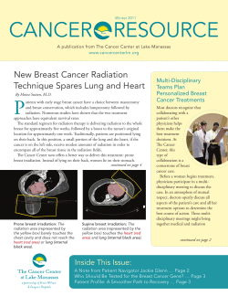


© Copyright 2026