
interview
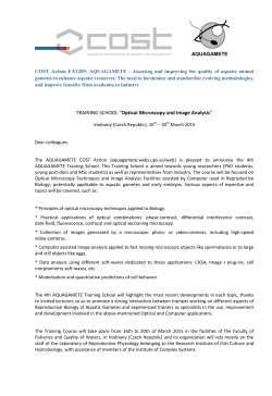
Society
Is microscopy
dangerous or immoral?
You might get this impression with respect to the criticism professor Morten
Laane got last year. Norway’s leading expert on microscopy (b. 1940) was, in the
media, even accused of presenting artifacts and trash science when revealing
his research done together with the zoologist Ivar Mysterud in their search for
answers to what is causing chronic disease in humans after tick bites.
Text Iver Mysterud
Photo Geir Holm og Shutterstock
Story at-a-glance
✓ The retired biologists Morten Laane and Ivar Mysterud were subject
to heated debates in the Norwegian media last year due to their use of
microscopy as a tool to examine blood from people who have become
chronically ill following tick bites.
✓ In this interview Morten Laane deepens central aspects around
microscopy. In the previous interview Ivar Mysterud presents his view of
the criticism.
✓ The microscopy methods used have deep historical roots, however,
are now considerably improved for detailed observations and digital
recordings of bacteria and parasites which reside in red blood cells.
✓ Everyone can easily look through a microscope; however, it demands
great and time-consuming work and experience to become decent at
microscopy. An understanding of the mechanisms of the microscope is
required, and the person executing the microscopy needs to possess
interdisciplinary experience when interpreting what is observed in the
specimens.
✓ Studying the presence of microorganisms in blood is thus demanding.
✓ In comparison to other test methods, microscopy shows the immediate situation in a specimen.
✓ Laane rejects the criticism that they have not observed real spirochetes (spiral-formed bacteria) and co-infections in the blood of patients
sick after tick bites. The structures are not protein fragments from blood
cells during decomposition, by some called pseudo spirochetes. He
has techniques to distinguish beyond any doubt the difference between
spirochetes and pseudo spirochetes and has filmed spirochetes that
actively move in and out of red blood cells. Laane also finds it incorrect
to call anything pseudo spirochetes and he challenges the argument
from opponents suggesting that general basic concepts of blood microbiology may be wrong.
✓ Laane is equally strong in his persuasion that microscopy is an invaluable tool for studying blood of people who have become chronically
sick after tick bites. The results from a new, ongoing research project
might indeed become useful for this group of patients.
78 l Helsemagasinet VOF l SEPTEMBER 2014
In spite of professional criticism, Laane
is equally strong in his persuasion that
microscopy is an invaluable tool for studying blood of chronically sick people. This
can provide a good indication of what is
really wrong with them.
It seems like microscopy as a method is
controversial in the eyes of some professionals. What is the reason for this?
– Lack of knowledge and loss of microbiological knowledge, Laane answers.
He explains that the first bacteria were
discovered in the 17th century by Antonie
van Leeuwenhoek (1632–1723). Among
lots of studies a “tooth spirochete” from a
biofilm in the oral cavity is described and
illustrated in his book Archana naturae
detecta, published in 1695. It is a collection of scientific letters to the Royal Society
of London. Laane emphasizes that this
meant approval at that time. – Physicians
who make statements about microscopy
should, at least, be able to repeat what van
Leeuwenhoek did, even if it is intricate,
Laane thinks.
The method you have used to identify the
presence of potential Borrelia spirochetes
is often presented as new. Why do you
disagree with this?
– Nothing is basically new. The methods
we are currently using are based on
insights in microscopy that gradually have
been developed throughout history, Laane
answers. – However, the techniques that
Morten Laane (left) and Ivar Mysterud study both pictures and film from microscopy on the screen.
for a long time have been used for
observing spirochetes have now been
greatly improved. It is important to
create conditions with reduced oxygen
levels in the living blood specimens.
The reduction of oxygen content seems
to stimulate inactive, hidden bacteria
and parasites to move out of the red
blood cells and become more easily
observed and imaged, the researcher
continues.1
Is it easy to do microscopy?
– Everyone can easily look through a
microscope. However, it demands a
great extent of work and long experience to become good. Only experience
will give a good understanding of what
can be observed. – Thus it is time consuming, Laane points out.
– Modern DNA analyses usually work
in a way that the specimen is sent to a
special laboratory and the sequences are
then returned. Thereafter you search
on a computer. This is to a great extent
less time consuming than working with
a microscope and becoming good at it.
Perhaps this is partly the reason why
microscopy is not widespread and is
less popular today, Laane wonders.
You have for decades lectured students
in the use of the microscope. What is
the main emphasis you have exhorted
them with when they use the microscope?
– A person has to understand how
the microscope works, an essential
“quality” in order to be able to use
it well. Practice and experience in
interpreting what you observe in
specimens is crucial, and frequent
use is necessary to become good,
Laane answers. – It is a pity that
education in traditional microscopy
generally is reduced both in schools,
universities and colleges. Highly
specialized, so-called confocal microscopes are popular and can be linked
to advanced molecular techniques;
however, you lose a great deal which
could have been detected by much
simpler microscopes. We need both
English translation of
article in Helsemagasinet
VOF no. 6/2014
This is a translation of an interview
published in September 2014 in
Helsemagasinet vitenskap & fornuft
(VOF; see www.vof.no) no. 6/2014
on pages 78–81. It directly follows
an interview with Ivar Mysterud on
pages 74–77 in the same issue.
It is translated by Iver Mysterud
and slightly edited in cooperation
with Morten Laane to better fit an
English-speaking audience.
types as they complement each other,
Laane says.
Demanding to study blood
The eager researcher explains that it
is demanding to study blood. Blood
is namely a very complex substance.
– First of all around 40 possible cell
types exist in blood, all of which you
www.vof.no l 79
Society
need to be able to identify. For instance,
if you want to detect a bacterium or
parasite, you need to know something
about “the background conditions”
in blood, he emphasizes. Laane tells
that one needs insight into what fibrin
looks like, i.e. thin threads of protein
in the blood, and changes in cell types
and other structures in blood that can
vary both under preparation and with
disease.
He further states that for optic reasons
red blood cells normally hide both
bacteria and parasites. They are small,
biconcave lenses, causing optic disturbances. Such problems are solved
through a careful swelling so that they
become more flat.
– The Borrelia bacteria at steep angles
may become so long and thin that it is
difficult to see them in a light microscope. By using another technique –
dark field microscopy – you send the
light in from the side, and bacteria and
other structures lighten up against a
black background. This complements
what you see through a light microscope
where the light is sent from below.
The syphilis bacteria and Borrelia bacteria have morphological similarities
of significance for simple detection. In
this textbook effective and very simple
microscopic techniques are described
which later have become more or less
abandoned in medicine. Laane’s father
used these methods as a military field
physician (officer) in the Norwegian
army during the Second World War,
and later when he resumed his private
practice in Tønsberg. At that time Tønsberg was a significant seaport. Many
needed to be tested because sailors
brought contagion from abroad. In
those days microscopic survey methods
were common and accepted amongst
Norwegian physicians. Laane’s interest
for microscopy arose when he observed
this at his father’s office in 1947, i.e.
when he was around seven years old.
At that time he also discovered Bruusgaard’s textbook.
This background has since led to
Microscopy being a large part of Laane’s
professional career, as a biological
researcher. Today he is internationally
recognized as a highly respected expert
in microscopy. Laane is also the author
of four textbooks in microscopy, the
microscope; however, instead observe
What can microscopy contribute with latest from 2007.3
protein fragments from blood cells
that other survey methods cannot?
– You observe the situation immedi- Unfortunately formal mistakes were during decomposition or other artiately in a specimen. You do not need committed in the research application facts, by some called pseudo spirocomplicated instruments. It is depen- for his criticized research project. This chetes. What is your comment to such
dent on understanding how the micro- is something Laane strongly regrets. arguments?
scope works and you need the skill to However, there is no resignation to be – Protein fragments from blood cells
prepare high quality specimens. In found in Laane. On the contrary, he during decomposition do not move in
addition you need broad basic insights is more concerned than ever trying to and out of red blood cells in orderly
in biology in order to interpret what get to the core of what is causing these patterns, Laane answers – the moveyou observe, Laane says. He is the first patients to be chronically sick. Laane ments we observe are typical for living
to admit that other types of survey is disappointed with the scientific structures. – We study living organisms
methods are equally important, for criticism which has emerged. – Why and have filmed spirochetes when they
example identifying the species and haven’t any of the most ardent critics move out of and into red blood cells.
strains of the bacteria being observed. confronted me directly to lead a fruit- The objection concerning pseudo spiroful discussion? Had they done that, chetes is misunderstood and irrelevant,
they could have had the opportunity to he continues.
Early influence
Laane himself was exposed to micros- see for themselves what we have found
copy early in life. His father was a through studies by microscope, he says How do you distinguish between spiphysician and used microscopy in his with slight resignation. – A scientific rochetes and pseudo spirochetes in the
practice. Laane’s father studied medi- debate rarely benefits from extensive microscope?
cine at the University of Oslo in the verbal abuse and criticism in the media, – It is actually incorrect calling something pseudo spirochetes. If you, by
1930s, followed the lectures of, among Laane adds.
pseudo spirochetes, mean protein
others, Professor E. Bruusgaard in
fragments from blood cells during
microbiology. One of the textbooks was False bacteria
Bruusgaard’s Forelesninger over syfilis Some of your critics have objected decomposition, you should call them
[=Lectures on syphilis] from 1930.2 that you do not see spirochetes in the that, Laane reasons. – The structures
80 l Helsemagasinet VOF l SEPTEMBER 2014
actually demonstrate that they are
bacteria, not protein fragments from
red blood cells. Human red blood cells
normally lack DNA (they do not have a
cell nucleus), and green spots usually
identify living bacteria, he says.
Laane has never doubted their observations in the blood of chronically sick
patients after tick bites: – It is without
doubt spirochetes, and they are probably Borrelia bacteria since the patients
have been bitten by ticks. However, the
species we cannot know with certainty
without using other survey techniques,
because microscopy is not sufficient for
this, he stresses.
”Old guys” can contribute
are of varied nature and are connected
to infectious processes and cell membrane events. The concept creates confusion and lack of understanding for
very complex processes linked to red
blood cells. Some “pseudo spirochetes”
emerge because the bacteria have two
membranes, and are able to shed the
outer membrane. In the microscope
this will be observed as a thin residual,
and it often has chromosomes inside,
detectable with specific stains. Thus,
this has nothing to do with protein fragments from red blood cells, however,
it involves parts of a spirochete and
sometimes also the red blood cells, the
experienced researcher explains.
– In a microscope you can thus observe
products from the cell membranes of
the red blood cells. They often contain
some DNA from bacteria that were
inside the blood cell. Such products
conduct so-called Brownian motions.
It is possible to film what you observe
in the microscope and filtrate Brownian motions. On the film you will then
Laane is retired, however, still actively
involved in research. After their previous project was closed by the order of
the Norwegian Board of Health Supervision – due to the regrettable, formal
mistakes in the project application – he
and Ivar Mysterud have now started
a new research project proposed and
directed by the Norwegian Institute of
Public Health – this time with all formalities in order. Their microscopic
method is to be tested and compared
see slow, clear spirochete movements, with other survey techniques. – I hope
Laane says.
that this research can become useful for
the chronic sick patients struggling, as
– Real spirochetes cannot just move out soon as possible, Laane says.
of and into red blood cells, they can also
move against the blood stream. One He adds that major medical advances
can prepare such stream on a micro throughout history have been done by
level in a blood specimen studied under non-physicians. One example is the
microscope, and only living bacteria chemist and bacteriologist Louis Pasteur
can move targeted, Laane emphasizes. (1822–1895), who still did important
discoveries after he had suffered a stroke.
Additionally, the researcher focuses – Also old guys can contribute, says
on a certain simple staining technique, Morten Laane, and hope the critics will
which is important for clarifying what calm down and look forward to seeing
”pseudo spirochetes” in reality are. – If the results of the new research project.
you use the dye “acridine orange” correctly, it binds to nucleic acids (DNA,
RNA), yet not to proteins. Single- Sources:
Laane MM, Mysterud I. A simple method for
stranded nucleic acid, i.e. RNA, emits 1.
the detection of live Borrelia spirochetes in human
orange-red light. A double-stranded blood using classical microscopy techniques.
nucleic acid, DNA, emits green light, Biological and Biomedical Reports 2013; 3: 15-28.
Laane explains. – Green dots are the http://www.biomedicalreports.org/index.php?jo
bacteria’s (simplified) cell nucleus. urnal=bbr&page=article&op=view&path%5B%
5D=98
You can clearly observe this in a fluo- 2. Bruusgaard E. Forelesninger over syfilis. Oslo:
rescence microscope. ”Pseudo spiro- Aeskulap forlag, 1930.
chetes” turn out, by this technique, to 3. Laane MM, Lie T. Moderne mikroskopi med
contain DNA within themselves, which enkle metoder. Oslo: Unipub, 2007.
www.vof.no l 81


© Copyright 2026