
Follicular thyroid neoplasms can be classified as low- and
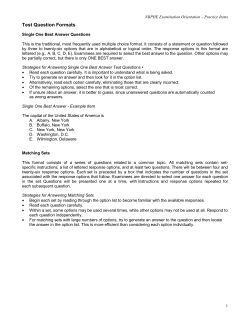
European Journal of Endocrinology (2011) 165 447–453
ISSN 0804-4643
CLINICAL STUDY
Follicular thyroid neoplasms can be classified as low- and
high-risk according to HBME-1 and Galectin-3 expression
on liquid-based fine-needle cytology
Guido Fadda1, Esther Diana Rossi1, Marco Raffaelli3, Alfredo Pontecorvi2, Stefano Sioletic1, Francesca Morassi1,
Celestino Pio Lombardi3, Gian Franco Zannoni1 and Guido Rindi1
Divisions of 1Anatomic Pathology and Histology, 2Endocrinology and 3Endocrine Surgery, ‘Agostino Gemelli’ School of Medicine, Catholic University of
Sacred Heart, Largo Francesco Vito, 1 – 00168 Rome, Italy
(Correspondence should be addressed to G Fadda; Email: [email protected])
Abstract
Design: Fine-needle aspiration biopsy (FNAB) is the most reliable diagnostic tool in the diagnosis of
thyroid nodules. A cytologic diagnosis of follicular neoplasm with atypical cells of undetermined
significance (FN/AUS) implies that the selection of patients between surgery and follow-up is difficult.
In this setting immunocytochemical stainings might be helpful. The efficacy of a panel made up of
HBME-1 and Galectin-3 antibodies is evaluated in cases processed by liquid-based cytology (LBC).
Methods: Out of 7091 thyroid FNAB processed by LBC method, 120 cases undergoing surgery
successively were selected. These cases were classified as benign lesion (BL, eight cases), FN, including
the ACUS category of the Bethesda classification (FN/AUS, 50 cases), suspicious for malignancy (SM,
59 cases), and malignant neoplasm (MN, three cases). Immunostains for HBME-1 and Galectin-3 were
carried out on the LBC slides.
Results: All MN and BL were histologically confirmed. FN/AUS and SM showed a malignancy risk of 24
and 72.9% respectively. The complete immunocytochemical panel was positive in 83.3% of the cases
resulting in malignancy and negative in 87.5% of cases resulting in benign histology. Among the
FN/AUS, the complete positive immunocytochemical panel was detected in 76.9% of cases resulting
as malignant and the complete negative immunocytochemical panel was observed in 96.8% of cases
resulting as benign at histology.
Conclusions: The expression of HBME-1 and Galectin-3 in cases classified as FN/AUS on LBC-processed
FNABs can effectively distinguish lesions, which need immediate surgery (high risk or FNH or Thy 3h)
from those which can be followed-up (low risk or FNL or Thy 3l).
European Journal of Endocrinology 165 447–453
Introduction
Fine-needle aspiration biopsy (FNAB) represents a valid
diagnostic choice combined with clinical and ultrasound evaluation, in the characterization of thyroid
nodules. It may achieve a precise interpretation of the
nature of thyroid lesions in more than 70% of cases and
it leads to a correct clinical strategy (mostly related
to the surgical indication) in more than 90% of cases
(1, 2). It is simple, safe and extremely cost-effective
especially when performed under sonographic guidance
(3, 4). Some problems may arise in the cytologic
diagnosis of lesions exhibiting a predominant microfollicular pattern. The real nature of these lesions can be
assessed only by the detection of capsular and vascular
invasion whereas the needle biopsy may sample only
the center of the nodule. Several studies (5–9) have
investigated different morphologic features (especially
the nuclear ‘atypia’ of the follicular cells) that may
q 2011 European Society of Endocrinology
enable a distinction between the most important
neoplasms, which are follicular adenoma (FA), follicular
carcinoma (FC), and the follicular variant of papillary
carcinoma (FVPC). However, this possibility is still
speculative and the majority of authors feel that the
diagnosis of indeterminate cytology is still unavoidable.
In thyroid pathology, immunohistochemistry has
been used for the differential diagnosis of follicular
and C cell-derived neoplasms. Recently, the expression of
markers of malignancy has also been used as an
additional tool for diagnosing malignant tumors,
regardless of the presence of capsular or vascular
invasion (10).
Among these markers of malignancy HBME-1 and
Galectin-3 have reached high values of specificity and
sensitivity in discriminating benign lesions (BL) from
malignancies.
Galectin-3 is a component of the b-galactosidebinding lectins whose function is still unclear. It appears
DOI: 10.1530/EJE-11-0181
Online version via www.eje-online.org
448
G Fadda and others
to be involved in the cell–cell and cell–matrix modulation and, according to some authors (11), in cell
growth and differentiation. Therefore, this antibody
could play a role in the malignant transformation of
thyroid cells and it is expressed in a high proportion
of carcinomas, especially of the papillary histotype
(11–13).
HBME-1 is an MAB directed against a still unknown
membrane antigen of the microvillar surface of the
mesothelioma cells. It yields positive in neoplastic
follicular cells, mostly from papillary carcinoma, and
negative in BL (14, 15).
However, none of them has shown a diagnostic
accuracy sufficient for using as single antibody in the
diagnosis of malignant thyroid neoplasms (10, 16).
Immunocytochemistry may find new application
fields in fine-needle aspiration cytology, especially
when a diagnosis of follicular neoplasm (FN/atypical
cells of undetermined significance (AUS) – Thy 3) is
made (4, 5). The latter category accounts for 10–25% of
all cases and represents a therapeutic problem because
of the low, though not irrelevant, risk of malignancy.
Therefore, the cases diagnosed as such are often
addressed to surgery that results in a large amount of
inappropriate thyroidectomies. In fact, about 80% of all
thyroid operations for FN/AUS yield a histologic
diagnosis of benign nodule (5–8).
Immunocytochemical techniques can be applied on
liquid-based cytology (LBC) (17–19). This technique
has been introduced to obtain thin-layer smears in
cervical cytology. The excellent results in this field
prompted a wider application to virtually all cytological
branches, including thyroid FNAB (20–27). The
current investigation has been carried out to evaluate
the effectiveness of a panel made up by HBME-1 and
Galectin-3 on LBC in identifying two subsets of FN/AUS
with low and high malignant risk that might be
managed differently to achieve a reduction of inappropriate surgery.
Materials and methods
During the period August 1, 2006–December 31, 2008,
7091 thyroid FNAB were carried out in the Divisions of
Endocrine Surgery, Endocrinology and Anatomic Pathology and Histology of the Catholic University, ‘Agostino
Gemelli’ School of Medicine and Hospital of Rome. The
diagnoses of FN/AUS were 953 (13.4%) and those of
suspicious for malignancy (SM) 227 (3.2%). The series
under investigation included 120 cases, 50 diagnosed
as FN (FN/AUS), corresponding to Thy 3 of the BTA
classification (28), which also includes oxyphilic
neoplasms of the NCI Bethesda classification (9) and
59 lesions classified as SM (Thy 4 of BTA). The
remaining 11 cases included eight cases diagnosed as
non-neoplastic lesion (Thy 2 of the BTA classification)
and three papillary carcinomas, which were used as
www.eje-online.org
EUROPEAN JOURNAL OF ENDOCRINOLOGY (2011) 165
controls. The following inclusion criteria were applied:
i) all cytologic diagnoses reviewed by an experienced
pathologist (G F); ii) presence of abundant material so
that immunocytochemical stainings could be carried
out; and iii) availability of the definitive histology
specimen. The morphologic criteria for the inclusion
of each case processed by LBC in the corresponding
cytologic category were presented in previous papers
of our group (26, 27). The series included 35 male and
85 female patients with a median age of 32.5 years
(range 13–78 years). All aspirations were performed
with 25–27G needles under sonographic guidance
with at least two until four consecutive passes for
each lesion (4).
After the aspiration, the needle was rinsed in Cytolit
solution and then the sample was processed by the
ThinPrep 2000 method (Hologic Cytyc Co., Marlborough, MA, USA), fixed with 95% ethyl alcohol and
stained with Papanicolaou.
Those cases showing a difficult cytologic picture were
discussed by three cytopathologists (E D R, G F, and G F
Z) until a final agreement was achieved.
Immunocytochemical staining was carried out with
the avidin–biotin–peroxidase complex on LBC slides
using the following antibodies: HBME-1 (Dako,
Glostrup, Denmark, 1:100 dilution) and Galectin-3
(Ventana, Tucson, AZ, USA, 1:100 dilution). The slides
were washed three times in PBS and then preincubated
in normal serum with PBS (1:50) for 20 min before
incubating at 4 8C with the primary antibody. Then the
slides were washed three times with PBS and incubated
with the biotinylated secondary antibody conjugated
with the avidin–biotin–peroxidase complex. The
reaction was developed using 3,3 0 -diamino-benzidine.
All slides were counterstained with hematoxylin for 5 s,
rinsed in water three times then mounted for the
microscopic examination. The positivity was assessed,
for each cytological case, when at least 50% of follicular
cells showed a convincing cytoplasmic positivity. The
cytodiagnostic groups of SM (Thy 4) and malignant
neoplasm (MN) (Thy 5) were referred to surgery
according with the BTA and ATA guidelines (29)
whereas the BL (Thy 2) cases were operated on when
esthetic or functional problems occurred.
For the FN (Thy 3) category, which shows a relevant
risk of carcinomatous occurrence, the same guidelines
Table 1 Cytohistological comparison (120 cases).
Histological diagnosis
Cytological
diagnosis
Benign
FA
Malignant
Overall
BL: Thy 2
FN/AUS: Thy 3
SM: Thy 4
MN: Thy 5
8 (100%)
20 (40%)
6 (10.2%)
0
0
19 (38%)
10 (16.9%)
0
0
11 (22%)
43 (72.9%)
3 (100%)
8
50
59
3
BL, benign lesion; FA, follicular adenomas; FN, follicular neoplasm; SM,
suspicious of malignancy; MN, malignant neoplasms.
Immunochemistry of thyroid follicular neoplasms
EUROPEAN JOURNAL OF ENDOCRINOLOGY (2011) 165
Table 2 Cytohistologic comparison for the immunocytochemical
results (all 120 cases).
Histological diagnosis
Immunocytochemistry
Benigna
Malignant
Overall
Concordant negative
HBME-1C, Galectin-3K
HBME-1K, Galectin-3C
Concordant positive
42 (87.5%)
10 (45.4%)
2 (100%)
8 (16.7%)
6 (12.5%)
12 (54.6%)
0
40 (83.3%)
48
22
2
48
a
Including follicular adenomas; P!0.001.
suggest that surgery is routinely advised after a
multidisciplinary discussion.
The surgical specimens were fixed in 10% buffered
formaldehyde, embedded in paraffin and the 5 mm thick
sections were stained with hematoxylin–eosin.
Immunostains for the above-mentioned antibodies,
particularly in those cases that did not show a complete
positivity of the panel in the preoperative cytology, were
carried out also on the histological sections. It was
performed by the ABC method with the same antibodies
used for the immunostains on LBC slides.
Statistical analysis
Sensitivity, specificity, and overall accuracy of FNAB
were calculated as follows: the BL category (Thy 2) were
defined as benign, the categories FN/AUS and SM (Thy 3
and 4), were included in the malignant lesions.
Inadequate specimens and microscopic carcinomas
incidentally found in the final pathologic examination
were excluded and the statistical analysis was performed considering only the histological diagnosis of the
sampled lesion.
Benign and malignant lesions were compared for
cytological and immunocytochemical pattern. Statistical analysis was performed by a commercially available
statistic software package (SPSS 10.0 for Windows –
SPSS, Inc., Chicago, IL, USA). The c2 test was used
for categorical variables up to 6 degrees of freedom.
A P value !0.05 was considered as significant.
All patients had been appropriately informed regarding the use of LBC method for processing their
aspiration samples and a written informed consent
was signed by each of them.
Results
The 120 cytological cases were diagnosed as follows: 8
BL (Thy 2), 50 FN/AUS, which did not include any
oxyphilic neoplasm (Thy 3); 59 SM (Thy 4), 3 papillary
carcinomas (PC) (Thy 5). All cases underwent surgery
and the histologic examination confirmed all three PC
(one showing an insular pattern) and all BL (three
revealed FAs at histology). Of the 50 FN/AUS, 11 (22%)
resulted as carcinoma at histology (ten papillary
449
carcinomas and one FC, Table 1). Forty-three out of
59 cases of SM (72.9%) were malignant; all but one
(which was an insular carcinoma arising from a
papillary carcinoma) was diagnosed as PC.
The application of the immunocytochemical stainings to the ThinPrep 2000 processed material showed
that 96 cases (80%) exhibited a concordant panel
(Table 2). In the group of PC, the complete panel was
positive in all cases and, accordingly, within the benign
group all cases were negative for HBME-1 and Galectin3. In the FN/AUS category (Thy 3, Table 3) the
concordant panel resulted positive in 13 cases (resulting
three benign and ten, 76.9%, MN) and negative in 31
cases (30 resulted benign neoplasms, including 14
adenomas, and one, 3.2%, was a PC). The results of IHC
for the subgroup of SM showed a concordant positivity
for HBME-1 and Galectin-3 in 32 cases (yielding five BL
and 27, 84.4%, malignancies) and a concordant
negativity in ten (five benign and five malignant,
Table 4 and Fig. 1). In the latter category, the low
efficacy of the immunocytochemistry compared with
morphology explains the relatively low values of
specificity and sensitivity for the complete series
(Table 5).
Regarding the cases with discordant panel, the figures
in Table 2 show that the best correlation between
cytoplasmic positivity and occurrence of malignancy
can be observed with HBME-1 instead of Galectin-3. In
fact, out of 22 cases of HBME-1 positivity and Galectin-3
negativity 13 resulted in malignancy (59.1%) whereas
in the opposite instance both cases were benign.
The correlation of the presence of the complete panel
with the histological results for the category of FN/AUS
(Table 3) shows that the simultaneous negativity of both
antibodies is almost always associated with a benign
diagnosis whereas complete positivity is associated with
an MN in 10 out of 13 cases (76.9%).
The descriptive statistical analysis, depicted in
Table 5, shows the differences in sensitivity, specificity,
diagnostic accuracy, positive, and negative predictive
values of the cytologic diagnosis alone, of the immunocytochemistry, of the category of FN/AUS (Thy 3
according to BTA guidelines) and of HBME-1 and
Galectin-3 alone.
In total, 19 cases (15.8%) were selected for the
repetition of the immunostains for HBME-1 and
Table 3 Cytohistological comparison for immunocytochemical
results (only FN: Thy 3; 50 cases).
Histological diagnosis
Immunocytochemistry
Benigna
Malignant
HBME-1C,
HBME-1C,
HBME-1K,
HBME-1K,
Overall
3 (23.1%)
5 (100%)
1 (100%)
30 (96.8%)
39 (78%)
10 (76.9%)
0
0
1 (3.2%)
11 (22%)
a
Galectin-3C
Galectin-3K
Galectin-3C
Galectin-3K
Including follicular adenomas; P!0.001.
www.eje-online.org
450
G Fadda and others
EUROPEAN JOURNAL OF ENDOCRINOLOGY (2011) 165
Table 4 Cytohistological comparison for immunocytochemical
results (only suspicious of malignancy diagnoses, Thy 4, 59
cases).
Histological diagnosis
Immunocytochemistry
Benigna
Malignant
HBME-1C,
HBME-1C,
HBME-1K,
HBME-1K,
Overall
5 (15.6%)
5 (31.3%)
1 (100%)
5 (50%)
16 (27.1%)
27 (84.4%)
11 (68.7%)
0
5 (50%)
43 (72.9%)
a
Galectin-3C
Galectin-3K
Galectin-3C
Galectin-3K
Including follicular adenomas; P!0.001.
Galectin-3 on the histological specimens and all were
confirmed. The selection criteria included 12 cases
exhibiting a discordant expression of the antibodies of
the panel, five FN/AUS and two cytologically BL.
Discussion
Thyroid nodules are a common finding in everyday
clinical practice, whereas only a small percentage of
them is malignant. The efficacy of FNAB in making a
correct diagnosis of both malignant and BL is definitely
stated in literature (1, 9, 30). On the other hand, in
those cases defined as FN, the cytologic examination is
not always able to distinguish malignant or suspicious
nodules requiring surgical treatment from those which
can be followed-up safely.
The category of FN (Thy 3 in the BTA classification)
represents an indeterminate category responsible for
a high rate of unnecessary thyroidectomies (5–8). The
problem may be due to the discrepancies between
the cytologic and the corresponding histologic pictures
because this category embraces BL showing scattered
nuclear pleomorphism (e.g. hyperplastic nodules in
Hashimoto’s thyroiditis and toxic adenomas) and
malignant lesions with moderate pleomorphism of the
follicular cells (e.g. well-differentiated FC).
The cytologic criteria for diagnosing FN are not
universally accepted and the category itself is debated.
Though the cytologic picture of a follicular-patterned
proliferation is well established, the diagnostic features
which are helpful to assess the malignant nature of the
nodule (e.g. nuclear atypia, nuclear clearing) are much
less reproducible (31–33).
To address the problem of the management of these
indeterminate diagnoses, immunohistochemical
procedures using antibodies against antigens associated
with malignancy were introduced. Among them
Galectin-3 and HBME-1 have shown the highest
reliability, particularly in differentiated carcinoma
(10–13, 34).
The quality of the immunocytochemical reaction
carried out on LBC slides is better than conventional
smears in terms of morphologic details and purity of
www.eje-online.org
background, as some studies have demonstrated,
including some from our group (17–19, 34).
The combination of the antibody panel made up of
HBME-1 and Galectin-3 results in a significant increase
of the diagnostic accuracy of the technique (78.3%
compared with 54.2% of the morphology alone) but this
improvement turns out even more striking in FN as
both the overall and the concordant panel results are
considered (82 and 91.1% respectively), as is also
depicted in Table 5. It is important to emphasize the
relevance of an appropriate cellularity to assess the
positivity or negativity of the immunocytochemical
reaction. In case of scant cellularity, which is defined as
not 6 groups of follicular cells, the immunocytochemical diagnosis should not be made.
An immunocytochemical-based classification of indeterminate thyroid lesions has never been attempted
because of the difficulty in performing immunocytochemistry on conventional smears. Nevertheless, the
uncertainty regarding the most appropriate treatment
of FNs (Thy 3 in the BTA classification, ACUS and FNs in
the Bethesda classification) that often sent to surgery
but result in being found as benign on the definitive
histology, has prompted the development of different
techniques to define the cancer risk preoperatively (e.g.
immunocytochemical and molecular studies (35–40)).
The results of these investigations are quite promising
but there is still a relevant amount of lesions that
undergo unnecessary surgery or, on the other hand,
may harbor an undiagnosed cancer. The current
investigation achieves results similar to the molecular
studies with a significantly cost-effective method still
based on the morphologic evaluation of the follicular
cells. The results of Nikiforov et al. (39, 40) compared
with the current data show a better positive predictivity for the molecular analysis compared with
90
P<0.001
80
70
60
50
%
Overall
40
Foll. Neopl.
30
Suspicious
20
10
0
Conc. Neg. Conc. Pos.
HB+GAL–
HB–GAL+
Antibody panel
Figure 1 Malignancy rates for different antibodies combinations in
the cytological categories. Conc. Neg., concordant negative panel;
Conc. Pos., concordant positive panel; HBCGALK, HBME-1
positive/Galectin-3 negative; HBKGALC, HBME-1 negative/
Galectin-3 positive; Foll. Neopl., follicular neoplasm.
Immunochemistry of thyroid follicular neoplasms
EUROPEAN JOURNAL OF ENDOCRINOLOGY (2011) 165
451
Table 5 Descriptive statistics for the morphologic and immunocytochemical yieldings in all cytologic categories and only in follicular
neoplasm lesions. Data are presented as %.
FN panels
a
Sensitivity
Specificity
Diagnostic accuracy
PPV
NPV
b
Cytology
ICC only
All
Conc.
HBME-1
Galectin-3
100
12.7
54.2
51
100
89.7
67.7
78.3
72.2
87.5
91.7
78.9
82
57.9
96.8
90.9
91.2
91.1
76.9
96.9
72.3
88
80
89.6
71
80
72.3
76.6
69
84
a
Follicular neoplasms; Thy 3, and suspicious of malignancy; Thy 4, are counted as malignant; ICC, immunocytochemistry.
All combinations of antibodies where at least one of them was positive were accounted as malignant; All panels, all possible combinations of HBME-1 and
Galectin-3 expressions (see Table 2); Conc. panels, HBME-1 and Galectin-3 are both positive or negative; FN, follicular neoplasm (Thy 3); PPV, positive
predictive value; NPV, negative predictive value.
b
immunocytochemistry in the indeterminate (FN/AUS)
lesions (100 vs 76.9%) but a lower negative predictive
value (16.2 vs 3.2%). As a rule in our institution,
lesions classified as FN (Thy 3) undergo immunocytochemical analysis for HBME-1 and Galectin-3
expression: those showing a simultaneous positivity
for both antibodies are referred to surgery whereas
those exhibiting a simultaneous negativity should be
carefully followed-up, with the surgical option being
held for potential future use.
Regarding the SM lesions, our results (Table 4) may
look somewhat surprising but they can be easily
explained. This category is usually cytologically
dyshomogeneous being composed of a mixture of
large atypical cells and smaller thyrocytes as is often
observed in cases of FVPC. Thus, the chosen cutoff of
50% expression of HBME-1 and Galectin-3 may result in
some false-negative cases that nonetheless do not
represent a true diagnostic problem as the inclusion of
a case in the SM category is mainly based on the
cytologic features.
As a consequence of these results, HBME-1 has a far
better diagnostic accuracy than Galectin-3 and, in case
of availability of only one antibody, the use of the former
as diagnostic aid in the classification of a thyroid nodule
is strongly suggested.
Based on the results (Fig. 1) of the present
investigation, the suggested management of a thyroid
lesion undergoing FNAB includes: i) accurate examination of clinical history and serum analysis to rule out
the possibility of a toxic adenoma or a Hashimoto’s
thyroiditis; ii) evaluation of the nuclear atypia of the
follicular cells to define the inclusion of the case in the
benign, FN/AUS, SM, and malignant categories; and iii)
if the lesion is classified as FN/AUS (Thy 3) the
assessment of the expression of HBME-1 and Galectin3 in the follicular cells would identify two additional
subtypes: low risk (Thy3l or FNL – resulting HBME-1
and Galectin-3 negative) and high risk (Thy 3h or FNH
– HBME-1 and Galectin-3 positive). These subgroups are
characterized by a significant difference in the risk that
a MN is being detected at histology (76.9% when the
concordant panel is positive and 3.2% when the same
panel is negative).
Therefore, the application of this antibody panel to
any cytological classification that includes the category
of FN/AUS may represent the first step in achieving a
correct preoperative diagnosis of thyroid nodules. It
improves the accuracy of the morphologic evaluation of
these indeterminate lesions enabling a better selection
of patients as candidates for surgery from those who
should be treated pharmacologically. In this perspective,
LBC is an improvement of conventional cytology as it
represents an invaluable storage for the cellular
material that can be successfully used for additional
investigations.
Declaration of interest
The authors declare that there is no conflict of interest that could be
perceived as prejudicing the impartiality of the research reported.
Funding
This research did not receive any specific grant from any funding
agency in the public, commercial or not-for-profit sector.
Acknowledgements
The authors thank Elena Visca, Flavia Barbetti, Laura Cammarota,
Alessia Maffione, and Antonella Evangelista for their invaluable
technical assistance.
References
1 Gharib H & Goellner JR. Fine-needle aspiration biopsy of the
thyroid. Annals of Internal Medicine 1993 118 282–289.
2 Galera-Davidson H & Gonzalez-Campora R. Thyroid. In Comprehensive Cytopathology, ch 23, 3rd edn, pp 633–670. Eds M Bibbo &
D Wilbur. Philadelphia: Saunders Elsevier, 2008.
3 Danese D, Sciacchitano S, Farsetti A, Andreoli M & Pontecorvi A.
Diagnostic accuracy of conventional versus sonography-guided
fine-needle aspiration biopsy of thyroid nodules. Thyroid 1998 8
15–21. (doi:10.1089/thy.1998.8.15)
4 Bishop-Pitman M, Abele J, Ali SZ, Duick D, Elsheikh TM, Brooke
Jeffrey R, Powers CN, Randolph G, Renshaw A & Scoutt L.
Techniques for thyroid FNA: a synopsis of the National Cancer
www.eje-online.org
452
5
6
7
8
9
10
11
12
13
14
15
16
17
18
G Fadda and others
Institute thyroid fine-needle aspiration state of the science
conference. Diagnostic Cytopathology 2008 36 407–424. (doi:10.
1002/dc.20829)
Baloch ZW & LiVolsi VA. Follicular-patterned lesions of the
thyroid: the bane of the pathologist. American Journal of Clinical
Pathology 2002 117 143–150. (doi:10.1309/8VL9-ECXYNVMX-2RQF)
Baloch ZW, Fleisher S, LiVolsi VA & Gupta PK. Diagnosis of
“follicular neoplasm”: a gray zone in thyroid fine-needle aspiration
cytology. Diagnostic Cytopathology 2002 26 41–44. (doi:10.1002/
dc.10043)
Poller DN, Ibrahim AK, Cummings MH, Mikel JJ, Boote D &
Perry M. Fine-needle aspiration of the thyroid. Importance of
an indeterminate diagnostic category. Cancer Cytopathology 2000
90 239–244. (doi:10.1002/1097-0142(20000825)90:4!239::
AID-CNCR7O3.0.CO;2-S)
Greaves TS, Olvera M, Florentine BD, Raza AS, Cobb CJ, TsaoWei DD, Groshen S, Singer P, Lopresti J & Martin SE. Follicular
lesions of thyroid. A 5-year fine-needle aspiration experience.
Cancer Cytopathology 2000 90 335–341.
Baloch ZW, LiVolsi VA, Asa SL, Rosai J, Merino MJ, Randolph G,
Vielh P, DeMay RM, Sidawy MK & Frable WJ. Diagnostic
terminology and morphologic criteria for cytologic diagnosis of
thyroid lesions: a synopsis of the National Cancer Institute
Fine-needle aspiration state-of-science conference. Diagnostic
Cytopathology 2008 36 425–437. (doi:10.1002/dc.20830)
Griffith OL, Chiu CG, Gown AM, Jones SJM & Wiseman SM.
Biomarker panel diagnosis of thyroid cancer: a critical review.
Expert Review of Anticancer Therapy 2008 8 1399–1413. (doi:10.
1586/14737140.8.9.1399)
Bartolazzi A, Gasbarri A, Papotti M, Bussolati G, Lucante T,
Khan A, Inohara H, Marandino F, Orlandi F, Nardi F, Vecchione A,
Tecce R & Larsson O. Application of an immunodiagnostic
method for improving preoperative diagnosis of nodular thyroid
lesions. Lancet 2001 357 1644–1650. (doi:10.1016/S01406736(00)04817-0)
Papotti M, Volante M, Saggiorato E, Deandreis D, Veltri A &
Orlandi F. Role of galectin-3 immunodetection in the cytological
diagnosis of thyroid cystic papillary carcinoma. European
Journal of Endocrinology 2002 147 515–521. (doi:10.1530/eje.
0.1470515)
Herrmann ME, LiVolsi VA, Pasha TL, Roberts SA, Wojcik EM &
Baloch ZW. Immunohistochemical expression of galectin-3 in
benign and malignant thyroid 5-year lesions. Archives of Pathology
& Laboratory Medicine 2002 126 710–713.
Miettinen M & Kovatich AJ. HBME-1 monoclonal antibody useful
in the differential diagnosis of mesothelioma, adenocarcinoma and
soft tissue and bone tumors. Applied Immunohistochemistry and
Molecular Morphology 1995 3 115–122.
Cheung CC, Ezzat S, Freeman JL, Rosen IB & Asa SL. Immunohistochemical diagnosis of papillary thyroid carcinoma. Modern
Pathology 2001 14 338–342. (doi:10.1038/modpathol.
3880312)
Rossi ED, Raffaelli M, Mule’ A, Miraglia A, Lombardi CP,
Vecchio FM & Fadda G. Simultaneous immunohistochemical
expression of HBME-1 and galectin-3 differentiates papillary
carcinomas from hyperfunctioning lesions of the thyroid.
Histopathology 2006 48 795–800. (doi:10.1111/j.1365-2559.
2006.02428.x)
Dabbs D, Abendroth CS, Grenko RT, Wang X & Gail ER.
Immunocytochemistry on the ThinPrep processor. Diagnostic
Cytopathology 1997 17 388–392. (doi:10.1002/(SICI)10970339(199711)17:5!388::AID-DC14O3.0.CO;2-J)
Tabbara SO, Sidaway MK, Frost AR, Brosky KR, Coles V, Hecht S,
Radcliffe G & Sherman ME. The stability of estrogen and
progesterone receptor expression on breast carcinoma cells stored
as preservCyt suspension and as ThinPrep slides. Cancer
Cytopathology 1998 84 355–360. (doi:10.1002/(SICI)10970142(19981225)84:6!355::AID-CNCR7O3.0.CO;2-H)
www.eje-online.org
EUROPEAN JOURNAL OF ENDOCRINOLOGY (2011) 165
19 Fadda G, Rossi ED, Mulè A, Miraglia A, Vecchio FM & Capelli A.
Diagnostic efficacy of immunocytochemistry on fine needle
aspiration biopsies processed by thin-layer cytology. Acta Cytologica 2006 50 129–135. (doi:10.1159/000325920)
20 Biscotti CV, Hollow JA, Toddy SM & Easley KA. ThinPrep
versus conventional smears cytological preparations in the analysis
of thyroid fine-needle aspiration 5-year specimens. American
Journal of Clinical Pathology 1995 104 150–153.
21 Frost AR, Sidawy MK, Ferfelli M, Tabbara SO, Bronner NA,
Brosky KR & Sherman ME. Utility of thin-layer preparations in
thyroid fine-needle aspiration. Cancer Cytopathology 1998 84
17–25. (doi:10.1002/(SICI)1097-0142(19980225)84:1!17::
AID-CNCR4O3.0.CO;2-C)
22 Michael CW & Hunter B. Interpretation of fine-needle aspirates
processed by ThinPrep technique: cytologic artifacts and diagnostic pitfalls. Diagnostic Cytopathology 2000 23 6–13. (doi:10.
1002/1097-0339(200007)23:1!6::AID-DC2O3.0.CO;2-F)
23 Scurry JP & Duggan MA. Thin layer compared to direct smear in
thyroid fine-needle aspiration. Cytopathology 2000 11 104–115.
(doi:10.1046/j.1365-2303.2000.00228.x)
24 Afify AM, Liu J & Al-Khafaji BM. Cytologic artifacts and pitfalls of
thyroid fine-needle aspiration using ThinPrep. Cancer Cytopathology 2001 93 179–186. (doi:10.1002/cncr.9027)
25 Cochand-Priollet B, Prat JJ, Polivka M, Thienpont L, Dahan H,
Wassef M & Guillausseau PJ. Thyroid fine needle aspiration: the
morphological features on ThinPrep slide preparations. Eighty
cases with histological control. Cytopathology 2003 14 343–349.
(doi:10.1046/j.0956-5507.2003.00098.x)
26 Fadda G, Rossi ED, Raffaelli M, Mulè A, Pontecorvi A, Miraglia A,
Lombardi CP & Vecchio FM. Fine-needle aspiration biopsy of
thyroid lesions processed by thin-layer cytology: one-year
institutional experience with histologic correlation. Thyroid
2006 16 975–981. (doi:10.1089/thy.2006.16.975)
27 Rossi ED, Raffaelli M, Zannoni GF, Pontecorvi A, Mulè A, Callà C,
Lombardi CP & Fadda G. Diagnostic efficacy of conventional as
compared to liquid-based cytology in thyroid lesions. Evaluation of
10,360 fine needle aspiration cytology cases. Acta Cytologica 2009
53 659–666. (doi:10.1159/000325407)
28 British Thyroid Association Guidelines for the Management of
Thyroid Cancer. 2nd edn, 2007.
29 Cooper DS, Doherty GM, Haugen BR, Kloos RT, Lee SL, Mandel SJ,
Mazzaferri EL, McIver B, Pacini F, Schlumberger M, Sherman SI,
Steward DL & Tuttle RM. Revised American Thyroid Association
management guidelines for patients with thyroid nodules and
differentiated thyroid cancer. Thyroid 2009 19 1–48. (doi:10.
1089/thy.2009.0110)
30 Ravetto C, Colombo L & Dottorini ME. Usefulness of fine-needle
aspiration in the diagnosis of thyroid carcinoma. A retrospective
study in 37,895 patients. Cancer Cytopathology 2000 90
357–363.
(doi:10.1002/1097-0142(20001225)90:6!357::
AID-CNCR6O3.0.CO;2-4)
31 Nga ME, Kumarasinghe MP, Tie B, Sterrett GF, Wood B, Walsh J,
Nguyen H, Whyte A & Frost FA. Experience with standardized
thyroid fine-needle aspiration reporting categories. Cancer Cytopathology 2010 118 423–433 (published online). (doi:10.1002/
cncy.20111)
32 Yang J, Schnadig V, Logrono R & Wassermann PG. Fine-needle
aspiration of thyroid nodules: a study of 4703 patients with
histologic and clinic correlations. Cancer 2007 111 306–315.
(doi:10.1002/cncr.22955)
33 Faquin WC & Baloch ZW. Fine-needle aspiration of follicular
patterned lesions of the thyroid: diagnosis, management and
follow-up according to NCI recommendations. Diagnostic Cytopathology 2010 38 731–739. (doi:10.1002/dc.21292)
34 Rossi ED, Raffaelli M, Minimo C, Mule’ A, Lombardi CP, Vecchio FM
& Fadda G. Immunocytochemical evaluation of thyroid neoplasms on thin-layer smears from fine-needle aspiration biopsies.
Cancer Cytopathology 2005 105 87–95. (doi:10.1002/cncr.
21026)
EUROPEAN JOURNAL OF ENDOCRINOLOGY (2011) 165
35 Sack MJ, Astengo-Osuna C, Lin BT, Battifora H & LiVolsi VA.
HBME-1 immunostaining in 5-year thyroid fine-needle aspirations: a useful marker in the diagnosis of carcinoma. Modern
Pathology 1997 10 668–674.
36 Beesley MF & McLaren KM. Cytokeratin 19 and galectin-3
immunohistochemistry in the differential diagnosis of solitary
thyroid nodules. Histopathology 2002 41 236–243. (doi:10.1046/
j.1365-2559.2002.01442.x)
37 Saggiorato E, De Pompa R, Volante M, Cappia S, Arecco F, Dei
Tos AP, Orlandi F & Papotti M. Characterization of thyroid
“follicular neoplasms” in fine-needle aspiration cytologic specimens using a panel of immunohistochemical markers: a proposal
for clinical application. Endocrine-Related Cancer 2005 12
305–317. (doi:10.1677/erc.1.00944)
38 Schmitt FC, Longatto-Filho A, Valent A & Vielh P. Molecular
techniques in cytopathology practice. Journal of Clinical Pathology
2008 61 258–267. (doi:10.1136/jcp.2006.044347)
Immunochemistry of thyroid follicular neoplasms
453
39 Nikiforov YE, Steward DL, Robinson-Smith TM, Haugen BR,
Klopper JP, Zhu Z, Fagin JA, Falciglia M, Weber K & Nikiforova MN.
Molecular testing for mutations in improving the fine-needle
aspiration diagnosis of thyroid nodules. Journal of Clinical
Endocrinology and Metabolism 2009 94 2092–2098. (doi:10.
1210/jc.2009-0247)
40 Ohori NP, Nikiforova MN, Schoedel KE, LeBeau SO, Hodak SP,
Seethala RR, Carty SE, Ogilvie JB, Yip L & Nikiforov YE.
Contribution of molecular testing to thyroid fine-needle aspiration
cytology of “follicular lesion of undetermined significance/atypia
of undetermined significance”. Cancer Cytopathology 2010 118
17–23. (doi:10.1002/cncy.20063)
Received 21 April 2011
Accepted 1 July 2011
www.eje-online.org


© Copyright 2026