
ENCCA
ENCCA DELIVERABLES
Project no. 261474
ENCCA
EUROPEAN NETWORK for CANCER research in CHILDREN and
ADOLESCENTS
Network of Excellence
Deliverable Number: 9.1
Title:
Common guidelines for diagnostic approaches
to leukemias
Due Delivery Date: M24
Actual Delivery Date: M24
Start date of project: 1 January 2011
Duration: 48 months
Project co-funded by the European Commission within the Seventh Framework Programme
(2011-2014)
Dissemination Level
Public
PU
1
ENCCA DELIVERABLES
Summary
In this deliverable we describe consensus recommendations on the diagnosis of childhood
acute leukemia from a European viewpoint based on surveys of the groups participating to
the AIEOP-BFM ALL 2009 study, a 7-country-based international frontline treatment trial on
acute lymphoblastic leukemia (ALL) in children and adolescents, a survey of the work group
DCOG Molecular Research of the Dutch Childhood Oncology Group, and the Biology &
Diagnosis Committee of the International BFM Study Group, the largest initiative on
childhood leukemias worldwide which unifies cooperative national study groups on
childhood leukemia from more than 30 different, mostly European countries. Topics covered
relate to cytomorphology, immunophenotyping, cyto- and molecular genetics, and the
evaluation of measures of treatment response.
The recommendations provide a common ground for systematic implementation of
molecularly-based diagnostic strategies in acute leukemias to be developed within ENCCA
and, therefore, can be seen as a prerequisite for the implementation of rational molecular
information-based treatment approaches in pediatric hematology and oncology.
The diagnostic guidelines will be published on the website of the international BFM study
group (http://www.bfm-international.org).
2
ENCCA DELIVERABLES
Description
Common guidelines for diagnostic approaches to leukemias
Correspondence to:
Susanne Kilian, PhD, MLaw
University Medical Center Schleswig-Holstein
Department of General Pediatrics ▪ Campus Kiel
Schwanenweg 20 ▪ 24105 Kiel, Germany
Tel.: +49 (0)431 597-3947 ▪ Fax: -3966
Email: [email protected]
Disclaimer:
While the advice and information in these guidelines is believed to be true and accurate,
neither the authors, the International BFM Study Group nor the European Network for
Cancer Research in Children and Adolescents accept any legal responsibility for the
content of this document.
Date of recommendation review:
January 2013
Writing group:
ENCCA WP9 and I-BFM Biology & Diagnosis Committee members
Declarations of Interest:
None of the authors are considered to have a conflict of interest with regards to the
recommendations made in this document.
3
ENCCA DELIVERABLES
Contents
Deliverable in context of WP9
5
Recommendations for the diagnosis of childhood acute lymphoblastic leukemia
General information
Clinical presentation and diagnosis
Prognostic factors and risk-adapted treatment
6
Diagnosis of childhood acute lymphoblastic leukemia
Diagnostic methods
Cytomorphology
Immunophenotyping
Cytogenetics and FISH
Molecular genetics
Treatment response evaluation
Biobanking
12
Recommendations for the diagnosis of childhood acute myeloid leukemia
General information
Clinical presentation and diagnosis
Prognostic factors and risk-adapted treatment
19
Diagnosis of childhood acute myeloid leukemia
Diagnostic methods
Cytomorphology
Immunophenotyping
Cytogenetics and FISH
Molecular genetics
Treatment response evaluation
Biobanking
22
References
24
4
ENCCA DELIVERABLES
Deliverable in context of WP9
WP9 aims to establish a harmonized and integrated approach to the rational introduction of
molecularly targeted treatment in clinical trials on leukemias. In order to achieve this
ultimate goal, the development of standardized comprehensive diagnostic approaches as
well as biobanking, and the establishment of a common pipeline for molecular diagnostics
in a European virtual laboratory on leukemias are necessary.
These recommendations are neither obligatory nor is it necessary to follow all of them.
5
ENCCA DELIVERABLES
Recommendations for the diagnosis of childhood acute lymphoblastic
leukemia (ALL)
General information
ALL represents the malignant proliferation of lymphoid cells blocked at an early stage of
differentiation and is the most common malignancy in children. It accounts for
approximately 25% of all childhood cancers and about 80% of childhood leukemias. The
annual incidence rate of childhood ALL varies world-wide between approximately one and
four new cases per 100,000 children younger than 15 years, with a peak incidence at
approximately two to five years of age. More affluent countries tend to have higher
incidence rates. However, incidence rates for childhood ALL do not only vary between
countries, but also by ethnicity within countries: in the USA rates are highest in Hispanic
children and higher in white compared to black children. More than 60% of patients
diagnosed with ALL are children. Treatment results in childhood ALL are one of the true
success stories of clinical oncology with current overall cure rates of approximately 80% in
developed countries. These results are reached by application of intensive multiagent
chemotherapeutic regimens and in specific patient subgroups additional radiotherapy
and/or hematopoietic stem cell transplantation (HSCT). Modern treatment regimens consist
of at least four phases: (1) an induction period aiming at an initial remission induction within
approximately 4 to 6 weeks through the use of multiple cancer chemotherapeutic drugs; (2)
consolidation/intensification and reinduction segments to eradicate residual leukemic blasts
in patients who are in remission by morphologic criteria; (3) extracompartment therapy such
as central nervous system (CNS) preventive therapy, and (4) a maintenance period to
further stabilize remission by suppressing re-emergence of a drug-resistant clone through
continuing reduction of residual leukemic cells. As certain clinically and biologically distinct
patient subgroups with ALL have a particular poor outcome on standard ALL treatment,
clinical protocols specifically addressing the potential therapeutic needs of these subgroups
have been initiated in the recent past (e.g., hybrid protocols for infants, and imatinibincluding regimens for BCR/ABL1-positive ALL).
Clinical presentation and diagnosis
The initial clinical presentation of a child with ALL largely depends on the extent of the
leukemic infiltration of the bone marrow and extramedullary sites. Typical clinical signs are
fever, pallor, fatigue, bruises, enlargement of liver, spleen and lymph nodes, and pain (e.g.,
bone pain). In most patients, white blood cell counts show anemia, thrombocytopenia and
granulocytopenia with or without concommitant leukocytosis. The diagnosis of ALL is
usually made by cytomorphological and cytochemical examination of a bone marrow
aspirate and in difficult cases by trephine biopsy and is established when at least 25%
lymphoblasts are present in the marrow. CNS involvement (CNS3 status) is diagnosed by
the presence of blasts in the cerebrospinal fluid (CSF; for definition see Table 1) or if
intracerebral infiltrates are detected by cross-sectional radiological imaging. Initial
diagnostics are complemented by flowcytometry-based immunophenotyping to gain
information on the blasts expression of lymphoid differentiation-associated antigens as
measure by the reactivity to specific monoclonal antibodies and to determine the cellular
6
ENCCA DELIVERABLES
DNA content of leukemic cells. In addition, a combined approach using cytogenetic and
molecular genetic techniques is used for the detection of genetic aberrations, such as nonrandom recurrent chromosomal translocations or their molecular equivalents (e.g., the
t(9;22) or the BCR/ABL1 fusion transcript). Molecular-genetic techniques and/or flow
cytometry are also used to monitor disease burden during therapy by measuring minimal
residual disease (MRD). A last important issue addresses the definition of what is called
complete remission and relapse: complete remission is defined as the absence of leukemic
blasts in blood and CSF, fewer than 5% lymphoblasts in bone marrow aspiration smears,
and no evidence of localized disease. Relapse is defined as the recurrence of lymphoblasts
or localized leukemic infiltrates at any site.
Prognostic factors and risk-adapted treatment
Continuing research on the clinical and biological aspects of ALL has identified numerous
features with prognostic potential some of which are displayed in Table 1. On modern
protocols, risk-adapted therapy reflecting the probability of treatment failure has become a
common feature in the clinical management of childhood ALL. For this purpose, the initially
assessed prognostic factors are used to estimate an individual patient’s risk of relapse and
to adjust the required treatment intensity by therapy stratification into different risk groups
(e.g., standard/low, intermediate, high). The prognostic significance of an inadequate early
reduction of leukemic blasts in the peripheral blood was first described by the BFM study
group and confirmed by several other study groups. Of importance, the specificity of
response evaluation might vary with the composition of the induction regimen and the time
point of response evaluation. However, although a poor early response to induction therapy
as described above is highly predictive of treatment failure, the majority of recurrences
occur in the large group of patients with an adequate morphological response to treatment.
Of advantage in this context, the sub-microscopic assessment of MRD is approximately
1000 to 10000-fold more sensitive compared to methods based on morphological detection
and provides excellent prognostic information. Although most of the experience on MRD in
clinical settings was gained through DNA-PCR-based detection of leukemic clone-specific
immunoglobulin and/or T-cell receptor gene rearrangements, it was shown that flowcytometry-based analyses by detection of specific antigen patterns of the leukemic clone
yield sensitive and reliable results comparable to PCR-based methods.
Remission induction treatment
Contemporary treatment approaches for childhood ALL aim at an initial remission induction
and restoration of normal hematopoiesis within approximately 4 to 6 weeks. In most study
groups this goal is achieved in approximately 98% of patients through the systemic
application of three drugs (glucocorticoid, vincristine, L-asparaginase) to which an
anthracycline may be added as a fourth drug. On ALL-BFM protocols, remission induction is
initiated by a 7-day monotherapy with orally administered prednisone (and one intrathecal
dose of intrathecal methotrexate on day 1), which isparticularly useful in avoiding
complications related to extensive tumor cell lysis. Undoubtedly, the dose intensity of the
induction phase can have a major impact on the overall treatment outcome. Nevertheless,
in specific subgroups of childhood ALL, the necessity of a four-drug induction regimen is
7
ENCCA DELIVERABLES
subject to debate and it is, for example, unclear if addition of an anthracycline to a threedrug induction regimen is of real benefit to certain low- or intermediate-risk patients.
Another frequently discussed issue addresses the choice of the glucocorticoid for optimal
induction. Despite some debate on a truly equivalent dose, compared to prednisolone,
dexamethasone appears to have a stronger antileukemic effect in vitro and has been shown
to provide better leukemic CNS control and lower relapse rates. However, dexamethasone
was also associated with increased side effects including severe infectious complications.
The 2% of patients not in remission after induction therapy will either have died of
treatment- or disease-related complications or display nonresponsive disease. The latter
group includes patients that will achieve only delayed remission or show resistant disease.
Because of the poor prognosis of this minor non-responsive patient population, alternative
therapeutic approaches should be considered early during the disease process.
Consolidation/intensification and reinduction treatment
Eradication of residual leukemic blasts in patients who are in remission by morphologic
criteria
is
the
primary
aim
of
consolidation/intensification
treatment.
Consolidation/intensification treatment is necessary as patients successfully induced into
remission, but not given additional treatment, usually relapse within months. A so-called
reinduction or delayed intensification treatment can further enhance the effect of previous
consolidation/intensification therapy. As consolidation/intensification phases administered in
protocols of the large study groups on treatment of childhood ALL are variable and may
differ, for example, with regard to amounts, timing, and number of drug doses, drug
composition and overall treatment context, the direct contributions of most of these
consolidation/intensification strategies and/or their individual components are difficult to
assess and associated with limited generalizability. Today, most protocols use high-dose
methotrexate (combined with folinic acid rescue) together with 6-mercaptopurine (6-MP)
and/or prolonged administrations of asparaginase in consolidation/intensification.
Reinduction treatment mainly consists of a late repetition of the initial remission induction
and early intensification phases. A randomized trial by the Children’s Cancer Group
applying an augmented BFM protocol showed that intensified consolidation and doubledelayed intensification can further improve the outcome of high-risk patients with a slow
initial treatment response. Of interest in this context, in a recent subsequent trial on higherrisk patients with a rapid marrow response to induction therapy by the same group, the
investigators demonstrated an improved event-free survival for more intensive but not for
longer postinduction intensification treatment. Unfortunately, further intensification of
treatment and the related higher doses of glucocorticoids have been associated with a high
incidence of osteonecrosis, especially in older children. Consequently, some investigators
suggest glucocorticoid administration in intensification/consolidation on alternate weeks for
children older than 10 years to improve complication rates.
Central nervous system-directed therapy
CNS-directed therapy has become a prerequisite for successful treatment of childhood ALL.
Before its introduction in the 1960s, more than 50% of children with ALL suffered from
disease recurrence originating from the CNS. This high rate could be reduced to less than
8
ENCCA DELIVERABLES
5% through the introduction of cranial irradiation, intrathecal chemotherapy with
methotrexate alone or in combination with other drugs (cytarabine, hydrocortisone), and
systemic application of chemotherapeutics with adequate penetration into the CNS (highdose methotrexate, dexamethasone, high-dose cytarabine). The intensity of CNS-directed
treatment is adjusted according to the risk of ALL relapse originating from the CNS, the
most important risk factor being overt CNS involvement at diagnosis (CNS3). Additional risk
factors include a high initial white blood cell count, pro-B or precursor T-cell
immunophenotype, t(9;22) or t(4;11), and a traumatic lumbar puncture with identifiable blast
cells present at diagnosis. CNS-directed therapy may differ in the number of intrathecal
injections and/or intrathecally applied drugs, as well as in the inclusion of cranial irradiation
at different doses. Excluding infants, most clinical protocols administering intensive
systemic therapy still recommend preventive cranial irradiation (12 or 18 Gy) for high-risk
patients and/or those with a precursor T-cell immunophenotype - at least for those with
white blood cell counts of 100.000/µl or more at diagnosis. Patients with CNS2 status or a
traumatic lumbar puncture are recommended to receive additional therapeutic doses of
intrathecal chemotherapy. Also CNS3 patients receive more intense intrathecal
chemotherapy and, in addition, are subject to therapeutic cranial irradiation (18 or 24 Gy
when ≥ two years of age; younger children should receive reduced doses). All other patients
(precursor B-cell ALL, CNS1, non HR) should receive preventive intrathecal chemotherapy.
Allogeneic hematopoietic stem cell transplantation
Results of frontline and relapse protocols have improved over time. At the same time, the
experience gained also led to advancements in HSCT procedures. The continuous parallel
developments in both fields complicate the description of the exact role of HSCT in
childhood ALL and elucidate the strong need for prospective clinical trials. Therefore, in
2003, the ALL-BFM and the ALL-REZ BFM study groups initiated a prospective,
international, multicenter trial (ALL-SCT-BFM 2003), which will now be extended to a larger
international consortium. In this trial exactly defined procedures on HLA-typing, donor
selection, conditioning regimen, graft versus host disease prophylaxis and therapy as well
as standards of supportive care ensure a high degree of standardization with regard to all
relevant components potentially associated with the heterogeneity in outcome observed in
the context of HSCT. It is expected that the results of such prospective trials will more
precisely determine the potential of the different HSCT procedures in high-risk or relapsed
childhood ALL. Meanwhile, HSCT in children with ALL in first remission should be confined
to patients whose disease is associated with poor prognostic features such as the t(9;22) or
a poor response to remission induction therapy.
Maintenance therapy
Hypothetically, maintenance treatment aims at a further stabilization of remission by
suppressing the re-emergence of a drug-resistant clone through consistently reducing the
pool of residual leukemic cells. The current standard of maintenance therapy consists of up
to two or three years of treatment (from initial time of diagnosis) with daily oral 6-MP and
weekly oral methotrexate. The combination of 6-MP with methotrexate acts synergistically
as methotrexate inhibits purine de novo synthesis, leading to a higher intracellular Availabi-
9
ENCCA DELIVERABLES
lity and increased incorporation of phosphorylated thiopurines in DNA and RNA. During
maintenance treatment, 6-MP and methotrexate doses are adjusted according to absolute
leukocyte or neutrophil and platelet counts. Important to note and a potential source of
heterogeneity with regard to outcome analyses, the starting dose as well as dose
adjustment guidelines while on therapy may differ between the different study groups. As
several reports suggested an improved outcome with bedtime administration, 6-MP is
commonly administered in the evening hours. Also, 6-MP should not be given in
combination with milk since the xanthine oxidase activity contained in milk decreases the
bioavailability of 6-MP. Of utmost clinical importance, at St. Jude Children’s Research
Hospital researchers have demonstrated that maintaining the highest tolerable dose of daily
6-MP in maintenance therapy is an important prognostic factor in childhood ALL.
Intensification of maintenance treatment by the administration of vincristine/dexamethasone
pulses was recently shown to provide no extra benefit. The reduction of maintenance below
two years (from the time point of initial diagnosis) was associated with an increased
frequency of leukemic relapses. Although it was proven disadvantageous to shorten
maintenance treatment, whether or not extended maintenance of up to three years is
offering any beneficial effect for particular subgroups in the context of different treatment
strategies is not completely evaluated. With regard to the debate on the better thiopurine,
three randomized studies compared the toxicity and efficacy of 6-thioguanine (6-TG) with 6MP in interim maintenance and maintenance therapy of childhood ALL. However, due to the
observation of dose-dependent high rates of severe hepatotoxic side effects associated
with the application of 6-thioguanine, the current thiopurine drug of choice for maintenance
treatment remains 6-MP.
10
ENCCA DELIVERABLES
Table 1. Important prognostic factorsa and their approximate incidences in childhood ALL
Factor
Favorable prognostic factors and their
Unfavorable or less favorable prognostic factors
approximate incidence (%)
and their approximate incidence (%)
Age at diagnosis
≥ 1 and < 10 years (77%)
< 1 year (3%) or ≥ 10 years (20%)
Gender
female (45%)
male (55%)
White blood cell count at diagnosis
Immunophenotype
< 50.000/µl (80%)
CD10-positive precursor B-cell ALL (83%)
CNS diseaseb
CNS 1 (80%)
Genetic featuresc
hyperdiploidy (20%),
TEL/AML1 positivity (20%)
≥ 50.000/µl (20%)
CD10-negative precursor B-cell ALL (4%),
T-ALL (13%)
CNS 3 (3%),
TLP+ (7%)
hypodiploidy (1%),
t(9;22) or BCR/ABL1 positivity (2%),
t(4;11) or MLL/AF4 positivity (2%)
Prednisone responsed
< 1000/µl blood blasts (90%)
≥ 1000/µl blood blasts (10%)
Early bone marrow response
< 5% blasts (M1) on day 15 of induction treatment
(60%)
≥ 25% blasts (M3) on day 15 of induction
treatment (15%)
Remission status after induction therapy in
the bone marrow (morphologically
assessed)
Minimal residual diseasee in the bone
marrow (molecularly assessed )
< 5% blasts (M1) after 4 to 5 weeks of induction
treatment (98%)
≥ 5% blasts (M2 or M3) after 4 to 5 weeks of
induction therapy (2%)
< 10-4 blasts after 5 weeks of induction treatment
(40%)
≥ 10-3 blasts after 12 weeks of treatment (induction
and consolidation) (10%)
a
prognostic factors are treatment dependent and, therefore, the selection presented in the table above cannot be entirely comprehensive; it reflects the current recommendations of AIEOP-BFM
ALL study group and the Biology & Diagnosis Committee of the I-BFM Study Group .
CNS1 (puncture nontraumatic, no leukemic blasts in the cerebrospinal fluid (CSF) after cytocentrifugation); CNS3 (puncture nontraumatic, >5 leukocytes/µL CSF with identifiable blasts); TLP+
(traumatic lumbar puncture with identifiable leukemic blasts); a TLP with no identifiable blasts is not an adverse factor; the prognostic impact of CNS2 status (puncture nontraumatic, ≤5
leukocytes/µL CSF with identifiable blasts) is debated. For cytomorphological examination, CSF samples should be analyzed after cytospin preparation, a method through which cellular components
within the CSF are concentrated by centrifugation.
c
hyperdiploidy defined as the presence of more than 50 chromosomes or a DNA index (the ratio of DNA content in leukemic G0/G1 cells to that of normal diploid lymphocytes) ≥1.16; hypodiploidy
defined by <45 chromosomes; the prognostic value of MLL gene rearrangements other than MLL/AF4 and presence of the E2A/PBX1 fusion transcript are debated.
d
after 7 days induction with daily prednisone and a single intrathecal dose of methotrexate on treatment day 1.
e
-4
assessed by molecular genetic techniques or flow cytometry; markers required to have a sensitivity of at least 10 .
b
11
ENCCA DELIVERABLES
Diagnosis of childhood acute lymphoblastic leukemia
The diagnosis of ALL should be made, or reviewed, in a laboratory providing the necessary
expertise and adequate technical equipment. A diagnostic setting in a clinical trial
environment on acute leukemias requires access to different resources which can be
centralized in a single place or be present in a decentralized structure in form of a
laboratory network structure.
Diagnostic methods
The diagnostic work-up of childhood ALL requires cytomorphology with cytochemistry,
immunophenotyping, karyotyping, FISH, and molecular genetic analyses of bone marrow
and/or peripheral blood leukemic cells. To tailor the intensity of CNS-directed treatment,
appropriate assessment of CNS involvement needs to be performed. The definition of CNS
disease is explained in Table 1.
Cytomorphology
The morphologic classification of ALL should be based on the French-American-British
(FAB) classification. Cytochemistry may help to assess lineage affiliation (e.g.,
myeloperoxidase [MPO]-positivity). In the presence of ambiguous morphology and
cytochemistry, immunophenotyping should be employed to define lineage. Reference
cytomorphology in the context of clinical trials may increase the quality of cytomorphological
assessments.
Immunophenotyping
In the context of ALL, international efforts within the I-BFM Study Group have led to a
consensus panel to be assessed for all ALL cases which should include iCD3, iCD22,
iCD79a, iIgM, iLysozyme, iMPO as intracellular markers (prefix “i” stands for intracellular
staining; in combination with CD45) as well as CD2 (PE recommended), CD3, CD5, CD7;
CD10, CD19, CD20; CD11b, CD11c, CD13, CD14, CD33, CD64, CD65 (available only in
FITC), CD117; CD34, CD56, and HLA-DR as surface markers (in combination with CD45).
In an integrated approach, the respective lineage-associated MRD-tubes should then be
added according to the primary lineage and, in addition, markers for lineage subclassification be added as appropriate (e.g., if T-ALL: CD1a, TCRab, TCRgd, CD4, CD8; if
B-ALL: CD15; if B-IV suspected: Kappa and Lambda (on surface after pre-washing or
intracellular); if biphenotypic acute leukemia (BAL) according to general panel: CD24, iTdT).
In case of double-platform approaches, screening tubes must include in all cases the
following markers: CD19, CD10, iCD79a, iCD22, iCD3, CD7, iMPO, and CD45.
Permeabilization reagents for intracellular stainings should be applied in a standardized
fashion in clinical trial groups. Bone marrow is preferred over peripheral blood for analyses.
The latter may be used for assessing the immunophenotype if bone marrow is not available
or of poor quantity/quality. There is consensus that lysis preparation is preferred. Additional
usage of SYTO-16 or -41 is recommended to distinguish non-nucleated events
(erythrocytes) from blasts which is particularly relevant in cases of very low CD45
expression of blasts. Immunological blast gating should be performed based on a
12
ENCCA DELIVERABLES
CD45/SSC backbone strategy and lineage-defined gating wherever appropriate. The
percentage of blasts among total nucleated cells of the sample must be recorded. Notably,
formal diagnosis of leukemia and treatment indication usually depends on morphologic blast
enumeration respecting conventional thresholds (e.g. ≥25% as per AIEOP-BFM ALL 2009
protocol, or as per current WHO definition which respects a usual threshold of 20%). This
threshold requirement is, however, independent of the process of immunophenotyping of
the leukemia. Hence, there is no formal cut-off of blast percentage below which the
immunophenotype may not be determined – the leukemic immunophenotype can be
determined as long as the blast population can be clearly distinguished from normal
background cells. In such case with a malignant blast percentage <25% by flow cytometry,
however, the immunophenotypic report should not anticipate formal diagnosis of ALL but
state e.g. “malignant lymphoblasts suggestive of ALL with low percentage of blasts”. In
leukemia with maturing cells in addition to immature blasts (usually not an issue in ALL –
not relating to B-I to B-III transition; but common in AML with maturation, e.g., FAB M2 or
M4), detailed immunophenotype reporting relates only to the population of immature blasts
(most frequently defined by CD34 and/or CD117 expression), but the phenotypic
peculiarities of the maturing leukemia cells (as far as unambiguously belonging to the
leukemia) should be reported in the descriptive summary (including % among all nucleated
cells). In case of truly bilineal leukemia, the detailed phenotype should be reported for the
larger clone, and the details of the second blast population should be summarized in the
descriptive summary (including % among all nucleated cells, lineage and subtype
assignment, antigen expression pattern as different from the larger clone). A minimum of 30
000 nucleated events should be acquired per tube. The definition of antigen distribution and
expression levels should employ ratings which use assessment of antigen expression on
blasts relative to an appropriate negative population. By determining the degree of overlap
(prevalence) of the blast population with the negative control subset, expression is to be
rated as either negative (overlap in ≥90%, i.e. non-overlap in <10% of blasts), weak positive
(non-overlap=positivity in ≥10% to <50% of blasts, i.e. low expression with a major overlap
with negative cells or positive in only a minor separate subset of blasts), or strong positive
(non-overlap=positivity in ≥50% of blasts, i.e. all kinds of expression with non-overlap of the
majority of blasts). In addition to that a more detailed further description of antigen
distribution in combination with expression intensity relative to the appropriate control
population is a recommended optional output. This fine-rating should be employed
according to adapted Bethesda criteria which make use of a descriptive and semiquantitative scale rather than an exact enumeration procedure. Hence, the focus in
interpretation of phenotyping data on the descriptive scale lies more on robustness in daily
routine and less on biologic accuracy. Among “weak positive” distinguish dim positive from
partially positive in <50% of blasts (= partially positive “1”). Among “strong positive”
distinguish medium, bright, heterogeneous or partially positive in ≥50% of blasts (= partially
positive “2”).
13
ENCCA DELIVERABLES
Flow cytometry for determination of DNA index
Determination of DNA index by flow cytometry is frequently employed to indicate the
presence of hidden high hyperdiploid clones (not detected by cytogenetics). These poor risk
abnormalities are often accompanied by a population comprising a doubling of the
hypodiploid clone, which may represent the predominant population. In near-haploid cases
the doubled population may be misclassified as classical high hyperdiploidy. As these two
abnormalities represent poor and good risk groups, respectively, their accurate distinction is
vital and the technique recommended to be employed in the diagnostic wotk-up of clinical
trials on treatment of childhood ALL.
Cytogenetics and FISH
Cytogenetic analysis of bone marrow blasts allows for the identification of significant
numerical and structural chromosomal abnormalities in acute leukemias. In childhood ALL,
the poor risk group currently comprises patients with t(9;22)(q32;q11), translocations
involving the MLL gene, primarily t(4;11)(q21;q23), near-haploidy and iAMP21. FISH is one
of the most successful techniques to be integrated into routine clinical practice recently. It
offers a valuable complementary approach to cytogenetics and one main advantage is that
it provides information in addition to the result of the specific test for which it is applied. For
diagnostic purposes, FISH should always be carried out in accredited laboratories with a
high throughput of samples. Preparation and analysis should always be performed in
accordance with national quality assessment schemes. In diagnostic assessment
procedures, highest priority should be given to the high-risk abnormalities, in particular:
BCR-ABL1; MLL rearrangements, particularly t(4;11)(q21;q23) giving rise to the MLL-AFF1
fusion; iAMP21; and near-haploidy. The good risk abnormalities, ETV6-RUNX1 fusion and
hidden high hyperdiploidy, are also recommended to be assessed with high priority for
testing as they are cryptic at the cytogenetic level. In addition, their presence does not only
impact on patient management but the same probes are used for the detection of the poor
risk anomalies: iAMP21 and near-haploidy.
Molecular genetics
Molecular genetic techniques have revolutionized leukemia diagnostics and have the
advantage of making use of DNA or RNA extracted directly from bone marrow without the
need for cell culture to produce metaphases or laborious chromosomal analysis. With
increasing numbers of significant chromosomal translocations and their partner genes
identified, techniques of reverse transcriptase PCR (RT-PCR) were developed for their
detection. The advantage of this approach is that it requires only small amounts of RNA to
demonstrate the expression of the fusion transcript arising from the significant
translocations. RT-PCR is used as a complementary or alternative technique to cytogenetic
analysis in a number of protocols for the rapid detection of specific translocations. It
provides a highly sensitive and accurate method, even in a multiplex reaction, for the
identification of specified abnormalities among patients with a failed cytogenetic result or
variant and complex chromosomal rearrangements. One major disadvantage is that
cooperating abnormalities cannot be simultaneously identified. Recently, Multiplex Ligation-
14
ENCCA DELIVERABLES
dependent Probe Amplification (MLPA) became an important technique in the molecular
work-up of specific losses and gains of genetic material in acute leukemias.
Due to the integrated use of cytogenetic and molecular genetic techniques in a
complementary way to assess the most relevant recurrent genetic aberrations, both
technical approaches are described below in context of those recurrent aberrations
recommended to be assessed.
15
ENCCA DELIVERABLES
BCR-ABL1 fusion
Routine cytogenetic analysis and RT-PCR readily detects the t(9;22)(q34;q11)/BCR-ABL1
fusion in the majority of patients. Those with a normal or “ill-defined” karyotype, a failed
cytogenetic result or a discrepancy between the cytogenetic and molecular result should be
tested by FISH. The use of a dual colour, dual fusion probe is recommended. The detection
of two fusion signals indicates a positive result with both fusion products present, for which
the false positive rate is extremely low. At the same time these probes will show the
presence of a second copy of the Philadelphia (Ph) chromosome, which is a characteristic
feature of Ph-positive ALL and indicate deletions from the derivative chromosome 9
involving the reciprocal ABL1-BCR fusion. These probes will also detect the NUP214-ABL1
fusion, found as a secondary change in T-ALL and other rarely occurring ABL1
translocations, which may be responsive to imatinib treatment. It could be argued that the
presence of the BCR-ABL1 fusion is the significant event; therefore FISH or RT-PCR could
replace cytogenetic analysis for the detection of this abnormality. However, for the purpose
of monitoring patients following treatment with imatinib, it is important to be aware of
secondary or chromosomal changes acquired during therapy.
MLL fusion
For risk stratification in current childhood ALL treatment trials, the translocation
t(4;11)(q21;q23)/MLL-AFF1 fusion is classified as high-risk, although the prognosis of the
other MLL partners may become significant in the future. As for BCR-ABL1, the
t(4;11)(q21;q23)/MLL-AFF1 fusion is readily detectable by cytogenetics and RT-PCR.
Multiplex molecular approaches may be applied for the detection of this fusion in
conjunction with other common MLL translocations. A dual colour breakapart probe
provides a highly effective approach for the detection of all chromosomal rearrangements
involving MLL by FISH, including variant t(4;11) translocations and cryptic insertions. This
FISH probe also detects 3’ deletions associated with MLL translocations. This confirms, that
as for BCR-ABL1, those samples with a normal or “ill-defined” karyotype, a failed
cytogenetic result or a discrepancy between the cytogenetic and molecular result should be
tested by FISH.
ETV6-RUNX1 fusion
This fusion arises from the translocation t(12;21)(p13;q22). Although the fusion transcript
can be detected by RT-PCR, the translocation is kryptic at the cytogenetic level. An extra
signal or dual colour fusion probe is usually used for detection by FISH. The advantage of
the FISH approach is that fusions arising from rare variant breakpoints are also identified,
which may not be detected by RT-PCR. In ETV6-RUNX1 positive cases, FISH provides
information on the status of the second ETV6 homologue: whether it is retained or deleted,
as well as indicating the number of RUNX1 and fusion signals. Deletion of the second ETV6
allele and additional fusion signals are important secondary events in this ALL subtype,
which may be linkable to outcome in future stuies and, therefore, are recommended to be
assessed.
16
ENCCA DELIVERABLES
Hidden high hyperdiploidy
In ETV6-RUNX1-negative cases, additional RUNX1 signals are frequently observed in
interphase. In high hyperdiploid cases, these usually correspond to the additional copies of
chromosome 21 seen by cytogenetic analysis, which are a characteristic finding in this
cytogenetic subgroup. Thus it can be inferred that additional RUNX1 signals seen in
interphase cells of patients with normal of failed cytogenetic results, when negative for the
three abnormalities: ETV6-RUNX1, BCR-ABL1 fusions and MLL rearrangements, represent
additional copies of chromosomes 21, likely within a hidden high hyperdiploid clone. When
using the ETV6-RUNX1 probe, the ETV6 signals will indicate the number of copies of
chromosome 12, acting as an internal control for RUNX1. Chromosomes 9, 11 and 22 are
less frequently gained than chromosome 21 within high hyperdiploid karyotypes, therefore
the relative numbers of signals seen with the BCR-ABL1 and MLL probes will rule out the
presence of triploid or tetraploid populations. Subsequent FISH hybridisation with
centromeric probes specific for the simultaneous trisomies of chromosomes 4, 10, and 17 is
recommended to identify the good risk group of high hyperdiploid patients as defined by the
Children’s Oncology Group.
iAMP21
iAMP21-positive samples are negative for the fusion, while in addition to the two normal
copies of the ETV6 signal, show multiple RUNX1 signals (3 or more additional signals) with
the ETV6-RUNX1 probe. In metaphase, one signal is located to the normal chromosome
21, while the others are seen in tandem duplication along an abnormal chromosome 21. In
interphase, the signals are clustered together, except for one signal representing the normal
chromosome 21 which is usually located apart. The observation that the commonly
amplified region always includes the RUNX1 gene, confirms that probes directed to RUNX1
provide a reliable detection method for this abnormality. It was shown by FISH, using a
probe specific to the 21q subtelomeric region, and aCGH that the abnormal chromosome
21 frequently shows a deletion of the subtelomeric region. Alternatively a normal pattern is
seen. In rare cases where a 21q subtelomere is gained, the level of RUNX1 amplification is
always greater. This means that accurate distinction of iAMP21 from the gains of whole
chromosomes 21 or isochromosomes 21 in interphase can be made by calculating the ratio
of RUNX1 to subtelomeric signals. When whole chromosomes 21 are gained or an
isochromosome 21 is present, the number of RUNX1 and 21q subtelomeric signals will be
equal in number, with a ratio of 1. In iAMP21 the number of RUNX1 signals will always
exceed the number of signals from 21q, with a ratio greater than 1. Thus application of
these two probes directed to chromosome 21 provides an accurate interphase FISH
diagnostic test for iAMP21.
Hidden near haploidy
Although near-haploidy is classified as a high risk abnormality in childhood ALL, it is rare.
Its characteristic features are the gain of specific chromosomes onto the haploid
chromosome set and, in the majority of patients, the presence of a population of cells with
an exact doubling of this chromosome number. This doubled population is often the
predominant one. The majority of near-haploid cases are identified from cytogenetic analy17
ENCCA DELIVERABLES
sis or their presence is inferred by the finding of the doubled population. This mixture of
near-haploid and doubled populations makes FISH detection of hidden populations difficult.
Probes specific for chromosomes which would be expected to be present as a single copy
in the near-haploid clone need to be applied in parallel with probes specific for
chromosomes which would be expected to have four copies within the doubled population.
As chromosomes 3, 7, 9, 11 and 22 are rarely gained in near-haploidy, centromeric probes
to chromosomes 3 and 7, in addition to the BCR-ABL1 and MLL probes should identify the
near-haploid clone. In these cases flow cytometry to measure the DNA index of the two
populations may provide a more appropriate and accurate detection method.
IGH@ translocations
Translocations involving IGH@ at 14q32 are emerging as a significant subgroup in
childhood ALL. Dual colour breakapart probes can identify these translocations and in
metaphase will identify their partners allowing studies to inform of their biological and
prognostic significance. This probe will also indicate gains of chromosome 14 in hidden high
hyperdiploid cases.
t(17;19)(q22;p13)
The translocation, t(1;19)(q23;p13)/TCF3-PBX1, fusion is now regarded as a standard risk
abnormality in childhood BCP-ALL. It has been shown that not all translocations involving
t(1;19)(q23;p13) at the cytogenetic level involve this fusion. This has been demonstrated by
FISH using a breakapart probe to TCF3. In addition this probe is able to detect the
presence of variant translocations involving TCF3. Of note is the t(17;19)(q22;p13) with the
TCF3-HLF fusion, which even on current therapies has a very dismal outcome. Although
this translocation is rare, it is usually visible by cytogenetic analysis and primers exist for its
detection by RT-PCR. However, in cases with failed and normal cytogenetic results FISH
with the TCF3 probe would be valuable to ensure that all cases are identified in view of the
high risk associated with this abnormality.
CDKN2A deletions
FISH with a commercially available probe to CDKN2A provides a reliable method for the
detection of deletions around this gene. Although this is known to be an important
secondary abnormality in both BCP- and T-ALL, its relative incidence among the different
cytogenetic subgroups and its relative prognostic significance remain unclear. Routine FISH
screening within different treatment protocols will be of value to inform the design of future
treatment protocols.
IKZF1 and CRLF2
As further evidence is accumulating regarding their potential inclusion in future riskstratification algorithms, deletions of IKZF1 and the PAR region on chromosome X including
CRLF2 is recommended to be assessed by MLPA. In addition, other techniques for the
assessment of aberrant IKZF1 and/or CRLF2 (e.g., real-time quantitative PCR, fusion gene
expression bei RT-PCR, and cell surface expression by flow cytometry are strongly encou-
18
ENCCA DELIVERABLES
raged to be evaluated in clinical trials on ALL as their exact prognostic value is yet to be
defined.
TPMT
To prevent prolonged hematopoietic toxicity for TPMT deficient individuals upon exposure
towards thiopurine drugs (6-MP, 6-TG), all patients with ALL should be genotyped for the
most common variant TPMT alleles before introduction of thiopurine medications (TPMT*2,
and *3A, *3C).
Treatment response evaluation
Response to treatment is the most important indicator of outcome in childhood ALL. Most
study groups evaluate treatment response by cytomorphology in the bone marrow after
treatment days 8, 14, or 15, and after the end of induction therapy (e.g., days 28 or 33).
Measures of cytomorphological treatment response are relevant for stratification on many
protocols. The difficulties of assessing hypoplastic bone marrow are well acknowledged
here. Besides cytomorphological response assessment, MRD analyses by mainly two
technical approaches, DNA-based PCR of clone-specific Ig or TCR gene rearrangements or
assessment of an aberrant immunophenotype by flow cytometry (see also section on
immunophenotyping), have become the main stratification elements in childhood ALL. It has
to be stressed that MRD analyses should be incorporated in the context of clinical trials
and, if appropriate, for treatment stratification. However, the latter can only be
recommended in association with the existing international study groups on MRD to assure
comparable and quality-controlled procedures.
Biobanking
Biobanking of spare diagnostic material in the context of clinical trials according to
standardized operating procedures is recommended. Consent issues should be addressed
already in the trial protocols.
19
ENCCA DELIVERABLES
Recommendations for the diagnosis of childhood acute myeloid
leukemia (AML)
General information
Childhood AML is rarer than ALL and accounts for up to 20% of childhood leukemias. The
annual incidence rate of this disease varies around a half new case per 100,000 children
younger than 15 years. Mainly through joint efforts of national cooperative trial groups,
survival rates during the last decades increased to 70%. These results are reached by
application of intensive multiagent chemotherapeutic regimens and in specific patient
subgroups additional radiotherapy and/or hematopoietic stem cell transplantation (HSCT).
Treatment regimens basically consist of three main components: (1) an induction period; (2)
consolidation/intensification elements; and (3) central nervous system (CNS) preventive
therapy. Treatment duration is approximately 18 months. Certain clinically and biologically
distinct patient subgroups with AML receive treatment specifically adapted to their needs
(e.g., ATRA-based maintenance treatment in promeyelocytic leukemia, adapted
chemotherapeutic protocols for patients with Down syndrom).
Clinical presentation and diagnosis
Similar to ALL, the initial clinical presentation of a child with AML largely depends on the
extent of the leukemic infiltration of the bone marrow and extramedullary sites. The
diagnosis of ALL is usually made by cytomorphological and cytochemical examination of a
bone marrow aspirate. CNS involvement (CNS3 status) is diagnosed by the presence of
blasts in the cerebrospinal fluid (CSF) or if intracerebral infiltrates are detected by crosssectional radiological imaging. Initial diagnostics are complemented by flowcytometry-based
immunophenotyping to gain information on the blasts expression of myeloid differentiationassociated antigens as measured by the reactivity to specific monoclonal antibodies. In
addition, a combined approach using cytogenetic and molecular genetic techniques is used
for the detection of genetic aberrations, such as non-random recurrent structural and, less
frequently, numerical chromosomal aberrations (e.g., different MLL rearrangements, t(8;21),
inv(16), t(15;17), t(7;12), t(6;9), monosomy 7). Molecular-genetic techniques are employed
to detect additional somatic aberrations (e.g.; FLT3 internal tandem duplications (ITD),
mutations of WT1, N-RAS, K-RAS, PTPN11, and c-KIT).
Prognostic factors and risk-adapted treatment
Large research efforts on the clinical and biological aspects of AML have led to the
identification of several prognostic factors – mostly genetic aberrations – which are used for
risk-adapted therapeutic treatment stratification in the clinical management of childhood
AML. Response to the first course of treatment and genetics are the most important
prognostic factors and are instrumental for most of today’s risk group stratification
strategies. Most study groups evaluate treatment response morphologically in the bone
marrow after the first and second induction course (e.g.; treatment days 15 and 28). MRD in
AML can be analyzed by morphology, immunophenotyping, and quantification of molecular
aberrations as well as specific gene expression. The definite clinical implications of the
different MRD measurements are still under debate.
20
ENCCA DELIVERABLES
Remission induction treatment
Contemporary treatment approaches for childhood AML mostly apply two courses of
induction therapy which routinely consist of three drugs – an anthracycline (eg.;
daunorubicin or idarubicin), cytarabine, and etoposide or 6-TG – and lead to achievement of
complete remission in more than 85% of patients.
Consolidation/intensification treatment
Eradication of residual leukemic blasts in patients who are in remission by morphologic
criteria is the primary aim of consolidation/intensification treatment. In childhood AML this is
achieved by application of chemotherapy courses consisting of drug combinations similar to
those given during induction. The value of applying more than three consolidation courses
is unclear.
Central nervous system-directed therapy
CNS-directed therapy has become a prerequisite for successful treatment of childhood
AML. Similar to ALL, preventive CNS treatment is given to all patients and traditionally
consisted of intrathecal chemotherapy (cytarabine or methotrexate, or triple drug
combinations of cytarabine, methotrexate, and hydrocortisone). Depending on the
protocols, intrathecal chemotherapy was given alone or combined with cranial irradiation.
During the last decade, however, systemic chemotherapy regimens with adequate CNS
intensity together with the lack of evidence of superiority for cranial irradiation led to
abandonment of irradiation by all major study groups. A CNS3 status in AML is not a crucial
factor within the AML risk group stratification because it does not affect overall survival.
However, those with CNS involvement relapse more frequently in the CNS and, therefore,
require intensified intrathecal therapy. Although most study groups have added CNS
irradiation to the regimen of these patients, recent observations suggest that frequent
intrathecal chemotherapy combined with intensive systemic chemotherapy may yield similar
results.
Allogeneic hematopoietic stem cell transplantation
The role of allogeneic HSCT in first complete remission is not clearly defined, but HSCT in
second CR is generally considered.
21
ENCCA DELIVERABLES
Diagnosis of childhood acute myeloid leukemia
The diagnosis of AML should be made, or reviewed, in a laboratory providing the necessary
expertise and adequate technical equipment. A diagnostic setting in a clinical trial
environment on acute leukemias requires access to different resources which can be
centralized in a single place or be present in a decentralized structure in form of a
laboratory network structure.
Diagnostic methods
The diagnostic work-up of childhood AML requires cytomorphology with cytochemistry,
immunophenotyping, karyotyping, FISH, and molecular genetic analyses of bone marrow
and/or peripheral blood leukemic cells. To tailor the intensity of CNS-directed treatment,
appropriate assessment of CNS involvement needs to be performed. The definition of CNS
disease is explained in Table 1.
Cytomorphology
AML classification is based on lineage-associated phenotype (undifferentiated, myeloid,
monoblastic, erythroblastic, or megakaryoblastic) and defined according to FAB.
Cytochemistry confirms lineage affiliation and classifies myeloid (MPO-positivity) and
monoblastic differentiation (nonspecific esterase-positivity). In the presence of ambiguous
morphology and cytochemistry, immunophenotyping may further support the lineage
definition. Acute megakaryoblastic leukemia (AMKL, FAB M7) and minimally differentiated
AML (FAB M0) have to be confirmed by immunophenotyping, although the former may
show typical morphologic features. The presence of myelofibrosis frequently associated
with acute megakaryoblastic leukemia, and consequent sampling problems may lead to an
underestimation of blasts by both morphology and immunophenotyping. In case of a low
blast count (< 20%), repeated bone marrow sampling, including biopsy, needs to be
performed.
Immunophenotyping
At present, there is no standardization of antibody panels used for immunophenotyping
among the large trial groups. However, upcoming standards suggest the use of multicolor
monoclonal antibody combinations that include CD45 to enable optimal gating and analysis
of the blast population within the complex context of residual hematopoiesis. In addition,
these procedures might improve flow cytometry techniques for assessing MRD in childhood
AML. Another methodologic transition concerns the interpretation of immunophenotypic
expression data. Rigid cut-off points for determining marker positivity (e.g., 10% or 20%)
are being replaced by biologically more meaningful semiquantitative estimates of blast
population appearances compared with those of normal populations. The mandatory
minimal panel required to fulfill WHO and EGIL criteria for AML includes CD34, CD117,
CD11b, CD11c, CD13, CD14, CD15, CD33, CD64, CD65, iMPO, i-lysozyme, CD41, and
CD61; and for MPAL: CD19, iCD79a, iCD22, CD10, and iCD3.
22
ENCCA DELIVERABLES
Cytogenetics and FISH
Cytogenetic analysis is required to allow detection of specific recurrent structural and
numerical cytogenetic abnormalities present in childhood AML. The most frequent
chromosomal abnormalities in children with AML include t(8;21)(q22;q22),
inv(16)(p13.1q22)
(together
referred
as
core
binding
factor
[CBF]-AML),
t(15;17)(q22;q21)/PML-RARA, and 11q23/MLL-rearranged abnormalities (up to 25%),
which together account for approximately 50% of childhood AML cases. Importantly, the
2008 WHO classification categorizes t(9;11)(p22;q23)/MLL-MLLT3(AF9) as an entity and
recommends that partners in the variant MLL translocations should be identified.
Frequently, 11q23/MLL rearrangements are complex and/or cryptic. For example, the
t(10;11)(p12;q23)/MLL-MLLT10(AF10) or t(6;11)(q27;q23)/MLL-MLLT4(AF6) can generate
the fusion via inversions, insertions, or translocations involving other chromosomes. The
best method to evaluate these cases is the sequential G-banding to FISH with a MLL probe
or RT-PCR, using specific primers. Here, cytogenetics have to be part of an integrated
diagnostic apprach. Monosomy 7, monosomy 5/5q deletions, and aberrations of 12p are
rare events (seen in 3%-5% of patients) that occur in nearly all subtypes of childhood AML.
Trisomies 8 und 21 are often associated with additional aberrations. Overall, routine
evaluation should include the evaluation of prognostically relevant genetic aberrations by
cytogenetics/FISH, including at least the following fusion genes at diagnosis: RUNX1RUNX1T1, CBFB-MYH11, PML-RARA, and MLL rearrangements. Other rare fusion genes
should be traced to determine adverse risk patients in the context of clinical trials.
Molecular genetics
Several gene mutations and aberrantly expressed genes have been described in childhood
AML. In AML with a normal karyotype (CN-AML), several mutations, such as NPM1, FLT3,
WT1, and biallelic CEPBA mutations, are clinically relevant and should be included in
standard diagnostics. Mutations in the WT1 gene are found mainly in CN-AML and are
often associated with FLT3-ITD mutations. The frequency of activating mutations of tyrosine
kinase receptor genes, such as FLT3 (predominantly in CN-AML, t(15;17)(q22;q21)/PMLRARA and t(5;11)(q35;p15.5)/NUP98-NSD1), increases with age. Point mutations in the
activating loop domain of the FLT3 receptor (frequency 2%-8%) are mutually exclusive of
FLT3-ITD mutations. Mutations in genes involved in the RAS-RAF-ERK signal transduction
pathway (PTPN11, NF-1, N-RAS, K-RAS) occur in 5%-21% of children with AML, more
frequently in those with core binding factor (CBF)-AML, and in young children with MLLrearranged AML. C-KIT mutations occur in approximately 25% of children with CBF-AML,
but in only 5%-8% of those with other leukemia types. Furthermore, it is regarded desirable
to assess prognostically relevant MLL fusions genes. At a minimum consesus, routine
evaluation should include the evaluation of a prognostically relevant and potentially
targetably selected set of molecular genetic markers FLT3-ITD, WT1, C-KIT, CEBPA
(double mutation), NPM1, and further specific MLL-abnormalities with favorable or very
poor prognosis (e.g., MLL-AF1Q, AF6, AF10).
23
ENCCA DELIVERABLES
Treatment response evaluation
Response to the first course of treatment is an important indicator of outcome. Most study
groups evaluate treatment response by cytomorphology in the bone marrow after the first
(e.g., on treatment days 15 or 28) and second induction courses and employ the
information for risk group stratification. The difficulties of assessing hypoplastic bone
marrow are well acknowledged here. Although the overall definitve prognostic value of MRD
analyses in AML still under debate, MRD monitoring can be applied in subsets of childhood
AML by employing different technical approaches. MRD assessment by
immunophenotyping can be done in up to 96% of children with AML. However, the
heterogeneity of leukemia-associated immunophenotypes and frequent antigen shifts over
time can limit the sensitivity and specificity of immunophenotypic detection of MRD. Recent
technological advances, such as multi-color flow cytometry, may overcome these
limitations. However, standardization of methods and quality control measures are of
utmost importance to assure comparability of data. At the transcript level, the high
specificity and sensitivity of real-time quantitative PCR for the detection of AML fusion
genes (e.g., RUNX1(AML1)-RUNX1T1(ETO), CBFB-MYH11, PML-RARA, and
MLLT3(AF9)-MLL) represents a monitoring option for aaproximately one third of patients.
After evident assessment of their clinical value, these measurements may provide guidance
for decision making on early HSCT or re-introduction of chemotherapy. Another option for
MRD monitoring employs monitoring specific mutations as a clone-specific marker (e.g.,
NPM1, FLT3-ITD, or GATA1s). Overall, MRD measurement should be incorporated in the
context of clinical trials and used, if appropriate, for treatment stratification.
Biobanking
Biobanking of spare diagnostic material in the context of clinical trials according to
standardized operating procedures is recommended. Consent issues should be addressed
already in the trial protocols.
24
ENCCA DELIVERABLES
References
Abrahamsson J, Forestier E, Heldrup J, et al. Response-guided induction therapy in pediatric acute myeloid
leukemia with excellent remission rate. J Clin Oncol 29:310-315, 2011
Alford KA, Reinhardt K, Garnett C, et al. Analysis of GATA1 mutations in Down syndrome transient
myeloproliferative disorder and myeloid leukemia. Blood 18:2222-2238, 2011
Alonzo TA, Wells RJ, Woods WG, et al. Postremission therapy for children with acute myeloid leukemia: the
Children’s Cancer Group experience in the transplant era. Leukemia 19:965-970, 2005
Arico M, Basso G, Mandelli F, Rizzari C, Colella R, Barisone E, et al. Good steroid response in vivo predicts a
favorable outcome in children with T-cell acute lymphoblastic leukemia. Cancer 75:1684-1693, 1995
Aricò M, Valsecchi MG, Camitta B, Schrappe M, Chessells J, Baruchel A, et al. Outcome of treatment in
children with Philadelphia chromosome-positive acute lymphoblastic leukemia. N Engl J Med 342:998-1006,
2000
Aricò M, Valsecchi MG, Rizzari C, Barisone E, Biondi A, Casale F, et al. Long-term results of the AIEOP-ALL95 Trial for Childhood Acute Lymphoblastic Leukemia: insight on the prognostic value of DNA index in the
framework of Berlin-Frankfurt-Münster based chemotherapy. J Clin Oncol 26:283-289, 2008
Balduzzi A, Valsecchi MG, Uderzo C, De Lorenzo P, Klingebiel T, Peters C, et al. Chemotherapy versus
allogeneic transplantation for very-high-risk childhood acute lymphoblastic leukaemia in first complete
remission: comparison by genetic randomisation in an international prospective study. Lancet 366:635-642,
2005
Balgobind BV, Hollink IH, Arentsen-Peters ST, et al. Integrative analysis of type-I and type-II aberrations
underscores the genetic heterogeneity of pediatric acute myeloid leukemia. Haematologica 96:1478-1487,
2011
Balgobind BV, Hollink IH, Reinhardt D, et al. Low frequency of MLL-partial tandem duplications in paediatric
acute myeloid leukaemia using MLPA as a novel DNA screenings technique. Eur J Cancer 46:1892-1899,
2010
Balgobind BV, Lugthart S, Hollink IH, et al. EVI1overexpression in distinct subtypes of pediatric acute myeloid
leukemia. Leukemia 24:942-949, 2010
Balgobind BV, Raimondi SC, Harbott J, et al. Novel prognostic subgroups in childhood 11q23/MLL-rearranged
acute myeloid leukemia: results of an international retrospective study. Blood 114:2489-2496, 2009
Balgobind BV, Van den Heuvel-Eibrink MM, De Menezes RX, et al. Evaluation of gene expression signatures
predictive of cytogenetic and molecular subtypes of pediatric acute myeloid leukemia. Haematologica 96:221230, 2011
Behm FG, Raimondi SC, Frestedt JL, Liu Q, Crist WM, Downing JR, et al. Rearrangement of the MLL gene
confers a poor prognosis in childhood acute lymphoblastic leukemia, regardless of presenting age. Blood
87:2870-2877, 1996
Bene MC, Castoldi G, Knapp W, Ludwig WD, Matutes E, Orfao A, et al. Proposals for the immunological
classification of acute leukaemias. European group for the immunological characterisation of leukaemias
(EGIL). Leukemia 9:1783-1786, 1995
25
ENCCA DELIVERABLES
Bene MC, Nebe T, Bettelheim P, et al. Immunophenotyping of acute leukemia and lymphoproliferative
disorders: a consensus proposal of the European LeukemiaNet Work Package 10. Leukemia 25:567-574,
2011
Bennett JM, Catovski D, Daniel MT, Flandrin G, Galton DA, Gralnick HR, et al. Proposals for the classification
of the acute leukaemias. French-American-British (FAB) cooperative group. Br J Haematol 33:451-458, 1976
Berman JN, Gerbing RB, Alonzo TA, et al. Prevalence and clinical implications of NRAS mutations in
childhood AML: a report from the Children’s Oncology Group. Leukemia 25:1039-1042, 2011
Betts DR, Ammann RA, Hirt A, et al. The prognostic significance of cytogenetic aberrations in childhood acute
myeloid leukaemia: a study of the Swiss Paediatric Oncology Group (SPOG). Eur J Haematol 78:468-476,
2007
Borkhardt A, Cazzaniga G, Viehmann S, Valsecchi MG, Ludwig WD, Burci L, Mangioni S, et al. Incidence and
clinical relevance of TEL/AML1 fusion genes in children with acute lymphoblastic leukemia enrolled in the
German and Italian multicenter therapy trials. Associazione Italiana Ematologia Oncologia Pediatrica and the
Berlin-Frankfurt-Munster Study Group. Blood 90:571-577, 1997
Borowitz MJ, Devidas M, Hunger SP, Bowman WP, Carroll AJ, Carroll WL, et al. Clinical significance of
minimal residual disease in childhood acute lymphoblastic leukemia and its relationship to other prognostic
factors: a Children's Oncology Group study. Blood 111:5477-5485, 2008
Bostrom BC, Sensel MR, Sather HN, Gaynon PS, La MK, Johnston K, et al. Dexamethasone versus
prednisone and daily oral versus weekly intravenous mercaptopurine for patients with standard-risk acute
lymphoblastic leukemia: a report from the Children’s Cancer Group. Blood 101:3809-3817, 2003
Brisco MJ, Condon J, Hughes E, Neoh SH, Sykes PJ, Seshadri R, et al. Outcome prediction in childhood
acute lymphoblastic leukaemia by molecular quantification of residual disease at the end of induction. Lancet
343:196-200, 1994
Brown P, McIntyre E, Rau R, et al. The incidence and clinical significance of nucleophosmin mutations in
childhood AML. Blood 110:979-985, 2007
Bürger B, Zimmermann M, Mann G, Kühl J, Löning L, Riehm H, et al. Diagnostic cerebrospinal fluid (CSF)
examination in children with acute lymphoblastic leukemia (ALL): Significance of low leukocyte counts with
blasts or traumatic lumbar puncture. J Clin Oncol 21:184-188, 2003
Cario G, Izraeli S, Teichert A, Rhein P, Skokowa J, Möricke A, et al. High interleukin-15 expression
characterizes childhood acute lymphoblastic leukemia with involvement of the CNS. J Clin Oncol 25:48134820, 2007
Cave H, van der Werff ten Bosch J, Suciu S, Guidal C, Waterkeyn C, Otten J, et al. European Organization for
Research and Treatment of Cancer–Childhood Leukemia Cooperative Group. Clinical significance of minimal
residual disease in childhood acute lymphoblastic leukemia. N Engl J Med 339:591-598, 1998
Cazzaniga G, Dell’Oro MG, Mecucci C, et al. Nucleophosmin mutations in childhood acute myelogenous
leukemia with normal karyotype. Blood 106:1419-1422, 2005
Childhood ALL Collaborative Group. Duration and intensity of maintenance chemotherapy in acute
lymphoblastic leukaemia: overview of 42 trials involving 12 000 randomised children. Lancet 347:1783-1788,
1996
26
ENCCA DELIVERABLES
Ciudad J, San Miguel JF, López-Berges MC, Vidriales B, Valverde B, Ocqueteau M, et al. Prognostic value of
immunophenotypic detection of minimal residual disease in acute lymphoblastic leukemia. J Clin Oncol
16:3774-3781, 1998
Clarke M, Gaynon P, Hann I, Harrison G, Masera G, Peto R, et al. CNS-directed therapy for childhood acute
lymphoblastic leukemia: Childhood ALL Collaborative Group overview of 43 randomized trials. J Clin Oncol
21:1798-1809, 2003
Conter V, Valsecchi MG, Silvestri D, Campbell M, Dibar E, Magyarosy E, et al. Pulses of vincristine and
dexamethasone in addition to intensive chemotherapy for children with intermediate-risk acute lymphoblastic
leukaemia: a multicentre randomised trial. Lancet 369:123-131, 2007
Coustan-Smith E, Sancho J, Hancock ML, Boyett JM, Behm FG, Raimondi SC, et al. Clinical importance of
minimal residual disease in childhood acute lymphoblastic leukemia. Blood 96:2691-2696, 2000
Creutzig U, Kaspers GJ. Revised recommendations of the International Working Group for diagnosis,
standardization of response criteria, treatment outcomes, and reporting standards for therapeutic trials in
acute myeloid leukemia. J Clin Oncol 22:3432-3433, 2004
Creutzig U, Ritter J, Zimmermann M, Schellong G. Does cranial irradiation reduce the risk for bone marrow
relapse in acute myelogenous leukemia? Unexpected results of the Childhood Acute Myelogenous Leukemia
Study BFM-87. J Clin Oncol 11:279-286, 1993
Creutzig U, van den Heuvel-Eibrink MM, Gibson B, Dworzak MN, Adachi S, de Bont E et al. Diagnosis and
management of acute myeloid leukemia in children and adolescents: recommendations from an international
expert panel. Blood 120:3187-3205, 2012
Creutzig U, Zimmermann M, Bourquin JP, et al. CNS irradiation in pediatric acute myleoid leukemia: equal
results by 12 or 18 Gy in studies AMLBFM98 and 2004. Pediatr Blood Cancer 57:986-992, 2011
Creutzig U, Zimmermann M, Bourquin JP, et al. Second induction with high-dose cytarabine and
mitoxantrone: different impact on pediatric AML patients with t(8;21) and with inv(16). Blood 118:5409-5415,
2011
Creutzig U, Zimmermann M, Ritter J, et al. Definition of a standard-risk group in children with AML. Br J
Haematol 104:630-639, 1999
Dahl GV, Simone JV, Hustu HO, Mason C. Preventive central nervous system irradiation in children with acute
nonlymphocytic leukemia. Cancer 42:2187-2192, 1978
Damm F, Thol F, Hollink I, et al. Prevalence and prognostic value of IDH1 and IDH2 mutations in childhood
AML: a study of the AML-BFM and DCOG study groups. Leukemia 25:1704-1710, 2011
Dervieux T, Hancock ML, Evans WE, Pui CH, Relling MV. Effect of methotrexate polyglutamates on
thioguanine nucleotide concentrations during continuation therapy of acute lymphoblastic leukemia with
mercaptopurine. Leukemia 16:209-212, 2002
Dworzak MN, Froschl G, Printz D, Mann G, Pötschger U, Mühlegger N, et al. Prognostic significance and
modalities of flow cytometric minimal residual disease detection in childhood acute lymphoblastic leukemia.
Blood 99:1952-1958, 2002
Evans AE, Gilbert ES, Zandstra R. The increasing incidence of central nervous system leukemia in children.
Cancer 26:404-409, 1970
27
ENCCA DELIVERABLES
Fletcher JA, Lynch EA, Kimball VM, Donnelly M, Tantravahi R, Sallan SE. Translocation t(9;22) is associated
with extremely poor prognosis in intensively treated children with acute lymphoblastic leukemia. Blood 77:435439, 1991
Flohr T, Schrauder A, Cazzaniga G, Panzer-Grümayer R, van der Velden V, Fischer S, et al. Minimal residual
disease-directed risk stratification using real-time quantitative PCR analysis of immunoglobulin and T-cell
receptor gene rearrangements in the international multicenter trial AIEOP-BFM ALL 2000 for childhood acute
lymphoblastic leukemia. Leukemia 22:771-782, 2008
Foroni L, Harrison JC, Hoffbrand AV, Potter MN. Investigation of minimal residual disease in childhood and
adult acute lymphoblastic leukemia by molecular analysis. Br J Haematol 105:7-24, 1999
Gajjar A, Harrison PL, Sandlund JT, Rivera GK, Ribeiro RC, Rubnitz JE, et al. Traumatic lumbar puncture at
diagnosis adversely affects outcome in childhood acute lymphoblastic leukemia. Blood 96:3381-3384, 2000
Gajjar A, Ribeiro R, Hancock ML, Rivera GK, Mahmoud H, Sandlund JT, et al. Persistence of circulating blasts
after 1 week of multiagent chemotherapy confers a poor prognosis in childhood acute lymphoblastic leukemia.
Blood 86:1292-1295, 1995
Gamis AS, Alonzo TA, Gerbing RB, et al. Natural history of transient myeloproliferative disorder clinically
diagnosed in Down syndrome neonates: a report from the Children’s Oncology Group Study A2971. Blood
118:6752-6759, 2011
Gaynon PS, Bleyer WA, Steinherz PG, Finklestein JZ, Littman P, Miller DR, et al. Day 7 marrow response and
outcome for children with acute lymphoblastic leukemia and unfavorable presenting features. Med Pediatr
Oncol 18:273-279, 1990
Gaynon PS, Trigg ME, Heerema NA, Sensel MG, Sather HN, Hammond GD. Children's Cancer Group trials in
childhood acute lymphoblastic leukemia: 1983-1995. Leukemia 14:2223-2233, 2000
Gerr H, Zimmermann M, Schrappe M, et al. Acute leukaemias of ambiguous lineage in children:
characterization, prognosis and therapy recommendations. Br J Haematol 149:84-92, 2010
Gibson BE, Wheatley K, Hann IM, et al. Treatment strategy and long-term results in paediatric patients treated
in consecutive UK AML trials. Leukemia 19:2130-2138, 2005
Giverhaug T, Loennechen T, Aarbakke J. Increased concentrations of methylated 6-mercaptopurine
metabolites and 6-thioguanine nucleotides in human leukemic cells in vitro by methotrexate. Biochem
Pharmacol 55:1641-1646, 1998
Goemans BF, Zwaan CM, Miller M, et al. Mutations in KIT and RAS are frequent events in pediatric corebinding factor acute myeloid leukemia. Leukemia 19:1536-1542, 2005
Grenier MA, Lipshultz SE. Epidemiology of anthracycline cardiotoxicity in children and adults. Semin Oncol
25:72-85, 1998
Gustafsson G, Schmiegelow K, Forestier E, Clausen N, Glomstein A, Jonmundsson G, et al. Improving
outcome through two decades in childhood ALL in the Nordic countries: the impact of high-dose methotrexate
in the reduction of CNS irradiation. Nordic Society of Pediatric Haematology and Oncology (NOPHO).
Leukemia 14:2267-2275, 2000
28
ENCCA DELIVERABLES
Harbott J, Ritterbach J, Ludwig W-D, Bartram CR, Reiter A, Lampert F. Clinical significance of cytogenetic
studies in childhood acute lymphoblastic leukemia: experience of the BFM trials. Recent Results in Cancer
Research 131:123-132, 1993
Harrison CJ, Haas O, Harbott J, Biondi A, Stanulla M, Trka J, Izraeli S et al. Detection of prognostically
relevant genetic abnormalities in childhood B-cell precursor acute lymphoblastic leukaemia: recommendations
from the Biology and Diagnosis Committee of the International Berlin-Frankfurt-M+nster study group. Br J
Haematol. 151:132-42, 2010
Harms DO, Göbel U, Spaar HJ, Graubner UB, Jorch N, Gutjahr P, et al. Thioguanine offers no advantage over
mercaptopurine in maintenance treatment of childhood ALL: results of the randomized trial COALL-92. Blood
102:2736-2740, 2003
Harms DO, Janka-Schaub GE. Co-operative study group for childhood acute lymphoblastic leukemia
(COALL): long-term follow-up of trials 82, 85, 89 and 92. Leukemia 14:2234-2239, 2000
Harris MB, Shuster JJ, Carroll A, Look AT, Borowitz MJ, Crist WM, et al. Trisomy of leukemic cells
chromosomes 4 and 10 identifies children with B-progenitor cell acute lymphoblastic leukemia with a very low
risk of treatment failure: a Pediatric Oncology Group study. Blood 79:3316-3324, 1992
Harrison CJ, Hills RK, Moorman AV, et al. Cytogenetics of childhood acute myeloid leukemia: United Kingdom
Medical Research Council Treatment trials AML 10 and 12. J Clin Oncol 28:2674-2681, 2010
Hasle H, Alonzo TA, Auvrignon A, et al. Monosomy 7 and deletion 7q in children and adolescents with acute
myeloid leukemia: an international retrospective study. Blood 109:4641-4647, 2007
Hasle H, Niemeyer CM, Chessells JM, et al. A pediatric approach to the WHO classification of myelodysplastic
and myeloproliferative diseases. Leukemia 17:277-282, 2003
Heerema NA, Nachman JB, Sather HN, Sensel MG, Lee MK, Hutchinson R, et al. Hypodiploidy with less than
45 chromosomes confers adverse risk in childhood acute lymphoblastic leukemia: a report from the Childrens’
Cancer Group. Blood 94:4036-4045, 1999
Hiddemann W, Wormann B, Ritter J, Thiel E, Gohde W, Lahme B, et al. Frequency and clinical significance of
DNA aneuploidy in acute leukemia. Ann N Y Acad Sci 468:227-240, 1986
Ho PA, Alonzo TA, Gerbing RB, et al. Prevalence and prognostic implications of CEBPA mutations in pediatric
acute myeloid leukemia (AML): a report from the Children’s Oncology Group. Blood 113:6558-6566, 2009
Ho PA, Kutny MA, Alonzo TA, et al. Leukemic mutations in the methylation-associated genes DNMT3A and
IDH2 are rare events in pediatric AML: a report from the Children’s Oncology Group. Pediatr Blood Cancer
57:204-209, 2011
Hollink IH, Van den Heuvel-Eibrink MM, Arentsen-Peters ST, et al. NUP98/NSD1 characterizes a novel poor
prognostic group in acute myeloid leukemia with a distinct HOX gene expression pattern. Blood 118:36453656, 2011
Hollink IH, Van den Heuvel-Eibrink MM, Zimmermann M, et al. Clinical relevance of Wilms tumor 1 gene
mutations in childhood acute myeloid leukemia. Blood 113:5951-5960, 2009
Hollink IH, Zwaan CM, Zimmermann M, et al. Favorable prognostic impact of NPM1 gene mutations in
childhood acute myeloid leukemia, with emphasis on cytogenetically normal AML. Leukemia 23:262-270, 2009
Howard SC, Metzger ML, Wilimas JA, Quintana Y, Pui CH, Robison LL, et al. Childhood cancer epidemiology
in low-income countries. Cancer 112:461-472, 2008
29
ENCCA DELIVERABLES
Hurwitz CA, Silverman LB, Schorin MA, Clavell LA, Dalton VK, Glick KM, et al. Substituting dexamethasone
for prednisone complicates remission induction in children with acute lymphoblastic leukemia. Cancer
88:1964-1969, 2000
Igarashi S, Manabe A, Ohara A, Kumagai M, Saito T, Okimoto Y, et al. No advantage of dexamethasone over
prednisolone for the outcome of standard- and intermediate-risk childhood acute lymphoblastic leukemia in the
Tokyo Children’s Cancer Study Group L95-14 protocol. J Clin Oncol 23:6489-6498, 2005
Janka-Schaub GE, Stührk H, Kortüm B, Graubner U, Jürgens H, Spaar HJ, et al. Bone marrow blast count at
day 28 as the single most important prognostic factor in childhood acute lymphoblastic leukemia. Haematol
Blood Transfus 34:233-237, 1992
Jones B, Freeman AI, Shuster JJ, Jacquillat C, Weil M, Pochedly C, et al. Lower incidence of meningeal
leukemia when prednisone is replaced by dexamethasone in the treatment of acute lymphocytic leukemia.
Med Pediatr Oncol 19:269-275, 1991
Jones LK, Saha V. Philadelphia positive acute lymphoblastic leukaemia of childhood. Br J Haematol 130:489500, 2005
Kamps WA, Bökkerink JP, Hakvoort-Cammel FG, Veerman AJ, Weening RS, van Wering ER, et al. BFMoriented treatment for children with acute lymphoblastic leukemia without cranial irradiation and treatment
reduction for standard risk patients: results of DCLSG protocol ALL-8 (1991-1996). Leukemia 16:1099-1111,
2002
Kaspers GJ, Veerman AJ, Popp-Snijders C, Lomecky M, Van Zantwijk CH, Swinkels LM, et al. Comparison of
the antileukemic activity in vitro of dexamethasone and prednisolone in childhood acute lymphoblastic
leukemia. Med Pediatr Oncol 27:114-121, 1996
Klusmann JH, Creutzig U, Zimmermann M, et al. Treatment and prognostic impact of transient leukemia in
neonates with Down’s syndrome. Blood 111:2991-2998, 2008
Langebrake C, Creutzig U, Dworzak M, et al. Residual disease monitoring in childhood acute myeloid
leukemia by multiparameter flow cytometry: the MRD-AML-BFM Study Group. J Clin Oncol 24:3686-3692,
2006
Lie SO, Abrahamsson J, Clausen N, et al. Treatment stratification based on initial in vivo response in acute
myeloid leukaemia in children without Down’s syndrome: results of NOPHOAML trials. Br J Haematol
122:217-225, 2003
Linabery AM, Ross JA. Trends in childhood cancer incidence in the U.S. (1992-2004). Cancer 112:416-432,
2008
Loh ML, Reynolds MG, Vattikuti S, et al. PTPN11mutations in pediatric patients with acute myeloid leukemia:
results from the Children’s Cancer Group. Leukemia 18:1831-1834, 2004
Loh ML, Silverman LB, Young ML, Neuberg D, Golub TR, Sallan SE, et al. Incidence of TEL/AML1 fusion in
children with relapsed acute lymphoblastic leukemia. Blood 15:4792-4797, 1998
Ludwig W-D, Harbott J, Bartram CR, Riehm H. Incidence and prognostic significance of immunophenotypic
subgroups in childhood acute lymphoblastic leukemia: experience of the BFM study 86. In: Recent advances
in cell biology of acute leukemia: impact on clinical diagnosis and therapy (Ludwig W-D, Thiel E, eds.), Berlin:
Springer Verlag, 269-282, 1993
Mahmoud HH, Rivera GK, Hancock ML, Krance RA, Kun LE, Behm FG, et al. Low leukocyte counts with blast
cells in cerebrospinal fluid of children with newly diagnosed acute lymphoblastic leukemia. N Engl J Med
329:314-319, 1993
30
ENCCA DELIVERABLES
Manabe A, Ohara A, Hasegawa D, Koh K, Saito T, Kiyokawa N, et al. Significance of the complete clearance
of peripheral blasts after 7 days of prednisolone treatment in children with acute lymphoblastic leukemia: the
Tokyo Children's Cancer Study Group Study L99-15. Haematologica 93:1155-1160, 2008
Mann G, Reinhardt D, Ritter J, et al. Treatment with all-trans retinoic acid in acute promyelocytic leukemia
reduces early deaths in children. Ann Hematol 80:417-422, 2001
Mastrangelo R, Poplack D, Bleyer A, Riccardi R, Sather HN, D’Angio G. Report and recommendations of the
Rome workshop concerning poor-prognosis acute lymphoblastic leukemia in children: Biologic bases for
staging, stratification, and treatment. Med Pediatr Oncol 14:191-194, 1986
Mattano LA, Sather HN, Trigg ME, Nachman JB. Osteonecrosis as a complication of treating acute
lymphoblastic leukemia in children: a report from the Children's Cancer Group. J Clin Oncol 18:3262-3272,
2000
Matutes E, Pickl WF, Van’t Veer M, et al. Mixed phenotype acute leukemia: clinical and laboratory features
and outcome in 100 patients defined according to the WHO 2008 classification. Blood 117:3163-3171, 2011
Mejstrikova E, Volejnikova J, Fronkova E, et al. Prognosis of children with mixed phenotype acute leukemia
treated on the basis of consistent immunophenotypic criteria. Haematologica 95:928-935, 2010
Mercher T, Busson-Le Coniat M, Nguyen KF, et al. Recurrence of OTT-MAL fusion in t(1;22) of infant AMLM7. Genes Chromosomes Cancer 33:22-28, 2002
Meshinchi S, Alonzo TA, Stirewalt DL, et al. Clinical implications of FLT3 mutations in pediatric AML. Blood
108:3654-3661, 2006
Meshinchi S, Appelbaum FR. Structural and functional alterations of FLT3 in acute myeloid leukemia. Clin
Cancer Res 15:4263-4269, 2009
Mitchell CD, Richards SM, Kinsey SE, Lilleyman J, Vora A, Eden TO, et al. Benefit of dexamethasone
compared with prednisolone for childhood acute lymphoblastic leukaemia: results of the UK Medical Research
Council ALL 97 randomized trial. Br J Haematol 129:734-745, 2005
Moghrabi A, Levy DE, Asselin B, Barr R, Clavell L, Hurwitz C, et al. Results of the Dana-Farber Cancer
Institute ALL Consortium Protocol 95-01 for children with acute lymphoblastic leukemia. Blood 109:896-904,
2007
Möricke A, Reiter A, Zimmermann M, Gadner H, Stanulla M, Dördelmann M, et al. Risk-adjusted therapy of
acute lymphoblastic leukemia can decrease treatment burden and improve survival: treatment results of 2169
unselected pediatric and adolescent patients enrolled in the trial ALL-BFM 95. Blood 111:4477-4489, 2008
Nachman JB, Heerema NA, Sather H, Camitta B, Forestier E, Harrison CJ, et al. Outcome of treatment in
children with hypodiploid acute lymphoblastic leukemia. Blood 110:1112-1125, 2007
Nachman JB, Sather HN, Sensel MG, Trigg ME, Cherlow JM, Lukens JN, et al. Augmented post-induction
therapy for children with high-risk acute lymphoblastic leukemia and a slow response to initial therapy. N Engl
J Med 338:1663-1671, 1998
Niewerth D, Creutzig U, Bierings MB, Kaspers GJ. A review on allogeneic stem cell transplantation for newly
diagnosed pediatric acute myeloid leukemia. Blood 116:2205-2214, 2010
31
ENCCA DELIVERABLES
Nyvold C, Madsen HO, Ryder LP, Seyfarth J, Svejgaard A, Clausen N, et al. Precise quantification of minimal
residual disease at day 29 allows identification of children with acute lymphoblastic leukemia and an excellent
outcome. Blood 99:1253-1258, 2002
Perel Y, Auvrignon A, Leblanc T, et al. Impact of addition of maintenance therapy to intensive induction and
consolidation chemotherapy for childhood acute myeloblastic leukemia: results of a prospective randomized
trial, LAME 89/91. Leucamie Aique Myeloide Enfant. J Clin Oncol 20:2774-2782, 2002
Pession A, Valsecchi MG, Masera G, Kamps WA, Magyarosy E, Rizzari C, et al. Long-term results of a
randomized trial on extended use of high dose L-asparaginase for standard risk childhood acute lymphoblastic
leukemia. J Clin Oncol 23:7161-7167, 2005
Pession A. The open issue of central nervous system disease in pediatric acute myeloid leukemia. Pediatr
Blood Cancer 55:399-400, 2010
Pieters R, Schrappe M, De Lorenzo P, Hann I, De Rossi G, Felice M, et al. A treatment protocol for infants
younger than 1 year with acute lymphoblastic leukaemia (Interfant-99): an observational study and a
multicentre randomised trial. Lancet 370:240-250, 2007
Pinkel, D. Five year follow-up of “Total Therapy” of childhood lymphocytic leukemia. JAMA 216:648-652, 1971
Pui CH, Boyett JM, Relling MV, Harrison PL, Rivera GK, Behm FG, et al. Sex differences in prognosis for
children with acute lymphoblastic leukemia. J Clin Oncol 17:818-824, 1999
Pui CH, Carroll WL, Meshinchi S, Arceci RJ. Biology, risk stratification, and therapy of pediatric acute
leukemias: an update. J Clin Oncol 29:551-565, 2011
Pui CH, Evans WE. Treatment of childhood acute lymphoblastic leukemia. N Engl J Med 354:166-178, 2006
Pui CH, Howard SC. Current management and challenges of malignant disease in the CNS in paediatric
leukaemia. Lancet Oncol 9:257-268, 2008
Pui CH, Rivera GK, Hancock ML, Raimondi SC, Sandlund JT, Mahmoud HH, et al. Clinical significance of
CD10 expression in childhood acute lymphoblastic leukemia. Leukemia 7:35-40, 1993
Pui CH, Sandlund JT, Pei D, Campana D, Rivera GK, Ribeiro RC, et al. Improved outcome for children with
acute lymphoblastic leukemia: results of Total Therapy Study XIIIB at St Jude Children's Research Hospital.
Blood 104:2690-2696, 2004
Pui CH, Williams DL, Raimondi SC, Rivera GK, Look AT, Dodge RK, George SL, Behm FG, et al.
Hypodiploidy is associated with a poor prognosis in childhood acute lymphoblastic leukemia. Blood 70:247253, 1987
Redaelli A, Laskin BL, Stephens JM, Botteman MF, Pashos CL. A systematic literature review of the clinical
and epidemiological burden of acute lymphoblastic leukaemia (ALL). Eur J Cancer Care (Engl) 14:53-62, 2005
Relling MV, Hancock ML, Boyett JM, Pui CH, Evans WE. Prognostic importance of 6-mercaptopurine dose
intensity in acute lymphoblastic leukemia. Blood 93:2817-2823, 1999
Riehm H, Gadner H, Henze G, Langermann H-J, Odenwald E. The Berlin childhood acute lymphoblastic
leukemia therapy study, 1970-1976. Am J Pediatr Hematol Oncol 2:299-306, 1980
32
ENCCA DELIVERABLES
Riehm H, Reiter A, Schrappe M, Berthold F, Dopfer R, Gerein V, et al. The in vivo response on corticosteroid
therapy as an additional prognostic factor in childhood acute lymphoblastic leukemia (therapy study ALL-BFM
83). Klin Pädiatr 199:151-160, 1987
Rivard GE, Infante-Rivard C, Dresse MF, Leclerc JM, Champagne J. Circadian time-dependent response of
childhood lymphoblastic leukemia to chemotherapy: a longterm follow-up study of survival. Chronobiol Int
10:201-204, 1993
Rizzari C, Valsecchi MG, Aricò M, Conter V, Testi A, Barisone E, et al. Associazione Italiano Ematologia
Oncologia Pediatrica. Effect of protracted high-dose L-asparaginase given as a second exposure in a BerlinFrankfurt-Münster-based treatment: results of the randomized 9102 intermediate-risk childhood acute
lymphoblastic leukemia study–a report from the Associazione Italiana Ematologia Oncologia Pediatrica. J Clin
Oncol 19:1297-1303, 2001
Rubnitz JE, Inaba H, Dahl G, et al. Minimal residual disease-directed therapy for childhood acute myeloid
leukaemia: results of theAML02 multicentre trial. Lancet Oncol 11:543-552, 2010
Rubnitz JE, Link MP, Shuster JJ, Carroll AJ, Hakami N, Frankel LS, et al. Frequency and prognostic
significance of HRX rearrangements in infant acute lymphoblastic leukemia: a Pediatric Oncology Group
study. Blood 84:570-573, 1994
Schlieben S, Borkhardt A, Reinisch I, Ritterbach J, Janssen JW, Ratei R, et al. Incidence and clinical outcome
of children with BCR/ABL-positive acute lymphoblastic leukemia (ALL). A prospective RT-PCR study based on
673 patients enrolled in the German pediatric multicenter therapy trials ALL-BFM 90 and CoALL-05-92.
Leukemia 10:957-963, 1996
Schmiegelow K, Glomstein A, Kristinsson J, Salmi T, Schroder H, Bjork O. Impact of morning versus evening
schedule for oral methotrexate and 6-mercaptopurine on relapse risk for children with acute lymphoblastic
leukemia. Nordic Society for Pediatric Hematology and Oncology (NOPHO). J Pediatr Hematol Oncol 19:102109, 1997
Schrappe M, Reiter A, Zimmermann M, Harbott J, Ludwig WD, Henze G, et al. Longterm results of four
consecutive trials in childhood ALL performed by the ALL-BFM study group from 1981 to 1995. Leukemia
14:2205-2222, 2000
Schrauder A, von Stackelberg A, Schrappe M, Cornish J, Peters C. Allogeneic hematopoietic SCT in children
with ALL: current concepts of ongoing prospective SCT trials. Bone Marrow Transplant 41:Suppl 2, 2008
Seibel NL, Steinherz PG, Sather HN, Nachman JB, DeLaat C, Ettinger LJ, et al. Early postinduction
intensification therapy improves survival for children and adolescents with high-risk acute lymphoblastic
leukemia: a report from the Children's Oncology Group. Blood 111:2548-2555, 2008
Shih LY, Liang DC, Huang CF, et al. Cooperating mutations of receptor tyrosine kinases and Ras genes in
childhood core-binding factor acute myeloid leukemia and a comparative analysis on paired diagnosis and
relapse samples. Leukemia 22:303-307, 2008
Silverman LB, Gelber RD, Dalton VK, Asselin BL, Barr RD, Clavell LA, et al. Improved outcome for children
with acute lymphoblastic leukemia: results of Dana-Farber Consortium Protocol 91-01. Blood 97:1211-1218,
2001
Smith LA, Cornelius VR, Plummer CJ, et al. Cardiotoxicity of anthracycline agents for the treatment of cancer:
systematic review and metaanalysis of randomised controlled trials. BMC Cancer 10:337, 2010
33
ENCCA DELIVERABLES
Sofianou-Katsoulis A, Khakoo G, Kaczmarski R. Reduction in bioavailability of 6-mercaptopurine on
simultaneous administration with cow's milk. Pediatr Hematol Oncol 23:485-487, 2006
Spix C, Eletr D, Blettner M, Kaatsch P. Temporal trends in the incidence rate of childhood cancer in Germany
1987-2004. Int J Cancer 122:1859-1867, 2008
Staffas A, Kanduri M, Hovland R, et al. Presence of FLT3-ITD and high BAALC expression are independent
prognostic markers in childhood acute myeloid leukemia. Blood 118:5905-5913, 2011
Stahnke K, Boos J, Bender-Gotze C, Ritter J, Zimmermann M, Creutzig U. Duration of first remission predicts
remission rates and long-term survival in children with relapsed acute myelogenous leukemia. Leukemia
12:1534-1538, 1998
Steinherz PG, Gaynon PS, Breneman JC, Cherlow JM, Grossman NJ, Kersey JH, et al. Cytoreduction and
prognosis in acute lymphoblastic leukemia - the importance of early marrow response: report from the
Childrens Cancer Group. J Clin Oncol 14:389-398, 1996
Stork LC, Sather H, Hutchinson RJ, Broxson EH, Matloub Y, Yanofsky R, et al. Comparison of mercaptopurine
(MP) with thioguanine (TG) and IT methotrexate (ITM) with IT “triples” (ITT) in children with SR-ALL: results of
CCG-1952. Blood 100:156a, 2002
Swaminathan R, Rama R, Shanta V. Childhood cancers in Chennai, India, 1990-2001: incidence and survival.
Int J Cancer 122:2607-2611, 2008
Testi AM, Biondi A, Lo CF, et al. GIMEMAAIEOPAIDA protocol for the treatment of newly diagnosed acute
promyelocytic leukemia (APL) in children. Blood 106:447-453, 2005
Thiede C, Creutzig E, Reinhardt D, Ehninger G, Creutzig U. Different types of NPM1 mutations in children and
adults: evidence for an effect of patient age on the prevalence of the TCTG-tandem duplication in NPM1-exon
12. Leukemia 21:366-367, 2007
Trka J, Zuna J, Hrusak O, Kalinova M, Muzikova K, Lauschman H, et al. Impact of TEL/AML1-positive patients
on age distribution of childhood acute lymphoblastic leukemia in Czech Republic. Leukemia 12:996-1007,
1998
Trueworthy R, Shuster J, Look T, Crist W, Borowitz M, Carroll A, et al. Ploidy of lymphoblasts is the strongest
predictor of treatment outcome in B-progenitor cell acute lymphoblastic leukemia of childhood: a Pediatric
Oncology Group Study. J Clin Oncol 10:606-613, 1992
Tubergen DG, Gilchrist GS, O'Brien RT, Coccia PF, Sather HN, Waskerwitz MJ, et al. Improved outcome with
delayed intensification for children with acute lymphoblastic leukemia and intermediate presenting features: a
Childrens Cancer Group phase III trial. J Clin Oncol 11:527-537, 1993
van der Velden V, Sluijs-Geling A, Gibson BE, et al. Clinical significance of flowcytometric minimal residual
disease detection in pediatric acute myeloid leukemia patients treated according to the DCOG
ANLL97/MRCAML12 protocol. Leukemia 24:1599-1606, 2010
van Dongen JJ, Seriu T, Panzer-Grümayer ER, Biondi A, Pongers-Willemse MJ, Corral L, et al. Prognostic
value of minimal residual disease in acute lymphoblastic leukaemia in childhood. Lancet 352:1731-1738, 1998
34
ENCCA DELIVERABLES
Veerman AJ, Hählen K, Kamps WA, Van Leeuwen EF, De Vaan GA, Solbu G, et al. High cure rate with a
moderately intensive treatment regimen in non-high-risk childhood acute lymphoblastic leukemia. Results of
protocol ALL VI from the Dutch Childhood Leukemia Study Group. J Clin Oncol 14:911-918, 1996
Viehmann S, Teigler-Schlegel A, Bruch J, Langebrake C, Reinhardt D, Harbott J. Monitoring of minimal
residual disease (MRD) by real-time quantitative reverse transcription PCR (RQ-RTPCR) in childhood acute
myeloid leukemia with AML1/ETO rearrangement. Leukemia 17:1130-1136, 2003
Vilmer E, Suciu S, Ferster A, Bertrand Y, Cavé H, Thyss A, et al. Long-term results of three randomized trials
(58831, 58832, 58881) in childhood acute lymphoblastic leukemia: a CLCG-EORTC report. Children
Leukemia Cooperative Group. Leukemia 14:2257-2266, 2000
von Neuhoff C, Reinhardt D, Sander A, et al. Prognostic impact of specific chromosomal aberrations in a large
group of pediatric patients with acute myeloid leukemia treated uniformly according to trial AML-BFM 98. J
Clin Oncol 28:2682-2689, 2010
Vora A, Mitchell CD, Lennard L, Eden TO, Kinsey SB, Lilleyman J, et al. Toxicity and efficacy of 6-thioguanine
versus 6-mercaptopurine in childhood lymphoblastic leukaemia: a randomised trial. Lancet 368:1339-1348,
2006
Waber DP, Carpentieri SC, Klar N, Silverman LB, Schwenn M, Hurwitz CA, et al. Cognitive sequelae in
children treated for acute lymphoblastic leukemia with dexamethasone or prednisone. J Pediatr Hematol
Oncol 22:206-213, 2000
Wechsler J, Greene M, McDevitt MA, et al. Acquired mutations in GATA1 in the megakaryoblastic leukemia of
Down syndrome. Nat Genet 32:148-152, 2002
Wells RJ, Woods WG, Lampkin BC, et al. Impact of high-dose cytarabine and asparaginase intensification on
childhood acute myeloid leukemia: a report from the Childrens Cancer Group. J Clin Oncol 11:538-545, 1993
Wheatley K, Burnett AK, Goldstone AH, et al. A simple, robust, validated and highly predictive index for the
determination of risk-directed therapy in acute myeloid leukaemia derived from the MRC AML 10 trial: United
Kingdom Medical Research Council’s Adult and Childhood Leukaemia Working Parties. Br J Haematol
107:69-79, 1999
Wood BL, Arroz M, Barnett D, et al. Bethesda International Consensus recommendations on the
immunophenotypic analysis of hematolymphoid neoplasia by flow cytometry: optimal reagents and reporting
for the flow cytometric diagnosis of hematopoietic neoplasia. Cytometry B Clin Cytom 72(Suppl 1):S14-S22,
2007
Woods WG, Neudorf S, Gold S, et al. A comparison of allogeneic bone marrow transplantation, autologous
bone marrow transplantation, and aggressive chemotherapy in children with acute myeloid leukemia in
remission. Blood 97:56-62, 2001
Zwaan CM, Reinhardt D, Zimmerman M, et al. Salvage treatment for children with refractory first or second
relapse of acute myeloid leukaemia with gemtuzumab ozogamicin: results of a phase II study. Br J Haematol
148:768-776, 2010
35


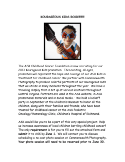


© Copyright 2026