
EVALUATING CHEST PAIN IN CHILDREN A R
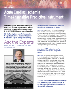
Dr Terence Prendiville, MB MRCPI, Department of Paediatric Cardiology and
Dr Colin J McMahon, MB FRCPI MRCP(UK) FAAP, Consultant Paediatric Cardiologist,
Our Lady’s Hospital for Sick Children, Crumlin, Dublin.
INTRODUCTION
Chest pain in children represents approximately 5%
of all referrals to paediatric cardiology services in a
tertiary referral hospital.1 It is a common symptom in
children and adolescents. Although the aetiology is
benign and non-cardiac in the majority of cases,
chest pain as a complaint often leads to anxiety both
in the child and parent, school absenteeism and
restrictions on physical activity. Extensive public
awareness on the association of chest pain with
ischaemic heart disease and myocardial infarction in
the adult has led to an erroneous assumption that
the same holds true for the paediatric patient. In
Driscoll’s prospective study of chest pain in children,
52% interviewed by a child psychiatrist believed their
chest pain to be cardiac in origin.2
EPIDEMIOLOGY AND PRESENTATION
Paediatric chest pain as a primary complaint to the
accident and emergency department has an
occurrence rate of 0.249% and 0.288% per patient
visit.2,3 There is no gender predilection in patients
presenting and in the largest prospective study to date
the average age at presentation with this complaint is
at a mean age of 11.9 years and a median age of 12.5
years.1,4 In the same study by Selbst et al, chest pain
was described as acute (less than 48 hours duration)
in 43% of cases and chronic (greater than six months’
duration) in 7%. In contrast to these figures, a study by
Pantell et al in adolescents (mean age 16.2 years,
67% female) presenting to an out-patient’s department
setting described an incidence of chronic chest pain of
36%.5
TYPES OF CHEST CAGE AND CHEST
WALL PAIN
Costochondritis
Costochondritis involves two to four contiguous
costochondral or costosternal junctions. Usually it is
unilateral. More commonly it involves the more
cephalad joints. The pain is described as sharp, lasting
several seconds to several minutes, and it is
exacerbated by deep breathing. The joints are not
inflamed and there is no swelling of the joints. Pushing
HEARTWISE SUMMER 2007
on the joint can reproduce the pain.
Tietze’s syndrome
This syndrome is quite uncommon in children. It
involves inflammation of one costochondral junction.
The area involved is warm, swollen and tender.
Non-specific chest wall pain (idiopathic
chest pain)
Non-specific chest wall pain may be the most common
type of chest pain in children and adolescents. The
pain is described as sharp. When asked to point to
the pain, the patient will usually point to the centre of
the chest or the infranipple area. The pain lasts
several seconds to several minutes and is exacerbated by deep breathing. Sometimes squeezing
the thoracic cage or gently pressing the sternum can
reproduce the pain. Frequently the pain cannot be
reproduced by palpation or pushing on chest
structures. The costochondral and costosternal joints
are non-tender.
Precordial catch syndrome
Precordial catch syndrome consists of a brief (several
seconds), sharp pain inferior to the left nipple or at the
lower sternal border. It is frequently pleuritic and can
be accentuated by bending forward. This syndrome
frequently forces the patient to breathe shallowly. Its
cause is unknown.
Slipping rib syndrome
Slipping rib syndrome is quite rare, but it does produce
rather intense pain, usually involving the eighth, ninth
and tenth ribs. These ribs do not attach directly to the
sternum but, rather, attach to each other. It has been
postulated that trauma to the chest results in
disruption of the connection of these ribs to each
other and subsequent movement produces pain. A
positive ‘hooking manoeuvre’ is said to be characteristic of this problem. The hooking manoeuvre is
performed by the examiner putting his or her fingers
under the inferior rib margin and pulling interiorly. This
action will reproduce the pain and may produce a
clicking sound.
ARTICLE
EVALUATING CHEST PAIN
IN CHILDREN
ARTICLE
Hypersensitive xiphoid syndrome
Hypersensitive xiphoid syndrome is uncommon in
children. It can be diagnosed easily by digital pressure
on the xiphoid reproducing the pain.
Trauma and muscle strain
Obviously, injury to the chest wall produces chest wall
pain. The history of prior trauma is suggestive and
usually the pain can be reproduced by palpation of the
chest wall. The same trauma, of course, could also
produce myocardial contusion and, possibly, a haemopericardium, both of which can cause chest pain.
ECG showing ST segment elevation in chest leads V2V6 characteristic of acute pericarditis.
Gastrointestinal
Sickle cell disease
Sickle cell crisis can produce chest wall bone pain.
In addition, chest pain in children and adolescents
with sickle cell disease can be of cardiac and
pulmonary origin.
OTHER CAUSES OF CHEST PAIN
Asthma
Wiens et al described findings of reversible airways
disease in 72.7% of children who had been referred to
a cardiology out-patients’ clinic with no prior history of
asthma or congenital heart disease.6 The mean age of
children who underwent the treadmill test was 12.4
years old. Inhaled salbutamol resulted in a subjective
improvement in chest pain in 97% and an objective
improvement in pulmonary function tests in 70%.
Reactive airways disease should be considered in
patients with chest pain, particularly if there is a
history of asthma, eczema, shortness of breath with
exercise, exercise-associated chest pain, exertional
cough, wheezing or a family history of atopy.
Infection
A number of infective processes can be associated
with chest pain. Lower respiratory infections of all
types can produce chest pain. Herpes zoster can
produce chest pain frequently before the appearance
of the typical vesicopustular rash.
Pericarditis
Classically, pericarditis is associated with chest pain,
whether due to an infectious aetiology or a noninfectious inflammatory cause. In general, the pain
associated with pericarditis is described as fairly
sudden in onset, located over the anterior chest wall
and more severe than other forms of chest pain. It is
frequently pleuritic in nature, being sharp and
exacerbated with deep breathing. The pain may
decrease in intensity when the patient sits up and
leans forward and may radiate, especially to one or
both trapezius ridges. Pericarditis is associated with
typical electrocardiographic findings of generalised ST
segment elevation.
Gastro-oesophageal reflux disease (GORD) and
oesophagitis can and frequently do cause chest
pain. Additional gastrointestinal (GI) symptoms (e.g.
reflux, heartburn) at the time of clinical presentation
can greatly aid and are quite specific in the
diagnosis of GORD as the aetiology of the chest
pain. In an adult study by Mousavi et al, GORD was
found in 44.8% of patients presenting with noncardiac chest pain.7
Berezin et al, investigating idiopathic chest pain in
children, described a GI cause in 78%.8 Oesophageal endoscopy and manometry may detect
abnormalities in children with chest pain, even
without other GI symptoms. This was demonstrated
in a study of 83 children with chest pain who
underwent these procedures.9 A total of 57% had
normal oesophageal histology and normal motility.
Among the others, 18% had oesophagitis on
histology but normal motility, 15.6% had normal
histology but gut dysmotility and 9.6% had both
oesophagitis and dysmotility. It is likely that if GI
causes of chest pain were sought more vigorously,
this diagnosis would be made more frequently.
Pneumothorax
Among patients with chest pain, pneumothorax is
uncommon; however, a pneumothorax is very frequently associated with chest pain, and the abrupt
onset of severe chest pain with or without dyspnoea
should alert the clinician to this differential, especially
as the clinical signs of a small pneumothorax can be
difficult to appreciate.
Pulmonary embolism
The most frequent discharge diagnosis in patients
who initially presented with a clinically suspected (and
subsequently out-ruled) pulmonary embolus is nonspecific chest pain.10 By the time of presentation to
hospital, it can frequently be difficult to separate
clinically pulmonary embolus from lower respiratory
tract infection; however, careful history may give the
clues to the initial symptoms of a pulmonary embolus
being dyspnoea and/or pleuritic chest pain.11
HEARTWISE SUMMER 2007
ARTICLE
In a retrospective study by Bernstein et al over a 15year period, the incidence of pulmonary embolism in
adolescents was repor ted as 78 per 100,000
hospitalised adolescents.12 Females presented with
twice the frequency of males. Common complaints
were chest pain, dyspnoea, cough and haemoptysis.
Major risk factors were oral contraceptive use and
elective abortion in 75% of female patients and trauma
in 67% of male patients. In children, the presence of a
central venous catheter is the most frequent underlying
risk factor.13 Pulmonary angiography is still the gold
standard in diagnosing pulmonary embolism with anticoagulation the mainstay of therapy.
331 children with isolated MVP for one month to eight
years (mean 2.7 years).14 Chest pain developed in 12
children. Ohara et al studied the incidence of
symptoms in 108 children with MVP.15 Chest pain was
the most common symptom, occurring in 11 children
(10.2%). The chest pain was non-exertional, located in
the left chest and intermittent. More recently, Van der
Ham et al describes a cohort of 45 children with
echocardiographically-proven mitral valve prolapse.16
There was no statistical difference in incidence among
the sexes. The most commonly reported symptoms
were shortness of breath and fatigue and not chest
pain. Most of the children were asymptomatic.
CARDIAC CAUSES OF CHEST PAIN
TOXIC EXPOSURE
Cardiac conditions are a rare but potentially serious
cause of chest pain in children. Cardiac disease is more
likely if chest pain occurs during exertion and is
recurrent. Most conditions will be associated with an
abnormal cardiac examination or co-existing symptoms.
In patients with known heart disease, chest pain may
indicate progression of the underlying condition.
• Severe left ventricular outflow tract obstruction
caused by aortic stenosis (subvalvar, valvar or
supravalvar), obstructive cardiomyopathy or coarctation of the aorta.
• Aortic root dissection associated with Marfan
syndrome, Turner syndrome, Ehlers-Danlos syndrome,
chronic systemic hypertension, homo-cysteinuria, rare
familial aortopathies or cystic medial necrosis.
• Pericarditis and myocarditis, in which chest pain
typically occurs with concomitant pericarditis.
• Coronary artery abnormalities, including congenital
disorders or acquired conditions (e.g., coronary
aneurysm or stenosis following Kawasaki disease;
coronary stenosis after coronary re-implantation
following arterial switch operation). Anomalous origin
of the left coronary artery from the main pulmonary
artery (ALCAPA) usually presents in infancy but can
become symptomatic later in childhood.
• Ruptured sinus of valsalva aneur ysm, a rare
condition caused by congenital absence of media
in the aortic wall behind the sinus of valsalva. The
aneurysm typically ruptures into the right ventricle
or right atrium, leading to intra-cardiac shunting
and myocardial ischaemia.
• Tachyarrhythmias (e.g. supraventricular tachycardia
with or without underlying Wolff-Parkinson-White
syndrome, ventricular tachycardia).
• Coronary thrombosis and acute myocardial infarct can
occur in premature arteriosclerosis, paradoxical
embolus or hypercoagulable state.
Exposure to vasoconstrictive agents, such as
cocaine, can cause chest pain that is likely ischaemic
in origin. Hollander et al described a prospective
cohort of 246 adult patients (median age 33 years)
who had chest pain following cocaine use.17 A total of
5.7% had suffered myocardial infarcts. Chest pain
began a median of 60 minutes after cocaine use and
persisted for a median of 120 minutes. Chest pain
was most frequently described as substernal (71.3%)
and pressure-like (46.7%)
MITRAL VALVE PROLAPSE
Whether mitral valve prolapse (MVP) is associated with
chest pain is controversial. Greenwood et al studied
MEDICAL EVALUATION
The evaluation of chest pain requires a thorough
history and careful physical examination.
The details of the history should be obtained, paying
specific attention to the onset, frequency, and
precipitating and relieving factors, as well as the
characteristic duration and location of the chest pain.
Associated features that would heighten suspicion for a
HEARTWISE SUMMER 2007
ARTICLE
true cardiac aetiology would be exercise intolerance,
palpitations or shortness of breath with activity,
presyncope or syncope, or a family history of congenital
heart disease or sudden cardiac death. In addition, it
may be helpful to know whether other family members
have chest pain, such as a parent or grandparent who
experiences angina. This information might heighten
the concerns of chest pain in the child.
A complete cardiovascular, respirator y and
abdominal examination should be per formed. The
physical examination of the patient presenting with
chest pain should initially focus on the vital signs.
After documenting a stable regular heart rate and
rhythm, respirations and blood pressure, a thorough
physical examination should focus on finding noncardiac causes. The initial evaluation should include
inspecting for trauma and bruises or abrasions on
the chest wall. Palpation should focus on bony
abnormalities and localised chest swellings and on
the site of the pain indicated by the patient. There
should be an attempt to reproduce the pain by
palpation of the location indicated by the patient.
Reproducible pain, particularly at the costochondral
junction or over a rib, points to costochondritis as
the aetiology of the pain. A cardiac cause of chest
pain may be suggested by auscultation of abnormal
heart sounds or a cardiac murmur or abnormal pulse
or blood pressure. Signs of left ventricular outflow
obstruction include a systolic ejection murmur at the
right upper sternal border and occasionally along the
left sternal border. Co-arctation of the aorta is
associated with elevated blood pressure in the arms
and a lower blood pressure in the legs. If the coarctation is long standing (present for more than five
to seven years), collateral vessels may form that
connect the upper and lower portions of the aorta;
these vessels create a continuous murmur over the
lateral aspect of the ribs.
In patients with pericarditis, pain increases when
manual pressure is applied to the sternal region. The
pain typically improves with sitting up and leaning
forward. Signs of pericarditis depend upon the size of
the pericardial effusion. Patients with a small effusion
typically have an audible pericardial friction rub, caused
by rubbing together of the inflamed parietal and visceral
pericardial surfaces. The rub is often continuous in
systole and diastole. It is easier to hear with the
diaphragm of the stethoscope when the patient is
sitting and leaning forward. A rub will not be heard if the
effusion is large because the two pericardial surfaces
of the pericardium are not in contact with each other. A
large effusion may result in cardiac tamponade,
manifested by a narrow pulse pressure, elevated
pulsus paradoxus (>10mmHg), elevated jugular venous
pressure, distant heart sounds, hepatomegaly, ascites,
and peripheral oedema.
Signs of MVP are a constant, mid-systolic apical
click and, occasionally, an apical systolic murmur of
mitral regurgitation. Both auscultatory findings are
more prominent when the patient is in the standing
rather than supine position.
In most cases, the aetiology of the pain will be
apparent after the history and physical examination.
DIAGNOSTIC TESTS
Most patients with chest pain have a normal physical
examination or findings consistent with a musculoskeletal aetiology. Further investigations are not
needed in those cases.
Diagnostic studies may help establish a diagnosis in
patients with abnormal physical findings or with
associated symptoms that suggest organic disease.
Although cardiac causes of chest pain are uncommon in
children, patients with dyspnoea, palpitations, anginal
pain and pain with exertion that cannot be attributed to
respiratory disease or syncope should be referred to a
paediatric cardiologist for further evaluation.
• A chest x-ray may show cardiomegaly, pulmonary
vasculature, infective infiltrates, hyperinflation and
pneumothoraces.
• An electrocardiogram can aid in the diagnosis of
arrhythmias, left ventricular outflow tract obstruction,
pericarditis, ALCAPA, pulmonary hypertension and
pulmonary embolus. If an arrhythmia is intermittent,
a 24-hour Holter monitor or King of Hearts event
recorder may be needed.
• In diagnoses of suspected cardiac aetiology, an
echocardiogram can confirm cardiac structure,
pericardial effusions and tamponade, cardiac
function, anomalous coronary anatomy, sinus of
valsalva aneurismal rupture and aortic root pathology.
• GI evaluation in children with chest pain should be
performed under the remit of a paediatric gastroenterologist. Investigation of the upper GI tract may
reveal histological oesophagitis or gut dysmotility.
• Other tests should be based upon associated signs
and symptoms and clinical suspicion. Fur ther
evaluation may include pulmonary function testing, a
ventilation-perfusion scan, cardiac catheterisation,
exercise stress testing, a full blood count, serum
reactive markers, appropriate cultures and
toxicology screening.
OUTCOME OF CHILDREN WITH
CHEST PAIN
Two studies of the outcome of chest pain in children
have been reported. Selbst et al reported on the
outcome of an initial 407 children with chest pain seen
in the accident and emergency department.18 Thirtyfour per cent of the original diagnoses were altered,
usually in favour of a non-organic aetiology. A new
organic aetiology was uncovered in only 12 of 149
HEARTWISE SUMMER 2007
667-71.
13. Van Ommen CH, Peters M. Acute pulmonary embolism
in childhood. Thromb Res 2006; 118: 13-25.
14. Greenwood RD. Mitral valve prolapse in childhood.
Hosp Pract (Off ed) 1986; 21: 41-4.
15. Ohara N, Mikajima T, Takagi J et al. Mitral valve
prolapse in childhood: the incidence and clinical
presentations in different age groups. Acta Paediatr
Jpn 1991; 33: 467-75.
IN SUMMARY
The primary role of the evaluating cardiologist is to rule
out the unlikely prospect of serious cardiac pathology.
An equally important role is to provide reassurance and
support to the patient and family. Most patients will not
have a serious underlying medical problem. A thorough
and thoughtful history and physical examination help
allay fears and are important in reassuring the patient
and family.
16. Van der Ham DP, de Vries JK, Van der Merwe PL. Mitral
valve prolapse: a study of 45 children. Cardiovasc J S
Afr 2003; 14: 191-4.
17. Hollander JE, Hoffman RS, Gennis P et al. Prospective
multicenter evaluation of cocaine-associated chest
pain. Cocaine Associated Chest Pain (CPCHPA) Study
Group. Acad Emerg Med 1994; 1: 330-9.
18. Selbst SM, Ruddy R, Clark BJ. Chest pain in children.
Follow-up of patients previously reported. Clin Pediatr
(Phila) 1990; 29: 374-7.
REFERENCES
1.
Geggel R. Conditions leading to pediatric cardiology
FURTHER READING
consultation in a tertiary academic hospital.
•
2.
Driscoll D, Glicklich L, Gallen W. Chest pain in
Vetter V. Pediatric Cardiology: Requisites (Requisites in
Pediatrics). Mosby, 2005.
Pediatrics 2004; 114: e409-17.
•
Moss and Adams. Heart Disease in Infants, Children,
children: a prospective study. Pediatrics 1976; 57:
and Adolescents (sixth ed). Lippincott Williams &
648-50.
Wilkins, 2001.
3.
Selbst S. Chest pain in children. Pediatrics 1985; 75:
4.
Selbst S, Ruddy R, Clark B et al. Pediatric chest pain:
1068-70.
a prospective study. Pediatrics 1998; 82: 319-23.
5.
Pantell R, Goodman B. Adolescent chest pain: a
prospective study. Pediatrics 1983; 71: 881-7.
6.
Wiens L, Sabath R, Ewing L et al. Chest pain in
otherwise healthy children and adolescents is
frequently caused by exercise-induced asthma.
Pediatrics 1992; 90: 350-3.
7.
Mousavi S, Tosi J, Eskandarian R et al. Role of
clinical presentation in diagnosing reflux-related
non-cardiac chest pain. J Gastroenterol Hepatol
2007; 22: 218-21.
8.
Berezin S, Medow MS, Glassman MS et al. Chest
pain of gastrointestinal origin. Arch Dis Child. 1988;
63: 1457-60.
9.
Glassman MS, Medow MS, Berezin S et al. Spectrum
of esophageal disorders in children with chest pain.
Dig Dis Sci 1992; 37: 663-6.
10. Bernard Bagattini S, Bounameaux H, Perneger T et al.
Suspicion of pulmonary embolism in outpatients: nonspecific chest pain is the most frequent alternative
diagnosis. J Intern Med 2004; 256: 153-60.
11. Soderberg M, Hedstrom U, Sjunnesson M. Initial
symptoms in pulmonary embolism differ from those
in pneumonia: a retrospective study during seven
years. Eur J Emerg Med. 2006; 13: 225-9.
12. Bernstein D, Coupey S, Schonberg SK. Pulmonary
embolism in adolescents. Am J Dis Child 1986; 140:
HEARTWISE SUMMER 2007
Correspondence to: Dr Colin McMahon,
Consultant Paediatric Cardiologist, Our Lady’s
Hospital for Sick Children, Crumlin, Dublin 12.
Email: [email protected]; Tel: (01)
4096160; Fax (01) 4096181
ARTICLE
cases, with only one having a heart abnormality (MVP).
Chest pain resolved in 57% of those followed. Driscoll
et al had a similar rate of resolution of chest pain in
his cohort of 43 patients, with 60% pain-free four to
eight weeks later on telephone follow-up.2 Of note, in
the group of Driscoll’s patients with a diagnosis of
idiopathic chest pain, only 30% of them had resolution
of chest pain at follow-up.


© Copyright 2026