
Synergy
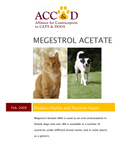
Synergy Summer 2011 Animal Cancer Care and Research Program DIRECTOR’S CORNER Clinical trials in veterinary oncology at the University of Minnesota I Michael G. Conzemius, D.V.M., Ph.D., Diplomate ACVS Tata Group Endowed Professor, Veterinary Clinical Sciences am pleased to introduce this issue of Synergy. As director of the College of Veterinary Medicine’s Clinical Investigation Center (CIC), I am excited about the breadth we have achieved in the area of oncology clinical trials. Articles in this newsletter describe ongoing, completed, and planned clinical trials focused on diverse tumors and therapies, both home-grown and acquired through collaborations. We also are delighted to highlight the work of the Center for Translational Medicine and the infrastructure they provide to translate discoveries by University of Minnesota investigators into Phase I clinical studies. At the College of Veterinary Medicine, the CIC plays a key role in promoting research by executing and supporting clinical and translational research based on patients from the University of Minnesota Veterinary Medical Center, the most advanced, full-service referral care center for large and small animals in Minnesota. Michael G. Conzemius The mission of the CIC is to develop and facilitate veterinary clinical trials and translational research studies that lead to new drugs, devices, procedures, and treatments for the benefit of animals and humans alike. We do this by providing scientific and clinical expertise, facilities, technical staff, and overall study coordination. The CIC supports the clinical and research goals of the College of Veterinary Medicine, which strives to sustain “One Medicine.” The concept of One Medicine is commonly attributed to Rudolf Virchow (1821-1902), a physician who wrote, “Between animal and human medicine there is no dividing line – nor should there be. The object is different but the experience obtained constitutes the basis of all medicine.” Our vision is for the CIC to be the premier veterinary research center for clinical trials in the United States. The best medical centers not only apply the most current information, they create it. To this end, clinical research is inseparable from the provision of state-of-the-art health care. The University of Minnesota College of Veterinary Medicine is recognized as a world leader in clinical research, where new knowledge is created from each patient and applied to the next patient. As you will read in the following pages, there is reason for optimism. Our success is your success, and we value your partnership. Synergy Animal Cancer Care and Research Program COTC016: A pilot study to assess feasibility of tissue collections and molecular profiling for future comparative oncology personalized medicine studies Kathy Stuebner, B.S., CVT, Research Coordinator, Clinical Investigation Center, College of Veterinary Medicine, and Michael S. Henson, D.V.M., Ph.D., Diplomate ACVIM (Oncology), Department of Veterinary Clinical Sciences, College of Veterinary Medicine and Masonic Cancer Center T he University of Minnesota is a member of the Comparative Oncology Trials Consortium (COTC), a network of 20 academic comparative oncology centers that collaborate in multi-center trials managed by the Center for Cancer Research, which is part of the National Cancer Institute at the National Institutes of Health. While most COTC trials evaluate novel therapies for cancer, the main goal of this trial is to evaluate the capability of the COTC network of hospitals and laboratories around the country to collect and process samples quickly and accurately enough to be useful in personalized medicine trials in the future. Personalized medicine involves tailoring cancer treatment and prevention to the specific molecular nature of the individual and their cancer (i.e., target the mutations that drive malignant transformation, resistance to treatment, risk for toxicity, etc.). The hope is that targeted therapies will be more effective with less risk for the patient than traditional treatments. For personalized medicine to be useful, we need to accurately determine the molecular characteristics (genomic, proteomic, and epigenetic profile) of the patient rapidly enough for treatments to be designed and implemented for patient care, ideally in less than a week. In addition to assessing sample collection, viability, and the clinical turnaround of results, we hope that this study will identify potential therapeutic targets in specific canine malignancies to allow the design of future studies in dogs. Since canine malignancies share many characteristics with those that occur in humans, if personalized medicine is proven efficacious in dogs, the results may advance development of more efficient targeted therapies for people, too. Once dogs are enrolled at the University of Minnesota Veterinary Medical Center, participation in the study involves a single collection of samples, specifically blood, saliva, and a biopsy of the tumor. After sample collection, owners can choose whatever treatment option is best for the family. The study pays for sample collection and provides $1,000 that can be used to cover the cost of evaluation and future expenses at the Veterinary Medical Center. At this time, we are specifically seeking golden retrievers with multicentric lymphoma, Scottish terriers with transitional cell carcinoma, and any breed of dog with oral melanoma. Amber Winter of the College of Veterinary Medicine’s Clinical Investigation Center holds Luke, a study dog, as fourth-year veterinary student Raeana Rice examines the springer spaniel. Photo by Sue Kirchoff A dog with oral melanoma, a type of skin cancer. Scientists with the Animal Cancer Care and Research program are seeking dogs with oral melanoma for a study that will help both dogs and people with cancer. 2 • Synergy Summer 2011 Synergy Animal Cancer Care and Research Program Nationwide study to evaluate novel therapeutic agent for the treatment of solid tumors in dogs Michael S. Henson, D.V.M., Ph.D., Diplomate ACVIM (Oncology), Department of Veterinary Clinical Sciences, College of Veterinary Medicine and Masonic Cancer Center, and Kathy Stuebner, B.S., CVT, Research Coordinator, Clinical Investigation Center, College of Veterinary Medicine C ancer occurs when a cell in the body acquires enough mutations to lose control of its growth and divides (replicates itself) several billions of times to form a visible tumor. To grow beyond a few millimeters in diameter, tumors also grow their own blood supply. However, because cancer cells and the environment in the tumor are so abnormal, the blood vessels that form within tumors are usually abnormal as well. These abnormal blood vessels, combined with rapid growth and high pressure within solid tumors, result in poor delivery of oxygen and nutrients to the center of the tumor. Tumors eventually outgrow their own blood supply, resulting in cancer cells that struggle to survive or die in the center of the tumor while the outer rim continues to grow and invade the normal surrounding tissues. This characteristic of solid tumors is one reason traditional therapies such as chemotherapy are not as effective as we hope they could be. The abnormal blood supply in tumors results in poor delivery of anti-cancer treatment to all parts of the tumor. Some areas of the tumor do not receive the treatment and therefore may survive. Most cancer therapies focus on the wellvascularized outer shell of the tumor, but few target the oxygenstarved (hypoxic) inner core. We will soon be enrolling dogs in a nationwide study to evaluate a novel cancer treatment that does just that. The therapeutic agent is a modified anaerobic bacterium. Anaerobic bacteria die or become inactive in air or in tissues with normal oxygen levels, but they grow in environments with low oxygen levels, such as the center of a tumor, resulting in inhibition of tumor growth or tumor shrinkage in cancer patients. Preliminary studies are encouraging. Common side effects include fever, nausea, and inflammation in the tumor. The primary goals of this study are to assess the safety and potential effectiveness of a new therapeutic agent in dogs with melanoma, osteosarcoma, soft tissue sarcoma, or squamous cell carcinoma. These cancer types are biologically similar in humans, so if this therapy proves safe and effective in dogs, this research may advance the development of treatments for people with these tumor types. Dogs are eligible if they meet the following inclusion criteria: • Diagnosis of melanoma, osteosarcoma, soft tissue sarcoma, or squamous cell carcinoma (biopsy or needle aspirate) • Measurable tumor (primary or metastasis) that is at least 1 cm in diameter • Generally feeling well (i.e., eating, drinking, Dr. Michael Henson examines Beckham, a patient in the Oncology Service at the University of ambulating on Minnesota Veterinary Medical Center. Photo by their own, etc.) Sue Kirchoff • No evidence of an active bacterial infection requiring antibiotics (other than topical medications) in the past seven days • No anti-cancer therapy within the past 21 days (including chemotherapy, radiation therapy, corticosteroids, or immunotherapy) • No tumors where abscess (infection) would result in major symptoms The study will cover all costs associated with the therapy as well as some diagnostic tests, including laboratory tests, chest X-rays, and abdominal ultrasound. Once enrolled, dogs will be treated with a single dose of the novel therapy and closely monitored for at least six hours afterward, with recheck visits on days 2, 4, 7, 14, 28 and 56. In the event that side effects are noted and attributed to the therapeutic agent, the study will pay for medical management of the side effects. 3 • Synergy Summer 2011 Synergy Animal Cancer Care and Research Program OSTEOSARCOMA Gene therapy for osteosarcoma – what’s next? Jaime F. Modiano, V.M.D., Ph.D., Department of Veterinary Clinical Sciences, College of Veterinary Medicine, and Masonic Cancer Center A nimal Cancer Care and Research (ACCR) members from the University of Minnesota were key participants in a recently completed clinical trial testing a gene therapy approach to treat canine osteosarcoma. This trial, supported through grants from the National Cancer Institute of the National Institutes of Health and the AKC Canine Health Foundation, was meant to define the safety, efficacy, and mechanisms of Fasaret®, a formulation for the Fas ligand gene expressed in a viral vector and produced by ApopLogic Pharmaceuticals, Inc.* Fas ligand is a protein that is normally made by cells of the immune system. It is the prototypical member of a family of proteins that promote “programmed death” (also called programmed suicide or apoptosis) of unwanted cells. However, Fas ligand has a variety of other functions, many of which are not fully understood. Scientists at ApopLogic, including the author, had shown that this therapy was safe and effective to treat tumors in laboratory animal models, so it was time to take a leap forward and translate it to the clinic. Translation was a large project that involved board-certified veterinary surgeons, veterinary medical oncologists, veterinary radiologists, veterinary pathologists, veterinary nurses, clinical coordinators, immunologists, statisticians, and exceptional scientific and technical support staff from four institutions at a cost of over $1 million. The results of the trial were reported to the scientific community in May 2011 at the 98th Annual Meeting of the American Association of Immunologists in San Francisco, California. Fifty-six dogs were enrolled. The results showed the dosing schedule used in the trial was safe. Moreover, they highlighted mechanisms through which this therapy would improve outcome for bone cancer patients. Next steps for this therapy are validation in community-based trials, and possibly its application to other types of cancer. Visit the CVM Clinical Investigation Center website at www.cvm.umn.edu/cic/ for more updates on results of this trial and next steps, as well as for applications to enroll dogs in other clinical trials, or to make a contribution to support this research. * Dr. Modiano holds an equity interest in and serves as a consultant for ApopLogic Pharmaceuticals, Inc., the developer of Fasaret, a product that was the subject of the research described in this report. These relationships have been reviewed and managed by the University of Minnesota in accordance with its conflict of interest policies. Osteosarcoma cells can be infected with adenovirus vectors with high efficiency. When this method is used to deliver the Fas ligand (FasL) gene, some tumor cells are highly susceptible to its death-promoting effects, while others are resistant. The images show two osteosarcoma cell lines, OSCA-2 and OSCA-36, infected with a control adenovirus encoding a green fluorescent protein (Ad-GFP) gene or the therapeutic adenovirus encoding the FasL gene (Ad-FasL). The adenovirus itself has no negative effects on cell viability or growth, as shown by the presence of healthy cells infected with Ad-GFP. OSCA-2 cells were similarly unaffected by infection with Ad-FasL, whereas OSCA-36 cells all were killed (through the effects of FasL) after infection with Ad-FasL. One question that this project sought to answer was whether there was a relationship between the susceptibility of the tumor cells to FasL-mediated death and the patient’s response to therapy. Reprinted from Modiano et al, “Naturally Occurring Translational Models for Development of Cancer Gene Therapy.” Gene Therapy and Molecular Biology, vol. 10, pp. 31-40, 2006. 4 • Synergy Summer 2011 Synergy Animal Cancer Care and Research Program OSTEOSARCOMA Bone cancer immunotherapy using a genetically modified Salmonella Vicki Wilke, D.V.M., Ph.D., Diplomate ACVS1,2, and Emily Lipsitz, M.D. (Board Certified in Pediatric Hematology Oncology)3 1 Department of Veterinary Clinical Science, College of Veterinary Medicine; 2Masonic Cancer Center; 3 Department of Pediatrics, School of Medicine S almonella is an organism that strikes fear due to its potential to cause severe gastrointestinal illness, but it is not a word many people associate with cancer therapy. Think again. Dr. Dan Saltzman of the University of Minnesota Medical School and his colleagues have taken Salmonella organisms, weakened them using genetic engineering to eliminate their disease-causing potential, and developed a new drug by adding Interleukin-2 (IL-2), a series of proteins that act as “flavors” to activate the immune system. Salmonella organisms like to grow in environments such as those created by tumors, and they seem to have special affinity for bone cancer. The development of Salmonella-IL2 has also led to a special collaboration between the University of Minnesota Medical School and the College of Veterinary Medicine to utilize it in our canine patients. Osteosarcoma (OSA) is the most common bone tumor in dogs. In approximately 90 percent of dogs with OSA, the cancer has already spread by the time they are diagnosed. With surgery alone, dogs have a median survival of three to four months. With chemotherapy, the median survival can increase to about nine to fourteen months. To improve survival in patients with OSA, we must develop more effective treatments. One treatment type that has shown promise is immunotherapy – the use of the immune system to kill metastatic tumor cells. IL2 is a pro-inflammatory protein made by the immune system that aids in cell death, forming the basis for its use in cancer immunotherapy. In a new study underway at the College of Veterinary Medicine, we will evaluate safety and efficacy of a genetically modified Salmonella carrying the IL2 gene (Salmonella-IL2) in the treatment of dogs with spontaneous bone cancer. Salmonella-IL2 has been shown to be safe and to reduce tumor volume (size) in various animal species. There is an ongoing early clinical trial in human patients with no toxicities noted thus far. If successful, this treatment will be further developed to improve the outcome of dogs with osteosarcoma. Dogs need to meet certain criteria to qualify for the study. Dogs that have untreated osteosarcoma that has not spread beyond the primary site and are otherwise in good health may be eligible. Dogs cannot have received coticosteroids (prednisone, dexamethasone) for two weeks prior to entering the study, and dogs that have already been treated with surgery, chemotherapy, or radiation therapy are not eligible. Similarly, dogs that have consumed raw foods or have a positive fecal culture for Salmonella are not eligible for this study. Color-enhanced scanning electron micrograph showing Salmonella typhimurium (red) invading cultured human cells (Rocky Mountain Laboratories, NIAID, NIH) Eligible dogs will undergo a specialized form of imaging called positron emission tomography/ computed tomography (PET/CT). On day one patients will undergo a bone biopsy and receive their first oral dose of Salmonella IL2. Recheck evaluations and diagnostic blood work, urine tests, and fecal culture will be performed on days three and seven. Amputation will be performed on day 10. Dogs will then have a recheck evaluation and second dose of Salmonella IL2 on day 21, when chemotherapy with doxorubicin will be initiated. A final recheck and diagnostic blood work will occur on day 28, which is the official end of the study. However, dogs will continue to receive doxorubicin and Salmonella IL2 therapy every three weeks for five total treatments. A recheck PET/CT will be performed at six months to assess efficacy. There is a financial incentive for participation. Specifically, the study will cover the costs of the study visits on day one (PET/ CT, bone biopsy, and Salmonella IL2 therapy), rechecks, and the costs associated with amputation, up to a total of $2,000. All costs associated with PET/CT imaging will also be covered by the study. An opening date for this trial is pending. Check the CVM Clinical Investigation Center website at www.cvm.umn.edu/cic/ for more information, applications to enroll dogs in clinical trials, or to make a contribution to support this research. 5 • Synergy Summer 2011 Synergy Animal Cancer Care and Research Program OSTEOSARCOMA Preclinical evaluation of adjuvant Minnelide in dogs with appendicular osteosarcoma – a pilot study Antonella Borgatti, D.V.M., M.S., Dipl ACVIM (oncology), Dipl ECVIM (oncology)1,2, Subbaya Subramanian, PhD2,3, and Ashok Saluja, Ph.D.2,3 1 Department of Veterinary Clinical Sciences, College of Veterinary Medicine; 2Masonic Cancer Center; 3 Division of Basic and Translational Research, Department of Surgery, School of Medicine O steosarcoma (OSA) is the most common malignant bone tumor of dogs, typically occurring in the limbs. It is an incurable cancer characterized by rapid progression and metastasis (spread). It shares many features with osteosarcoma of children and has been often used as a spontaneous model of the pediatric disease, thus providing information to advance care for these patients. Dogs with OSA treated with surgery have an expected survival time of approximately four to six months. This increases to about one year with the addition of chemotherapy. However, less than 25 percent of dogs receiving this standard of care live two or more years, and virtually all dogs die of their disease due to the inevitable development of metastasis. Chemotherapy is declined by many dog owners due to the cost and the perception of risks outweighing benefits. Therefore, development of new, more attractive, and cost-effective treatment options to improve clinical and survival outcome is critical. Minnelide (a water-soluble prodrug of triptolide) is a promising compound that inhibits heat shock proteins (HSP70) implicated in cancer formation, progression, and metastasis. Minnelide possesses highly selective anti-tumor activity against OSA in cell culture and in laboratory models and was well-tolerated with no toxicity in dogs when administered at doses predicted to show anti-tumor activity. The pilot study will investigate the use of Minnelide in dogs with OSA to establish its safety and efficacy as a single agent added to surgery. For this trial, dogs will undergo limb amputation and vascular port placement to allow delivery of Minnelide, intravenously, for 60 consecutive days starting from the day of surgery. We will monitor the patients for possible treatmentrelated toxicities as well as to establish the effects of the therapy to inhibit its intended targets. Routine radiographs and positronemission tomography (PET-CT) will be used to determine disease progression (metastatic spread) throughout the study. Canine osteosarcoma under the microscope. Animal Cancer Care and Research program scientists are looking for new, cost-effective treatments for this common cancer in dogs. We expect that adjuvant Minnelide treatment will be welltolerated in dogs with OSA, significantly improve the dogs’ quality of life, and result in survival outcomes that will equal or exceed those of conventional chemotherapy regimens. This pilot study will provide a foundation for future clinical trials using Minnelide in combination with cytotoxic chemotherapy for this disease and will also justify and expedite further testing of this novel drug in children with OSA. Financial incentives will include defrayment of costs associated with amputation and vascular port placement, Minnelide and its administration, and recheck visits during the study period. An opening date for this trial is pending. Check the CVM Clinical Investigation Center website at www.cvm.umn.edu/cic/ for more information, applications to enroll dogs in clinical trials, or to make a contribution to support this research. 6 • Synergy Summer 2011 Synergy Animal Cancer Care and Research Program GLIOMA A gene therapy clinical trial for dogs with glioma 1 G. Elizabeth Pluhar, D.V.M., Ph.D., Dip ACVS1, John R. Ohlfest, Ph.D.2, and Matthew A. Hunt, M.D.3 Department of Veterinary Clinical Sciences, College of Veterinary Medicine; 2Department of Pediatrics, School of Medicine; 3Department of Neurosurgery, School of Medicine G liomas, which arise from the “gluey,” or supportive, tissue of the brain, are common malignant brain tumors. For both dogs and people with these aggressive tumors, the prognosis is dismal. People with gliomas are usually treated by surgical removal of the tumor, a full course of radiation therapy, and chemotherapy. Despite this combination of therapies, the tumors grow back in five to six months, and life expectancy is 12 to 14 months. Dogs with this type of tumor have rarely been treated. We are currently conducting clinical trials to find new and better ways to treat gliomas. In the past 12 months, we have recruited 10 dogs for a gene therapy study. Preliminary data hint at improved times of progression-free as well as overall survival in dogs treated with gene therapy. At present, median progression-free survival in dogs treated with gene therapy is six months and overall survival is about a year. This compares to progression-free and overall survival time of 55 days in dogs in the control group (no gene therapy). However, a definitive interpretation must await the final statistical analysis that will be performed at the end of this project. Dr. G. Elizabeth Pluhar with her patient, Dakota. Photo by Sue Kirchoff Neither the surgical tumor removal nor the gene therapy injections into the brain appear to have any adverse effects. Most dogs were well enough to be discharged from the hospital within 24 to 48 hours after surgery. At most, they experienced only mild weakness on one side of the body, which resolved within days. All the dogs in the study received chemotherapy with a drug called temozolomide, which is the standard of care used in people with gliomas. Some dogs became nauseous from the temozolomide, but this was controlled by giving the drug at night on an empty stomach and with anti-nausea medication. We have not seen adverse side effects on blood cells or the major organs of the body. Similar to people undergoing surgery for brain tumors, these dogs are at a higher risk for developing pneumonia, which will resolve without complication if it is diagnosed and treated early in the disease process. We are currently recruiting dogs into this clinical trial. Eligible dogs must have a tentative diagnosis of a glioma based on MRI showing a tumor with the right characteristics. We also have another clinical trial for dogs with gliomas using vaccine therapy in addition to surgical removal of the tumor, similar to that described for dogs with meningiomas (see page 9). We are looking for 35 more dogs for these trials. Visit the CVM Clinical Investigation Center website at www.cvm.umn.edu/cic/current/ braintumortrials for more information, applications to enroll dogs in clinical trials, or to make a contribution to support this research. 7 • Synergy Summer 2011 Synergy Animal Cancer Care and Research Program MENINGIOMA Immunotherapy for canine meningioma (the meningioma vaccine trial) 1 G. Elizabeth Pluhar, D.V.M., Ph.D., Dip ACVS1, John R. Ohlfest, Ph.D.2, and Matthew A. Hunt, M.D.3 Department of Veterinary Clinical Sciences, College of Veterinary Medicine; 2Department of Pediatrics, School of Medicine; 3Department of Neurosurgery, School of Medicine M eningiomas are the most common brain tumor in dogs. Surgical removal is the mainstay of treatment, and most of these tumors are managed with local therapy. Historically, the survival time of dogs with meningiomas treated with surgery alone has been four and one-half to seven months. Systemic therapies so far have had limited effectiveness. However, based on preclinical data from our research program, we embarked on the development of anti-tumor vaccinations to treat dogs with spontaneous meningiomas after surgical removal. Thirteen dogs have been treated with surgical removal of the tumor followed by a series of vaccinations made from the dog’s own tumor cells (called an “autologous tumor lysate” vaccine), along with one of two immune stimulators. The vaccines were injected into the skin on the back of the dog’s neck two weeks after surgery at suture removal. A total of six vaccinations were given at two-week intervals. All 13 dogs have completed the vaccination series. Magnetic resonance imaging (MRI) of the brain was performed immediately after surgery to assess whether the tumor mass was completely removed, and 3, 6, and 12 months after surgery to check for tumor recurrence. Blood collected from each dog during the visits for the MRIs was analyzed for an immune response against the tumor. High-grade malignant tumors were found in two dogs, and low-grade tumors were found in the remaining 11 dogs. A survival analysis comparing dogs treated with surgery and vaccines to dogs that had surgical removal alone demonstrated a significant survival improvement in the dogs receiving the vaccine therapy. To date, the mean survival time in dogs treated with surgery and vaccines is about one year, with many of the treated dogs still alive and tumor-free, compared to a survival time of approximately 200 days in dogs treated with surgery alone. We also have not seen severe toxicities or side effects in the vaccine-treated dogs. Immune testing demonstrated an antibody response as well as enhanced tumor cell killing by the treated dogs’ lymphocytes. A patient is prepared for MRI at the University of Minnesota Veterinary Medical Center. Our results indicate that immunotherapy using autologous tumor lysate and immune adjuvant after tumor removal elicits specific immune responses against residual tumor cells and increases survival over historical controls. Since this therapy appears to work so well in dogs with meningiomas, it may also be effective in humans with meningiomas that have failed to respond to the standard therapy or are at high risk of recurrence. Although the pilot phase for this trial is complete and we are not currently recruiting dogs with meningiomas, we are waiting to hear whether two grant proposals to continue our work on dogs with meningiomas will be funded. Check the CVM Clinical Investigation Center website at www.cvm.umn.edu/cic/current/ braintumortrials for more information, applications to enroll dogs in clinical trials, or to make a contribution to support this research. 8 • Synergy Summer 2011 Synergy Animal Cancer Care and Research Program LYMPHOMA “LICKing Lymphoma” – A clinical trial for spontaneous diffuse large B cell lymphoma in dogs Amber Winter, B.S., CVT, Clinical Investigation Center, College of Veterinary Medicine T he concept behind LICKing Lymphoma came from our recently reported discovery of a subset of cells we have provisionally called lymphoma-initiating cells (LIC), which may be responsible for initiating and maintaining lymphoma. We plan to exploit properties that make these cells responsible for drug resistance and eventual relapse, using them as the targets that will help us improve outcomes. We are using a drug provided by our partners at Novartis, Inc., called PSC-833 (valspodar), which inhibits the multidrug resistance protein P-glycoprotein (ABCB1) as a means to sensitize LICs to killing by conventional chemotherapy. By eliminating LICs, we expect to extend durable remissions and reduce both disease- and treatment-related morbidities in diffuse large B-cell lymphoma. If successful, the application of this Lymphoma-Initiating CellKilling (LICKing) approach could be extended to other common tumors that are known to harbor similar tumor-initiating cells in dogs and humans. This is the first veterinary translational medicine project supported under an agreement between the Clinical and Translational Science Institutes at the University of Minnesota and Indiana University-Purdue University. It is being conducted as a multi-institutional collaboration led by Drs. Jaime Modiano (Minnesota), Michael Childress (Purdue), and Nicola Mason (Pennsylvania). Additional dogs are undergoing screening. Enrollment will continue until the study has accrued a total of 20 dogs (total for the three sites). Owners receive a $2,500 incentive for allowing their dog to participate in this study. This covers most, if not all of the chemotherapyrelated treatment costs. Visit the CVM Clinical Investigation Center website at www. cvm.umn.edu/ cic/ for more information and for applications to enroll dogs in this and other ongoing clinical trials. Copper, a dog enrolled in the LICKing Lymphoma study Once a patient has been diagnosed with lymphoma, we start the screening/staging process. This process takes about two to three days to determine eligibility. The staging diagnostics include clinical pathology, thoracic radiographs, abdominal ultrasound, bone marrow aspirate, and lymph node biopsy. This is a double-blinded study. When inclusion criteria are fulfilled, patients are randomized to either the experimental drug group or the placebo group. The dog owners administer the treatment article to their dog daily for four days, when they return with their dog for a recheck exam, biopsy, blood collection, and the first chemotherapy administration. The drug is given for one more day. One week later, they bring their dog back for a final study recheck exam and blood sample. The dogs then complete standard chemotherapy using five treatments of doxorubicin. Currently, six cases have completed the LICKing Lymphoma study process with no severe treatment-related toxicity. Flow cytometry analysis of a malignant lymph node from a dog with B-cell lymphoma. A small number of progenitor cells (LICs) are present within malignant CD22+ B-cells (rectangle gate). 9 • Synergy Summer 2011 Synergy Animal Cancer Care and Research Program LYMPHOMA COTC007b: Preclinical comparison of three indenoisoquinolines candidates in tumor-bearing dogs Antonella Borgatti, D.V.M., M.S., Dipl ACVIM (Oncology), Dipl ECVIM (Oncology), Department of Veterinary Clinical Sciences, College of Veterinary Medicine and Masonic Cancer Center C OTC007b: Preclinical Comparison of Three Indenoisoquinolines Candidates in TumorBearing Dogs, a clinical trial sponsored by the National Cancer Institute, assesses the safety and efficacy of three newly developed chemotherapy agents (indenoisoquinolines) when given to dogs with lymphoma. This class of topoisomerase inhibitors has shown efficacy against a variety of cancers, and is currently being evaluated in human patients as agents with improved drug stability and measurable blood levels. Topoisomerase I (topo-I) is an enzyme that facilitates DNA replication. The complexes between the enzyme and the drug are reversible under normal conditions. Other topo inhibitors like topotecan (Camptothecin) cause irreversible DNA damage; however, they have a number of limitations that reduce their overall efficacy. An indenoisoquinoline (NSC 314622) was discovered in 1997 as a potential topo-I inhibitor unrelated to topotecan, which might be able to overcome the limitations associated with this drug, while at the same time maintaining the specificity and potency of the class. Dr. Antonella Borgatti with Copper, a dog enrolled in the LICKing Lymphoma study (see page 10) Three analogs were chosen for this study based on their chemical and clinical properties. The present trial is divided into two phases, which include a dose-finding phase for safety and a validation phase. Anti-cancer activity against canine lymphoma will be assessed in both phases. The study will pay for test drugs, chemotherapy administration, and sample collection procedures, and will provide a $1,000 incentive for participation. The observation period will be 28 days. Dogs with objective responses (tumor shrinkage) will be eligible for a subsequent treatment cycle. Dogs with a stable disease (no tumor shrinkage) or progressive disease will have the option to pursue a different treatment option. Information from this study will not only provide potential benefit for dogs with lymphoma, but also will assist in prioritization of indenoisoquinoline lead candidates for Phase II clinical trials in humans. Lymphoma under the microscope. Animal Cancer Care and Research program scientists are searching for new treatments for this cancer of the lymphatic cells of the immune system. 10 • Synergy Summer 2011 Synergy Animal Cancer Care and Research Program LYMPHOMA Development of new lymphoma treatment strategy with a selective inhibitor of nuclear export Daisuke Ito, D.V.M., Ph.D., Department of Veterinary Clinical Sciences and Masonic Cancer Center A ccording to the Leukemia & Lymphoma Society, approximately 65,000 people were newly diagnosed with non-Hodgkin lymphoma (NHL) in the U.S. in 2010, and an estimated 20,000 patients died from the disease. This unacceptably high death rate persists even after the introduction of rituximab, one of the most successful cancer treatments in recent decades, highlighting the critical need to develop better treatments for NHL. proteins from the cell nucleus to the cytoplasm. Inhibition of CRM1 by KPT-SINE causes retention of multiple tumor suppressor proteins in the nucleus, resulting in the selective death of tumor cells—while normal cells undergo transient, but reversible proliferation arrest. This makes KPT-SINE unique among drugs in current use or undergoing trials for lymphoma treatment, which carries as an additional benefit the potential to be used as a single agent or in combination with other therapies. NHL also occurs in dogs. It is one of the most common tumors seen in dogs, and as is true in humans, cures for this disease are elusive. To circumvent this problem, we have worked diligently to establish new therapeutic strategies for lymphoma. One such strategy is the approach described in the article by Amber Winter on page 10. However, we strongly believe that success will require a multi-pronged approach, and recently collaborated with scientists and oncologists at Karyopharm Therapeutics, Inc. in Natick, Massachusetts, to introduce their newly developed selective inhibitors of nuclear export (SINE), to clinical trials for NHL. This collaboration between Karyopharm and the Animal Cancer Care and Research program has a basic laboratory component and a planned clinical trial. The basic project will be led by Dr. Daisuke Ito, who will study the specific mechanisms of action that promote selective killing of canine lymphoma cells by KPT-SINE. The multi-site clinical trial will be conducted at the University of Minnesota and at The Ohio State University. We anticipate that this clinical trial will open for enrollment in 2011. As we enhance the options that exist to successfully treat dogs with NHL, we can translate these findings to human patients with the same disease and move forward in our mission to improve the health and well-being of animals and their human companions. Our trials take advantage of the similarities between human and canine NHL. The disease has similar biology and clinical behavior in both species; in many or most cases, the diseases have identical histology and comparable response to therapy. Dogs and humans also have similar physiology and genome organization, and are largely exposed to the same environment. Yet, the more rapid progression of disease in dogs provides opportunities to answer questions in a shorter time frame than comparable trials in humans. Thus, therapy trials in dogs with NHL not only benefit dogs by helping to develop new potential treatments, but also can be translated to clinical trials in human patients with NHL. The Karyopharm platform (KPT-SINE) inhibits the function of CRM1, a protein that exports nearly all known tumor suppressor CRM1 Inhibi+on Forced Nuclear Reten#on by Blocking Nuclear Export p53 FOXO pRB APC NPM PAR-‐4 p21 p27 IκB Ac#va#on of Key Tumor Suppressor & Growth Regulatory Proteins Cell Cycle Arrest à Ini#a#on of Genome Survey Tumor Cell Apoptosis The Karyopharm platform (KPT-SINE) inhibits the function of CRM1, a protein that exports nearly all known tumor suppressor proteins. 11 • Synergy Summer 2011 Synergy Animal Cancer Care and Research Program HEMANGIOSARCOMA Success in translation – a new clinical trial for canine hemangiosarcoma S Jaime F. Modiano, V.M.D., Ph.D.1,2 and Daniel A. Vallera, Ph.D.2,3 1 Department of Veterinary Clinical Sciences, College of Veterinary Medicine; 2Masonic Cancer Center; 3 Department of Therapeutic Radiology cience is full of examples where success comes from unexpected sources. This one arose from a hallway conversation among five curious people. Sometime around 2008, a casual conversation was struck up by Dr. Antonella Borgatti, a recently recruited assistant clinical professor of oncology; Jill Schappa, then a second-year DVM student working on a summer project in the Modiano lab; Megan Duckett, an assistant scientist in the Modiano lab who was working with them; Dr. Jaime Modiano, Perlman Professor of Animal Oncology and director of the Animal Cancer Care and Research program; and Dr. Dan Vallera, a professor in the Department of Therapeutic Radiology. Borgatti was interested in developing targeted therapies for sarcomas. Schappa had developed an interest in specific aspects of canine hemangiosarcoma. Vallera, an expert in targeted immunotherapies, had developed a bispecific ligand-targeted toxin (BST), where two proteins (ligands) that bind receptors commonly found in human cancers and the blood vessels that feed them were linked to a lethal bacterial toxin. Together, they found a unifying link that moved them all in a new direction. Vallera had developed the concept so BSTs would home to tumors (like “smart bullets”), which express high levels of the targeted receptors. By homing to the tumors, the lethal payload would be delivered with high specificity, and most Several different bispecific normal cells and tissues would ligand-targeted toxins have been published. The EGF-ATF toxin, remain unharmed. Vallera’s illustrated above, is being used in group also had shown that a clinical trial. the approach was feasible in laboratory animal models. Borgatti and Schappa realized that this approach would uniquely be able to target sarcomas, which are notoriously difficult to treat and occur only rarely in people, but frequently in dogs. Evaluating the therapy in dogs could be a win-win situation, as we might find an effective therapy to treat these cancers in our trusted companions and help to develop the therapy to treat these rare diseases in humans. HHMI-Burroughs Wellcome fellow Jill Schappa with her mentors, Drs. Jaime Modiano and Dan Vallera. Laboratory testing ensued, with support for Schappa through a Howard Hughes Medical Institute and Burroughs Wellcome Foundation Fellowship. The results of her work were presented to the scientific community at a Keystone meeting in March 2011, where the results garnered the attention of Dr. Corrie Painter, founder and director of Angiosarcoma Awareness, Inc. A new partnership began, where Angiosarcoma Awareness has agreed in principle to support a clinical trial to test the safety and efficacy of this therapeutic approach in dogs with hemangiosarcoma. We anticipate the trial will open for enrollment in the fall of 2011. Financial incentives will include the payment of all costs associated with the experimental treatment and partial defrayment of costs associated with subsequent standard of care. Check the CVM Clinical Investigation Center website at www.cvm.umn.edu/cic/ for more information, for applications to enroll dogs in clinical trials, or to make a contribution to support this research. Canine hemangiosarcoma under the microscope. Animal Cancer Care and Research program scientists are exploring new therapies for this aggressive cancer of blood vessel cells. 12 • Synergy Summer 2011 Synergy Animal Cancer Care and Research Program Bridging the gap from new discovery to new therapy Robert Schumacher, Ph.D., Scientific Director, Center for Translational Medicine T he translation of new discoveries into clinical use is challenging, expensive, and time-consuming. Preclinical development, that portion of the development process between the discovery of a potential new treatment and clinical trials, is focused on demonstrating that the new therapy is safe enough for the FDA to approve it for testing in patients. The preclinical phase of development can be especially challenging for scientists at academic institutions because they may not have the time, resources, or expertise necessary to successfully navigate the complex and highly regulated drug development process. Recognizing these challenges, the University of Minnesota's Academic Health Center created the Center for Translational Medicine. Our mission is to facilitate the development and implementation of novel, investigator-initiated treatment therapies. We offer a comprehensive portfolio of services and provide the infrastructure, expertise, and resources necessary to translate innovative drugs, biological reagents, and devices into clinical trials. The Center solicits and evaluates promising research leads from throughout the University and provides expertise for their preclinical evaluation and testing. It also supports the manufacture of clinical products and the design and implementation of the first clinical trial in humans for that product. The Center has a lab to conduct preclinical studies as well as a full-time staff, which includes a scientific director and four scientists. We also collaborate with other University of Minnesota departments and centers to complete the preclinical testing and manufacturing necessary to move a new therapy into the clinical testing stage. Since human and animal diseases are often comparable, animal models of human disease are a critical component of preclinical development. Our ability to leverage the work and expertise of veterinarians and researchers with experience in these areas allows us to develop new therapies more efficiently and effectively. Working with scientists from across the University, including those from the College of Veterinary Medicine, Department of Surgery, College of Pharmacy, Experimental Surgery Services, and the Masonic Cancer Center, we have advanced four candidate therapies toward clinical testing. Two of the new products are cancer treatments, and an application to begin testing one of these in humans will be submitted to the FDA in the fall of 2011. Although the Center for Translational Medicine was established with the goal of moving promising new therapies in to Phase 1 clinical trials in humans, whenever possible, these therapies can be applied to improve animal health as well. Recent success stories in veterinary medicine, such as Palladia (a c-Kit inhibitor used to treat mast cell tumors) and ONTEC (a therapeutic “vaccine” for canine melanoma) show that the pathways used to develop new treatments for humans also can allow for approval of products that benefit companion animals with cancer. In addition, our expertise and experience is available on a feefor-service basis to support the development of new therapies specifically designed to benefit companion animals. By focusing University of Minnesota resources and expertise, the Center for Translational Medicine is facilitating preclinical development and helping innovators turn discoveries into new therapies for patient treatment. Synergy Synergy is designed to provide you with information about the research and treatment efforts of the College of Veterinary Medicine’s Animal Cancer Care and Research program and how these efforts may be of service to you and the animals you care about. The Animal Cancer Care and Research program is part of the University of Minnesota College of Veterinary Medicine and Masonic Cancer Center. Its mission is to advance knowledge in cancer biology that can be translated and implemented into treatment that will reduce the incidence of cancer and improve the outcomes for animals and humans with cancer. For more information about the Animal Cancer Care and Research program, visit www.cvm.umn.edu/accr. Published by the University of Minnesota Animal Cancer Care and Research program. Copy editing and design: Sue Kirchoff Copyright 2011. All rights reserved. The University of Minnesota is an equal-opportunity educator and employer. 13 • Synergy Summer 2011


© Copyright 2026