
18 Severe Malaria Anaemia in Children Ayodotun Olutola and Olugbenga Mokuolu
18 Severe Malaria Anaemia in Children Ayodotun Olutola and Olugbenga Mokuolu University of Maryland/Institute of Human Virology Nigeria 1. Introduction Severe malaria anaemia is defined as haemoglobin concentration <5g/dl associated with Plasmodium falciparum parasitaemia.(WHO, 1986) Other causes of anaemia have to be excluded as asymptomatic falciparum parasitaemia is common in endemic areas.(Salako et al,1990) Severe anaemia may exist alone or in combination with other complications particularly cerebral malaria and respiratory distress in which it portends worse prognosis.(WHO, 2004) It is a significant cause of morbidity and mortality in children below five years of age. Children below 3 years are predominantly affected with a mean age of 1.8 years.(Krause,2000) Available data suggests that severe malaria anaemia is the commonest complication of malaria in areas subjected to high inoculation rates throughout the year.(Newton & Krishna, 1998). It accounts for between 26 and 62% of severe malaria admissions in malaria endemic countries (Satpathy et al,2004, Mockenhaupt et al,2004) and up to 29% of total hospital admissions as reported in Ilorin(Ernest,2002) and Kenya.(Lackritz et al,1992). Hospital based data of deaths from anaemia ranges between 11.2% in Sierra Leone and 14% in Kenya for children below 5 years.(Brabin et al,2002). Most deaths occur within 2 days of admission underscoring the importance of adequate blood banking facilities in our primary health care centres. (Newton &Krishna, 1998, Lackritz et al,1992). 2. Pathogenesis of severe malaria anaemia Multiple mechanisms may account for anaemia in children infected with malaria. Host factors, associations with parasitic infections, nutritional deficiencies of iron, Vitamins B12, Vitamin E, and folate, in addition to drug associated causes are important considerations. Children under 5 years are more prone to severe malaria anaemia. The following are some of the key pathogenetic mechanisms: 2.1 Host factors Age: Malaria affects all age groups and congenital malaria is relatively uncommon.(Stephen et al,1996, Falade et al,2007)) There is no sex predilection. It is relatively uncommon in the first few months of life due to the high concentration of haemoglobin F which is not favorable for parasite growth, presence of maternal immunoglobulins (Falade et al,2007) and selective shielding of infants from parasite. Exclusively breastfed infants are also deficient in paraaminobenzoic acid which the parasite thrives upon.(Gilles, 1957) www.intechopen.com 284 Anemia Immune status: Natural immunity occurs in subjects that are heterozygous for Haemoglobin S, thalassaemia, G6PD deficiency, Human Leucocyte Antigen classes I and II alleles, Band 3, Spectrin, Lewis, Kid Js red cell types.(Luzatto, 1974, Miller et al,1976, Allison 1954, Marsh, 2002) They are believed to be a protective mutation representing a balanced genetic polymorphism that occurred over the years in response to the threat of malaria. Individuals that are duffy negative also lack the receptor for vivax merozoites on their red cells and are resistant to vivax infection. (Miller et al, 1976) Although new evidence is beginning to contradict this age long position and there are increasing reports of P vivax infection in Duffy negative individuals.(Mercereau-Puijalon &Menard, 2010) Acquired immunity which may be passive as in transplacental transfer of maternal IgG to babies(Falade et al, 2007, Marsh,2002) or active immunity following repeated exposure to the parasite determine incidence and severity of infection. IgM and IgG immunoglobulin response to malaria particularly IgG protects against invasion of red cells by merozoites.(Anonymous, 1975) These immunoglobulins also promote phagocytosis of erythrocytes containing maturing schizonts. Blood group type: Parasite virulence has been found to be reduced in blood group O erythrocytes compared with groups A, B and AB suggesting that Blood group O may confer some resistance to severe falciparum malaria. A matched case-control study of 567 Malian children found that group O was present in only 21% of severe malaria cases compared with 44–45% of uncomplicated malaria controls and healthy controls. Group O was associated with a 66% reduction in the odds of developing severe malaria compared with the non-O blood groups.(Rowe et al, 2007) Others have confirmed that blood group A is a co receptor for plasmodium falciparum rossetting, a mechanism by which the parasite potentiates its virulence causing severe malaria.(Barragan et al, 2000) Nutritional status: Nutritional status of the host also plays a role as severe malaria has been reported to be uncommon in marasmic and kwarshiokor patients.(Goyal, 1991) Malnutrition is thought to contribute to 53% of under-5 mortality in the developing world.(Caulfield et al, 2004) The global distribution of malnutrition overlaps with that of malaria. However the relationship between malnutrition and malaria is unclear. The pathology of malaria is partly immune mediated requiring both cellular and humoral mechanisms for its evolution.(Jhaveri et al, 1997, Turrini et al, 2003, Sandau,2001) This partly explains why under-nutrition is widely believed to be protective for severe malaria.(Goyal, 1991) Genetic disorders: Haemoglobinopathies, membranopathies and inherited enzyme deficiencies particularly G6PD all contribute to the anaemia in affected children. In each case multiple mechanisms are at work though one or two mechanisms predominate. (Wickramasinghe & Abdallah,2000) Generally severe malaria anaemia is characterized by a low reticulocyte response and high erythropoietin levels.(Roberts et al, 2005) 2.2 Lysis of parasitized erythrocytes As part of the malaria life cycle, rupture of erythrocytes to release merozoites result in cell lysis. The merozoites destroy the red blood cell by its own protease enzyme. The released merozoites attack other red cells and through the repeated cycles of red cell lysis, anaemia ensues. This is particularly important for falciparum as it has the propensity to invade large cell populations of all age groups.(Miller, 1976) This also partly accounts for the reticulocytopenia as the reticulocytes are not spared of the direct invasion in addition to other mechanisms that suppress regeneration of red cell precursors in the bone marrow (dyserythropoeisis ) In contrast p vivax invades only the young and large cells with less www.intechopen.com Severe Malaria Anaemia in Children 285 severity of anaemia (Miller et al, 1976) Severe anaemia develops rapidly in children and the rate is directly proportional to the degree of parasitaemia in many cases.(WHO, 2004, Afolabi et al, 2002) However, parasitaemia is not a very reliable indicator of severity as a large number of parasitized red cells may be sequestered in capillaries and venules of vital organs and hence not detected in the peripheral blood film.(Silamut & While, 1993) Prior treatment and continuous immune lysis after parasitic clearance also impair the reliability of parasitaemia as a reliable indicator of severity of anaemia. 2.3 Immune mediated lysis There is evidence for immune mediated lysis of both parasitized and non parasitized red cells as specific antibodies are produced against them. Increased clearance of red blood cells still occur even after parasitic clearance.(Ouma et al, 2008, Edington & Gilles, 1976, Warell et al, 2002, Stouti et al, 2002) This occurs as a result production of antibodies to non parasitized cell, binding of soluble malaria antigens to the red cells, binding of immune complexes to red cell surface with subsequent removal by immune lysis and the erythrophagocytosis. (Warell et al, 2002) The observation of reduced complement particularly C3 during acute attacks, increased destruction of transfused cells in malaria patients also support the possibility of an immune mediated lysis. Complement mediated lysis is increased due to loss of complement regulatory protein CD-55 and CD -59 associated with malaria which protects inadvertent complement mediated lysis. (Stouti et al, 2003) In addition to this the increased production of immune complexes including malaria antigen and drug associated complexes increase complement activation. Non specific polyclonal activation of B cell is a common finding in malaria and may cause production of autoantibodies some of which could conceivably be directed at red cell antigens.(Jhaveri et al, 1997) Transfusion of malaria antigens alone in the absence of infection in animals giving rise to adverse effects on the red cells further strengthen the evidences for immune lysis. (Satpathy et al, 2004) 2.4 Removal by the reticuloendothelial system Removal, particularly in the spleen, of deformed, parasitized red cells and immune sensitized red cells appears to be the most significant mechanism and explains why majority of patients do not present with overt signs of hemolysis such as jaundice and dark colored urine as is commonly found in other causes of intravascular hemolysis. Cytokine dysregulation and increased Tumor Necrosis Factor (TNF - ) can activate macrophages which in a hyperactive stage may even reduce its threshold for amount of antibody coating needed for phagocytosis i.e. minimally sensitized red cells which otherwise would not have been phagocytosed now are actively phagocytosed due to activation of the monocytes and macrophages.(Turrini et al, 2003) 2.5 Bone marrow suppression Anaemia due to malaria is hyporegenerative along with mild to moderate shortening of red cell life span. It is characterized either by normochromic normocytic or hypochromic microcytic features and associated with hypoferinaemia, low total iron binding capacity, transferring, low reticulocyte count and raised levels of inflammatory proteins like fibrinogen.(Abdalla, 1990) These are all features of anaemia of chronic disorder. So long as the insult persists, administration of iron does not correct the anaemia. Interleukins (IL-1 www.intechopen.com 286 Anemia and IL-6) and TNF - have been found to be significantly elevated in patients with severe anaemia due to malaria and they have an inverse relationship to the degree of anaemia found in such patients.(Issifou et al, 2003, McDevitt et al, 2004) The bone marrow shows a non specific suppression of all cell lines and there is a sequestration of young erythrocytes and reticulocytes.(Edington & Gilles, 1976) 2.6 Iron shunting for parasite use The hypoferrinaemia discussed above can be explained by three mechanisms (Ghosh, 2007) 1. Locking of iron in macrophage stores 2. Synthesis of iron binding proteins with higher affinity for iron than transferrin by inflammatory cells leading to a mop up of available iron 3. Reduction in transferrin synthesis by the hepatocytes The overall effect is a reduction in iron (a growth promoting nutrient) available to the parasites and the other cells that require iron for their metabolism. This contributes to reduced erythropoiesis. Malaria parasite has an enormous need for iron for its life cycle and extracts iron from host by inserting parasite specific transferring - like receptors on the host red cell membrane. This significantly contributes to the picture of iron deficiency anaemia seen particularly in children with borderline or low iron stores.(Oppenheimer, 1989) 2.7 Hypersplenism Intense stimulation of monocyte macrophage system associated with malaria and hypersplenism persists 4 -6 weeks after parasitic clearance.(Looaressuwan et al, 1997)2 Some non parasitized red blood cells are removed as a result of changes in the membrane and increased osmotic fragility as a result of the changes in the chemical and immunological constituents of plasma.(Ghosh, 2007) Tropical splenomegaly syndrome following repeated or chronic falciparum malaria infection and may contribute to increased red cell removal by the reticulo -endothelial system. 2.8 Dyserythropoiesis High levels of erythropoietin and suppressed marrow response are paradoxically associated with malaria anaemia. The high erythropoietin levels is caused by high levels of hypoxia inducing factor (HIF) induced by a combination of high levels of TNF- .(Sandau et al 2001) Deficiency of IL -12 and IL - 10 have been found to correlate to the marrow suppression seen in malaria.(Weatherall et al 2002) Erythroid precursors like BFU – E ( Burst forming colonies) and CFU –E (Colony Forming colonies) are inhibited. It is believed that therapeutic application of IL-10 and IL-12 provides prospects for correcting severe malaria anaemia. The inability of young children to maintain IL-10 production in response to inflammatory processes contributes to the anaemia. This cytokine is a growth factor that stimulates the differentiation of haemopoietic progenitor cells in response to anaemia.(Angela O’Donnell et al 2007). Ouma et al investigated the polymorphic variability in innate immune response genes, susceptibility to malaria and circulating inflammatory mediator levels (i.e., IL-10, TNF-alpha, IL-6 and IL-12) in 375 Kenyan children. Results demonstrated that common IL-10 promoter haplotypes modulate susceptibility to severe malaria anaemia and functional changes in circulating IL-10, TNF-alpha, and IL-12 levels in children with falciparum malaria. The expression of these haplotypes have been found to be age dependent and may account for less erythropoeitic response to anaemia in the younger child. www.intechopen.com Severe Malaria Anaemia in Children 287 2.9 Role of nitric oxide Acute malaria is associated with increases in Nitric oxide (NO) production.(Clark et al, 1991) High levels of nitric oxide inhibit Na+/K+ ATPase in the red cell membrane and oxidizes the membrane lipids through generation of peroxiynitrate causing poor deformability of red cells. Overactivation of poly-ADP ribose polymerase-1 (PARP-1) by nitric oxide and other proinflammaotry cytokines causes rapid depletion of nicotinamide adenine dinucleotide (NAD) and adenosine triphosphate (ATP) from red cells.(Clark & Cowden,2003) Hence it can inhibit red cell glycolysis. Membrane-damaged red cells are removed by the spleen. Nitric Oxide also suppresses erythropoiesis by mitochondrial damage to erythroid progenitors and early erythroid.(Xie & Wolin, 1996) 2.10 Role of haemozoin Haemozoin, a product of catabolism of haemoglobin by malaria parasite contains iron in the ferrous state which catalyzes the production of free radicals. This leads to increased production of 15-hydroxy-eicosatetraenoic acid (15-HETE) and 4-Hydroxy Nonenal (4HNE) from red cell membrane lipids. These products increase red cell stiffness and shorten red cell life span.(Arese & Schwarzwer, 1997) 2.11 Pitting of parasitized red cell Spherocytes have been found in high prevalence in peripheral blood in malaria endemic areas, and a detailed study of mechanism of whole parasitized red cell removal versus pitting out the parasite from red cell along with some amount of red cell membrane (leading to spherocyte formation) proved that the later mechanism appears to be preferred by the human body.(Anyona et al, 2006) Pitting as one of the major parasite mechanisms is also suggested by high levels of parasite-related antigens on the unaffected spherocytic red cells in malarial infection.(Anyona et al, 2006) Spherocytosis in malarial infection can be caused by several mechanisms. Spherocytes are less compliant than normal red cells and are easily removed by the spleen. 2.12 Tropical splenomegaly syndrome (hyperactive malarial splenomegaly) In some patients with chronic exposure to malaria, P. falciparum leads to chronic and intense stimulation of splenic macrophages leading to gross splenomegaly, low levels of parasitaemia and very high levels of IgM in the serum. These patients have a defect in immunoglobulin class switching and a genetic predisposition to develop this condition. Clonal B cell proliferation in this syndrome has also been recognized.(Bates et al, 1991) Huge spleen and hyperactive reticulo-endothelial system chronically can cause significant anemia by red cell pooling. A small proportion of these patients develop non-Hodgkin lymphoma, which adds to the existent causes of anemia in this infection as a future consequence. 2.13 Endothelial injury Parasitized red cell develops special receptors to stick to endothelial cells. This property is seen particularly with P. falciparum infection. Attachment of these parasites can take place through CD-36 ligand (Gamain et al, 2001) or through interaction with endothelial cell chondroition sulphate-like molecule. Cytokine dysregulation could up regulate endothelial adhesion molecules and converts the anti-coagulant endothelium to a procoagulant surface. www.intechopen.com 288 Anemia Hence combination of these two mechanisms may cause intense red cell sequestration in deeper capillaries and disseminated intravascular coagulation (DIC) with hemorrhage. Both conditions can contribute to acute anemia in P. falciparum infection. A proportion of patients can also develop microangiopathic hemolysis. 3. Clinical features The features of severe malaria anaemia are those of malaria with or without features of cardio respiratory decompensation. The signs and symptoms of uncomplicated malaria are non specific and there exists a wide range of differential diagnosis for malaria in an endemic region. Many children with malaria parasitemia are asymptomatic particularly in malaria endemic regions. Threshold values of parasitemia based on epidemiological surveys are established for various regions of endemicity to be able to ascribe the clinical features to malaria in the presence of malaria parasitemia.(Salako et al, 1990, Krause, 2000) In malaria endemic regions, a threshold of 5,000 -10,000 parasites/ml is commonly quoted.(Snow et al, 2002) However clinical malaria can occur in the absence of detectable parasite in the blood particularly in falciparum malaria during the process of deep tissue schizogony where the maturing schizonts are sequestered in the capillaries of deep tissues like the muscle and bone marrow.(Taylor & Molyneux, 2002) Malaria is a febrile illness and majority of patients present with fever.(Ehrhardt et al, 2006) The fever may be of any pattern, the classical intermittent description of fever, within every 48 hours in falciparum, ovale, and vivax and within every 72 hours in malariae is seen if only one brood of parasite causes infection so that the cyclical rupture of erythrocytic schizonts are synchronous with the clinical symptomatology, otherwise fever may be continuous, high or low grade in nature. In cases unmodified by treatment, it starts with malaise, myalgia, anorexia, headache followed by an intense feeling of cold associated with shivering called the cold phase, core temperature is high and tachycardia is common. This lasts for about 15-30 minutes and followed by a rapid rise in temperature to as high as 410C, vomiting, headache, convulsion and delirium may occur while splenomegaly may be detected at this phase. This hot phase lasts 2-4 hours after which is the wet or defervescence where the child sweats profusely and feels better this lasts 2-4 hours. The whole cycle is repeated within 48 hours in falciparum, vivax and ovale malaria and within 72 hours in p. malariae. In the infant and younger child, symptoms are less specific and include irritability, refusal to feed, diarrhea cough and fever of any pattern predominating. (Ehrhardt et al, 2006) Vomiting, diarrhea, cough may occur and confuse the diagnosis with gastroenteritis or an acute upper respiratory tract infection. Anaemia, tachypnea, hepatosplenomegaly and dehydration are common in uncomplicated cases.(Grobusch & Kremsner, 2005) Altered consciousness, repeated convulsions, severe pallor, shock, jaundice, dark or coke colored urine, oliguria, prostration, respiratory distress and hyperpyrexia put patients at a high risk of dying and in addition to laboratory findings of hypoglycemia and hyperparasitemia are classified as forms of severe malaria.(WHO, 2004) The features of severe malaria anaemia are those enumerated above in addition to symptoms and signs of cardiorespiratory compromise in decompensated children, however many children have severe malarial anaemia with few or no life threatening symptoms (Lackritz et al, 1992, English et al, 2002) and this usually follows when anaemia has developed slowly. Other children with systemic organ diseases particularly cardiac, respiratory, renal diseases www.intechopen.com 289 Severe Malaria Anaemia in Children and sepsis may present with signs of decompensation at higher haematocrit levels. If anaemia occurs rapidly or when these compensatory mechanisms are overwhelmed, anaerobic metabolism commences with the generation of acids and development of the signs and symptoms of decompensation. The cardinal signs of decompensation are respiratory distress (tachypnea, chest in-drawing, acidotic breathing)(WHO, 2004, English et al, 1996) tachycardia with or without gallop rhythm and tender hepatomegaly. These signs are also those of congestive cardiac failure.(Afolabi et al, 2002) Others include features of hypovolemic shock, (English et al, 1996a,1996b) cold clammy extremities, weak thready pulses, delayed capillary refill time.(Pamba & Maitland, 2004) An overlap commonly occurs with severe anaemia coexisting with cerebral malaria, respiratory distress and other forms of severe malaria in which it portends worse prognosis.(WHO, 2004) Pathophysiology of Some Clinical Features of Severe Malaria Clinical Features Pathophysiology Fever Cytokine mediated Gastrointestinal symptoms (Nausea, Diarrhea) Jaundice and Dark urine Difficulty in breathing Tachypnea Tachycardia Hepatosplenomegaly Loss of consciousness Mechanism unclear Intestinal dysfunction secondary to hypoxia from parasitic sequestration in splanchnic bed Hemolysis Dehydration Hypoxia Low pH Lactic acidosis Pyrexia Hypoxia, Low pH Lactic acidosis Pyrexia Hypoxia Pyrexia Parasitic sequestration Reticuloendothelial hyperactivity Increased preload from right sided heart failure Cerebral hypoxia and ischaemia, Sludging as a result of parasitic sequestration Micro circulatory obstruction Increased capillary permeability Cerebral oedema 4. Biochemical and laboratory changes Anaemia is a common finding in malaria and the degree correlates with severity of parasitaemia. (Grobusch & Kremsner, 2005) In severe anaemia, haematocrit is less than 15% or haemoglobin concentration less than 5g/dL. Anaemia is usually haemolytic normochromic and normocytic though macrocytic picture is seen if there is folate deficiency or with marked reticulocytosis. Mean corpuscular volume, however, varies with age in www.intechopen.com 290 Anemia children and its interpretation has to be related to the expected for age. Leucopenia with a left shift is a common finding though leucocytosis may occur in the early stage of infection or when a concomitant bacterial infection coexists. Monocytosis and malaria pigments in form of granules are found in large monocytes.(Edington & Gilles, 1976, Warrell et al, 2002) Thrombocytopenia (Grobusch & Kremsner, 2005) with some degree of depletion of clotting factors and accumulation of fibrinogen degradation products is seen though disseminated intravascular coagulopathy is very rare.(Warrell et al, 2002) This is as a result of consumption coagulopathy triggered by parasite products, phagocytosis of platelets by the reticuloendothelial system and an inappropriate bone marrow response. The activation of the coagulation cascade is via the intrinsic pathway and has been found to be proportional to disease severity and is least severe in uncomplicated malaria cases.(Clemens et al, 1994) The peripheral blood film shows parasitized red cells, polychromasia, anisocytosis, poikilocytosis, target cells and in severe cases nucleated red blood cells. The presence of schizont and gametocytes in the peripheral blood film indicates severity of infection. Blood film may show few parasites as a result of deep tissue sequestration or following prior treatment with antimalarial drugs. The bone marrow shows erythroblastic hyperplasia with large eosinophilic normoblastic cells. Erythrocyte sedimentation rate (ESR) is increased in malaria cases and variation due to the intensity of infection can be expected.(Viroj, 2008, Karunaweera et al, 1998) It has been found that an increase in the mass of individual red cell due to inclusion bodies reduce the time for sedimentation.(Viroj, 2008) Malarial parasites act as inclusion bodies thereby increasing red cell mass (weight) and ESR. However ESR is a non specific hematological parameter that cannot be reliably used for diagnosis of malaria or monitoring of response to treatment. Oxygen delivery is determined by tissue blood flow and the arterial oxygen content.(Moroff & Dend, 1983) While tissue blood flow is dependent on the cardiac output (function of the stroke volume and the heart rate), the arterial oxygen content is a function of haemoglobin concentration and saturation and minimally the amount of oxygen dissolved in plasma. Stroke volume is determined by the preload (venous return), myocardial contractility and the afterload (resistance to flow). All these parameters are delicately regulated by a host of local autoregulatory, hormonal and neural mechanisms to maintain optimal oxygen delivery even in the face of disease. (William, 1997) In anaemic states the arterial partial oxygen pressure (pO2) reduces and pCO2 is elevated. These factors in addition to low pH, fever, lactate, potassium and a host of others are potent stimuli for arteriolar vasodilation increasing tissue blood flow in vital organs like the brain and the heart. (William, 1997b) Autonomic discharges from the sympathetic system results in generalized vasoconstriction and venoconstriction, increase in blood pressure and a reduction in blood pool in the capacitance vessels culminating in an increase in venous return. More importantly in children the sympathetic discharge increases the heart rate by a direct stimulatory effect on the sinoatrial node and a reduction in the vagal inhibitory pulses. Additional effect of the sympathetic discharge on the renal vessels result in increased production of renin by the juxtaglomerular apparatus with subsequent activation of the Renin-Angiotensin-Aldosterone system resulting in water and salt retention further accentuating the preload.( William, 1997b) Blood viscosity which is a function of the haematocrit drops with a progressive reduction in haematocrit such that resistance to flow further reduces in the blood vessels contributing to the increase in tissue blood flow and the venous return.67 Children in contrast to adults have a greater capacity to increase cardiac output by increasing heart rate than by increasing stroke volume; therefore tachycardia is a more prominent feature in children. Oxygen extraction from www.intechopen.com Severe Malaria Anaemia in Children 291 the arterial circulation is enhanced by the higher concentration of 2,3 diphosphoglycerate in children particularly in high oxygen consumption and supply dependent organs which include the brain and the heart(Marsh, 2002) while more is synthesized within 24-36hours of onset of anaemia.(Card & Brain, 1973) This results in a wider arteriovenous oxygen differential across the tissues. Depending on how rapid the anaemia develops in children, these mechanisms are brought to play so that a 50% reduction in oxygen carrying capacity results in less than 25% reduction in tissue oxygen availability.(Moroff & Dend,1983) 5. Treatment Blood transfusion with 10ml/kg of packed cells should be given over 2-4 hours with diuretic therapy to prevent volume overload (Newton et al, 1992) while whole blood transfusion is advocated for patients with proven hypovolemia. There is an increasing tendency towards whole blood transfusion based on evidence that many patients with severe malaria anaemia are actually hypovolemic with hypotension, delayed capillary refill time and low central venous pressures.(Maitland et al, 2003) Based on these findings, this school of thought postulates that cardiac failure does not occur and its features are rather those of a compensated hypovolemic shock,(WHO, 2004) thus they advocate whole blood transfusion or initial volume expansion with colloid to improve tissue perfusion and correct acidosis while awaiting blood transfusion.(Maitland et al, 2003) Severe malaria has been found to be associated with about 6.7% reduction in total body water, a loss slightly more than mild dehydration such that overzealous volume expansion may be detrimental.(Planche et al, 2004) Blood transfusion is fraught with many risks of immediate and long term complications which must be weighed against their potential benefits. Such risks include transmission of infections including HIV, Hepatitis, Ebstein Barr, cytomegalovirus and other pathogens in screened and unscreened blood. (Halim et al, 1999, Anderson & Weinstein, 1990) Risk of transmitting HIV via transfusion of screened blood in the window period has been estimated to be 1 in 3 x 10⁵ – 2 x 10⁶ while hepatitis is 1 in 1 x 10⁵ transfusions in the USA. (Anderson & Weinstein, 1990) Others include volume overload, electrolyte abnormalities, transfusion reactions, alloimmunization in females and graft vs host disease rarely in immunocompetent hosts. (Anderson & Weinstein, 1990) Various studies have been done with the aim of increasing the threshold for blood transfusion to limit transfusion to only those at the risk of dying while advocating conservative management for the others with potent anti malarial agents with or without haematinics. (Holzer et al, 1993, Bojang et al, 1997) These studies defined decompensation as respiratory distress or a combination of tachycardia and tachypnea in addition to tender hepatomegaly. Cochrane review of 2 of 42 such studies involving 230 children found no significant tendency towards dying among 2 groups of patients randomized for transfusion and non transfusion. (Meremikwu & Smith, 2004) In one study in Gambia with severe anaemia, those with haematocrit <12 and all those with severe anaemia in association with respiratory distress and cardiac failure were transfused while others were randomized for transfusion and conservative management with antimalarial medication and iron therapy. No stastistical significance in mortality was found and haematologic restoration viz a viz haematocrit at 28 days was better in those treated conservatively.(Bojang et al, 1997) However the need for close monitoring was reduced as well as shortened hospital stay. A study in Tanzania recruited patients with similar criteria and conservative management was with antimalarial alone. They achieved similar result except that haematocrit at 28 days was not significantly different in both groups probably due to the omission of haematinics in their treatment protocol. (Holzer et al, 1993) www.intechopen.com 292 Anemia Antimalarial drug of choice are Quinine and Artemether. Artemether has a faster parasitic clearance than quinine and the additional advantage of less potential side effects.(Taylor et al, 1998) In comparison to sulphadoxine/pyrimethamine it has both a shorter parasite and fever clearance time but high recrudescence rate.(Salako et al, 1994) Artemisinin based combination therapy is being advocated as a result of this and are now available. However they are expensive, mostly come in oral preparations and may not be easily applicable in emergency situations.(WHO, 2002) 6. Findings from a clinical study on severe malaria anaemia in Ilorin, Nigeria A cross sectional study was carried out in the Emergency Paediatric Unit of the University of Ilorin Teaching Hospital over a ten month period from February to November 2006 to document the clinical profile and haematological indices of children with severe anaemia due to malaria. The study attempted to determine the: 1. Hospital prevalence of severe anaemia in children. 2. Clinical presentation of children with severe anaemia due to malaria 3. Haematological indices (Hb, PCV,MCV,MCH, MCHC), Genotype, and Blood group of children with severe anaemia due to malaria. 4. Factors associated with risk of cardiac decompensation in children with severe anaemia due to malaria Children between 6 months and 12 years of age with suspected malaria anemia were enrolled into a case control study at the University of Ilorin Teaching Hospital from February to November 2006. Severe malaria anaemia was defined as haemoglobin concentration <5g/dl associated with Plasmodium falciparum parasitaemia in the absence of other identifiable cause of anaemia. The controls were age and sex matched children with similar characteristics above who had malaria without severe anaemia (Haematocrit >15%). Detailed history and physical examination were done on all recruited subjects. Venous samples were collected for haematocrit check, haemoglobin electrophoresis, Mean Corpuscular Volume (MCV), Mean Corpuscular Haemoglobin(MCH) and blood film analysis for malaria parasite prior to blood transfusion. Investigations were done using Sysmex 18 auto analyzer and Volkam SAE 2761 electrophoretic tank. Presence of malaria parasitaemia was noted while specie of parasite is detected on the thin film. Parasite count was determined by calculating the number of parasitized red blood cells corresponding to 200 white blood cells multiplied by the total white cell count divided by 200. All the subjects in the study received blood transfusion. Intravenous fluids and sodium bicarbonate were used as required. Artemether or quinine was administered at the appropriate dose. Data analysis was done using Epi-info 2004 software package on a microcomputer. For simple proportion, frequency tables were generated. Chi square test and student’s ‘t’ test was used to test for significance of the difference between categorical and continuous variable respectively. Yates correction of Chi-square and Fisher’s exact were used when appropriate. A p-value of <0.05 was regarded as significant. 7. Results A total of nine hundred and eighty one (981) children were admitted into the Emergency Paediatric Unit from February to November 2006 of which 209 (21.3%) were cases of severe www.intechopen.com 293 Severe Malaria Anaemia in Children malaria. Among the children admitted with severe malaria, 96 (45.9%) had severe anaemia; thus severe anaemia due to malaria accounted for 9.8% of total admissions in the emergency paediatric unit. One hundred and eighty six children were recruited for the study, 93 each in the subject and control groups. There were 49 males and 44 females in each group with a male to female ratio of 1.1:1. (Table 1) The mean age for the subjects was 24.03 + 14.2 onths (range 6 – 60 onths) compared to 23.97 ± 14.3 onths in the controls and both were comparable (p = 0.91) About a third (32.3%) of the subjects were infants less than 12 months of age while 5.4% were children older than 48 months. 19.4% of the subjects presented to the hospital less within 3 days of onset of illness. A significantly higher proportion of the subjects presented later than 3 days compared to the controls (2 = 21.24; p = 0.001). Subjects n % Controls n % Male 49 52.3 49 52.3 Female 44 47.7 44 47.7 6 -12months 30 32.3 30 32.3 13-24months 25 26.9 27 29.0 24-36months 18 19.4 17 18.3 37-48months 15 16.1 14 15.1 49-60 months 5 5.4 5 5.4 18 19.4 48 51.6 75 80.6 45 47.4 Parameter P Sex Age in months Duration of illness <3 days 3 days Mean age (months) 24.01 ± 14.2 0.01 23.97 ± 14.3 Table 1. Sex and age distribution of the subjects and controls. Figure 1 shows that forty five (45%) percent of the subjects were within social class IV-V while thirty two (32%) percent were in social class III using the Oyedeji Social classification Scheme.110 A statistically higher proportion of the subjects were in the lower socio economic classes IV –V compared to the controls. (2 - 9.16; p = 0.002) The predominant symptoms in both subjects and controls groups were fever, vomiting and refusal of feeds with comparable proportions as shown in Table 2. Breathlessness and convulsion were significantly prominent among the subjects than controls while easy fatigability, abdominal swelling and loss of consciousness were seen only among the subjects. www.intechopen.com 294 Anemia Socio-Economic Class of Subjects and Controls 40 34.4 35 Proportion (%) 30 33.3 28 24.7 25 23.7 Subject 20 Control 15.1 15 11.8 11.8 10.8 10 6.4 5 0 I II III IV V Socio-Economic Class Fig. 1. Socioeconomic class of the subjects and controls. Symptom Subjects Controls n % n % 2 p Fever 91 97.8 86 92.5 2.90 0.088 1.6(1.1 -2.3) Vomiting 51 54.8 43 46.2 1.38 0.241 1.18(0.89-1.6) Refusal of feeds 68 73.1 56 60.2 3.48 0.062 1.32(1.0 -1.8) Diarrhea 20 21.5 18 19.4 0.13 0.72 1.07(0.74 -1.5) Cough 22 23.7 17 18.3 0.88 0.35 1.19(0.8 -1.8) Breathlessness 25 26.9 4 4.3 18.0 0.001 4.1(1.6 -10.3) Convulsion 26 30.0 3 3.2 21.6 0.001 5.5(1.88 -16.3) Easy fatigability 3 3.2 0 0 3.05 0.81 0.42(0.42 -0.6) Abdominal swelling 6 6.5 0 0 6.2 0.03 0.48(0.4 -0.6) Loss of consciousness 13 14.0 0 0 13.9 0.01 0.46(0.39 -0.5) OR(CI) Table 2. Symptoms among subjects and controls. A combination of tachycardia and tachypnea was found in 33.3% of the subjects and 19.4% of the controls and the difference was statistically significant (2 = 4.22; p = 0.04). Among the subjects, 28% had a combination of tachycardia, tachypnea and tender hepatomegaly while 5.4% of the controls demonstrated similar signs and the difference was statistically significant.( 2 = 17.07; p = 0.001) (Table 3). www.intechopen.com 295 Severe Malaria Anaemia in Children Sign n Subject % n Control % 2 p OR( CI) Temperature <37.50C 19 20.4 14 15.1 0.92 0.33 1.45 (0.64 – 43.31) 37.5-38.50C 41 44.1 43 46.2 0.09 0.76 0.92 (0.49 – 1.70) >38.50C 33 35.5 33 38.7 0.02 0.88 1.00 (0.52 – 1.91) 62 66.7 74 79.6 3.74 0.06 0.51 (0.25 – 1.05) 27 29 19 20.4 1.85 0.17 1.59 (0.77 – 3.30) 4 4.3 0 0 4.09 0.04 2.04 (1.76 – 2.37) Hydration Status Normal Mild – Moderate Dehydration Severe Dehydration Weight(% of expected <60% 1 1.1 0 0 1.01 0.31 - 60-80% 27 29 20 21.5 1.40 0.24 1.49 (0.73 – 3.07) >80% 65 69.9 73 78.5 1.80 0.18 0.64 (0.31 – 1.02) Acidotic breathing 18 19.4 7 7.5 5.6 0.01 2.95(1.1-8.3) Tachypnea 33 35.5 17 18.3 7.0 0.01 2.46( 1.2-5.1) Tachycardia 37 39.8 30 32.3 1.14 0.28 1.39(0.73 -2.65) Gallop rhythm 15 16.1 2 2.2 9.32 0.001 8.75(1.83 -57.2) Blood pressure for age Hypotension 2/31 Normal 29/31 6.5 93.5 0/42 42/42 0 100 0.89 0.4 - Hepatomegaly 87 93.5 54 58.1 31.92 0.001 2.3(1.8 -2.9) Splenomegaly 58 62.4 43 46.2 4.87 0.03 1.4(1.03-1.1.84) 5.4 8.7 85.9 0 0 93 0 0 100 3.29 6.4 13.98 0.06 0.01 0.001 - 33.3 18 19.4 4.68 0.03 2.1(1.0- 4.3) 17.07 0.001 3.52(1.56 -7.9) Glasgow Coma Score ≤ 10 5 11-14 8 15 80 Tachycardia + Tachypnea 31 Tachycardia+Tachypnea+Tender hepatomegaly 26 28 5 5.4 Table 3. Physical findings in the subjects and controls at presentation. www.intechopen.com 296 Anemia Table 4 shows the relationship between selected features and signs of decompensation defined as a combination of tachycardia, tachypnea and tender hepatomegaly among the subjects. Parameter Age Compensated Decompensated n (%) n (%) <36 months 48(71.6%) ≥36months 19(73.1%) 19(28.4%) Male 36(84.7%) Female 31(82.9%) 13(15.3%) p OR( CI) 0.02 0.89 1.1(0.50-2.21) 13(17.1%) 0.10 0.75 1.04(0.8-1.34) I-II 13(81.3%) 3(18.8%) 0.08 0.77 1.5(0.35-7.53) III 23(71.9%) 9(28.1%) 0.3 0.58 0.76(0.26-2.25) IV-V 34(75.6%) 11(24.4%) 0.01 0.95 1.03(0.36-2.93) ≤3days 32(71.1%) >3days 35(72.9%) 13(28.9%) 13(27.1%) 0.04 0.03 1.1(0.56-2.0) <80% 47(72.3%) ≥80% 20(71.4%) 18(27.7%) 8(28.6%) 0.01 0.93 0.97(0.48-2.0) Normal 48(75.0%) 14(25.0%) 2.09 0.14 1.93(0.72-5.19) Mild to Moderate dehydration 17(65.5%) 10(34.5%) 0.44 0.51 0.73(0.26 -2.05) Severe dehydration 1(25.0%) 3(75.0%) 1.79 0.18 0.11(0.1-1.27) 1.99 0.16 0.76(0.56-1.1) 1.16 0.28 1.5(0.70-3.2) 2.41 0.01 0.1(0.00-0.72) Sex Social Class Duration of illness Weight for age 7(26.9%) 2 Hydration Status Temperature PCV PCV (Controls) ≥38.9 30(45.5%) 36(54.5%) <38.9 8(29.6%) 19(70.4%) ≤ 12% 41(68.3%) >12% 26(78.8%) 19(31.7%) <15% 67(72%) 26(28%) 15 -20% 26(96.3%) 1(3.7%) 7(21.2%) Table 4. Factors associated with features of decompensation among the subjects. www.intechopen.com 297 Severe Malaria Anaemia in Children Cardiac decompensation was not significantly affected by age, sex and social class. However children older than 36 months (OR 1.1(0.5-2.21) and male sex (OR 1.2 (0.5 -2.9) demonstrated an increased risk for decompensation though these were not statistically significant. Though approximately half (49.5%) of the subjects had PCV less than 12%, a higher proportion (31.7%) of the children in this group decompensated compared to 21.2% who decompensated in those with PCV greater than 12%. The difference however was not statistically significant. (p = 0.28 OR 1.5 CI 0.7 -3.2)). A statistically significant relationship was seen between duration of illness and risk of cardiac decompensation.(p = 0.03 OR – 1.1 CI 0.56 – 2.0) Fifty percent of the subjects had PCV equal or greater than 12% while 36.6% and 12.9% had PCV of 9-11%.and less than 9% respectively. Among the controls, 70.3% had PCV 21-30% while approximately equal proportions had PCV 16-20% and greater than 30%. (Figure 2) 80 70.3 70 60 Proportion (%) 50.5 50 36.6 40 66 30 47 20 12.9 10 15.2 14.1 14 13 34 12 0 `6 - 8 `9 - 11 `12 - 15 Subjec 16 - 20 `21 - 30 ≥30 Contro Fig. 2. PCV among subjects and controls. About a fifth (21.5%) of the patients in both groups had comparable MCV values less than normal for age. However a significantly higher proportion of the subject group had high MCV values for age when compared with those with normal MCV (2 = 5.59; p = 0.02). (Table 5) A significantly higher proportion of the subjects had low MCH values with 26.8% and 11.8% of subjects and controls respectively demonstrating low MCH values for age. (2 = 7.92; p = 0.004). Leucocytosis was prominent in 53.7% of the subjects and 41.2% of the controls and a significantly lower proportion of the subjects (4.4%) had WBC less than 4000/mm3 compared to the 17.2% among the controls.(2= 8.07;p = 0.01) www.intechopen.com 298 Anemia Thromobocytopenia was significantly found in 76.4% of the subjects (2 = 6.30; p =0.01). Parameter Subject Controls p 2 20(21.5%) 55(59.1%) 18(19.4%) 18(19.3%) 68(73.2%) 7(7.5%) 0.72 0.04 0.02 0.13 4.06 5.59 25(26.8%) 66(71%) 2(2.2%) 10(11.8%) 82(88.2%) 1(1.1%) 0.004 7.92 0.003 8.47 0.50 0.34 4(4.4%) 39(41.9%) 50(53.7%) 16(17.2%) 35(37.6%) 42(41.2%) 0.001 8.07 0.55 0.36 0.24 1.38 71(76.4%) 20(21.5) 2(2.2%) 55(59.2%) 37(38.7%) 1(1.1%) 0.01 6.30 0.006 7.31 1.0 0.34 28(30.1%) 30(32.3%) 33(35.5%) 2(2.2%) 33(35.5%) 27(29%) 32(34.4%) 1(1.1%) 0.43 0.63 0.88 0.56 0.61 0.23 0.02 0.34 78(83.8%) 14(15.1%) 1(1.1%) 73(78.5%) 20(21.5%) 0 0.35 0.26 0.32 0.88 1.30 1.01 11.2 ± 2.24 79.3 ± 10.28 26.5 ± 4.08 31.0 ± 3.68 15.1 ±1 2.93 25.6 ± 5.01 81.5 ± 6 .92 28.4 ± 3.64 31.4 ± 2.84 10.9 ± 6.94 0.00 0.06 0.001 0.6 0.03 25.0 1.9 3.44 0.59 3.1 0.01 3.5 t MCV Low Normal Elevated MCH Low Normal Elevated WBC Count(cells/mm3) <4000 4-11000 >11000 Platelet Count (cells/mm3) < 150,000 150-450000 >450000 Blood Group A B O AB HbGenotype AA AS AC Mean Values PCV(%) MCV(µm3) MCH(pg/cell) MCHC(%Hb/cell) WBC Count (cells/mm3) Platelet Count(cells/mm3) Table 5. Haematological Indices. www.intechopen.com 112.8 89.47 ± 151.6 86.94 ± 299 Severe Malaria Anaemia in Children Fifty three percent (53%) of the subjeects and 18(19.4%) of the controls had parasite count greater than 250,000. (Figure 3) The mean parasite count for children in the subject group was 499,450.43 ± 449,018.98 parasites/ml (range 8000 – 3,100,000) while for controls was 283,646 ± 357,224 parasites/ml (range 23000 -730000). Mean parasite count was significantly higher among subjects compared to the controls.(2 = 2.52; p = 0.014). 60 57 Proportion (%) 50 38.7 40 Subjects 28 30 19.4 20 10 0 Controls 24.7 23.6 8.6 0 <50000, 2 = 7.79, p =0.01 50000-250000 250000-500000 2= 6.23, p=0.00 2 = 1.91, p=0.17 2= 26.25, p = 0.00 >500000 Parasite Count Fig. 3. Parasite Count in the subjects and controls Table 6 shows the distribution of subjects and controls based on their parasite density across all age bands. Parasite count greater than 250,000 parasites/ml was significantly prominent among children less than 24 months. Sixty percent and forty eight percent of children in age bands 6 -12 months and 12-24 months respectively had parasite density higher than 250,000 parasites/ml ( p = 0.01). Among older children the proportions with parasite density higher than 250,000 parasites/ml were comparable with controls. www.intechopen.com 300 Anemia Parasite density Age Range (months) 6-12 13-24 25-36 37-28 49-61 <250,000 n(/%) >250,000 n( %) Subjects Controls 12(40%) 22(73%) 18(60%) 89(27%) Subjects 13(52%) 12(48%) Controls 23(85%) 4(15%) Subjects 10(56%) 8(44%) Controls 14(82%) 3(18%) Subjects 8(53%) 7(47%) Controls Subjects Controls 13(93%) 3(43%) 3(60%) 1(7%) 4(57%) 2(40%) 2 p 6.79 0.01 6.71 0.01 2.91 0.09 6.77 0.05 0.00 1.00 Table 6. Parasite count and age distribution among subjects and controls. 8. Discussion Severe anaemia due to malaria accounted for 9% of all admissions into the Emergency Paediatric Unit (EPU) of the University of Ilorin Teaching Hospital similar to earlier reports of 5.2% in the same facility. (Ernest SK et al) This is comparable to 11.3% reported from Ibadan, Western Nigeria. (Orimadegun et al,2007) and 9.5% from Zambia ( Biemba et al, 2000) Several studies in holoendemic regions have documented severe anaemia as the predominant presentation of severe malaria including this study where it accounted for 45.9% of all cases of severe malaria.( Orimadegun et al,2007, Biemba et al, 2000) This is similar to the WHO report of a multicentre study that attributed a 51.2% contribution of severe anaemia to severe malaria burden in Ibadan.(WHO Report 2002) However Schellenberg et al in Tanzania and Modiano et al in Burkina Faso reported 24 and 21% prevalence of severe anaemia among severe malaria cases respectively. The huge burden of severe malaria and particularly severe malaria anaemia in malaria endemic region may be underestimated as a result of limited access to hospitals in developing countries. Sixty percent (60%) of the children in the study were less than 24 months of age. This finding supports previous evidence that severe malaria anaemia is seen more frequently below 3 years with a mean age of 1.8 years.(WHO 2004) A reduction in the frequency of severe anaemia beyond 24 months of life with a two fold increase in prevalence of cerebral malaria across same age band had earlier been reported in Ghana and Mali.(Oduro et al, 2007, Ranque et al, 2008) The fact that most cases of severe anaemia are seen in children less than 2years of age is attributable to the smaller red cell mass and the relatively lower immunity to the malaria parasite compared to the older children.(Newton & Krishna, 1998) This study demonstrated a progressive and consistent reduction in proportion of severe malaria anaemia with increasing age supporting the assertion that repeated exposure to malaria with advancing age increased acquired immunity to the parasite with a reduction in severity of malaria presentation. There was a slight male preponderance among children www.intechopen.com Severe Malaria Anaemia in Children 301 with severe anaemia with a male to female ratio of 1.1:1. This simulates the pattern among general hospital admissions as has been reported in most studies on severe malaria.(Berkley et al, 1999, Chessebrough, 1998) More than half of the children were from the lower socioeconomic classes III-V. Hedberg et al in Kinshasha found low socioeconomic status to be independently associated with anaemia. The children of parents with high socioeconomic status are likely to be relatively shielded from mosquitoes, live in environments with little or no breeding grounds for mosquitoes, have access to malaria prevention methods, early diagnosis and treatment. Poor nutrition, high cost and or unavailability of health services contribute to poor health seeking behaviour in this group of children of parents who belong to low socio economic classes. Severe malaria anaemia in many cases presents as an acute illness as found in this study with 48% of the patients presenting to the hospital within 3 days after onset of illness. The mean duration of illness of the subjects was 4.3 days. Studies in The Gambia and Burkina Faso reported mean duration of illness of 2 and 3.1 days respectively for children with severe malaria.(Jaff et al, 1997, Modiano et al, 1999) It has been established that severe malaria is rapidly progressive in children with most deaths occurring within 24 hours thus definitive intervention must be undertaken within first 24 -48 hours of the illness if mortality is to be prevented. (Ernest et al, 2002, Greenwood, 1997) Fever was the commonest symptom reported in 97% of patients and pyrexia was documented at presentation in 77.4% of the children with severe malaria anaemia. Vomiting and refusals of feeds were equally prominent symptoms in this study. These are largely nonspecific symptoms and are found in most febrile illnesses in children. In this study, fast breathing and convulsion were more prominent in the patients with severe anaemia than controls. A third of the children in this study were underweight while Kwashiokor and Marasmic Kwashiokor were not common findings. The low weight for age may be related to the socio economic status of the study group and suggests an interplay between nutritional anaemia and malaria amongst them. Several studies have however reported a high incidence of severe anaemia due to malaria among children with low weight for age.(Oduro et al, 2007, Hedberg et al, 1993) The rarity of Marasmus and Marasmic Kwashiokor further confirms earlier findings that severe malaria particularly cerebral malaria is not commonly seen in children with severe malnutrition. Severe malaria anaemia is partly immune mediated requiring both cellular and humoral mechanisms for its evolution.(Turrini et al, 2003, Sandau et al, 2001) Immunosuppression depresses this mechanism and account for the uncommon presentation of severe malaria among children with severe malnutrition. Hepatomegaly, splenomegaly, pyrexia and tachycardia were the most prominent signs. Hepatomegaly and splenomegaly occurred in 94% and 58% of children respectively. A strong correlation between parasitaemia and organ enlargement has been reported.(Hedberg et al, 1993, Mongensen et al, 2006) This is explained by the congestion of parasite infested red cells, hypertrophy and erythrophagocytosis in the reticuloendothelial system particularly the spleen and liver.(Taylor & Molyneux, 2002) The higher preponderance of hepatomegaly over splenomegaly suggests additional mechanism for organ enlargement particularly fluid retention as may occur following anaemic heart failure in decompensated children. Tachycardia was more prominent among children with severe malaria anaemia (40%) than controls (32%) though this was not found to be statistically significant. The pathophysiology of tachycardia in severe malaria is multifactorial particularly with in the presence of fever, www.intechopen.com 302 Anemia dehydration and anaemia. Therefore tachycardia in isolation may not necessarily imply cardiac decompensation. Slow decline in haematocrit allows for effective adaptation to anaemic states and may reduce the severity of tachycardia seen among the children with severe malaria anaemia who presented late for treatment. (Moroff & Dend, 1983, Card & Brabin, 1973) Less than a third of the children in both groups had signs of dehydration. Gastrointestinal symptoms (diarrhea, vomiting, anorexia) and late presentation contribute to dehydration. The reduced body water and intravascular space also contribute to tachycardia. In one study, severe malaria was found to be associated with about 6.7% reduction in total body water, a loss, marginally greater than values for mild dehydration such that overzealous volume expansion may be detrimental (Planche et al, 2004) Increased insensible loss from pyrexia, sweating and tachypnea are contributory factors to fluid loss in the study population. Despite the longer duration of illness and prominence of tachypnea, many of the children with severe malaria anaemia showed no or minimal signs of dehydration. We postulate that activation of the renin angiotensin aldosterone system secondary to hypoxia and hypovolemia result in compensatory fluid retention. Decompensation occurs as a result of a breakdown in maintenance of tissue oxygenation and manifests as signs of cardiorespiratory decompensation or cardiac failure. These signs are tachycardia tachypnea and tender hepatomegaly with or without cardiomegaly. (Orimadegun et al, 2007( A combination of tachycardia, tachypnea and tender hepatomegaly was seen in 28% of the children while the majority of children in the study (72%) were stable. Therefore it may be possible to avoid blood transfusion with its attendant risks among majority of children with SMA if facilities for close observation and early identification of need for transfusion are available. This has been severally reported that many children with severe malaria anaemia remain stable.( Mulenga M et al, 2005, Lackritz et al, 1992, English et al, 2002) The mean PCV of the subjects was 11.2% and half of the children had PCV less than 12%. Signs of cardiac decompensation were demonstrated by similar proportions of subjects with PCV ≤ 12% and those with higher PCV however, among the 26 children who decompensated, seven (27%) had haematocrit levels higher than 12% while 73% had levels 12%. A study found no difference in mortality among children with a mean of PCV 14.1% randomized for blood transfusion or a more conservative management.(Holzer et al,1993) Mortality was significantly higher in children who had a combination of Hb < 4.7g/dL and clinical findings of respiratory distress. (Lackritz et al, 1992) Similar findings of high mortality in children with PCV < 14% with or without respiratory distress was documented in a study by English et al in 1996. A strict PCV threshold may be insufficient for instituting blood transfusion across board. This study has shown that signs of decompensation become more prominent when PCV declines below 15% compared to higher levels of PCV among controls. However other factors contribute to the risk of decompensation as majority of children remain stable irrespective of haematocrit. Presentation at a health facility later than 3 days after onset of illness significantly reduced the risk of decompensation (p = 0.03). Decompensation occurred within first 72 hours of onset of illness in many cases. This can be attributed to the sudden onset of illness before compensatory mechanisms were well established. Lackritz et al reported that blood transfusion given after a delay of 2 days did not significantly affect mortality suggesting that www.intechopen.com Severe Malaria Anaemia in Children 303 deaths due to anaemia may be considerably greater in the communities than in hospitals where prompt treatment is administered.(Lacritz et al, 1992) In Ilorin, North Central Nigeria a reduction in mortality in children with severe anaemia who did not receive blood transfusion when they survived longer than 24 hours was found.(Ernest et al, 2002) All these confirm that severe malaria is a rapidly progressing disease and early intervention is required within the first 48hours of illness if mortality is to be limited.(Greenwood, 1997) This also accounts for the relative stability observed in the majority of the study subjects as majority presented later than 72 hours to the facility. Less occurrence of features of decompensation in the children who presented later than 3 days after onset of illness may also be attributed to the possible slower decline in haematocrit during the course of the illness and the gradual deployment of compensatory mechanisms particularly synthesis of 2, 3 diphosphoglycerate which allows for increased oxygen extraction from haemoglobin. (Tuman, 1990) Age at presentation did not significantly contribute to the risk of decompensation when comparing children younger or older than 36 months. This contrasts with previous works that younger children tolerate anemia better. (Ehrhardt et al, 2006, Tuman,1990) A conclusion however can not be made on the protective value of age as no comparison was made with older children and adults. Dehydration was not significantly associated with decompensation in this study. Ranque et al in Mali reported that SMA was less associated with dehydration than cerebral malaria. (Ranque et al, 1990) Conversely in another study, volume contraction and reduced central venous pressure were found in decompensated children advocating for volume expansion or whole blood transfusion for children presenting with severe anaemia due to malaria.(Marsh et al, 1995) The degree of dehydration is variable and may also be affected by other factors including reduced intake, vomiting and hyperpyrexia. It is therefore difficult to give general recommendations for fluid therapy in severe malaria anaemia cases without individual assessment of hydration status. Other factors that showed no relationship with decompensation include sex, socio economic status and temperature at presentation. High grade pyrexia was seen in 29% of the children, among whom 70.4% were observed to have decompensated compared to 55% that decompensated among children with low grade pyrexia. However this finding was not statistically significant and may be as a result of the small number of children in the category with high grade pyrexia. It does however suggests the contribution of temperature to degree of tachycardia as it is known that heart rate increases by 2.44 beats for every 0.6oC rise above normal temperature.(Mackowaik et al, 1992) The management of pyrexia is therefore of utmost importance not only in prevention of febrile convulsions but also to reduce its contribution to cardiac decompensation. Hyperparasitaemia was a common laboratory finding in children with severe anaemia in this study as 24.7% and 52.7% of children with severe malaria anaemia had parasite densities greater than 500,000parasites/mL and 250,000parasites/mL respectively. Sowunmi et al in Ibadan, South western Nigeria found age less than 5 years to be an independent risk factor for hyperparasitaemia. (Sowunmi et al, 2004 ) Hyperparasitaemia was significantly prominent in children with severe malaria anaemia who were less than 24 months compared with the controls. This finding can be explained by the waning of maternal antibody protection and increased exposure to the mosquito as the child becomes more ambulant. The prominence of hyperparasitemia in this age range reiterates the need for www.intechopen.com 304 Anemia appropriate chemotherapy for malaria parasitaemia to prevent severe malaria. Leucocytosis was the predominant finding among children with severe anaemia. The possibility of superimposed bacterial sepsis may explain the prominence of leucocytosis in these children as over half of the subjects presented later than 3 days after onset of illness. Researchers have documented 7.8 – 15.6% prevalence of bacteraemia complicating severe malaria particularly in children under 3 years. (Berkley et al, 1999, Enwere et al, 1998) A great overlap exists between severe malaria and the incidence of bacteremia as the two conditions are commoner in younger children.(Newton et al, 1997) Severe malaria potentiates this by reducing splanchnic perfusion and causing intestinal and hepatic hypoxia. This effect is brought about by sequestration of parasites in splanchnic beds and the shunting of blood away from the gastrointestinal tract to essential organs as a compensation for hypoxia associated with severe anaemia. Entry of gram negative organisms and endotoxins from the gut lumen is thus enhanced coupled with the reduction in normal hepatic filtration of these toxins and bacteria.(Usawattanahul et al, 1985) Reduced gastric acidity and immaturity of the gut lymphoid tissue in young children contribute to this.(Miller et al, 1995) Gram negative organisms particularly Non Typhoidal Salmonella sp have been reported in association with severe malaria.(Ayoola et al, 2005) They also found Escherichia coli as the commonest organism isolated in association with malaria parasitaemia. The predisposition to bacteraemia may be related to low socioeconomic status of the study group with associated poor living conditions particularly as enteral organisms have been commonly reported by many studies. WHO recommends that, threshold for administration of broad spectrum antibiotics should be low in severe malaria because of the diagnostic overlap between severe malaria and septicaemia.(WHO Library, 2006) The use of antibiotics for children with severe malaria who demonstrate poor response to potent anti malaria chemotherapy is justified to reduce mortality. Thrombocytopenia was found in 76.4% of the children, however none presented with features of disseminated coagulopathy such as bleeding diathesis. This has been reported to be a feature of falciparum malaria and is more profound in severe forms.(Viroj, 2008) Reasons for thrombocytopenia include reduced platelet survival, bone marrow suppression, destruction by anti platelet antibodies and most significantly sequestration of platelets and removal by the spleen. Disseminated intravascular hemolysis is uncommon and if present suggests an additional pathology in most instances particularly gram negative septicaemia. Normocytic normochromic anaemia with normal MCV and MCH for age was found in majority of the children with severe malaria anaemia. A fifth (21.5%) of the children had microcytic anaemia with low MCV for age. A similar proportion had low MCH values. Isolated SMA presents as normocytic normochromic anaemia however co morbidities like nutritional deficiencies and parasitic infections particularly hook worm infestations may complicate the picture with microcytic or macrocytic features. Iron deficiency anaemia, a common consequence of nutritional deficiency of iron may not be unexpected as a third of children had low weight for age while majority of the children were from low socio economic class. Premji et al reported that majority of children infected with P. falciparum were iron deficient. (Premji et al, 1995) In a randomized study in Tanzania, children given iron supplementation had low incidence of severe anaemia than those who had placebo.( Menendez et al, 1997) These findings support the multifactorial aetiology of severe anaemia and the use of iron supplementation to reduce incidence of severe anaemia in children living in malaria endemic regions. In this study www.intechopen.com Severe Malaria Anaemia in Children 305 Children with SMA had a significantly higher incidence of microcytic anaemia than controls with 19.4% presenting with high MCV for age. A co morbid state of megaloblastic anaemia or reticulocytosis following rapid bone marrow response to hemolysis may account for these findings. AS genotype was found in a smaller proportion (15.1%) of children with severe malaria anaemia compared to controls (22%) though the difference showed no statistical significance. Traditional knowledge AS genotype has been established to protect against the progression to severe forms of malaria. The mechanisms by which mutant haemoglobins protect against severe malaria have not been definitively established and are likely multifactorial. In vitro culture experiments have shown that parasitized AS erythrocytes are more likely to sickle and support reduced parasite growth rates than their non parsitized counterparts under conditions of low oxygen tension.(Pasvol, 1980) The protection is however at the expense of early clearance of parasitized red blood cell by the spleen and contributes to some degree of anemia seen among children with AS genotype in malaria endemic regions. In this study, the lesser proportion of AS genotype found among the children with severe malaria anaemia strengthens the assertion. The ABO blood group also demonstrated no protection against severe malaria anaemia as a similar blood group distribution in the general population was found among children with severe anaemia due to malaria. This is at variance with the findings of other workers who found that parasite virulence is reduced in blood group O erythrocytes compared with groups A, B and AB suggesting that Blood group O may confer resistance to severe falciparum malaria. A matched case-control study of 567 Malian children found that group O was present in only 21% of severe malaria cases compared with 44–45% of uncomplicated malaria controls and healthy controls. Group O was associated with a 66% reduction in the odds of developing severe malaria compared with the non-O blood groups.(Mercereau & Menard, 2010) Others have confirmed that blood group A is a co receptor for plasmodium falciparum rossetting, a mechanism by which the parasite potentiates its virulence causing severe malaria.(Barragan et al, 2000) The sample size may be too small to demonstrate this effect in the study in addition to other findings that suggest the multifactorial aetiology of severe anaemia in children with malaria. 9. Conclusion This study has demonstrated the clinical burden of severe malaria anaemia as a common indication for admission of young children into emergency units particularly children less than 24 months of age. The common clinical and laboratory presentation of this disease were also highlighted. This study suggests a multifactorial aetiology for SMA and also confirming that majority of affected children are clinically stable without signs of decompensation. Clinical stealth needs to be applied in determining need for transfusion in the current era of blood borne morbidities particularly HIV and hepatitis in malaria prone regions. A larger randomized study with alternative therapies needs to be conducted to prescribe clinical and laboratory guides for transfusion interventions. But in the interim, a higher threshold has been sensitized particularly in 3 situations; a. clinical stable children with severe malaria anaemia with PCV >12%, www.intechopen.com 306 Anemia b. presence of capacity for close monitoring of patient’s clinical profile for emergency transfusion while undergoing conservative management c. availability of potent appropriate anti malaria interventions. Appropriate preventive strategies should focus on early recognition and prompt treatment of malaria to prevent progression to severe malaria, provision of free or cheap anti malaria medications to reduce progression of malaria to severe forms and improvement in the living conditions particularly appropriate feeding practices to reduce incidence of malaria and anaemia associated with iron deficiency. 10. Acknowledgement We acknowledge the contributions of individuals, paediatricians who have contributed to the body of knowledge on severe malaria and haematological conditions in children. We thank all those whose active or passive participation enriched the process particularly the published works on severe malaria. 11. References Abdalla SH. Hematopoiesis in human malaria. Blood Cells 1990; 16: 401–416. Afolabi BM, Salako LA, Mafe AG, Ovwigbo UB, Rabiu KA, Sanyaolu NO et al. Malaria in the first six months of life in urban African infants with anaemia. Am J Trop Med Hyg 2002; 65: 822-827. Allen SJ, O’Donnell A, Alexander ND, Alpers MP, Peto TE, Clegg JB et al. thalassaemia protects children against disease caused by other infections as well as malaria. Proceedings of the National Academy of sciences of the USA. 1997; 94: 14736-14741. Allison AC. Protection afforded by sickle cell trait against sub tertian malaria infection.Br Med J 1954; 1: 290-294. Angela O’Donnell, Premawardhena A, Arambepola M, Allen SJ, Peto EA, Fisher CA et al. Age related changes in adaptation to severe anaemia in childhood in developing countries. Proc Natl Acad Sci USA 2007;104:9440-9444. Anderson KC, Weinstein HS. Transfusion associated graft vs host disease. N Eng J Med 1990; 323: 315-321. Anonymous: Developments in malaria immunology. WHO Technical Report Series, 1975; 579:1-45. Anyona SB, Schrier SL, Gichuki CW, Waitumbi JN. Pitting of malaria parasites and spherocytes formation. Malaria Journal 2006; 5: 64-65. Arese P, Schwarzwer E. Malarial pigment (haemozoin): a very active “inert” substance. Ann Trop Med Parasitol 1997; 91:501–516. Ayoola OO, Adeyemo AA, Osinusi K. Concurrent bacteraemia and malaria in febrile Nigerian infants. Trop Doct 2005; 35: 34 -36. Barragan A, Kremsner PG, Wahlgren M, Carlson J. Blood group A antigen is a coreceptor in Plasmodium falciparum rosetting. Infect Immun 2000; 68: 2971-2975. Bates I, Bedu Addo G, Bevan DH. Use of immunoglobin gene rearrangement to show clonal lymphoproliferative is hypereactive malarial splenomegaly. Lancet 1991; 337:505– 507. www.intechopen.com Severe Malaria Anaemia in Children 307 Berkley J, Nwarumba S, Bramham K, Lowe B, Marsh K. Bacteraemia complicating severe malaria in children. Trans R Soc Trop Med Hyg 1999; 93: 283-286. Biemba G, Dolmans D, Thuma PE, Weiss G, Gordeuk VR. Severe anaemia in Zambian children with Plasmodium falciparum malaria. Trop Med Int Health. 2000; 5: 9-16. Bojang KA, Palmer A, Boelevan Hensbroek M, Banya WA, Greenwood BM.Management of severe malarial anaemia in Gambian children. Trans R Soc Trop Med Hyg 1997; 91: 557-561. Brabin BJ, Premji Z, Verhoeff F. An analysis of anaemia and child mortality. J Nutr 2002; 131: 636-645. Card R, Brain M. The ‘‘anaemia’’ of childhood, evidence for physiologic response to hyperphosphatemia. N Eng J Med 1973; 28: 388-392. Caulfield LE, Richard SA, Black RE: Undernutrition as an underlying cause of malaria morbidity and mortality in children less than five years old. Am J Trop Med Hyg 2004; 71:55-63. Cheesebrough M. Haematological tests. In Cheesbrough M (eds). District Laboratory Practice in Tropical countries. Cambridge: University Press UK; 1998. pg 320-329.. Clark IA, Cowden WB. The pathophysiology of falciparum malaria. Pharmacol Ther 2003; 99: 221–260. Clark IA, Rockett KA, Cowden WB. Proposed link between cytokines, nitric acid and human cerebral malaria. Parasitol Today 1991; 7:205–207. Clemens R, Pramoolsinaap C, Lorenz R, Pukrittayakamee S, Bock HL, white NJ. Activation of the coagulation cascade in severe falciparum malaria through the intrinsic pathway. Bri J Haem 1994; 87:100-105. Edington GM, Gilles HM. Malaria. In: Pathology in tropics. 2nded. London: Edward Arnold Ltd; 1976. pg 10-34. Ehrhardt S, Burchard GD, Mantel C, Cramer JP, Kaiser S, Kubo M et al. Malaria, anemia, and malnutrition in african children - defining intervention priorities. J Infect Dis. 2006; 194: 108-114. English M, Waruiru C, Amukoye E, Murph S, Crawley J, Mwangi I et al. Deep acidotic breathing in children with severe anaemia: Indicator of metabolic acidosis and poor outcome. Am J Trop Med Hyg 1996; 55: 521-524. English M, Waruiru C, Marsh K. Transfusion for life threatening respiratory distress in severe childhood malaria. Am J Trop Med Hyg 1996; 55: 525-530. English M, Ahmed M, Ngando C, Berkley J, Ross A. Blood transfusion for severe anaemia in children in a Kenyan hospital. Lancet 2002; 359: 494-5Ernest SK, Anunobi NE, Adeniyi A. Correlates of emergency response and mortality from severe anaemia in childhood. West Afr J Med 2002; 21: 177-179. Enwere G, Hensbroek MBV, Adegbola R, Palmer A. Bacteraemia in cerebral malaria. Ann Trop Paediatr 1998; 18: 275-278. Falade C, Mokuolu O, Okafor H, Orogade A, Falade A, Adedoyin O et al. Epidemiology of congenital malaria in Nigeria: a multi-centre study. Trop Med Int Health. 2007; 12 :1279-1287. Gamain B, Smith JD, Miller LH, Baruch OI. Modification of CD 36 binding domain of P. falciparum variant antigen are responsible for the inability of chondrointin sulphate adherent parasites to bond CD 36. Blood 2001; 97: 3268–3274. www.intechopen.com 308 Anemia Ghosh K. Pathogenesis of anaemia in malaria, a concise review Parasitol Res. 2007; 101:14631469. Gilles HM. The development of malaria infection in breastfed Gambian infants. Ann Trop Med Parasitol 1957; 51: 58-59. Grobusch MP, Kremsner PG. Uncomplicated Malaria. Currr Top Microbiol Immunol 2005; 295: 83-104. Guidelines for red blood cell and plasma transfusion for adults and children. Can Med Ass J 2004; 156: 121-134. Halim NKD, Offor E, Ajayi OI. Epidemiological study of seroprevalence of Hepatitis B surface antigen and HIV 1 in blood donors. Niger J Clin Pract 1999; 2: 1-34. Hedberg K, Shaffer N, Davachi F, Hightower A, Lyamba B, Paluku KM et al. Plasmodium falciparum-associated anemia in children at a large urban hospital in Zaire. Am J Trop Med Hyg. 1993; 48: 365-371. Holzer B, Egger M, Teuscher T. Chidhood anaemia in Africa: To transfuse or not transfuse. Acta Trop 1993; 5: 47-51. Issifou S, Mavoungou E, Borrmann S, Bouyou-Akotet Mk, Matsiegui PI, Kremsner PG, et al. Severe malarial anaemia associated with increased soluble Fas ligand concentrations in Gabonese children. Eur Cytokine Netw 2003; 14: 238-41. Jaff S, Hensbroek MBV, Palmer A, Schneider G, Greenwood B. Predictors of a fatal outcome following cerebral malaria. Am J Trop Med Hyg 1997; 57: 20-24. Jhaveri KN, Ghosh K, Mohanty D, Parmar BD, Surati RR, Camoens HM, Joshi SH et al. Autoantibodies immunoglobulins, complement and circulating immune complexes in acute malaria. Natl Med J India 1997; 54: 23–29. Karunaweera ND, Carter R, Grau GE, Mendis KN. Demonstration of anti-disease immunity to Plasmodium vivax malaria in Sri Lanka using a quantitative method to assess clinical disease. Am J Trop Med Hyg 1998; 58: 204-210. Krause PJ. Malaria. In: Behrman RE, Kligman RM, Jenson HB (eds). Nelson textbook of Paediatrics, 18th Ed. Philadelphia: WB Saunders; 2000. pg 1477-1484. Lackritz EM, Campbell CC, Ruebush TK, Hightower AW, Wakube W, Steketee RW et al. Effect of blood transfusion on survival among children in a Kenyan hospital. Lancet.1992; 340: 524-528. Looareesuwan S, Ho M, Wattanagoon Y, White NJ, Warrell DA, Bunnaq D et al. Dynamic alterations in splenic function during acute falciparum malaria. N Engl J Med 1997; 317: 678–681. Luzzattto L. Genetic factors in malaria. Bull WHO 1974; 50: 195-202. Maitland K, Pamba A, Newton CR, Levin M. Response to volume resuscitation in children with severe anaemia. Paedtr Crit Care Med 2003; 4: 426-431. Mackowaik PA, Wasseman SS, Levine MM. A critical appraisal of 98.6F, the upper limit of the normal body temperature and other legacies of Carl Reinhold August Wunderlich. JAMA. 1992;268:1578-1580. Marsh K. Immunology of malaria. In: Warell DA, Gilles HM(ed). Essential Malariology 4th Ed. London: Arnold Publications; 2002. pg 252 – 267. Marsh K, Forster D, Waruiru C, Mwangi I, Wistanley M, Marsh V et al. Life threatening malaria in African children; clinical spectrum and simplified prognostic criteria. N Eng J Med 1995; 322: 1399-1404. www.intechopen.com Severe Malaria Anaemia in Children 309 McDevitt MA, Xie J, Gordeuk V, Bucala R. The anaemia of malaria infection: role of inflammatory cytokines. Curr Haematol Rep 2004; 3: 97-106. Menendez C, Kahigwa E, Hirt R, Vounatsou P, Aponte JJ, Font F et a. Randomised placebocontrolled trial of iron supplementation and malaria chemoprophylaxis for prevention of severe anaemia and malaria in Tanzanian infants. Lancet. 1997; 350: 844-850. Mercereau-Puijalon O, Ménard D Plasmodium vivax and the Duffy antigen: a paradigm revisited Transfus Clin Biol. 2010 Sep;17(3):176-83. Epub 2010 Jul 23. Meremikwu M, Smith HJ. Blood transfusion for treating malarial anaemia. In: The Cochrane Library, 2004; 3:1-76. Miller SI, Hohmann EL, Pegues DA. Salmonella (including Salmonella typhi ). In: Mandell GL, Bennett JE, Dolin R. (eds). Mandell, Douglas and Bennett’s principles and practice of infectious diseases, 4th edn. New York, Churchill Livingstone,1995: 2013 Miller LH, Mason SJ, Clyde DF, McGinniss MH. Innate resistance in malaria. Exp Parasitol 1976; 40: 132-146. Miller LH, Mason SJ, Clyde DF, McGinniss MH. The resistance factor to plasmodium vivax in blacks. The Duffy-blood-group genotype, Fy Fy. N Engl J Med 1976; 295: 302304. Mockenhaupt FP, Eghardt S, Buckhardt J, Bosomtwe SY, Laryea S, Anemana SD, et al. Manifestations and outcome of severe malaria in children in Northern Ghana: Am J Trop Med Hyg 2004; 71: 167-72. Modiano D, Sirima BS, Sawadogo A, Sanou I,Pare J,Konate A et al. Severe malaria in Burkina Faso: urban and rural environment. Parasitologia 1999; 41: 251-254. Mongensen CB, Soerensen J, Bjorkman A, Montgomery SM. Algorithm for the diagnosis of anaemia without laboratory facilities among small children in a malaria endemic of rural Tanzania. Acta Trop 2006; 99: 119-125. Moroff G, Dend D. Characterization of biochemical changes occurring with storage of red cells; comparative studies with CPD and CPDA-1. Transfusion 1983; 23: 484-489. Mulenga M, Malunga P, Bennett S, Thuma PE, Shulman C, Fielding K et al. Factors associated with severe anaemia in Zambian children admitted with Plasmodium falciparum malarial anaemia. Ann Trop Paediatr 2005; 25: 87-90. Newton CR, Krishna S. Severe falciparum malaria in children. Current understanding of pathophysiology and supportive treatment. Pharmacol Ther 1998; 79: 1-53. Newton CR, Marsh K, Peshu N, Mwayi I. Blood transfusion For severe anaemia in African children. Lancet 1992; 340: 917-918. Newton CR, Warn PA, Winstanley PA, Peshu N, Snow RW, Pasvol G et al. Severe anaemia in children living in a malaria endemic area of Kenya. Trop Med Int Health. 1997; 2: 165-178. Oduro AR, Koram KA, Rogers W, Atuguba F, Ansah P, Anyorigiya T et al. Severe falciparum malaria in young children of the Kassena-Nankana district of northern Ghana. Malar J. 2007; 6: 96-97. Okiro EA, Al-Taiar A, Reyburn H, Idro R, Berkley JA, Snow RW. Age patterns of severe paediatric malaria and their relationship to plasmodium falciparum intensity. Malar J. 2009; 8: 4 -5. Oppenheimer SJ. Iron and malaria. Parasitol Today 1989; 5:77–82. www.intechopen.com 310 Anemia Orimadegun AE, Fawole O, Okereke JO, Akinbami FO, Sodeinde. Increasing burden of childhood severe malaria in a Nigerian tertiary hospital: implication for control. J Trop Pediatr. 2007; 53: 185-189.WHO Malaria action programme; severe and complicated malaria. Trans R Soc Trop Med & Hyg 1986; 80:1-76. Ouma C, Davenport GC, Awandare GA, Keller CC, Were T, Otieno MF et al. Polymorphic variability in the interleukin (IL)-1beta promoter conditions susceptibility to severe malaria anaemia and functional changes in IL-1 beta susceptibility. J Infect Dis. 2008;198:1219-1226. Pamba A, Maitland K. Capillary refill: Prognostic value in Kenyan children. Arch Dis Child 2004; 89: 950-955. Pasvol G. the interaction between sickle haemoglobin and the malaria parsite. Trans R Soc Trop Med Hyg 1980; 74:701-750. Planche T, Onanga M, Schwnk A, Dzeing A, Borrman S, Faucher J et al. Assessment of volume depletion in children with malaria. Plos Med 2004; 1:18-20. Premji Z, Hamisi Y, Shiff C, Minjas J, Lubega P, Makwaya C. Anaemia and Plasmodium falciparum infections among young children in an holoendemic area, Bagamoyo, Tanzania. Acta Trop. 1995; 59: 55-64. Ranque S, Poudiougou B, Traoré A, Keita M, Oumar AA, Safeukui I et al. Life-threatening malaria in African children: a prospective study in a mesoendemic urban setting: Pediatr Infect Dis J. 2008; 27: 130-135. Roberts DJ, Casals-Pascul C, Weatherall DJ The clinical and pathophysiological features of malarial anemia. Curr Trop Microbiol Immunol 2005; 295:137–167. Ronald G. Strauss. Blood and blood component transfusion. In: Behrman RE, Kligman RM, Jenson HB(editors). Nelson textbook of paediatrics 18th Ed. Philadelphia: WB Saunders; 2000. pg 2055 – 2056. Rowe JA, Handel IG, Thera MA, Deans AM, Lyke KE, Koné A et al. Blood group O protects against severe Plasmodium falciparum malaria through the mechanism of reduced resetting. Proc Natl Acad Sci U S A 2007; 104: 17471-17476 Salako LA, Ajayi FO, Sowunmi A, Walker O. Malaria in Nigeria: a revisit. Ann Trop Med Parasitol 1990; 84: 435-45. Salako LA, Walker O, Sowunmi A, Omokhodion SJ, Adio R, Oduola AM. Artemether in moderately severe and cerebral malaria in Nigerian children. Trans R Soc Trop Med Hyg 1994; 88: 13-15. Sandau KB, Zhou J, Kietzmann T, Brune B. Regulation of hypoxia-inducible factoralpha by the inflammatory mediators nitric oxide and tumor necrosis, factor alpha in contrast to desferroxamine and phenylarsine oxide. J Biol Chem 2001; 276: 39805– 39811. Satpathy SK, Mohanby N, Naida P, Samal G. Severe falciparum malaria. Indian J Pediatr 2004; 71: 133-5. Schellenberg D, Menendez C, Kahigwa E, Font F, Galindo C, Acosta C. African Children with Malaria in Area of Intense Plasmodium Falciparum Transmission : Features on admission to the hospital and risk factors for death. Ann. J Trop Med Hyg 1999; 61: 431-438. Severe falciparum malaria(WHO).communicable diseases cluster. Trans R Soc Trop Med Hyg 2004; 94:1-90. www.intechopen.com Severe Malaria Anaemia in Children 311 Silamut K, White NJ. Relation of the stage of parasite development in the peripheral blood to prognosis in severe falciparum malaria. Trans Roy Soc Trop Med Hyg 1993; 87: 436-443. Snow RW, Gilles HM. Sinden RE, Gilles HM. The malaria parasite, In: Warell DA, Gilles HM(ed). Essential Malariology 4th Ed. London: Arnold Publications; 2002. pg 85106. Sowunmi A, Adedeji AA, Fateye BA, Babalola CP. Plasmodium falciparum hyperparasitaemia in children. Risk factors, treatment outcomes, and gametocytaemia following treatment. Parasite 2004; 11: 317-323. Stephen CR, Wirima JJ, Steketee RW. Transplacental transmission of p. falciparum in rural Malawi. Am J Trop Med Hyg 1996; 55: 57-60. Stouti JA, Odindo AO, Owuor BO, Mibei EK, Opollo MO, Waitumbi JN et al. Loss of red blood cell compliment regulatory proteins and susceptibility to severe malarial anemia. J Infect Dis 2003; 187:522–525. Taylor TE, Molyneux ME. Clinical features of malaria in children. In: Warell DA, Gilles HM(ed). Essential Malariology 4th Ed. London: Arnold Publications; 2002. pg 206218. Taylor TE, Willis BA, Courval JK, Molyneux ME. Intramuscular artemether vs intravenous quinine: an open randomized trial in Malawian children with cerebral malaria. Trop Med Int Health 1998; 3: 3-8. Tuman KJ. Tissue oxygen delivery. The physiology of anaemia\a. Anaesthesiol Clin North Am 1990; 9: 451-469. Turrini F, Giribaldi G, Carta F, Mannu F, Arese P. Mechanism of band-3 oxidation and clustering in the phagocytosis of Plasmodium falciparum-infected erythrocytes. Rodex Report 2003; 8: 300–303. Usawattanakulb W, Tharavanij S, Warrell DA, Looareesuwan S, White NJ, Supvej S et al. Factors contributing to the development of cerebral malaria II Endotoxin. Clin Exp Immun 1985; 61:562-568. Viroj Wiwanitkit. Malarial parasite infection and increase of erythrocyte sedimentation rate. Irn J Med Hypotheses Ideas 2008; 2: 20-23. Warell DA, Turner GDH, Francis N. Pathology and pathophysiology of human malaria. In: Warell DA, Gilles HM(ed). Essential Malariology 4th Ed. London: Arnold Publications; 2002. pg 236-251. Weatherall DJ, Miller LH, Barruch DI, Marsh K, Doumbo OK, Casals-Pasual C et al. Malaria and red cell. In “Hematology: ASH education programme book” 2002; 35:1–25. Wickramasinghe SN, Abdalla SH. Blood and bone marrow changes in malaria. Clin Haematol 2000; 13:277–299. William F. G.. Cardiovascular regulatory mechanisms. In: Review of medical physiology. 18th ed. Norwalk: Appleton and Lange; 1997. pg 553-566. William F. G. Endocrine functions of the kidney, heart and pineal gland. . In: Review of medical physiology. 18th ed. Norwalk: Appleton and Lange; 1997. pg 425-434. World Health Organization. Antimalarial Drug Combination Therapy. Report of a WHO Technical Consultation( document WHO 2002). WHO Library. WHO guidelines for treatment of malaria. 2006. WHO Report. Clinical, Behavioral and Socioeconomic factors related to severe malaria. A multicenter study in African Region. 2002. www.intechopen.com 312 Anemia World Health Organization. Severe falciparum malaria. Trans Roy Soc Trop Med Hyg 2004; 94 : 1-90. Xie YW, Wolin MS. Role of nitric oxide and its interaction with superoxide in the suppression of cardiac muscle mitochondrial respiration. Involvement in response to hypoxia/ reoxygenation. Circulation 1996; 94: 2580–2586. www.intechopen.com Anemia Edited by Dr. Donald Silverberg ISBN 978-953-51-0138-3 Hard cover, 440 pages Publisher InTech Published online 29, February, 2012 Published in print edition February, 2012 This book provides an up- to- date summary of many advances in our understanding of anemia, including its causes and pathogenesis, methods of diagnosis, and the morbidity and mortality associated with it. Special attention is paid to the anemia of chronic disease. Nutritional causes of anemia, especially in developing countries, are discussed. Also presented are anemias related to pregnancy, the fetus and the newborn infant. Two common infections that cause anemia in developing countries, malaria and trypanosomiasis are discussed. The genetic diseases sickle cell disease and thalassemia are reviewed as are Paroxysmal Nocturnal Hemoglobinuria, Fanconi anemia and some anemias caused by toxins. Thus this book provides a wide coverage of anemia which should be useful to those involved in many fields of anemia from basic researchers to epidemiologists to clinical practitioners. How to reference In order to correctly reference this scholarly work, feel free to copy and paste the following: Ayodotun Olutola and Olugbenga Mokuolu (2012). Severe Malaria Anaemia in Children, Anemia, Dr. Donald Silverberg (Ed.), ISBN: 978-953-51-0138-3, InTech, Available from: http://www.intechopen.com/books/anemia/severe-malaria-anaemia-in-children InTech Europe University Campus STeP Ri Slavka Krautzeka 83/A 51000 Rijeka, Croatia Phone: +385 (51) 770 447 Fax: +385 (51) 686 166 www.intechopen.com InTech China Unit 405, Office Block, Hotel Equatorial Shanghai No.65, Yan An Road (West), Shanghai, 200040, China Phone: +86-21-62489820 Fax: +86-21-62489821
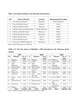


© Copyright 2026