
Document 62426
S c i e n t i f i ca n dT e c h n i c a l P u b l i c a t i o nNo . 6 3 3 Sur v ei l l anceofBact er i al Pneumoni aand Meni ngi t i si nChi l dr enAgedUnder5Year s Fi el dGui de Surveillance of Bacterial Pneumonia and Meningitis in Children Aged Under 5 Years Field Guide Scientific and Technical Publication No. 633 PAN AMERICAN HEALTH ORGANIZATION Regional Office of the WORLD HEALTH ORGANIZATION 525 Twenty-third Street, N.W. Washington, D.C. 20037 2010 Also published electronically in Spanish (2009) as: Vigilancia de las neumonías y meningitis bacterianas en menores de 5 años: guía práctica (ISBN 978-92-75-31633-7) PAHO HQ Library Cataloging-in-Publication Data: Pan American Health Organization Surveillance of bacterial pneumonia and meningitis in children aged under 5 years: Field Guide Washington, D.C.: PAHO © 2009 (Scientific and Technical Publication No. 633) ISBN 978-92-75-11633-3 I. Title II. Series 1. EPIDEMIOLOGICAL SURVEILLANCE – standards 2. BACTERIAL PNEUMONIA – diagnosis 3. PNEUMOCOCCAL PNEUMONIA – immunology 4. BACTERIAL MENINGITIS – diagnosis 5. PNEUMOCOCCAL MENINGITIS – immunology 6. PRESCHOOL AGE CHILDREN 7. EPIDEMIOLOGICAL SURVEILLANCE SERVICES NLM WC217 This guide was prepared by the Comprehensive Family Immunization Project of the Family and Community Health (FCH/IM) Area, with support from the Essential Medicines and Biologicals Project of the Technology, Health Care, and Research (THR/AES) Area at the Pan American Health Organization (PAHO). The Pan American Health Organization welcomes requests for permission to reproduce or translate its publications, in part or in full. Applications and inquiries should be addressed to the Publications Program, Pan American Health Organization, Washington, D.C., U.S.A., which will be glad to provide the latest information on any changes made to the text, plans for new editions, and reprints and translations already available. © Pan American Health Organization (2010) Publications of the Pan American Health Organization enjoy copyright protection in accordance with the provisions of Protocol 2 of the Universal Copyright Convention. All rights are reserved. The designations employed and the presentation of the material in this publication do not imply the expression of any opinion whatsoever on the part of the Secretariat of the Pan American Health Organization concerning the status of any country, territory, city, or area or of its authorities, or concerning the delimitation of its frontiers or boundaries. The mention of specific companies or of certain manufacturers’ products does not imply that they are endorsed or recommended by the Pan American Health Organization in preference to others of a similar nature that are not mentioned. Errors and omissions excepted, the names of proprietary products are distinguished by initial capital letters. CONTENTS Acknowledgments . . . . . . . . . . . . . . . . . . . . . . . . . . . . . . . . . . . . . . . . . . . . . v About the immunization field guides . . . . . . . . . . . . . . . . . . . . . . . . . . . . . vii Preface . . . . . . . . . . . . . . . . . . . . . . . . . . . . . . . . . . . . . . . . . . . . . . . . . . . . . . ix 1. Bacterial pneumonia and meningitis in children aged under 5 years . . . . 1 1.1 Epidemiological situation in the Region of the Americas . . . . . . . . . . . . . . . . 1 1.2 Epidemiology of infectious agents . . . . . . . . . . . . . . . . . . . . . . . . . . . . . . . . . . 2 2. Bacterial pneumonia . . . . . . . . . . . . . . . . . . . . . . . . . . . . . . . . . . . . . . . . . . 9 2.1 Clinical aspects. . . . . . . . . . . . . . . . . . . . . . . . . . . . . . . . . . . . . . . . . . . . . . . . . 9 2.2 Radiological diagnosis . . . . . . . . . . . . . . . . . . . . . . . . . . . . . . . . . . . . . . . . . . 10 2.3 Laboratory diagnosis . . . . . . . . . . . . . . . . . . . . . . . . . . . . . . . . . . . . . . . . . . . 12 2.4 Treatment. . . . . . . . . . . . . . . . . . . . . . . . . . . . . . . . . . . . . . . . . . . . . . . . . . . . 12 3. Bacterial meningitis . . . . . . . . . . . . . . . . . . . . . . . . . . . . . . . . . . . . . . . . . . 13 3.1 Clinical aspects. . . . . . . . . . . . . . . . . . . . . . . . . . . . . . . . . . . . . . . . . . . . . . . . 13 3.2 Laboratory tests. . . . . . . . . . . . . . . . . . . . . . . . . . . . . . . . . . . . . . . . . . . . . . . 13 3.3 Complications . . . . . . . . . . . . . . . . . . . . . . . . . . . . . . . . . . . . . . . . . . . . . . . . 15 3.4 Treatment. . . . . . . . . . . . . . . . . . . . . . . . . . . . . . . . . . . . . . . . . . . . . . . . . . . . 15 4. Laboratory procedures . . . . . . . . . . . . . . . . . . . . . . . . . . . . . . . . . . . . . . . 17 4.1 Specimen collecting, storing, and transporting. . . . . . . . . . . . . . . . . . . . . . . 17 4.2 General biosafety recommendations . . . . . . . . . . . . . . . . . . . . . . . . . . . . . . . 19 4.3 Quality assurance . . . . . . . . . . . . . . . . . . . . . . . . . . . . . . . . . . . . . . . . . . . . . 19 5. Surveillance of bacterial pneumonia and meningitis . . . . . . . . . . . . . . . . 21 5.1 Surveillance objectives . . . . . . . . . . . . . . . . . . . . . . . . . . . . . . . . . . . . . . . . . . 21 5.2 Surveillance strategies . . . . . . . . . . . . . . . . . . . . . . . . . . . . . . . . . . . . . . . . . . 21 5.3 Sentinel hospital-based bacterial pneumonia surveillance . . . . . . . . . . . . . . 22 5.4 Sentinel hospital-based bacterial meningitis surveillance . . . . . . . . . . . . . . . 24 5.5 Data analysis . . . . . . . . . . . . . . . . . . . . . . . . . . . . . . . . . . . . . . . . . . . . . . . . . 27 iii iv PAN AMERICAN HEALTH ORGANIZATION 5.6 Information flow and reporting periodicity . . . . . . . . . . . . . . . . . . . . . . . . . 29 5.7 Functional structure of the surveillance system . . . . . . . . . . . . . . . . . . . . . . 30 5.8 Evaluating the surveillance system . . . . . . . . . . . . . . . . . . . . . . . . . . . . . . . . 31 5.9 Feedback . . . . . . . . . . . . . . . . . . . . . . . . . . . . . . . . . . . . . . . . . . . . . . . . . . . . 31 5.10 Investigating meningitis cases . . . . . . . . . . . . . . . . . . . . . . . . . . . . . . . . . . . 31 5.11 Intervention measures . . . . . . . . . . . . . . . . . . . . . . . . . . . . . . . . . . . . . . . . . 32 6. Vaccines . . . . . . . . . . . . . . . . . . . . . . . . . . . . . . . . . . . . . . . . . . . . . . . . . . . . 35 6.1 Haemophilus influenzae type b (Hib) vaccine . . . . . . . . . . . . . . . . . . . . . . . . . . 35 6.2 Pneumococcal vaccine . . . . . . . . . . . . . . . . . . . . . . . . . . . . . . . . . . . . . . . . . . 37 6.3 Meningococcal vaccines. . . . . . . . . . . . . . . . . . . . . . . . . . . . . . . . . . . . . . . . . 40 Glossary. . . . . . . . . . . . . . . . . . . . . . . . . . . . . . . . . . . . . . . . . . . . . . . . . . . . . 43 Bibliography . . . . . . . . . . . . . . . . . . . . . . . . . . . . . . . . . . . . . . . . . . . . . . . . . 45 Annex 1. Bacterial pneumonia case report form . . . . . . . . . . . . . . . . . . . . . . . . . 57 Annex 2. Bacterial meningitis case report form . . . . . . . . . . . . . . . . . . . . . . . . . 59 Annex 3. Specimen collecting, storing, and transporting . . . . . . . . . . . . . . . . . . 61 Annex 4. Processing specimens for culture . . . . . . . . . . . . . . . . . . . . . . . . . . . . . 65 Annex 5. Functions of those responsible for surveillance . . . . . . . . . . . . . . . . . . 71 Annex 6. Data collection forms . . . . . . . . . . . . . . . . . . . . . . . . . . . . . . . . . . . . . . 75 ACKNOWLEDGMENTS The Pan American Health Organization is grateful for the valuable contributions to the preparation of this guide provided by personnel of the countries’ ministries of health to the staff of the Family and Community Health (FCH) Area (Comprehensive Family Immunization Project) and the Technology, Health Care, and Research Area (Essential Medicines and Biologicals Project). Coordination: Lúcia Helena de Oliveira, FCH/IM, PAHO Jean Marc Gabastou, THR/AES, PAHO Preparation: Maria Tereza da Costa Oliveira, FCH/IM, PAHO Review/ collaboration: Desirée Shepherd, FCH/IM, PAHO Gustavo Chamorro, Laboratory Public Health Central, Asunción, Paraguay Critical reading: Clara Inés Agudelo, National Institute of Health, Bogotá, Colombia Jon Andrus, FCH/IM, PAHO María Cristina Brandileone, Instituto Adolfo Lutz, São Paulo, Brazil Elizabeth Castañeda, National Institute of Health, Bogotá, Colombia Roxana Castle, Ecuador Carolina Danovaro, FCH/IM, PAHO Brendan Flannery, CDC/CCID/NCIRD, United States Jorge Alirio Holguin, University of Cali, Valle, Colombia Beryl Irons, FCH/IM, PAHO Ana Paula Lemos da Silva, Institute Adolfo Lutz, São Paulo, Brazil Greta Miño, Ecuador Farzana B. Muhib, PneumoADIP Linda Ojo, CDC/CCID/NCIRD, United States Rosalyn O’Loughlin, CDC/CCID/NCIRD, United States Cuauhtémoc Ruiz Matus, FCH/IM, PAHO Cristiana Toscano, WHO Cynthia Ruiz Matus Whitney, CDC/CCID/NCIRD, United States v ABOUT THE IMMUNIZATION FIELD GUIDES The Expanded Program on Immunization is viewed as one of the most successful public health experiences in the Americas because it has played a pivotal role in reducing infant mortality from vaccine-preventable diseases in the Region. In fact, since the program was launched, our countries stopped the transmission of wild poliovirus in the Region in 1991 and interrupted indigenous measles transmission in November 2002; they also are making significant gains in the battle to eliminate rubella and congenital rubella syndrome. In addition, national immunization programs are undertaking extraordinary efforts to identify at-risk populations and overcome inequities in vaccination. To maintain these advances and to cope with new challenges, such as the introduction of new vaccines, partnerships will have to be strengthened among governments, donor agencies, the private sector, scientific associations, and society as a whole. To this end, PAHO is promoting the best technical quality by issuing these practical field guides, which have been prepared by the Immunization Unit in the Family and Community Health Area. The most recent techniques presented in the field guides, coupled with useful illustrations, will aid health workers in their efforts to control, eliminate, or eradicate diseases such as poliomyelitis, neonatal tetanus, yellow fever, diphtheria, pertussis, tetanus, Haemophilus influenzae type b infections, hepatitis B, measles, and rubella. The field guides also include standardized methods and procedures for conducting epidemiologic surveillance and maintaining an up-to-date information system that will make it possible to make timely and effective decisions. These field guides are based on the latest scientific information, and they pool the experience of prominent health professionals in the field. As a result, they are particularly suitable for promoting strategies that have already proven to be effective. The strengthening of prevention activities, the reduction of health inequities, and the promotion of technical expertise in vaccination services were the principles that guided the preparation of the guides. The Expanded Program on Immunization, a joint effort by all the countries of the Americas, effectively contributes to the attainment of the Millennium Development Goals. Mirta Roses Periago Director Pan American Health Organization vii PREFACE This field guide was prepared by PAHO to support health workers participating in the epidemiological surveillance of bacterial pneumonia and meningitis. Pneumonia is among the leading causes of hospitalization and death for children aged under 5 years in the Region of the Americas. In developed countries, the majority of pneumonias are believed to be of viral origin; however, the etiology of pneumonia is almost always bacterial in developing countries. Bacterial meningitis, although not as frequent as pneumonia, is always a serious disease, given the risk of sequelae and its high case-fatality rate. Three bacteria are principally responsible for the diseases: Haemophilus influenzae (Hi) type b (Hib), Neisseria meningitidis (meningococcus), and Streptococcus pneumoniae (pneumococcus). The introduction of the Hib vaccine in countries of the Region produced a dramatic decline of invasive disease due to this bacterium, and pneumococcus is now the principal etiologic agent responsible for bacterial pneumonia and meningitis in children aged under 5 years. This guide describes the clinical and epidemiological aspects of bacterial pneumonia and meningitis, the role of radiology in pneumonia diagnosis, and the confirmatory role of laboratory tests for both diseases. It also describes recommended procedures for epidemiological surveillance of these diseases, and for their prevention and control. The guide is designed to standardize thinking and procedures, so as to strengthen epidemiological surveillance and provide data on the burden of these diseases. The data will be used for decision-making on incorporating new vaccines into national vaccination schedules, to assess the impact of vaccines already used, and to provide guidelines for the rational use of antimicrobial drugs. The introduction of new vaccines to prevent these diseases will be an important step in meeting one of the Millennium Development Goals, that of reducing the under-5 mortality by two thirds between 1990 and 2015. ix 1. BACTERIAL PNEUMONIA AND MENINGITIS IN CHILDREN AGED UNDER 5 YEARS 1.1 Epidemiological Situation in the Region of the Americas Since 1993, the Region has had a network of laboratories for the regional surveillance of bacterial pneumonia and meningitis known as SIREVA (Sistema Regional de Vacunas/Regional Vaccine System). SIREVA was organized by PAHO with financial and technical support from the Canadian International Development Agency (CIDA). Twenty countries currently participate in the network, which has identified the three main bacterial agents causing pneumonia and bacterial meningitis: Haemophilus influenzae (Hi), Neisseria meningitidis (meningococcus), and Streptococcus pneumoniae (pneumococcus). It has also succeeded in identifying the serotypes and circulating serogroups of these bacteria, and their susceptibility to the most commonly used antibiotics. The Program for External Evaluation of Performance (Programa de Evaluación Externa del Desempeño/PEED) has been responsible for verifying the quality of the network’s laboratory procedures. Between 1993 and 2004, the network processed over 11,000 pneumococcal isolates from invasive infections in children aged under 6 years. Isolates from Argentina, Brazil, Chile, Colombia, Mexico, and Uruguay between 2000 and 2005 pointed to serotype 14 as the predominant serotype, representing 29.2% of isolates, followed by serotypes 6B, 5, and 1 (Figure 1). For the 2000-2005 period, 13 of 20 countries reported Figure 1. Principal Pneumococcus Serotypes Isolated from Invasive Infections in Children Aged Under 6 Years in Six serotype 14 as the priority serotype, followed in Latin American Countries, 2000-2003 decreasing order by 6B, 5, and 1. The same 6B 6A order had prevailed in 2000-2003. 7F 9.2% 4.0% 5 2.5% 6.1% 9V Another important finding is the increase in 4 3.0% 1.7% the proportion of pneumococcal isolates not 3 3.5% susceptible to penicillin—from 14.7% in 1993 to 1 30.6% in 1999 and to 39.8% in 2003. 6.0% 14 29.2% Before the introduction of the conjugate vacOther cines, H. influenzae type b was the most common 15.9% 18C agent responsible for bacterial meningitis 23F 4.2% 6.0% 19A 19F among children aged under 5 years in the Amer3.1% 5.6% icas. After the introduction of the Hib vaccine, there was a spectacular decline in disease caused by this bacterium, as documented in some Source: García S, Levine OS, Cherian T, Gabastou JM, Andrus J, Working Group members. Pneumococcal disease and vaccination in the Americas: an agenda for accelercountries of the Region that have surveillance ated vaccine introduction. Panam J Public Health 2006;19(5):340-348. 1 2 PAN AMERICAN HEALTH ORGANIZATION systems for the disease. Assuming 95% efficacy for the vaccine, and 20,000 cases annually prior to vaccine introduction, it has been estimated that meningitis due to Hib had decreased by nearly 85% in Latin America and the Caribbean as of 2005. Currently, the principal etiologic agent is pneumococcus, followed by meningococcus. In light of the increased pneumococcus vaccine availability on the market, it has become necessary to strengthen the Regional surveillance of the corresponding diseases in order to supplement laboratory data with standardized epidemiological information and facilitate comparison and analysis in different countries. Thus, more uniform and systematic participation from all the countries is desired. The information generated will be used for making decisions on the introduction of new vaccines and for monitoring their impact. To address this need, PAHO launched the SIREVA II project (Network Surveillance System for the Bacterial Agents Responsible for Pneumonia and Meningitis) in 2005, with support from the GAVI Alliance’s PneumoADIP (Pneumococcal Vaccines Accelerated Development and Introduction Plan). 1.2 Epidemiology of Infectious Agents 1.2.1 Description of the Infectious Agents Haemophilus influenzae (Hi) is a Gram-negative coccobacillus. Six encapsulated antigenic serotypes have been identified (they are designated as “a” to “f”). Both the encapsulated and non-encapsulated strains are potentially pathogenic for human beings, but they differ in their virulence and pathogenic mechanisms. H. influenzae serotype b (Hib) is the most pathogenic. Neisseria meningitidis (meningococcus) is a Gram-negative diplococcus. Thirteen meningococcus serogroups of this bacterium have been identified, and the six most frequently isolated are A, B, C, W 135, Y, and Z. Serogroups B and C are predominant in the Region of the Americas. Streptococcus pneumoniae (pneumococcus) is a lanceolate Gram-positive diplococcus. Although 90 serotypes and over 40 pneumococcal subgroups have been identified, 11 of the most common serotypes are responsible for approximately 75% of all invasive infections in children worldwide. 1.2.2 Reservoir Human beings are the sole reservoir of Hi, meningococcus, and pneumococcus. 1.2.3 Transmission The transmission of Hi, meningococcus, and pneumococcus is from person-toperson via droplets of saliva or by contact with the nasopharyngeal secretions of infected individuals. SURVEILLANCE OF PNEUMONIA AND MENINGITIS UNDER 5 YEARS 3 1.2.4 Distribution and Seasonality The distribution of Hi, meningococcus, and pneumococcus is global. Hi does not generally display well-defined seasonality. However, studies conducted in the pre-vaccine era describe peaks in the fall and spring months in countries with temperate climates. The incidence of meningococcus in Europe and the United States peaks in the winter and spring months. In sub-Saharan Central Africa, there has long been a wide area with elevated incidence of meningococcal meningitis and prevalence tends to increase during the dry season. Pneumococcus is present in all seasons and under any climate. Temperate countries experience a higher incidence of pneumococcal pneumonia in the winter and spring months. 1.2.5 Susceptibility Susceptibility to Hi, meningococcus, and pneumococcus infection is presumed to be universal. In other words, all people are susceptible to the infections caused by these agents. However, the younger the child, the more susceptible he or she will be to these bacteria and the invasive illnesses that they cause. The risk of Hi infection is greatest between 2 months and 3 years of age, and declines thereafter. In developing countries, the greatest incidence is in children under 6 months of age, while developed countries experience a peak in children aged 6 to 12 months. Infection is infrequent after the age of 5. The highest attack rates of meningococcus are seen in children aged under 1 year, with a peak in this group in the 3-to-5-month range. However, adolescents and young adults are also vulnerable, especially if they live in overcrowded conditions (barracks and other institutional settings). In addition to age, risk factors for meningococcus infection include asplenia and acquired or congenital immunodeficiency. Other conditions, such as dense population, poverty, active or passive exposure to tobacco smoke, and concurrent upper respiratory tract infections, increase the risk of meningococcal infection. Pneumococcus infection is most frequent between 2 months and 3 years of age, although it declines after 18 months. The risk rises again after age 65. Carriers of some chronic diseases are at greater risk of infection by pneumococcus. For further details, see Chapter 6, Section 2. 1.2.6 Immunity Immunity to Hi, meningococcus, and pneumococcus can be acquired passively through the placenta, or actively by previous infection or immunization. As of age 5 years, the majority of unvaccinated children have H. influenzae anticapsular antibodies due to exposure to the bacterium. 4 PAN AMERICAN HEALTH ORGANIZATION Regarding meningococcus, group immunity of unknown duration follows clinical or subclinical infection. As for pneumococcus, newborns can have antibodies due to passive transmission from the mother. However, they disappear within months, as the incidence of the invasive disease increases. After age 18 months, children show specific immune responses to the majority of the circulating pneumococcus serotypes, as a result of repeated exposure. 1.2.7 Carrier Status Hi, pneumococcus, and meningococcus are generally nasopharyngeal colonizing agents in asymptomatic individuals, who are considered carriers. Between 1% and 5% of non-immunized individuals are estimated to be Hi carriers. The percentage is higher among preschool children. Hi can remain in the nasopharynx for months. Between 5% and 15% of adolescents and young adults can carry meningococcus in the nasopharynx. It is unusual for young children to be meningococcus carriers, and it is rare in adults (1%). The prevalence of pneumococcus carriers is higher among children, especially children who attend daycare, and in adults who have close contact with these children. It is estimated that this bacterium colonizes the nasopharynx of as many as 40% of children aged under 2 years. The pneumococcal serotypes most frequently isolated, both in carriers and in people with invasive disease, are 14, 6B, and 23F. Carrier status is associated with the emergence of diseases such as otitis, sinusitis, meningitis, pneumonia, and septicemia. Table 1 summarizes the epidemiological characteristics of the three etiologic agents. 1.2.8 Disease Burden of Pneumonia and Meningitis In developing countries, acute respiratory infections (ARIs), especially pneumonias contracted in the community, are the principal cause of hospitalization and death among children aged under 5 years. In 2004, the World Health Organization (WHO) estimated the incidence of clinical pneumonia in developing countries at 0.29 episodes per child per year, or 29 episodes per 100 children annually, which translates into 150.7 million new cases annually, 11 to 20 million (7%-13%) of which require hospitalization. A number of population studies show that the incidence of pneumonia contracted by children aged under 5 years in communities of developed countries is approximately 0.026 episodes per child per year. In other words, over 95% of all episodes of clinical pneumonia in young children worldwide occur in developing countries. SURVEILLANCE OF PNEUMONIA AND MENINGITIS UNDER 5 YEARS 5 Table 1. Epidemiological Characteristics of the Principal Bacteria Responsible for Pneumonias and Meningitis in Children Aged Under 5 Years ETIOLOGIC AGENT Type of bacterium HAEMOPHILUS INFLUENZAE (HI) Gram-negative coccobacillus Reservoir Fall and spring Susceptibility Immunity (PNEUMOCOCCUS) Gram-negative diplococcus Lanceolate Gram-positive diplococcus Winter and spring Winter and spring From person to person via nasal and pharyngeal secretions While in the respiratory tract and until 24 hours after the start of specific antibiotic therapy Carrier Incubation (MENINGOCOCCUS) Worldwide Transmission Transmission period STREPTOCOCCUS PNEUMONIAE Human beings Distribution Seasonality NEISSERIA MENINGITIDIS Yes 2-4 days Greatest risk is between the ages of 2 months and 3 years, but it declines after age 2. Infrequent above age 5. 1-10 days (usually <4) Susceptibility is greatest among infants, peaking between 3 and 5 months. Adolescents and young adults are also vulnerable, especially those living in overcrowded conditions. 1-3 days Incidence is highest between the ages of 2 months and 3 years, but it declines from 18 months on. Incidence increases again after age 65. Carriers of some chronic diseases are at increased risk. Immunity can be acquired passively through the placenta, or actively by prior infection or immunization Source: Heymann DL (ed.). Control of communicable diseases manual, 18th ed. Washington, DC: American Public Health Association. According to various studies, mortality from ARIs is directly associated with bacterial infection, primarily pneumococcus and Hib. Pneumococcus causes infections of various types and in various ways, including colonization of the nasopharynx; non-invasive direct dissemination of microorganisms in the mucous membranes of the middle ear, in the paranasal sinuses, in the trachea, and in the bronchia and lungs; and dissemination in the bloodstream, which causes bacteremia without an apparent focus. It can also spread to the central nervous system, joints, bones, heart valves, and pleura. Most cases of pneumococcal pneumonia are not accompanied by bacteremia. On the other hand, the vast majority of invasive pneumococcus infections involve pneumonia with bacteremia. Figure 2 shows the distribution of invasive pneumococcal diseases proportionally by type of disease (first circle) and pneumococcal pneumonia with and without bacteremia (second circle). Otitis media, or inflammation of the middle ear, is a type of non-invasive bacterial infection frequent in children aged 6 to 36 months. Nearly three fourths of the 6 PAN AMERICAN HEALTH ORGANIZATION Figure 2. Percentage of Invasive Infections and Pneumonia Caused by Pneumococcus Pneumococcal Pneumonia Pneumonia with bacteremia 20% Invasive Pneumococcal Disease Others: meningitis (5-10%), artritis, pleuritis 10% Neumon ía con bacteriemia Pneumonia without bacteremia 80% Pneumonia with bacteremia 90% Adapted from Fedson DS, Musher DM. Pneumococcal polysaccharide. In: Orenstein WA, Plotkin SA (eds.). Vaccines, 4th ed. Philadelphia, PA: W. B. Saunders; 2004: 538. population has at least one otitis media episode between those ages. Pneumococcus is found in 40% of positive cultures of middle ear secretions. Pneumococcus is the leading cause of pneumonias contracted in the community and requiring hospitalization, and is responsible for nearly 800,000 deaths worldwide each year in children aged under 5 years. Bacterial meningitis in children aged under 5 years is caused by Hib, meningococcus, and pneumococcus in over 90% of cases. However, as mentioned above, Hib disease has declined in the Region of the Americas. Thus, pneumococcus is currently the principal cause of bacterial meningitis, followed by meningococcus. 1.2.9 Economic Studies Economic studies of health variables are needed, and countries should take the findings into account when deciding whether to introduce a vaccine or to expand the age group selected to receive the vaccines administered in each country. A cost analysis can provide a measure of the economic burden of vaccine-preventable diseases in a country. Pneumococcal pneumonia bears major economic implications for health systems, families, and society in general, given its high disease burden and mortality. Direct expense for care is one type of cost. It includes medical consultations, hospitalization, diagnostic tests, and treatment, whether paid for by a country’s health system, by families, or both. Other direct expenses include family expenditures on transportation, food, lodging, and the like in connection with hospital visits. Rehabilitation is another direct expense in case of sequelae and disabilities such as postmeningitic deafness, mental retardation, motor anomalies, convulsions, and visual disorders. SURVEILLANCE OF PNEUMONIA AND MENINGITIS UNDER 5 YEARS 7 Indirect, or social, costs include reduced production due to absenteeism and intangible costs such as pain, suffering, and stigma. Studies are also needed to estimate the cost of incorporating a vaccine in an immunization program and calculate the resources that the program will need for financial sustainability in the mid- and long-term. This is a crucial component of decision-making in national immunization programs. 2. BACTERIAL PNEUMONIA 2.1 Clinical Aspects Pneumonia is an infection of the pulmonary parenchyma that can be caused by a variety of microorganisms (viral, bacterial, and other). Pneumonias with different etiologies may present very similar clinical symptoms. In infants and young children, pneumonia tends to begin with acute fever. Numerous studies have attempted to determine what specific and sensitive clinical signs most reliably indicate the presence of pneumonia. Most such studies have settled on tachypnea (rapid breathing) as the most effective predictive sign. The strategy known as Integrated Management of Childhood Illness (IMCI) classifies pneumonia according to its clinical manifestations, that is (i) pneumonia, (ii) severe pneumonia, and (iii) very severe pneumonia. Pneumonia is suspected when physical examination reveals that the child is coughing and has difficulty breathing, as well as rapid breathing. The latter is defined as follows: • Before age 2 months: over 60 breaths/minute; • Age 2 to 11 months: over 50 breaths/minute; • Age 12 months to 5 years: over 40 breaths/minute. Other signs that can be detected through thoracic auscultation include crepitant stertors, reduced respiratory sounds, or areas of bronchial breathing. Pneumonia is considered severe when any of the following symptoms is present in addition to cough, difficulty breathing, and rapid breathing: • Retraction of the inferior thoracic wall (subcostal retraction); • Nasal flaring; • Whizzing sounds (in younger infants). Very severe pneumonia presents with, in addition to the above, the following symptoms: • • • • • Central cyanosis; Inability to breast-feed or drink; Vomiting of everything ingested; Convulsions, lethargy, or loss of consciousness; Severe difficulty breathing (for example, with head nodding). Generally speaking, the most severe pneumonia cases are of bacterial origin, and these are responsible for most hospitalizations and deaths of children aged under 5 years. 9 10 PAN AMERICAN HEALTH ORGANIZATION 2.1.1 Differential Diagnosis Respiratory viral infections are common in children aged under 5 years and tend to cause cough, fever, mouth breathing, and nasal secretion, but not tachypnea, intercostal retraction, or other signs of severity. Young children may present with episodes of wheezing. Respiratory syncytial virus (RSV), influenza, adenovirus, and parainfluenza are viruses known to cause viral pneumonia. However, they produce upper respiratory tract illness more often than pneumonia. Viral pneumonia may also appear in measles, influenza, and chickenpox. Some common childhood diseases, such as bronchiolitis and asthma, can have symptoms similar to those caused by bacterial pneumonia. Bronchiolitis is a viral infection of the lower respiratory tract. It is frequent and relatively severe in infants. Most cases are caused by RSV. The disease is characterized by obstruction of the respiratory tract and episodes of wheezing, with little response to bronchodilators. Secondary bacterial infection may occur. Asthma is a chronic inflammatory disorder with reversible obstruction of the respiratory tract. It is characterized by recurrent episodes of wheezing with cough, and sometimes by lower intercostal retraction and tachypnea. In the absence of viral or bacterial infection, there is no fever. Bronchodilators and anti-inflammatory drugs are effective treatments. 2.1.2 Complications Pleural effusion, empyema, atelectasis, and hypertensive pneumothorax are among the complications from bacterial pneumonia. If a child with severe pneumonia does not receive proper and timely treatment with specific antibiotics, respiratory insufficiency can become acute and cause death. 2.2 Radiological Diagnosis Radiographic analysis is an important tool in diagnosing severe and very severe pneumonia because it helps to differentiate between bacterial and viral etiologies, and to determine whether complications such as pleural effusion or atelectasis are present. The typical radiological alterations associated with bacterial pneumonia are single- or multi-focus condensations. SURVEILLANCE OF PNEUMONIA AND MENINGITIS UNDER 5 YEARS 11 According to criteria and definitions established by WHO for interpreting chest X-rays of children with pneumonia, bacterial pneumonia presents a dense cottony appearance (the alveolar infiltrate), reflecting that one or more segments or pulmonary lobes, or a complete lung, are compromised. With these infiltrates, there are often areas of air bronchogram (see Glossary), sometimes in conjunction with pleural effusion. Figure 3 shows a radiological image compatible with bacterial pneumonia. Figure 3. Chest X-ray Compatible with Bacterial Pneumonia in Upper Right Lobe Lobar consolidation Source: WHO Lite. Pneumonia Vaccine Trial Investigator’s Group: Training Programme for the Standardized Diagnosis of Radiological Pneumonia. The compromising of the pleura can show up as effusions of various sizes. A small effusion may be difficult to see in a chest X-ray, but it will be clear when it produces a shift of the mediastinum toward the contralateral side (right or left), or if there is a blurring of the costofrenic angles. A lateral chest X-ray is called for if pleural effusion is suspected, since it can help to discern the effusion. Atelectasis is seen in an X-ray as a reduction in the volume of the distal parenchyma and a shift of the scissures, mediastinum, and diaphragm toward the affected side (see Glossary). Radiological manifestations appear when clinical symptoms have already emerged, and can persist as long as three months after clinical symptoms disappear. 2.2.1 X-ray Quality A well-taken and well-interpreted X-ray is crucial for the radiological diagnosis of pneumonia. In analyzing an X-ray, the following aspects of its technical quality should be kept in mind: • Proper exposure: A good exposure will show differences of density that make it possible to differentiate bones, soft tissue, and lungs. • Correct position: The medial and terminal parts of the patient’s clavicles should be approximately equidistant from the midline. 12 PAN AMERICAN HEALTH ORGANIZATION • Developing: There should be black space between the body and the edges of the plate, and the densest areas, such as the most distal portion of the thoracic spine behind the heart, should appear white. 2.3. Laboratory Diagnosis A blood specimen for culture should be collected as soon as possible in order to isolate the etiologic agent. Depending on the severity of the case, this should be done before antibiotic treatment has begun. Once antibiotic therapy starts, all data on the antibiotic type and dosage should be collected and recorded. Hemoculture is not a highly sensitive diagnostic test, and only a small percentage of hemocultures (under 20%) will be positive. However, collecting blood specimens and culturing them is extremely important, in that when the culture is positive the etiologic agent can be identified with certainty, and antibiograms can be conducted to determine the susceptibility of the bacterium to the customary antimicrobial drugs. It also makes it possible to monitor the serotypes/serogroups of bacteria that are in circulation. Pleural fluid cultures are a highly sensitive laboratory test (as much as 80%) for bacteria. Thus, whenever thoracocentesis is indicated, a pleural fluid specimen should be collected for culturing. The hemogram is a supplementary, though nonspecific, test, and can help to suggest what bacterial etiology may be behind the infection. The laboratory tests and the types of specimens that they require are detailed in Chapter 4. 2.4. Treatment In general, children with pneumonia can be treated on an outpatient basis, with general care in the home following medical instructions specific to each case. Children with severe or very severe pneumonia should be treated in a hospital setting. For more details on recommended treatment, consult IMCI manuals or national protocols for managing bacterial pneumonia patients. 3. BACTERIAL MENINGITIS 3.1 Clinical Aspects Meningitis is an inflammation of the membranes around the brain, the cerebellum and the bone marrow, the anatomical sites surrounded by the subarachnoid space, where the cerebrospinal fluid (CSF) circulates. It is characterized by fever and signs of meningeal inflammation. Children under 1 year of age usually present some nonspecific symptoms such as reduced appetite or vomiting. At least one of the following specific signs is present: • • • • Bulging fontanelle; Convulsion; Irritability without justification or clinical reason; Lethargy. Children age 1 year and over, as well as adults, also have nonspecific symptoms such as photophobia and headache, plus at least one of the following specific signs: • • • • • Altered state of wakefulness; Convulsion; Stiff neck, other signs of meningeal inflammation, or both; Prominent signs of hyperactivity or lethargy; Projectile vomiting. Meningococcemia is accompanied by an initially erythematous and macular cutaneous exanthema that rapidly leads to petechial eruption and ultimately ecchymosis. 3.2 Laboratory Tests Although several microorganisms can cause bacterial meningitis, and the clinical symptoms associated with them are similar, therapy must be specific to the pathogen. Thus, an etiological diagnosis must be achieved by culturing the CSF and blood. This calls for immediate systematic collection of specimens—if possible, before beginning antibiotic treatment. Once antibiotic therapy is begun, information on the type of antibiotic used, doses, and dates should be recorded. Obtaining a CSF specimen (Figure 4) is indispensable as a first step in determining etiology in cases of bacterial meningitis and conducting other tests immediately thereafter. 13 Figure 4. Position for Lumbar Puncture Source: MedlinePlus Medical Encyclopedia [cited 23 January 2007]. Available at: http:/nlm.nih.gov/medilineplus/ency/imagespages/19078.htm. 14 PAN AMERICAN HEALTH ORGANIZATION It is advisable to conduct at least the following laboratory tests on CSF specimens where meningitis is suspected: cytochemical, Gram stain, and culture. In bacterial meningitis, the CSF presents the following visual characteristics and cytochemistry: • • • • Turbidity; Increased leukocytes (>100/mm3); Elevated protein levels (>100 mg/dl); Decreased glucose levels (<40 mg/dl). A Gram stain of the CSF examined under the microscope will suggest the bacterial etiology of the infection. Where bacterial meningitis is present, the Gram stain will show: • • • • Gram-negative coccobacilli (H. influenzae); Gram-negative intracellular or extracellular diplococci (meningococci); Lanceolate Gram-positive diplococci (pneumococci); Other. In doing cultures, both CSF and blood specimens are used, in order to isolate the bacterium in at least one of the two. Culturing makes it possible to determine with certainty what bacterium is causing the disease. Isolating the strain also makes it possible to identify the bacterial serotype/serogroup involved, and to do antibiograms and determine the bacterium’s susceptibility to proven antimicrobial drugs. Culture is considered “the gold standard” for diagnosing bacterial etiology. • Cytochemical examination of the CSF establishes whether bacterial meningitis may be present. • The Gram stain suggests the type of bacterium present. • Cultures determine with certainty what bacterium is causing the disease. • Antibiograms establish the susceptibility of the agent to proven antibiotics. Even if the CSF is apparently normal and does not show cytochemical alterations compatible with bacterial meningitis, it is imperative that it be cultured. Whenever possible, a molecular or immunological (antigen-detecting) test such as polymerase chain reaction (PCR), latex particle agglutination, or counterimmunoelectrophoresis should be performed in order to obtain an immediate etiological diagnosis. For cases where meningitis is suspected despite the absence of cytochemical alterations in the CSF and despite the fact that CSF or blood cultures have identified no pathogen, the country’s protocols should include a detailed propaedeutic that 15 SURVEILLANCE OF PNEUMONIA AND MENINGITIS UNDER 5 YEARS can be consulted to establish a diagnosis and determine the proper management of the patient. Identifying the bacterium causing bacterial meningitis and determining its antimicrobial susceptibility allows for the following steps: • Confirming the clinical diagnosis; • Deciding on specific treatment; • Taking immediate measures to protect the patient’s contacts where advised (see Chapter 5, Section 11); • Identifying the circulating serotype. For more details on these tests, see Chapter 4. 3.3 Complications As many as 30% of bacterial meningitis survivors can suffer permanent sequelae. The most frequent of these is sensorineural hearing loss. Others include language disorders, mental retardation, motor anomalies, convulsions, and visual disorders. Sequelae are more frequent in meningitis caused by pneumococcus, as shown in Table 2. Table 2. Percentage of Bacterial Meningitis Sequelae for Cases with Different Etiologic Agents Etiologic agent H. influenzae N. meningitidis S. pneumoniae Deafness or hypoacusis 10 6 28 Sequelae (%) Mental Spasticity/ retardation paresis 6 5 2 2 17 12 Convulsions 6 6 14 Source: Adapted from Baraff LJ, Reads SI, Schriger DL. Outcomes of bacterial meningitis in children: A meta-analysis. Pediatr. Infect. Dis. J. 1993;12:392. Meningitis can evolve rapidly toward stupor, coma, and death. Case fatality rates range from 3% to 6% for Hib meningitis, from 8% to 15% for meningococcal meningitis, and, highest of all, from 10% to 30% for pneumococcal meningitis. 3.4 Treatment Every child with meningitis should be referred to the closest hospital for treatment. For more details on the treatment of bacterial meningitis, consult IMCI manuals or national protocols for the management of patients with bacterial meningitis. 4. LABORATORY PROCEDURES The main purpose of laboratory tests in this context is to make etiological diagnoses of bacterial pneumonia and meningitis, and to determine the antimicrobial susceptibility of the bacterium (through disc diffusion and determination of minimum inhibitory concentration), as a basis for surveillance of these two diseases (see Chapter 5). In cases of probable bacterial pneumonia or suspected cases of bacterial meningitis in children aged under 5 years, the hospital technical team responsible for sentinel surveillance should proceed as follows: • Collect CSF and blood specimens from patients with suspected meningitis; • Collect blood specimens for culture in all probable cases of bacterial pneumonia, and in all cases involving pleural fluid with pleural effusion where thoracocentesis is indicated; • Send the specimens to the laboratory immediately for processing. Figure 5 presents a flow chart for the Figure 5. Flow Chart for Laboratory Diagnosis of Bacterial network involved in laboratory diagno- Pneumonias and Meningitis sis, with competencies by level. Sentinel Hospital Laboratory The hospital laboratory will process CSF: Pleural fluid, the specimens and report the findings Blood cytochemistry, Gram stain, culture to the hospital’s technical team as soon Gram stain, culture culture as they are obtained. Positive culture / antibiogram Send Haemophilus Influenzae, Strains of H. influenzae (Hi), meningomeningococcus, and pneumococcus strains (with corresponding report sheets) coccus, or pneumococcus isolated should be sent to the national reference laboraNational Reference Laboratory tory for serotyping, and to determine Antimicrobial susceptibility Typing their antimicrobial susceptibility. The Quality control Program for External Evaluation of Performance national laboratory will provide feedback on its findings to the hospital’s Subregional Laboratory sentinel team as soon as possible, and Quality control Program for External Evaluation of Performance will send a percentage of the isolated strains to one of the two subregional reference laboratories for quality conRegional Laboratories trol as a part of the PEED. (See section Program for External Evaluation of Performance on quality assurance in this chapter). 4.1 Specimen Collecting, Storing, and Transporting Certain steps should be taken in obtaining good quality specimens of CSF, blood, and pleural fluid that allow for strain isolation. Whenever possible, specimens 17 18 PAN AMERICAN HEALTH ORGANIZATION should be collected for culturing before beginning antibiotic treatment, since specimens taken later may produce false negatives. Specimens should always be taken in standardized aseptic conditions, and the technical personnel conducting the procedure should use sterile gloves and masks. These steps should be reviewed and followed in order to ensure successful laboratory diagnosis (see Annex 3). 4.1.1 Specimen Storing and Transporting • Specimens taken for culture should be sent to the laboratory immediately. • The maximum time for delivery of blood, CSF, and pleural fluid specimens to the laboratory is two hours. During that period, they should remain at room temperature. • If specimens must be kept longer, it is recommended that they be kept in an incubator at 35o C to 36o C. (Never refrigerate them.) • CSF and pleural fluid specimens should not be sent in any culture media. In exceptional cases, it may be necessary to send specimens from the laboratory of the sentinel hospital for culturing at the national reference laboratory. The transportation of these specimens should meet the following criteria: • Blood, CSF, and pleural fluid specimens for culture should not be refrigerated or frozen during transport. • Specimens should be correctly identified, and an epidemiological case record should be included. • The bottle, wrapped in aluminum foil or in sterile packaging paper, should be packed and placed in a box, well protected with Styrofoam or paper to cushion it and avoid breakage. An arrow should indicate the direction in which the bottles have been packed, in order to prevent specimens from being turned over and spilled. • The box should be labeled with sender and recipient information. • The box should bear an indication of the type of biological material that it contains. 4.1.2 Strain Transporting Since isolated strains are nutritionally demanding microorganisms and require special conditions to remain viable, the following factors must be kept in mind while storing and transporting them: • Specimens should be kept in proper culture media at room temperature, and should be shipped to the national reference laboratory in a transport media SURVEILLANCE OF PNEUMONIA AND MENINGITIS UNDER 5 YEARS 19 such as Amies with activated charcoal, along with the isolation report for confirmation and serotyping as soon as possible. • Transportation should meet biosafety criteria to reduce possible risks of contamination. The strains should be transported in boxes according to IATA’s (International Air Transport Association) standards, and have labels indicating that infectious substances are within. 4.2 General Biosafety Recommendations In order to reduce the risk of transmitting viral agents (human immunodeficiency virus [HIV], hepatitis B, etc.), bacterial agents, and protozoan agents, or other parasites that may be present in any biological material (including blood, CSF, and pleural fluid), the following recommendations should be followed: • Exercise care in collecting and transporting specimens. • Label the containers holding the material to be transported to indicate that they contain specimens of biological materials. • Seal the containers with the specimens hermetically. • Clean the outsides of the containers with a disinfectant. A hypochlorite solution with 0.1% of available chlorine (1g/liter, 1,000 ppm) can be used. Make sure that it does not become contaminated with blood or any other biological material. In emergencies (spillage, etc.) it is advisable to use sodium hypochlorite with available chlorine at a concentration of 4 or 5 g/L. Use waterproof latex or vinyl gloves. • Wash hands with soap and water immediately after removing gloves. • Place the used needles and syringes in a puncture-resistant container. • If pricked by a needle, or in any other case of puncture or skin wound, wash abundantly with soap and water, and bleed the wound. • Report any contamination of hands or body with blood or other biological material, as well as any prick or other skin wound, to the supervisor and the health service, in order to obtain proper treatment. 4.3 Quality Assurance A key component of the SIREVA project is its quality assurance program, which was created to address participants’ specific needs and includes the following elements: • A process to monitor the shipment of individual isolates to the National Centre for Streptococcus in Edmonton, Alberta, Canada, and to the Instituto de Salud Carlos III in Madrid, Spain, where confirmation, additional investigation, or both may occur; 20 PAN AMERICAN HEALTH ORGANIZATION • A quality control program to ascertain the capacities of the participating laboratories. The quality control program currently has two subregional reference centers: the Microbiology Group at Colombia’s National Institute of Health, and the Toxigenic and Biogenic Bacteria Section of the Microbiology Department at the Institute Adolfo Lutz in São Paulo, Brazil. In October 2003, the SIREVA project’s quality assurance program was renamed Program for External Evaluation of Performance (Programa de Evaluación Externa del Desempeño/PEED), and the relevant elements were updated and standardized accordingly. Among the main quality control functions of the national and subregional reference laboratories, the following should be mentioned: • Training the staff of participating laboratories; • Ensuring a system of safe air transport for shipment of isolates, in order to prevent contamination, loss of viability, or both, by following the recommendations of the United Nations Committee of Experts on the Transport of Dangerous Goods. These recommendations are incorporated in the regulations of the Universal Postal Union (UPU), the International Civil Aviation Organization (ICAO), and the IATA, all of which are advised by WHO; • Ensuring proper storage of the isolates sent by participating laboratories; • Conducting serotyping and antimicrobial susceptibility tests for all the Hi, meningococcus, and pneumococcus isolates sent by the laboratories; and • Conducting outside performance evaluations for interlaboratory systems. 5. SURVEILLANCE OF BACTERIAL PNEUMONIA AND MENINGITIS 5.1 Surveillance Objectives The objectives of bacterial pneumonia and meningitis surveillance in children aged under 5 years in the Region are the following: • To obtain standardized epidemiological data on bacterial pneumonia and meningitis; • To identify Hi, meningococcus, and pneumococcus, and to describe the strains of those agents in circulation, as well as changes of serotypes/serogroups as they emerge; • To monitor antimicrobial susceptibility patterns and contribute to establishing technical standards for the use of antimicrobial drugs; • To generate information on the basis of which to introduce new vaccines and monitor their impact. 5.2 Surveillance Strategies 5.2.1 Target Population for Surveillance The target population for surveillance of bacterial pneumonia and meningitis is children aged under 5 years. 5.2.2 Type of Surveillance It is recommended that surveillance be conducted through sentinel hospitals where sentinel teams will be created for that purpose (see Chapter 5, Section 8: Evaluating the Surveillance System). The rationale behind using this kind of surveillance is as follows: 1. Patients aged under 5 with bacterial pneumonia are frequently hospitalized; 2. Every case of bacterial meningitis is considered serious and requires hospitalization; 3. Specimens are cultured by sentinel hospital laboratories; and 4. The sentinel hospitals’ radiology services facilitate diagnosis of probable cases of bacterial pneumonia. Criteria for the selection of sentinel hospitals Each country must decide how many hospitals to involve in sentinel surveillance, given existing logistical and operational capacities. It is advisable to begin with a small number of hospitals, adding others after evaluating the surveillance conducted by the initial sentinel units. 21 22 PAN AMERICAN HEALTH ORGANIZATION The following criteria are recommended in selecting a sentinel hospital: 1. Priority should be given to hospitals that cover a demographically and geographically defined population. 2. The hospital should be a reference hospital for the population that is the target of the surveillance. 3. The hospital should be geographically and economically accessible to that population. 4. There should be high demand for the hospital’s services. 5. The hospital should have a radiology department. 6. The hospital should have a bacteriological laboratory that does isolations of H. influenzae, meningococcus, and pneumococcus. 7. The hospital should have the human and logistical resources needed for surveillance. 8. The hospital should have a strong sense of institutional commitment. 5.3 Sentinel Hospital-based Bacterial Pneumonia Surveillance 5.3.1 Case Definitions For the purpose of epidemiological surveillance, the following case definitions should be considered: Suspect case of pneumonia Every patient aged under 5 hospitalized with a diagnosis of pneumonia contracted in the community. A “hospitalized patient” means any patient for whom hospital treatment is indicated. Probable case of bacterial pneumonia Every suspect case with a chest X-ray showing a pattern compatible with bacterial pneumonia. Confirmed case of bacterial pneumonia Every probable case of bacterial pneumonia in which H. influenzae, S. pneumoniae, or another bacterium in the blood or pleural fluid has been identified or cultured. Discarded case of bacterial pneumonia1 Every suspect case with a chest X-ray that does not show a radiological pattern compatible with bacterial pneumonia. 1 Bacterial pneumonia will be discarded only for the purposes of epidemiological surveillance. For case diagnosis and appropriate patient management, it is recommended that clinical protocols be consulted. SURVEILLANCE OF PNEUMONIA AND MENINGITIS UNDER 5 YEARS 23 Inadequately investigated case of pneumonia Every suspect case without a chest X-ray. 5.3.2 Steps of Sentinel Hospital-based Bacterial Pneumonia Surveillance 1. The physician or nurse seeing patients in the emergency room or in an inpatient area will report to the hospital’s head of epidemiology every suspect pneumonia case contracted in the community by children aged under 5 years where hospital treatment is indicated and will start filling out the case report. 2. The head of epidemiology will take the case, i.e., put it in his/her files and follow up on it. 3. The physician or nurse will assess whether the case meets the criteria for epidemiological surveillance of severe acute respiratory infection (SARI), following the generic influenza surveillance protocols prepared by PAHO in collaboration with the Centers for Disease Control and Prevention of the United States, and will collect the appropriate specimens. 4. The physician will order a chest X-ray. 5. If the plate is suggestive of bacterial pneumonia, the physician will classify the case as probable bacterial pneumonia. 6. In every probable case of bacterial pneumonia, the physician will take a blood specimen for hemoculture, if possible before beginning antibiotic therapy, depending on the severity of the case. 7. In patients where thoracocentesis is required because pleural effusion is present, the physician will collect a specimen of pleural fluid for culture. 8. If the patient received antibiotics before specimen collection, the physician will record the information in the case report. 9. The physician or nurse will send the specimens to the hospital laboratory immediately, with a copy of the case report. 10. The head of the laboratory will inform the physician and epidemiologist immediately on the culture results and susceptibility to proven antimicrobial drugs. 11. The physician will confirm an etiological diagnosis of bacterial pneumonia when the blood or pleural fluid culture is positive. 12. If culture results from different specimens differ, the sentinel team will determine which results to use to decide on the final classification of the case. 13. The head of the laboratory will send the isolated Hi or pneumococcus strain to the national reference laboratory for characterization. 24 PAN AMERICAN HEALTH ORGANIZATION 14. Once the national reference laboratory has reported its results, the head of the laboratory will report the results regarding strain identification and susceptibility to antimicrobial drugs to the sentinel team. 15. When the patient is discharged, the head of epidemiology will complete the case report by entering the final classification of the case. 16. The head of epidemiology will consolidate the data and use them to provide feedback to the entire hospital team periodically. A consolidated monthly report is suggested. 17. The head of epidemiology at the hospital should send the relevant data to the superior level periodically, following each country’s established guidelines. 18. The head of epidemiological surveillance at national level will report these data to PAHO on a monthly basis. 5.3.3 Required Data for Sentinel Hospital-based Bacterial Pneumonia Surveillance Data on children aged under 5 years that should be collected monthly and shared within the Region of the Americas include the following: a. Number of hospitalizations for any cause. b. Number of hospitalizations in suspect pneumonia cases. c. Number of suspect pneumonia cases investigated by chest X-ray and with a complete epidemiological report. d. Number of probable bacterial pneumonia cases. e. Number of probable bacterial pneumonia cases with blood specimens collected for culture. f. Number of probable bacterial pneumonia cases with pleural fluid specimens collected for culture. g. Number of confirmed bacterial pneumonia cases due to Hib, Hi (non-Hib), S. pneumoniae, or other bacteria. h. Number of deaths from bacterial pneumonia.2 5.4. Sentinel Hospital-based Bacterial Meningitis Surveillance 5.4.1 Case Definitions For purposes of epidemiological surveillance, cases are to be defined as follows: 2 The number of bacterial pneumonia cases is calculated by adding the probable cases and the confirmed cases, making sure not to count any case twice. In other words, a probable case that is later confirmed should be counted only once. SURVEILLANCE OF PNEUMONIA AND MENINGITIS UNDER 5 YEARS 25 Suspect case of meningitis Every patient aged under 5 hospitalized with a diagnosis of meningitis. Probable case of bacterial meningitis Every suspect case in which CSF findings are compatible with bacterial meningitis, i.e., where at least one of the following characteristics is present: • Turbidity; • Increased leukocyte count (>100/mm3); • Leukocyte count between 10-100 mm3 and elevated protein levels (>100 mg/ dl) or reduced glucose levels (<40 mg/dl). Confirmed case of bacterial meningitis Every suspect case in which a bacterium (Hib, non-Hib Hi, meningococcus, pneumococcus, or other) was identified or cultured from CSF or blood. Bacterial meningitis discarded3 Every suspect case with CSF findings not compatible with bacterial etiology, and without bacterium culture from the CSF or blood. Inadequately investigated case of suspect meningitis Every suspect case without collection of a CSF specimen. 5.4.2 Steps in Sentinel Hospital-based Bacterial Meningitis Surveillance 1. The physician or nurse seeing patients in the emergency room or in a hospital room will report to the hospital’s head of epidemiology on every suspect meningitis case contracted in the community by children aged under 5 years where hospital treatment is indicated, and will begin the process of filling out the case report. 2. The head of epidemiology will take the case, i.e., put it in his/her files and follow up on it. 3. The physician will obtain a CSF specimen and will collect or order a blood specimen for culture and other tests, if possible before beginning antibiotic therapy, depending on the severity of the case. 4. If the patient received antibiotics before specimen collection, the physician will record the information in the case report. 5. The physician or nurse will send the specimens to the hospital laboratory immediately, with a copy of the case report. 3 Bacterial meningitis will be discarded solely for the purposes of epidemiological surveillance. For proper patient diagnosis and management, clinical protocols should be consulted. 26 PAN AMERICAN HEALTH ORGANIZATION 6. The head of the laboratory will report the findings from the cytochemical study of the CSF immediately to the physician handling the case and to the epidemiologist. 7. The physician will classify the case as probable bacterial meningitis based on clinical examination and CSF (appearance and cytochemical findings). 8. The head of the hospital laboratory will immediately report the results of the Gram stain and findings from any fast test available to the physician and epidemiologist. 9. The head of the hospital laboratory will report to the physician and epidemiologist the national reference laboratory’s characterization of the strain and its susceptibility to antimicrobial drugs. 10. The physician will confirm an etiological diagnosis of bacterial meningitis when the bacterium is identified in or cultured from the CSF or blood. If, as may happen in some cases, the bacterium is identified in the blood but not in the CSF, the case should be classified as a confirmed bacterial meningitis case. 11. Depending on the etiologic agent identified, the epidemiologist will indicate what action is needed to protect people who have been in contact with the patient. 12. If culture results from different specimens differ, the sentinel team will determine which results to use to decide on the final classification of the case. 13. The head of the laboratory will send the isolated Hi, meningococcus, or pneumococcus strain to the national reference laboratory for characterization. 14. When the patient is discharged, the head of epidemiology will complete the case report by recording the final classification of the case. 15. The head of epidemiology will consolidate the data and use them to provide feedback to the entire hospital team periodically. A consolidated monthly report is suggested. 16. The head of epidemiology at the hospital should send the relevant data to his or her superior in the epidemiological chain of command periodically, following each country’s established guidelines. 17. The head of epidemiological surveillance at the national level will report these data to PAHO on a monthly basis. 5.4.3 Data to Be Collected for Sentinel Hospital-based Bacterial Meningitis Surveillance Data on children aged under 5 years that should be collected monthly and shared within the Region of the Americas include the following: a. Number of hospitalizations for any cause. b. Number of hospitalizations for suspected meningitis. SURVEILLANCE OF PNEUMONIA AND MENINGITIS UNDER 5 YEARS 27 c. Number of suspect meningitis cases investigated with a CSF sample and with a complete epidemiological report. d. Number of probable bacterial meningitis cases among the cases investigated. e. Number of confirmed bacterial meningitis cases due to Hib, non-Hib Hi, meningococcus, pneumococcus, or another bacterium. f. Number of deaths from bacterial meningitis. 5.5 Data Analysis The purpose of periodic data analysis is to determine disease patterns and to monitor and evaluate the surveillance system. Collecting and consolidating bacterial pneumonia surveillance data makes it possible to calculate the following indicators (BP = bacterial pneumonia): • Percentage of hospitalizations for BP: Number of hospitalizations for BP X 100 Total hospitalizations of children aged under 5 years • Percentage of suspect BP cases investigated: Number of suspect BP cases investigated Number of suspect cases of pneumonia X 100 • Percentage of probable BP cases investigated: Number of probable BP cases Number of suspect cases investigated X 100 • Percentage of probable BP cases with hemoculture: Number of probable BP cases with hemoculture Number of probable BP cases X 100 • Percentage of probable BP cases with pleural fluid culture: Number of probable BP cases with pleural fluid culture Number of probable BP cases X 100 • Percentage of confirmed BP cases: Number of confirmed BP cases Number of suspect BP cases X 100 • Percentage of confirmed BP cases for specific bacterium identified: Number of cases with given bacterium isolated Number of confirmed BP cases X 100 • Case-fatality rate among hospitalized BP patients: Number of hospitalized BP patients who died X 100 Number of BP patients hospitalized 28 PAN AMERICAN HEALTH ORGANIZATION Similarly, based on bacterial meningitis (BM) surveillance data collected and consolidated, the following indicators can be calculated: • Percentage of hospitalizations for BM: Number of hospitalizations for BM X 100 Total of hospitalizations of children aged under 5 years • Percentage of suspect BM cases investigated: Number of suspect BM cases investigated Number of suspect cases of BM X 100 • Percentage of probable BM cases: Number of probable BM cases Number of suspect BM cases investigated X 100 • Percentage of confirmed BM cases: Number of confirmed BM cases X 100 Number of suspect BM cases • Percentage of confirmed BM cases for specific bacteria identified: Number of cases with given bacterium isolated Number of confirmed BM cases X 100 • Case-fatality rate among hospitalized BM patients: Number of hospitalized BM patients who died Number of BM patients hospitalized X 100 Note: To calculate the number of hospitalized cases of bacterial pneumonia or bacterial meningitis, the probable cases and confirmed cases should be added up, making sure not to count any case twice, i.e., probable cases that are later confirmed are handled differently from cases that remain classified as probable. The sum of probable cases and confirmed cases is the total number of hospitalized cases of bacterial pneumonia or meningitis. The results should then be analyzed to answer the following questions: • What percentage of total hospital admissions of children aged under 5 years is due to bacterial pneumonia and bacterial meningitis? • In what percentage of probable cases of bacterial pneumonia and bacterial meningitis are specimens collected for culture in each sentinel hospital? It is suggested, as a parameter, that specimens be collected for culture in at least 80% of probable cases. • What is the percentage of confirmed cases of bacterial pneumonia and bacterial meningitis? • What bacteria are most frequently cultured? • What serotypes/serogroups are most frequent among the bacteria cultured? SURVEILLANCE OF PNEUMONIA AND MENINGITIS UNDER 5 YEARS 29 • What is the susceptibility of the cultured agents to proven antimicrobial drugs? • What is the case-fatality rate for hospitalized cases (i.e., probable cases plus confirmed cases) of bacterial pneumonia and bacterial meningitis? Distribution by time, place, and person should be described for the suspect, probable, and confirmed cases of each disease, with spreadsheets and charts showing the following: • The epidemiological week of disease onset; • The age of the children; and • The place where the cases appeared. A consolidated statement of the data, along with the distribution analysis, should be prepared monthly. Knowing the distribution of cases will make it possible to determine whether the infections follow seasonal patterns and whether cases are isolated occurrences or part of an outbreak in a daycare center, another institution, or in the community. For economic studies (cost-benefit and cost-effectiveness studies) that a country needs to evaluate whether to introduce new vaccines, the average number of days of hospitalization needed to treat bacterial pneumonia and meningitis in each sentinel hospital will be very useful information. Additional analyses can be conducted to meet other needs that a particular country might have. 5.6. Information Flow and Reporting Periodicity When a physician reports a suspect case, he or she should fill out the original epidemiological case report. The information collected will be entered in the database of the hospital’s epidemiology department. After a patient is discharged, the head of epidemiology will complete the report with information on the final case classification and its evolution. If possible, a definitive diagnosis should be reached no later than two weeks after admission. The hospital’s epidemiology department will send the information from its database, as well as additional data on hospitalizations in the target population monitored by the surveillance program, to the local epidemiological center (immediate supervising level) or to the supervising level called for in the national protocol. The local epidemiology office will send the data to the regional level. The regional office will consolidate the data from the sentinel hospitals reporting to it, and send this information to the national office (Figure 6). In countries without regional offices, data will be sent from the local level directly to the national office. 30 PAN AMERICAN HEALTH ORGANIZATION Figure 6. Flow and Periodicity of Information from Hospital Sentinel Surveillance of Pneumonia and Meningitis Sentinel Hospital Local Level Regional Level National Level Monthly International - PAHO Each country must establish the data reporting flow and schedule regarding data reporting, so that data move properly through the levels of the country’s surveillance system. Data analysis should occur at all health system levels involved in the process. Data from all the country’s sentinel hospitals will be consolidated at the national level, and statistics will be reported to PAHO monthly so that by the 10th of each month, PAHO will receive reports on the previous month’s cases. PAHO in turn will consolidate the data from the Region’s reporting countries and will provide them with periodic feedback. Annex 6 contains the forms used to consolidate monthly data on surveillance of bacterial pneumonia and meningitis. 5.7 Functional Structure of the Surveillance System The surveillance system for bacterial pneumonia and meningitis should be an integral part of the national epidemiological surveillance system, following its flow pattern for case notification (this includes sending epidemiological reports and biological specimens for case confirmation) and providing the relevant feedback. Each country has its own functional structure. However, the recommendation is to form a national team and a local team in each sentinel hospital (as a minimum), with their respective coordinators. Teams can be formed at various levels: local, regional, and/or other, depending on the country’s health system structure. SURVEILLANCE OF PNEUMONIA AND MENINGITIS UNDER 5 YEARS 31 Each team of a sentinel hospital should include the heads of the hospital’s various areas: clinical, nursing, epidemiology, and laboratory. The functions of each must be well defined to ensure the generation of data and proper data flow among the different levels (Annex 6). 5.8 Evaluating the Surveillance System The team at each sentinel hospital, as well as the teams at different levels (local, jurisdictional, and central) that might have been formed, should evaluate the surveillance data monthly. The national coordinator will consolidate these data at the national level for an evaluation that includes analyzing the surveillance process itself and its performance. If there is more than one sentinel hospital, their levels of performance can be compared. One of the indicators that can be used to evaluate the surveillance process is the percentage of suspect pneumonia and meningitis cases with complete reports along with chest X-rays for pneumonia cases and CSF specimens for meningitis cases. It is also important to evaluate the timeliness and regularity with which information is received from each sentinel hospital. Recommendations to improve the surveillance process will be made on the basis of the evaluation. 5.9 Feedback Epidemiological feedback should include all levels of the system. That is, the information flow that begins at the sentinel hospital and eventually reaches PAHO should ultimately return to the hospital. In addition, the country’s network of sentinel laboratories, composed of hospital laboratories and the national reference laboratory, should be included in the process. Information from other countries should also reach the sentinel hospitals twice a year. Various forms of communication facilitate feedback. They include working meetings, fora for discussion, electronic exchange of information, websites, surveillance bulletins, and specific bulletins. Such types of communication are necessary for the components of the surveillance system to work in an integrated manner. 5.10 Investigating Meningitis Cases Every suspect meningitis case calls for clinical investigation. When meningitis due to Hib, meningococcus, or pneumococcus is confirmed, an epidemiological investigation 32 PAN AMERICAN HEALTH ORGANIZATION should also be conducted in order to decide on measures to protect contacts where this is indicated. The steps in investigating a suspect meningitis case are as follows: 1. Fill out the initial case report data; 2. Analyze the CSF findings; 3. If the CSF findings indicate an etiologic agent, or if fast tests have identified the agent, the risk to the patient’s close contacts should be assessed. This includes family members and people in institutions where the child was present, such as daycare centers or hospitalization rooms; 4. Also determine the risk for the contacts who are carriers of immunosuppressive diseases, as well as very young children, especially children aged under 2 years; 5. Ascertain the vaccination status of the patient and the patient’s contacts. Children aged under 5 years who have not been vaccinated or who have been inadequately vaccinated against Hib should be vaccinated. To control a meningitis outbreak due to meningococcus, the vaccine for the relevant serogroup should be considered; 6. Institute chemoprophylaxis in indicated cases, if possible within 24 hours of identifying each case (see Section 11). Vaccination does not replace chemoprophylaxis for close contacts, given the time needed for an immunity response to develop after vaccination; 7. Investigate whether there are similar cases in the geographical area or institution involved; and 8. Follow the steps for final classification of meningitis cases. Identifying Hib cases in the Americas is important at this time. Considering the high vaccine efficacy and high coverage reported, it is expected that very few cases of infection due to this microorganism occur in countries of the Region. 5.11 Intervention Measures 5.11.1 Protecting Contacts Pneumococcal Pneumonia and Meningitis In cases of pneumococcal pneumonia and meningitis, it is advisable to follow universal infection control precautions. Respiratory isolation is indicated in hospital settings during the first 24 hours after antibiotic therapy is begun, in order to avoid transmission to people at high risk for pneumococcus infection. Home contacts and SURVEILLANCE OF PNEUMONIA AND MENINGITIS UNDER 5 YEARS 33 other close contacts should be monitored, with special attention to early signs and symptoms of the disease in children aged under 5 years, adults 65 years and older, and immunosuppressed people, so that proper treatment can be initiated quickly if needed. Meningococcal and Hib Meningitis To prevent secondary infection in cases of meningococcal and Hib meningitis, respiratory isolation is indicated during the first 24 hours after antibiotic therapy is begun for all close contacts of the patient and those exposed to the patient in hospitalization rooms. Thorough monitoring of home and other close contacts, with attention to early signs of the disease, is indispensable, so as to ensure timely treatment where necessary. For close contacts, the chemoprophylaxis described below is also indicated. (Decisions on who is to receive prophylactic treatment should not be dependent on culturing pharyngeal discharges to identify the source of the infection and determine whether the patient’s contacts are infected because this would delay prophylaxis.) Chemoprophylaxis Meningococcal meningitis/meningococcemia: Prophylactic administration of an effective chemotherapeutic agent (these agents are listed in all the countries’ hospital protocols) is advised for close contacts, members of the family unit, and people with whom the patient shares a room, as well as for people who have been exposed directly to the patient’s oral secretions. Treatment should begin immediately, ideally within 24 hours of identifying the case. Hib meningitis: It is also advisable to administer a prophylactic agent to all contacts in daycare centers and in the homes of index cases with children aged under 12 months, immunosuppressed children, or children aged 1 to 3 years old who have not been adequately immunized against Hib. Vaccination The next chapter covers the vaccines that are available to prevent infection by Hib, meningococcus, and pneumococcus. 6. VACCINES The first Hib, pneumococcus, and meningococcus vaccines were the polysaccharide vaccines. Their effectiveness is limited in the following important ways: • • • • They do not induce an adequate immunity response in children aged under 2. They are not effective in carriers. Protection seems to diminish within a few years. They do not generate an immunological memory response, and as a result there is no booster effect. The first conjugate vaccines, produced in the 1980s and composed of polysaccharides united to a transport protein, overcame these limitations. Conjugate vaccines are inactivated vaccines that have a transport protein attached or conjugated to the polysaccharide of the capsule of the bacterium (which is antigenically active). Various transport proteins are used, including the diphtheria toxoid, the tetanus toxoid, the outer membrane of the meningococcus, and a mutant Corynebacterium diphtheriae protein. The conjugation makes it possible for the immune system of children aged under 2 to identify the protein, and leads to a good and lasting antibody seroconversion. Furthermore, it has been shown that these vaccines produce group immunity (herd effect) by diminishing bacterial colonization of the respiratory tract among the vaccinated, thus reducing transmission to third parties, including adults. 6.1 Haemophilus influenzae type b (Hib) Vaccine The Hib vaccine is the only conjugate vaccine widely incorporated in the vaccination schedules of the Region’s countries. Table 3 shows the types of conjugate vaccines on the market that currently are used in the Region. Table 3. Available Hib Vaccines Vaccine Description PRP-OMP OMP = conjugated with the protein complex of the outer membrane of N. meningitidis PRP-T T = conjugated with the tetanus toxoid PRP-CRM (HbOC)197 CRM197 = conjugated with a mutant C. diphtheriae protein PRP: Polyribosylribitol phosphate. Adapted from Wenger JD, Ward JI, Haemophilus influenzae vaccine. In: Orenstein WA, Plotkin SA (eds.). Vaccines, 4th ed. Philadelphia, PA: W. B. Saunders; 2000: 229-268. A vaccine based on synthetic PRP conjugated with tetanus toxoid has recently been registered in Cuba. 35 36 PAN AMERICAN HEALTH ORGANIZATION 6.1.1 Indications The Hib vaccine is indicated for all children aged under 5 years, with a principal focus on children aged under 2, and for children over 5 with risk factors such as anatomical or functional asplenia and immunosuppression (including infection by HIV/AIDS). 6.1.2 Contraindications • Anaphylactic reaction to previous doses is an absolute contraindication for administration of a new dose. • If a child presents acute febrile disease, it is advisable to await recovery before vaccinating. 6.1.3 Vaccination Schedule Most of the countries in the Region use a three-dose schedule, administered during the first year of life at two-month intervals beginning at 2 months, in combination with the DTP and hepatitis B (pentavalent) vaccines. In addition, some countries administer a booster in the second year of life. 6.1.4 Form of Administration The vaccine is administered intramuscularly or as specified by the manufacturing laboratory. 6.1.5 Immunity Response The currently available conjugate Hib vaccines prescribed for children produce a response of circulating antibodies that are deemed protective, as well as immunological memory in all age groups. In addition to individual protection, conjugate Hib vaccines provide collective or community protection, since by protecting against diseases of the mucous membranes they reduce oropharyngeal colonization, and consequently reduce the number of carriers in the community and the transmission of the bacterium from person to person. The duration of protection in children who have received the complete primary vaccination is not well defined. However, in most cases children will be protected during the years of their greatest susceptibility to invasive Hib infection. 6.1.6 Adverse Reactions The conjugate Hib vaccines are remarkably safe and well tolerated, though they can produce erythema, pain, and induration at the site of application in up to 25% of cases. These reactions begin a day after vaccination, and tend to last between one SURVEILLANCE OF PNEUMONIA AND MENINGITIS UNDER 5 YEARS 37 and three days. Children can also present fever and irritability, although this occurs less frequently and for shorter periods. Administering the conjugate vaccines at the same time as the DPT vaccine does not increase the frequency of adverse reactions to the latter. 6.2 Pneumococcal Vaccine The first tests of a pneumococcal polysaccharide vaccine were conducted in 1945. The 1970s saw the introduction of a polysaccharide pneumococcal vaccine covering 14 serotypes. In the 1980s, a vaccine for 23 serotypes was introduced and is still used for persons aged over 2 years old. In 2000, the heptavalent conjugate pneumococcus vaccine currently on the market was licensed for use in several countries. Other conjugate vaccines covering 10 and 13 serotypes are also being developed. 6.2.1. 23-valent Polysaccharide Vaccine The 23-valent polysaccharide vaccine covers serotypes 1, 2, 3, 4, 5, 6B, 7F, 8, 9N, 9V, 10A, 11A, 12F, 14, 15B, 17F, 18C, 19A, 19F, 20, 22F, 23F, and 33F. Indications The 23-valent polysaccharide vaccine is indicated for the following risk groups: • Persons aged 65 years or older; • Persons at high risk of contracting pneumococcal diseases, such as those with a history of pneumonia, functional or surgical asplenia, hematological tumors or other generalized cancers, diabetes mellitus, alcoholism, chronic liver disease, cardiovascular disease, pulmonary or renal disease, cerebrospinal fluid fistula, hemoglobinopathies, and acquired or congenital immunodeficiencies, as well as recipients of organs or hematopoietic cells and patients who have received immunosuppressive treatments. The vaccine is not recommended for children aged under 2 years, due to its low immunogenicity in this group. Children in this age bracket, as well as those between 2 and 5 years with the aforementioned diseases, should receive the conjugate vaccine. Contraindications • Anaphylactic reaction to previous doses; • Moderate or severe acute disease; • Pregnancy (the safety of this vaccine is not yet proven; it is recommended for women at high risk of pneumococcal infection before pregnancy). 38 PAN AMERICAN HEALTH ORGANIZATION Vaccination Schedule A single dose is generally recommended for immunocompetent people, except for individuals aged over 65 years who received their first dose five years earlier or more. A second dose is recommended for the immunosuppressed. Children aged 10 years or younger should receive their second dose three years after the first one; children aged over 10 years and adults should receive the second dose five years after the first one. Form of Administration The preferred form of administration is intramuscular, although subcutaneous injection is possible if compatible with the manufacturing laboratory’s specifications. Immunity Response The vaccine induces production of specific antibodies for each serotype in more than 80% of healthy young adults, and prevents between 60% and 70% of invasive infections in this population. Response to the vaccine in the immunosuppressed is generally poorer. The efficacy of the vaccine in preventing invasive diseases in adults aged over 65 years is estimated to be between 50% and 80%. Adverse Reactions The most frequent adverse reactions are pain, induration, and erythema at the site of injection within 48 hours of vaccination. These occur in 30% to 50% of people. Anaphylaxis is extremely rare. 6.2.2 Conjugate Vaccines The heptavalent pneumococcal conjugate vaccine that is on the market and has been approved for use in over 70 countries covers serotypes 4, 6B, 9V, 14, 18C, 19F, and 23F. Conjugate pneumococcus vaccines for other serotypes are in the final stage of development, as follows: • The decavalent (10-valent) vaccine covers the heptavalent vaccine serotypes plus serotypes 1, 5, and 7F; • The 13-valent covers serotypes 6A and 19A in addition to the above. The heptavalent vaccine covers an average of approximately 60% of the circulating serotypes isolated in Latin America by the SIREVA network in the 2000-2003 period, while the decavalent will cover 73.5% and the 13-valent 84.1%. The decavalent vaccine may be on the market by 2008 and the 13-valent by 2010. Other conjugate vaccines, as well as a vaccine with a common protein in its formulation, are in the initial stages of development. SURVEILLANCE OF PNEUMONIA AND MENINGITIS UNDER 5 YEARS 39 Indications • The vaccine is indicated for all children aged under 2 years and for children aged between 2 and 5 years who are at high risk of pneumococcal infection. This includes children with a history of pneumonia, carriers of functional or surgical asplenia, and those with hematological tumors and other generalized cancers, diabetes mellitus, chronic liver disease, cardiovascular disease, pulmonary disease and renal disease, cerebrospinal fluid fistulas, hemoglobinopathies, and acquired or congenital immunodeficiencies, as well as recipients of organs or hematopoietic cells and patients who have received immunosuppressive treatments, including systemic corticosteroids. • WHO regards it as a priority to include the conjugate pneumococcus vaccine in national immunization programs for children. Contraindications • Anaphylactic reaction to previous doses is the only contraindication for administration. Vaccination Schedule WHO recommendations call for a primary vaccination schedule with three doses of the heptavalent vaccine at a minimum interval of four weeks, starting at 6 weeks of age. In the industrialized countries, vaccination at 6, 10, and 14 weeks of age has been shown to have the same immunizing effect as administration at 2, 4, and 6 months. A booster between 12 and 15 months can induce an immunity response, with a notable reduction in nasopharyngeal carriers. When introducing the pneumococcus vaccine into immunization programs, WHO recommends beginning with a single dose for all children aged between 12 and 24 months, as well as one for children aged between 2 and 5 years who are at high risk of pneumococcus infection. Form of Administration The indicated form of administration is intramuscular. Immunity Response The heptavalent pneumococcal conjugate vaccine induces a response of circulating antibodies that are deemed protective, and induces immunological memory. It protects against systemic infections and infections of the mucous membranes by the serotypes that the vaccine covers, and prevents nasopharyngeal colonization by these serotypes, diminishing transmission in the community. The vaccine’s efficacy in preventing invasive disease in children is higher than 90%, and it seems to be effective for a prolonged period. For non-invasive disease, it is less effective, especially above the age of 2. Efficacy for acute otitis media is estimated at 57%. 40 PAN AMERICAN HEALTH ORGANIZATION Protection against invasive disease caused by the pneumococcus serotypes currently present in the vaccine lasts at least two to three years following the primary vaccination. However, immunogenicity data on the heptavalent vaccine, as well as experience with other conjugate vaccines, suggest longer-term protection. Adverse Reactions The heptavalent pneumococcal conjugate vaccine is safe and well tolerated. Over 20 million children have been vaccinated in the United States without any significant adverse reaction being reported, though mild induration and sensitivity can occur at the site of injection. Transitory fever of 39°C or more has been reported in as many as 4.7% of those receiving the vaccine. 6.3. Meningococcal Vaccines The development of meningococcal vaccines has a 30-year history, but none offers protection against all the pathogenic serogroups. Two types of meningococcus vaccines are available today: polysaccharide and conjugate. 6.3.1 Polysaccharide Vaccines The polysaccharide vaccines can be monovalent or bivalent for serogroups A and C, or tetravalent for serogroups A, C, W135, and Y. A vaccine is also produced in Cuba for serogroups B and C. Indications These vaccines are only recommended to control epidemic outbreaks produced by the specific serogroups, and for use in hyperendemic areas. Contraindications There are no specific contraindications for the meningococcal vaccine, but the general contraindications for vaccines apply, namely: • Moderate or severe acute disease; and • Severe reaction to a previous dose. Pregnant women can be vaccinated when the risk of meningococcus infection is so high that it clearly is more of a danger than the possible harmful effects of the vaccine on the fetus. Vaccination Schedule A single dose is indicated starting at the age of 2. SURVEILLANCE OF PNEUMONIA AND MENINGITIS UNDER 5 YEARS 41 Form of Administration The indicated form of administration is intramuscular or subcutaneous, as specified by the manufacturing laboratory. Immunity Response The immunity response of children aged under 2 years for serogroup C is very poor. The vaccine’s effectiveness is low among children aged between 2 and 5 years, protective antibody titers developing only in approximately 40%. Immunity lasts 3 years, and the older the child when vaccinated, the faster the decline in immunity. There are signs that this vaccine can produce immunological tolerance or hyporesponse to this antigen in a subsequent vaccination, especially when the first vaccination is at a very early age. The effectiveness of the tetravalent vaccine for serogroups W135 and Y is not well established. Adverse Reactions Adverse reactions are infrequent. Mild and transitory local reactions can occur in 25% to 26% of cases, including pain, erythema, and induration in the 24 to 48 hours following vaccination. Mild and moderate systemic reactions are infrequent, and fewer than 5% of those vaccinated develop fever. Anaphylactic reactions are rare. 6.3.2 Conjugate Vaccines Three monovalent conjugate vaccines for serogroup C are on the market. Since 2005, a tetravalent meningococcus vaccine conjugated with diphtheria toxoid and covering serogroups A, C, Y, and W135 has been licensed for use in the United States. There is no conjugate vaccine for meningococcus serogroup B at this time. Indications WHO recommends that these conjugate vaccines be used in regular immunization programs to protect people at high risk, as well as in outbreaks. Deployment will depend on each country’s epidemiological situation, public health priorities, and economic conditions. Vaccination of close contacts in outbreaks should accompany chemoprophylaxis when a vaccine for the specific serogroup involved is available. Contraindications • Severe anaphylactic reaction to previous doses; • Moderate or severe acute disease; • Pregnancy. 42 PAN AMERICAN HEALTH ORGANIZATION Table 4 shows the available conjugate meningococcus vaccines. Vaccination Schedule Table 4. Conjugate Meningococcus Vaccines In the United Kingdom, the Vaccine Description monovalent conjugate vaccine C Conjugated with tetanus toxoid for meningococcus serogroup C Conjugated with CRM197 C was incorporated in the Y, C, W135, Y Conjugated with diphtheria toxoid routine vaccination schedule in 1999. It is given to children Source: Harrison LH, Prospects for vaccine prevention of meningococcal infection. starting at 2 months, in three Clin Microbiol Rev, Jan. 2006:142-164. Asociación Española de Pediatría. Módulo II doses at one-month inter- a. Vacunas habituales contenidas en calendario. 5. Vacunas contra meningococo. Available at: http://www.aeped.es/vacunas/pav/modulo2/ PDFs/Modulo2_5.pdf vals. A single dose is recom- [accessed 10 October 2008]. mended for children 1 year old and more, and for adults. In the United States, the tetravalent conjugate vaccine for serogroups Y, C, and W135 has been licensed. It is administered in a single dose to people between the ages of 11 and 55. Form of Administration The indicated form of administration for the conjugate meningococcal vaccine is intramuscular. Immunity Response Studies on the efficacy of the conjugate vaccine against meningococcus C in the United Kingdom found 88% to 98% effectiveness among different age groups in the year following vaccination. With three doses in the first year of life, at 2, 3, and 4 months of age, effectiveness declined 81% within a year. A booster at 12 months of age was found to generate immunological memory and reduce the number of carriers, with a consequent decline in the incidence of the disease among the unvaccinated (herd immunity). Adverse Reactions Adverse reactions are rare. They are similar to those occurring with other conjugate vaccines, and generally resolve within 48 hours. GLOSSARY Air bronchogram: a radiolucent image (black) consisting of branching lines. It results from the contrast between the air content of a bronchus and the dense image produced by alveolar infiltrates surrounding it. Alveolar infiltrate: dense pulmonary infiltrate of a homogeneous spongy or cottony appearance, indicating that there is fluid (pus, edema) in the alveolar air space. Alveolus/alveoli: small air space(s) where the exchange of O2 and CO2 gases occurs. Atelectasis: loss of lung volume due to absorption of air in the lung tissue distal to an obstruction of the airway (e.g., mucus plug). The pulmonary tissue collapses in a fan-like shape, and the X-ray shows a dense band, usually triangular with the vertex pointing towards the hilar area. Bacteremia: temporary presence of bacteria in the blood. Condensation: confluent alveolar infiltrate, dense, usually uniform or cottony in appearance, that compromises a complete pulmonary lobe, segment, or part of a segment, and that usually contains air bronchograms and is accompanied by silhouette sign (see below). Sometimes associated with pleural effusion. Costofrenic angle: The angular space formed by the diaphragm and ribs on both sides of the thorax. When free of fluid, it can be seen in X-rays. Hypoacusis: loss or reduction of hearing in one or both ears. Infiltrate: any pathological density of the lung fields appearing in a chest X-ray. Interstice: pulmonary tissue located outside the alveoli and bronchia. It includes the supporting tissue, blood vessels, and lymph vessels. Interstitial infiltrate: linear or reticular density in a radiological image, corresponding to vascular and bronchial structures and reflecting a process that compromises the interstitial structure of the lung. It is usually diffuse, giving the chest X-ray a “dirty” appearance. Nodding: head movement accompanying inspiration that indicates the use of accessory muscles in cases of severe difficulty breathing. Septicemia or sepsis: infection in the blood, generally from a very large quantity of bacteria entering the bloodstream and not eliminated by the white blood cells. It is associated with severe clinical symptoms and can be fatal (septic shock). 43 44 PAN AMERICAN HEALTH ORGANIZATION Silhouette sign: blurring of an edge or normally well-defined contour. For example, the opacity that appears with pneumonia makes it difficult to see the silhouette of the lung, or the borders of neighboring structures such as the heart, diaphragm, etc. Tachypnea: accelerated respiratory rate: in infants under 2 months, a rate over 60 breaths/minute; between 2 and 11 months, a rate over 50 breaths/minute; in children 12 months to 5 years of age, over 40 breaths/minute. BIBLIOGRAPHY Agudelo CI, Moreno J, Sanabria OM, Ovalle MV, di Fabio JL, Castañeda E, Grupo Colombiano de Trabajo en Streptococcus pneumoniae. Streptococcus pneumoniae: evolución de los serotipos y los patrones de susceptibilidad antimicrobiana en aislamientos invasores en 11 años de vigilancia en Colombia (1994-2004). Biomedica 2006;26(2):234-249. Almeida AECC, de Filippis I, Abreu AO, Ferreira DG, Gemal AL, Marzochi KBF. Occurrence of Haemophilus influenzae strains in three Brazilian states since the introduction of a conjugate Haemophilus influenzae type b vaccine. Braz J Med Biol Res 2005;38(5):777-781. American Academy of Pediatrics. Recommendations for care of children in special circumstances. In: Pickering LK (ed.). Red Book 2003 report of the Committee on Infectious Diseases, 26th ed. Elk Grove Village, IL: American Academy of Pediatrics; 2003. Amieta JME, De León JM. Sensibilidad y especificidad de la frecuencia respiratoria aumentada y el tiraje subcostal en el diagnóstico de neumonía en el Hospital Infantil “Napoleón Franco Pareja” de Cartagena, Colombia. In: Benguigue Y, Valenzuela C (eds.). Investigaciones operativas sobre el control de las infecciones respiratorias agudas (IRA) en niños en América Latina y el Caribe. Washington, DC: Pan American Health Organization; 1998: 67-75. Anderson MS, Glodé MP, Smith AL. Meningococcal disease. In: Feigin RD, Cherry JD, Demmler GJ, Kaplan S (eds.). Textbook of pediatric infectious diseases, 5th ed. Philadelphia, PA: W.B. Saunders; 2004: 1265-1279. Andrus JK, de Quadros CA (eds.). Recent advances in immunization, 2nd ed. Washington, DC: Pan American Health Organization; 2006. Baraff LJ, Lee SI, Schriger DL. Outcomes of bacterial meningitis in children: A metaanalysis. Pediatr Infect Dis J 1993;12:389-394. Beutels P, Thiry N, Van Damme P. Convincing or confusing? Economic evaluations of childhood pneumococcal conjugate vaccination–a review (2002-2006). Vaccines 2007;25:1355-1367. Black S, Eskola J, Whitney C, Shinefield H. Pneumococcal conjugate vaccine and pneumococcal common protein vaccines. In: Plotkin SA, Orenstein WA, Offit PA (eds.). Vaccines, 5th ed. Philadelphia, PA: Elsevier Inc.; 2008: 531-567. Borrow R, Goldblatt D, Andrews N, Southern J, Ashton L, Deane S et al. Antibody persistence and immunological memory at age 4 years after meningococcal 45 46 PAN AMERICAN HEALTH ORGANIZATION group C conjugate vaccination in children in the United Kingdom. JID 2002;186:1353-1357. Brandileone MCC, Andrade ALSS, Di Fabio JL, Guerra MLLS, Austrian R. Appropriateness of a pneumococcal conjugate vaccine in Brazil: Potential impact of age and clinical diagnosis, with emphasis on meningitis. J Infect Dis 2003;187:1206-1212. Brandileone MCC, Casagrande ST, Guerra MLLS, Zanella RC, Andrade ALSS, Di Fabio JL. Increase in numbers of ß-lactam-resistant invasive Streptococcus pneumoniae in Brazil and the impact of conjugate vaccine coverage. J Med Microbiol 2006;55:567-574. Brandileone MCC, Vieira VSD, Zanella RC, Landgraf IM, Melles CEA, Taunay AE, et al. Distribution of serotypes of Streptococcus pneumoniae isolated from invasive infections over a 16 year period in the greater São Paulo Area, Brazil. Braz J Clin Microbiol 1995;3(10):2789-2791. Bricks LF, Berezin E. Impact of pneumococcal conjugate vaccine on the prevention of invasive pneumoccocal diseases. J Pediatr (Rio J) 2006;82(3 Suppl):S67-S74. Cavalieri S, et al. Manual of antimicrobial susceptibility testing. Washington, DC: American Society for Microbiology; 2005. Centers for Disease Control and Prevention. Haemophilus b conjugate vaccines for prevention of Haemophilus influenzae type b disease among infants and children two months of age and older: Recommendations of the Advisory Committee on Immunization Practices. MMWR 1991;40(RR01):1-7. Centers for Disease Control and Prevention. Haemophilus influenzae type b. In: Atkinson W, Wolfe C (eds.). Epidemiology and prevention of vaccine-preventable diseases, 10th ed. Atlanta, GA: CDC; 2008:115-127. Centers for Disease Control and Prevention. Prevention and control of meningococcal and meningococcal disease and college students: Recommendations of the Advisory Committee on Immunization Practices. MMWR 2000;49(RR-7):11-20. Centers for Disease Control and Prevention. Pneumococcal disease. In: Atkinson W, Wolfe C (eds.). Epidemiology and prevention of vaccine-preventable diseases, 10th ed. Atlanta, GA: CDC; 2008: 257-270. Centers for Disease Control and Prevention. Preventing pneumococcal disease among infants and young children: Recommendations of the Advisory Committee on Immunization Practices. MMWR 2000; 49(RR-9). SURVEILLANCE OF PNEUMONIA AND MENINGITIS UNDER 5 YEARS 47 Centers for Disease Control and Prevention. Prevention and control of meningococcal disease: Recommendations of the Advisory Committee on Immunization Practices. MMWR 2005;54(RR-7):1-21. Centers for Disease Control and Prevention. Recommended immunization schedules for persons age 1 to 18 years–United States, 2008. MMWR 2008;57(1). Chandran A, Watt JP, Santosham M. Haemophilus influenzae vaccines. In: Plotkin SA, Orenstein WA, Offit PA (eds.). Vaccines, 5th ed. Philadelphia, PA: Elsevier Inc.; 2008: 157-176. Cochi SL, O’Mara D, Preblud SR. Progress in Haemophilus type b polysaccharide vaccine use in the United States. Pediatrics 1988;81(1):166-168. Coll MT, Uriz MS, Pineda V, Fontanals D, Bella F, Nava JM, et al. Meningococcal meningitis with ‘normal’ cerebrospinal fluid. J Infect 1994;29:289-294. Dagan R, Greenberg M, Jacobs MR. Pneumococcal infections. In: Feigin RD, Cherry JD, Demmler GJ, Kaplan S (eds.). Textbook of pediatric infectious diseases, 5th ed. Philadelphia, PA: W.B. Saunders; 2004: 1204-1258. Danovaro-Holliday C, García S, de Quadros C, Tambini G, Andrus JK. Progress in vaccination againt Haemophilus influenzae type b in the Americas. PloS Med. 2008;5(4). de Filippis I, Nascimento CRS, Clementino MBM, Sereno AB, Rebelo C, Souza NNF, Riley LW. Rapid detection of Neisseria meningitidis in cerebrospinal fluid by onestep polymerase chain reaction of the nspA gene. Diagn Microbiol Infect Dis 2005;51:85-90. di Fabio JL, Castañeda E, Agudelo MI, De La Roz F, Hortal M, Camou T, et al. Evolution of Streptococcus pneumoniae serotypes and penicillin susceptibility in Latin America, Sireva-Vigía Group, 1993 to 1999. Pediatr Infect Dis J 2001;20(10):959967. di Fabio JL, Hortal M. Invasive pneumococcal diseases in Uruguayan children: Comparison between serotype distribution and conjugate vaccine formulations. Vaccine. 2003;21:2093-2096. Faccone D, Andres P, Galas M, Tokumoto M, Rosato A, Corsoi A. Emergence of a Streptococcus pneumoniae clinical isolate highly resistent to telithromycin and fluoroquinolones. Clin Microbiol Rev 2006;43(11):5800-5803. Ferreira OS, Britto MCA. Pneumonia aguda–tema que todos devemos estudar. J Pediatr (Rio J) 2003;79(6):478-479. 48 PAN AMERICAN HEALTH ORGANIZATION Gamboa L, Camou T, Hortal M, Castañeda E, SIREVA-Vigía Working Group. Dissemination of Streptococcus pneumoniae Clone Colombia5-19 in Latin America. J Clin Microbiol 2003;40(11):3942-3950. García S, Levine OS, Cherian T, Gabastou JM, Andrus J, Working Group members. Pneumococcal disease and vaccination in the Americas; an agenda for accelerated vaccine introduction. Panam J Public Health 2006;19(5):340-348. Gomes FMS, Valente MH. Pneumonias bacterianas agudas. In: Isler H, Leone Claudio, Marcondes E (eds.). Pediatria na atenção primária. São Paulo: Sarvier; 2002: 286-292. Granoff DM, Harrison LH, Borrow R. Meningococcal vaccines. In: Plotkin SA, Orenstein WA, Offit PA (eds.). Vaccines, 5th ed. Philadelphia, PA: Elsevier Inc.; 2008: 399-434. Harrison LH. Prospects for vaccine prevention of meningococcal infection. Clin Microbiol Rev 2006;19(1):142-164. Heymann DL (ed.). Control of communicable diseases manual, 18th ed. Washington, DC: American Public Health Association; 2004:356-366 [Meningoccocal infection]. Hortal M, Camou T. Molecular epidemiology of Streptococcus pneumoniae. Rev Chil Infect 2001;18(Suppl.1):25-30. Hortal M, Camou T, Palacio R, Didarboure H, García A. Ten-year review of invasive pneumococal diseases in children and adults from Uruguay: Clinical spectrum, serotypes, and antimicrobial resistance. Int J Infect Dis 2000;4(2):91-95. Hortal M, Contera M, Mogdasy C, Russi JC. Acute respiratory infections in children from a deprived urban population from Uruguay. Rev Inst Med Trop S Paulo 1994;36(1):51-57. Hortal M, Ruvinsky R, Rossi A, Agudelo CI, Castañeda E, Brandileone C, et al. Impact of Streptococcus pneumoniae on pneumonia in Latin American children. Grupo SIREVA-Vigía. Panam J Public Health 2000;8(3):185-195. Instituto Nacional de Salud. Área Microbiología. Manual de procedimientos de laboratorio. Bogotá, Colombia; 2005. Jackson, LA, Neuzil KM. Pneumococcal polysaccharide. In: Plotkin SA, Orenstein WA, Offit PA (eds.). Vaccines, 5th ed. Philadelphia, PA: Elsevier Inc.; 2008: 569604. Kertesz DA, Di Fabio JL, Brandileone MCC, Castañeda E, Echaniz-Aviles G, Heitmann I. Invasive Streptococcus pneumoniae infection in Latin American children: SURVEILLANCE OF PNEUMONIA AND MENINGITIS UNDER 5 YEARS 49 Results of the Pan American Health Organization Surveillance Study. CID 1998;26:1355-1361. Laforce FM. Conjugate meningoccocal vaccines for Africa. In: de Quadros CA (ed.) Vaccines: Preventing Disease, Protecting Health. Washington, DC: Pan American Health Organization; 2004: 99-103. Lagos R, Di Fabio JL, Moënne K, Muñoz AM, Wasserman S, de Quadros C. The use of chest X-rays for surveillance of bacterial pneumonias in children in Latin America. Panam J Public Health 2003;13(5):294-302. Lagos R, Muñoz A, Valenzuela MT, Heitmann I, Levine MM. Population-based surveillance for hospitalized and ambulatory pediatric invasive pneumococcal disease in Santiago, Chile. Pediatr Infect Dis J 2002;21(12):115-123. Lagos RZ, San Martin OB, Erazo AL, Avendaño B, Levine MM, Grupo de Trabajo Colaborativo para el Diagnóstico de Infecciones Pneumocóccicas del Niño. Rev Chil Infect 2001;18(Suppl.1):15-21. Levine OS, Lagos R, Munoz A, Villaroel J, Alvarez AM, Abrego P, Levine M. Defining the burden of pneumonia in children preventable by vaccination against Haemophilus influenzae type b. Pediatr Infect Dis J 1999;18(12):1060-1064. Lin J, Kaltof MS, Brandao AP, Echaniz-Aviles G, Brandileone MCC, Hollingshead SK, Benjamin WH, Nahm MH. Validation of a multiplex pneumococcal serotyping assay with clinical specimens. J Med Microbiol 2006:44(2):383-388. Lucarevschi B, Baldacci ER, Bricks LF, Bertoli CJ, et al. Oropharyngeal carriage of Streptococcus pneumoniae by children attending day care centers in Taubaté, SP: Correlation between serotypes and conjugated heptavalent pneumococcal vaccine. J Pediatr (Rio J) 2003;79(3l):215-220. Mantese OC, Paula A, Moraes AB, Moreira TA, et al. Prevalencia de serotipos e resistência antimicrobiana de cepas invasivas de Streptococcus pneumoniae. J Pediatr (Rio J) 2003;79(6):537-542. Mesa Española de Normalización de la Sensibilidad y Resistencia a los Antimicrobianos (MENSURA). Recomendaciones del grupo MENSURA para la selección de antimicrobianos en el estudio de la sensibilidad y criterios para la interpretación del antibiograma. Rev Esp Quimioter 2000;13:73-86. Ministerio de Salud Pública y Bienestar Social. Laboratorio Central de Salud Pública. Departamento de Bacteriología Referencial. Manual de procedimientos de laboratorio. Asunción, Paraguay; 2005. 50 PAN AMERICAN HEALTH ORGANIZATION Ministerio de Salud. Vigilancia epidemiológica centinela del Haemophilus influenzae y Streptococcus pneumoniae en menores de cinco años. Lima, Peru; 2001. Ministério de Saúde. Secretaria de Vigilância em Saúde. Guia de vigilância epidemiológica. 6th ed. Brasília, Brasil: Ministério de Saúde; 2005. Mulholand K, Levine O, Nohynek H, Greenwood BM. Evaluation of vaccines for the prevention of pneumonia in children in developing countries. Epidemiol Rev 1999;21(1):43-55. Nascimento-Carvallho CM, Andrade ALS. Haemophilus influenzae type b vaccination: Long term protection. J Pediatr (Rio J) 2006; 82(3 Suppl):S109-S114. Nascimento-Carvalho CM, Freitas-Souza LS, Moreno-Carvalho OA, Alves NN, et al. Invasive pneumococcal strains isolated from children and adolescents in Salvador. J Pediatr (Rio J) 2003;79(3):209-214. Nascimento-Carvalho CM, Lopes AA, Gomes MDBS, Magalhaes MP et al. Community acquired pneumonia among pediatric outpatients in Salvador. BJID 2001,5:13-20. Oselka G. Prevencão da doenca pneumocócica no Brasil. J Pediatr (Rio J). 2003;79(3):195-196. Pan American Health Organization. Control of diphtheria, pertussis, tetanus, Haemophilus influenzae type b, and hepatitis B: Field guide. Scientific and Technical Publication No. 604. Washington, D.C.: PAHO; 2006. Pan American Health Organization. IMCI Program. World Health Organization. UNICEF. Meningitis. In: Diagnóstico y tratamiento de enfermedades prevalentes graves de la infancia. Washington, DC: PAHO; 2004. Pan American Health Organization. IMCI Program. World Health Organization. UNICEF. Neumonía. In: Diagnóstico y tratamiento de enfermedades prevalentes graves de la infancia. Washington, DC: PAHO; 2004. Pan American Health Organization. IMCI Program. World Health Organization. Referencia urgente. In: Manual de atención integrada a las enfermedades prevalentes de la infancia. Washington, DC: PAHO; 2004. Pan American Health Organization. Informe Regional de SIREVA II: Datos por país y por grupos de edad sobre las características de los aislamientos de Streptococcus pneumoniae, Haemophilus influenzae y Neisseria meningitidis, en procesos invasores, 2000-2005. Washington, DC: PAHO; 2007. Pan American Health Organization. Instituto Nacional de Salud de Colombia. Programa de Vigilancia de los Serotipos y Resistencia Antimicrobiana de SURVEILLANCE OF PNEUMONIA AND MENINGITIS UNDER 5 YEARS 51 Streptococcus pneumoniae y Haemophilus influenzae. Manual de procedimientos. June 2004 revision. Bogotá, Colombia. Pan American Health Organization. Training Course on Effective Management of the Expanded Program on Immunization (EPI). Module I: EPI Diseases. Washington, DC: PAHO; 2006. Pan American Health Organization. Training Course on Effective Management of the Expanded Program on Immunization (EPI). Module II: EPI Vaccines. Washington, DC: PAHO; 2006 Peltola H. Haemophilus influenzae type b disease and vaccination in Latin America and the Caribbean. Pediatr Infect Dis J 1997;16(8):780-787. Pio A. Standard case management of pneumonia in children in developing countries: The cornerstone of the acute respiratory infection program. Bull World Health Organ 2003;81(4):298-300. Popovic T, Ajello GW, Facklam RR. Laboratory methods for the diagnosis of meningitis caused by Neisseria meningitidis, Streptococcus pneumoniae and Haemophilus influenzae. Atlanta, GA: Centers for Disease Control, World Health Organization; 1998. Proposal for an epidemiological surveillance system for bacterial meningitis and pneumonia in the Region. XIII Meeting of the PAHO Technical Advisory Group on Vaccine Preventable Diseases. Hull, Quebec, Canada. April 12-16, 1999. Rodriguez RS. Infecções respiratórias agudas das vias superiores. In: Benguigui Y, Antuñano FJL, Schmunis G, Yunes J (eds.). Infecções respiratórias em crianças. Washington, DC: Pan American Health Organization; 1998: 190-194. Rudan I, Tomaskovic L, Boschy Pinto, Campbell C, on behalf of the WHO Child Health Epidemiology Reference Work Group. Global estimate of the incidence of clinical pneumonia among children under five years of age. Bull World Health Organ. 2004;82(12):895-903. Ruvinsky R, Balanzat AMC. Neumonías bacterianas y virales. In: Benguigui Y, Antuñano FJL, Schmunis G, Yunes J eds. Infecciones respiratorias en niños. Washington, DC: Pan American Health Organization; 1997:215-243. Ruvinsky R, Gentile A, Regueira M, et al. Infecciones invasivas por Streptococcus pneumoniae: Estudio epidemiológico e importancia del desarrollo de un sistema de vigilancia. Rev Chil Pediatr 2004;75(1):77-79. Safadi MAP, Barros AP. Meningococcal conjugate vaccines: Efficacy and new combinations. J Pediatr (Rio J). 2006; 82 (3 Suppl):S35-S44. 52 PAN AMERICAN HEALTH ORGANIZATION Sarria E, Fisher GB, Lima JAB, Barreto SSM, Flores JAM, Sukiennik R. Interobserver agreement in radiological tract infections in children. J Pediatr (Rio J). 2003; 79(6):497-503. Schrag SJ, Beall B, Dowell S. Resistant pneumococcal infections: The burden of disease and challenges in monitoring and controlling antimicrobial resistance. Geneva: World Health Organization; 2001. Sinha A, Levine O, Knoll MD, Muhib F, Lieu T. Cost-effectiveness of pneumococcal conjugate vaccination in the prevention of child mortality: An international economic analysis. Lancet 2007;369:389-396. Sociedad Latinoamericana de Infectología Pediátrica. Manual de vacunas de Latinoamérica. SLIPE; 2005: 148-257. Solórzano-Santos F, Ortiz-Ocampo LA, Miranda-Novales MC, Echániz-Avilés G, et al. Serotipos prevalentes de Streptococcus pneumoniae colonizadores de nasofaringe en niños del Distrito Federal. Salud Pública de México 2005;47(4):276-281. Swingler G, Fransman D, Hussey G. Conjugate vaccines for preventing Haemophilus influenzae type b infections. In: Cochrane Library Plus 2006. Available at: http:// www.update-software.com/AbstractsES/ab001729-ES.htm [accessed 10 October 2008]. Vela MC, Fonseca N, Di Fabio JL, Castañeda E. Presence of international multiresistant clones of Streptococcus pneumoniae in Colombia. Microb Drug Resist 2001;7(2):153-163. Ward J. Haemophilus influenzae. In: Feigin RD, Cherry JD, Demmler GJ, Kaplan S (eds.). Textbook of pediatric infectious diseases, 5th ed. Philadelphia, PA: W.B. Saunders; 2004: 1636-1654. Wedege E, Hoiby EA, Rosenqvist E, Froholm LO. Serotyping and subtyping of Neisseria meningitidis isolates by co-agglutination, dot-blotting and ELISA. J Med Microbiol 1990;31:195-201. Weycker D, Richardson E, Oster G. Childhood vaccination against pneumococcal otitis media and pneumonia: An analysis of benefits and costs. Am J Manag Care 2000;6(10 Suppl):S526-535. William MJ, Joan SK. Neisseria and Moraxella catarrhalis. In: Murray PR, Baron EJ, Jorgensen JH, Pealler MA, Yolken RH (eds.). Manual of clinical microbiology, 8th ed. Washington, DC: American Society of Microbiology;2003: 585-608. World Health Organization. Department of Vaccines and Biologicals. Bacterial meningitis (including Haemophilus influenzae type b (Hib), Neisseria meningitidis, and SURVEILLANCE OF PNEUMONIA AND MENINGITIS UNDER 5 YEARS 53 Streptococcus pneumoniae). In: WHO-recommended standards for surveillance of selected vaccine-preventable diseases; 2003: 4-9. Available at: http://www.who.int/vaccinesdocuments/DocsPDF06/843.pdf [accessed 10 October 2008]. World Health Organization. Meeting of the Immunization Strategic Advisory Group of Experts, November 2006–Conclusions and recommendations. Wkly Epidemiol Rec 2007;82(1/2). Available at: http://www.who.int/wer/2007/wer8201_02.pdf [accessed 9 October 2008]. World Health Organization. Meningococcal vaccines: Polysaccharide and polysaccharide conjugate vaccines. WHO position paper. Wkly Epidemiol Rec 2002;40(77). Available at: http://www.who.int/docstore/wer/pdf/2002/ wer7740.pdf [accessed 9 October 2008]. World Health Organization. Pneumococcal conjugate vaccines for childhood immunization–WHO position paper. Wkly Epidemiol Rec 2007;82(12):93-104. World Health Organization. Pneumococcal vaccines. WHO position paper. Wkly Epidemiol Rec 2003;14(78). Available at: http://www.who.int/wer/2003/ en/ wer7814.pdf [accessed 9 October 2008]. World Health Organization. Vaccine Trial Investigators Group. Standardization of interpretation of chest radiographs for the diagnosis of pneumonia in children. Geneva: WHO, 2001. World Health Organization. WHO position paper on Haemophilus influenzae type b conjugate vaccines. Wkly Epidemiol Rec 2006;81(47):445-452. Zurita J. Recolección y transporte de muestras en microbiología clínica. Washington, DC: PAHO; 2004. ANNEXES Annex 1. Bacterial Pneumonia Case Report Form Annex 2. Bacterial Meningitis Case Report Form Annex 3. Specimen Collecting, Storing, and Transporting Annex 4. Processing Specimens for Culture Annex 5. Functions of Those Responsible for Surveillance Annex 6. Data Collection Forms ANNEX 1. BACTERIAL PNEUMONIA CASE REPORT FORM Date admitted: ………/………/……… Reporting date: …….…/………/……… HOSPITAL: Case no.: Clinical history no.: 1. PATIENT DATA Name and surname: Sex: M From: District F Birth date:...../......./....... Age: years Department : months days Date of symptoms onset: Reported in: Emergency room Hospital room Diagnosis on admission: ………/………/……… Intensive care unit Vaccination history: Vaccination card: Yes No No Does not know Hib: Yes No. of doses: ______ Date of last dose ………/………/……… Meningococcus: Yes which ___________________________ No. of doses: _____ Date of last dose ……/……/…… No Does not know Pneumococcus: Yes which___________________________ No. of doses: _____ Date of last dose ……/……/…… No Does not know Influenza: Yes No. of doses: _____ Date of last dose ……/……/…… No Does not know Antibiotics taken within last week: Yes Oral Parenteral Both No Does not know If “Yes”: what antibiotic(s)? ________________________________________________________________________ Date of first dose: ………/………/……… Date of last dose: ………/………/……… Does the patient suffer from any chronic diseases? Yes Describe _______________________________________ No 2. RADIOLOGY FINDINGS Was X-ray taken? Yes Date ……/……/…… No Mark with X if detected: Consolidation Pleural effusion Other Describe: 3. LABORATORY DATA 3.1. SPECIMENS COLLECTED: Check those tests requested Blood for hemoculture Date collected: ……/……/…… Air bronchogram Pleural fluid for culture Interstitial infiltrate Date collected: ……/……/…… 3.2 RESULTS Pleural fluid Gram stain: Date of results ……/……/…… Hemoculture Gram stain: Date of results ……/……/…… Hi Spn Another bacterium: __________________________________________________ No Hemoculture: Date of results ……/….…/……. Serotype ________________________________________________ Hi Spn Another bacterium: ________________________________________________ No Pleural fluid: Date of results ……/….…/…… Method used (culture or other; specify): _________________________ 3.3 ANTIMICROBIAL SUSCEPTIBILITY: List antimicrobial drugs according to susceptibility Sensitive: Intermediate: Resistant: 4. COURSE OF ILLNESS Admitted to ICU? Yes No. of days _________ No Discharged cured Diagnosis on discharge:_________________________________________ Date: ……/….…/……. Voluntary discharge Date: ……/….…/……. Referred to another hospital: ( ) Date: ……/….…/……. Died Date: ……/….…/……. 5. FINAL CLASSIFICATION OF CASE Inadequately investigated Discarded Probable BP BP confirmed (describe etiologic agent and method)___________________________________ Classification:____________ Remarks: Person filling out case report: 57 ANNEX 2. BACTERIAL MENINGITIS CASE REPORT FORM Date admitted: ..….…/…….../.……... Reporting date: ……../……../…….. HOSPITAL: 1. PATIENT DATA Name and surname: Sex: M F From: District: Case no.: Clinical history nb.: Birth date: ……/……/…… Department: Age: Years Months Days Current address and telephone: Instructions to locate house: Reported in: Emergency room Hospital room Diagnosis on admission: Intensive care unit Date of symptom onset: ………/………/……… Vaccination history: Vaccination card: Yes No Hib: Yes No. of doses: ______ Date of last dose ……/……/…… No Does not know Meningococcus: Yes Which __________________________________ No. of doses: _____ Date of last dose ……/……/…… Does not know No Pneumococcus: Yes Which___________________________________ No. of doses: _____ Date of last dose ……/……/…… No Does not know Influenza: Yes No. of doses: _____ Date of last dose ……/……/…… No Does not know Antibiotics taken within last week: Yes Oral Parenteral Both No Does not know If “Yes”: what antibiotic(s)? Date of first dose: Date of last dose: ......../........./....... ......../........./....... Does the patient suffer from any chronic diseases? Yes Describe No Signs and symptoms Complications Yes No Unknown Vomiting Yes No Unknown Fever (_______ degrees) Yes No Unknown Lethargy Yes No Unknown Stiff neck Irritability or altered state of wakefulness, Bulging fontanelle (infant) Yes No Unknown stupor, coma Yes No Unknown Sucking difficulty (infant) Yes No Unknown Convulsions (fits) Yes No Unknown Petechial exanthema Yes No Unknown Other meningeal signs Yes No Unknown hypoacusis visual deficiency motor alterations Other___________________ Other complications or sequelae 2. LABORATORY DATA 2.1. SPECIMENS COLLECTED – Check those tests requested Blood for hemoculture Date collected: ……/……/…… Cerebrospinal fluid for direct tests Culture Date collected: ……/……/…… 2.2 RESULTS – Note results from those tests requested CSF cytochemistry: Glucose: Leukocytes: Date of results: ……/……/…… Proteins: Red blood cells: CSF Gram stain: Hemoculture Gram stain: Hi Spn SL Another bacterium: ______________________________________________ No Hemoculture: Date of results ……/……/…… Serotype: _____________________________ Hi Spn SL Another bacterium: ______________________________________________ No CSF: Date of results ……/……/…… Serotype: ________________________________________________ Appearance: Method used (culture or other; describe) ____________________________________________________ 2.3. ANTIMICROBIAL SUSCEPTIBILITY – List antimicrobial drugs according to susceptibility Sensitive: Intermediate: Resistant: 3. COURSE OF ILLNESS: Admitted to ICU? Yes No. of days: _________ No Discharged cured Diagnosis on discharge:____________________________ Date: ……./……/…… Voluntary discharge Date: ……./……/…… Referred to another hospital Date: ……./……/…… Died Date: ……./……/…… 4. FINAL CLASSIFICATION OF CASE Inadequately investigated Discarded Probable BM BM confirmed (describe etiologic agent and method) _______________________________________________________________________ Remarks: Person filling out case report: 59 ANNEX 3. SPECIMEN COLLECTING, STORING, AND TRANSPORTING Steps in collecting CSF, blood, and pleural fluid specimens Whenever possible, specimens should be collected for culture before beginning antibiotic treatment, since otherwise false negatives can result. • Specimens should always be obtained under standardized aseptic conditions, and the technical personnel conducting the procedure should use sterile gloves and mask. • The best quantity of each type of specimen for testing is: — Children’s blood: 1-3 ml in bottles maintaining the 1:10 ratio as per international standards; — CSF: 3 ml (1 ml for cytochemical analysis and 2 ml for culture); — Pleural fluid: 3 ml. • Wash the selected site with water and soap. • Use a cotton ball impregnated with a 70% solution of alcohol or iodized alcohol, rubbing concentrically outward from the place where the specimen is going to be extracted. • Wait for the iodized compound to dry, so that the oxidizing agent can work, and avoid touching the area with the fingers, or speaking or coughing while the specimen is extracted. Iodized alcohol should not be used in patients allergic to iodine. • The bottles should be sterile and of specific types: — For CSF, they should have screw tops, and be without anticoagulants; — For pleural fluid, they should have screw tops and contain anticoagulant; — For blood, they should have threadless covers and contain the specified culture medium and anticoagulant. • The specimen should be sent to the hospital laboratory immediately (at most within two hours after specimen collection). • The specimen should be accompanied by a case investigation form including the patient’s identifying data (name, age, sex) and information on the specimen (type of specimen, date and hour of collection, diagnostic hypothesis). The bottle should be labeled with the name of the patient and the date. • When bar codes are used, care must be taken not to damage them in labeling the bottle. 61 62 PAN AMERICAN HEALTH ORGANIZATION Recommendations for collecting blood specimens • Obtain two blood specimens for culture as soon as possible from two different anatomical sites, with an interval of one hour (minimum half an hour) between the two. • Locate the most appropriate peripheral vein and perform the puncture. • Immediately inoculate the culture bottle with the blood to avoid clotting in the syringe. Tip the bottle slowly to mix the blood with the medium. • Using a 70% solution of alcohol, disinfect the cap of the bottle where the blood is to be inoculated. • The adequate hemoculture media are trypticase soy broth, brain-heart infusion broth, and Columbia agar base. The selected culture medium should contain sodium polyanethol sulfonate (SPS) in a final concentration of 0.025% as an anticoagulant. Recommendations for collecting pleural fluid specimens • The pleural tap is an invasive procedure, so it should be performed only by an experienced physician. • Before performing a thoracocentesis, it is important to make sure that the procedure room is equipped with all the necessary material for draining the pleura, and that it has an oxygen supply system and resuscitation equipment (laryngoscope and intubation tube or cannula). • To prevent coagulation of the pleural fluid and ensure that clots that form do not trap the microorganisms present in them, add a small quantity of SPS (approximately 0.025%) to the tube where the specimen is to be collected. Recommendations for collecting CSF specimens • Direct examination of the CSF, cultures, and other available relevant tests (cytochemical examination, Gram stain, direct tests such as latex, counterimmunoelectrophoresis, and PCR) should be considered indispensable and extremely urgent. • Obtaining CSF is an invasive procedure and should be performed only by an experienced physician in a health care facility that has the appropriate conditions for the procedure. This technique requires precautions similar to those involved in surgery, and the puncture site must be properly prepared. • In order to prevent contamination of the CSF specimen to be cultured, the CSF should be collected separately in two sterile screw-top tubes. A tube with at least 2 ml of CSF will be used for the bacteriological testing, and another with at least 1 ml will be used for the cytochemical tests. SURVEILLANCE OF PNEUMONIA AND MENINGITIS UNDER 5 YEARS 63 Storing and transporting the specimens • The specimen for culture should be sent to the laboratory immediately. • The maximum time for delivering blood, CSF, and pleural fluid specimens to the laboratory is two hours. In the meantime, they should remain at room temperature. • If it is necessary to keep the specimen longer, it is recommended that it be kept in an incubator at a temperature of 35oC to 36oC. (It should never be refrigerated.) • CSF and pleural fluid specimens should not be sent without their culture medium. In exceptional cases, it may be necessary to send specimens from the laboratory of the sentinel hospital for culturing at the national reference laboratory. The transportation of these specimens should meet the following requirements: • The blood, CSF, and pleural fluid specimens to be cultured should not be refrigerated or frozen during transportation. • Specimens must be correctly identified, and each should be accompanied by an epidemiology form. • The bottle, wrapped in aluminum foil or sterile packaging paper, should be packed and placed in a well-protected box with Styrofoam or paper to cushion it and prevent breakage. An arrow should indicate the direction in which the bottles have been placed in the box, to avoid the specimens being turned over and spilling. • The box should be labeled with sender and recipient information. • The box should bear an indication of the type of biological material that it contains. Transporting strains Since isolated strains are nutritionally demanding microorganisms and require special conditions to remain viable, the following factors must be taken into account in holding and transporting them: • Strains should be kept in the appropriate culture medium, and should be kept at room temperature. They should be shipped to the central laboratory in a transport medium such as Amies with activated charcoal, along with the isolation form for confirmation and serotyping as soon as possible. • The transportation should meet biosafety criteria to reduce possible risk of contamination. Strains should be transported in boxes complying with IATA standards with labels indicating the presence of infectious substances. ANNEX 4. PROCESSING SPECIMENS FOR CULTURE 1. Hemoculture Inoculating hemoculture bottles The inoculation of hemoculture bottles is conducted as follows: • If the hemoculture bottle has a diaphragm, disinfect it with a 70% alcohol solution or iodized alcohol, and inoculate the culture with the blood. • Move the bottle gently several times to dilute the blood thoroughly. • Discard the needle and syringe in a puncture-resistant container. • Clean the diaphragm again. • Label the bottle with the patient name and identification code, date and hour, and the approximate quantity of blood inoculated. (Even if difficulties in collecting a specimen result in a smaller amount than desired, the specimen should not be discarded.) In order to neutralize the normal bactericidal properties of blood and possible antimicrobial agents, it is advisable to use media with inhibitors such as resins or other chemical inhibitors. Hemocultures of children older than 2 should be diluted as follows: 1-2 ml of blood to 20 ml of culture broth (i.e., a ratio of between 1:10 and 1:20). Hemoculture incubation For a hemoculture to be performed, the following steps are necessary: • Immediately incubate the inoculated broths to 36oC. • Reseed at the 18-hour point if common hemoculture bottles are used, and then continue to incubate the bottle, observing it daily for five days. Any turbidity or lysis of the erythrocytes suggests growth, and in such cases subcultures should be performed. As S. pneumoniae has a tendency to autolysize, subcultures should be conducted early (at eight hours) and repeated until the sixth day, regardless of the appearance of the hemoculture bottles. • Observe the hemocultures daily. Subcultures require the following steps: • Clean the rubber cap of the hemoculture bottle with a 70% solution of alcohol or iodized alcohol. • With a needle and syringe, aspire a small amount of the material (approximately 0.5 ml). • Inoculate. 65 66 PAN AMERICAN HEALTH ORGANIZATION • Reseed in chocolate agar and incubate in an atmosphere of 5% CO2 in the presence of turbidity, gas, and hemolysis. • In addition, prepare a smear for Gram stain. • Continue to observe the hemoculture daily. There are devices to facilitate daily reseeding. The following table summarizes the steps involved in hemoculture processing. Table 1. Processing Hemocultures Specimens 18 hours 48 hours 72 hours 4th day I • Gram stain • Reseeding in CHOC Observe Observe Observe II • Gram stain • Reseeding Observe Observe Observe 5th day Observe Report Observe Report 6th day • Gram stain • Reseeding • Gram stain • Reseeding Hemoculture results If there is bacterial growth, follow the algorithm to identify the bacterial agent. In performing tests of susceptibility to antimicrobial drugs, follow the international standards established by the Algorithm 1. Flow Chart for Processing Blood Samples Collected for Culture of Hospitalized Children Under 5 Clinical and Laboratory StanProbable bacterial pneumonia or suspected meningitis dard Institute (CLSI), which Take sample for hemoculture (1-2 ml of blood for 20-ml bottle) can be consulted at http:// Incubate 18-24 hours at 36°C If turbidity appears www.clsi.org. Gram stain Culture in chocolate agar The isolated strain should Incubate 18-24 h 36°C; CO2 5% then be sent to the national Positive culture reference laboratory, and to the regional laboratory, folConduct identification tests according to bacterial morphology found in Gram stain lowing specified protocols. Spn: optochin - solubility in bile Hi: factors V and X Nm: oxidase (CTA or MH) Conduct tests of antimicrobial susceptibility (antibiogram) Spn: chloramphenicol, erythromycin, oxacillin, SXT, vancomycin (MH + blood 5%) MIC for SXT, cefotaxime or ceftriaxone, erythromycin, penicillin, vancomycin Hi: ampicillin, β-lactamase, chloramphenicol, cefotaxime, rifampicin, SXT (HTM) Nm: no antibiogram Keep strain to send to National Reference Laboratory Hi: Haemophilus influenzae, Nm: Neisseria meningitidis, Spn: Streptococcus pneumoniae 67 SURVEILLANCE OF PNEUMONIA AND MENINGITIS UNDER 5 YEARS 2. Pleural fluid cultures Processing pleural fluid specimens The steps in processing pleural fluid specimens are as follows: • Centrifuge for 15 minutes at 10,000 revolutions per minute (RPM) or for 20 minutes at 5,000 RPM, and then process the sediment. Clearly purulent specimens should be examined directly. • Prepare two smears, one Gram-stained, the other not. Gram staining is a critical test for rapid presumptive diagnosis of infectious agents, and is also used to assess a specimen’s quality. • Inoculate dishes of supplemented chocolate agar with the specimen fluid. Incubate in a candle flask with 5% to 7% of CO2 for 24 to 48 hours at 36oC. It is also possible to incubate the tube with the specimen and reseed the material after a few hours. Algorithm 2. Flow Chart for Processing Pleural Fluid Specimens in • Observe the cultures daily. Probable Cases of Bacterial Pneumonia in Hospitalized Children Aged Under 5 Years Pleural fluid culture results If there is bacterial growth, proceed as indicated in the algorithm for the identification of the bacterial agent, and perform tests of susceptibility to antimicrobial drugs, following the international standards established by the CLSI. Then send the isolated strain to the national reference laboratory and from there to the regional laboratory, as called for in national protocols. The flow for the processing of pleural fluid specimens is shown at right. Do pleural tap - 3 ml in sterile tube with screw top Culture in chocolate agar Gram stain Incubate 18-24 hours at 36°C; CO2 5% Positive culture Conduct identification tests according to bacterial morphology found in Gram stain Spn: optochin—solubility in bile Hi: factors V and X Conduct tests of antimicrobial susceptibility (antibiogram) Spn: chloramphenicol, erythromycin, oxacillin, SXT, vancomycin (MH + blood 5%) CIM for SXT, cefotaxime or ceftriaxone, erythromycin, penicillin, vancomycin Hi: ampicillin, β-lactamase, cloramphenicol, cefotaxime, rifampicin, SXT (HTM) Keep strain to send to National Reference Laboratory Hi: Haemophilus influenzae, Spn: Streptococcus pneumoniae 68 PAN AMERICAN HEALTH ORGANIZATION CSF tests CSF specimens should be processed in the bacteriological laboratory no more than two hours after they are collected. Table 2 shows the types of tests and the corresponding specimens that must be collected. Table 2. Recommended Laboratory Tests for Diagnosing Meningitis Specimen to Collect Type of Test Cytochemical and microscopic tests (Gram) Container Care of Specimen Before Sending to Laboratory • Send to laboratory immediately, or keep at room temperature (three hours maximum) • For more than three hours, keep at 4o C 1-2 ml 1 sterile tube Culture 0.5-1 ml 1 tube chocolate MH • Keep at 35-36o C Latex 0.5 ml 1 sterile tube • Keep at 4o C for up to five days PCR 0.5 ml 1 sterile tube • Keep at 20o C • Seed immediately 10%-20% Hemoculture bottle (BHI or TSB/SPS) CSF Blood Volume Needed Culture • Seed immediately/three hours • Keep subcultures at 36o C • Up to 5-7 days at 35o C to 36o C Processing cerebrospinal fluid specimens for culture Once specimens are received, they should be processed as follows: • Centrifuge the CSF for 15 minutes at 10,000 RPM, remove supernatant, and use sediment to perform two smears for Gram staining. • Seed the specimen on chocolate agar. • Incubate to 35o C 18-48 hours in a 5%-7% CO2 atmosphere. If microorganisms grow, they will be identified using traditional techniques, and tests of antimicrobial susceptibility will be performed. The results will make it possible to adjust the initial treatment, whether empirical or based on direct examination. SURVEILLANCE OF PNEUMONIA AND MENINGITIS UNDER 5 YEARS 69 Algorithm 3. Flow Chart for Processing CSF Specimens of Children Aged Under 5 Years in Suspect Meningitis Cases Do lumbar puncture (3 ml of CSF in 2 sterile tubes with screw tops) Cytochemistry Centrifuge CSF and sediment Gram stain Cultivate in chocolate agar Incubate 18-24 hours at 36°C; CO2 5% Positive culture Conduct identification tests according to bacterial morphology found in Gram stain Spn: optochin—solubility in bile Hi: factors V and X Nm: oxidase (CTA or MH) Conduct tests of antimicrobial susceptibility (antibiogram) Spn: chloramphenicol, erythromycin, oxacillin, SXT, vancomycin (MH + blood 5%) MIC for SXT, cefotaxime or ceftriaxone, erythromycin, penicillin, vancomycin Hi: ampicillin, β-lactamase, chloramphenicol, cefotaxime, rifampicin, SXT (HTM) Nm: no antibiogram Keep strain to send to National Reference Laboratory Hi: Haemophilus influenzae, Nm: Neisseria meningitidis, Spn: Streptococcus pneumoniae ANNEX 5. FUNCTIONS OF THOSE RESPONSIBLE FOR SURVEILLANCE 1. National team Overall coordinator The person designated by each country as overall coordinator of the epidemiological surveillance system for bacterial pneumonia and meningitis in children aged under 5 years will have the following duties and responsibilities: — As needed, arrange to train the sentinel team and staff responsible for epidemiology and laboratory and promote their awareness. — Monitor performance in each sentinel hospital, identifying any problems that arise, and collaborate in finding solutions. — Periodically evaluate the data obtained. — Prepare the national report in collaboration with the staff responsible for epidemiology and laboratory. — Disseminate the report widely to the various areas within the Ministry of Health. — Disseminate the information monthly through PAHO. Epidemiology coordinator The epidemiology coordinator will perform the following functions: — With the national coordinator, as needed, promote training and awareness activities for the team. — Ensure that the information generated in all of the country’s sentinel hospitals is consolidated and analyzed. — Prepare the national report in collaboration with the surveillance coordinator and the laboratory coordinator. — Provide feedback for the country’s sentinel hospital network. Laboratory coordinator The head of the laboratory will perform the following functions: — With the national coordinator and the epidemiology coordinator, as needed, arrange to train and promote the awareness of the team. — Be the national technical reference source for the laboratory diagnosis of these diseases. 71 72 PAN AMERICAN HEALTH ORGANIZATION — Make sure that laboratory supplies are available continually to ensure uninterrupted surveillance. — Act in coordination with the hospital laboratories to ensure that isolated strains are properly forwarded to the next level. — Perform quality control for the sentinel hospital laboratories that process specimens. — Perform serotyping of cultured strains, and determine minimum inhibitory concentrations (MICs). — Report the results of these tests to the sentinel laboratory as soon as they are available. — Work with others responsible for surveillance and evaluate the data obtained. — Participate in the preparation of the monthly report. The national sentinel team should meet monthly to do the following: — — — — Consolidate the data from the sentinel hospitals. Evaluate the monitoring process in each of the sentinel hospitals. Analyze the data and prepare the monthly report. Plan and provide feedback. It is recommended that the team at each level be equipped with facilities and competencies similar to those of the national team. 2. Sentinel hospital teams Each hospital team should include the person in charge of the hospital’s clinical and nursing departments and the local laboratory, as well as an epidemiologist in charge of the information. It also should have a coordinator (it is suggested that the epidemiologist assume this role) and, where possible, a radiologist. This team will have the following duties and responsibilities: Clinical coordinator — Work with individuals responsible for epidemiology, laboratory, radiology, and nursing to train the various shifts of the hospital team participating in surveillance. — With the nursing coordinator, ensure that proper and timely data from the emergency room and inpatient areas are obtained. — Supervise the participation of clinical personnel. — Monitor the percentage of suspect cases that are reported. — Collaborate in the data analysis and in preparing the monthly report. SURVEILLANCE OF PNEUMONIA AND MENINGITIS UNDER 5 YEARS 73 Nursing coordinator — Work with individuals responsible for the clinical area and epidemiology, as well as the laboratory coordinator, to train the various shifts of the hospital team participating in surveillance. — With the clinical coordinator, follow up on the suspect cases identified. — Ensure proper and timely collection of specimens and taking of chest X-rays. — With the clinical coordinator, ensure that data from the emergency room and inpatient areas are obtained on a timely basis to fill out the case investigation forms. — Follow up on the cases identified in inpatient areas. — Supervise the participation of nursing personnel. — Participate in the data analysis and in preparing the monthly report. Epidemiology coordinator — Coordinate the activities of the sentinel team. — Work with individuals responsible for the clinical area, radiology, nursing, and laboratory to train the various shifts of the hospital team participating in surveillance. — Identify cases in the emergency room and inpatient areas that are eligible for surveillance. — Collect the data generated in the clinical area (hospital records) and laboratory to complete the data in the case reporting form, and enter the information in a database. — Send the data up the hierarchical chain (local-regional-national) according to the periodicity established by the country. — On the first day of each month, consolidate the data on the suspect cases entered into the system during the previous month. — Conduct a monthly analysis of the data, including an evaluation of the surveillance indicators. — Prepare a monthly report, along with the clinical and nursing coordinators and the head of the laboratory. — Send the report to the hospital’s administrator and technical team. — Send the monthly local report to the general coordinator of the system for epidemiological surveillance of pneumonias and bacterial meningitis at the next highest level of the hierarchy by the 5th of each month. 74 PAN AMERICAN HEALTH ORGANIZATION Hospital laboratory coordinator — Work with the individuals responsible for the clinical area and nursing, as well as the epidemiology coordinator, to train the various shifts of the hospital’s team participating in surveillance. — Receive blood, pleural fluid, and CSF specimens. — Store the specimens properly. — Perform diagnostic tests on a timely basis. — Report test findings to the clinical and epidemiology coordinators. — When Hi, meningococcus, or pneumococcus is isolated, send the positive strains to the national reference laboratory for serotyping and determination of MICs. — Ensure that strains isolated are transported to the reference laboratory properly. — Receive the findings of the studies conducted on these strains and report to the team. — Collaborate in the data analysis and in preparing the monthly report. Each hospital team should meet monthly to analyze the cases identified by the surveillance, and do the following: — — — — — Discuss the weaknesses and progress of the surveillance process; Answer questions; Propose amendments; Evaluate data and make decisions on action; and Prepare the monthly report and ensure its dissemination. 3. Training All personnel participating in surveillance at the different levels of the health system should be trained, with special attention to the personnel at the hospitals selected for the country’s sentinel surveillance. Facilitating contact and communication with the personnel of the sentinel hospitals and providing them constant support is essential in order to foster commitment and a participatory attitude among all participating in the surveillance effort. 75 Jan Year: Feb Mar * Refers to cases with pleural effusion where thoracocentesis is indicated. ** Probable and confirmed cases. • Other bacteria 8. Number of BP cases** that ended in death. • Spn • Hi (non-Hib) • Hib 7. Number of confirmed BP cases due to: 6. Number of probable BP cases with pleural fluid specimens for culture* 5. Number of probable BP cases with a blood specimen for culture 4. Number of probable BP cases 3. Number of suspect pneumonia cases with chest X-ray and completed epidemiological forms 2. Number of suspect pneumonia cases 1. Number of hospitalizations in children aged <5 years HOSPITALIZED CASES BP IN CHILDREN AGED UNDER 5 YEARS Sentinel hospital: SENTINEL HOSPITAL SURVEILLANCE OF BACTERIAL PNEUMONIA (BP) Apr May Jun Country: Jul Aug Sep Oct Nov Dec Total ANNEX 6. DATA COLLECTION FORMS * Probable and confirmed cases. 6. Number of BM* cases that ended in death. • Other bacteria • Spn • BM • Hi (non-Hib) • Hib 5. Number of confirmed BM cases due to: 4. Number of probable BM cases 3. Number of suspect meningitis cases with CSF specimens and completed epidemiological forms 2. Number of suspect meningitis cases 1. Number of hospitalizations of children aged <5 years HOSPITALIZED CASES BM IN CHILDREN AGED UNDER 5 YEARS Sentinel hospital: Jan Year: Feb HOSPITAL SENTINEL SURVEILLANCE OF BACTERIAL MENINGITIS (BM) Mar Apr May Jun Country: Jul Aug Sep Oct Nov Dec Total 76 PAN AMERICAN HEALTH ORGANIZATION


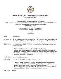


© Copyright 2026