
Common Injuries of the Foot and Ankle Gerard A. Malanga, MD ,
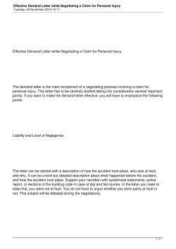
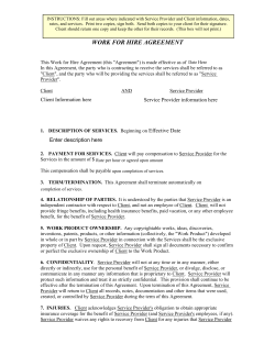
Phys Med Rehabil Clin N Am
19 (2008) 347–371
Common Injuries of the Foot and Ankle
in the Child and Adolescent Athlete
Gerard A. Malanga, MDa,b,c,*,
Jose A. Ramirez – Del Toro, MDd
a
Department of Physical Medicine and Rehabilitation, University of Medicine and Dentistry,
New Jersey Medical School, 30 Bergen Street, Newark, NJ 07101, USA
b
Mountainside Hospital, 1 Bay Avenue, Montclair, NJ 07042, USA
c
Department of Rehabilitation Medicine, Pain Management Center, Overlook Hospital,
MAC II Building, Suite B110, 11 Overlook Road, Summit, NJ 07091, USA
d
Sports Medicine and Spinal Intervention, New Jersey Sports Medicine Institute,
Montclair, NJ, USA
One of the most commonly injured parts of the body in adolescent athletes
is the foot and ankle. It can account for up to 30% of visits to sports medicine
clinics [1,2]. Ankle sprains alone account for 10% of all injuries seen in the
emergency room [3]. Different sports can cause different types of injuries in
the foot and ankle. In basketball, for example, foot and ankle injuries have
been shown to account for 44% to 45% of all injuries in adolescent athletes,
and in the adolescent football player, the foot and ankle make up 13% to 16%
of all injuries [4–6]. Most of these injuries are lateral ankle sprains. Long
distance runners report foot injuries as the most common injury they sustain
[7–9], and adolescent runners are susceptible to overuse type of injuries [10].
Young dancers and gymnasts also have a high percentage of foot and ankle
injuries, with their specific sports mechanics often predisposing to acute
fractures and fatigue fractures [11,12]. It is important that physicians feel
comfortable with the common injuries that can occur in the foot and ankle
and be able to identify these injuries in the young athlete.
When treating young athletes, physicians must keep in mind the anatomic
developmental differences that exist between the skeletally mature and the
skeletally immature foot and ankle. These anatomic differences predispose
young athletes to an entirely different set of injuries than the adult athlete.
A thorough understanding of general bony, ligamentous, and muscular
* Corresponding author.
E-mail address: [email protected] (G.A. Malanga).
1047-9651/08/$ - see front matter Ó 2008 Elsevier Inc. All rights reserved.
doi:10.1016/j.pmr.2007.11.003
pmr.theclinics.com
348
MALANGA & RAMIREZ – DEL TORO
anatomy of the foot and ankle is also valuable to be able to accurately devise
a differential diagnosis based on symptom location.
In this article we first address, define, and explain the types of injuries and
injury patterns germane to the developing skeletally immature athlete. Next,
before discussing all the common injuries, we present a brief anatomic review
of the basic bony anatomy of the ankle and foot for reference purposes. In an
anatomically oriented fashion, we outline the most common injuries noted in
the pediatric and adolescent ankle and foot. There is emphasis on history,
physical examination, diagnosis, and basic treatment guidelines. We look at
the lateral ankle, the medial and anterior ankle, and the hindfoot, midfoot,
and forefoot.
Injury patterns in the developing athlete
The presence of a growth plate, known as the physis or the epiphyseal
plate, is a major difference in developing musculoskeletal structures that is
not seen in the fully mature skeleton. The long bones of children contain
the physis between the metaphysis and the epiphysis (Fig. 1). Bone is laid
down for growth in the physis; however, this area is not a stable and strong
area because it is constantly changing and remodeling. There is relative
weakness at the growth plate and its surrounding bony structures as compared with the ligamentous structures about the pediatric and adolescent
foot and ankle [1]. The epiphyseal plate is also less resistant to shear and
tensile forces than the adjacent bony structures [13]. In adults, the opposite
is true. The bone is strong, and the sites of injuries are in the ligamentous
and muscular structures because they are the weaker points [14]. For example,
where an inversion rollover injury would cause ligamentous injury in an adult,
it is much more likely to cause injury at the growth plate in a child, possibly
leading to a fracture of the physis or epiphyseal plate.
Fig. 1. A pictorial depiction of the metaphysis, physis, and epiphysis of the developing bone.
(Adapted from Canale ST. Physeal injuries. In: Green NE, Swiontkowski MF, editors. Skeletal
trauma in children. 3rd edition. Philadelphia: WB Saunders; 2003. p. 17–56; with permission.)
COMMON INJURIES OF THE FOOT AND ANKLE
349
Generally, the injuries seen in skeletally immature athletes can be divided
into three main categories: (1) injuries related to growth, (2) overuse injuries,
and (3) acute presentations [13–15]. Pain related to growth stems from bony
coalitions or accessory ossification centers that may be abnormally developing. Overuse injuries include osteochondroses, apophysitis, and stress
fractures. Acute injuries include the full spectrum of ligament, tendon, and
muscle injuries and acute fractures. Epiphyseal injuries can be from overuse
and acute traumatic events. An overview is provided, and a more comprehensive discussion of the specific injuries follows.
Growth-related problems: coalitions and accessory ossicles
A coalition is a connection or fusion of two or more bones. It can be
a bony, cartilaginous, or fibrous connection [16–18]. Endochondral ossification is the formation of new bone from tissues such as cartilage, and there
are two ossification centers in bone. The primary ossification center is
located in the diaphysis, and the secondary ossification center is the physis
(physeal plate, epiphyseal plate, or growth plate) located between the diaphysis and the epiphysis [19]. Because coalitions are composed of bony,
fibrous, or cartilaginous tissues, they can act as their own ossification
centers. When they ossify, they become painful where the tissues are placed
under stress, particularly in highly active adolescent athletes [1,16]. The
most common coalitions are talocalcaneal and calcaneonavicular coalitions.
Accessory ossicles are separate ossification centers located extrachondrally. They differ from coalitions because they do not form a connection
between two bones but exist at the end of certain bones. Accessory ossicles
usually appear at age 8 to 10 years and usually fuse approximately 1 year
after their formation. When they do not fuse, they become symptomatic
[16]. The most common sites for accessory ossification center formation
are at the posterior talus, known as os trigonum, the medial malleolus,
and the navicular. The navicular ossification center sometimes can form
an entirely new bone known as an accessory navicular.
Overuse injuries: apophysitis and osteochondroses
Overuse injuries in sports have been defined as chronic injuries related to
constant repetitive stress without adequate recovery time [20]. The cause is
believed to be repetitive application of a submaximal stress to normal tissue
that overwhelms the normal repair process [15,20–22]. These types of injuries
can develop in one of three ways in the adolescent athlete population [20].
First, they can occur in athletes who increase their activity level rapidly
without adequate training, as in individuals who begin preseason workouts
without having practiced the sport for long periods. Second, they can occur
in ill-prepared children who lack good mechanical sport-specific skills. Third,
they can occur in vigorous athletes who do not provide their body with
350
MALANGA & RAMIREZ – DEL TORO
adequate rest from activity. Overuse injuries in the adolescent foot and ankle
can present at (1) the insertion of the tendon to the bone, which is known as
the apophysis, (2) the articular cartilage, which causes what is known as an
osteochondrosis injury, and (3) the growing bone itself, which presents as
a stress fracture [15,23].
The apophysis is the area of junction between a tendon/musculotendinous
unit and the epiphysis. Sometimes these sites of attachment also cross the
physeal growth plate. These areas are constantly placed under stress from
repeated contractions and traction at the site, which can lead to irritation
or inflammation at the physis, known as apophysitis [1,24]. The most common sites for the occurrence of apophysitis that we discuss are at the calcaneus
(Sever’s disease) and at the base of the fifth metatarsal (Iselin’s disease).
Osteochondroses refer to lesions thought to be related to overuse,
although it also believed that osteonecrosis may play a role in their development [15,25]. What is known is that they are lesions of the ossification
centers that eventually undergo recalcification [1]. The two most common
lesions that we discuss are osteochondrosis of the tarsal navicular, known
as Kohler’s disease, and osteochondrosis of the second or third metatarsal
heads, known as Freiberg’s infarction. It is worth noting that an osteochondral lesion of the talusda complication of lateral ankle sprainsdis not
technically considered an osteochondrosis or overuse injury, although the
pathology is located in the talar dome articular cartilage. These injuries
can occur with up to 6.5% of ankle sprains and are discussed as a chronic
presentation of an acute injury [26].
Overuse injuries: stress fractures
A stress fracture can occur anywhere in the pediatric and adolescent foot
and ankle and is believed to be the ultimate overuse injury [16,27]. It has
been referred to as a process that leads to fatigue or insufficiency failure of
bone that occurs when the bone’s reparative abilities have been surpassed
[13,16,28] and the bone is unable to withstand chronic repetitive submaximal
loads [29]. These injuries account for up to 15% of all athletic injuries in
young athletes [30]. Stress fractures are most commonly seen in adolescent
runners [10,20] but are associated with almost any sport in which repetitive
running and cutting movements occur [29,31].
Multiple risk factors exist for the development of stress fractures, including sudden increases in training, poor mechanics, improper or worn-out
footwear, young age, and poor nutrition with low bone mineral density
[1,20,32–36]. Recently there has been an increase in stress fractures in young
female athletes, and a connection has been made between anorexia, amenorrhea, and osteoporosis and the incidence of stress fractures [10,15]. This
population is also at increased risk. In the foot and ankle, stress fractures
can occur anywhere, but the most common sites are the metatarsals and
the tibial diaphysis [20]. Stress fractures of the medial and lateral malleolus
COMMON INJURIES OF THE FOOT AND ANKLE
351
can occur in adolescents but are more common in adult athletes. Tarsal
navicular stress fractures are also common and difficult to treat.
In a study on military recruits, the occurrence of stress fractures was most
prominent in the first month of training, when the increased training and
repetitive loads led to increased osteoclastic activity and the osteoblastic
activity had not caught up with the remodeling process [36]. Research
indicated that bone mineral content increased after 14 weeks of training,
possibly acting to prevent continued occurrence of stress fractures. This finding argues in favor of evidence that accelerated bone remodeling during the
time when overuse is occurring is directly associated with stress fracture
development.
Patients who have stress fractures commonly present with insidious onset
of pain that worsens with increased activity and dissipates once the activity
is stopped [37]. There is usually a history of an increase in the amount of
training that coincides with the onset of symptoms; therefore, they are
thought to be overuse injuries [13]. On physical examination, palpation
can recreate symptoms depending on the location of the fracture. There
may be point tenderness with no history of acute discrete traumatic event.
Radiographs often do not show evidence of the fractures initially [38]. It
has been reported that only 10% of initial radiographs showed abnormalities [29,39]. It may take 3 to 4 weeks for the reactive process associated with
stress fractures to become visible on radiographs, and often the first sign of
this reactive process is subperiosteal new bone formation [13]. Results of
radiographs also may remain normal if athletic activity is decreased [38].
In cases in which the diagnosis is suspected, a three-phase bone scan is
most sensitive in detecting the stress fracture [38].
Proper treatment of stress fractures, as with most overuse injuries,
requires a period of 2 to 4 weeks of relative rest, with temporary cessation
of running. Usually partial weight bearing is tolerated, unless the symptoms
are present during walking and light activity. During this period of modified
rest, the osteoblastic activity catches up and restores balance [1]. It is important to maintain some level of cardiopulmonary fitness program, including
non–land-based training, such as pool activities or cycling. Progression to
running depends on symptoms and is individualized. Please note that these
specifications do not apply to navicular stress fractures, whose management
is somewhat different.
Acute problems: epiphyseal fracture classification
Acute fractures of the ends of long bones in children are common because
of the relative weakness of the epiphysis in relation to the surrounding soft
tissues. The literature is full of different ways to attempt classify these fractures. Some systems attempt to define the position of the foot with relation
to the leg, whereas other systems attempt to define the fracture patterns in
terms of the direction of the force placed on the leg [15,40–45]. These
352
MALANGA & RAMIREZ – DEL TORO
systems can be useful, but unless one is communicating with specialists,
there is not good communication regarding the injury.
One system that is widely used by specialists and primary care physicians to
communicate about growth plate fractures is the Salter-Harris classification
system (Fig. 2) [46]. It is essential to have a grasp on this way of referring to
physeal plate fractures. This system not only gives an anatomic and radiologic
way of describing these injuries unique to the child and adolescent population
but also provides useful prognostic implications that may affect treatment
and the potential for growth disturbances [47,48]. For example, Salter-Harris
type I fractures of the distal fibula are rarely complicated by growth arrests,
whereas Salter-Harris type II fractures of the distal tibia do have a significant
incidence of growth arrest [14,49].
Salter-Harris fractures of the foot and ankle most commonly are seen in
the distal tibia and distal fibula and the phalanges [15]. The most common
acute injury of the adolescent foot and ankle is a Salter-Harris type I
Fig. 2. Salter-Harris classification of physeal injury. (Adapted from Canale ST. Physeal injuries.
In: Green NE, Swiontkowski MF, editors. Skeletal trauma in children. 3rd edition. Philadelphia:
WB Saunders; 2003. p. 17–56; with permission.)
COMMON INJURIES OF THE FOOT AND ANKLE
353
fracture of the distal fibula, which has been called the childhood equivalent
of the lateral ankle sprain in skeletally mature patients [14]. Type I SalterHarris fractures are confined to the growth plate, and they do not involve
either the metaphysis or the epiphysis. Salter-Harris type II fractures involve
the growth plate and usually a margin of the metaphysis. The epiphysis is
not involved. These fractures are by far the most common types of growth
plate fractures seen [15]. Salter-Harris type III fractures occur when a fracture line extends vertically or obliquely through a section of the epiphysis
and proximally to reach the growth plate. In these fractures, the metaphysis
is not involved. Salter-Harris type IV fractures extend vertically through the
epiphysis, into the physeal growth plate, and into the metaphysis. Type V
Salter-Harris injuries usually result from a compressive or crushing force.
They are rare and often have no radiographic abnormality.
Treatment of physeal growth plate fractures depends on multiple factors,
including the location of the injury, the Salter-Harris classification, the age
of the child, and the potential pitfalls and complications of each injury [48].
The age of the child is particularly important because the growth plate may
be fully open if the child is young or may be closing if the child is older. In
the latter case, there is less concern for growth arrest and significant leglength discrepancy because there is likely little growth remaining. If the child
is younger, premature physeal closure is a concerning complication. These
fractures usually heal within 4 to 6 weeks [50].
General guidelines state that for Salter-Harris type I and II fractures,
closed reduction and cast immobilization with a short leg walking cast for 3
to 4 weeks are usually the initial treatments of choice, unless there is any level
of displacement of the fracture, in which case maintenance of reduction must
be undertaken, sometimes necessitating wire placement [14,15,50]. The
patient is followed with serial radiographs to ensure that no complications
occur. These fractures have been thought to be fairly uncomplicated.
Recently, however, studies illuminated that premature physeal closure may
be more common than previously thought [51]. Salter-Harris types III and
IV always require closed reduction, but if their displacement is more than
2 mm, usually open reduction with internal fixation is favored. Two specific
kinds of Salter-Harris fractures are presented later in this article: the Tillaux
fracture and the triplane fracture.
Brief anatomic review of foot and ankle
The most pertinent bony anatomy is as follows: the ankle joint is a synovial joint composed of three bones: the tibia, fibula, and talus (Fig. 3). The
proximal articulating surface of the ankle is composed of the concave end of
the distal tibia and its medial malleolus and the lateral fibular malleolus [52].
This proximal articulating surface extends more distally in its posterior and
lateral borders. It forms a mortise-type shape into which the distal articulating surface of the ankledthe talusdarticulates; there is inherent stability in
354
MALANGA & RAMIREZ – DEL TORO
Fig. 3. (A) Dorsal views of the foot and ankle bony anatomy. (B) Lateral view of the ankle and
foot bony anatomy. (Reprinted with permission from Netter Anatomy Illustration Collection,
Ó Elsevier Inc. All rights reserved.)
COMMON INJURIES OF THE FOOT AND ANKLE
355
this type of formation. The hindfoot is composed of the talus and the calcaneous bones. The articulation between these two is the subtalar joint. The
midfoot is composed of the navicular, the cuboid, and the three cuneiforms:
medial, intermediate, and lateral. The articulation of the distal talus and calcaneus with the proximal navicular and cuboid bones forms the midtarsal
joint. The forefoot is composed of the five metatarsals and the 14 phalanges.
The articulation of the three cuneiforms and the cuboid distally with the
proximal metatarsals forms Lisfranc’s joint. Note the following three articulations: (1) the talus with the calcaneous (ie, subtalar joint), (2) the talus
with the navicular, and (3) the calcaneous with the cuboid. (Articulations
2 and 3 are known as the transverse tarsal joints.) These three joints are collectively known as the three-joint complex, and they are crucial structures
for the dissipation of forces throughout the ankle and foot during gait
mechanics [53].
Anatomic location of injury presentations
Lateral ankle
Acute injuries
Lateral ankle sprain. In the lateral ankle, developmental or overuse type of
injuries are uncommon. The possibility exists of an ossification center in
the fibular region or peroneal tendonitis overuse issues, but they are rare.
The most common predominant injury in a child’s lateral ankle, after the
Salter-Harris type I physeal fracture of the distal fibula, which was discussed
earlier, is the lateral ankle sprain. It is less common in younger children,
because they are more apt to injure the fibular physeal plate for the aforementioned reason that the epiphysis and the growth plate are inherently
weak points in the childhood ankle. In more mature adolescents, however,
the bone is stronger and the growth plate is ossifying and closing, so injuries
of the ligaments are common [14].
The main function of the anterior talofibular ligament (ATFL) and calcaneofibular ligament (CFL) complex is the prevention of excessive lateral
or varus translation of the ankle [52]. The typical mechanism of a lateral ankle sprain occurs when the ankle is subjected to an inversion and internal
rotation force while in plantarflexion [54,55]. The ATFL is the first ligament
injured, and the CFL is also at risk of injury, although it is more likely injured if the ankle is in dorsiflexion. One common method of injury involves
landing on another athlete’s foot while coming down from a jump. Patients
complain of immediate pain and, depending on the severity of the sprain,
may have swelling over the area. Depending on the severity of the injury,
the patient may be unable to bear weight on it.
Physical examination usually reveals tenderness over the ATFL or the
CFL with palpation. There may be ecchymosis and swelling and tenderness
with active and passive range of motion of the ankle. Two maneuvers can be
356
MALANGA & RAMIREZ – DEL TORO
performed to assess the integrity of the ATFL and the CFL, respectively: the
anterior draw test and the talar tilt or varus stress test [56]. A positive anterior draw is more than 3 to 5 mm difference in anterior displacement of the
ankle in side-to-side comparisons. Results of the talar tilt test are positive
when there is more than 23 of angulation or more than 10 of difference
side to side. The specificity and sensitivity for the anterior draw are 80%–
94% and 74%–84% respectiviely. These have not been reported for the talar
tilt [57]. Severity of injury is usually based on a three-point grading system.
Grade 1 sprains are mild and involve a partial tear of the ATFL with intact
CFL. There is usually tenderness to palpation, but only mild swelling. The
anterior draw and the talar tilt tests usually have negative results. Grade 2
sprains are moderate and involve complete tears of the ATFL and mild tears
of the CFL. There is diffuse swelling and ecchymosis and a large anterior
shift on the anterior draw test. The talar tilt test usually has negative results.
Grade 3 sprains are severe and involve tears of the ATFL and the CFL, with
both tests producing positive results [58].
Radiographic imaging in lateral ankle sprain-type injuries in children is
somewhat controversial. In adults, the Ottawa Ankle Rules (OAR) were
devised by Stiell and colleagues because too many ankle radiographs were
being obtained in emergency departments [59,60]. The sensitivity of the
OAR is 100% and the specificity is 40% for the detection of ankle fractures.
If a patient does not meet the rules, then no imaging is necessary [60]. With
OAR, however, all patients were older than 18 years. In children younger
than18 years, there may be issues of weak growth plates and the possibility
of physeal fractures. Some physicians have argued that nearly all children
with ankle pain merit an evaluation with plain radiographs to assess the
integrity of the bony structures and to look for congenital or developmental
anomalies [61]. Clark and colleagues [59] tried to apply the OAR to children.
They came up with a sensitivity of 83%, specificity of 50%, positive predictive value of 28%, and negative predictive value (NPV) of 93% and concluded that the OAR could not be applied to children.
In another prospective study, Boutis and colleagues [62] used a specific set
of physical examination parameters for children and adolescents with ankle
sprains who presented to the emergency department. Their low-risk clinical
examination was defined as isolated tenderness, with or without edema or
ecchymosis of the distal fibula below the level of the joint line and/or over
the adjacent lateral ligaments (ATFL, posterior talofibular ligament, CFL).
All other findings on examination were believed to be high risk. They stratified
all injuries into low risk and high risk based on their examination and obtained radiographic views on all subjects to confirm their findings. It is worth
noting that the diagnoses that they considered to be low risk included sprains,
contusions, lateral talar avulsion fractures, and fractures of the distal fibula,
including nondisplaced Salter-Harris I and II and epiphyseal avulsion fractures. These fractures were considered low risk because they are stable injuries
that carry excellent prognosis and their management is usually based on
COMMON INJURIES OF THE FOOT AND ANKLE
357
maximizing comfort, according to the authors. All other fractures were classified as high risk. They found that none of the 381 enrolled children with lowrisk examinations had high-risk fractures (sensitivity 100% and NPV 100%).
They concomitantly applied the OAR to all children and analyzed how the
OAR would have fared as far as limiting the number of radiographs. They
found that with their low-risk examination, 63% of radiographs could have
been omitted, whereas only a 12% reduction in radiographs would have
occurred with the OAR [62].
Proper treatment is essential for adequate return to competition in youth
sports and to prevent future negative sequelae associated with improperly
managed ankle sprains [63,64]. Some guidelines have been set forth in the
literature, but much of the basic concepts are the same [65–67]. It also has
been noted that early mobilization may promote better healing by producing
better orientation of the collagen fibers when compared with an immobilized
joint [1,68]. The basic guidelines of the PRICE acronym are used at first:
protection, rest, ice, compression, and elevation. Patients may bear weight
as tolerated with protective support and crutches. As patients progress
from the acute to subacute phase of the injury and their pain at rest decreases,
the goal should be to attempt to increase pain-free range of motion. Cardiovascular fitness comprised of upper extremity or non–weight-bearing aerobic
work also should be undertaken. As the swelling and pain diminish, progressive weight bearing should be performed. Strengthening also should begin to
progress from isometric to isotonic and isokinetic exercises based on patient
symptomatology. Proprioceptive training also should be initiated to improve
neuromuscular signaling and decrease future ankle instability. Once patients
reach full range of motion with minimal or no pain on vigorous activity,
sport-specific functional progression should occur [65]. The overall goal of
the rehabilitative program should be to return athletes to full strength and
range of motion and attempt to decrease the recurrence rates of sprains
with improved proprioceptive neuromuscular control and confidence in the
ankle.
Talar osteochondral defects. Talar dome injuries are common complications
of lateral ankle sprains and occur in up to 6.5% of cases [26]. The mechanism
by which the lesions develop is still not fully elucidated, but the belief is that
poor healing after an ankle sprain or other ankle trauma leads to poor circulation to the subchondral bone of the talus, which in turn leads to these focal
lesions of almost necrotic bone fragments [69,70]. The injury is most common
in the second decade of life, and up to 100% of the lateral lesions are believed
to be from previous ankle sprains or trauma, whereas 64% of patients with
medial lesions had a history of trauma [69,71].
The typical history is an adolescent athlete with ankle pain and either
a persistent effusion or the occurrence of intermittent swelling of the joint.
There may be a history of the ankle catching or locking and some instability
and episodes of giving way [72]. Inevitably, further probing detects a history
358
MALANGA & RAMIREZ – DEL TORO
of ankle sprains at some point in the athletic career of the patient and usually
one that was not properly rehabilitated. Along the same lines, when an ankle
sprain does not respond to 6 to 8 weeks of conservative treatment, then talar
dome osteochondral lesions must be highly suspected and ruled out [72].
Radiographs often demonstrate the lesions fairly clearly, particularly with
mortise views. Berndt and Harty [73] developed a classification system for
these lesions based on radiographic appearance, which helps to guide management of the lesions. Stage I lesions show localized trabecular compression.
Stage II lesions are incompletely separated fragments. Stage III lesions are
undetached, undisplaced fragments. Stage IV lesions demonstrate a displaced
or inverted fragment floating free within the joint. When radiographs do not
demonstrate the lesions but clinical suspicion remains high, MRI can serve as
a valuable tool for diagnosis. MRI helps to distinguish among stable lesions,
loose in situ lesions, and loose lesions [74]. Stable lesions correlate with
Berndt and Harty stage I and II lesions that have healed. Loose in situ and
loose lesions correlate with stages III and IV lesions in the Berndt and Harty
classification system.
Treatment for the stage I, stage II, and medial stage III lesions is nonoperative, short leg cast immobilization with limited or non–weight bearing for
6 to 8 weeks, whereas surgery is indicated for lateral sided stage III lesions
and all stage IV lesions [75]. The authors believe that the best treatment for
this lesion is prevention of its occurrence, which may be accomplished if
a comprehensive therapy and rehabilitative program for lateral ankle
sprains (much like the one outlined previously) is performed, with a focus
on early mobilization, range-of-motion training, proprioceptive training,
and progressive strengthening.
Medial and anterior ankle
Growth-related problems
Medial malleolus ossification center. The medial malleolus ossification center
is present in all children. It usually appears at 1 to 2 years of age and closes by
age 12 [16]. This center occasionally persists into adulthood but is usually
asymptomatic. It becomes a problem in adolescents when they are overly
active athletically [16,76]. The usual presentation is pain, point tenderness,
and swelling over the medial malleolus without a significant history of trauma
or any acute injury to the area. Anteroposterior radiographs of the ankle
demonstrate an irregular ossification center with an associated ossicle
[14,16,76]. Treatment includes rest from athletic activities with at least 3 to
6 weeks of short leg cast immobilization [76]. If no improvement occurs
with this regimen, surgery for removal of the ossicle may be indicated.
Overuse injuries
Anterior ankle impingement syndrome. Bony anterior ankle impingement is
a painful condition seen in many young athletes. It is an irritation of the
COMMON INJURIES OF THE FOOT AND ANKLE
359
periosteum on the talar neck that leads to bony exostosis, which in turn
leads to impingement [14,16]. It is commonly seen in athletes, such as ballet
dancers and gymnasts, who are constantly in extremes of dorsiflexion; it is
considered an overuse type of injury. Patients present with pain in the anterior ankle and a history of participating vigorously in a sport or activity that
lends itself to repeated dorsiflexion moments. Pain is usually exacerbated by
dorsiflexion movements, such as pliés in ballet dancers. Dancers often
complain of limited dorsiflexion range of motion in the affected ankle, which
may be noted during the examination [11]. Radiographs demonstrate an
anterior tibial or talar neck osteophyte that has developed from the exostosis from overuse [11]. Conservative treatment consists of stretching of the
Achilles tendon to attempt to improve range of motion and strengthening
of the dorsiflexors [14]. Rest from activity and icing may help. According
to one author, however, by the time an adolescent dancer presents with
this problem, it usually cannot be solved with conservative measures, and
a surgical excision of the osteophyte is needed [11]. The dancer usually
can return to full plié position in 3 to 4 months if adequate postsurgical
rehabilitation is conducted.
Acute injuries
Tillaux fractures. A Tillaux fracture is the most common Salter-Harris type
III fracture seen in adolescents [14,15]. It is an isolated fracture of the distal
anterolateral tibial physis. During normal development, the medial and posterior tibial physeal plates close first, and then the anterolateral areas close.
This fracture occurs late in the teen years in the period when the medial and
posterior plates have closed and the anterior growth plate is still open [15].
The most common mechanism of injury is a forceful external rotation-type
injury. As the ankle is stressed medially, the pull of the anterior tibiofibular
ligament results in an avulsion fracture of the anterolateral aspect of the distal tibial epiphysis over the area of the physeal plate that is still not ossified
[15]. Because that physeal plate is not yet closed, it remains a structural
weak link. The medial and posterior parts of the epiphysis are not affected
because the growth plate already has ossified and closed and it is not a weak
point any more [15]. Patients present with anterior ankle pain and swelling
in the setting of an external rotation trauma. Radiographs reveal a vertical
line that extends from the anterior ankle joint proximally through the epiphysis to the growth plate. Treatment depends on the amount of displacement
of the growth plate. If it is less than 2 mm, then closed reduction and a short
leg walking cast for 4 to 6 weeks are favored, as noted in the discussion on
Salter-Harris fractures. Fracture displacements that are more than 2 mm
necessitate open reduction and internal fixation [15,77].
Triplane fractures. Triplane fractures represent yet another type of fracture
of the distal anterior tibial epiphysis and physeal growth plate. These fractures are similar to Tillaux fractures in two main ways. First, they normally
360
MALANGA & RAMIREZ – DEL TORO
occur in the period when the anterolateral plate is still open and other areas
of the distal tibial growth plate have closed. The pattern of what has and has
not been ossified is what determines the extent of the injury [78]. Second, the
triplane fracture occurs from external rotation forces of the ankle, which
cause shearing and avulsion. Some authors speculate that plantarflexion
may contribute to their occurrence and to their irregularity in presentation
and on radiographs [79]. They differ from Tillaux fractures in that the extent
of involvement of the terminal bone is greater and involves the metaphysis,
physis, and epiphysis. Triplane fractures are more difficult to diagnose on
plain radiographs because their full extent may not be seen on regular views
of the ankle. CT scans usually delineate the lesion well [15]. Treatment is
same as for Tillaux fractures, and if surgery is performed, adequate visualization of all fracture fragments is paramount to successful outcomes
[15,80].
Hindfoot
Growth-related problems
Talocalcaneal coalition. Tarsal coalitions are fusions of two or more of the
tarsal bones [1,16]. The incidence of coalitions may be as high as 1% to 3%
in the population and are bilateral 50% of the time [1,81]. The most common
tarsal coalitions are the talocalcaneal and the calcaneonavicular coalitions,
which account for 90% of all coalitions [1]. The subtalar joint, the talocalcaneal articulation, and the calcaneocuboid articulation form the three-joint
complex. This complex is responsible for many foot motions during the gait
cycle. The presence of a talocalcaneal and calcaneonavicular coalition
severely affects the motion at the three-joint complex [81].
The typical presentation of a painful coalition occurs in the developing
mid- to late teenage athlete who participates in vigorous activity. It is then
that the presence of the congenital coalition first becomes evident. Increasing
activity, combined with maturing ossification, leads to motion alteration and
pain. Pain is usually located vaguely around the ankle based on the location of
the coalition and based on which motion segment in the three-joint complex is
mostly affected. There may or may not be a history of previous lateral ankle
sprains [81]. Physical examination reveals findings associated with decreased
motion of the hindfoot. The hindfoot is held in rigid valgus, and there is
absence of heel varus on tiptoes. There is often rigid flat foot, and peroneal
tightness and spasticity can be seen in their attempts to overcome the rigid
flat foot. Pain is also present with foot inversion [1,81,82].
Radiographically, the talocalcaneal coalition is difficult to identify on plain
radiographs, although the calcaneonavicular one is usually well visualized.
CT is considered the gold standard imaging modality for the diagnosis of
tarsal coalitions, however [83]. Treatment initially targets symptom control.
If athletic activity worsens the symptoms, then the activity should be
decreased or temporarily stopped. Orthotics can help control mild symptoms.
COMMON INJURIES OF THE FOOT AND ANKLE
361
Cast immobilization for 6 weeks in a short leg walking cast is indicated, particularly with painful and stiff joints [81]. Failure of conservative therapy is
marked by continued pain and inability to participate in sports. Surgical
options include excision of the coalition, calcaneal osteotomy, and arthrodesis
of the joint [1,81].
Os trigonum. A normal ossification center can often be located at the posterior aspect of the talus. It usually appears at 9 to 12 years of age and fuses
1 year after its appearance. When it does not ossify, an ossicle develops,
which is known as the os trigonum [16]. It has been reported to be present
in as much as 10% of the population, and it is usually unilateral [16,84]. It
becomes symptomatic in young athletes who perform repeated ankle plantarflexion, such as ballet dancers, gymnasts, and ice skaters. One of the
mechanisms by which it is believed to cause pain is from mechanical
impingement of the posterior talus between the posterior tibia and the
calcaneous when the foot is in plantarflexion [16,84]. The presentation is
an active athlete who has pain in the posterolateral ankle that is reproducible on palpation and active plantarflexion. The os trigonum is usually seen
on lateral plain radiographs as an ossicle located posterior to the calcaneus.
It has been recommended that plantarflexion views also be obtained for
verification [16]. Treatment is conservative. Plantarflexion must be avoided
as much as possible. If the pain continues with resumption of the athletic
activity, then surgical resection may be indicated. Some authors recommend
early resection in competitive young athletes as the best way to resolve the
symptomatology and expedite return to play safely [85].
Overuse injuries
Sever’s apophysitis. Apophyses are bony attachment sites that develop as
accessory ossification centers and mimic the maturation of an epiphyseal
plate [1,24]. The calcaneal apophysis serves as the attachment site for the
Achilles tendon superiorly and the plantar fascia inferiorly [16]. Inflammation of the calcaneal apophysis, known as Sever’s disease, is one of the most
common overuse injuries seen in the young athletic population, accounting
for approximately 8% of all overuse injuries in this group [1,86]. It has been
referred to as the ankle equivalent of Osgood-Schlatter’s disease of the knee
[16]. The typical presentation is that of an athlete who has just begun the
season or has increased running activity recently. Pain is at the heel, particularly with running and jumping. Physical examination is often positive for
tight Achilles’ heel cord and weakness of the ankle dorsiflexors [1,87]. There
also may be swelling and induration over the calcaneal apophysis. Diagnosis
is usually clinical, and radiographs are usually not helpful. Treatment is
multifaceted. First, causative activity should cease. Short-term icing and
nonsteroidal anti-inflammatory drugs can be helpful for controlling the
pain. A comprehensive program of heel cord stretching and dorsiflexor
strengthening should be initiated. Barefoot walking should be avoided
362
MALANGA & RAMIREZ – DEL TORO
because this prolonged traction is what leads to apophysitis. Occasionally,
a heel insert or a heel lift is recommended to remove tensile forces on the
tendon while the inflammation decreases [24,87].
Plantar fasciitis. The plantar fascia stretches from the calcaneal tuberosity
and fans out to attach around the plantar aspect of the proximal phalanges
[1]. Current literature has noted that this is not a true inflammatory condition but rather the result of repetitive microtrauma from continued athletic
activity overuse [1,88,89]. Much like lateral epicondylitis, it seems that plantar fasciitis is not an ‘‘-itis’’ but rather an ‘‘-osis,’’ a loss of normal tendon
integrity. Young athletes involved with speed work, jumping, or hill running
are at increased risk of developing this condition [16]. In young athletes,
plantar fasciitis usually coincides with calcaneal apophysitis, but in adolescent athletes with closed physes, it can exist by itself and presents as medial
arch or heel pain. Patients give a history of heavy athletic involvement and
medial arch or heel pain, particularly with the first step out of bed every day.
Physical examination shows tenderness over the anteromedial aspect of the
heel, particularly with the foot in dorsiflexion [1]. This, like Sever’s disease,
is a clinical diagnosis, because radiographs are often not helpful. Treatment
involves conservative measures, including relative rest, ice massage, arch
supports, heel pads, and heel cord stretching and strengthening, both of
which are paramount to successful rehabilitation. The decision to inject
corticosteroids in the area is currently controversial in the literature, particularly in adolescent athletes. Evidence exists to support injection in adults,
but complications such as tendon rupture and fat pad atrophy are real
and must be monitored. Studies are not solid in adolescents [16].
Midfoot
Growth-related problems
Calcaneonavicular coalition. The two most common coalitions are the
talocalcaneal and the calcaneonavicular coalitions. The calcaneonavicular
coalition is a bony fusion between the talus and the calcaneus, and it is the
second most common coalition behind the talocalcaneal coalition (Fig. 4).
It has a similar presentation as the talocalcaneal coalition, with decreased
range of motion across the hindfoot and the three-joint complex. Diagnosis
and treatment are also similar.
Accessory navicular. An accessory navicular bone is the most common
accessory bone in the foot [16]. It is an ossicle that develops like all other
ossicles as a separate extrachondral ossification center, and it is located at
the site of the tibialis posterior tendon insertion [14,16,90]. When it fails
to ossify fully, it becomes an accessory navicular bone. It has been reported
to occur in anywhere from 4% to 14% of the population [16]. It is not until
adolescence, when athletes increase their participation, that these conditions
COMMON INJURIES OF THE FOOT AND ANKLE
363
Fig. 4. (A) Pictorial of calcaneonavicular coalition. (B) Oblique radiograph shows the calcaneonavicular fusion. (Reprinted with permission from Netter Anatomy Illustration Collection,
Ó Elsevier Inc. All rights reserved.)
actually become painful. Patients present with pain medially along the arch
of the foot, and on physical examination there is often a prominence along
the arch of the foot that is tender with shoe wear [14]. Also on examination
there is often evidence of pes planus. One theory about the development of
pes planus is that the tibialis posterior tendon, which is a dynamic stabilizer
of the medial longitudinal arch of the foot, inserts onto the accessory navicular instead of into the native navicular. Because it is a weaker insertion
point, there is a drop in the longitudinal arch, which causes the pes planus
[16]. Radiographic imaging is often diagnostic, but in cases in which it is not,
MRI can help elucidate the lesion and define the anatomic points of insertion of the tibialis posterior tendon [72,91]. Treatment involves conservative
management initially, with orthotics and casting and doughnut cut-outs for
the painful parts over the enlarged navicular. If these measures fail and an
athlete continues to have pain, there are well documented surgical procedures for the excision of these ossicles with varying degrees of handling of
the posterior tibial tendon [14,92].
Overuse injuries
Navicular stress fractures. Aside from the previous discussion on stress fractures in general, one stress fracture in particular must be emphasized: the
navicular stress fracture, which has been documented to have an incidence
anywhere from 0% to 29% in young track and field athletes [15,93]. Navicular stress fractures are difficult to diagnose. The presentation is usually
vague onset of foot pain along the dorsomedial area [10]. History elicits
risk factors such as overuse with an increase in exercise duration and intensity and poor nutrition. Examination reveals palpatory tenderness over the
dorsomedial navicular and may show mechanical configurations that may
increase the risk of these fractures, such as a tight gastrocnemius complex
or a Morton foot with a long second ray [10]. As with all stress fractures,
radiographs are frequently normal, and a bone scan is required for definitive
diagnosis. There must be a high incidence of suspicion for these fractures
364
MALANGA & RAMIREZ – DEL TORO
because the navicular has poor blood supply over the middle one third, and
if the fracture is untreated, poor healing with delayed union or nonunion is
a possibility [10]. Treatment is more aggressive than with other stress fractures. Non–weight-bearing cast immobilization for 6 weeks is recommended
by most physicians to prevent malunion, and if no progress is made, then
surgical internal fixation is recommended. The usual time to return to athletic
activity can be as long as 5 to 6 months [15,94].
Kohler’s osteochondrosis. Osteochondroses are overuse lesions of the osteochondral ossification centers that are idiopathic. Osteochondrosis of the
tarsal navicular is termed Kohler’s disease. It typically occurs in children
aged 5 to 9 years, and patients present with pain over the midfoot region
that worsens with weight bearing [1]. Swelling of the area also may be
present. Bilateral lesions have been known to occur, and one such case of
bilateral lesions in twins led those authors to speculate on the possibility
of a genetic predisposition to its occurrence [95]. Radiographs may be difficult to interpret because many children have irregular-appearing navicular
bones that are normal and asymptomatic [1], but because patients have
such good outcomes, further imaging has little use and does not change
management. Treatment is conservative, with the use of the RICE technique
and shoe supports for mild cases. Casting may be necessary over a 4- to
8-week period for more severe cases [95]. Patients typically fare well with
this conservative treatment. In one series in the literature, 100% of patients
became asymptomatic after conservative treatment, and the navicular
returned to its normal appearance on radiographs [96]. There have been
a few reports of children with Kohler’s abnormality persisting into adulthood
clinically and radiographically [97].
Acute injuries
Lisfranc injury. Lisfranc’s joint is the tarsometatarsal articulation of the
three cuneiforms and cuboid with the proximal five metatarsals. The keystones of this joint are the first and second metatarsals articulating with
the first and second cuneiforms. Transverse ligaments connect the bases of
the lateral four metatarsals; however, no such transverse ligament exists
between the base of the first and second metatarsals. The second metatarsal
proximally has small articulations with the three cuneiforms that support
the tarsometatarsal articulation [1,98]. This is a precarious yet highly important anatomic location. Injuries at Lisfranc’s joint can be in the form of
sprains of the transverse ligaments or even fracture-dislocations. The most
common mechanism of injury is an axial loading through the foot as the
foot is forcefully plantarflexed and slightly rotated, which causes the
proximal second metatarsal to dislocate dorsally [1,98,99]. Given this mechanism, many of the Lisfranc joint injuries seen in adolescents occur while
playing football, with a large percentage of those injuries occurring in
COMMON INJURIES OF THE FOOT AND ANKLE
365
linemen [1,100]. Linemen are often in situations in which other linemen step
on their toes, which cause a heavy axial load while they are attempting to
explode forward onto their toes.
The typical presentation involves an athlete with pain over the dorsum of
the midfoot associated with swelling and an inability to bear weight through
the midfoot, particularly on the tiptoes [1]. Plantar bruising often is associated
with this injury, and if this sign is noticed, clinical suspicion should be raised.
Radiographs are needed to make the diagnosis, particularly weight-bearing
films (Fig. 5). There is a significantly high incidence of missed diagnoses
[101]. One should look for malalignment between the first metatarsal and
the first cuneiform or between the second metatarsal and the second
cuneiform [99,101]. Severity grading is like other sprains, based on ligamentous tearing and amount of dislocation. Bone scans may help make the
diagnosis in patients with negative radiographic results and continued high
suspicion. Treatment of these injuries is based on the degree of severity.
Stretch injuries or partial tears with less than 2 mm of malalignment should
be treated conservatively with cast immobilization or a walking boot for 4
to 6 weeks [1,100]. Sprains that are more severe require operative reduction
with internal fixation [1,102].
Forefoot
Overuse injuries
Iselin’s apophysitis. Iselin’s disease is an apophysitis that occurs at the
tuberosity of the fifth metatarsal. The apophysis at this site appears at
ages 9 to 14 and is located within the insertion of the peroneus brevis tendon
Fig. 5. Lisfranc injury. Note the malalignment between the metatarsals and the cuneiforms.
(Reprinted with permission from Netter Anatomy Illustration Collection, Ó Elsevier Inc. All rights
reserved.)
366
MALANGA & RAMIREZ – DEL TORO
Fig. 6. (A) Fifth metatarsal avulsion fracture. (B) Jones fracture. Note how it extends to the
metaphyseal-diaphyseal junction. (Adapted from Brodsky JW, Krause JO. Stress fractures of
the foot and ankle. In: DeLee JC, Drez D, Miller MD, editors. Orthopaedic sports medicine:
principles and practice. 2nd edition. Philadelphia: Saunders Elsevier; 2003. p. 2391–409; with
permission.)
[16]. Presentation is similar to other cases of apophysitis in terms of history
of overuse in athletically active older children. There is usually a presentation
of insidious onset of pain over the lateral foot, with no history of trauma, in
the setting of overuse-type activities. Radiographically, the apophysis
appears as a diagonal or longitudinal line parallel to the long axis of the shaft
of the fifth metatarsal, an important distinction from acute fifth metatarsal
avulsion fractures, which are usually transverse in nature (see later discussion)
[16]. Treatment is conservative, with decrease of activity and stretching and
strengthening of the peroneal muscles. This treatment is usually effective until
bony union eventually occurs [16].
Acute injuries
Fifth metatarsal fractures. Young child and adolescent athletes who present
with pain along the lateral aspect of the foot near the fifth metatarsal present
a challenging dilemma. History should be able to differentiate whether an
acute or overuse injury has occurred; however, matters are not always that
clear-cut. Three possible types of fractures are seen in the area of the base
of the fifth metatarsal: include fifth metatarsal stress fractures, fifth metatarsal
acute avulsion fractures, and Jones fractures. Metatarsal stress fractures are
overuse injuries, which are the most common types of stress fractures seen
in adolescent feet and ankles. Their management was reviewed previously.
Most young athletes return to their sport in 4 to 6 weeks [16].
The fifth metatarsal acute fracture is the most common metatarsal fracture
in children, accounting for 45% of all metatarsal fractures [16]. It occurs after
an inversion-type injury, when the peroneus brevis tendon is avulsed from its
attachment at the base of the fifth metatarsal [14]. Radiographs show the
lesion, which can be distinguished from apophysitis because the fracture is
transverse along the bone, whereas apophysitis of the fifth metatarsal (Iselin’s
367
COMMON INJURIES OF THE FOOT AND ANKLE
Table 1
Summary of common injuries of the foot and ankle in the child and adolescent athlete
Lateral
ankle
Growth
problems
Overuse
injuries
Acute
injuries
Medial &
anterior
ankle
Medial
malleolus
ossification
center
Anterior
ankle
impingement
syndrome
Salter-Harris
Tillaux
fractures
fractures
Lateral
Triplane
ankle
fractures
sprains
Osteochondritis
dissecans
Hindfoot
Midfoot
Forefoot
Talocalcaneal Calcaneonavicular
coalition
coalition
Os trigonum Accessory
navicular
Sever’s
Navicular
Metatarsal
apophysitis
stress
stress
Plantar
fracture
fractures
fasciitis
Kohler’s
Iselin’s
apophysitis
disease
Lisfranc
Fifth
injury
metatarsal
avulsion
fracture
Jones
fracture
disease) occurs along the diagonal plane from the bone (Fig. 6A) [16].
Treatment is usually conservative unless there is more than 2 to 3 mm of
displacement, in which case surgical open reduction and internal fixation
should be performed [14].
The Jones fracture is actually a fracture that occurs at the metaphysealdiaphyseal junction of the base of the fifth metatarsal. The average age and demographics of occurrence of this fracture involve 15- to 21-year-old athletes
[16]. Patients usually present with pain along the fifth metatarsal, particularly
upon weight bearing, and give a history of an acute injury. Radiographs
usually reveal the fracture (Fig. 6B). Similar to the navicular stress fracture,
this lesion has higher rates of nonunion because it has poor blood supply to
that area. Treatment involves a non–weight-bearing cast for 6 to 8 weeks
and possible surgical screw fixation for nonhealing fractures [16].
Summary
Myriad problems in the foot and ankle are specific to child and adolescent
athletes. The anatomy of young athletes with respect to the presence of
a growth plate makes their injury patterns different from those seen in adults.
The main general injury patterns seen in the feet and ankles of children are
related to growth and development or occur from overuse syndromes or
acute trauma. We have outlined in an anatomically oriented manner most
of the common problems in this population. They are also summarized in
Table 1.
368
MALANGA & RAMIREZ – DEL TORO
References
[1] Pommering TL, Kluchurorski L, Hall SL. Ankle and foot injuries in pediatric and adult
athletes. Prim Care 2005;32(1):133–61.
[2] Mahaffey D, Hilts M, Fields KB. Ankle and foot injuries in sports. Clinics in Family Practice 1999;1(1):233–50.
[3] Frey C, Bell J, Teresi L, et al. A comparison of MRI and clinical examination of acute
lateral ankle sprains. Foot Ankle Int 1996;17(9):533–7.
[4] Damore DT, Metzl JD, Ramundo M, et al. Patterns in childhood sports injury. Pediatr
Emerg Care 2003;19(2):65–7.
[5] Ruda SC. Common ankle injuries in the athlete. Nurs Clin North Am 1991;26(1):167–80.
[6] Garrick JB. The frequency of injury, mechanism of injury, and epidemiology of ankle
sprains. Am J Sports Med 1977;5(6):242–50.
[7] Barr KP, Harrast MA. Evidence-based treatment of foot and ankle injuries in runners. Phys
Med Rehabil Clin N Am 2005;16:779–99.
[8] Epperly T, Fields KB. Epidemiology of running injuries. In: O’Connor FG, Wilder RP,
Nirschl R, editors. Textbook of running medicine. New York: McGraw-Hill; 2001.
p. 3–9.
[9] Taunton JE, Ryan MB, Clement DB, et al. A retrospective case-control analysis of 2002
running injuries. Br J Sports Med 2002;36(2):95–101.
[10] Kennedy JG, Knowles B, Dolan M, et al. Foot and ankle injuries in the adolescent runner.
Curr Opin Pediatr 2005;17(1):34–42.
[11] Kadel NJ. Foot and ankle injuries in dance. Phys Med Rehabil Clin N Am 2006;17:813–26.
[12] Chilvers M, Donahue M, Nassar L, et al. Foot and ankle injuries in elite female gymnasts.
Foot Ankle Int 2007;28(2):214–8.
[13] Wojtys EM. Sports injuries in the immature athlete. Orthop Clin North Am 1987;18(4):
689–708.
[14] Chambers HG. Ankle and foot disorders in skeletally immature athletes. Orthop Clin
North Am 2003;34(3):445–59.
[15] Pontell D, Hallivis R, Dollard MD. Sports injuries in the pediatric and adolescent foot and
ankle: common overuse and acute presentations. Clin Podiatr Med Surg 2006;23(1):209–31.
[16] Omney ML, Micheli LJ. Foot and ankle problems in the young athlete. Med Sci Sports
Exerc 1999;31(Suppl 7):S470–86.
[17] Elkus RA. Tarsal coalition in the young athlete. Am J Sports Med 1986;14(6):477–80.
[18] O’Neill DB, Micheli LJ. Tarsal coalition: a follow-up of adolescent athletes. Am J Sports
Med 1989;17:544–9.
[19] MacGregor J. The skeletal system. In: MacGregor J, editor. Introduction to the anatomy
and physiology of children. London: Routledge; 2000. p. 17–34.
[20] Hogan KA, Gross RH. Overuse injuries in pediatric athletes. Orthop Clin North Am 2003;
34(3):405–15.
[21] Herring SA, Nilson KL. Introduction to overuse injuries. Clin Sports Med 1987;6(2):
225–39.
[22] Stanitski CL. Overuse injuries. In: Stanitski CL, DeLee JC, Drez D, editors. Pediatric and
adolescent sports medicine, vol 3. Philadelphia: WB Saunders Company; 1994. p. 94–9.
[23] Micheli LJ. Overuse injuries in children’s sports: the growth factor. Orthop Clin North Am
1983;14(2):337–60.
[24] Carr KE. Musculoskeletal injuries in young athletes. Clin Fam Pract 2003;5(2):385–406.
[25] Resnick DR. Osteochondroses. In: Resnick DR, editor. Diagnosis of bone and joint
disorders. 4th edition. Philadelphia: WB Saunders Company; 2002. p. 3686–741.
[26] Farmer JM, Martin DF, Boles CA, et al. Chondral and osteochondral injuries: diagnosis
and management. Clin Sports Med 2001;20(2):299–320.
[27] Hershman EB, Mailly T. Stress fractures. Clin Sports Med 1990;9(1):183–214.
[28] Monteleone GP. Stress fractures in the athlete. Orthop Clin North Am 1995;26(3):423–32.
COMMON INJURIES OF THE FOOT AND ANKLE
369
[29] Coady CM, Micheli LJ. Stress fractures in the pediatric athlete. Clin Sports Med 1997;
16(2):225–38.
[30] Beck BR, Matheson GO. Common stress fractures. Clin Fam Pract 1999;1(1):251–74.
[31] Orava S, Jromakka E, Hulkko A. Stress fractures in young athletes. Arch Orthop Trauma
Surg 1981;98:271.
[32] Verma RB, Sherman O. Athletic stress fractures. Part I: history, epidemiology, physiology,
risk factors, radiology, diagnosis and treatment. Am J Orthop 2001;30(11):798–806.
[33] Milgrom C, Finestone A, Shlamkovitch N, et al. Youth as a risk factor for stress fracture:
a study of 783 infantry recruits. J Bone Joint Surg 1994;76-B(1):20–2.
[34] Frusztajer NT, Dhuper S, Warren MP, et al. Nutrition and the incidence of stress fractures
in ballet dancers. Am J Clin Nutr 1990;51(5):779–83.
[35] Giladi M, Milgorm C, Simkin A, et al. Stress fractures: identifiable risk factors. Am J Sports
Med 1991;19(6):647–52.
[36] Margulies JY, Simkin A, Leichter I, et al. Effect of intense physical activity on the bonemineral content in the lower limbs of young adults. J Bone Joint Surg Am 1986;68(7):
1090–3.
[37] Stanitski C. Management of sports injuries in children and adolescents. Orthop Clin North
Am 1998;19:689–98.
[38] Rettig AC, Shelbourne KD, Beltz HF, et al. Radiographic evaluation of foot and ankle
injuries in the athlete. Clin Sports Med 1987;6(4):905–19.
[39] Matheson GO, Clement DB, McKenzie DC, et al. Stress fractures in athletes: a study of 320
cases. Am J Sports Med 1987;15(1):46–58.
[40] Gross RH. Foot and ankle injuries and disorders. Adolescent Medicine State of the Art
Reviews 1998;9(3):599–609.
[41] Ashhurst APC, Bromer RS. Classification and mechanism of fractures of the leg bones
involving the ankle. Arch Surg 1922;4:121–9.
[42] Dias LS, Giegerich CR. Fractures of the distal tibial epiphysis in adolescence. J Bone Joint
Surg 1983;65(4):438–44.
[43] Dias L, Tachdjian M. Physical injuries of the ankle in children. Clin Orthop 1978;136:
230–3.
[44] Landin L, Danielsson L. Children’s ankle fractures. Acta Orthop Scand 1983;54:634–40.
[45] Lauge-Hansen N. Fractures of the ankle. II: combined experimental surgical and
experimental roentgenologic investigations. Arch Surg 1950;60:957.
[46] Salter R, Harris W. Injuries involving the epiphyseal plate. J Bone Joint Surg 1963;45:
587–622.
[47] Brown JH, DeLuca SA. Growth plate injuries: Salter-Harris classification. Am Fam
Physician 1992;46(4):1180–4.
[48] Devalentine SJ. Epiphyseal injuries of the foot and ankle. Clin Podiatr Med Surg 1987;4(1):
279–310.
[49] Goldberg VM, Aadalen R. Distal tibial epiphyseal injuries: the role of athletics in 53 cases.
Am J Sports Med 1978;6(5):263–8.
[50] Pizzutillo PD, Chandler JB, Maxwell T. Pediatric orthopedics: fractures of the growth
plate. In: Griffin LY, editor. Essentials of musculoskeletal care. 3rd edition. Rosemont
(IL): American Academy of Orthopedic Surgeons; 2005. Section 9. p. 865–7.
[51] Rohmiller MT, Gaynor TP, Pawelek J, et al. Salter-Harris I and II fractures of the distal
tibia: does mechanism of injury relate to premature physeal closure? J Pediatr Orthop
2006;26(3):322–8.
[52] Norkin CC, Levangie PK. The ankle-foot complex. In: Norkin CC, Levangie PK, editors.
Joint structure and function: a comprehensive analysis. Philadelphia: FA Davis Company;
1992. p. 379–418.
[53] Kitaoka HB, Crevoisier XM, Hansen D, et al. Foot and ankle kinematics and ground
reaction forces during ambulation. Foot Ankle Int 2006;27(10):808–13.
370
MALANGA & RAMIREZ – DEL TORO
[54] Bennett WF. Lateral ankle sprains. Part I: anatomy, biomechanics, diagnosis, and natural
history. Orthop Rev 1994;23(5):381–7.
[55] Cohen RS, Balcom TA. Current treatment options for ankle injuries: lateral ankle sprain,
Achilles tendonitis, and Achilles rupture. Curr Sports Med Rep 2003;2(5):251–4.
[56] Young CC, Niedfeldt MW, Morris GA, et al. Clinical examination of the foot and ankle.
Primary Care: Clinics in Office Practice 2005;32(1):105–32.
[57] Hyman GS, Solomon J, Dahm D. Physical examination of the foot and ankle. In: Malanga
GA, Nadler SF, editors. Musculoskeletal physical examination: an evidence-based
approach. Philadelphia: Elsevier Mosby; 2006. p. 315–43.
[58] Brown DP, Freeman ED, Cuccurullo SJ. Musculoskeletal medicine. In: Cuccurullo SJ,
editor. Physical medicine and rehabilitation board review. New York: Demos; 2004.
p. 131–293.
[59] Clark KD, Tanner S. Evaluation of the Ottawa ankle rules in children. Pediatr Emerg Care
2003;19(2):73–8.
[60] Stiell IG, Greenberg GH, McKnight RD, et al. A study to develop clinical decision rules for
the use of radiography in acute ankle injuries. Ann Emerg Med 1992;21(4):384–90.
[61] Churchill JA, Mazur JM. Ankle pain in children: diagnostic evaluation and clinical decision
making. J Am Acad Orthop Surg 1995;3(4):183–93.
[62] Boutis K, Komar L, Jaramillo D, et al. Sensitivity of a clinical examination to predict need
for radiography in children with ankle injuries: a prospective study. Lancet 2001;358(9299):
2118–21.
[63] Smith RW, Reischl SF. Treatment of ankle sprains in young athletes. Am J Sports Med
1986;14(6):465–71.
[64] Bennett WF. Lateral ankle sprains. Part II: acute and chronic treatment. Orhtop Rev 1994;
23(6):504–10.
[65] Safran MR, Zachazewski JE, Benedetti RS, et al. Lateral ankle sprains: a comprehensive
review. Part 2: treatment and rehabilitation with an emphasis on the athlete. Med Sci Sports
Exerc 1999;31(7):S438–47.
[66] Stonnington MJ. Lower leg, ankle and foot. In: Buschbacher RS, editor. Practical guide to
musculoskeletal disorders: diagnosis and rehabilitation. 2nd edition. Boston: Butterworth
Heinemann; 2002. p. 229–54.
[67] Mascaro TB, Swanson LE. Rehabilitation of the foot and ankle. Orthop Clin North Am
1994;25(1):147–60.
[68] Safran MR, Benedtti RS, Bartolzzi AR, et al. Lateral ankle sprains: a comprehensive review
part 1: etiology, pathoanatomy, histopathogenesis and diagnosis. Med Sci Sports Exerc
1999;31(7):S429–37.
[69] Sullivan JA. Ankle and foot injuries in the pediatric athlete. Instr Course Lect 1993;42:
545–51.
[70] Pizzutillo P. The osteochondroses. In: Sullivan JA, Grana WA, editors. The pediatric
athlete. Park Ridge (IL): American Academy of Orthopaedic Surgeons; 1990. p. 211–33.
[71] Canale ST, Belding RH. Osteochondral lesions of the talus. J Bone Joint Surg 1980;62(4):
97–102.
[72] Canale ST. Osteochondroses and related problems of the foot and ankle. In: DeLee JC,
Drez D, Miller MD, editors. Orthopedic sports medicine: principles and practice. 2nd
edition. Philadelphia: Saunders; 2003. p. 2587–623.
[73] Brendt AL, Harty M. Transchondral fractures (osteochondritis dissecans) of the talus.
J Bone Joint Surg 1959;41(4):988–1020.
[74] Smith DK, Gilley JS. Imaging of sports injuries of the foot and ankle. In: DeLee JC, Drez
D, Miller MD, editors. Orthopedic sports medicine: principles and practice. 2nd edition.
Philadelphia: Saunders; 2003. p. 2190–224.
[75] Sullivan JA. Ligament injuries of the foot/ankle in the pediatric athlete. In: DeLee JC, Drez
D, Miller MD, editors. Orthopedic sports medicine: principles and practice. 2nd edition.
Philadelphia: Saunders; 2003. p. 2376–91.
COMMON INJURIES OF THE FOOT AND ANKLE
371
[76] Stanitski CL, Micheli LJ. Observations on symptomatic medial malleolar ossification
centers. J Pediatr Orthop 1993;13(2):164–8.
[77] Hunter-Griffin LY. Injuries to the leg, ankle and foot. In: Sullivan JA, Granna WA, editors.
The pediatric athlete. Park Ridge (IL): American Academy of Orthopaedic Surgeons; 1990.
p. 187–98.
[78] Cooperman D, Spiegel P, Laros G. Tibial fractures involving the ankle in children. J Bone
Joint Surg 1978;60(8):1040–6.
[79] Lynn M. The triplane distal epiphyseal fracture. Clin Orthop 1972;86:187–90.
[80] Cone R, Nygayen V, Fluornoy J, et al. Triplane fracture of the distal tibial epiphysis.
Radiology 1984;153(3):763–7.
[81] Bohne WHO. Tarsal coalition. Curr Opin Pediatr 2001;13(1):29–35.
[82] Cowell HR. Talocalcaneal coalition and new causes of peroneal spastic flatfoot. Clin
Orthop 1972;85:16–22.
[83] Harty MP. Imaging of pediatric foot disorders. Radiol Clin North Am 2001;39(4):733–48.
[84] Marotta JJ, Micheli LJ. Os trigonum impingement in dancers. Am J Sports med 1992;20(5):
533–6.
[85] Quirk R. Talar compression syndrome in dancers. Foot Ankle 1982;3(2):65–8.
[86] Maffulli N, Wong J, Almekinders LC. Types and epidemiology of tendinopathy. Clin
Sports Med 2003;22(4):675–92.
[87] Christopher NC, Cogeni J. Overuse injuries in the pediatric athlete: evaluation, initial
management, and strategies for prevention. Clinical Pediatric Emergency Medicine 2002;
3(2):118–28.
[88] Lemont H, Ammirati KM, Usen N. Plantar fasciitis: a degenerative process (fasciosis)
without inflammation. J Am Podiatr Med Assoc 2003;93(3):234–7.
[89] Wapner KL, Bordelon RL. Heel pain. In: DeLee JC, Drez D, Miller MD, editors. Orthopedic
sports medicine: principles and practice. 2nd edition. Philadelphia: Saunders; 2003.
p. 2446–72.
[90] Grogan DP, Gasser SI, Ogden JA. The painful accessory navicular: a clinical and
histopathological study. Foot Ankle 1989;10(3):164–9.
[91] Kitter E, Edrag N, Karatosun V, et al. Tibialis posterior tendon abnormalities in feet with
accessory navicular bone and flat foot. Acta Orthop Scand 1999;70:618–21.
[92] Ugolini PA, Raikin SM. The accessory navicular. Foot Ankle Clin 2004;9(1):165–80.
[93] Bennell KL, Brukner PD. Epidemiology and site specificity of stress fractures. Clin Sports
Med 1997;16(2):179–96.
[94] Khan KM, Fuller PJ, Brukner PD. Outcome of conservative and surgical management of
navicular stress fracture and athletes. Am J Sports Med 1992;20(6):657–66.
[95] Tsirikos AI, Riddle EC, Kruse R. Bilateral Kohler’s disease in identical twins. Clin Orthop
Relat Res 2003;409:195–8.
[96] Williams GA, Cowell HR. Kohler’s disease of the tarsal navicular. Clin Orthop 1981;158:
53–8.
[97] Sharp RJ, Calder JD, Saxby TS. Osteochondrosis of the navicular: a case report. Foot
Ankle Int 2003;24(6):509–13.
[98] Kay MR, Tang CW. Pediatric foot fractures: evaluation and treatment. J Am Acad Orthop
Surg 2001;9(5):308–19.
[99] Kolodin EL, Vitale T. Foot disorders. In: DeLisa JA, editor. Physical medicine and
rehabilitation: principles and practice and practice. 4th edition. Philadelphia: Lipincott
Williams and Wilkins; 2005. p. 873–94.
[100] Nunley JA, Vertullo CJ. Classification, investigation and management of midfoot sprains.
Am J Sports Med 2002;30(6):871–8.
[101] Sherief TI, Mucci B, Greiss M. Lisfranc injury: how frequently does it get missed? And how
can we improve? Injury 2007;38(7):856–60.
[102] Philbin T, Rosenberg G, Sferra JJ. Complications of missed or untreated Lisfranc injuries.
Foot Ankle Clin N Am 2003;8(1):61–71.


© Copyright 2026