
Approach to the Pediatric Patient with Warranted? Andrew J. Bauer
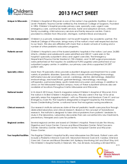
S P E C I A L A p p r o a c h F E A T U R E t o t h e P a t i e n t Approach to the Pediatric Patient with Graves’ Disease: When Is Definitive Therapy Warranted? Andrew J. Bauer Accreditation and Credit Designation Statements The Endocrine Society is accredited by the Accreditation Council for Continuing Medical Education to provide continuing medical education for physicians. The Endocrine Society has achieved Accreditation with Commendation. The Endocrine Society designates this Journal-based CME activity for a maximum of 1 AMA PRA Category 1 Credit™. Physicians should only claim credit commensurate with the extent of their participation in the activity. Learning Objectives Upon completion of this educational activity, participants should be able to • Distinguish the differences in clinical presentation between pre- and post-pubertal children with Graves’ disease • Identify and discuss the available treatment options for Graves’ disease and convey the advantages and disadvantages for each therapeutic option • Define the predictors associated with a decreased likelihood of remission in the pediatric patient • Debate the benefits and limitations of prolonged use of antithyroid medications compared to definitive therapy Target Audience This Journal-based CME activity should be of substantial interest to endocrinologists. Disclosure Policy Authors, editors, and Endocrine Society staff involved in planning this CME activity are required to disclose to learners any relevant financial relationship(s) that have occurred within the last 12 months with any commercial interest(s) whose products or services are discussed in the CME content. The Endocrine Society has reviewed all disclosures and resolved or managed all identified conflicts of interest, as applicable. Disclosures for JCEM Editors are found at http://www. endo-society.org/journals/Other/faculty_jcem.cfm. The following individuals reported NO relevant financial relationships: Andrew J. Bauer, M.D., and Leonard Wartofsky, M.D., reported no relevant financial relationships. Endocrine Society staff associated with the development of content for this activity reported no relevant financial relationships. Acknowledgement of Commercial Support This activity is not supported by grants, other funds, or inkind contributions from commercial supporters. Privacy and Confidentiality Statement The Endocrine Society will record learner’s personal information as provided on CME evaluations to allow for issuance and tracking of CME certificates. No individual performance data or any other personal information collected from evaluations will be shared with third parties. Method of Participation This Journal-based CME activity is available in print and online as full text HTML and as a PDF that can be viewed and/or printed using Adobe Acrobat Reader. To receive CME credit, participants should review the learning objectives and disclosure information; read the article and reflect on its content; then go to http://jcem.endojournals.org and find the article, click on CME for Readers, and follow the instructions to access and complete the post-activity test questions and evaluation. The estimated time to complete this activity, including review of material, is 1 hour. If you have questions about this CME activity, please direct them to [email protected]. Activity release date: March 2011 Activity expiration date: March 2012 Pediatric Endocrinology, Department of Pediatrics, Walter Reed Army Medical Center, Washington, D.C. 20307; Uniformed Services University, Bethesda, Maryland 20814; and The Thyroid Center, Division of Endocrinology, Children’s Hospital of Philadelphia, Philadelphia, Pennsylvania 19104 Pediatric Graves’ disease accounts for 10 –15% of thyroid disorders in patients less than 18 yr of age. The onset of symptoms may be insidious and subsequently associated with a delay in diagnosis. Decreased concentration and poor school performance are frequent complaints and can be quite frustrating for the patient and family. Severe ophthalmopathy is uncommon. The diagnosis is established by the findings of an increased heart rate and goiter in the setting of a suppressed TSH and elevated T3 and/or T4. The majority of pediatric patients are initially placed on antithyroid medications and maintained on these medications for prolonged periods of time in hopes of achieving remission. Unfortunately, for many children and adolescents remission is unattainable, ultimately occurring in only 15–30% of patients. Several recent studies have suggested that the age of the patient, the degree of thyrotoxicosis at diagnosis, the initial response to therapy, and the level of TSH receptor antibodies serve as reasonable predictors of remission and relapse. However, a consensus on the utility of these markers has not been reached. The present clinical case describes an adolescent with Graves’ disease and highlights the negative impact that prolonged medical therapy can have on quality of life and school performance; it reviews pertinent data on the diagnosis, comorbidities, and treatment options; and it identifies gaps in knowledge for when definitive therapy should be pursued. The case serves as a reminder that earlier discussion and decision for definitive therapy should be more commonplace in caring for our pediatric patients with Graves’ disease. (J Clin Endocrinol Metab 96: 580 –588, 2011) ISSN Print 0021-972X ISSN Online 1945-7197 Printed in U.S.A. Copyright © 2011 by The Endocrine Society doi: 10.1210/jc.2010-0898 Received April 19, 2010. Accepted November 8, 2010. 580 jcem.endojournals.org 15-yr-old girl was referred to our service for reevaluation of Graves’ disease (GD). She had been diagnosed with hyperthyroidism 3 yr earlier and treated with methimazole (MMI) titrated to maintain euthyroidism. Two months before the referral, a trial period off medication had been attempted, but the patient experienced a return of symptoms, including fatigue, palpitations, heat intolerance, increased appetite, decreased sleep, and poor concentration. The patient was a hard-working and successful student, in addition to being a star member of her A Abbreviations: AITD, Autoimmune thyroid disease; DTC, differentiated thyroid cancer; GD, Graves’ disease; MMI, methimazole; MPO-ANCA, myeloperoxidase-antineutrophil cytoplasmic antibody; PTU, propylthiouracil; TRAb, TSHR antibody; TSHR, TSH receptor. J Clin Endocrinol Metab, March 2011, 96(3):580 –588 The Endocrine Society. Downloaded from press.endocrine.org by [${individualUser.displayName}] on 22 August 2014. at 13:52 For personal use only. No other uses without permission. . All rights reserved. J Clin Endocrinol Metab, March 2011, 96(3):580 –588 TABLE 1. Signs and symptoms of GD in children Signs Goiter Tachycardia Weight loss Heat intolerance Tremor Systolic hypertension Increased pulse pressure Hair loss Secondary enuresis (nocturia) Advanced bone age Ophthalmopathy—pain, exposure keratitis, lid lag, proptosis Symptoms Hyperactivity Palpitations Sleep disturbance Fatigue Poor school performance Emotional lability Neck fullness or lump Irritability and nervousness Increased stool frequency Increased appetite school’s soccer team and swim team. Family history was significant for autoimmune disease in her mother (hypothyroidism), maternal grandmother (GD), and paternal grandfather (type 1 diabetes mellitus). The patient and parents denied a history of smoking or regular exposure to secondhand smoke. On physical examination, the patient had a heart rate of 105 beats per minute, blood pressure of 114/55 mm Hg, weight of 61 kg, and a body mass index of 22 kg/m2. The patient was restless and fidgety with warm, moist skin. There was no evidence of ophthalmopathy, specifically no upper eyelid retraction, scleral injection, edema, or proptosis. Thyroid exam revealed a moderately sized nontender, asymmetric goiter estimated to be about 30 g. One year earlier, the family reported being counseled regarding radioiodine ablation but decided to continue with MMI and not pursue definitive treatment in the hope that the GD would resolve on its own. Background GD accounts for approximately 10 –15% of childhood thyroid disease (1). Signs and symptoms are similar to adults (Table 1), but in contrast with adults there is often a delay in diagnosis (2– 4). The majority of children do not suffer longterm consequences. However, delayed diagnosis may be associated with impaired neurodevelopmental outcome (5) and altered skeletal maturation, including craniosynostosis and advanced bone age in younger children (6). In addition, for school-aged children, particularly adolescent patients, a decrement in school performance is common, can be quite significant, and can cause a great deal of anxiety and frustration for patients and their parents. The incidence of GD is believed to be between 0.1 and 3 per 100,000 children (7) with a prevalence of 1 in 10,000 children in the United States (8). GD is rare under the age of 5 yr and has a peak incidence at 10 –15 yr of age, more jcem.endojournals.org 581 commonly affecting female patients (1, 9). Genetic and environmental factors play a role in the pathogenesis of GD, reflected by an increased association with other autoimmune disorders and syndromes, both in the individual patient and in other family members (10 –13). Linkage analysis from families with a history of autoimmune thyroid disease (AITD; GD and autoimmune hypothyroidism) has provided evidence for involvement of several loci, including the human leukocyte antigen (HLA) region on chromosome 6p21, cytotoxic T lymphocyte antigen 4 (CTLA-4) on chromosome 2q33, and lymphoid protein tyrosine phosphatase (PTPN22), each individually conferring a 1.4- to 4-fold relative risk for disease. In addition, several other regions have been identified on 2q36, 11p15, 18p11, 5q23, and Xp11; however, no single locus has been found to explain the familial association of AITD (14). The pathophysiology of AITD involves diffuse infiltration of lymphocytes into the thyroid gland with loss of tolerance to multiple thyroid antigens, including the TSH receptor (TSHR), thyroglobulin, and thyroperoxidase (11). For unknown reasons, activated T cells invade the thyroid and release cytokines, leading to dysregulation of B cells and subsequent production of autoantibodies. In GD, the predominant antibodies are directed against the TSHR (TRAb). The TRAbs are heterogeneous and can either stimulate or inhibit thyroid hormone secretion. In addition, they lead to an increase in follicular cell growth and increased vascularity (15). Fluctuating levels of the stimulatory and inhibitory antibodies can result in an alternating clinical presentation between hyper- and hypothyroidism, making it difficult to correlate TRAb levels with clinical status. Over the last 40 yr, multiple assays have been developed in an effort to accurately identify the causative factors leading to the hyperthyroxinemia. Unfortunately, the mixture of antibodies directed at the TSHR, the lack of standardization in technique and nomenclature, and the varying availability of specific tests have led to a fair degree of confusion over which assay affords the greatest utility in predicting the course of disease (16, 17). Clinical Considerations: Presentation and Evaluation The symptoms of GD often develop insidiously and may initially be interpreted as common complaints of childhood and adolescence, in particular moodiness and emotional lability, fatigue, sleep disturbance, and increased appetite. School-aged children, particularly adolescents, may be referred for evaluation of attention-deficit hyperactivity disorder after a teacher or parent raises concern over a decreased ability to concentrate and complete The Endocrine Society. Downloaded from press.endocrine.org by [${individualUser.displayName}] on 22 August 2014. at 13:52 For personal use only. No other uses without permission. . All rights reserved. 582 Bauer Treatment of Graves’ Disease in Children school work with resultant poor school performance. This later symptom can be particularly troublesome, depending on the length of delay in diagnosis. The question of whether the presentation differs between prepubertal and pubertal children continues to be debated (2– 4). Some clinicians believe that prepubertal children more commonly present with poor weight gain and frequent stooling, whereas adolescents typically present with irritability, fatigue, palpitations, heat intolerance, fine tremor, and a goiter (3). Others believe the differences are individually based and are independent of age (2). A consistent observation is that younger patients have up to a 2-fold greater delay in diagnosis that is often associated with increased height, advanced bone age, and lower weight (2– 4, 18, 19). Coincident with the delay in diagnosis, prepubertal children typically have greater severity of biochemical hyperthyroidism with more extreme elevations in T4 and T3 and associated levels of TRAbs (2– 4, 18, 19). Whether this is an associated finding or a reflection of the disease process or is simply due to delayed diagnosis is not known. In peripubertal children, with proper treatment, pubertal progression can be maintained, and final predicted height can be preserved (19). Our patient’s presentation of increased fatigue, irritability, and poor school performance was typical of our experience with recurrent GD in the adolescent population. Once our patient stopped her MMI, the return of symptoms was fairly rapid, worsening over a 2-month period. The symptoms that were most bothersome to the patient included fatigue, exercise intolerance, and decreased ability to concentrate. Early on, the family attributed the symptoms as secondary to the busy life of an adolescent, but when the symptoms worsened, with decreased school performance despite continued commitment to her studies, and several missed soccer games due to decreased physical performance, the family returned for reevaluation. In regard to the biochemical and radiological evaluation, at a minimum we send a TSH level and, if suppressed, add a T4 or free T4 and a T3. The addition of T3 provides a useful screen for identifying isolated T3 toxicosis, which is a common presentation in pediatric patients, most notably in prepubertal patients (3, 4). In fact, in a prepubertal child, the degree of T3 elevation is a useful predictor for identifying patients that are unlikely to experience early remission (20). We do not routinely perform radioiodine uptake and scan unless the biochemical data are inconclusive in the face of a suppressed TSH or at the time definitive therapy is pursued. The chronicity of symptoms and an uptake and scan are often helpful in distinguishing between subacute thyroiditis, hashitoxicosis, toxic multinodular goiter, or a toxic adenoma, although discerning hashitoxicosis from GD can be quite challenging due to J Clin Endocrinol Metab, March 2011, 96(3):580 –588 overlap in presentation, antibody profiles, and even I-123 uptake and scans (21). The laboratory evaluation for our patient revealed a TSH of 0.06 IU/ml (normal, 0.27– 4.2), with free T4 of 2.25 ng/dl (normal, 1.01–1.79), T3 of 4.67 nmol/liter (normal, 1.3– 3.10), thyroid-stimulating Ig of 187% (normal, ⱕ125), and a TSH-binding inhibitor Ig of 48.3% (normal, ⱕ16%). These results, along with the classic signs and symptoms of hyperthyroxinemia, confirmed our suspicion of recurrent GD. On examination, the thyroid gland size was estimated at two times normal for age based on the 1960 World Health Organization (WHO) criteria that define goiter size by comparison of the thyroid gland to the distal phalanges of the patient’s thumb. This estimate, along with the 1994 WHO criteria that use a two-grade classification, either visible/palpable or not, provides a field-expedient, systematic method for documentation and communication of thyroid gland size (22). An enlarged gland is easily visible, palpable, and is estimated on one half increments above normal size (i.e. enlarged 1.5, 2.0, 2.5…. times). With the thyroid not only being enlarged but also asymmetric, before discussion on therapeutic options, a thyroid ultrasound was performed to determine whether a nodule(s) was responsible for the asymmetric goiter noted on physical examination. In general, we perform an ultrasound for any GD patient with thyroid gland asymmetry or a palpable nodule. If a nodule is confirmed, fine-needle aspiration biopsy should be considered, as well as I-123 or Tc-99 scan. Although uncommon, patients with GD, or an autonomous nodule, may have concurrent differentiated thyroid cancer (DTC). In the adult literature, there is continued debate about whether DTC in the setting of GD follows a more aggressive or a more indolent course (23– 25). In the adult literature, DTC is frequently reported as an incidental discovery on review of pathology after thyroidectomy. In children, this association has been infrequently reported (26). However, we and others have had several adolescents with GD present with clinically aggressive DTC, including diffuse cervical lymph node metastasis and invasion and pulmonary metastasis (our unpublished observation). Given the debate on whether nodules found in the setting of AITD have an increased risk of malignancy and/or aggressive features, a thorough evaluation of the nodule(s), including fine-needle aspiration biopsy, is warranted. Fortunately for this particular patient, the thyroid ultrasound showed a diffusely, mildly heterogeneous, hypoechoic gland without nodules. Clinical Consideration: Treatment Options We were now faced with an all too common situation in treating pediatric patients with GD; when is definitive The Endocrine Society. Downloaded from press.endocrine.org by [${individualUser.displayName}] on 22 August 2014. at 13:52 For personal use only. No other uses without permission. . All rights reserved. J Clin Endocrinol Metab, March 2011, 96(3):580 –588 therapy the better treatment option? For our particular patient, despite treatment with MMI for 3 yr, disease recurred within 2 months of stopping medication. All three treatments—restarting antithyroid medication, radioiodine ablation, and thyroidectomy—are effective, with the ultimate decision to be made by the patient and the family. In our case, during the follow-up visit to review the laboratory and ultrasound results, the family voiced hesitancy to pursue definitive therapy, either radioiodine ablation or surgery, opting to restart MMI with the hope that remission was still possible. Although we reviewed the unlikely scenario of remission with the family, we wondered whether earlier counseling on remission rates for GD in pediatric patients and an earlier discussion on the length of therapy before consideration for definitive therapy would have lessened the anxiety for the patient and her family as they faced this transition point; it is a rhetorical question for this patient, but of potential benefit for the next. With rare exception, at the time of initial diagnosis, we place patients on an antithyroid medication. In addition, patients with relapse who ultimately choose definitive therapy may be started on antithyroid medications as a temporizing measure until a workable time for radioiodine or surgery can be found. With the recent association between propylthiouracil (PTU) and severe liver failure and the subsequent black-box warning from the Food and Drug Administration, MMI (0.1–1.0 mg/kg 䡠 d or 15–20 mg/m2 䡠 d) is now our first-choice antithyroid medication (27). In countries where MMI is not available, carbimazole (0.5– 0.7 mg/kg 䡠 d), a drug that is biologically converted to MMI, may be substituted (9). We add a betablocker, such as propranolol (1–2 mg/kg 䡠 d) or atenolol (0.5–1.2 mg/kg 䡠 d) depending on the severity of tachycardia, palpitations, or tremor. Follow-up labs are sent 4 – 6 wk after initiation of therapy and repeated every 2–3 months once the appropriate dose has been determined. TSH often remains suppressed for an extended period of time, so adjustments in medication are made based on thyroid hormone levels (T4 or T3). The discussion over the best approach to dosing follows two basic approaches: “titrate” and “block-and-replace.” Those in favor of block-and-replace, i.e. use a higher dose of antithyroid medication and add levothyroxine to achieve euthyroidism, suggest that euthyroidism is more readily achieved and with less laboratory follow-up. Those in favor of titrate, i.e. adjusting the antithyroid medication dose to achieve euthyroidism, suggest improved compliance and decreased likelihood of toxicity and side effects associated with higher doses of antithyroid medications (Table 2) (28 –30). Our general approach is to titrate the dose, but in practice we’ve occasionally resorted to block-and-replace due to difficulty achieving a titrated jcem.endojournals.org 583 TABLE 2. Side effects of antithyroid medications (MMI and carbimazole) Minor Rash Pruritus Hives Hair loss Nausea Decreased taste Joint pain Arthralgia Severe Agranulocytosis Neutropenia Thrombocytopenia Stevens-Johnson syndrome Cholestatic jaundice Hepatitis dose. In either case, achieving euthyroidism as quickly as possible is desirable for the patient’s well-being and may increase the likelihood for remission (31). Although reinitiation of MMI (or carbimazole) is a reasonable choice, the unfortunate reality is that pediatric patients treated with antithyroid medications have a lower likelihood of remission when compared with adults, 30 vs. 50%, respectively (9). In addition, the longer the period of watchful waiting, the higher the likelihood of decreased compliance (20). Despite these fairly known and accepted data, many clinicians caring for pediatric-aged patients are hesitant to recommend definitive therapy, often keeping patients, especially younger patients, on antithyroid medications for prolonged periods of time in hopes of ultimately achieving remission. This is not to suggest that antithyroid medications are not an effective choice. A review of eight studies shows a range of remission rates varying from 25– 65% of pediatric patients (32–37). However, extended periods of time are frequently required for pediatric patients to achieve remission, with one study suggesting an approximately 25% remission rate for every 2 yr of continued antithyroid medication therapy (38). In addition, whereas more than 50% will remain in remission, relapse is common, varying from 36 – 47% of patients (34, 35). In contrast, the data from adult patients, a group with a higher overall likelihood of achieving spontaneous remission, suggest that if remission is not achieved within 12–18 months of medical therapy, then definitive therapy (radioiodine or surgery) should be pursued (29). In the absence of comparative data in children, the question over defining a time limit for maximum benefit from antithyroid medications for the pediatric population remains unanswered. For the individual pediatric patient, however, the question as to whether the risks and benefits between watchful waiting and definitive therapy are equivalent, including a comparison in potential changes to qualityof-life issues, should be regularly discussed with the patient and family during follow-up visits. If and when a decision for definitive therapy has been reached, both radioiodine therapy and near-total thyroidectomy are tenable options, with the goal being to achieve The Endocrine Society. Downloaded from press.endocrine.org by [${individualUser.displayName}] on 22 August 2014. at 13:52 For personal use only. No other uses without permission. . All rights reserved. 584 Bauer Treatment of Graves’ Disease in Children hypothyroidism. The limiting factor for thyroidectomy is access to a high-volume thyroid surgeon, defined by performance of more than 30 cervical endocrine procedures per year (39). The surgical complication rate is more directly related to the number of procedures the surgeon performs annually rather than the training of the surgeon, and whether they are general surgeons or ear, nose, and throat surgeons, and pediatric or adult surgeons. Although adult surgeons will clearly have increased numbers of thyroid procedures, surgery must still be performed in a hospital capable and comfortable with the postoperative care of children, including availability of pediatric anesthesia and intensive care support. When these basic requirements are met, the surgical option is reasonable and should be presented to the patient and family as an equivalent option to radioiodine ablation. In general, we recommend surgery over radioiodine ablation in patients younger than 10 yr of age, in patients or situations where compliance with precautions of radioiodine and follow-up are questionable, and for larger glands (more than two times normal for age). Severe eye disease or a late adolescent that is considering pregnancy are additional situations were surgery may be preferred because radioiodine ablation can be associated with a transient increase of TRAb levels and with persistence of detectable antibody levels for at least 5 yr after ablation (40). Several steps can be taken to decrease the risk of surgical complications. To decrease risk of anesthesia, patients should achieve euthyroidism with antithyroid medication and remain on a therapeutic dose until surgery is completed. A 7- to 10-d course of concentrated iodine solution given the week before surgery will help to decrease thyroid hormone production with the added benefit of decreasing thyroid vascularity (41). The typical dose is three to five drops of concentrated iodine, 50 –150 mg/dose, taken three times per day for 7–10 d. Taking the iodine with food or milk will help improve the palatability. Lastly, the availability of rapid PTH testing, drawn at the end of the procedure, has proven to be extremely helpful in accurately predicting the occurrence of postoperative hypocalcemia (42– 45). We routinely use the value to stratify intensity and location of postoperative care, admission to the ward vs. intensive care unit, and to decide whether we should initiate calcitriol and oral calcium replacement. If the test is not available, empiric treatment with calcium and vitamin D in the postoperative period is equally as effective but may be associated with a higher risk of hypercalcemia (45). In the United States, I-131 ablation is the most common choice for definitive therapy, with variability in recommendation based on geographic location, bias of physician training, and availability of resources. Some families and patients prefer I-131 ablation over thyroidectomy due J Clin Endocrinol Metab, March 2011, 96(3):580 –588 to concerns over the cosmetic appearance of a surgical scar, particularly adolescents as well as patients with a personal or family history of keloid formation. The greatest long-term concern we hear from families is the potential association of I-131 therapy with secondary malignancy. In the absence of a national registry prospectively following pediatric patients treated with radioiodine over their lifetime, the retrospective evidence suggests a very low risk of adverse events. Specifically, in studies reporting data for pediatric patients followed for up to 36 yr after radioiodine treatment, there was no evidence of decreased fertility, increased congenital anomalies, increased spontaneous pregnancy loss, or an increase in malignancy above the general population (46 – 48). Although these data are reassuring, the data from the Chernobyl nuclear accident suggest that children exposed to radioiodine are at an increased risk for developing a thyroid malignancy (49). Although many radioisotopes were released into the atmosphere, the increased incidence of pediatric thyroid malignancy after Chernobyl is believed to be secondary to exposure to low doses of radioiodine (I-131) in a population with baseline dietary iodine insufficiency (50). Interestingly, the risk appeared to be age dependent, greatest for children less than 5 to 6 yr of age and decreasing progressively through 12 yr of age (49). With this in mind, whereas there are no absolute cut-offs for age, we rarely recommend treating GD patients under the age of 10 yr with I-131, based on the potential long-term risk of radioiodine-induced malignancy as well as the inherent increased risk of exposure to family members caring for a child in a less-independent age group. For pediatric patients with GD-associated ophthalmopathy, radioiodine can be used but consultation with pediatric ophthalmology for pre- and postablation evaluation and treatment is recommended. In adolescents with more severe ophthalmopathy, those with persistent lid retraction, proptosis, chemosis, or corneal involvement despite achieving a euthyroid state, oral glucocorticoids should be considered, started 1 d after ablation and tapered over a 1- to 3-month time frame (51, 52). An abridged NOSPEC classification has been proposed, but neither the NOSPEC severity assessment nor the clinical activity score has been validated in the pediatric population (53). In general, children and adolescents appear to have the same incidence of GD-associated ophthalmopathy, but it is typically less severe than adults. Elevated TRAb titers and smoking are associated with an increased incidence and increased severity for eye disease in both adults and children (54, 55). In regard to I-131 therapy, most agree that hypothyroidism is the target of therapy, but there is continued discussion over an appropriate dose. Although the major- The Endocrine Society. Downloaded from press.endocrine.org by [${individualUser.displayName}] on 22 August 2014. at 13:52 For personal use only. No other uses without permission. . All rights reserved. J Clin Endocrinol Metab, March 2011, 96(3):580 –588 TABLE 3. Thyroid gland weight by age Age (yr) 3– 4 10 –14 15–19 Adult Weight (g) 4⫾2 10 ⫾ 6 14 ⫾ 5 20 ⫾ 5 Adapted from Ref. 6. ity of articles list the Quimby-Marinelli formula or a modified version of the formula based on 24-h uptake, one is hard-pressed to find normative values for thyroid weight in children or a reliable method of estimating thyroid weight based on physical examination or ultrasound dimensions. For postpubertal adolescents, using factor estimates based on comparison to the standard thyroid weight of an adult in an iodine-sufficient area (15–20 g) is a reasonable approach, with consideration of total dose based on gland size and 24-h uptake. In prepubertal children, the estimates are more difficult. Previous published estimates of normal thyroid gland size for age are listed in Table 3 (56), and estimated weight can be adjusted based on physical examination. As a general approach, the dose of I-131 needed to achieve remission in greater than 95% of patients is believed to be between 220 and 275 Ci/g, increased to 300 Ci/g for larger goiters or less elevated radioiodine uptake (57). In practice, the total dose that we use is typically between 12 and 15 mCi. Calculated dosing should be considered if radioiodine therapy is prescribed for a patient with a goiter size more than two times normal for age (58). Of interest and concern are the data showing decreased efficacy of radioiodine ablation after the use of antithyroid drug therapy (30). In the absence of a large gland and an increased risk of thyroid storm, strategies to increase the likelihood of a successful ablation include stopping antithyroid medications 5–7 d before and avoiding their use for at least 1 wk after radioiodine treatment (30). In this approach, continuation of the beta-blocker will help lessen symptoms. Opponents of this approach argue that increasing the dose of radioiodine by approximately 20% will result in successful ablation without increasing the risk of thyroid storm associated with stopping the antithyroid medication. Neither approach has been adequately studied in children. Once an appropriate dose has been determined and administered, postablation hypothyroidism is achieved within 1 to 3 months, at times delayed out to 6 months. Follow-up thyroid function testing should begin at about 1 month and then be repeated every 4 – 6 wk to determine appropriate timing of thyroid hormone replacement therapy. After discussion of the pros and cons of definitive therapy, the family and patient elected to restart the MMI (15 jcem.endojournals.org 585 mg orally twice a day). Within several weeks, the patient reported decreased symptoms. However, approximately 7– 8 wk after restarting MMI she began to complain of intermittent knee pain. There was no known trauma, and the patient had actually decreased participation in sports as she tried to improve her grades in school. She denied fever, rash, abdominal pain, or sore throat. Although joint pain is common in athletic teenagers, in the setting of GD one must consider the potential association between GD, antithyroid medications, and myeloperoxidase-antineutrophil cytoplasmic antibody (MPOANCA)-associated vasculitis (59, 60). MPO-ANCAs may be present before initiation of antithyroid medications, but initiation of PTU, and to a lesser extent MMI (59, 61), increases MPO-ANCA titers often associated with multiorgan vasculitis, including involvement of the kidneys, respiratory tract, joints, eyes, gastrointestinal tract, and brain (59, 60). Cross-reactivity between PTU and MMI may occur if attempts are made to switch medications once symptoms present (62), a less frequent issue in pediatric patients now with the current recommendation to avoid PTU therapy due to an increased risk of liver failure (8). On examination, the patient appeared well, was afebrile, and had a normal exam, including the absence of a rash or joint effusion, erythema, or calor. Laboratory evaluation revealed a normal complete blood count, normal liver function panel, normal urinalysis and negative antinuclear antibody titer. However, the MPO-ANCA level was positive at 273 EU/ml (normal range, ⬍20 EU/ml), consistent with MMI-induced vasculitis as the etiology for the patient’s arthralgia. Controversies in Treatment: When Should Definitive Therapy Be Pursued? Predicting remission and establishing the appropriate timing for definitive therapy remains elusive and controversial. For the majority of pediatric patients, initiation of an antithyroid medication at the time of diagnosis is a reasonable plan. It affords an opportunity to determine the course of disease and to decrease symptoms in a relatively short period of time, and, for the most part, antithyroid medications, in particular MMI, have a relatively low incidence of side effects or adverse events. Unfortunately, despite good intentions, children often remain on antithyroid medications for extended periods of time because clinicians and parents are hesitant to pursue definitive therapy. With the watchful-waiting approach, 15–30% of the time a subgroup of pediatric patients will enter remission; however, this may take between 2 and 10 yr to achieve, and 35– 60% of patients may experience a relapse after medications are discontinued (9, 20, 35, 38). In addi- The Endocrine Society. Downloaded from press.endocrine.org by [${individualUser.displayName}] on 22 August 2014. at 13:52 For personal use only. No other uses without permission. . All rights reserved. 586 Bauer Treatment of Graves’ Disease in Children tion, although less common than with PTU, prolonged use of MMI is not without risk of associated side effects, including hives, Stevens-Johnson syndrome, neutropenia, cholestatic jaundice, and vasculitis-associated symptoms, such as hematuria, proteinuria, dyspnea and hemoptysis, purpuric rash, arthralgia, uveitis, myalgia, and others (59, 63). For years, clinicians have attempted to identify predictive measures for remission and to determine a maximum time limit to duration of therapy after which time the likelihood of remission is unlikely. Although no single marker has been found that provides 100% predictability, there are several observations and markers that are associated with a decreased likelihood of achieving and maintaining remission. Theoretically, the measurement of TRAbs should provide the closest approximation to disease activity. In practice, however, the heterogeneity of the antibody and the nonstandardized approach to measurement have led to mixed results in the published literature. Because of this, many clinicians do not rely on its level, or even measure the level, in an attempt to predict disease remission. Despite these shortfalls, the presence of elevated TRAbs (thyroidstimulating Ig, TSH-binding inhibitor Ig, and/or TRAb) identifies patients at increased risk of relapse. However, the opposite is not true. Normal values do not predict patients that will remain in remission (9, 64, 65). Translated into practice, the following patient characteristics should lead to consideration for earlier definitive therapy: younger age (prepubertal vs. pubertal), greater degree of biochemical thyrotoxicosis at presentation (T3 and/or T4/free T4), an extended period of time before achievement of euthyroid status after initiation of antithyroid medications (⬎3 months), and persistent elevation of TRAbs (9, 20). TRAb levels should be measured at the time one is considering stopping antithyroid medications, and if elevated, patients and families should be closely followed, provided anticipatory guidance for seeking reevaluation, and counseled on the high likelihood that definitive therapy will be required. In patients with low levels of TRAbs, stopping medication is reasonable, assuming the family is provided anticipatory guidance on signs and symptoms that suggest relapse. With the lower rate of remission in children and the inherent risks of toxicity and decreasing compliance associated with prolonged antithyroid medication therapy, there should be an increasing focus in pediatric endocrinology on determining whether the adult paradigm of moving to definitive therapy after a maximum of 12–18 months is clinically beneficial (3, 29). In adolescent patients, where continued symptoms can negatively impact school performance and quality of life, a discussion and recommendation to move toward definitive therapy J Clin Endocrinol Metab, March 2011, 96(3):580 –588 Graves’ Disease -low TSH, elevated T4 and/or T3 Antithyroid Medication Beta-blocker if symptoms Treatment x 18-24 months Evidence of active disease - Inability I bilit to t decrease d ATD dose d and/or - Elevated TSHR Antibody level No Yes Trial off ff off medication Definitive therapy -Anticipatory Guidance for re-evaluation Near-total thyroidectomy -high high volume surgeon, surgeon pediatric setting Recurrence Or Radioiodine ablation -consider stopping ATD prior FIG. 1. Treatment options for GD. ATD, Anti-thyroid drug. should be considered sooner, as early as 18 –24 months after initiation of antithyroid medications, if remission has not been achieved (Fig. 1). Returning to the Patient With increased joint pain and evidence of MPO-ANCA vasculitis, continued difficulties with school performance, and persistent elevation in TRAbs, the family elected to pursue radioiodine ablation. The MMI was discontinued, and 7 d later the 4-h radioiodine uptake was 63.4% (normal, 5–15%). The following day, the patient was administered 15.3 mCi of I-131. Within 7 wk of ablation, hypothyroidism was achieved, and the patient was started on thyroid hormone replacement. Three months after definitive therapy, the patient reported improved school performance, decreased fatigue, and reinitiation of physical activities. Conclusion GD is the most common cause of hyperthyroidism in the pediatric population. There is little controversy on the use of antithyroid medications as a first-line agent in the treatment of pediatric GD. Once euthyroidism is established, a subset of patients will spontaneously enter remission; however, the watchful-waiting approach should have limits. For the subgroup of patients that do not achieve remission within 18 –24 months of initiation of anti-thyroid drug therapy or who experience toxicity or difficulties with compliance, definitive therapy with radioiodine or surgery should be offered. Patient preference must be strongly considered, but a frank, open discussion on the The Endocrine Society. Downloaded from press.endocrine.org by [${individualUser.displayName}] on 22 August 2014. at 13:52 For personal use only. No other uses without permission. . All rights reserved. J Clin Endocrinol Metab, March 2011, 96(3):580 –588 lower likelihood of remission for pediatric patients should be a routine part of follow-up visits. Risk factors associated with a decreased likelihood of remission include younger age at presentation with a higher degree of biochemical thyrotoxicosis and patients with persistence of elevated TRAbs. In addition, when definitive therapy is targeted to achieve hypothyroidism, adolescent patients may also benefit from earlier definitive therapy due to reduced complexity of follow-up and improved quality of life. These issues are as salient to the pediatric and adolescent patient as they are to the adult population. Despite an agreement on a lower rate of remission for pediatric patients, controversy remains on the timing for definitive therapy, with patients often remaining on antithyroid medication for extended periods of time. Cooperative, multicenter, prospective studies are needed to assess the long-term risks and benefits of the therapeutic options, to define a maximum length of benefit for antithyroid medication therapy, and to determine the potential positive impact that earlier definitive therapy may have on quality-of-life considerations for our pediatric population. Acknowledgments Address all correspondence and requests for reprints to: Dr. Andrew J. Bauer, Uniformed Services University of the Health Sciences, Pediatric Endocrinology, Bethesda, Maryland 20814. E-mail: [email protected]. The opinions or assertions contained herein are the private views of the author and are not to be construed as official or as reflecting the opinions of Walter Reed Army Medical Center, the Uniformed Services University of the Health Sciences, the Department of the Army, or the Department of Defense. Disclosure Summary: The author has no conflicts of interest to report. References 1. Zimmerman D, Lteif AN 1998 Thyrotoxicosis in children. Endocrinol Metab Clin North Am 27:109 –126 2. Poyrazoðlu S, Saka N, Bas F, Isguven P, Dogu A, Turan S, Bereket A, Turan A, Sarikaya S, Adal E, Cizmecioglu F, Cizmeci F, Saglam H, Ercan O, Memioglu N, Günöz H, Bundak R, Darendeliler F, Yildiz M, Guran T, Akcay T, Akin L, Hatun S 2008 Evaluation of diagnosis and treatment results in children with Graves’ disease with emphasis on the pubertal status of patients. J Pediatr Endocrinol Metab 21:745–751 3. Lazar L, Kalter-Leibovici O, Pertzelan A, Weintrob N, Josefsberg Z, Phillip M 2000 Thyrotoxicosis in prepubertal children compared with pubertal and postpubertal patients. J Clin Endocrinol Metab 85:3678 –3682 4. Shulman DI, Muhar I, Jorgensen EV, Diamond FB, Bercu BB, Root AW 1997 Autoimmune hyperthyroidism in prepubertal children and adolescents: comparison of clinical and biochemical features at diagnosis and responses to medical therapy. Thyroid 7:755–760 jcem.endojournals.org 587 5. Segni M, Leonardi E, Mazzoncini B, Pucarelli I, Pasquino AM 1999 Special features of Graves’ disease in early childhood. Thyroid 9:871– 877 6. Wong GW, Lai J, Cheng PS 1999 Growth in childhood thyrotoxicosis. Eur J Pediatr 158:776 –779 7. Lavard L, Ranløv I, Perrild H, Andersen O, Jacobsen BB 1994 Incidence of juvenile thyrotoxicosis in Denmark, 1982–1988. A nationwide study. Eur J Endocrinol 130:565–568 8. Rivkees SA, Mattison DR 2009 Propylthiouracil (PTU) hepatoxicity in children and recommendations for discontinuation of use. Int J Pediatr Endocrinol 2009:132041 9. Kaguelidou F, Alberti C, Castanet M, Guitteny MA, Czernichow P, Léger J 2008 Predictors of autoimmune hyperthyroidism relapse in children after discontinuation of antithyroid drug treatment. J Clin Endocrinol Metab 93:3817–3826 10. Wasniewska M, Corrias A, Messina MF, Crisafulli G, Salzano G, Valenzise M, Mussa A, De Luca F 2010 Graves’ disease prevalence in a young population with Turner syndrome. J Endocrinol Invest 33:69 –70 11. Michels AW, Eisenbarth GS 2010 Immunologic endocrine disorders. J Allergy Clin Immunol 125:S226 –237 12. De Luca F, Corrias A, Salerno M, Wasniewska M, Gastaldi R, Cassio A, Mussa A, Aversa T, Radetti G, Arrigo T 2010 Peculiarities of Graves’ disease in children and adolescents with Down’s syndrome. Eur J Endocrinol 162:591–595 13. Birrell G, Cheetham T 2004 Juvenile thyrotoxicosis; can we do better? Arch Dis Child 89:745–750 14. Taylor JC, Gough SC, Hunt PJ, Brix TH, Chatterjee K, Connell JM, Franklyn JA, Hegedus L, Robinson BG, Wiersinga WM, Wass JA, Zabaneh D, Mackay I, Weetman AP 2006 A genome-wide screen in 1119 relative pairs with autoimmune thyroid disease. J Clin Endocrinol Metab 91:646 – 653 15. Kaguelidou F, Carel JC, Léger J 2009 Graves’ disease in childhood: advances in management with antithyroid drug therapy. Horm Res 71:310 –317 16. Ajjan RA, Weetman AP 2008 Techniques to quantify TSH receptor antibodies. Nat Clin Pract Endocrinol Metab 4:461– 468 17. Baloch Z, Carayon P, Conte-Devolx B, Demers LM, Feldt-Rasmussen U, Henry JF, LiVosli VA, Niccoli-Sire P, John R, Ruf J, Smyth PP, Spencer CA, Stockigt JR 2003 Laboratory medicine practice guidelines. Laboratory support for the diagnosis and monitoring of thyroid disease. Thyroid 13:3–126 18. Bossowski AT, Reddy V, Perry LA, Johnston LB, Banerjee K, Blair JC, Savage MO 2007 Clinical and endocrine features and long-term outcome of Graves’ disease in early childhood. J Endocrinol Invest 30:388 –392 19. Cassio A, Corrias A, Gualandi S, Tato’ L, Cesaretti G, Volta C, Weber G, Bona G, Cappa M, Bal M, Bellone J, Cicognani A 2006 Influence of gender and pubertal stage at diagnosis on growth outcome in childhood thyrotoxicosis: results of a collaborative study. Clin Endocrinol (Oxf) 64:53–57 20. Glaser NS, Styne DM 2008 Predicting the likelihood of remission in children with Graves’ disease: a prospective, multicenter study. Pediatrics 121:e481– e488 21. Nabhan ZM, Kreher NC, Eugster EA 2005 Hashitoxicosis in children: clinical features and natural history. J Pediatr 146:533–536 22. Zimmermann M, Saad A, Hess S, Torresani T, Chaouki N 2000 Thyroid ultrasound compared with World Health Organization 1960 and 1994 palpation criteria for determination of goiter prevalence in regions of mild and severe iodine deficiency. Eur J Endocrinol 143:727–731 23. Cappelli C, Braga M, De Martino E, Castellano M, Gandossi E, Agosti B, Cumetti D, Pirola I, Mattanza C, Cherubini L, Rosei EA 2006 Outcome of patients surgically treated for various forms of hyperthyroidism with differentiated thyroid cancer: experience at an endocrine center in Italy. Surg Today 36:125–130 24. Pellegriti G, Belfiore A, Giuffrida D, Lupo L, Vigneri R 1998 Out- The Endocrine Society. Downloaded from press.endocrine.org by [${individualUser.displayName}] on 22 August 2014. at 13:52 For personal use only. No other uses without permission. . All rights reserved. 588 25. 26. 27. 28. 29. 30. 31. 32. 33. 34. 35. 36. 37. 38. 39. 40. 41. 42. 43. 44. 45. Bauer Treatment of Graves’ Disease in Children come of differentiated thyroid cancer in Graves’ patients. J Clin Endocrinol Metab 83:2805–2809 Yano Y, Shibuya H, Kitagawa W, Nagahama M, Sugino K, Ito K, Ito K 2007 Recent outcome of Graves’ disease patients with papillary thyroid cancer. Eur J Endocrinol 157:325–329 Niedziela M, Korman E 2002 Thyroid carcinoma in a fourteenyear-old boy with Graves disease. Med Pediatr Oncol 38:290 –291 Rivkees SA 12 July 2010 63 years and 715 days to the “boxed warning”: unmasking of the propylthiouracil problem. Int J Pediatr Endocrinol 10.1155/2010/658267 Cooper DS 2005 Antithyroid drugs. N Engl J Med 352:905–917 Abraham P, Avenell A, McGeoch SC, Clark LF, Bevan JS 2010 Antithyroid drug regimen for treating Graves’ hyperthyroidism. Cochrane Database Syst Rev 1:CD003420 Walter MA, Briel M, Christ-Crain M, Bonnema SJ, Connell J, Cooper DS, Bucher HC, Müller-Brand J, Müller B 2007 Effects of antithyroid drugs on radioiodine treatment: systematic review and meta-analysis of randomised controlled trials. BMJ 334:514 Laurberg P 2006 Remission of Graves’ disease during anti-thyroid drug therapy. Time to reconsider the mechanism? Eur J Endocrinol 155:783–786 Barnes HV, Blizzard RM 1977 Antithyroid drug therapy for toxic diffuse goiter (Graves disease): thirty years experience in children and adolescents. J Pediatr 91:313–320 Barrio R, López-Capapé M, Martinez-Badás I, Carrillo A, Moreno JC, Alonso M 2005 Graves’ disease in children and adolescents: response to long-term treatment. Acta Paediatr 94:1583–1589 Gorton C, Sadeghi-Nejad A, Senior B 1987 Remission in children with hyperthyroidism treated with propylthiouracil. Long-term results. Am J Dis Child 141:1084 –1086 Hamburger JI 1985 Management of hyperthyroidism in children and adolescents. J Clin Endocrinol Metab 60:1019 –1024 Raza J, Hindmarsh PC, Brook CG 1999 Thyrotoxicosis in children: thirty years’ experience. Acta Paediatr 88:937–941 Vaidya VA, Bongiovanni AM, Parks JS, Tenore A, Kirkland RT 1974 Twenty-two years’ experience in the medical management of juvenile thyrotoxicosis. Pediatrics 54:565–570 Lippe BM, Landaw EM, Kaplan SA 1987 Hyperthyroidism in children treated with long term medical therapy: twenty-five percent remission every two years. J Clin Endocrinol Metab 64:1241–1245 Sosa JA, Tuggle CT, Wang TS, Thomas DC, Boudourakis L, Rivkees S, Roman SA 2008 Clinical and economic outcomes of thyroid and parathyroid surgery in children. J Clin Endocrinol Metab 93:3058 –3065 Laurberg P, Wallin G, Tallstedt L, Abraham-Nordling M, Lundell G, Tørring O 2008 TSH-receptor autoimmunity in Graves’ disease after therapy with anti-thyroid drugs, surgery, or radioiodine: a 5-year prospective randomized study. Eur J Endocrinol 158:69 –75 Erbil Y, Ozluk Y, Giriþ M, Salmaslioglu A, Issever H, Barbaros U, Kapran Y, Ozarmaðan S, Tezelman S 2007 Effect of lugol solution on thyroid gland blood flow and microvessel density in the patients with Graves’ disease. J Clin Endocrinol Metab 92:2182–2189 McLeod IK, Arciero C, Noordzij JP, Stojadinovic A, Peoples G, Melder PC, Langley R, Bernet V, Shriver CD 2006 The use of rapid parathyroid hormone assay in predicting postoperative hypocalcemia after total or completion thyroidectomy. Thyroid 16:259 –265 Alía P, Moreno P, Rigo R, Francos JM, Navarro MA 2007 Postresection parathyroid hormone and parathyroid hormone decline accurately predict hypocalcemia after thyroidectomy. Am J Clin Pathol 127:592–597 Higgins KM, Mandell DL, Govindaraj S, Genden EM, Mechanick JI, Bergman DA, Diamond EJ, Urken ML 2004 The role of intraoperative rapid parathyroid hormone monitoring for predicting thyroidectomy-related hypocalcemia. Arch Otolaryngol Head Neck Surg 130:63– 67 Sabour S, Manders E, Steward DL 2009 The role of rapid PACU parathyroid hormone in reducing post-thyroidectomy hypocalcemia. Otolaryngol Head Neck Surg 141:727–729 J Clin Endocrinol Metab, March 2011, 96(3):580 –588 46. Read Jr CH, Tansey MJ, Menda Y 2004 A 36-year retrospective analysis of the efficacy and safety of radioactive iodine in treating young Graves’ patients. J Clin Endocrinol Metab 89:4229 – 4233 47. Safa AM, Schumacher OP 1976 Letter: Follow-up of children treated with 131-I. N Engl J Med 294:54 48. Rivkees SA, Sklar C, Freemark M 1998 Clinical review 99: The management of Graves’ disease in children, with special emphasis on radioiodine treatment. J Clin Endocrinol Metab 83:3767–3776 49. Nikiforov Y, Gnepp DR, Fagin JA 1996 Thyroid lesions in children and adolescents after the Chernobyl disaster: implications for the study of radiation tumorigenesis. J Clin Endocrinol Metab 81:9 –14 50. Cardis E, Kesminiene A, Ivanov V, Malakhova I, Shibata Y, Khrouch V, Drozdovitch V, Maceika E, Zvonova I, Vlassov O, Bouville A, Goulko G, Hoshi M, Abrosimov A, Anoshko J, Astakhova L, Chekin S, Demidchik E, Galanti R, Ito M, Korobova E, Lushnikov E, Maksioutov M, Masyakin V, Nerovnia A, Parshin V, Parshkov E, Piliptsevich N, Pinchera A, Polyakov S, Shabeka N, Suonio E, Tenet V, Tsyb A, Yamashita S, Williams D 2005 Risk of thyroid cancer after exposure to 131I in childhood. J Natl Cancer Inst 97:724 –732 51. Krassas GE, Bartalena L, Benbassat C 2010 Graves’ ophthalmopathy (GO), are the extrathyroidal manifestations of Graves’ disease. Foreword. Pediatr Endocrinol Rev 7(Suppl 2):173 52. Ponto KA, Zang S, Kahaly GJ 2010 The tale of radioiodine and Graves’ orbitopathy. Thyroid 20:785–793 53. Chan W, Wong GW, Fan DS, Cheng AC, Lam DS, Ng JS 2002 Ophthalmopathy in childhood Graves’ disease. Br J Ophthalmol 86:740 –742 54. Acuna OM, Athannassaki I, Paysse EA 2007 Association between thyroid-stimulating immunoglobulin levels and ocular findings in pediatric patients with Graves disease. Trans Am Ophthalmol Soc 105:146 –150; discussion 150 –151 55. Gogakos AI, Boboridis K, Krassas GE 2010 Pediatric aspects in Graves’ orbitopathy. Pediatr Endocrinol Rev 7(Suppl 2):234 –244 56. Stocker T, Dehner LP, eds. 1992 Pediatric pathology. 1st ed. Philadelphia: JB Lippincott Company 57. Rivkees SA, Cornelius EA 2003 Influence of iodine-131 dose on the outcome of hyperthyroidism in children. Pediatrics 111:745–749 58. Kobe C, Eschner W, Sudbrock F, Weber I, Marx K, Dietlein M, Schicha H 2008 Graves’ disease and radioiodine therapy. Is success of ablation dependent on the achieved dose above 200 Gy? Nuklearmedizin 47:13–17 59. Noh JY, Yasuda S, Sato S, Matsumoto M, Kunii Y, Noguchi Y, Mukasa K, Ito K, Ito K, Sugiyama O, Kobayashi H, Nihojima S, Okazaki M, Yokoyama S 2009 Clinical characteristics of myeloperoxidase antineutrophil cytoplasmic antibody-associated vasculitis caused by antithyroid drugs. J Clin Endocrinol Metab 94:2806 – 2811 60. Aloush V, Litinsky I, Caspi D, Elkayam O 2006 Propylthiouracilinduced autoimmune syndromes: two distinct clinical presentations with different course and management. Semin Arthritis Rheum 36: 4 –9 61. Kawachi Y, Nukaga H, Hoshino M, Iwata M, Otsuka F 1995 ANCA-associated vasculitis and lupus-like syndrome caused by methimazole. Clin Exp Dermatol 20:345–347 62. Ahmed K, Rao S, Simha V 2010 Antineutrophil cytoplasmic antibody-positive vasculitis in a patient with Graves’ disease: cross-reaction between propylthiouracil and methimazole. Endocr Pract 16: 449 – 451 63. Rivkees SA, Stephenson K, Dinauer C 2010 Adverse events associated with methimazole therapy of Graves’ disease in children. Int J Pediatr Endocrinol 2010:176970 64. Shibayama K, Ohyama Y, Yokota Y, Ohtsu S, Takubo N, Matsuura N 2005 Assays for thyroid-stimulating antibodies and thyrotropinbinding inhibitory immunoglobulins in children with Graves’ disease. Endocr J 52:505–510 65. Glaser NS, Styne DM 1997 Predictors of early remission of hyperthyroidism in children. J Clin Endocrinol Metab 82:1719 –1726 The Endocrine Society. Downloaded from press.endocrine.org by [${individualUser.displayName}] on 22 August 2014. at 13:52 For personal use only. No other uses without permission. . All rights reserved.


© Copyright 2026