
severe ards in great britain
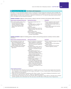
Original articles
© The Intensive Care Society 2013
Individualising management of severe
respiratory failure and the specialist
commissioned severe respiratory failure
service for England
N Barrett, L Camporota, C Langrish, G Glover, R Beale
Despite the improvement in the survival rate in patients with acute respiratory distress syndrome, there is a cohort of
patients with severe hypoxaemia and hypercapnia who offer a significant therapeutic challenge and may require some of
the more contentious rescue therapies, including prone positioning, high-frequency oscillatory ventilation and
extracorporeal support. It is essential to implement a protocolised pathway for diagnosis and individualised treatment for
these patients. In 2011, the English National Specialist Commissioning Service established a number of severe respiratory
centres for England including the provision of extracorporeal membrane oxygenation. Early referral is essential for the
successful use of rescue therapy as the evidence indicates that the time of mechanical ventilation prior to rescue therapy
is a key predictor of mortality. Guy’s and St Thomas’ NHS Foundation Trust has been commissioned as one of the severe
respiratory failure services and we describe the process of assessment and management that we have instituted to
manage patients with severe respiratory failure.
Keywords: adult ARDS; mechanical ventilation; lung injury; recruitment; extracorporeal membrane oxygenation
Introduction
The current operative definition of acute respiratory distress
syndrome (ARDS) has been well established for the last 15
years.1 Over this time, significant research into the mechanisms
and management of patients with ARDS has led to a gradual
improvement in patient mortality from 90% in the 1970s, to
40% in the mid-late 1990s, down to 25-30% in the most recent
trials.2-4 Besides the general improvement in the standard of
care provided in the intensive care units (ICUs), the only
specific management that has consistently been shown to
reduce mortality in ARDS is the provision of mechanical
ventilation with static inspiratory pressures (plateau pressure)
of less than 30 cm H2O and low tidal volumes normalised to
predicted body weight (PBW) according to a concept known as
‘lung-protective ventilation’ (LPV). This strategy has been
shown to reduce ventilator-induced lung injury (VILI), and is
linked to improvements in short- and long-term outcomes for
the majority of patients.5
Despite this global improvement in survival, there is a
cohort of patients with severe hypoxaemia (PaO2/FiO2 ratio
<13.3 kPa) and hypercapnia (leading to a pH <7.20) who offer
a significant therapeutic challenge. This group of patients, even
when managed with optimal recruitment and LPV, have a
significantly higher mortality than patients with a higher
PaO2/FiO2 ratio or lower PaCO2 for a given minute
114
ventilation.6-8 The identification of a subgroup of patients with
‘severe’ ARDS, has been made more explicit in the new ‘Berlin
definition of ARDS’,9-11 allowing for targeted therapeutic
interventions and further clinical studies.
Furthermore, this particular cohort of patients may be the
group who benefit from some of the more contentious rescue
therapies, including prone positioning, steroids, high-frequency
oscillatory ventilation (HFOV) and extracorporeal support –
such as extracorporeal CO2 removal (ECCO2R) and
extracorporeal membrane oxygenation (ECMO).
A model for severe respiratory failure
management
ARDS is the end result of a series of pathophysiological
processes caused by a wide variety of triggering conditions.
The pathological hallmarks are diffuse alveolar damage and
alveolar-capillary junction dysfunction causing initial egress of
fluid into the alveoli (exudative phase), followed by a
proliferative phase then either recovery or progressive
fibrosis.12 The clinical sequelae consist of significant
hypoxaemia, hypercapnia, poor respiratory system compliance
and progressive alveolar infiltration on chest imaging, that are
not exclusively due to cardiogenic pulmonary oedema. Despite
the apparent pathophysiological similarities, ARDS is
essentially the final common pathway of a wide range of
Volume 14, Number 2, April 2013 JICS
Original articles
Computed tomography
Diagnostic volume scan with
contrast recruitment CT scan
Diagnosis
ECMO HFOV LPV &
ECCO2R
LPV & LPV
prone
Investigations
Vasculitic screen
Autoimmune screen
HIV test
CMV and HSV viral load
Urinary pneumococcal
and Legionella antigens
Bacterial pneumonia
16
2
4
Viral pneumonia
1
1
Asthma
1
3
Aspiration pneumonitis
1
1
1
Mycobacterial infection
1
Interstitial lung disease
1
1
1
1
Malignancy
1
1
Primary cardiac disease 1
1
Broncho-alveolar lavage
Cardiac investigations
Bacterial culture
Respiratory virus PCR panel
CMV/HSV inclusions
Fungal culture
TB screen and culture
Screening transthoracic
echocardiogram
Additional echocardiography or
coronary angiography as appropriate
Table 1 Initial routine investigations performed. More targeted
investigations are requested based on history and initial
investigations.
known triggering conditions. Each triggering disease has its
own natural history, morbidity and mortality and requirement
of specific treatments beyond the common ground of the
supportive therapies. An acute viral pneumonitis, such as seen
in the 2009-2011 influenza A (H1N1) pandemics is a different
disease process to a bacterial pneumonia, to an aspiration
pneumonitis, to acute pancreatitis, to transfusion-associated
lung injury, and so on.13 Furthermore, each disease has a
slightly different expression in each patient, almost certainly
due to the host response having a causative role in the
sequelae of ARDS. When considering severe respiratory
failure however it is important to remember that, although
patients may present with refractory hypoxaemia, poor
respiratory system compliance and progressive alveolar
infiltrates that are potentially consistent with severe ARDS, the
underlying diagnosis can at times be pathologically
inconsistent with ARDS (eg lymphangitis carcinomatosa,
primary pulmonary Kaposi’s sarcoma and non-specific
interstitial pneumonitis are recent cases managed at our
institution). These conditions need to be diagnosed and
specific management plans instituted.
Although a conventional, evidence-based management
strategy which limits tidal volume, plateau pressures and fluid
administration, and aims to optimise PEEP have been
demonstrated to impact upon the progression of the lung
injury,8,14-19 the best course of management for patients with
refractory and life-threatening hypoxaemia or hypercapnia
despite LPV remains unclear. The options include the
administration of nitric oxide, LPV in prone position, HFOV,
and LPV with ECCO2R or ECMO. Although these modalities
have a well-demonstrated benefit on improving gas exchange,
the data supporting their benefit on mortality outcomes are
still limited and often conflicting.20 Each treatment has a
different risk profile, degree of invasiveness and resource
utilisation. In addition, the level of expertise and training
required dictate and limit their availability. There is also no
evidence supporting the use of a particular ‘rescue’ therapy
JICS Volume 14, Number 2, April 2013
12
1
1
1
Table 2 Breakdown of diagnoses by form of rescue support. Five
patients had more than one support, all eventually requiring
ECMO.
over any other for an unselected patient population. Given the
heterogeneous population of patients with severe respiratory
failure, how does one choose which therapy will best suit an
individual patient?
At Guy’s and St Thomas’ Hospitals (GSTT) we have
developed a pathway for the management of severe respiratory
failure (Figure 1). The key elements of the pathway are
protocolised diagnostic investigations, optimisation of LPV and
haemodynamics, and a graded approach to therapy, based
around the complexity of the intervention, likelihood of
response to that intervention and the individual patient’s risk
profile suggested by their co-morbidities.
The GSTT severe respiratory failure pathway —
diagnosis
The diagnosis of the triggering disease in patients presenting
with severe respiratory failure can be relatively straightforward
and based upon the presenting history, physical examination
and the initial investigations. However, it is not always so. We
use a multimodal diagnostic pathway incorporating radiology,
broncho-alveolar lavage (BAL) and serology to attempt to
delineate the underlying diagnosis (Table 1). All patients with
severe respiratory failure routinely have a high resolution chest
CT as early as possible in their admission, usually as they are
being retrieved into our institution from referring centres. The
CT is subsequently considered at our respiratory radiology
multi-disciplinary meeting, along with any previous
radiological investigations that are available. There is early and
sustained
microbiology,
virology
and
rheumatology
involvement in the management and investigation of patients.
All patients also undergo an early screening trans-thoracic
echocardiogram (TTE) and when necessary more detailed
cardiac investigation. Routine TTE has significant advantages
in this population, giving guidance on filling status with
inferior vena caval compressibility and the appearances of the
left ventricle as well as assisting in the diagnosis of valvular
disease, ventricular failure and the presence of intracardiac
shunts. Bronchoscopy is routinely used for this patient cohort.
Although it may introduce temporary physiological instability,
it can allow both investigations (cultures and cytology from
broncho-alveolar lavage) and management (the identification
and removal of inspissated secretions) to be addressed. This
115
Original articles
GSTT ICU Severe Respiratory Failure Protocol
Basic Sepsis Management
Basic ARDS Management
Identify cause
Source identification/treatment
Antibiotic treatment as per departmental guidelines
Adequate oxygenation/ventilation
Treatment MODS
Lung protective ventilation
Lung oedema reduction
Optimise ventilation and haemodynamic management
Optimise haemodynamics
Optimise ventilator settings
PEEP — optimise, provided
Vt < 6mL/kg(IBW) AND
PPlat >30 cm H2O
I:E ratio 1:1
RR 20-30 breaths/min
Drain pnemothorax
Measure IAP and optimise (<20)
ScvO2 >65%
Lactate <2
Hb >8
ITBVI 850-1000 mL/m2
MAP >65 mm Hg
EVLWI <15
AND
No
Assess fluid
responsiveness
Yes
Negative
fluid balance
Assess
PaO2/FiO2 <10 kPa
or
PaO2/FiO2 >10 kPa and Pplat >30 cm H2O
or
PaO2/FiO2 >10 kPa and pH <7.20 (respiratory)
No
Adjust ventilator settings
to achieve targets
Consider recruitment
manoeuvre
Yes
HFOV
ECMO fast track
Recruitment manoeuvre
Bias flow 40L/min
Initial frequency 6Hz
CDP <30 cm H2O
PaO2/FiO2 <10kPa and
Significant barotrauma
or HFOV precluded
Or
Reassess after 12-24 hours
PaO2/FiO2 <7 kPa then ECMO
At 48-72 hours
PaO2/FiO2 >10 kPa
CDP <30 cm H2O
pH >7.20 (freq >6Hz)
PaO2/FiO2 >38%
Continue
HFOV
PaO2/FiO2 <20 kPa
and
pH <7.20 (respiratory)
(freq >6Hz)
HFOV
+
pECLA
PaO2/FiO2 <10 kPa
or PaO2/FiO2 10-20 kPa and pH <7.20 (respiratory)
or PaO2/FiO2 <38%
or PaO2/FiO2 >10 and CDP >30 cm H2O
ECMO
Figure 1 The GSTT severe respiratory failure protocol. ΔPaO2/FiO2 is the change in PaO2/FiO2 ratio after a recruitment manoeuvre
compared with that prior to the first recriutment manoeuvre on HFOV, expressed as a percentage change. ARDS=Acute respiratory
distress syndrome; MODS=Multiple organ dysfunction syndrome; Vt=Tidal volume; Pplat=Plateau pressure; IBW=Ideal body weight;
RR=Respiratory rate; IAP=Intra-abdominal pressure; I:E=Inspiratory to expiratory ratio; ScvO2=Central venous oxygen saturation;
Hb=Haemoglobin; ITBVI=Intra-thoracic blood volume index; MAP=Mean arterial pressure; EVLWI=Extra-vascular lung water index;
PaO2/FiO2=Partial pressure oxygen to fraction of inspired oxygen ratio; HFOV=High frequency oscillatory ventilation; CDP=Continuous
distending pressure; ECMO=Extracorporeal membrane oxygenation; PECLA=Pumpless extracorporeal lung assist; ΔPaO2/FiO2=Change in
partial pressure of oxygen to fraction of inspired oxygen ratio compared with baseline; PEEP=Positive end expiratory pressure.
approach has allowed for the early diagnosis and appropriate
treatment of a number of patients with significant underlying
disease processes that were not recognisable from the
116
presenting history (Table 2).
Of the last 50 patients with severe respiratory failure
admitted to our service, all presented with features consistent
Volume 14, Number 2, April 2013 JICS
Original articles
with acute bacterial or viral lung infections or with a history
suggestive of aspiration pneumonitis and although, in the
majority of cases, the initial diagnosis was confirmed by the
described protocolised approach, in some cases alternative
diagnoses have been found (malignancy, interstitial lung
disease and primary cardiac pathology) which required specific
interventions and were not compatible with the denomination
of ARDS.
The GSTT severe respiratory failure pathway –
management
The aim of our severe respiratory failure pathway is firstly to
optimise conventional ventilation using the established
evidence base then attempt to offer the optimal rescue therapy
(Figure 1). Choosing which form of support to use can be a
very difficult decision.
The first step is to assess patient recruitability, in other words
the possibility of obtaining improvement in gas exchange and
lung mechanics following a ‘high pressure’ strategy as a
consequence of increasing the proportion of aerated lung.
Patients only undergo recruitment assessment strategies if there
is demonstrable hypoxaemia on the current ventilatory regime
and rescue strategies are being considered. Although the benefit
of a recruitment manoeuvre remains unclear and may entail risk
of both hypotension and acute desaturation,21 given the relative
risks of rescue therapies, we find that this can assist in
identifying patients who will be able to be managed with less
aggressive therapies. Recruitability is also assessed using
changes in lung mechanics in response to recruitment
manoeuvres (RM) modelled on the protocol by Gattinoni et al,7
which involves a lung CT scan at two levels of PEEP 5 and
45 cm H2O. This is only performed if, in the judgement of the
consultant managing the patient, the patient is stable enough to
tolerate the manoeuvre. Anecdotally we have found that there is
a correlation with patients who recruit well on CT and
subsequently recruit well on HFOV and we have been using this
while considering our initial rescue strategy. More recently, we
have been measuring end-expiratory lung volume at two PEEP
levels pre- and post-RM using the nitrogen wash-out/wash-in
technique to quantify lung recruitability, and electrical
impedance tomography (EIT) to assess at the bedside the
regional distribution of ventilation and change in regional
compliance post RM. If patients are demonstrated to be
‘recruitable’ then patients may persist with either conventional
LPV with higher PEEP or HFOV. If patients with refractory
hypoxaemia are ‘non-recruitable’ they are fast-tracked to ECMO.
If patients are ‘recruitable’, we measure trans-pulmonary
pressure using oesophageal manometry to titrate plateau
pressure and PEEP to ensure lung protection with an open
lung strategy. If the inspiratory plateau trans-pulmonary
pressures are >25 cm H2O, patients are treated with either
HFOV or consider LPV with extracorporeal CO2 removal
(ECCO2R) to further reduce tidal volumes until the transpulmonary inspiratory pressure is <25 cm H2O.22 If the patient
responds well to HFOV (ie, relative increase in PaO2/FiO2 ratio
by more than 38%) then they are predicted from our analysis
of 102 cases to respond to HFOV and they remain with this
support.23 If however the PaO2/FiO2 and PaCO2 have not
JICS Volume 14, Number 2, April 2013
improved we convert them to ECMO. A small number of
patients do improve their oxygenation sufficiently on HFOV or
LPV but have significant refractory hypercapnia. In this group
we do arterio-venous ECCO2R for these patients in
combination with HFOV or LPV. The optimal role of ECCO2R
is however currently unclear and although it may be of benefit
to use it earlier as an addition to conventional ventilation,
further studies are required to support this.
Prone positioning is not a clear step within our guidelines.
Prone positioning has had a number of studies over the years,
many of which have demonstrated significant physiological
benefit. A recent meta-analysis by Gattinoni et al has
demonstrated a mortality benefit associated with prone
positioning in severe respiratory failure.24 We also use
conventional ventilation or HFOV in prone position in those
patients with severe ARDS and with lung recruitability, but
large regional inhomogeneities as suggested by CT or EIT,
especially with a significant basal distribution of disease. If
there is limited or no improvement, we convert to other forms
of support within 24-48 hours.
Neither inhaled nitric oxide, nor inhaled prostacyclin are
steps within our guideline due to the evidence for
physiological improvement without mortality benefit
associated with these approaches.25,26 We do however use nitric
oxide in cases where there is pulmonary hypertension and
right ventricular failure after the correction of hypoxaemia and
hypercapnoea. Whether this approach confers a mortality
benefit for patients is unknown.
Using this approach in the period December 2011-March
2012 we have admitted 26 patients with severe respiratory
failure. Ten patients underwent ECMO, nine HFOV, three
Novalung and four patients had conventional ventilation in the
prone position. Overall 20/26 survived (77%), 8/10 ECMO, 7/9
HFOV, 1/3 Novalung and 4/4 conventional. However, of the
deaths, all had uncorrectable underlying disease such as
metastatic malignancy, non-specific interstitial pneumonitis or
severe chronic obstructive pulmonary disease. These
comorbidities or their severity were not known at the time of
referral to our service. Although these are small numbers and it
is difficult to make meaningful comparisons, the mortality in
the published literature for patients with severe respiratory
failure is 40-60% in large series.
Although some of our approach is based on the available
evidence, the pathway in its entirety is not. However, there are
relatively few trials examining specific therapies and even fewer
comparing the relative merits of one form of support over
another. Furthermore, given the complex nature of the patients
and the wide range of comorbidities and aetiologies of the
respiratory failure, each patient presenting with severe
respiratory failure really needs tailored management to optimise
the benefit and minimise the risks of therapy. This in turn may
allow improvement in survival. Over time our approach will be
further refined as more evidence becomes available. In
particular techniques such as EIT have the potential to improve
our ability to predict patients who will respond to recruitment
or prone positioning. We also need to understand how the
initial supportive therapy interacts with medium- and long-term
outcomes for survivors. The evidence from patients managed
117
Original articles
with either ECMO or LPV is that there is a medium- and longterm improvement in respiratory function with persisting
neuromuscular weakness and psychological problems lasting
out to at least five years.27 Clearly long-term observational
studies are required to assess any sustained difference in
outcome between patients managed with different rescue
therapies and this in turn needs to be considered when selecting
supportive therapy during the acute illness.
The Severe Respiratory Failure Service for England
The National Specialist Commissioning Service in the UK held
a tender process over 2011 to establish a number of severe
respiratory centres for England. The results of that process
were announced in December 2011 with five centres being
commissioned to provide management for patients with severe
respiratory failure – Glenfield Hospital in Leicester,
Wythenshawe Hospital in Manchester, Papworth Hospital in
Cambridge, the Royal Brompton Hospital and Guy’s and St
Thomas’ Hospitals in London (Figure 2). This aims to give the
UK a flexible and distributed tertiary capacity for patients with
severe respiratory failure with the provision of ECMO and
retrieval services included.
What is the rationale for this approach? Firstly the decision
to have a small number of centres with higher volumes is based
on the available evidence, where there has been an association
between improved outcomes and volume of patients in a
diverse range of fields – cardiology, oncology, cardiac surgery
and indeed in the outcomes from mechanical ventilation.28-31
Secondly, a key component, the ability to provide ECMO, is
particularly resource intensive. In part it requires significant
capital to set up and maintain a programme, but mainly it is
very heavily dependent upon having appropriate numbers of
trained staff – nursing, perfusion, medical, physiotherapy and
dietetics as well as the need for on-site back-up services
including vascular and cardiothoracic surgery. Hence it is
difficult to set up and maintain ECMO without a substantial
investment in staffing and education in order to manage
patients and potential complications.
The referral process for the network is straightforward. All
regions in England have a nominated centre to which they
refer. The referral criteria are patients with severe respiratory
failure that is believed to be reversible, with a Murray score of
3 or more,32 with seven days or less of a plateau pressure
greater than 30 cm H2O and a FiO2 of greater than 0.8. Early
referral is essential for the successful use of rescue therapy as
the evidence indicates that the time of mechanical ventilation
prior to rescue therapy is a key predictor of mortality.23,33
This is thought to relate to the progression of ventilatorinduced lung injury. Patients accepted into the service will
be retrieved by the accepting centre either by using a
conventional approach or using mobile ECMO. It is the
centre’s responsibility to find a bed in the event that they have
insufficient resources.
Retrieval of patients with severe respiratory failure is well
established internationally with retrieval services in Australia,
the US and Europe routinely transporting and managing
patients on ECMO.34-37 Depending on the country involved
these are either institution-dependent or centrally organised.
118
This map represents CCNs to whom a unit provides advice and has an
ongoing relationship. Referrals from CCN go via the allocated unit
English commissioned service, available to Scottish, Welsh and Northern Irish patients
Adult Respiratory ECMO providers
Guy’s and St Thomas’ Hospitals NHS Foundation Trust
Papworth Hospital NHS Foundation Trust
Royal Brompton and Harefield NHS Foundation Trust
University Hospitals of Leicester NHS Trust
University Hospital of South Manchester NHS Foundation Trust
ECMO centre
Figure 2 The regional coverage of England by the National
Severe Respiratory Failure Service.
The UK has for many years had an excellent, though regional,
specialist primary retrieval network able to respond to trauma,
search and rescue and major incidents. Inter-ICU transfers
have been performed in the UK on an ad-hoc basis, often
utilising trainees with an on-demand ambulance organised by
the referring hospital. In the case of severe respiratory failure
referrals all retrievals are performed by a consultant or senior
trainee with significant experience in the management of
patients with severe respiratory failure and have to meet
national standards of governance and audit. Such a system has
been demonstrated to be safe for patients. CESAR
demonstrated a 2.2% transport-related mortality.38 In our own
series of cases we have successfully undertaken inter-hospital
transport for 56 patients with severe respiratory failure over the
last 18 months and had suffered no transport-related mortality.
Horizon scanning
There are a number of research and technological advances
that will be impacting the management of severe respiratory
failure in the next few years. Firstly, two major HFOV studies
(OSCAR and OSCILLATE) will report their findings. Secondly,
there are a number of new extracorporeal CO2 removal devices
that will be available, including A-LungTM, Novalung ActivveTM
and HaemodecTM. Although each offers different technical
solutions, all use a veno-venous approach to provide CO2
clearance with a relatively low blood flow. At present there is
little robust data to demonstrate that this technology improves
patient outcomes, although there are case series and registry
data sets demonstrating physiological improvements. It is
intuitively appealing that these devices may well assist in the
management of hypercapnic respiratory failure and may
provide some benefit in the management of ventilated patients
with severe respiratory failure.
Declaration of interests
The authors have no personal financial interests to disclose.
References
1. Bernard GR, Artigas A, Brigham KL et al. The American-European
Consensus Conference on ARDS. Definitions, mechanisms, relevant
Volume 14, Number 2, April 2013 JICS
Original articles
outcomes, and clinical trial coordination. Am J Respir Crit Care Med 1994;
149:818-24.
2. Erickson SE, Martin GS, Davis JL et al. Recent trends in acute lung
injury mortality: 1996-2005. Crit Care Med 2009;37:1574-79.
3. Rice TW, Wheeler AP, Thompson BT et al. Enteral omega-3 fatty acid,
gamma-linolenic acid, and antioxidant supplementation in acute lung
injury. JAMA 2011;306:1574-81.
4. Rice TW, Wheeler AP, Thompson BT et al. Initial trophic vs full enteral
feeding in patients with acute lung injury: the EDEN randomized trial.
JAMA 2012;307:795-803.
5. Moloney ED, Griffiths MJ. Protective ventilation of patients with acute
respiratory distress syndrome. Br J Anaesth 2004;92:261-70.
6. Britos M, Smoot E, Liu KD et al. The value of positive end-expiratory
pressure and Fio(2) criteria in the definition of the acute respiratory
distress syndrome. Crit Care Med 2011;39:2025-30.
7. Gattinoni L, Caironi P, Cressoni M et al. Lung recruitment in patients
with the acute respiratory distress syndrome. N Engl J Med 2006;354:
1775-86.
8. Nuckton TJ, Alonso JA, Kallet RH et al. Pulmonary dead-space fraction
as a risk factor for death in the acute respiratory distress syndrome. N
Engl J Med 2002;346:1281-86.
9. Ranieri VM, Rubenfeld GD, Thompson BT et al. Acute respiratory
distress syndrome: the Berlin Definition. JAMA 2012;307:2526-33.
10.Ferguson ND, Fan E, Camporota L et al. The Berlin definition of ARDS:
an expanded rationale, justification, and supplementary material. Intensive
Care Med 2012;38:1573-82.
11.Camporota L, Ranieri VM. What's new in the ‘Berlin’ definition of acute
respiratory distress syndrome? Minerva Anestesiol 2012;78:1162-66.
12.Ware LB, Matthay MA. The acute respiratory distress syndrome. N Engl J
Med 2000;342:1334-49.
13.Nehra D, Goldstein AM, Doody DP et al. Extracorporeal membrane
oxygenation for nonneonatal acute respiratory failure: the Massachusetts
General Hospital experience from 1990 to 2008. Arch Surg 2009;144:42732; discussion 432.
14.Briel M, Meade M, Mercat A et al. Higher vs lower positive endexpiratory pressure in patients with acute lung injury and acute
respiratory distress syndrome: systematic review and meta-analysis. JAMA
2010;303:865-73.
15.Hager DN, Krishnan JA, Hayden DL, Brower RG. Tidal volume
reduction in patients with acute lung injury when plateau pressures are
not high. Am J Respir Crit Care Med 2005;172:1241-45.
16.Sakr Y, Vincent JL, Reinhart K et al. High tidal volume and positive fluid
balance are associated with worse outcome in acute lung injury. Chest
2005;128:3098-108.
17.Thompson BT, Bernard GR. ARDS Network (NHLBI) studies: successes
and challenges in ARDS clinical research. Crit Care Clin 2011;27:459-68.
18.Wiedemann HP, Wheeler AP, Bernard GR et al. Comparison of two fluidmanagement strategies in acute lung injury. N Engl J Med 2006;354:
2564-75.
19.Mercat A, Richard JC, Vielle B et al. Positive end-expiratory pressure
setting in adults with acute lung injury and acute respiratory distress
syndrome: a randomized controlled trial. JAMA 2008;299:646-55.
20.Patroniti N, Bellani G, Pesenti A. Nonconventional support of
respiration. Curr Opin Crit Care 2011;17:527-32.
21.Fan E, Checkley W, Stewart TE et al. Complications from recruitment
maneuvers in patients with acute lung injury: secondary analysis from the
lung open ventilation study. Respir Care 2012;57:1842-49.
22.Grasso S, Terragni P, Birocco A et al. ECMO criteria for influenza A
(H1N1)-associated ARDS: role of transpulmonary pressure. Intensive Care
Med 2012;38:395-403.
23.Camporota L, Sherry T, Smith J et al. High-frequency oscillatory
ventilation for ARDS in adults: a cohort study. Intensive Care Med
2005;31(Supplement 1):A004 - S005-S013.
24.Gattinoni L, Carlesso E, Taccone P et al. Prone positioning improves
survival in severe ARDS: a pathophysiologic review and individual patient
meta-analysis. Minerva Anestesiol 2010;76:448-54.
JICS Volume 14, Number 2, April 2013
25.Adhikari NK, Burns KE, Friedrich JO et al. Effect of nitric oxide on
oxygenation and mortality in acute lung injury: systematic review and
meta-analysis. BMJ 2007;334:779.
26.Abraham E, Baughman R, Fletcher E et al. Liposomal prostaglandin E1
(TLC C-53) in acute respiratory distress syndrome: a controlled,
randomized, double-blind, multicenter clinical trial. TLC C-53 ARDS
Study Group. Crit Care Med 1999;27:1478-85.
27.Herridge MS, Tansey CM, Matte A et al. Functional disability 5 years
after acute respiratory distress syndrome. N Engl J Med. 2011;364:
1293-304.
28.Kahn JM, Goss CH, Heagerty PJ et al. Hospital volume and the outcomes
of mechanical ventilation. N Engl J Med 2006;355:41-50.
29.Pasquali SK, Li JS, Burstein DS et al. Association of center volume with
mortality and complications in pediatric heart surgery. Pediatrics 2012;
129:e370-376.
30.von Meyenfeldt EM, Gooiker GA, van Gijn W et al. The relationship
between volume or surgeon specialty and outcome in the surgical
treatment of lung cancer: a systematic review and meta-analysis. J Thorac
Oncol 2012;1170-78.
31.Zevin B, Aggarwal R, Grantcharov TP. Volume-outcome association in
bariatric surgery: A systematic review. Ann Surg 2012;256:60-71
32.Murray JF Matthay MA Luce JM et al. An expanded definition of the
adult respiratory distress syndrome. Am Rev Respir Dis 1988;138:720-23.
33.Brogan TV, Thiagarajan RR, Rycus PT et al. Extracorporeal membrane
oxygenation in adults with severe respiratory failure: a multi-center
database. Intensive Care Med 2009;35:2105-14.
34.Ciapetti M, Cianchi G, Zagli G et al. Feasibility of inter-hospital
transportation using extra-corporeal membrane oxygenation (ECMO)
support of patients affected by severe swine-flu(H1N1)-related ARDS.
Scand J Trauma Resusc Emerg Med 2011;19:32.
35.Forrest P, Ratchford J, Burns B et al. Retrieval of critically ill adults using
extracorporeal membrane oxygenation: an Australian experience.
Intensive Care Med 2011;37:824-30.
36.Isgro S, Patroniti N, Bombino M et al. Extracorporeal membrane
oxygenation for interhospital transfer of severe acute respiratory distress
syndrome patients: 5-year experience. Int J Artif Organs 2011;34:
1052-1060.
37.Philipp A, Arlt M, Amann M et al. First experience with the ultra
compact mobile extracorporeal membrane oxygenation system
Cardiohelp in interhospital transport. Interact Cardiovasc Thorac Surg
2011;12:978-81.
38.Peek GJ, Mugford M, Tiruvoipati R et al. Efficacy and economic
assessment of conventional ventilatory support versus extracorporeal
membrane oxygenation for severe adult respiratory failure (CESAR): a
multicentre randomised controlled trial. Lancet 2009;374:1351-63.
Nicholas Barrett Consultant in Critical Care
[email protected]
Luigi Camporota Consultant in Critical Care
[email protected]
Chris Langrish Consultant in Critical Care
[email protected]
Guy Glover Consultant in Critical Care
[email protected]
Richard Beale Consultant in Critical Care
[email protected]
Department of Adult Critical Care, Guy’s and St Thomas’ NHS
Foundation Trust, King’s Health Partners
119


© Copyright 2026