
PDF hosted at the Radboud Repository of the Radboud University
PDF hosted at the Radboud Repository of the Radboud University Nijmegen The following full text is a publisher's version. For additional information about this publication click this link. http://hdl.handle.net/2066/100586 Please be advised that this information was generated on 2014-12-22 and may be subject to change. Genetic and clinical aspects of chronic pancreatitis Aura van Esch ISBN/EAN 978-94-6191-433-0 Cover design and lay-out: In Zicht Grafisch Ontwerp, Arnhem Printed by: Ipskamp Drukkers, Enschede Financial support by Tramedico BV, Ferring BV, Dr. Falk Pharma Benelux BV, Olympus Nederland BV, Vifor Pharma Nederland BV and Abbott BV for publication of this thesis is gratefully acknowledged. © A.A.J. van Esch All rights reserved. No part of this publication may be reproduced or transmitted in any form or by any means, electronic or mechanical, by print or otherwise, without permission in writing from the author Genetic and clinical aspects of chronic pancreatitis Proefschrift ter verkrijging van de graad van doctor aan de Radboud Universiteit Nijmegen op gezag van de rector magnificus prof. mr. S.C.J.J. Kortmann, volgens besluit van het college van decanen in het openbaar te verdedigen op woensdag 31 oktober 2012 om 13.30 uur precies door Aura Adriana Josepha van Esch Geboren op 5 juni 1975 te Nijmegen Promotor Prof. dr. J.P.H. Drenth Manuscriptcommissie Prof. dr. J. de Graaf (voorzitter) Prof. dr. J.H.J.M. van Krieken Dr. M.G. Besselink, Academisch Medisch Centrum, Amsterdam Contents Chapter 1General introduction and outline of this thesis 1a: Problem statement and purpose of the studies underlying this thesis 1b: Introduction in chronic pancreatitis as a clinical entity Part I 9 11 14 Genetic aspects of pain in chronic pancreatitis Chapter 2Catechol-O-methyltransferase (COMT) gene variants and pain in chronic pancreatitis 25 Chapter 3Polymorphisms in gene encoding TRPV1-receptor involved in pain perception are unrelated to chronic pancreatitis 39 Part II Clinical aspects of chronic pancreatitis Chapter 4Specific radiological imaging findings in patients with hereditary pancreatitis during a long follow-up of disease 55 Chapter 5Comparative analysis of 1777 chronic pancreatitis patients reveals important differences between India and the Western world 71 Chapter 6Recurrent idiopathic pancreatitis in familial adenomatous polyposis: report of a case-series and review of the literature Part III 87 Diagnosis and treatment of chronic pancreatitis Chapter 7A wide variation in diagnostic and therapeutic strategies in chronic pancreatitis: A Dutch national survey 103 Chapter 8 Pharmacological management of pain in chronic pancreatitis 125 Chapter 9 Summary and future perspectives 145 AppendixComparison of intragastric versus intrajejunal feeding in chronic pancreatitis; effect on pain and abdominal symptoms 163 Nederlandse samenvatting 175 Dankwoord 191 Curriculum Vitae 195 Thesis series of the Institute for Genetic and Metabolic Disease 197 1 General introduction and outline of this thesis general introduction and outline of this thesis Background Chronic pancreatitis (CP) is characterized by an inflammatory process of the pancreas that leads to irreversible morphological changes. It represents a progressive fibroinflammatory disease that can be categorized in a large-duct (often with intraductal calculi) or small-duct form. CP is considered to be a difficult topic for research for the reason that it is a multifaceted disease with respect to cause and clinical presentation. It is associated with a large number of risk factors (ranging from environmental to genetic) and the clinical presentation varies from asymptomatic to a highly complicated disease course. Though this variation is not unique to CP, the relatively low prevalence of the disease and its association with alcohol as a major precipitating factor has left CP as a model for research in the limelight. However, the chief presentation requiring medical attention by CP patients is chronic pain. We took this symptom as the guiding theme of our studies and asked ourselves a number of questions. Questions relevant to this thesis: 1. Why do some CP patients develop pain while others are left unscathed? 2. What is the clinical presentation of a subset of patients with CP, such as patients with hereditary or idiopathic forms of CP? 3. How do we need to treat these patients? Disease model In order to address these issues we chose to have patients with CP as a disease model. As we have a large outpatient clinic where we see these patients on a regular basis this came to us as a natural choice. We designed a clinical database that contained a large number of clinical variables of our CP patients and took that as an entry point. This enabled us to design a number of genetic case control studies that examined our first question. Next, we established a number of case series that allowed us to delineate specific elements of CP as a disease entity. Lastly, we went back to the literature in order to examine the issue how we should treat our patients and checked whether we as clinicians adhere to the existing guidelines. These endeavors led to the following structure of this thesis. 11 1 chapter 1 Outline of this thesis This thesis focuses on three different aspects of CP. In the first section, we focus on genetic aspects of CP. We questioned ourselves if we could detect genetic alterations that could explain the differences in the experience of pain in CP. In the second section, we studied clinical aspects of CP. We studied the phenotype and imaging findings in a specific group of CP in order to learn more about etiology and disease course. In the third section, we focus on diagnosis and management of CP. Since guidelines on CP are scarce, we wanted to know more about current practice in the diagnosis and management of CP in the Netherlands? Furthermore, we reviewed the management of pain in CP. Question 1: Why do some CP patients develop pain while others are left unscathed? In the first section, we focus on genetic aspects of pain in CP. Since genetic factors may play a role in a patient’s pain experience, we investigated if the Catechol-O-methyltransferase (COMT) gene and the transient receptor potential vanilloid receptor 1 (TRPV1) gene might be involved in pain in CP patients. The following research questions will be addressed: • What is the effect on COMT gene variants on the presence and severity of pain in CP? (Chapter 2) • Could modifications in the TRPV1 gene modify the presence and the phenotypical expression of CP? (Chapter 3) Question 2: What is the clinical presentation of a subset of patients with CP, such as patients with hereditary or idiopathic forms of CP? In the second section, we focus on clinical aspects of CP. In these chapters, we focus on different clinical aspects on specific types of pancreatitis: hereditary pancreatitis, idiopathic CP in the Western world and in India and pancreatitis in patients with familial adenomatous polyposis (FAP). In this specific subset of patients, we asked ourselves: • Are there specific radiological findings in patients with hereditary pancreatitis and what is the evolution of pancreatic abnormalities during the disease course? (Chapter 4) • Is the phenotype of CP and idiopathic CP in India different from CP and idiopathic CP in the Western world? (Chapter 5) • Is there a relationship between FAP en CP and may SPINK1 mutations contribute to the risk of pancreatitis? (Chapter 6) 12 general introduction and outline of this thesis Question 3: How do we need to treat these patients? In the third section of this thesis, we focus on diagnosis and treatment of CP. There are many controversies in the diagnosis and treatment of CP and pain in CP due to lack of evidence. Our objectives with regards to this theme were: • To evaluate current practice in the Netherlands by evaluating decisions regarding the diagnosis, management and screening in CP. (Chapter 7) • To discuss the suggested mechanisms for pain in CP and review the therapeutic options. (Chapter 8) In the appendix we focus on the difficulties we met in adopting a different study strategy, i.e. a randomized clinical trial. A randomized clinical trial is generally accepted as the most reliable evidence of whether a treatment is effective. Therefore, we designed a clinical trial on the effectiveness of nasogastric and nasojenunal feeding in CP patients with abdominal pain as a primary outcome measure. In the appendix we describe two patients who were included and focus on the results of the single patient who completed the entire protocol. The appendix highlights the reasons for difficult accrual of patients for this trial. 13 1 chapter 1 Chapter 1b. Introduction in chronic pancreatitis as a clinical entity This chapter describes the clinical aspects of CP and serves as an introduction to the studies described in this thesis. Incidence of CP The prevalence of CP is 26 per 100,000 in Europe and the USA.1 The reported incidence of CP in industrialized countries ranges from 3.5 to 10 per 100,000 population.2 The overall incidence is stable over three decades (1977–2006).3 In the USA, there is an increase in the incidence of alcoholic CP.4 In Japan, there is a gradual increase of the annual incidence (to 11.9/100,000), especially in alcoholic CP.5 There is a male predominance in alcoholic CP and idiopathic CP (>71%) but not in hereditary CP (46%).6 The median age at onset differs between etiology; 36 years in case of alcoholic CP, 10 years in case of hereditary CP. Idiopathic CP has an early onset or ‘juvenile’ form (median age at onset 23 years) and a late onset or ‘senile’ form (at 62 years).6,7 The mean age of onset of CP in Japan is 59.4 years.5 Etiology The predominant cause of CP in Western countries is excessive alcohol consumption, accounting for approximately 70% of all cases. However, since <10% of chronic alcoholics develop CP, other predisposing factors besides alcohol are involved.8 Thus, alcohol requires additional cofactors to result in CP.3 About 20% of cases is considered to relate to idiopathic pancreatitis, while the remaining 10% is categorized as ‘other’. New classification systems have been established, for instance the M-ANNHEIM classification system.9 This classification is based on the assumption that CP results from the interaction of multiple (M) risk factors. The risk factors are categorized into the major subcategories of alcohol consumption (A), nicotine consumption (N), nutritional factors (N), hereditary factors (H), efferent pancreatic duct factors (E), immunological factors (I), and various rare miscellaneous and metabolic (M) factors. Another classification is the TIGAR-O risk factor classifications system.10,11 The risk factors are categorized according to causes that have a toxic-metabolic, idiopathic, genetic, autoimmune, recurrent severe acute pancreatitis-associated and obstructive background. There is a clear relationship between cigarette smoking and pancreatitis. Smoking is an important independent risk factor for CP and influences progression of acute pancreatitis to CP.3 Genetic factors play an important role in the etiology of CP. There is a rare form of CP, hereditary pancreatitis, with an autosomal dominant inheritance caused by 14 general introduction and outline of this thesis cationic trypsinogen (PRSS1) gene mutations.12,13 Furthermore, genetic factors can explain a significant proportion of other CP cases. Genes like SPINK1, CTRC and CFTR are associated with CP and should be considered as predisposing or modifying rather than directly causative. SPINK1-mutations are associated with ~20% of idiopathic CP cases but only ~5% of the alcoholic CP cases. Mutations in the Chymotrypsine C-gene are seen in only 2% of CP cases.14 Clinical Features The three major clinical features of CP are: • Pain • Pancreatic exocrine insufficiency • Pancreatic endocrine insufficiency. Pain Pain is the major presenting symptom of CP and the majority of patients will have pain at a given time during the course of their disease. It is the most frequent reason for CP patients to consult their physician. Pain in CP can occur as attacks that mimic acute pancreatitis or as constant and disabling pain. In a recent survey in Japan, 60.6% of the CP patients experienced abdominal pain.5 In alcoholic CP patients pain was present more frequently (alcoholic 65.0% vs. nonalcoholic 53.0%).5 Often, the onset of CP is heralded by a severe painful attack, indistinguishable from an acute pancreatitis attack. After the first attack, patients become symptom free. However, with progression of the disease, the attack frequency increases and the symptom free periods progressively shorten. The inter- and intraindividual variation of pain in CP is high, with pain duration varying from intermittent to persistent, and pain intensity ranging from mild to disabling. The pain is usually epigastric in location (although more diffuse pain in the upper abdomen can occur) and may radiate to the left infrascapular region. The pain can be accompanied by nausea and vomiting and can be partially eased by sitting up and leaning forward or by application of local heat or other counterirritants to the dorsal spine or epigastrium. Amman and Muellhaupt distinguished two typical pain patterns in alcoholic CP.15 The type A pain pattern, typically observed in acute relapsing pancreatitis, is short-lived and pain episodes usually last less than 10 days and are separated by long pain-free intervals of several months to a year. It is predominant in late-onset idiopathic CP and hereditary CP. B-type pain pattern, seen in more than 50% of patients, is characterized by prolonged periods of persistent pain or clusters of recurrent severe pain exacerbations, lasting two or more days per week for at least two months, and requiring frequent hospitalizations. Type B pain predominates in alcoholic CP and is associated with local complications, often requiring surgery. 15 1 chapter 1 The pathogenesis of pancreatic pain is poorly understood and probably multi factorial. It can be a result from extrapancreatic (e.g. bile duct stenosis) or intra pancreatic complications (e.g. pseudocysts). Surgical or endoscopic complications can be involved. A few theories explaining the pathogenesis of pain in CP have been postulated: 1) the increased intrapancreatic pressure in the pancreatic duct or parenchyma; 2) inflammation of the pancreas; and 3) alterations in pancreatic nerves; pancreatic neuropathy.16 The neuropathic pain-hypothesis is supported by evidence from experimental human pain research that in many of these patients pain processing in the central nervous system is abnormal and mimics that seen in neuropathic pain disorders. Probably genetic factors also play a role in a patient’s pain experience, similar to the involvement of genes in neuropathic pain disorders. Pancreatic exocrine and endocrine insufficiency The other clinical features of CP include exocrine and endocrine insufficiency. The main clinical manifestation of pancreatic exocrine insufficiency is malnutrition, resulting in low circulating levels of micronutrients, fat soluble vitamins and lipoproteins, which have been related to a high morbidity and mortality secondary to an increased risk of malnutrition-related complications and cardiovascular events.17 Steatorrhea does not occur until pancreatic lipase secretion is reduced to less than 10% of normal. Diabetes mellitus may develop in the long-term course of the disease and is characterized by destruction of both insulin- and glucagon-producing cells. The diabetes is classified as type IIIc according to the American Diabetes Association.18 The overall prevalence of diabetes in CP is 47%.19 The incidence of diabetes increase to more to more than 80% 25 years after onset. 20 Diagnosis of CP The diagnosis of CP is based on a combination of clinical symptoms, pancreatic function tests and imaging. In the diagnosis of CP different imaging modalities are used; transabdominal ultrasonography (TUS), CT-scanning, MRI, MRCP, secretinenhanced-MRCP and endoscopic ultrasound (EUS). TUS is able to identify thinning of pancreatic parenchyma, irregularity of the pancreatic margins, dilatation of the MPD and of side branches and ductal calcified stones. Therefore it can be used to confirm the diagnoses advanced CP. 21 However, in diagnosis of CP TUS is not very useful. Early CP can best be diagnosed by gadolinium enhanced MR imaging combined with MRCP. 21 MRCP and increasingly EUS have become the screening methods of choice. 22 16 general introduction and outline of this thesis Treatment The treatment of CP consists of different aspects: • Treatment of pain • Correction of metabolic disorders (diabetes, malnutrition) • Treatment of pancreatic exocrine insufficiency • Management of complications (e.g. pseudocysts) • Behavior modification: cessation of alcohol consumption and cigarette smoking Treatment of pain The treatment of pain in CP still is difficult, despite new strategies, new drugs and new interventional options. The treatment of pain in CP requires a multidisciplinary approach of gastroenterologists, surgeons, radiologists, anesthesiologists and psychiatrists. As advised in the AGA guidelines, treatable complications of chronic pancreatitis, such as pseudocysts, bile duct obstruction or duodenal obstruction should be excluded. 23 The conservative management of CP included abstinence of alcohol and smoking. Alcohol abstinence improves the prognosis of CP and frequently results in a reduction of pain. The management of pain in CP include pharmacological, endoscopic and surgical treatment. In the treatment of pain, there are several pharmacological options, with different effectiveness. Pharmacological treatment of pain in CP In the most recent guidelines of diagnosis and treatment of CP, this pharmacological options are mentioned:21,22 • Analgesics: acetaminophen, non-steroidal anti-inflammatory drugs are recommended as firs approach in pain in CP, followed by opioids such as tramadol. • Antioxidant therapy (selenium, beta-carotene, ascorbic acid and tocopherol) may be useful to prevent painful recurrences of CP. • Tricyclic antidepressants, selective serotonin re-uptake inhibitors and combined serotonin and norepinephrine re-uptake inhibitors, will alleviate co-existent depression and may ameliorate pain and potentiate the effects of opiates. • Opioid receptor agonists: the ‘classic’ opioids such as morphine, methadone, fentanyl. Ideally, ongoing regular opioid analgesia is reserved for those in whom endoscopic or surgical therapies are not appropriate and symptoms are intractable. There are other pharmacological options in the management of pain in CP, such as octreotide, loxiglumide, secretine, oral protease inhibitors and leukotriene receptor antagonist, but the data of effectiveness are inconsistent and therefore cannot be routinely advised. A promising new drug in the treatment of pain is pregabalin, a gabapentoid effective in treating other causes of neuropathic pain, which has shown effective as adjuvant analgesic in pain control in patients with CP. 24 17 1 chapter 1 Other therapeutic options and neuropathic pain treatments include nerve blocks, transcutaneous electronic nerve stimulation, acupuncture and intrathecal pumps for infusion of opioids and anesthetic agents, the latter to block afferent pain nerves accompanying sympathetic nerves to the central nervous system. 25 Endoscopic therapy for pain in CP For treating painful uncomplicated CP, the ESGE recommends extracorporeal shockwave lithotripsy (ESWL)/endoscopic retrograde cholangiopancreatography (ERCP) as the first-line interventional option. 26 This is only indicated in case of pancreatic duct dilation.21 In case of no response, surgical options should be considered. ESWL is recommended in case of radiopaque stones ≥5 mm obstructing the main pancreatic duct (MPD), followed by endoscopic extraction of stone fragments. In case of dominant MPD strictures, the ESGE recommends inserting a single 10-Fr plastic stent, with stent exchange planned within one year. If the ductal strictures persist after 12 months, other therapeutic options such as endoscopic placement of multiple simultaneous MPD stents or surgery should be discussed. 26 Recently, two trials who compared endoscopic intervention to surgical intervention were reviewed. 27 This review showed that in patients with obstructive CP and dilated MPD surgery is superior to endoscopy in terms of pain control. In a prospective randomized trial with a long-term follow-up was shown that symptomatic patients with advanced CP who underwent surgery as the initial treatment for pancreatic duct obstruction had more relief from pain, with fewer procedures, than patients who were treated endoscopically. Moreover, almost half of the patients who were treated with endoscopy eventually underwent surgery. 28,29 Surgical therapy for pain in CP If endoscopic therapy fails in the treatment of CP, or if there are complications of CP such as bile duct and duodenal obstructions, surgical therapy is indicated. Ideally, pancreatic surgery should be performed before narcotic addiction. In case of CP with MPD dilation (≥ 7 mm) a drainage procedure should be chosen (lateral pancreaticojejunostomy procedure proposed by Partington and Rochelle).30 When there is an inflammatory mass, a pancreatic resection is indicated. When there is a head mass-forming CP, mixed surgery (drainage and limited resection) can be performed.21 Treatment of exocrine insufficiency A fat restriction should not longer be routinely advised, but frequent meals of low volume are still recommended.17 Adequate pancreatic enzyme therapy is required to avoid malnutrition-related complications such as osteoporosis. Clinical indications for initiating enzyme supplementation are steatorrhea, weight loss or diarrhea. A minimum dose of pancreatic enzyme in the form of enteric-coated minimicro- 18 general introduction and outline of this thesis spheres of 20 000- 40 000 U of lipase per meal and 10 000-20 000 U of lipase with snacks is required.17 Inhibition of gastric acid secretion by use of proton pump inhibitors can be added if steatorrhea is not controlled by pancreatic enzyme suppletion.21 In case of persistent signs of maldigestion, the dose of pancreatic enzymes should be increased. Therapy with pancreatic enzymes is not effective as treatment for pain in CP. A meta-analysis showed no significant benefit of pancreatic enzyme therapy on the relief op CP associated pain.31 19 1 chapter 1 References (1) Braganza JM, Lee SH, McCloy RF, McMahon MJ. Chronic pancreatitis. Lancet 2011 Apr 2;377(9772): 1184-97. (2) Witt H, Apte MV, Keim V, Wilson JS. Chronic pancreatitis: challenges and advances in pathogenesis, genetics, diagnosis, and therapy. Gastroenterology 2007 Apr;132(4):1557-73. (3) DiMagno MJ, DiMagno EP. Chronic pancreatitis. Curr Opin Gastroenterol 2012 Jul 9. (4) Yadav D, Timmons L, Benson JT, Dierkhising RA, Chari ST. Incidence, prevalence, and survival of chronic pancreatitis: a population-based study. Am J Gastroenterol 2011 Dec;106(12):2192-9. (5) Hirota M, Shimosegawa T, Masamune A, Kikuta K, Kume K, Hamada S, et al. The sixth nationwide epidemiological survey of chronic pancreatitis in Japan. Pancreatology 2012 Mar;12(2):79-84. (6) Mullhaupt B, Truninger K, Ammann R. Impact of etiology on the painful early stage of chronic pancreatitis: a long-term prospective study. Z Gastroenterol 2005 Dec;43(12):1293-301. (7) Drenth JP, te Morsche R, Jansen JB. Mutations in serine protease inhibitor Kazal type 1 are strongly associated with chronic pancreatitis. Gut 2002 May;50(5):687-92. (8) Dufour MC, Adamson MD. The epidemiology of alcohol-induced pancreatitis. Pancreas 2003 Nov; 27(4):286-90. (9) Schneider A, Lohr JM, Singer MV. The M-ANNHEIM classification of chronic pancreatitis: introduction of a unifying classification system based on a review of previous classifications of the disease. J Gastroenterol 2007 Feb;42(2):101-19. (10) Etemad B, Whitcomb DC. Chronic pancreatitis: diagnosis, classification, and new genetic developments. Gastroenterology 2001 Feb;120(3):682-707. (11) Stevens T, Conwell DL, Zuccaro G. Pathogenesis of chronic pancreatitis: an evidence-based review of past theories and recent developments. Am J Gastroenterol 2004 Nov;99(11):2256-70. (12) Comfort MW, Steinberg AG. Pedigree of a family with hereditary chronic relapsing pancreatitis. Gastroenterology 1952 May;21(1):54-63. (13) Rebours V, Levy P, Ruszniewski P. An overview of hereditary pancreatitis. Dig Liver Dis 2012 Jan;44(1):8-15. (14) Derikx MH, Drenth JP. Genetic factors in chronic pancreatitis; implications for diagnosis, management and prognosis. Best Pract Res Clin Gastroenterol 2010 Jun;24(3):251-70. (15) Ammann RW, Muellhaupt B. The natural history of pain in alcoholic chronic pancreatitis. Gastroenterology 1999 May;116(5):1132-40. (16) Drewes AM, Krarup AL, Detlefsen S, Malmstrom ML, Dimcevski G, Funch-Jensen P. Pain in chronic pancreatitis: the role of neuropathic pain mechanisms. Gut 2008 Nov;57(11):1616-27. (17) Dominguez-Munoz JE. Pancreatic exocrine insufficiency: diagnosis and treatment. J Gastroenterol Hepatol 2011 Mar;26 Suppl 2:12-6. (18) Genuth S, Alberti KG, Bennett P, Buse J, Defronzo R, Kahn R, et al. Follow-up report on the diagnosis of diabetes mellitus. Diabetes Care 2003 Nov;26(11):3160-7. (19) Wang W, Guo Y, Liao Z, Zou DW, Jin ZD, Zou DJ, et al. Occurrence of and risk factors for diabetes mellitus in Chinese patients with chronic pancreatitis. Pancreas 2011 Mar;40(2):206-12. (20) Malka D, Hammel P, Sauvanet A, Rufat P, O’Toole D, Bardet P, et al. Risk factors for diabetes mellitus in chronic pancreatitis. Gastroenterology 2000 Nov;119(5):1324-32. (21) Frulloni L, Falconi M, Gabbrielli A, Gaia E, Graziani R, Pezzilli R, et al. Italian consensus guidelines for chronic pancreatitis. Dig Liver Dis 2010 Nov;42 Suppl 6:S381-S406. (22) Bornman PC, Botha JF, Ramos JM, Smith MD, Van der Merwe S, Watermeyer GA, et al. Guideline for the diagnosis and treatment of chronic pancreatitis. S Afr Med J 2010 Dec;100(12 Pt 2):845-60. (23) Warshaw AL, Banks PA, Fernandez-Del CC. AGA technical review: treatment of pain in chronic pancreatitis. Gastroenterology 1998 Sep;115(3):765-76. (24) Olesen SS, Bouwense SA, Wilder-Smith OH, van Goor H, Drewes AM. Pregabalin reduces pain in patients with chronic pancreatitis in a randomized, controlled trial. Gastroenterology 2011 Aug;141(2): 536-43. 20 general introduction and outline of this thesis (25) DiMagno MJ, DiMagno EP. Chronic pancreatitis. Curr Opin Gastroenterol 2011 Sep;27(5):452-9. (26) Dumonceau JM, Delhaye M, Tringali A, Dominguez-Munoz JE, Poley JW, Arvanitaki M, et al. Endoscopic treatment of chronic pancreatitis: European Society of Gastrointestinal Endoscopy (ESGE) Clinical Guideline. Endoscopy 2012 Jul 2. (27) Ahmed Ali U, Pahlplatz JM, Nealon WH, van Goor H, Gooszen HG, Boermeester MA. Endoscopic or surgical intervention for painful obstructive chronic pancreatitis. Cochrane Database Syst Rev 2012 Jan 18;1:CD007884. (28) Cahen DL, Gouma DJ, Nio Y, Rauws EA, Boermeester MA, Busch OR, et al. Endoscopic versus surgical drainage of the pancreatic duct in chronic pancreatitis. N Engl J Med 2007 Feb 15;356(7):676-84. (29) Cahen DL, Gouma DJ, Laramee P, Nio Y, Rauws EA, Boermeester MA, et al. Long-term outcomes of endoscopic vs surgical drainage of the pancreatic duct in patients with chronic pancreatitis. Gastroenterology 2011 Nov;141(5):1690-5. (30) Partington PF, Rochelle RE. Modified Puestow procedure for retrograde drainage of the pancreatic duct. Ann Surg 1960 Dec;152:1037-43. (31) Brown A, Hughes M, Tenner S, Banks PA. Does pancreatic enzyme supplementation reduce pain in patients with chronic pancreatitis: a meta-analysis. Am J Gastroenterol 1997 Nov;92(11):2032-5. 21 1 Part I Genetic aspects of pain in chronic pancreatitis 2 Catechol-O-methyltransferase (COMT) gene variants and pain in chronic pancreatitis AAJ van Esch E de Vries RHM te Morsche MGH van Oijen JBMJ Jansen JPH Drenth The Netherlands Journal of Medicine 2011 July-August;69(7):330-334 chapter 2 Abstract Background Pain is the major symptom of chronic pancreatitis (CP). The role of genetics in pancreatic pain is unclear. Catechol-O-methyltransferase (COMT) regulates enkephalin levels and influences pain perception. The COMT gene contains functional polymorphisms that have been found to influence human pain perception. The aim of our study was to investigate COMT single-nucleotide polymorphisms (SNPs) and diplotypes in CP patients and healthy controls. Methods We genotyped four COMT gene SNPs: c.1-98A>G (rs6269), c.186C>T (p.=) (rs4633), c.408C>G (p.=) (rs4818) and c.472G>A (p.Val158Met) (rs4680) using a dual-colour discrimination assay in 240 CP patients and 445 controls. We generated five diplotypes with a frequency >0.5% and compared prevalence between patients and controls. Results There was no significant association between the SNPs in the COMT gene and CP. The diplotype ATCA/ACCG was more prevalent in controls compared to patients (OR 0.48, 95% CI 0.24-0.93, p=0.03) where the most common diplotype GCGG/ ATCA served as reference. However, after correction for multiple testing, this is not a significant difference. The distribution of other diplotypes was not significantly different between patients and controls. Conclusion COMT SNPs and diplotypes are not associated with CP. As a consequence, our results do not support a significant role for the COMT gene in CP. 26 The COMT gene and chronic pancreatitis Introduction Chronic abdominal pain is the major presenting symptom of chronic pancreatitis (CP) and the majority of patients will have pain at a given time during the course of their disease. A large majority of patients with CP presented with pain in a survey of the Asia-Pacific region, varying from 60% in Japan, to 90% in Australia, South Korea and South India and 100% in Singapore.1 The inter- and intra-individual variation of pain in CP is high, with pain duration varying from intermittent to persistent and pain intensity ranging from mild to disabling. 2 The inter-individual differences in the response to pain suggest that genetic factors can be involved in its modulation.3,4 Recently, several studies have investigated the association between the CatecholO-methyltransferase (COMT) gene and pain sensitivity.5-12 In some studies, there was a positive association between COMT gene SNPs and pain.5,10-13 This was not confirmed by other studies.6-8 Other studies have focused on the association between COMT and the efficacy of pain therapy, such as morphine.14,15 The COMT enzyme metabolizes catecholamines, thereby acting as a key modulator of dopaminergic and adrenergic/noradrenergic neurotransmission.16,17 Low activity of COMT is associated with activation of dopaminergic neurons, a reduction in the neuronal content of enkephalin and an increase in the regional concentration of µ-opioid system receptors. The µ-opioid system system is activated in response to stressors, pain and other salient environmental stimuli, typically reducing pain and stress responses.9,18 COMT inhibition results in increased pain sensitivity via a β2/3 adrenergic mechanism.19 The COMT gene is located on the long arm of chromosome 22, at the gene map locus of 22q11.2. The human COMT gene encodes two distinctive proteins: soluble COMT (S-COMT) and membrane-bound (MB-COMT) through the use of alternative translation initiations sites and promoters. 20 There are different single-nucleotide polymorphisms (SNPs) in the COMT gene, which induce important functional alterations of the enzyme. The COMT gene contains a common functional polymorphism: c.472G>A (p.Val158Met) (rs4680). This substitution is associated with a reduction in thermostability and activity of the enzyme. 21 Individuals with the Val158/Val158 genotype have the highest activity of COMT and have found to be less susceptible to pain compared with other genotypes. Individuals with the Met158/ Met158 genotype showed diminished regional µ-opioid system responses to pain compared with heterozygotes.9 The exact mechanism by which diminished COMT activity influences pain perception is not known. However, associations between the low-activity Met158 allele are often inconsistent. 22 This suggests that additional SNPs in the COMT gene modulate COMT activity. There are three other SNPs in the COMT gene that exhibit a strong linkage disequilibrium with the Val158Met variation. One is located in the S-COMT promoter region: c.1-98A>G (rs6269). The two other 27 2 chapter 2 SNPs are located in the MB-COMT coding region: c.186C>T (p.=) (rs4633) and c.408C>G (p.=) (rs4818). 23 Furthermore, haplotypes of the COMT gene that have functional consequences with respect to COMT enzyme activity have been revealed. Diatchenko identified three different haplotypes formed by the four different SNPs. 23 The use of haplotype reconstruction is preferred because of combinations of SNPs might have a synergistic effect on COMT protein function. Since each person has two haplotypes for each gene, one can determine the variation on both haplotypes simultaneously; the diplotype. The aim of this study was (1) to compare four COMT SNPs and the diplotypes between patients with CP and controls and (2) examine the effect of COMT gene variants on presence and severity of pain in CP. Materials and methods Subjects We included patients diagnosed with CP who visited the outpatient clinic at the Department of Gastroenterology and Hepatology of the Radboud University Nijmegen Medical Centre between 1980 and 2009. We sampled patients and performed a cross-sectional study. Therefore we collected venous blood samples for DNA-analysis in these patients on our outpatient clinic. The clinical diagnosis of CP was based on one or more of the following criteria: presence of typical complaints (recurrent upper abdominal pain, radiating to the back, relieved by leaning forward or sitting upright and increased after eating), suggestive radiological findings, such as pancreatic calcifications or pseudocysts, and pathological findings (pancreatic ductal irregularities and dilatations) revealed by endoscopic retrograde pancreaticography or magnetic resonance imaging of the pancreas before and after stimulation with secretin. We collected data regarding the cause of pancreatitis. Patients who had an estimated intake of alcohol of more than 60 g (females) or 80 g (males) daily for more than two years were classified as CP of alcoholic origin. The diagnosis hereditary pancreatitis was established by fulfilling the international diagnostic criteria for hereditary pancreatitis: two first-degree relatives or three or more second-degree relatives, in two or more generations with recurrent acute pancreatitis, and/or CP for which there were no known precipitating factors.24 Idiopathic pancreatitis was diagnosed if precipitating factors such as alcohol abuse, bile stones, trauma, medication, infection, metabolic disorders, and a positive family history were absent. Patients with other causes of pancreatitis, such as anatomic or tropical, were classified as miscellaneous causes. The controls were unrelated, healthy individuals from the Netherlands who were not suffering from pancreatic disease. We matched cases and controls on gender while gender is a significant covariate in genetic studies of human pain. A positive family history 28 The COMT gene and chronic pancreatitis for pancreatic diseases was absent in all controls. In addition there was no chronic alcohol abuse (< 60 g for females and < 80 g for males) in our population. These data were collected through interviews. Ethics The study was conducted in accordance with the principles of the Helsinki Declaration. The study protocol was approved by the local medical ethics review committee, the Institutional Review Board from the Radboud University Nijmegen Medical Center (CWOM-nr 0011-0242). All subjects gave their informed consent. The informed consent was obtained verbally in presence of a witness and documented in the patient’s medical file. Genotyping All patients donated a venous blood sample. Genomic DNA was extracted from 300 μL whole blood using the Puregene® genomic DNA isolation kit (Gentra Systems, Minneapolis, USA). The 4 COMT SNPs (c.1-98A>G , c.186C>T (p.=), c.408C>G (p.=) and c.472G>A) were analysed by a dual-colour discrimination assay, using the iCycler iQ Multicolour Real-Time Detection System (Bio-Rad Laboratories; Hercules, USA). The PCR amplifications were carried out in a final volume of 25 μL, which contained 200 ng of genomic DNA, 10 mM Tris/HCl (pH 9.0), 50 mM KCl, 0.1% Triton X-100, 3 mM MgCl2 0.25 mM dNTP’s, 200 nM of forward and reverse primer, 200 nM of both probes complementary to the two alleles of each SNP labelled at the 5’ end with the fluorophore Fam or Hex and at the 3’ end with BHQ1 as quencher (primer sequences available on request) and 3.0 units of Taq-DNApolymerase. Genomic DNA was denatured at 95 °C for 5 minutes. 40 Cycles were carried out, each composing denaturation for 30 seconds at 95 °C, annealing for 30 seconds at 63 °C, and extension for 30 seconds at 72 °C. Genotype assignment was conducted using the iCycler iQ Optical System Software version 3.1. (Bio-Rad Laboratories; Hercules, USA) using the final fluorescent signals. Statistical methods After testing for Hardy-Weinberg equilibrium (HWE) among controls, frequency tables were provided for the distribution of the four studied SNPs. 25 Differences between continuous variables were tested using Student’s t-test and categorical variables by the chi-square test. Combination of haplotypes, diplotypes, were generated based on the four studied SNPs, missing SNPs were imputed. The relative risk associated with minor alleles was estimated as an odds ratio (OR) with a 95% confidence interval (CI) with the most common diplotype as a reference. Statistical significance was defined as p < 0.05. For diplotypes that were only present in either the patient population or healthy controls, no odds ratios could be calculated. 29 2 chapter 2 Statistical analysis was carried out with SPSS 16.0 for Windows. Pairwise linkage disequilibrium estimations between polymorphisms and haplotype reconstruction were performed with Haploview version 4.0. 26 Results Characteristics of patients and controls Samples of 685 subjects were included in our study cohort. The characteristics of the patients and controls are shown in Table 1. The cohort consisted of 240 CP patients (157 males, 83 females), with a mean age of 48 years (range 17-78 years). We included 445 controls (294 male, 150 female) with a mean age of 53 years (range 19-90 years). Patients and controls are Caucasians. Forty-four percent of the patients had alcohol related CP. Healthy controls were significantly three years older than patients. The genotyping completion rate was 100%. The observed and expected frequencies of the different SNPs in controls were in Hardy-Weinberg equilibrium. The allele frequencies of the four SNPs in CP patients and healthy controls are shown in Table 2. There was no significant association between the SNPs and CP. Table 1 D emographic and clinical characteristics of chronic pancreatitis patients and healthy controls n Age (mean, range, in years) Sex (male:female) Patients Controls 240 445 48 (17-78) 53 (19-90) 0.001* 157;83 294;150; 1 N/A 0.833 Tobacco use Smoking 158 Non-smoking 63 Unknown 19 Cause of chronic pancreatitis Alcoholic 30 p value 106 Hereditary 14 Idiopathic 103 Miscellaneous 17 445 The COMT gene and chronic pancreatitis Table 2 A llele frequencies of the four SNPs in chronic pancreatitis patients and 2 healthy controls Alleles Patients (n=240) Controls (n=445) rs6269 p value 0.25 A/A 84 (35%) 164 (37%) A/G 123 (51%) 202 (45%) G/G 33 (14%) 79 (18%) rs4633 0.14 T/T 70 (29%) 122 (27%) T/C 127 (53%) 214 (48%) C/C 43 (18%) 109 (25%) rs4818 0.26 C/C 82 (34%) 165 (37%) C/G 126 (53%) 206 (46%) G/G 32 (13%) 74 (17%) rs4680 0.18 A/A 70 (29%) 120 (27%) A/G 127 (53%) 218 (49%) G/G 43 (18%) 107 (24%) Diplotype analysis Linkage analysis between the four SNPs showed that they were closely linked (figure 1). We then determined haplotypes and combinations of haplotypes (diplotypes). Based on the SNP distribution, five diplotypes with a frequency >0.5% were generated, three of them representing 84% of all diplotypes observed in this study. Diplotype GCGG/ATCA is most prevalent in both groups, but more frequent in patients compared to controls (47.5% vs. 38.4%). This haplotype served as reference in calculating the odds ratios for the remaining diplotypes (figure 2). ATCA/ACCG was more prevalent in controls compared to patients (9.2% vs. 5.4%, OR 0.48, 95% CI 0.24-0.93, p=0.03). After correction for multiple testing, this was no longer a significant difference. The distribution of other diplotypes was not significantly different between patients and controls. 31 chapter 2 Figure 1 L inkage disequilibrium plot Linkage disequilibrium (LD) plot across the COMT. The box at the top indicates the COMT gene with the four investigated SNPs. The LD plot is based on the measure of D'. Each diamond indicates the pair wise magnitude of LD, with dark grey diamonds indicating strong LD (D' > 0.8). LD: linkage disequilibrium is the non-random association of alleles at two or more loci, not necessarily on the same chromosome. Linkage disequilibrium describes a situation in which some combinations of alleles or genetic markers occur more or less frequently in a population than would be expected from a random formation of haplotypes from alleles based on their frequencies. Discussion This study investigated the association between four SNPs in the COMT gene in a large cohort of patients with CP and healthy controls. We considered the COMT gene is a candidate in CP because of several reasons. First, COMT has been associated with several chronic pain conditions, such as fibromyalgia syndrome, neuropathic pain and temporomandibular disorder. Second, pain is a major symptom in CP that ultimately will be present in nearly all patients and it causes substantial impairments in health-related quality of life in these patients. Gene association studies in CP have so far focussed on the presence vs. absence of the disease. 27 For example, mutations in pancreatic serine protease inhibitor Kazal type 1 (SPINK 1) are enriched in patients with idiopathic CP as well in alcoholic pancreatitis in comparison to background population. 28 Likewise, the G191R variant of anionic trypsinogen gene (PRSS2) affords protection against various forms of CP 32 The COMT gene and chronic pancreatitis Figure 2 D iplotype distribution of chronic pancreatitis patients and healthy controls Distribution of diplotypes in patients with CP and healthy controls. The diplotypes are compared to the most prevalent diplotype GCGG/ATCA (reference). when compared to healthy controls. 29 We tried to take this further and search for genetic variants that determine an important symptom in CP: pain. In our study, we investigated if COMT polymorphisms are associated with CP, but we were actually interested in the question whether “COMT polymorphisms are associated with pain in patients with CP?”. COMT itself has no role in the etiology of CP per se, but its genetic variants have a role in altered pain perception. Our CP group consisted of patients experiencing pain varying from intermittent to persistent and pain intensity ranging from disabling to no pain or mild pain. We did not directly quantify pain, which makes it difficult to study the exact correlation between COMT and pain due to CP in this population. It is very complex to investigate pain, due to different levels of pain that patients experience, the use of analgetic drugs and different pain scales. Furthermore, the difficulty in measuring pain is that there is no validated objective measurement of pain associated with CP. This is partially due to the unpredictable course of CP with relapses and remission. Pain in CP is highly 33 2 chapter 2 variable and it varies greatly during the lifetime of the disease. But ultimately, the majority of the patients with CP will experience pain. Moreover, there are several confounding variables, such as dependence of analgetic drugs and the use of alcohol or other narcotic agents. However, since almost every patient with CP will experience pain during the course of their disease, we lumped patients and investigated COMT in CP patients from our cohort. We did not limit ourselves to a single COMT SNP, but rather elected to perform haplotype (and diplotype) association studies. Haplotype and diplotype reconstruction, rather than individual SNPs, better predicts variability in pain sensitivity. Diplotype GCGG/ATCA is most prevalent in both groups and more frequent is patients than in controls. ATCA/ACCG was more prevalent in controls compared to patients (OR 0.48, 95% CI 0.24-0.93). However, after correction for multiple testing this is not a significant difference. Furthermore, we demonstrated no association between the SNPs c.1-98A>G (rs6269), c.186C>T (p.=) (rs4633), c.408C>G (p.=) (rs4818) and c.472G>A (p.Val158Met) (rs4680) and CP. As a consequence, our results do not support a significant role for the COMT gene in the CP. A possible limitation of our study is that we do not have detailed insights in nicotine and alcohol use in our healthy controls. Numerous studies have explored the association of COMT with alcohol dependence. The Met158 allele has been associated with late onset alcoholism in men, but not in the development of early-onset alcoholism with severe antisocial behavior. 30,31 Second, the Met158 allele has also been associated with elevated weekly alcohol consumption in male social drinkers.32 However, these findings are not consistent, because others failed to find evidence to support an association between alcohol dependence and variation in COMT.33 In addition, we don’t know if the pain pattern is different between patients with idiopathic and alcoholic CP. In conclusion, our study shows that the SNPs of the COMT gene are not associated with CP. Because our results do not answer the complete complex of pain, future studies are needed to characterize the joint effect of multiple genes affecting pain. 34 The COMT gene and chronic pancreatitis References (1) Garg PK, Tandon RK. Survey on chronic pancreatitis in the Asia-Pacific region. J Gastroenterol Hepatol 2004 Sep;19(9):998-1004. (2) van Esch AA, Wilder-Smith OH, Jansen JB, van Goor H, Drenth JP. Pharmacological management of pain in chronic pancreatitis. Dig Liver Dis 2006 Jul;38(7):518-26. (3) Mogil JS. The genetic mediation of individual differences in sensitivity to pain and its inhibition. Proc Natl Acad Sci U S A 1999 Jul 6;96(14):7744-51. (4) Reimann F, Cox JJ, Belfer I, Diatchenko L, Zaykin DV, McHale DP, et al. Pain perception is altered by a nucleotide polymorphism in SCN9A. Proc Natl Acad Sci U S A 2010 Mar 16;107(11):5148-53. (5) Diatchenko L, Nackley AG, Slade GD, Bhalang K, Belfer I, Max MB, et al. Catechol-O-methyltransferase gene polymorphisms are associated with multiple pain-evoking stimuli. Pain 2006 Dec 5;125(3):216-24. (6) Hagen K, Pettersen E, Stovner LJ, Skorpen F, Zwart JA. The association between headache and Val158Met polymorphism in the catechol-O-methyltransferase gene: the HUNT Study. J Headache Pain 2006 Apr;7(2):70-4. (7) Hagen K, Pettersen E, Stovner LJ, Skorpen F, Zwart JA. No association between chronic musculoskeletal complaints and Val158Met polymorphism in the Catechol-O-methyltransferase gene. The HUNT study. BMC Musculoskelet Disord 2006;7:40. (8) Armero P, Muriel C, Santos J, Sanchez-Montero FJ, Rodriguez RE, Gonzalez-Sarmiento R. COMT (Val158Met) polymorphism is not associated to neuropathic pain in a Spanish population. Eur J Pain 2005 Jun;9(3):229-32. (9) Zubieta JK, Heitzeg MM, Smith YR, Bueller JA, Xu K, Xu Y, et al. COMT val158met genotype affects mu-opioid neurotransmitter responses to a pain stressor. Science 2003 Feb 21;299(5610):1240-3. (10) Vargas-Alarcon G, Fragoso JM, Cruz-Robles D, Vargas A, Vargas A, Lao-Villadoniga JI, et al. CatecholO-methyltransferase gene haplotypes in Mexican and Spanish patients with fibromyalgia. Arthritis Res Ther 2007;9(5):R110. (11) Jensen KB, Lonsdorf TB, Schalling M, Kosek E, Ingvar M. Increased sensitivity to thermal pain following a single opiate dose is influenced by the COMT val(158)met polymorphism. PLoS One 2009;4(6):e6016. (12) McLean SA, Diatchenko L, Lee YM, Swor RA, Domeier RM, Jones JS, et al. Catechol O-methyltransferase haplotype predicts immediate musculoskeletal neck pain and psychological symptoms after motor vehicle collision. J Pain 2011 Jan;12(1):101-7. (13) Lee PJ, Delaney P, Keogh J, Sleeman D, Shorten GD. Catecholamine-o-methyltransferase polymorphisms are associated with postoperative pain intensity. Clin J Pain 2011 Feb;27(2):93-101. (14) Reyes-Gibby CC, Shete S, Rakvag T, Bhat SV, Skorpen F, Bruera E, et al. Exploring joint effects of genes and the clinical efficacy of morphine for cancer pain: OPRM1 and COMT gene. Pain 2006 Dec 5. (15) Rakvag TT, Klepstad P, Baar C, Kvam TM, Dale O, Kaasa S, et al. The Val158Met polymorphism of the human catechol-O-methyltransferase (COMT) gene may influence morphine requirements in cancer pain patients. Pain 2005 Jul;116(1-2):73-8. (16) Cumming P, Brown E, Damsma G, Fibiger H. Formation and clearance of interstitial metabolites of dopamine and serotonin in the rat striatum: an in vivo microdialysis study. J Neurochem 1992 Nov;59(5):1905-14. (17) Li PP, Warsh JJ, Godse DD. Formation and clearance of norepinephrine glycol metabolites in mouse brain. J Neurochem 1984 Nov;43(5):1425-33. (18) Zubieta JK, Smith YR, Bueller JA, Xu Y, Kilbourn MR, Jewett DM, et al. Regional mu opioid receptor regulation of sensory and affective dimensions of pain. Science 2001 Jul 13;293(5528):311-5. (19) Nackley AG, Tan KS, Fecho K, Flood P, Diatchenko L, Maixner W. Catechol-O-methyltransferase inhibition increases pain sensitivity through activation of both beta2- and beta3-adrenergic receptors. Pain 2007 Apr;128(3):199-208. (20) Mannisto PT, Kaakkola S. Catechol-O-methyltransferase (COMT): biochemistry, molecular biology, pharmacology, and clinical efficacy of the new selective COMT inhibitors. Pharmacol Rev 1999 Dec;51(4):593-628. 35 2 chapter 2 (21) Lotta T, Vidgren J, Tilgmann C, Ulmanen I, Melen K, Julkunen I, et al. Kinetics of human soluble and membrane-bound catechol O-methyltransferase: a revised mechanism and description of the thermolabile variant of the enzyme. Biochemistry 1995 Apr 4;34(13):4202-10. (22) Nackley AG, Shabalina SA, Lambert JE, Conrad MS, Gibson DG, Spiridonov AN, et al. Low enzymatic activity haplotypes of the human catechol-O-methyltransferase gene: enrichment for marker SNPs. PLoS One 2009;4(4):e5237. (23) Diatchenko L, Slade GD, Nackley AG, Bhalang K, Sigurdsson A, Belfer I, et al. Genetic basis for individual variations in pain perception and the development of a chronic pain condition. Hum Mol Genet 2005 Jan 1;14(1):135-43. (24) Howes N, Lerch MM, Greenhalf W, Stocken DD, Ellis I, Simon P, et al. Clinical and genetic characteristics of hereditary pancreatitis in Europe. Clin Gastroenterol Hepatol 2004 Mar;2(3):252-61. (25) Website of Institute für Humangenetik: www.genetik.charite.de; tests for Hardy-Weinberg equilibrium. (26) Website of Broad Institute: Haploview: www.broadinstitute.org/scientific-community/science/programs/ medical-and-population-genetics/haploview/haploviete.org (27) Derikx MH, Drenth JP. Genetic factors in chronic pancreatitis; implications for diagnosis, management and prognosis. Best Pract Res Clin Gastroenterol 2010;(2010), doi:10.1016/j.bpg.2010.02.001. (28) Drenth JP, te Morsche R, Jansen JB. Mutations in serine protease inhibitor Kazal type 1 are strongly associated with chronic pancreatitis. Gut 2002 May;50(5):687-92. (29) Witt H, Sahin-Toth M, Landt O, Chen JM, Kahne T, Drenth JP, et al. A degradation-sensitive anionic trypsinogen (PRSS2) variant protects against chronic pancreatitis. Nat Genet 2006 Jun;38(6):668-73. (30) Hallikainen T, Lachman H, Saito T, Volavka J, Kauhanen J, Salonen JT, et al. Lack of association between the functional variant of the catechol-o-methyltransferase (COMT) gene and early-onset alcoholism associated with severe antisocial behavior. Am J Med Genet 2000 Jun 12;96(3):348-52. (31) Tiihonen J, Hallikainen T, Lachman H, Saito T, Volavka J, Kauhanen J, et al. Association between the functional variant of the catechol-O-methyltransferase (COMT) gene and type 1 alcoholism. Mol Psychiatry 1999 May;4(3):286-9. (32) Kauhanen J, Hallikainen T, Tuomainen TP, Koulu M, Karvonen MK, Salonen JT, et al. Association between the functional polymorphism of catechol-O-methyltransferase gene and alcohol consumption among social drinkers. Alcohol Clin Exp Res 2000 Feb;24(2):135-9. (33) Foroud T, Wetherill LF, Dick DM, Hesselbrock V, Nurnberger JI, Jr., Kramer J, et al. Lack of association of alcohol dependence and habitual smoking with catechol-O-methyltransferase. Alcohol Clin Exp Res 2007 Nov;31(11):1773-9. 36 The COMT gene and chronic pancreatitis 2 37 3 Polymorphisms in gene encoding TRPV1-receptor involved in pain perception are unrelated to chronic pancreatitis AAJ van Esch MP Lamberts RHM te Morsche MGH van Oijen JBMJ Jansen JPH Drenth BMC Gastroenterology 2009 December 24;9:97 chapter 3 Abstract Background The major clinical feature in chronic pancreatitis (CP) is pain, but the genetic basis of pancreatic pain in CP is poorly understood. The transient receptor potential vanilloid receptor 1 (TRPV1) gene has been associated with pain perception, and genetic variations in TRPV1 may modify the presence and phenotype of CP. The aim of our study was to investigate the genetic variation of TRPV1 in Dutch patients with CP and healthy controls. Methods We genotyped four SNPs (rs222749, rs222747, rs224534 and rs8065080) in 228 CP-patients and 207 healthy controls by PCR, followed by restriction-fragmentlength-polymorphism analysis and DNA sequencing. We generated 27 diplotypes and compared prevalence between patients and controls. Results There was no significant difference in allele frequency of the four TRPV1 gene SNPs in patients with CP and healthy controls. Distribution of diplotypes was not statistically significantly different between patients and controls. Conclusion TRPV1 diplotypes are not associated with CP. 40 The TRPV1 gene and chronic pancreatitis Background Chronic pancreatitis (CP) is an inflammatory process that leads to a progressive and irreversible destruction of the pancreatic parenchyma. The major presenting clinical feature is abdominal pain.1 Some 60-100% of patients report abdominal pain at a given time during the course of their disease, with pain duration varying from intermittent to persistent and pain intensity ranging from mild to disabling. 2 Consequently, patients with CP experience substantial impairments in health-related quality of life.3 The mechanism of pancreatitis-induced pain is unknown.1 The inter-individual differences in the response of pain suggest that genetic factors may be involved. 2, 4, 5 A few studies suggest the transient receptor potential vanilloid receptor 1 (TRPV1) gene might be involved in pancreatitis.6-9 The TRPV1 receptor is a nonselective calcium permeant cation channel that belongs to the transient receptor potential family (TRP). It is expressed predominantly in nociceptors that participate in the detection of noxious chemical and thermal stimuli in the dorsal root ganglia and peripheral sensory nerve endings.6, 7 Capsaicin, red pepper, is its natural agonist. Activation of TRPV1 on neurons cause the release of pro-inflammatory neuropeptide substance P and calcitonine G related peptide (CGRP) in the dorsal horn of the spinal cord which is critical for transmitting pain signals from the periphery to the central nervous system.7 Substance P then binds to the neurokinin-1 receptor (NK1-R) on endothelial cells and promotes extravasation of plasma and proteins into the interstitial tissue and neutrophil infiltration, a process called neurogenic inflammation.10 There is evidence that activation of TRPV1 is implicated in CP. In a cerulein-induced pancreatitis model, activation of TRPV1 on sensory neurons promoted neurogenic pancreatic inflammation.10 This effect was blocked by administration of the selective TRPV1 antagonist capsazepine. In another experimental CP model, pancreatic TRPV1 receptor mediated inward currents have shown to be greatly enhanced. Moreover, systemic administration of the TRPV1 antagonist SB-366791 markedly reduced both visceral pain behavior and referred somatic hyperalgesia in rats with CP, but not in control animals.6 TRPV1 is localized on chromosome 17, and consists of 16 exons.11 HapMap analysis reveals that there are at least eight non-synonymous single nucleotide polymorphisms (SNPs) that have a heterozygosity rate that exceeds 10%. At least four SNPs affect structural domains of TRPV1: p.P91S (rs222749) affects the intracellular amino terminus of TRPV1; p.I315M (rs222747) is localized in the ankyrin repeat-containing domain which is predicted to play a role in mediating protein-protein interactions and homotetramerization of the channel; p.T469I (rs224534) is predicted to be located in the extracellular loop between membrane-spanning helices 1 and 2; and p.I585V (rs8065080) is predicted to reside within membrane-spanning helix 5 and affects the transmembrane domain which confers responsiveness to capsaicin 41 3 chapter 3 (Figure 1). Apart from structural changes, at least two SNPs (p.P91S, p.I315M) modify the functional properties of the channel and induce increased TRPV1 protein expression due to an increased copy number.6 Collectively, these data suggest a role for TRPV1 in CP, and we hypothesized that TRPV1 could act as a modifier for CP and that therefore genetic variations in TRPV1 could modify the presence and/ or the phenotypical expression of CP. The aim of our study was to investigate the genetic variation of TRPV1 in Dutch CP patients and healthy controls. Methods Subjects We enrolled patients diagnosed with CP, who visited the outpatient clinic of the Department of Gastroenterology and Hepatology of the Radboud University Nijmegen Medical Centre in the Netherlands between 1999 and 2008. The clinical diagnosis of CP was based on one or more of the following criteria: presence of typical complaints (recurrent upper abdominal pain, radiating to the back, relieved by leaning forward or sitting upright and increased after eating), suggestive radiological findings, such as pancreatic calcifications or pseudo cysts, and pathological findings (pancreatic ductal irregularities and dilatations) revealed by endoscopic retrograde pancreaticography or magnetic resonance imaging of the pancreas before and after stimulation with secretin. We collected data with special emphasis on the cause of CP. We specified for the cause of pancreatitis. Patients who had an estimated intake of alcohol of more than 60 g (females) or 80 g (males) daily for more than two years were classified as CP of alcoholic origin. Hereditary pancreatitis was defined when CP was present in two or more family members.12 Idiopathic pancreatitis was diagnosed when precipitating factors such as alcohol abuse, bile stones, trauma, medication, infection, metabolic disorders, and a positive family history were absent. Patients with other causes of pancreatitis, such as anatomic or tropical, were classified as miscellaneous causes. Ethics The study was conducted in accordance with the principles of the Helsinki Declaration and was approved by the local medical ethics review committee. All subjects gave their informed consent. Genotyping For genotyping, a venous blood sample from each subject was collected. Genomic DNA was extracted from peripheral blood leukocytes by standard techniques. We 42 The TRPV1 gene and chronic pancreatitis Figure 1 T he TRPV1 gene 3 Genomic organization of the TRPV1 gene with genomic structures, positions of splice junction sites. p.P91S (rs222749), p.I315M rs(222747), p.T4691 (rs224534) and p.I585V (rs8065080) selected four non-synonymous TRPV1 gene SNPs with a heterozygosity rate exceeding 10%. Primers flanking the SNPs were designed on the basis of publicly available nucleotide sequence of human TRPV1. Amplification was performed in a 25 µl mixture containing 200 ng of genomic DNA 10 mM Tris-HCl (pH 9.0), 50 mM KCl, 0.1% Triton X-100, 0.25 mM dNTPs, 0.20 µM each primer, 2.5 U Taq polymerase, and 1.5 mM MgCl2, except the mixture of rs224534 which contained a concentration of 2.0 mM MgCl2. The protocol consists of an initial denaturation at 95 ºC for 4 minutes, followed by 40 cycles of denaturation at 95 ºC for 30 seconds, annealing at a particular temperature for each exon for 30 seconds as shown in table 1, and extension at 72 ºC for 30 seconds, followed by a final extension at 72 ºC for 7 minutes. To detect SNP rs222749 (c.271C>T) PCR products were digested with Sau96I (New England Biolabs, Ipswich, USA) resulting in the following fragments: C/C, 322 + 99 bp; C/T, 421 + 322 + 99 bp; T/T, 421 bp. SNP rs222747 (c.945C>G) was detected using BsaBI (New England Biolabs, Ipswich, USA) digestion of the PCR products resulting in the following fragments: C/C, 269 + 162 bp; C/G, 431 + 269 + 162 bp; G/G, 431 bp. To detect SNP rs224534 (c.1406C>T) PCR products were digested with BsrI (New England Biolabs, Ipswich, USA) resulting in the following fragments: 43 chapter 3 C/C, 191 + 115 + 21 bp; C/T, 191 + 136 + 115 + 21 bp; T/T, 191 + 136 bp. All digested products were subjected to electrophoresis on a 3.5% pronarose MS-8 gel (Hispanagar, Burgos, Spain) and detected by ethidium bromide. SNP rs8065080 (c.1753A>G) was detected by direct sequencing and analysed by the sequence facility of the Radboud University Nijmegen Medical Centre in the Netherlands. Statistical analysis Frequency tables were provided for the distribution of the four studied SNPs and compared between cases and controls by Pearson’s chi-squared test (two-sided Fisher’s exact test was used in case values in any of the cells within the table was below 10). We tested for Hardy-Weinberg equilibrium (HWE) among controls using a calculator available on the internet.13 Diplotypes were generated based on the four studied SNPs, missing genes were imputed. The relative risk associated with rare alleles was estimated as an odds ratio (OR) with a 95% confidence interval (CI) with the most common diplotype as a reference. For diplotypes that were only present in either the patient population or healthy controls, no odds ratios were calculated. We used Haploview 4.0 software to construct a figure of the linkage disequilibrium (LD) plot.14 Results Characteristics of patients and controls Samples from a total of 435 subjects were included in our study cohort. The clinical characteristics of the patients and controls are shown in Table 1. The cohort of 228 patients with CP included 146 male and 82 female patients and the mean age was 47 years (range 17-78 years). 42% of the patients had alcohol related CP. There were 207 healthy, unrelated controls (79 male and 128 female) with a mean age of 39 (range 18-86 years). In the control group, participants were significantly more often of female gender than in the patient group and they were younger of age. SNP The allele frequencies of the four TRPV1 gene SNPs in CP patients and healthy controls are shown in Table 2. There was no evidence that the genotype frequencies of the four SNPs among the CP patients and healthy controls deviated from those expected under Hardy-Weinberg equilibrium (p > 0.05), except for one SNP (rs8065080) in CP patients. There was no significant difference in allele frequency between CP patients and healthy controls. 44 The TRPV1 gene and chronic pancreatitis Table 1 Demographic and clinical characteristics of chronic pancreatitis patients and healthy controls Patients Controls 228 207 47 (17-78) 39 (18-86) p=0.002 146/82 79/128 p<0.001 96 (42.1%) 0 Hereditary 14 (6.1%) 0 Idiopathic 100 (43.9%) 0 18 (7.9%) 0 n Age (mean, range) Sex (M/F) p-value Cause of chronic pancreatitis Alcoholic Miscellaneous Table 2 Allele frequencies of the four TRPV1 gene SNPs in chronic pancreatitis patients and healthy controls Alleles Patients Controls rs222749 p value 0.251 C/C wildtype 204 (89.5%) 189 (92.6%) C/T heterozygote 24 (10.5%) 15 (7.4%) T/T homozygote 0 0 rs222747 0.946 C/C wildtype 118 (52%) 106 (51.2%) C/G heterozygote 86 (37.9%) 78 (37.7%) G/G homozygote 23 (22.0%) 23 (11.1%) rs224534 0.135 C/C wildtype 83 (39.1%) 65 (39.4%) C/T heterozygote 91 (42.9%) 82 (49.7%) T/T homozygote 38 (17.9%) 18 (10.9%) rs8065080 0.512 A/A wildtype 85 (38.2%) 68 (35.0%) A/G heterozygote 86 (38.7%) 86 (44.3%) G/G homozygote 51 (23.0%) 40 (20.6%) 45 3 chapter 3 Diplotype analysis Based on the SNP distribution, 27 diplotypes were generated which were present in chronic pancreatitis patients and in controls, of which 17 diplotypes were present in both groups. Diplotype CCCA/CCCA is most prevalent in both groups. This was therefore considered as the reference in calculating the odds ratios for the remaining diplotypes. Distribution of diplotypes was not statistically significantly different between patients and controls (Figure 2 and 3). Figure 2 Diplotype distribution of chronic pancreatitis patients and healthy controls Distribution of diplotypes in patients with CP and healthy controls. The diplotypes are compared to the most prevalent diplotype CCCA/CCCA (reference) 46 The TRPV1 gene and chronic pancreatitis Figure 3 L D plot D'/LOD 3 Linkage disequilibrium (LD) plot across the TRPV1. The box at the top indicates the TRPV1 gene with the four investigated SNP’s. The LD plot is based on the measure of D'. Each diamond indicates the pair wise magnitude of LD, with dark grey diamonds indicating strong LD (D' > 0.8) and light grey: uninformative. LD: linkage disequilibrium is the non-random association of alleles at two or more loci, not necessarily on the same chromosome. Linkage disequilibrium describes a situation in which some combinations of alleles or genetic markers occur more or less frequently in a population than would be expected from a random formation of haplotypes from alleles based on their frequencies. Discussion and conclusion We considered genetic variants of TRPV1 as a candidate gene for CP, as we hypothesized that TRPV1 could act as a modifier for CP and that therefore genetic variations in TRPV1 could modify the presence and the phenotypical expression of CP. In this study, we investigated the association between four SNPs in the TRPV1 gene and CP in a large cohort of adult CP patients. We found that the genotypic distribution of none of these four SNPs was significantly different between the CP and control group. Furthermore, based on the SNP distribution 17 diplotypes were generated. There was no significant difference in distribution of diplotypes between patients and controls. Our study seems to accord with data from TRPV1 knockout animals. There, mice lacking TRPV1 were not protected against pancreatic inflammation induced by 47 chapter 3 cerulein. Because the knockout mice lack functional TRPV1, it seems likely that they developed an alternate inflammatory response pathway that compensates for the loss of TRPV1 signaling. However, compensation by other receptors in this model could not be excluded. The investigators addressed TNFα as a potential candidate for the compensatory response. This suggests that the role of TRPV1 in the generation of pancreatitis is smaller than anticipated on experimental data from other studies.15 We should remember that this is a study on an acute pancreatitis model that does not reflect CP. The pathogenesis in acute pancreatitis is probably to some extent dissimilar from CP. Therefore, one cannot extrapolate results from a secretagogue-induced acute pancreatitis to CP. So far, the majority of case-control studies has focused on the association between various TRPV1 gene SNPs, most often rs222747 and rs8065080, and pain. One study failed to identify a significant association between these SNPs and cold/heat pain sensitivity in European Americans.16 Few studies have investigated the role of TRPV1 gene and human CP. Further credibility to the role of TRPV1 is provided by histochemistry studies showing that TRPV1 is significant upregulated in human CP and in pancreatic cancer in comparison with patients with a normal pancreas. However, TRPV1 expression was related to the intensity of pain reported by cancer patients, but not to the intensity of pain reported by CP patients.17 In our study, we investigated if TRPV1 polymorphisms are associated with CP, but we were actually interested in the question whether “TRPV1 polymorphisms are associated with pain in patients with CP?”. Our CP group consisted of patients experiencing pain varying from intermittent to persistent and pain intensity ranging from disabling to no pain or mild pain. We did not directly quantify pain, which makes it difficult to study the exact correlation between TRPV1 and pain due to CP in this population. It is very complex to investigate pain, due to different levels of pain that patients experience, the use of analgetic drugs and different pain scales. Moreover, there are several confounding variables, such as dependence of analgetic drugs and the use of alcohol or other narcotic agents. However, since almost every patient with CP will experience pain during the course of their disease, we lumped patients and investigated TRPV1 in CP patients from our cohort. Xu and colleagues investigated functional effects of nonsynonymous SNPs in the human TRPV1 gene.18 They found that polymorphisms rs222747 and perhaps rs222749 resulted in markedly increased abundance of the variant TRPV1 protein at the level of whole-cell expression, and at the level of expression at the cell surface. The increment in rs222747 mRNA level was probably not sufficient to account for the marked change in protein expression. Our study does have certain limitations. First, with our current sample size, we were able to detect a 10 percent difference with 80% power and a 0.05 two-sided significance level. As a consequence, we cannot rule out the existence of a smaller 48 The TRPV1 gene and chronic pancreatitis than 10% difference between patients with pancreatitis and controls. The clinical relevance of a difference below 10% is rather low. Moreover, we combined four SNPs into diplotypes, making the study even more robust excluding chance results. Second, we have no insights in alcohol use in our healthy controls. Recently other investigators found that the TRPV1 receptor is activated by ethanol and has a role in specific behavioral effects of ethanol.19 Since a large proportion of CP patients developed CP as a result of liberal alcohol use, these variables may likewise be important. In conclusion, our results suggest that these four SNPs in do not seem to modify the presence and phenotypical expression of CP. 49 3 chapter 3 References (1) Conwell DL, Banks PA. Chronic pancreatitis. Curr Opin Gastroenterol 2008 September;24(5):586-90. (2) van Esch AA, Wilder-Smith OH, Jansen JB, van Goor H, Drenth JP. Pharmacological management of pain in chronic pancreatitis. Dig Liver Dis 2006 July;38(7):518-26. (3) Lieb JG, Forsmark CE. Review article: pain and chronic pancreatitis. Aliment Pharmacol Ther 2009 April 1;29(7):706-19. (4) Garg PK, Tandon RK. Survey on chronic pancreatitis in the Asia-Pacific region. J Gastroenterol Hepatol 2004 September;19(9):998-1004. (5) Lazarev M, Lamb J, Barmada MM, Dai F, Anderson MA, Max MB et al.Does the pain-protective GTP cyclohydrolase haplotype significantly alter the pattern or severity of pain in humans with chronic pancreatitis? Mol Pain 2008;4:58. (6) Xu GY, Winston JH, Shenoy M, Yin H, Pendyala S, Pasricha PJ. Transient receptor potential vanilloid 1 mediates hyperalgesia and is up-regulated in rats with chronic pancreatitis. Gastroenterology 2007 October;133(4):1282-92. (7) Liddle RA. The role of Transient Receptor Potential Vanilloid 1 (TRPV1) channels in pancreatitis. Biochim Biophys Acta 2007 August;1772(8):869-78. (8) Noble MD, Romac J, Wang Y, Hsu J, Humphrey JE, Liddle RA. Local disruption of the celiac ganglion inhibits substance P release and ameliorates caerulein-induced pancreatitis in rats. Am J Physiol Gastrointest Liver Physiol 2006 July;291(1):G128-G134. (9) Wick EC, Hoge SG, Grahn SW, Kim E, Divino LA, Grady EF et al. Transient receptor potential vanilloid 1, calcitonin gene-related peptide, and substance P mediate nociception in acute pancreatitis. Am J Physiol Gastrointest Liver Physiol 2006 May;290(5):G959-G969. (10) Hutter MM, Wick EC, Day AL, Maa J, Zerega EC, Richmond AC et al. Transient receptor potential vanilloid (TRPV-1) promotes neurogenic inflammation in the pancreas via activation of the neurokinin-1 receptor (NK-1R). Pancreas 2005 April;30(3):260-5. (11) Xue Q, Yu Y, Trilk SL, Jong BE, Schumacher MA. The genomic organization of the gene encoding the vanilloid receptor: evidence for multiple splice variants. Genomics 2001 August;76(1-3):14-20. (12) Drenth JP, te Morsche R, Jansen JB. Mutations in serine protease inhibitor Kazal type 1 are strongly associated with chronic pancreatitis. Gut 2002 May;50(5):687-92. (13) Website of Institute für Humangenetik: www.genetik.charite.de; Website of Institute für Humangenetik; tests for Hardy-Weinberg equilibrium. (14) Website of Broad Institute: Haploview: www.broadinstitute.org/scientific-community/science/ programs/medical-and-population-genetics/haploview/haploviete.org (15) Romac JM, McCall SJ, Humphrey JE, Heo J, Liddle RA. Pharmacologic disruption of TRPV1-expressing primary sensory neurons but not genetic deletion of TRPV1 protects mice against pancreatitis. Pancreas 2008 May;36(4):394-401. (16) Kim H, Lee H, Rowan J, Brahim J, Dionne RA. Genetic polymorphisms in monoamine neurotransmitter systems show only weak association with acute post-surgical pain in humans. Mol Pain 2006;2:24. (17) Hartel M, di Mola FF, Selvaggi F, Mascetta G, Wente MN, Felix K et al. Vanilloids in pancreatic cancer: potential for chemotherapy and pain management. Gut 2006 April;55(4):519-28. (18) Xu H, Tian W, Fu Y, Oyama TT, Anderson S, Cohen DM. Functional effects of nonsynonymous polymorphisms in the human TRPV1 gene. Am J Physiol Renal Physiol 2007 December;293(6):F1865-F1876. (19) Blednov YA, Harris RA. Deletion of vanilloid receptor (TRPV1) in mice alters behavioral effects of ethanol. Neuropharmacology 2009 March;56(4):814-20. 50 The TRPV1 gene and chronic pancreatitis 3 51 Part II Clinical aspects of chronic pancreatitis 4 Specific radiological imaging findings in patients with hereditary pancreatitis during a long follow-up of disease AAJ van Esch JPH Drenth JJ Hermans Submitted chapter 4 Abstract Background Hereditary pancreatitis (HP) is a rare disease which is diagnosed based on clinical findings, family history, genetic studies and imaging. Radiological imaging is used in diagnosing HP and complications during disease course. There is not much known about specific imaging findings in HP. The aim of this study was to describe specific imaging findings in HP and to assess the evolution of pancreatic abnormalities on different imaging modalities during the clinical course of HP from childhood to adulthood. Methods We included children and adults with HP if any data of disease course at childhood and in which serial imaging (≥ 2) were available. We reviewed all radiological imaging studies. Results We included 15 HP patients, with a mean age of 32.5 years (range 9-61) and mean disease duration of 24.1 years (range 6-42). In total 152 imaging studies (mean 10.1, range 3-21) were performed from 1979 until 2012, mostly transabdominal ultrasonography or MR. Some 73% of patients had a dilated main pancreatic duct (MPD) (width 3.5-18 mm). There was a large variation in size of MPD during the disease course, with only a temporary reduction in diameter after a drainage procedure. A very wide MPD often coincided with presence of intraductal stones (33%; size 1-12 mm). In 73% a variable degree of atrophy was visible. This did not correlate with the presence of exocrine or endocrine insufficiency. Conclusion There was a large variation on pancreatic imaging in HP patients, with as most remarkable finding an increased diameter of the MPD, often accompanied by large intraductal stones, with a large variation in MPD size inter- and intraindividually during the disease course of HP. 56 Hereditary pancreatitis and radiological imaging Introduction Hereditary pancreatitis (HP) is a rare disease with an autosomal dominant inheritance and an incomplete penetrance of 80-93%.1,2,3 It is most frequently caused by cationic trypsinogen (PRSS1) gene mutations.3 Furthermore, mutations in the serine protease inhibitor, Kazal type I (SPINK1) are associated with HP.4 The diagnosis of HP is made through a combination of clinical findings, family history, genetic studies and imaging. Radiological imaging of the pancreas in search for findings such as pancreas atrophy, calcifications or pancreatic duct dilatation is the cornerstone in diagnosis and staging of chronic pancreatitis (CP). There is a number of imaging modalities available for pancreatitis such as transabdominal ultrasonography (TUS), computed tomography (CT), magnetic resonance (MR) and magnetic resonance cholangiopancreatography (MRCP) and (previously) endoscopic retrograde cholangiopancreatography (ERCP).5 It is unclear which role the various imaging modalities play in the diagnostic workup of HP. Furthermore, not much is known about the correlation between abnormalities on pancreatic imaging, the disease course of pancreatitis and the presence of complications. Along the same lines, there is no evidence of the correlation between pancreatic atrophy and the presence of endocrine and exocrine insufficiency. Lastly, there is no systematic grading system of the degree of atrophy on imaging. The aim of this study was to describe specific findings on pancreatic imaging in HP and to assess the evolution of radiological abnormalities of the pancreas on different imaging modalities evolving with the clinical course of HP from childhood to adulthood. Materials and methods We screened all HP patients known in the Department of Pediatrics and Gastro enterology & Hepatology of the Radboud University Nijmegen Medical Centre in the period 1995-2011 for the inclusion criteria (figure 1). The HP diagnosis was defined by genetic criteria (the presence of a detected cationic trypsinogen gene mutation) or if they met the EUROPAC criteria for HP: two first-degree relatives, or at least three second-degree relatives, in two or more generations, with CP for which there is no other etiology. 2,6 General characteristics including date of birth, sex, type of mutation, age at onset and operations and interventions were recorded. Furthermore, we recorded the presence of endocrine insufficiency, use of pancreatic enzyme suppletion and the presence of diabetes. We reviewed all radiological imaging studies of patients during the disease course which were available in our hospital. We included all radiological imaging modalities; TUS, CT, MR, MRCP, 57 4 chapter 4 s ecretin-enhanced MRCP and ERCP. We only included patients if two or more serial imaging studies were available to assess the evolution of radiological abnormalities during the disease course. Each study was reviewed by an experienced radiologist (JH). We reassessed the following aspects systematically: the size of the main pancreatic duct (MPD), the presence of intraductal stones and calcifications and the presence of atrophy. A normal diameter of the MPD was less than 2 mm on US and 3 mm on MRCP.7,8 The degree of atrophy is classified as: 1) ‘none’; no signs of atrophy, 2) ‘moderate’; some signs of atrophy with decreased transverse diameter of the pancreas, and 3) ‘severe atrophy’; almost no visible pancreatic tissue. If there was more than one study performed in one year, the average size of pancreatic duct and other aspects were used as a value. Statistical analysis The mean age, age of onset, duration of disease with median and range were calculated with SPSS for Windows (version 18.0; SPSS Inc., Chicago, IL). Figure 2 was generated using GraphPad Prism 4 for Windows. Results Sample characteristics A total of 42 patients with HP were identified. For 17 HP patients, radiological imaging studies were available (figure 1). We excluded two patients, where only a single imaging study was available. A large proportion of the patients was only seen in our department for second opinion and genetic analysis and therefore no serial imaging was available. As a consequence, we included 15 HP patients in this study (table 1); two children and 13 adults. There were nine males (60%). The mean age of patients was 32.5 years (range 9-61). Thirteen patients (87%) had a mutation in the PRSS1-gene (seven R122H mutations and six N29I mutations). Two patients (13%) had a homozygous mutation in the SPINK-gene (N34S). In these two patients, the HP diagnosis was also based on a positive family history. The mean age of onset of disease was 8.6 years, but with a wide range (1-22). In one patient HP was diagnosed at adult age (22 years). In approximately half of patients, HP was diagnosed < 12 years-of-age. The mean duration of disease was 24.1 years (range 6-42). Eight (53%) patients underwent an intervention: surgical drainage in two patients (longitudinal pancreaticojejunostomy), combined drainage and resectionprocedure in two patients, cystogastrostomy in one patient and an ERCP with ESWL in one patient. In two patients, a bilateral thoracoscopic splanchnicectomy was performed because of severe pain. Two patients had diabetes mellitus and in one patient there was concomitant exocrine insufficiency However, exocrine 58 HEREDITARY PANCREATITIS AND RADIOLOGICAL IMAGING Figure 1 Flowchart of study population CP cohort CP patients from gastroenterology department CP patients from pediatric department HCP patients two or more radiologic imaging studies available 4 our study population insufficiency was not assessed systematically in all patients (by use of fecal elastase). Pancreatic enzyme suppletion was initiated in five patients, mostly based on presence of fatty stools. Imaging modalities In total 152 imaging studies were performed from 1979 until 2012 (table 2). There was a wide range in the number of imaging studies performed in each patient (range 3-21) with an average of 10.1 studies (median 10) per patient. The reason for imaging was variable: routine workup, imaging at exacerbation and imaging in case of pain. Particularly, TUS was used frequently; 71% (108) of all imaging studies were TUS. In a third of patients more than 10 TUS were performed. Furthermore, MR was performed frequently. In 80% (12/15) of patients a MR-study was performed: eight MRs, 10 MRCPs and seven secretin-enhanced MRCPs. In six patients a total of 10 ERCPs were performed. ERCPs were performed from 1978-2001. Since 2001 no ERCPs were performed in this cohort of HP patients. One patient was subjected to therapeutic ERCPs with ESWL with stone-extraction. 59 60 f m m f m f m f f m m m m m 2 3 4 5 6 7 8 9 10 11 12 13 14 15 32.5 9 33 17 61 35 29 45 47 36 42 33 25 15 * and positive family history mean 13 f 1 47 age (years) Patient Sex 8.6 1 10 11 22 6 4 11 17 16 4 5 1 7 13 2 24.1 8 23 6 42 30 25 34 30 20 38 28 24 8 34 11 R122H R122H R122H N29I N29I N29I N29I R122H N29I N29I R122H R122H R122H surgical drainageprocedure (t=24) combined drainage and resection (t=5) combined drainage and resection (t=1 and t=11) bilateral thoracoscopic splanchnicectomy surgical drainageprocedure (t=17) cystogastrostomy (t=14), ERCP with ESWL (t=13) ongoing untill 2005 bilateral thoracoscopic splanchnicectomy ongoing ongoing ongoing ongoing N34S/N34S* ongoing N34S/N34S* ongoing until 2010 until 2002 ongoing ongoing until 2008 ongoing ongoing age at Duration Mutation in Mutation in Follow-up Intervention diagnosis of disease PRSS1-gene SPINK-gene (years) (years) Table 1 Clinical characteristics chapter 4 32.5 8.6 3 108 71.1% 7.2% 21 3 10 6 6 14 13 14 19 8 12 3 11 3 152 Mean 10 (range 3-21) 2 3 4 5 6 7 8 9 10 11 12 13 14 15 11 1 3 1 2 2 1 TUS: transabdominal ultrasonography CT: computed tomography MR: magnetic resonance MRCP: magnetic resonance cholangiopancreatography ERCP: endoscopic retrograde cholangiopancreatography 10 2 11 6 13 11 9 9 2 4 6 3 1 7 9 1 12 TUS CT 24.1 Patient Total of imaging studies Table 2 Imaging modalities * and positive family history mean 5.3% 8 2 1 3 2 MR 6.6% 10 1 1 1 2 1 1 3 4.6% 7 1 1 1 1 1 1 1 MRCP secretinenhanced MRCP 6.6% 10 1 2 3 1 1 2 ERCP diagnostic ERCP diagnostic ERCP 1982-2010 (28) 1978-2011 (33) 2006-2011 (5) 1991-2004 (13) 2007-2012 (5) 1990-2012 (22) 1984-2012 (28) 1993-2011 (18) diagnostic ERCP diagnostic ERCP ERCP with ESWL 1990-2004 (14) 1996-2002 (6) 2002-2011 (9) 1997-2012 (5) 2004-2008 (4) 1979-2011 (32) 2003-2011 (8) Time-frame of studies Remarks performed (number of years) Hereditary pancreatitis and radiological imaging 4 61 chapter 4 Findings at imaging In three patients (age 9, 13 and 17 years) a normal pancreas was visible during a mean follow-up of disease of 6.3 years (range 4-8 years) (table 3). In one child at the age of 13 years already signs of CP were present with a dilated MPD, calculi and pancreatic atrophy. Pancreatic duct In 11 patients (73%) the MPD was dilated (table 3, figure 2). In 6/11 patients, there was a moderate dilatation of the MPD with a maximum diameter of 8.5 mm (mean 5.3 mm, range 3.5-8.5) during an average disease duration of 30.3 years (range 24-42 years). In 5/11 patients, there was a large dilatation of the MPD with a maximum diameter of 18 mm (mean 13.2 mm, range 10-18) during an average disease follow-up of 26.8 years (range 11-34 years). An extremely dilated MPD (>10 mm) often coincided with presence of pancreatic stones. In 33% of patients (5/15) stones in the MPD were present. The size of stones varied from 1-2 mm to 12 mm. The number of stones in the MPD could not be evaluated, but mostly more than one stone was present. Frequently there was a uniform dilatation of the MPD, both proximal and distal from intraductal stones. In 80% (4/5) of the patients with intraductal stones the diameter of the MPD was greater than 10 mm (mean 12.3 mm, range 10-18 mm). In 6/11 patients a dilated MPD was seen without intraductal stones. However, in this subset of patients, the MPD was less dilated than in patients with intraductal stones (mean 7.5 mm, range 5.5-17 mm). Six patients underwent an intervention either a surgical drainage procedure or therapeutic ERCP with ESWL (indicated by arrow in figure 1). After intervention, a decrease in MPD-size was seen in all patients, persistent during a few years after the procedure. However, in two patients although the MPD was not dilated, a surgical drainage procedure was performed. Eventually in 3/6 of patients there was a renewed increase of MPD size varying from 5 to 16 mm (patient 2), 2 to 5 mm (patient 8) and 5 to 10 mm (patient 9) during 10-22 years following an intervention. Overall, there was a progression of MPD dilatation during course of disease. Atrophy Signs of atrophy were visible in 73% of patients (11/15) (table 3). In nine patients atrophy was classified as moderate and in two patients there were signs of severe atrophy. There was no clear association between pancreatic atrophy and presence of exocrine or endocrine insufficiency. In two patients with severe atrophy one patient had no signs of exocrine or endocrine insufficiency. On the other hand, one patient with no signs of atrophy on imaging had clear signs of exocrine insufficiency (fecal elastase < 15 mg/g). In the group of patients with moderate atrophy (n=9), there were six patients who did not use supplementary pancreatic enzymes. Some 62 11/15 (73%) + + + + + + + + + + 3/15 (20%) 3 4 5 6 7 8 9 10 11 12 13 14 15 MPD: main pancreatic duct - + 2 - - + (5.5) + (8.5) + (10) + (10) + (17) + (4) + (3.5) + (4) + (6) - + (18) + (11) + 1 5/15 (33%) - - - - - + + - - + - - - + + Dilated MPD Intraductal (maximum stones diameter in mm) Patient Signs of CP Table 3 Imaging findings 2/15 (13%) - - - - + - - + - - - - - - - Calcifications (in the parenchyma) 2 24 8 13 24 2 8 First detection of calcifications or stones (time in years from diagnosis) 4/14 (27%) - + - + - - - - - - - + - - + suspicion of pancreas divisum suspicion of pancreas divisum Pseudocyst Other remarkable aspects Hereditary pancreatitis and radiological imaging 4 63 chapter 4 Figure 2 D iameter of MPD (mm) during course of disease (years) per patient This figure represents the variation of the size of the main pancreatic duct (MPD) during the disease course. After an intervention a decrease in MPD size is seen, but eventually in 3/6 of patients there was a renewed increase of MPD size. patients did use pancreatic enzymes without confirmation of exocrine insufficiency, but based on presumed presence of fatty stools. There was no clear relationship between the duration of disease and the presence of exocrine insufficiency. There was only one patient with diabetes; this patient underwent a resection procedure and had signs of severe atrophy on imaging. 64 Hereditary pancreatitis and radiological imaging Figure 3 4 Two imaging modalities in one patient (patient 10): MRCP March 2010 and CT-scan in April 2010. They show a dilated MPD (size 12.8 mm) with multiple intraductal stones. The stones are not calcified, as seen on the CT Other aspects of imaging In four patients there were pseudocysts. Small calcifications in small ductuli were seen (often seen on CT in the parenchyma) in two patients. In two patients a pancreas divisum was likely to be present as judged by MRCP and CT. Discussion In this study, we reviewed 152 radiological imaging studies in 15 HP patients during a disease course of 23.8 years. In some patients, imaging was repeated frequently, with a maximum of 21 imaging studies in one patient during a disease course of 34 years. The most often used imaging modality was TUS, followed by MR-scanning. At imaging a total of 73% of patients developed a dilated MPD. The size of MPD varied between 3.5 mm and 18 mm. This accords with the literature.9 The inter- but also intraindividual variation is remarkable, as shown in figure 1. Eighty percent of patients with a very wide MPD (>10 mm) also had intraductal stones. The size of MPD decreased in all patients who underwent a drainage procedure, but recurrence was frequent and occurred in about 50% of patients. This finding has clinical consequences as the indication for some drainage procedures depends on the dilation of the MPD (for example a Puestow procedure). 65 CHAPTER 4 Figure 4 In the same patient (patient 4) three imaging modalities: a CT-scan (2002), a transabdominal ultrasonography (2009) and a secretin-enhanced MRCP (June 2012). They show a dilated MPD and atrophy of the pancreas 66 Hereditary pancreatitis and radiological imaging Table 4 The presence of atrophy Patient Atrophy Diabetes Exocriene insufficiency Suppletion of pancreatic enzymes 1 moderate - - - 2 moderate - NR + 3 none - + + 4 severe - - - 5 moderate - - - 6 moderate - - - 7 moderate - - - 8 moderate - - - 9 severe + NR + 10 moderate - NR + 11 moderate - - - 12 moderate - NR + 13 none - - - 14 none - - - 15 none - - - 4 NR: not reported In our cohort we observed intraductal stones in a third of patients; frequently multiple and large stones, with a size of 1 mm to 12 mm. These findings accord with data from the literature that reports intraductal stones in up to 52% of HP patients.10,11 Graziani et al. described stones as large with a typical pattern at CT examination with hyperdense peripheral margins and a hypodense center due to the lack of calcium deposits in the central core, an aspect often described as a “bull’s-eye”.9 Pancreatic atrophy was classified as moderate in 60% of our patients, while 13% of patients had signs of severe atrophy. When signs of atrophy were detected on imaging, suppletion of pancreatic enzymes was started in only 5/11 patients with presumed exocrine insufficiency. When we evaluate the different imaging modalities used in our study, there are some important issues. TUS is used frequently in our study population, not only in advanced stages of disease, but also for the diagnosis of disease. TUS allows identification of thinning of pancreatic parenchyma, dilatation of the MPD and of side branches and intraductal (calcified) stones. Therefore it can be used to confirm 67 chapter 4 the diagnosis of advanced CP, but is probably not very accurate in diagnosing early CP.12 Dynamic MRCP during secretin administration is able to identify initial morphological changes of the pancreatic duct system and could therefore be used in the early diagnosis of CP.12,13 As a (variable) dilatation of the MPD and pancreas atrophy are the most remarkable imaging findings in our HP patients, we advise to perform a dynamic MRCP during secretin administration in the early diagnosis of HP, and during follow up in case of exacerbation and for analysis of exocrine function.14 Due to the young age of the patients and long term follow up, CT imaging should be minimized. Another advantage of MRCP, besides the lack of radiation, is the reproducible depiction of the entire gland and its ductal system, as opposed to the operator and patient dependent TUS. Limitations Our first limitation is the sample size. This is a small group with HP patients in which frequent/serial imaging is available. On the other hand, to the best of our knowledge, this is one of the first studies to report multiple imaging findings in HP patients with a long disease follow-up. Our second limitation is the revision of imaging studies. We had to revise imaging studies from 1979 onwards. Our reassessment depended on the static images of TUS that were recorded. Furthermore, the quality of imaging from 1979 until now has greatly improved. However, this was inherent to a retrospective analysis with a long duration of follow up. In conclusion, this is the first retrospective radiologic analysis of a cohort of 15 HP patients with a long term follow-up with mostly TUS and MR. Key findings in imaging were the dilatation of the MPD with a large intra- and interindividual variation during the course of the disease, often accompanied by large intraductal stones. 68 Hereditary pancreatitis and radiological imaging References (1) Comfort MW, Steinberg AG. Pedigree of a family with hereditary chronic relapsing pancreatitis. Gastroenterology 1952 May;21(1):54-63. (2) Howes N, Lerch MM, Greenhalf W, Stocken DD, Ellis I, Simon P, et al. Clinical and genetic characteristics of hereditary pancreatitis in Europe. Clin Gastroenterol Hepatol 2004 Mar;2(3):252-61. (3) Rebours V, Boutron-Ruault MC, Schnee M, Ferec C, Le MC, Hentic O, et al. The natural history of hereditary pancreatitis: a national series. Gut 2009 Jan;58(1):97-103. (4) Witt H, Luck W, Hennies HC, Classen M, Kage A, Lass U, et al. Mutations in the gene encoding the serine protease inhibitor, Kazal type 1 are associated with chronic pancreatitis. Nat Genet 2000 Jun;25(2): 213-6. (5) Bornman PC, Botha JF, Ramos JM, Smith MD, Van der Merwe S, Watermeyer GA, et al. Guideline for the diagnosis and treatment of chronic pancreatitis. S Afr Med J 2010 Dec;100(12 Pt 2):845-60. (6) Rebours V, Levy P, Ruszniewski P. An overview of hereditary pancreatitis. Dig Liver Dis 2012 Jan;44(1):8-15. (7) Sirli R, Sporea I. Ultrasound examination of the normal pancreas. Med Ultrason 2010 Mar;12(1):62-5. (8) Matos C, Metens T, Deviere J, Nicaise N, Braude P, Van YG, et al. Pancreatic duct: morphologic and functional evaluation with dynamic MR pancreatography after secretin stimulation. Radiology 1997 May; 203(2):435-41. (9) Graziani R, Tapparelli M, Malago R, Girardi V, Frulloni L, Cavallini G, et al. The various imaging aspects of chronic pancreatitis. JOP 2005 Jan;6(1 Suppl):73-88. (10) Choudari CP, Nickl NJ, Fogel E, Lehman GA, Sherman S. Hereditary pancreatitis: clinical presentation, ERCP findings, and outcome of endoscopic therapy. Gastrointest Endosc 2002 Jul;56(1):66-71. (11) Konzen KM, Perrault J, Moir C, Zinsmeister AR. Long-term follow-up of young patients with chronic hereditary or idiopathic pancreatitis. Mayo Clin Proc 1993 May;68(5):449-53. (12) Frulloni L, Falconi M, Gabbrielli A, Gaia E, Graziani R, Pezzilli R, et al. Italian consensus guidelines for chronic pancreatitis. Dig Liver Dis 2010 Nov;42 Suppl 6:S381-S406. (13) Sai JK, Suyama M, Kubokawa Y, Watanabe S. Diagnosis of mild chronic pancreatitis (Cambridge classification): comparative study using secretin injection-magnetic resonance cholangiopancreatography and endoscopic retrograde pancreatography. World J Gastroenterol 2008 Feb 28;14(8):1218-21. (14) Manfredi R, Perandini S, Mantovani W, Frulloni L, Faccioli N, Pozzi MR. Quantitative MRCP assessment of pancreatic exocrine reserve and its correlation with faecal elastase-1 in patients with chronic pancreatitis. Radiol Med 2012 Mar;117(2):282-92. 69 4 5 Comparative analysis of 1777 chronic pancreatitis patients reveals important differences between India and the Western world AAJ van Esch MH Derikx A Schneider AG Unnikrishnan V Thomas G Choudhuri P Veeraraju V Balakrishnan JPH Drenth Submitted chapter 5 Abstract Background & Aims Chronic pancreatitis (CP) has a heterogeneous aetiology. In the Western world, the predominant cause is alcoholism, while in India idiopathic chronic pancreatitis (ICP) including tropical chronic pancreatitis (TCP) is thought to be common. TCP has been defined as a form chronic pancreatitis, with unique epidemiological and clinical features. However, the clinical profile of CP and ICP in India is changing. The aim of our study was to investigate the phenotype of ICP in India compared to the phenotype of ICP in the Western World. Methods We included CP patients from three registries from India, Germany and the Netherlands. We compared data regarding age, age of onset, cause of CP and the presence of CP complications between the three cohorts. Results We included 1777 CP patients; (India n=1033; Germany n=386; Netherlands n=358). The majority (68%) of patients were male. Relative to the Western cohorts Indian CP patients were younger, had a younger age of onset and smoked less frequently. The majority of Indian subjects were diagnosed with ICP (65%) (Netherlands 40%, Germany 22%). Endocrine insufficiency and pancreatic calcifications were more frequently seen in Indian ICP patients. Pain was present in the large majority (> 85%) of all CP patients. Conclusions The phenotype of Indian CP patients is changing, as our analysis demonstrates that most of Indian patients now have a form of CP that bear phenotypic resemblance to ICP. Indian patients have younger onset of CP with more endocrine insufficiency and pancreatic calcifications, and there is a shift towards the phenotype of ICP in the Western world. 72 Chronic pancreatitis in India and in the Western world Introduction Chronic pancreatitis (CP) is a disease with a heterogeneous etiology characterized by progressive destructive changes of the pancreas. In the Western world, the predominant cause of CP is alcohol abuse, while in tropical countries like India, tropical chronic pancreatitis (TCP) has been reported as relatively common.1 TCP was first described in 1959 in a group of young malnourished diabetics from Indonesia. 2 TCP has been loosely defined as a form of “idiopathic chronic pancreatitis (ICP)”, with unique epidemiological and clinical features3. Initially, it was defined as “pain in childhood, diabetes in puberty and death at the prime of life”.4 Several reports of pancreatitis patients stemming from tropical regions followed which fed the idea that there is a specific form of CP unique to the tropics.4-8 Later studies described it as a form of CP characterized by recurrent abdominal pain, pancreatic calculi and diabetes mellitus, occurring mostly among poor children and young adults of developing nations. Other forms of CP such as caused by alcohol have been reported although earlier series from India are dominated by TCP.9,10 TCP is an enigmatic type of pancreatitis and the etiology is elusive. The classical phenotype of TCP as described in early publications is seen less frequently, while idiopathic CP (ICP) is described more commonly. Our hypothesis is that the phenotype of ICP in India now resembles the phenotype of ICP in the Western world. This is important as if our hypothesis is true, the results from Indian studies can be generalized to other populations and vice versa. The aim of our study was to investigate the phenotype of CP and ICP in India compared to the two large cohorts of CP patients from the Netherlands and Germany. Methods Databases This is a comparative analysis of the phenotype of CP cases recorded in three separate registries. The Dutch registry was initiated in 2000 by the Department of Gastroenterology & Hepatology of the Radboud University Nijmegen Medical Centre, The Netherlands. It was designed to be compatible with international registries and to satisfy the need for accurate phenotypical data of CP patients. The registry collects detailed demographic data, data on past medical history, in addition to clinical and procedural information. Furthermore, this registry records pharmaceutical therapy. The patients included in the database are regularly (> once a year) seen in the Department of Gastroenterology & Hepatology. The registry is updated continuously and for the purpose of this study in 2012 all records were scrutinized and compared against paper records and electronically recorded patient information.11 73 5 chapter 5 The Indian registry stems from the “Indian Pancreatitis Study group (IPANS)”. This registry was initiated by members of the study group. This registry collects data on Indian CP patients using an online data entry website as interface (www.ipans.org). Investigators of a national network of thirty-two centers participated in the study by entering clinical information on CP patients using the online interface.12 Investigators needed to enter data prospectively, consecutively and completely. The nationwide character of the registry was assured as major centres were targeted to enrol patients.12 A full list of participating sites appears at the end of the article. The German database originates from Germany and selected CP patients according to the M-ANNHEIM-criteria.13 Clinical data from patients with CP presenting to the Department of Medicine II, University Hospital of Mannheim, Germany, during the period 1997 until 2007 were retrospectively recorded from patient charts and, if possible, prospectively collected during daily clinical practice. All the three registries maintained the same definition for the diagnosis of CP. CP is based on the presence of typical complaints (recurrent upper abdominal pain, radiating to the back, relieved by leaning forward or sitting upright and increased after eating) and suggestive radiological findings, such as pancreatic ductal lesions and pancreatic calcifications revealed by ultrasonography, CT scan, MRI, magnetic resonance cholangiopancreatography (MRCP), endoscopic cholangiopancreato graphy (ERCP) or endoscopic ultrasound (EUS).13-15 Data collection Data were extracted from these three registries in order to create a merged database. We collected data regarding the cause of CP. Patients who had an estimated intake of alcohol of more than 60 g (females) or 80 g (males) daily for more than two years were classified as CP of alcoholic origin. The diagnosis of hereditary pancreatitis is defined by the presence of a detected cationic trypsinogen gene mutation (with or without clinical or radiological manifestations of CP) (genetic criterion) or by the presence of a CP with a familial history (genealogical and clinical criteria). A familial history is defined by recurrent acute pancreatitis or CP occurring in two first degree relatives or three or more second degree relatives, in two or more generations in the absence of precipitating factors after negative work-up for known CP aetiology.16 ICP was diagnosed if precipitating factors such as alcohol abuse, bile stones, trauma, medication, infection, metabolic disorders. In the Indian database, a distinction between alcoholic CP and ICP has been made. Patients with TCP have been included in the idiopathic group. TCP was defined based on three most distinctive features of the disease: 1) onset at less than 30 years of age; 2) a BMI less than 18 kg/m2; 3) 74 Chronic pancreatitis in India and in the Western world subjects with CP who also did not consume alcohol and who did not have any other specific cause for the pancreatitis.12 In the Indian database, pain was dichotomously recorded as present or not present. In the Dutch database, pain was assessed as type A and type B pain. The type A pain pattern, typically observed in acute relapsing pancreatitis, is short-lived and pain episodes usually last less than 10 days and are separated by long pain-free intervals of several months to a year. B-type pain pattern is characterised by prolonged periods of persistent pain or clusters of recurrent severe pain exacerbations, lasting two or more days per week for at least two months, and requiring frequent hospitalisations.17 In the German database, a classification of pancreatic pain according to the severity index was done by combining the observed pain patterns together with their treatment interventions.13 For the purpose of the current analysis we categorized “pain only during acute pancreatitis’ and ‘intermittent episodes of pain’ as type A pain, and “constant pain” as type B pain. Statistical analysis Statistical analysis was performed using SPSS for Windows (version 18.0; SPSS Inc., Chicago, IL). Differences between continuous variables were tested using the ANOVA test and categorical variables by the chi-square test. The analysis of our data was performed in two stages. First, we lumped cases of all causes from India, Germany and the Netherlands together and we compared the clinical characteristics and demographic variables between the three different cohorts. Next we separated the ICP patients from other causes and analyzed this cohort separately. Results All CP patients Demography We included a total of 1777 CP patients; 1033 patients from India, 386 patients from Germany and 358 patients from the Netherlands (table 1, figure 1). The mean age is 51.4 years (95% CI 50.7-52.2). CP patients from India are significantly younger than CP patients from Germany and the Netherlands; 45.9 years (95% CI 45.0-46.8) compared to 60.5 years (95% CI 59.0-62.1) and 57.2 years (95% CI 55.8-58.6) (p<0.05). There were 1213 male patients (68%). The gender balance was not different between the three cohorts. The age of onset of disease in Indian CP patients (36.4, 95% CI 35.6-37.3) was significantly lower than in German (47.4, 95% CI45.7-49.1) or Dutch CP patients (39.6 years, 95% CI 38.1-41.1) (p<0.05). There are more smokers in the Western European cohorts (Netherlands 65%, Germany 62%) compared to India (28%) (p<0.05). 75 5 76 1213 (68%) Sex (male) 665 (38%) 352 (43%)* 1041 (59%) 55 (3%) 337 (19%) Endocrine insufficiency Exocrine insufficiency Calcifications Carcinoma Operation * valid percent ** only reported in 95 patients of which 10 patients had exocrine insufficiency 763 (43%) 103 (6%) Miscellaneous Smoking 894 (51%) Idiopathic 39.7 (38.7-40.2) 55 (3%) Hereditary Age of onset disease (mean, 95% CI) 719 (41%) Alcoholic Cause 51.4 (50.7-52.2) 87 (8%) 42 (4%) 710 (69%) 10 (11%)** 418 (41%) 293 (28%) 36.4 (35.6-37.3) 11 (1%) 666 (65%) 0 351 (34%) 727 (70%) 45.9 (45.0-46.8) n=1033 (58%) n=1777 Age (mean, 95% CI) India All cases Table 1 Patients with chronic pancreatitis of all causes 50 (13%) 7 (2%) 149 (39%) 208 (54%) 132 (34%) 239 (62%) 47.4 (45.7-49.1) 66 (17%) 84 (22%) 22 (6%) 214 (55%) 262 (68%) 60.5 (59.0-62.1) n=386 (22%) Germany 200 (56%) 6 (2%) 182 (52%) 134 (40%) 115 (33%) 231 (65%) 39.6 (38.1-41.1) 26 (7%) 143 (40%) 33 (9%) 154 (43%) 223 (63%) 57.2 (55.8-58.6) n=358 (20%) The Netherlands < 0.001 < 0.001 < 0.001 < 0.001 < 0.001 < 0.001 < 0.001 < 0.001 0.058 < 0.001 p-value chapter 5 CHRONIC PANCREATITIS IN INDIA AND IN THE WESTERN WORLD Figure 1 The cohort of our study Cohort n=1777 Indian cohort German cohort Dutch cohort CP of all causes n=1033 CP of all causes n=368 CP of all causes n=358 ICP n=666 ICP n=84 ICP n=144 * valid percent ** only reported in 95 patients of which 10 patients had exocrine insufficiency 5 CP: chronic pancreatitis ICP: idiopathic pancreatitis Etiology Regarding etiology, overall 894 (51%) patients had ICP and 719 (41%) patients had ACP. ICP was more common in India (65%) compared to Germany (22%) and the Netherlands (40%). In the Indian ICP cohort, 39 (3.8%) patients fulfilled the criteria for TCP (data from a previous publication of the same cohort).12 For purpose of the analysis they were included in the ICP group. Hereditary CP was not very common: 22 (6%) German CP patients and 33 (9%) Dutch patients had hereditary CP. In Germany there was a substantial group ‘miscellaneous cause’ (17%): autoimmune pancreatitis, hypertriglyceridemia, anatomical variations. Phenotype Pain was present in the large majority of patients. In India, 94% of the CP patients experienced pain. In Germany and the Netherlands, the type of pain differed; in Germany, a large majority (80%) experienced type A-pain, while in the Netherlands 77 chapter 5 the majority (53%) has type B-pain. The presence of calcifications varied greatly between the cohorts. In India, 69% of the CP patients had calcifications, compared to 52% in the Netherlands and 39% in Germany (p<0.001). Endocrine insufficiency was more common in Indian CP patients (in 41%) than in German CP patients (in 34%) and Dutch CP patients (33%) (p<0.001). CP patients in the Netherlands were frequently subjected to surgery compared to Indian or German patients: 56% compared to 13% and 8%. In the Indian database the presence of exocrine insufficiency was only documented in a small subset of patients (95, 9.2%). In ten Indian CP patients exocrine insufficiency was present. In Germany and the Netherlands exocrine insufficiency was present in 54% and 40% of CP patients. Pancreatic adenocarcinoma was reported in 42 (4%) Indian CP patients, compared to seven (2%) German CP patients and six (2%) Dutch CP patients. ICP Demography Upon analysis of ICP patients the differences between the 3 cohorts are amplified (table 2). A total of 719 cases have been classified as ICP: 666 patients from India, 84 patients from Germany and 144 patients from the Netherlands. ICP patients overall were younger then patients with CP of all causes: 47.3 years (95% CI 46.2-48.5) compared to 51.4 years (95% CI 50.7-52.2). There was a large difference in age of onset of disease; in Indian ICP patients 32.8 years (95% CI 31.7-33.9), compared to 42.4 years (39.9-45.0) in Dutch ICP patients and 51.0 years (95% CI 47.0-55.0) in German ICP patients. The majority of ICP patients was male (56%), and this was equal in all cohorts. A small minority of the Indian ICP patients smoked (11%), compared to 51% of the Dutch ICP patients and 35% of German ICP patients. Phenotype As in CP of all causes, most ICP patients experienced pain: in 94% of the Indian ICP patients, in 78% of the German ICP patients and in 93% of the Dutch ICP patients. In the Netherlands, type B pain was more common than in Germany. Indian ICP patients had more endocrine insufficiency (39%) compared to German ICP patients (32%) and Dutch ICP patients (28%). There was a large difference in the presence of calcifications: 72% in the cohort from India, in contrast to 31% in Germany and 44% in the Netherlands 78 72 (51%) 7 (8%) * valid percent **only reported in 63 patients of which 9 patients had exocrine insufficiency 126 (14%) Operation 3 (2%) 3 (4%) 34 (4%) Carcinoma 62 (44%) 26 (31%) 569 (64%) Calcifications 49/132 (37%) 50/84 (60%) 9/63 (14%)** 108 (39%)* Exocrine insufficiency 39 (28%) 27 (32%) 259 (39%) 325 (36%) Endocrine insufficiency 73 (51%) 47 (7%) 28 (4.2%) 481 (72%) 73 (11%) 29 (35%) 175 (20%) Smoking 42.4 (39.9-45.0) 51.0 (47.0-55.0) 32.8 (31.7-33.9) 36.2 (35.1-37.3) Age of onset disease (mean, 95%CI) 82 (57%) 43 (51%) 372 (56%) 498 (56%) Sex (male) 57.0 (54.5-59.6) 64.1 (60.3-68.0) 42.8 (41.6-44.0) 47.3 (46.2-48.5) Age (mean, 95%CI) n=144 (16%) n=84 (9%) n=666 (75%) n=894 The Netherlands Germany India All ICP cases Table 2 Patients with Idiopathic chronic pancreatitis < 0.001 < 0.001 < 0.001 < 0.001 < 0.001 < 0.001 < 0.001 0.648 < 0.001 P-value Chronic pancreatitis in India and in the Western world 5 79 chapter 5 Discussion This large comprehensive issue aimed to compare the phenotype of CP in India to CP in Western countries. We found that Indian CP patients were younger and had a younger age of onset of disease than CP patients in Germany and the Netherlands. The majority of Indian patients were diagnosed with ICP and a minority with ACP. In Germany and the Netherlands this ratio is exactly reversed. There are regional differences in CP etiology. In Western counties, alcohol is the most frequent cause of CP. In India, TCP is known as a distinct type of disease. TCP is generally characterized by an advanced form of CP with large pancreatic calculi that affects very young malnourished individuals who often develop diabetes and have an aggressive course of the disease.4 Nowadays, the classical form of TCP appears to be less frequent. In a study of 411 CP patients, only 5.8% of the patients fulfilled the classical criteria for TCP.18 This is similar to the proportion of patients seen in our Indian cohort (3.8%).12 The majority of Indian patients in our cohort were diagnosed with ICP and a minority with ACP. When we compare ICP in India to ICP in the Western world, there are still important differences. The age of onset of ICP in Indian patients in our cohort was 32.8 years, more than a decade younger than in the Western cohort. In the last decades though, the age of onset of CP in India has shifted upwards. In a study in 1987, the mean age of onset of CP was 20.7 years.19 In a cohort in 2004, the age of onset was 30.6 years.3 In another cohort, the age of onset of ICP in India was 27.5 years.18 So ICP in India still occurs at a younger age, but there has already been a shift towards an older age of onset, as in ICP in the Western cohort. The prevalence of endocrine insufficiency in Indian CP patients has dropped with time. In 1987 diabetes was prevalent in 77% of cases, compared to 59.7 % in 2004.19,20 In 2010, diabetes was prevalent in 35.5% in ICP.18 In our study, diabetes was prevalent in about 40% in Indian CP and ICP patients, compared to approximately 30% in the Western cohort. So the prevalence of diabetes seems to decline in CP in India, equal to the Western world. Another remarkable aspect of TCP which has been reported repeatedly, is the high association between TCP and pancreatic carcinoma. 21,22 In our study, the frequency of pancreatic carcinoma in Indian CP patients was 4%, comparable with the incidence of pancreatic carcinoma in CP patients overall. 23 In the Indian ICP group the frequency of pancreatic carcinoma was also 4%. This is much lower than the elevated risk reported in TCP. The frequency in the Dutch group is relatively low (2%), but probably due to small numbers. All together, the phenotype of CP and ICP in India is rapidly being replaced by a more ‘Western’ type of disease (older onset of disease, less endocrine insufficiency, less frequent calcifications). This could be due to changes in environmental factors, 80 Chronic pancreatitis in India and in the Western world dietary and lifestyle patterns, such as industrialization, and environmental factors such as alcohol and smoking. 24 Furthermore, several genetic alterations have been discovered to be associated with ICP in Western counties, which has changed the understanding of the pathogenesis of CP. The change of phenotype of ICP in India towards the ICP phenotype in the Western world, suggests that genetic defects might play a role in ICP in India. Variants in at least two genes, SPINK1 and CTRC, are strongly associated with TCP. 25 A loss-of-function alteration in chymotrypsinogen C (CTRC) gene has been shown to be associated with TCP. 26 Furthermore, TCP was found to be strongly associated with the N34S mutation in the SPINK1 gene. 27 In that study, approximately 50% of the TCP patients were carriers for N34S, whereas 14% were homozygous for this mutation. Mutations in cationic and anionic trypsinogen gene were not found to play an important role in causing CP in Asia Pacific region.26 Thus, the genetic predisposition to TCP and ICP in India is partially similar to ICP in the Western world since TCP shares similar susceptibility loci with ICP in the West.25 This suggests that TCP and ICP in the West could be related disease entities. This study has several strengths and weaknesses. Obvious strengths include the large sample size. The first limitation is the use of three different cohorts, with occasionally different inclusion of data in the different countries and missing data. For instance, exocrine insufficiency is only reported in a minority of the Indian patients (95 of 1033), because in this aspect there was no uniformity of method across all centers. The second limitation may be a selection bias, whereas the CP population partially originates from tertiary referral centers. This is reflected by the large number of surgical procedures in the Dutch cohort. This can be explained by the large number of thoracoscopic splanchninectomy performed in Dutch patients (78), due to expertise in the local hospital. In conclusion, the phenotypes of Indian ICP patients used to include a significant proportion of patients with TCP, but now most of Indian patients have a form of CP that bear phenotypic resemblance to ICP. Indian ICP patients still have younger onset of CP with more endocrine insufficiency and pancreatic calcifications, but there is a shift towards the phenotype of ICP in the Western world, associated with various environmental factors and genetic alterations. 28 81 5 chapter 5 Acknowledgements: Indian Pancreatitis Study Group (IPANS). National Coordinator: V Balakrishnan; Assistant Coordinator: AG Unnikrishnan; Institutional IPANS Committee: P Nair, P Dhar, H Kumar, S Sudhindran, OV Sudheer. IPANS Writing Committee: V Balakrishnan, AG Unnikrishnan, P Dhar, SP Singh, and G Pai. IPANS Secretaries: Mr. N Sudhakaran, Ms. V Vidya, Ms. R Lakshmi, and Ms. AS Nair IPANS: Vallath Balakrishnan1, Ambika Gopalakrishnan Unnikrishnan1, Varghese Thomas2, Gourdas Choudhuri3, Peda Veeraraju4, Shivram Prasad Singh5, Pramod Garg6, Cannore Ganesh Pai7, Raveendranathan Nair Sobhana Devi8, Deepak Bhasin9, Venkataraman Jayanthi10, Narayanan Premalatha11, Ashok Chacko12, Premashish Kar13, Ramesh Roop Rai14, Ramesh Rajan15, Narayanan Subhalal15, Rajiv Mehta16, Sri Prakash Mishra17, Manisha Dwivedi17, Kattoor Ramakrishnan Nair Vinayakumar18, Ashok Kumar Jain19, Kalidas Biswas20, Sunil Mathai21, Jaison Varghese22, Hariharan Ramesh23, Thomas Alexander24, Jacob Philip25, Vigna Venugopal Raj26, Ankarath Vinodkumar27, Srikanth Mukevar28, Prabha Sawant29, Prem Nair1, Harish Kumar1, Surendran Sudhindran1, Puneet Dhar1, Othayil Vayoth Sudheer1, Karimassery Ramaiyer Sundaram1, Bailuru Vishwanath Tantri30, Devinder Singh31, Thekkayil Rajindra Nath32 Amrita Institute of Medical Sciences, Cochin; 2Medical College, Calicut; 3Sanjay Gandhi Postgraduate Institute of Medical Sciences, Lucknow; 4AMC Hospital, Vishakapatnam; 5SCB Medical College, Cuttack; 6All India Institute of Medical Sciences, New Delhi; 7Kasturba Medical College, Manipal; 8Medical College, Kottayam; 9Postgraduate Institute of Medical Education and Research, Chandigarh; 10 Stanley Medical College, Chennai; 11Medical College, Thrissur; 12Christian Medical College, Vellore; 13Maulana Azad Medical College, New Delhi; 14SMS Medical College, Jaipur; 15Medical College, Trivandrum; 16Shri Mahavir General Hospital, Surat; 17Motilal Nehru Medical College, Allahabad; 18Medical College, Thiruvananthapuram; 19Institute of Medical Sciences, BHU, Varanasi; 20Consultant Gastroenterologist, Kolkata; 21Medical Trust Hospital, Cochin; 22Devamatha Hospital, Koothatukulam; 23Lakeshore Hospital, Cochin; 24Holy Cross Hospital, Kottiyam; 25 Cosmopolitan Hospital, Trivandrum; 26Manipal Hospital, Bangalore; 27Ernakulam Medical Center, Cochin; 28Consultant Gastroenterologist, Nagpur; 29Lokmanya Tilak Medical College, Mumbai; 30Consultant Gastroenterologist, Mangalore; 31 Apollo Hospital, Bilaspur; 32Baby Memorial Hospital, Calicut. India 1 82 Chronic pancreatitis in India and in the Western world References (1) Chari ST, Mohan V, Jayanthi V, Snehalatha C, Malathi S, Viswanathan M, et al. Comparative study of the clinical profiles of alcoholic chronic pancreatitis and tropical chronic pancreatitis in Tamil Nadu, south India. Pancreas 1992;7(1):52-8. (2) Zuiderma PJ. Cirrhosis and disseminated calcification of the pancreas in patients with malnutrition. Trop Geogr Med 1959 Mar;11(1):70-4. (3) Balakrishnan V, Nair P, Radhakrishnan L, Narayanan VA. Tropical pancreatitis - a distinct entity, or merely a type of chronic pancreatitis? Indian J Gastroenterol 2006 Mar;25(2):74-81. (4) Geevarghese PH. Pancreatic diabetes; a clinicopathological study of growth onset diabetes with pancreatic calculi. Bombay: Popular Prakastan 1968. (5) Shaper AG. Chronic pancreatic disease and protein malnutrition. Lancet 1960 Jun 4;1(7136):1223-4. (6) Kinnear TW. The pattern of diabetes mellitus in a Nigerian teaching hospital. East Afr Med J 1963 May;40:288-94. (7) Rajasuriya K, Thenabadu PN, Leanage RU. Pancreatic calcification following prolonged malnutrition. Am J Dis Child 1970 Feb;119(2):149-51. (8) Chandraprasert S, Samranvej P, Arthachinta S, Isarasena S. Diabetes mellitus and tropical form of chronic calcific pancreatitis in Thailand. Aust N Z J Med 1976 Aug;6(4):316-20. (9) Balakrishnan V. Tropical chronic pancreatitis: a historical perspective. Gut 2011 Oct;60(10):1441-2. (10) Pipalia DH, Naik SR, Ratnam VJ, Plumber ST, Bapat RD, Bhandarkar SD. Chronic calcific pancreatitis (C.C.P.) in western Maharashtra (experience of 55 cases). J Postgrad Med 1987 Oct;33(4):201-5. (11) van Esch AA, Lamberts MP, Te Morsche RH, van Oijen MG, Jansen JB, Drenth JP. Polymorphisms in gene encoding TRPV1-receptor involved in pain perception are unrelated to chronic pancreatitis. BMC Gastroenterol 2009;9:97. (12) Balakrishnan V, Unnikrishnan AG, Thomas V, Choudhuri G, Veeraraju P, Singh SP, et al. Chronic pancreatitis. A prospective nationwide study of 1,086 subjects from India. JOP 2008;9(5):593-600. (13) Schneider A, Lohr JM, Singer MV. The M-ANNHEIM classification of chronic pancreatitis: introduction of a unifying classification system based on a review of previous classifications of the disease. J Gastroenterol 2007 Feb;42(2):101-19. (14) Buchler MW, Martignoni ME, Friess H, Malfertheiner P. A proposal for a new clinical classification of chronic pancreatitis. BMC Gastroenterol 2009;9:93. (15) Ammann RW. A clinically based classification system for alcoholic chronic pancreatitis: summary of an international workshop on chronic pancreatitis. Pancreas 1997 Apr;14(3):215-21. (16) Rebours V, Levy P, Ruszniewski P. An overview of hereditary pancreatitis. Dig Liver Dis 2012 Jan;44(1):8-15. (17) Ammann RW, Muellhaupt B. The natural history of pain in alcoholic chronic pancreatitis. Gastroenterology 1999 May;116(5):1132-40. (18) Midha S, Khajuria R, Shastri S, Kabra M, Garg PK. Idiopathic chronic pancreatitis in India: phenotypic characterisation and strong genetic susceptibility due to SPINK1 and CFTR gene mutations. Gut 2010 Jun;59(6):800-7. (19) Balakrishnan V. Pancreatic tropicale. BernadesP, Hugier M, Eds Progress en Hepatogastroenterologie :Maladies Du Pancreas Exocrine Paris: Doin editeurs 1987;207-27. (20) Balakrishnan V. Tropical pancreatitis - what is happening to it? In: V.Balakrishnan, Harishkumar, S.Sudhindran, A.G.Unnikrishnan, editors. Chronic pancreatitis and pancreatic diabetes in India. Bangalore: The Editors on behalf of the Indian Pancreatitis Study Group; 2006. p. 21-51. (21) Petersen JM. Tropical pancreatitis. J Clin Gastroenterol 2002 Jul;35(1):61-6. (22) Chari ST, Mohan V, Pitchumoni CS, Viswanathan M, Madanagopalan N, Lowenfels AB. Risk of pancreatic carcinoma in tropical calcifying pancreatitis: an epidemiologic study. Pancreas 1994 Jan;9(1):62-6. (23) Kudo Y, Kamisawa T, Anjiki H, Takuma K, Egawa N. Incidence of and risk factors for developing pancreatic cancer in patients with chronic pancreatitis. Hepatogastroenterology 2011 Mar;58(106):609-11. (24) Udayakumar N, Jayanthi V. Chronic pancreatitis in India: the changing spectrum. Postgrad Med J 2007 Sep;83(983):562-3. 83 5 chapter 5 (25) Witt H, Bhatia E. Genetic aspects of tropical calcific pancreatitis. Rev Endocr Metab Disord 2008 Sep;9(3):213-26. (26) Reddy DN, Prasad SS. Genetic basis of chronic pancreatitis in Asia Pacific region. J Gastroenterol Hepatol 2011 Mar;26 Suppl 2:2-5. (27) Bhatia E, Choudhuri G, Sikora SS, Landt O, Kage A, Becker M, et al. Tropical calcific pancreatitis: strong association with SPINK1 trypsin inhibitor mutations. Gastroenterology 2002 Oct;123(4):1020-5. (28) Derikx MH, Drenth JP. Genetic factors in chronic pancreatitis; implications for diagnosis, management and prognosis. Best Pract Res Clin Gastroenterol 2010 Jun;24(3):251-70. 84 Chronic pancreatitis in India and in the Western world 5 85 6 Recurrent idiopathic pancreatitis in familial adenomatous polyposis: report of a case-series and review of the literature AAJ van Esch JPH Drenth RHM te Morsche JBMJ Jansen FM Nagengast Familial Cancer 2007;6(3):275-280 chapter 6 Abstract Familial adenomatous polyposis (FAP) is characterized by the development of multiple adenomatous polyps predominantly in the colon but also in the duodenum. Scattered case reports indicate that there is a risk for pancreatitis in FAP. The most likely cause of pancreatitis in FAP is obstructing ampullary adenomas. We describe seven FAP patients who experienced one or more episodes of pancreatitis. Two patients experienced pancreatitis after endoscopic treatment of ampullary adenoma. The cause of the pancreatitis in five of seven patients could not be determined, as none of the patients had obstruction of the ampulla. Furthermore, other risk factors for pancreatitis such as pancreatic serine protease inhibitor Kazal type I (SPINK1) gene mutations were ruled out. A review of literature identified 20 FAP patients who developed the first episode of pancreatitis at a mean age of 45 years (range 23 to 72 years). Some 55% had recurrent episodes of pancreatitis. Eight patients had (peri) ampullary adenomas or carcinomas. In most cases, the course of pancreatitis was mild with an uneventful outcome, but one patient died after an episode of acute pancreatitis. 88 Pancreatitis and familial adenomatous polyposis Introduction Familial adenomatous polyposis (FAP) is an autosomal dominant inherited disorder, caused by germline mutations in the adenomatous polyposis coli (APC) tumor suppressor gene. Phenotypically, it is characterized predominantly by the development of multiple colonic adenomatous polyps. Apart from colonic adenomas, 58-90% of FAP patients develop duodenal adenomas, most notably in the proximity of the ampulla. The presence of duodenal adenomas is associated with an approximately 300 times higher risk for duodenal cancer compared to the general population and the lifetime risk is estimated to be 5%.1-5 Apart from the presence of colonic and duodenal adenomas, few case reports point to an association of FAP with recurrent acute pancreatitis.6-13 One reasonable hypothesis is that ampullary adenomas may cause pancreatitis by obstructing the common bile duct.6-13 On the other hand, not every FAP patient with duodenal adenomatosis will develop pancreatitis, and FAP patients without duodenal adenomas also appear to be at risk for pancreatitis. This suggests that other, as of yet unidentified, factors play a role. Pancreatic serine protease inhibitor, Kazal type I (SPINK1), is a potent inhibitor inhibitor of trypsin activity, and a recent study found that a SPINK1 allele was enriched in a population of patients with acute recurrent pancreatitis.14 The N34S mutation was present in a frequency of 7.8% in patients, but only in 2.6% of healthy controls, which suggests that SPINK1 may play a role in the susceptibility for acute pancreatitis. The purpose of this study is to report the clinical characteristics of seven FAP patients who presented with acute pancreatitis and to illustrate this condition with an overview of the current literature. In addition, we explored whether SPINK1 mutations contribute to the risk of pancreatitis in these patients. Materials and methods Patients The setting of the study was a tertiary referral centre for FAP in the Netherlands. We identified patients by a search of the morbidity database of the department of Gastroenterology and Hepatology, which includes the ICD-9 diagnoses of 114 FAP patients. FAP was diagnosed based upon the presence of more than 100 adenomatous colorectal polyps and a positive family history and/or by genetic testing of an APC mutation. We selected cases with a diagnosis of at least one documented episode of pancreatitis. We reviewed medical records of all cases including results of radiological, endoscopic or pathologic studies. Pancreatitis was diagnosed by clinical abdominal symptoms and elevated serum amylase and 89 6 chapter 6 defined as idiopathic in case precipitating factors such as alcohol abuse, trauma, medication, infection, metabolic disorders and/or a family history were all absent. Duodenal adenomas were detected by a side viewing duodenoscope (TJF-160 Olympus) and classified according to the modified Spigelman classification.15 This classification is based on the number and size of polyps, their histology, and the degree of dysplasia. Stage I and II signify mild duodenal polyposis while stage III and IV indicate severe disease.15 Literature We performed a MEDLINE literature search using the Pubmed interface (http:// www.ncbi.nlm.nih.gov/entrez) for articles focused on patients who had FAP and presented with acute pancreatitis in order to obtain a comprehensive overview of the condition. Our search strategy was performed with the following terms: familial adenomatous polyposis; FAP; acute pancreatitis; duodenal polyposis, for the period 1970–2005. Articles written in English, French, Italian, Dutch, or German were considered for inclusion in the analysis. The references of the traced articles were scrutinized for additional articles. Data were collected with special attention to the following items: demographic features, age at onset of FAP and pancreatitis, presence and localization of duodenal polyps, comorbidity, and follow-up. DNA studies We extracted genomic DNA from whole blood according to established protocols using the Puregene DNA isolation kit (GENTRA systems, Minneapolis, Mn, USA). We searched for two prevalent SPINK1 gene mutations: N34S and P55S. We sought for these mutations using an allele specific PCR as described elsewhere. Briefly, a 50μl reaction was prepared that contained 200 ng genomic DNA, 10 mM Tris-HCl (pH 9.0), 50 mM KCl, 0.1% TRITON, 2 mM MgCl2, 0.25 mM dNTP’s, 3.0 U Taq-DNApolymerase and 100 ng of sense, anti sense and mutation primers 16. Cycling conditions called for an initial step at 94C for 5 minutes then 35 cycles at 94C for 30 seconds, 59C for 30 seconds, 72C for 30 seconds, and a final elongation step at 72C for 5 minutes. The PCR product was subjected to gel electrophoresis and analysed by visual inspection. Results We identified seven FAP patients (five female, three male) with a mean age of 44 years (range 21–64 years) who had at least one single episode of acute pancreatitis (table 1). 90 M 1962 18 33 22 II >5* absent normal - Sex Date of birth Age at diagnosis FAP (yrs) Age at diagnosis duodenal adenomatosis (yrs) Age at diagnosis pancreatitis (yrs) Spigelman classification N° of episodes pancreatitis SPINK1 mutation N34S /P55S MRPC/ERCP Relevant comorbidity Interventions prior to pancreatitis 26 II 2 absent NP 33 III 2 absent NP 1 absent - Hyperparathyroidism 5 NP* II 51 - 30 28 53 - 20 28 31 - 1975 1972 1952 - F F F 4 3 2 - - normal absent 3 II 35 30 17 1971 F 5*1 *1described patient *3 patient developed pancreatitis after endoscopic laser treatment of an ampullary adenoma. *4 patient developed pancreatitis after ERCP with papillectomy because of ampullary adenoma with high grade dysplasia. *5 NP: not performed *6APC: Argon Plasma Coagulation 2 1 Patients Table 1 Clinical data of the seven FAP-patients from the Radboud University Medical Centre 3 Endoscopic papillectomy APC*5 of ampullary adenoma. ECRP with papillectomy not tested 1*4 III 60 55 42 1942 M 7 - NP not tested 1* II 50 44 24 1953 F 6 Pancreatitis and familial adenomatous polyposis 6 91 chapter 6 Case 1 A 34-yr-old woman (table 1) with FAP underwent a subtotal colectomy at 17 years of age. She was admitted to the hospital with acute abdominal pain and vomiting. She did not use any medication. Past history included occasional alcohol intake. Laboratory investigations demonstrated an elevated serum amylase (3955 IU/l; normal values < 100 IU/l). Other laboratory values including liver enzymes, peripheral blood count, serum calcium and triglyceride levels were within normal range. Ultrasound examination of the abdomen demonstrated signs of acute pancreatitis with fluid surrounding the pancreas, but no abnormalities to the liver or bile duct. A computed tomography (CT) revealed slight swelling of the pancreas and some infiltration in surrounding areas. Acute pancreatitis was diagnosed and treated conservatively. Because of an elevation of temperature and infection parameters antibiotics were added, although the CT did not show any signs of necrosis or infection. She recovered well and after seven days she was discharged from the hospital. She had two recurrences of pancreatitis, which were treated conservatively and the subsequent course was uneventful so far. The etiology of the recurrent episodes of pancreatitis could not be determined. Alcohol abuse was ruled out, and we failed to elicit a positive family history for the disease. Duodenoscopy showed approximately five small duodenal polyps, none in the proximity of the ampulla, classified as Spigelman stage II. Histological analysis showed only low-grade dysplasia. Magnetic resonance imaging (MRI-MRCP) revealed no abnormalities of the pancreatic ducts and no signs of gallstones. Molecular analysis demonstrated that she possessed two wild type copies of the SPINK1 gene, excluding the N34S and P55S mutations. Case series Table 1 summarizes the pertinent clinical data of all FAP patients who had at least one single episode of acute pancreatitis. Two patients experienced pancreatitis after endoscopic treatment of an ampullary adenoma. In one patient endoscopic snare papillectomy was performed because of an ampullary adenoma with high grade dysplasia. Immediately after the procedure he developed pancreatitis, which was treated conservatively and resolved without sequelae. Another patient experienced pancreatitis after endoscopic plasma coagulation treatment of an ampullary adenoma. This was treated conservatively and the subsequent course was uneventful. She underwent a surgical papillectomy one year later uneventful. We could exclude obvious causes for acute pancreatitis in all other cases. All patients have duodenal polyps, but there were no signs of obstruction of the ampulla. Gallstones either in the gallbladder or in the common bile duct were absent. Furthermore, there was no history of excessive alcohol consumption. At the time of pancreatitis, the patients did not use drugs associated with pancreatitis. 92 Pancreatitis and familial adenomatous polyposis One patient had concomitant hyperparathyroidism, with marginally elevated serum calcium concentrations (Calcium 2.73 mmol/l, albumin 32 g/L). Although hyperparathyroidism may be associated with acute pancreatitis, it is debatable whether it contributed to the development of this episode of pancreatitis. In most instances it occurs when hypercalcaemia is moderate to severe or if concomitant risk factors such as treatment with steroids and azathioprin are present. One patient had recurrent episodes of acute pancreatitis resulting in chronic pancreatitis, while pancreatitis resolved without sequelae in the remaining four patients. Genetic analysis of the SPINK1 gene showed no mutation in all cases. The patients with single bouts of pancreatitis after endoscopic treatment of ampullary adenoma were not genetically tested. Literature search We identified eight articles describing patients with FAP and pancreatitis.6-13 (Table 2) Most articles were case reports on one to three cases.6,8-12 Two articles described a series of patients.7,13 In a series of 141 patients with FAP, 5 patients with pancreatitis were reported.7 The total population of patients with pancreatitis from all articles combined consisted of 21 patients (13 female, four male, four unknown). All patients were diagnosed with FAP and underwent colectomy. The mean age of onset of pancreatitis was 45 years (range 23 to 72 years). The oldest patient (72 years) was diagnosed with pancreatitis after biopsies of a papillary polyp. A total of 12 of 20 patients experienced recurrent episodes of pancreatitis. The majority of patients were subjected to endoscopic retrograde cholangiopancreaticography (ERCP). Three patients had a peri-ampullary adenocarcinoma and five patients had an ampullary adenoma. Six patients had duodenal adenomas. Snare resection of the ampullary adenoma and subsequent sphincterotomy was performed in four patients. Sixteen patients underwent pancreatic surgery. The performed procedures ranged from drainage of a pancreatic abscess to Whipple’s resection and Roux-Y choledochojejunostomy. Eight patients eventually underwent a pancreatectomy or pancreaticoduodenectomy was performed. In most cases, the outcome was uneventful. Follow-up was not reported in all cases. One patient died after an episode of acute pancreatitis. 93 6 chapter 6 Table 2 Patients with FAP and pancreatitis Case report No. of patients with FAP and pancreatitis Sex Age of colectomy (years) Age of onset pancreatitis (years) Recurrent episodes of pancreatitis Endoscopy Clark et al.6 1978 1 M ? 42 Yes (2) peri-ampullary carcin Sener et al.7 1984 5 of 141 FAP patients 4F Range 23-50 Range 23-62 No 1: adenoma of the pa 1 NR* 1: ampullary adenoc 1: no duodenal polyp 2: endoscopic exami done Berk et al.8 1985 1 F 29 41 Yes (4) obstructing ampullar with carcinoma in situ Stevenson et al.9 1986 3 NR NR NR Yes 1: ampullary adenom 1: refused endoscop 1: dilated duct but no papilla on ERCP Burt et al.10 1987 1 M 34 51 No ampullary adenoma duodenal adenomas Nugent et al.11 1993 1 F 32 72 No >20 duodenal adeno an ampullary adenom Futami et al.12 1997 1 F 31 33 Yes ERCP: obstructing ad the inferior bile duct Wright et al.13 1999 8 6F NR NR Yes (6/8) 5: obstructive adeno disease: 4 diffuse adenomatous cha 1 focal adenoma c the ampulla, 7 sph 1 stent placed 2M 94 nset titis 3-62 Pancreatitis and familial adenomatous polyposis Recurrent episodes of pancreatitis Endoscopy Comorbidity Surgery Yes (2) peri-ampullary carcinoma No 1: adenoma of the pancreas 1: postoperative pancreatitis 1: laparotomy for drainage of pancreatic ascites. 1: ampullary adenocarcinoma 1: history of alcohol abuse, but not at the time of pancreatitis. 1: pancreatectomy for ampullary carcinoma 1: no duodenal polyps 1: laparotomy for lysis of intestinal lesions 2: endoscopic examination not done Yes (4) obstructing ampullary adenoma with carcinoma in situ Whipple’s resection of the periampullary duodenum and a repeat laparotomy with Roux-Y choledochojejunostomy. Yes 1: ampullary adenoma 1: partial pancreatectomy 1: refused endoscopy 1: dilated duct but normal papilla on ERCP No ampullary adenoma and 10 duodenal adenomas occasional alcohol intake and chlorthalidone use laparotomy for drainage of a pancreatic abscess and later on a duodenostomy No >20 duodenal adenomas and an ampullary adenoma iatrogenic pancreatitis after biopsies of ampullary adenoma explorative laparotomy Yes ERCP: obstructing adenoma of the inferior bile duct Yes (6/8) 5: obstructive adenomatous 2: pancreatitis attributed to other 5: pancreaticoduodenectomy disease: 4 diffuse causes (divisum, stones) 3: t ransduodenal adenomatous changes, sphincteroplasty 1 focal adenoma compressing the ampulla, 7 sphincterotomy, 1 stent placed 6 laparotomy: resection of the polyp 95 chapter 6 Discussion This report describes seven patients from one centre who developed acute pancreatitis in the context of FAP. Pancreatitis is a severe and potentially lethal complication in FAP patients and may contribute to the extracolonic causes of morbidity and mortality in FAP. Acute pancreatitis in itself is a rare event and it has been estimated that the incidence of acute pancreatitis (of all causes) lies around 5-10 per 100.000 (0.005-0.01%) in Western Europe.17 Our data along with data from the literature suggests that the risk for pancreatitis in FAP higher than that, and one series reported a frequency of pancreatitis in FAP of 3.5% (5/141).7 This suggests that pancreatitis is part of the extracolonic phenotype of FAP. Few aspects can be gleaned from literature review. First, the first episode of pancreatitis occurs at middle age, mean 45 years; range 23 to 72 years. Second, it appears that the majority of patients will have a recurrence of their pancreatitis. Further, eight of 21 patients had (peri) ampullary adenomas that might or might not have contributed to the pancreatitis. Lastly, although outcome was uneventful in most cases, one patient died after an episode of acute pancreatitis There appear to be a few major causes for pancreatitis in FAP patients. First, pancreatitis that arises from obstruction of the pancreatic duct / common bile duct because of ampullary adenomas. Duodenal adenomas are a common manifestation of FAP and it is estimated that 54% of FAP patients will develop duodenal adenomas twenty years after colectomy while at 75-years-of-age the prevalence may reach 98%.3,4,18 Advanced periampullary adenomas are seen in 20% of patients by 60years-of-age.18 Duodenal adenomas are not usually associated with symptoms, but there is an increased risk for development of duodenal and especially ampullary cancer. The incidence of duodenal cancer in FAP may reach 4%, which is 100 to 300 times higher than the background population. 2 The high risk for malignant degeneration underscores the necessity for screening, most preferably using side-viewing endoscopes. Lastly, we identified a sample of five of seven patients in whom no clear risk factor was apparent. We carefully searched for underlying risk factors but none of the patients had obstructing ampullary adenomas. Furthermore, other common risk factors for pancreatitis were absent. This contrasts the common dogma that FAP related pancreatitis is caused by obstruction by ampullary adenomas. We therefore sought for other host factors that might explain why these patients are at high risk for the development of acute pancreatitis. As a recent study demonstrated that recurrent idiopathic pancreatitis might be associated with a SPINK1 gene mutation, we searched for two of the most common SPINK1 gene mutations (N34S / P55S), but failed to detect them in our sample. Although this does not rule out that SPINK1 plays a role in FAP associated pancreatitis, it makes it less likely. 96 Pancreatitis and familial adenomatous polyposis In conclusion, we report seven FAP patients who presented with acute (recurrent) idiopathic pancreatitis without the presence of obstructing ampullary adenomas. This suggests that pancreatitis may be a manifestation of FAP although the actual mechanism is unclear. 6 97 chapter 6 References (1) Offerhaus GJ, Giardiello FM, Krush AJ, Booker SV, Tersmette AC, Kelley NC, et al. The risk of upper gastrointestinal cancer in familial adenomatous polyposis. Gastroenterology 1992 Jun;102(6):1980-2. (2) Kadmon M, Tandara A, Herfarth C. Duodenal adenomatosis in familial adenomatous polyposis coli. A review of the literature and results from the Heidelberg Polyposis Register. Int J Colorectal Dis 2001 Apr;16(2):63-75. (3) Bulow S, Bjork J, Christensen IJ, Fausa O, Jarvinen H, Moesgaard F, et al. Duodenal adenomatosis in familial adenomatous polyposis. Gut 2004 Mar;53(3):381-6. (4) Church JM, McGannon E, Hull-Boiner S, Sivak MV, Van SR, Jagelman DG, et al. Gastroduodenal polyps in patients with familial adenomatous polyposis. Dis Colon Rectum 1992 Dec;35(12):1170-3. (5) Stolte M, Pscherer C. Adenoma-carcinoma sequence in the papilla of Vater. Scand J Gastroenterol 1996 Apr;31(4):376-82. (6) Clarke DN, Smith JA, Norman JN, Brunt PW. Pancreatitis and duodenal obstruction due to periampullary carcinoma associated with familial polyposis coli. Postgrad Med J 1978 Jun;54(632):418-20. (7) Sener SF, Miller HH, DeCosse JJ. The spectrum of polyposis. Surg Gynecol Obstet 1984 Dec;159(6):525-32. (8) Berk T, Friedman LS, Goldstein SD, Marks GP, Rosato FE. Relapsing acute pancreatitis as the presenting manifestation of an ampullary neoplasm in a patient with familial polyposis coli. Am J Gastroenterol 1985 Aug;80(8):627-9. (9) Stevenson JK, Reid BJ. Unfamiliar aspects of familial polyposis coli. Am J Surg 1986 Jul;152(1):81-6. (10) Burt RW, Rikkers LF, Gardner EJ, Lee RG, Tolman KG. Villous adenoma of the duodenal papilla presenting as necrotizing pancreatitis in a patient with Gardner‘s syndrome. Gastroenterology 1987 Feb;92(2):532-5. (11) Nugent KP, Spigelman AD, Williams CB, Phillips RK. Iatrogenic pancreatitis in familial adenomatous polyposis. Gut 1993 Sep;34(9):1269-70. (12) Futami H, Furuta T, Hanai H, Nakamura S, Baba S, Kaneko E. Adenoma of the common human bile duct in Gardner‘s syndrome may cause relapsing acute pancreatitis. J Gastroenterol 1997 Aug;32(4):558-61. (13) Wright BE, Kozarek RA, Traverso LW, Wechter D, Thirlby R, Raltz SL. Recurrent pancreatitis in Gardner variant familial polyposis: etiology, diagnostic approach, and interventional results. Arch Surg 1999 Mar;134(3):311-5. (14) Tukiainen E, Kylanpaa ML, Kemppainen E, Nevanlinna H, Paju A, Repo H, et al. Pancreatic secretory trypsin inhibitor (SPINK1) gene mutations in patients with acute pancreatitis. Pancreas 2005 Apr;30(3):239-42. (15) Spigelman AD, Williams CB, Talbot IC, Domizio P, Phillips RK. Upper gastrointestinal cancer in patients with familial adenomatous polyposis. Lancet 1989 Sep 30;2(8666):783-5. (16) Drenth JP, te Morsche R, Jansen JB. Mutations in serine protease inhibitor Kazal type 1 are strongly associated with chronic pancreatitis. Gut 2002 May;50(5):687-92. (17) Banks PA. Epidemiology, natural history, and predictors of disease outcome in acute and chronic pancreatitis. Gastrointest Endosc 2002 Dec;56(6 Suppl):S226-S230. (18) Bjork J, Akerbrant H, Iselius L, Bergman A, Engwall Y, Wahlstrom J, et al. Periampullary adenomas and adenocarcinomas in familial adenomatous polyposis: cumulative risks and APC gene mutations. Gastroenterology 2001 Nov;121(5):1127-35. 98 Pancreatitis and familial adenomatous polyposis 6 99 Part III Diagnosis and treatment of chronic pancreatitis 7 A wide variation in diagnostic and therapeutic strategies in chronic pancreatitis: A Dutch national survey AAJ van Esch U Ahmed Ali H van Goor MJ Bruno JPH Drenth Journal of the Pancreas 2012 July 10; 13(4):394-401 chapter 7 Abstract Background Optimal diagnostic and treatment modalities in chronic pancreatitis (CP) are controversial due to lack of evidence. To evaluate current clinical practice, we conducted a survey with the primary objective to evaluate decisions regarding the diagnosis, management and screening in CP. Methods We developed a vignette survey. We surveyed Dutch gastroenterologists, internists, gastrointestinal surgeons and an international expert panel. Results A total of 110 questionnaires (31% gastroenterologists, 39% internists and 20% gastrointestinal surgeons) were returned out of the 1,324 sent (response 8.3%). There was a wide variation in strategies regarding diagnosis, treatment and screening in CP. As a diagnostic test, serum amylase is used frequently by internists, while gastro enterologists and experts often use fecal elastase. Most respondents preferred CT-scanning for diagnosis, while experts preferred transabdominal ultrasonography as an initial test. Respondents frequently use pancreatic enzymes for treatment of pain in CP. The majority advised to perform an intervention (endoscopic or surgical) in case of morphological changes of the pancreatic duct. Conclusions The results of our survey identify important differences between physicians in diagnosis and management of CP. This is often due to lack of evidence and consensus in literature. Certain wide-spread practices are in contrast with available vidence, and should be addressed by improved education and adherence to guidelines. 104 Dutch survey of medical strategies in chronic pancreatitis Introduction There are a number of challenges when it comes to the diagnosis and treatment of chronic pancreatitis (CP). The diagnosis of CP depends on interpretation of a variety of diagnostic tests, all of them aimed to detect structural and functional changes of the pancreas. Early in the disease course diagnosis is difficult, particularly when abdominal pain is the only symptom and the results of imaging tests are unequivocal.1 In order to guide the clinician, several groups have attempted to design classifications and scorings systems for CP.1-4 For example, the M-ANNHEIM classification formulates criteria for definitive, probably and borderline CP.5 This classification also has components that allow patients to be categorized according to etiology, clinical stage, and severity of CP. Even when the diagnosis is established, a clear treatment protocol for CP is lacking. Common opinion is that the treatment of CP should be guided by the clinical presentation and specific complaints of the patient. A major issue is the lack of evidence for treatment paradigms. Nevertheless, several recommendations on therapy in CP have been published.6-10 In addition two recent guidelines have been published.11,12 In 2010 the Italian Association for the Study of the Pancreas published an Italian consensus regarding diagnosis and treatment in CP. This consensus appraised the best available evidence combined with input from experts.11 After a consensus meeting several statements on the diagnosis and treatment on CP were made. The South African guidelines are based on best practice principles determined by the available evidence and the opinions of an expert group.12 All in all, despite guidelines, important controversies concerning the diagnosis and treatment of CP remain. Moreover, adherence to these guidelines is unclear. In an effort to revisit the most important issues, we developed a survey to evaluate current clinical practice in the Netherlands. The primary objective of this study was to evaluate decisions regarding the diagnosis, management and screening in CP. Methods 7 Vignette Survey Design We developed a vignette survey to evaluate decisions regarding essential aspects of the diagnosis and management of CP aided by representative scenarios in CP. The questionnaire included three clinical CP cases (vignettes), followed by multiple-choice and open questions. The three vignettes were designed to evaluate controversies in the diagnosis, treatment and screening of CP. The first clinical vignette assessed the use of diagnostic tests and the criteria to diagnose CP. The second vignette assessed therapeutic decision making in CP, regarding both 105 chapter 7 medical and interventional therapies. The third vignette assessed aspects regarding screening and follow-up of hereditary pancreatitis. We developed the vignettes in cooperation with CP experts of the Dutch Pancreatitis Study Group and the Scientific Institute for Quality of Healthcare of our institution.13,14 Each vignette included the patient’s history, physical examination and results of relevant additional investigations (e.g., laboratory investigation, imaging or other diagnostic tests). This was followed by a number of questions pertaining to diagnostic testing, treatment and follow-up decisions. The full vignettes are presented in the supplementary file. Furthermore, the questionnaire included several questions regarding the clinical experience and the setting in which the physician provides care: type of hospital (academic vs. community), the number of CP patients in their practice, and type of interventions performed, if any. Sampling Frame Three provider groups mainly involved in care for CP patients in the Netherlands were surveyed: 1) gastroenterologists; 2) specialists internal medicine (internists); and 3) gastrointestinal surgeons. Additionally, we established an expert panel comprised of seven health professionals and leading researchers in the field of CP as a fourth group (i.e., experts). Members of this panel were non-Dutch physicians selected on the basis of demonstration of knowledge and competence documented by an extensive publication record on CP. We surveyed all gastroenterologists registered as members of the Dutch Association of Specialists for Gastroenterology-Hepatology (n=344). We also surveyed all internists registered as members of the Netherlands Association of Internal Medicine, working in a non-academic hospitals (n=833). Furthermore, we surveyed gastrointestinal surgeons, registered as member of the Netherlands Society for Gastrointestinal Surgery (n=422). Survey Distribution and Follow-up Procedures A request to participate in the survey was sent to the gastroenterologists and internists directly by e-mail, accompanying by a link to an online questionnaire platform. Non-responders received two reminder e-mails. In the provider group of gastrointestinal surgeons, a request was forwarded by the secretary of Netherlands Society for Gastrointestinal Surgery. Statistics Results of all questions were analyzed separately and presented according to topic. Data are presented for the group of gastroenterologists, internists and gastrointestinal surgeons together and for the provider groups separately. We discuss differences of Dutch physicians and compare them to the strategies of non-Dutch experts. Statistical analysis was carried out by using the SPSS 18.0 for Windows. 106 Dutch survey of medical strategies in chronic pancreatitis Frequencies of proposed diagnostic or treatment strategies for each vignette were calculated and compared among the three groups (gastroenterologists, internists, gastrointestinal surgeons) using the chi-square test (asymptotic P values). When the expected values in any of the cells of a contingency table were below five, the Fisher’s exact test was used (exact p-values). We only performed a statistical analysis on diagnostic and therapeutical strategies of the gastroenterologists, internists and gastrointestinal surgeons. Statistical significance was defined as two-tailed P values less than 0.05. We discussed these outcomes in relation with the strategies of the experts. Results Sample Characteristics Table 1 displays the characteristics of the survey respondents. Requests for participation were sent to 1,599 physicians. 236 surveys sent to internists and gastro enterologists were returned because of incorrect addresses. Some 39 surveys were returned because the addressee did not treat CP patients. One hundred ten physicians of the remaining 1,324 requests for participation sent returned their surveys (response percentage 8.3%). From the respondents, 34 of the responders were gastroenterologists (30.9%), 43 internists (39.1%), 22 gastrointestinal surgeons (20.0%) and 11 (10.0%) respondents ‘other’ (e.g., five intensivists, one nephrologist and five not reported) (Table 1). The mean age of respondents was 47 years (range: 33-66 years). The majority of physicians provided care in a non-academic hospital (63/105, 60.0%) and had a clinical experience of 10-20 years (gastroenterologists 20/33, 60.6%; gastrointestinal surgeons: 13/22, 59.1%) or more than 20 years (internists 28/43, 65.1%). Seventy-three (71.6%) out of 102 respondents provided care for CP patients. Most indicated that they treat CP patients themselves, and only a minority (n=29, 28.4%) referred patients to specialized centers on a regular basis. A total of 12 (35.3%) responding gastroenterologists also indicated to perform endoscopic ultrasonography (EUS)-guided drainage of pancreatic fluid collections and 16 (47.1%) of the gastroenterologists performed endoscopic intervention in CP patients. Fourteen (63.6%) of all responding surgeons indicated that they did operate on CP patients. Diagnosis We presented a typical case of CP with continuous abdominal pain and frequent exacerbations and asked which laboratory test is an important part of the diagnostics. The most common test used was fecal elastase (54/110, 49.1%). Fecal elastase was most often chosen by gastroenterologists (25/34, 73.5%) and by 107 7 chapter 7 Table 1 Characteristics of the respondents GastroInternists Gastrointestinal enterologists surgeons Other* (n = 34) (n=43) (n = 22) Academic hospital 3 0 7 0 Non-academic teaching hospital 23 24 11 5 Non-academic non-teaching hospital 8 19 4 0 Number of valid responses 34 43 22 5 0-10 years 4 5 1 1 10-20 years 20 10 13 2 Practice Years in practice >20 years 9 28 8 3 Number of valid responses 33 43 22 6 Number of CP patients seen (yearly) 0-10 17 37 15 5 10-30 14 4 4 0 >30 2 1 3 0 Number of valid responses 33 42 22 5 *Other: 5 intensivists, 1 nephrologist, and 5 not reported experts in 71.4% (5/7). On the other hand, amylase was considered as a diagnostic tool in CP by 42.9% (3/7) of experts, compared to 38.2% (13/34) of the gastroenterologists, 27.3% (6/22) of the gastrointestinal surgeons and 72.1% (31/43) of the internists (Figure 1). When subsequently asked which imaging modality was used first at suspicion of CP, only a minority (22/91, 24.2%) of all respondents considered transabdominal ultrasonography as useful in diagnosing CP, compared to 71.4% (5/7) experts (Figure 2). The majority of the respondents (63/91, 69.2%) indicated they used CT instead as the confirmatory test, whereas only two out of seven (28.6%) experts would perform a CT in this case. Regarding the criteria for establishing CP, we noted large differences between the different categories of respondents (Table 2). Some 73.8% (62/84) of the respondents and all seven experts diagnosed CP in case of chronic abdominal pain and calcifications on a plain abdominal X-ray. In case of relapsing pseudocysts 78.8% (67/85) of all respondents regarded this as indicative for CP compared to 71.4% 108 Dutch survey of medical strategies in chronic pancreatitis Figure 1 In a clinical vignette diagnostic strategies are questioned. In this case, we ask the physicians which laboratory test plays an important part of their diagnostics Figure 2 R ole of imaging in the diagnosis of CP 7 In a clinical vignette, the respondents were asked which radiological modality they prefer first diagnostic tool in considering CP in a patient. TUS: transabdominal ultrasonography EUS: endoscopic ultrasonography CT: computed tomography MRI: magnetic resonance imaging EUS: endoscopic ultrasonography 109 110 1 (3%) 2 (7%) 25 (81%) 12 (40%) 17 (57%) 14 (47%) 22 (71%) 9 (31%) 6 (8%) 20 (25%) 62 (74%) 33 (58%) 47 (58%) 32 (41%) 67 (79%) 33 (41%) Chronic abdominal pain and elevated amylase Chronic abdominal pain and calcifications on abdominal X-ray Chronic abdominal pain and first complaints of steatorrhea Steatorrhea, improving with pancreatic enzyme supplementation Decreased fecal elastase Relapsing pseudocyts Dilated pancreatic duct 16 (42%) 33 (83%) 12 (33%) 22 (60%) 15 (42%) 29 (74%) 12 (32%) 3 (8%) Internists 7 (58%) 11 (92%) 4 (36%) 7 (58%) 5 (46%) 6 (50%) 5 (42%) 1 (9%) Gastrointestinal surgeons 2 (29%) 5 (57%) 0 2 (29%) 1 (14%) 7 (100%) 1 (14%) 2 (29%) Experts Overall: a total of gastroenterologists, internists and Gastrointestinal surgeons * P < 0.05 Willingness to diagnose CP (chronic pancreatitis) on the basis of symptoms and/or result of a diagnostic test in a CP-vignette. The respondents were asked: ‘When do you diagnose ‘chronic pancreatitis? Choose yes or no if you consider this as sufficient for diagnosing chronic pancreatitis’. The reported percentages represent the percentage of respondents who answered this question on diagnosis (missings excluded). The difference between the gastroenterologists, internists and gastrointestinal surgeons is calculated (P-value) Chronic ‘typical” abdominal pain without alternative diagnosis Gastroenterologist Overall Table 2 When do you diagnose chronic pancreatitis? chapter 7 Dutch survey of medical strategies in chronic pancreatitis (5/7) experts. Five out of 12 (41.7%) of the gastrointestinal surgeons and 32.4% (12/37) of the internists diagnosed CP in case of chronic abdominal pain and elevated amylase, compared to only 6.3% (2/30) of the gastroenterologists (p<0.010). Regarding etiology, 46.1% (41/89) of the respondents considered alcohol as a cause of CP at consumption of four or more standard drinks/day for men and three or more standard drinks/day for women during more than six months (by considering 12 g ethanol in each drink). Medical Treatment of Pain In general, all of the 34 gastroenterologists indicated that they prescribe pancreatic enzymes for CP, compared to 86.0% (37/43) of the internists and 40.9% (9/22) of the gastrointestinal surgeons (p<0.001). When a patient with uncomplicated CP presents with daily abdominal pain using only acetaminophen and non-steroidal anti-inflammatory agents (NSAIDs), pancreatic enzymes were prescribed as subsequent treatment for pain by half of the respondents: 58.8% (20/34) of the gastroenterologists, 55.8% (24/43) of the internists and 27.3% (6/22) of the gastrointestinal surgeons (p=0.046). This contrasts with the experts where only one out of seven experts (14.3%) would do so. A large majority of the respondents prescribed analgesics. When asked about type of analgesics commonly prescribed for CP patients, 42.7% (47/110) indicated to use acetaminophen and 26.4% (29/110) used NSAIDs. Different morphine derivates were prescribed in a frequency ranging between 6.4 and 50.9% of the 110 respondents: buprenorphine in 6.4% (n=7), morphine sulfate in 11.8% (n=13), fentanyl in 28.2% (n=31), oxycodon in 35.5% (n=39) and tramadol in 50.9% (n=56). Only few indicated the use of pregabalin (5/110, 4.5%) for CP. Analgesics usually were advised on continuous basis (73/110, 66.4% of the respondents) rather than on demand. Treatment of Pancreatic Exocrine and Endocrine Insufficiency Fifty percent of the respondents (42/84) indicated that in case of exocrine insufficiency their preferred initial dose of pancreatic enzymes would be 25,000 units of lipase per meal and 10,000 units of lipase with snacks. A higher initial dose (50,000 units of lipase per meal and 25.000 units of lipase with snacks) was more frequently (although not significantly, p=0.701) prescribed by experts (3/7, 42.9%) compared to the respondents (30/84, 35.7%). Diabetes secondary to CP was treated only by internists (38/43, 88.4%). Interventional Treatment In case of persistent pain in a CP patient, respondents but also experts had a low threshold for interventional treatment (Table 3). All 79 respondents and seven experts advised to perform an intervention in case of morphological changes of the 111 7 chapter 7 Table 3 Clinical vignette: Interventional treatment in chronic pancreatitis We present a patient with idiopathic chronic pancreatitis with persistent abdominal pain despite analgesic use, including opiods. We ask the respondents which additional treatment they consider in two different scenarios: with and without pancreatic duct dilation). Which additional treatment do you consider at this moment? In case of no dilated pancreatic duct p=0.097b Overall (n=110)a Gastroenterologi (n=34) Continue narcotics in a higher dose 37 (46%) 14 (48% Thoracoscopic splanchninectomy 13 (16%) 6 (20%) 5 (6%) 2 (7%) 18 (22%) 3 (10%) 8 (10%) 4 (14%) In case of a dilated pancreatic duct with intraductal stones (p<0.001 ) Overall Gastroenterologi Endoscopic treatment; lithotripsy and stenting of the pancreatic duct in case of stenosis 50 (63%) 13 (45% Enteral feeding (jejunal tube) Endoscopic therapy Surgical treatment b Thoracoscopic splanchninectomy Surgical treatment: pancreaticojejunostomy (Partington-Rochelle) 1 (1%) 1 (3%) 28 (35%) 15 (52% 0 0 I do not consider additional treatment at the moment a The 11 respondents other than gastroenterologists, internists and gastrointestinal surgeons are also included b Gastroenterologists, internists and gastrointestinal surgeons were compared pancreatic duct (e.g., dilation of pancreatic duct, intraductal stones). Endoscopic treatment (lithotripsy and stenting of the pancreatic duct in case of stenosis) was preferred by internists (31/36, 86.1%) and 4 out of 7 experts (57.1%). Surgical treatment (pancreaticojejunostomy) was preferred by gastroenterologists (15/29, 51.7%), gastrointestinal surgeons (8/12, 66.7%) and 2 out of 7 experts (28.6%). On the other hand, internists rarely referred for surgery in this case (5/36, 13.9%; p<0.001 among gastroenterologists, gastrointestinal surgeons and internists). In case of a CP patient with ongoing pain despite narcotics, but without dilated pancreatic duct or duct stones, still 22.2% (18/81) of the respondents and one out of seven experts (14.3%) considered endoscopic treatment. A majority of the experts (4/7, 57.1%) considered surgery, as would 9.9% (8/81) of the respondents. Few respondents (13/81, 16.0%) considered a thoracoscopic splanchnicectomy (p=0.097 among gastroenterologists, gastrointestinal surgeons and internists). 112 Dutch survey of medical strategies in chronic pancreatitis s pain treatment enosis Overall (n=110)a Gastroenterologists (n=34) Internists (n=43) Gastrointestinal surgeons (n=22) Experts (n=7) 37 (46%) 14 (48%) 15 (41%) 6 (46%) 2 (29%) 13 (16%) 6 (20%) 5 (14%) 2 (15%) 0 5 (6%) 2 (7%) 2 (5%) 1 (8%) 0 18 (22%) 3 (10%) 14 (38%) 1 (8%) 1 (14%) 8 (10%) 4 (14%) 1 (3%) 3 (23%) 4 (57%) Overall Gastroenterologists Internists Gastrointestinal surgeons Experts 50 (63%) 13 (45%) 31 (86%) 3 (33%) 4 (57%) 1 (1%) 1 (3%) 0 0 0 28 (35%) 15 (52%) 5 (14%) 8 (67%) 3 (43%) 0 0 0 0 0 re also included Furthermore, respondents differed in their timing of additional treatment (endoscopic or surgical treatment). Even in case of a CP patient with a dilated pancreatic duct and stones, 29.6% (24/81) of the respondents only considered additional treatment if the patient still experiences pain (despite a maximum dose of narcotics). On the other hand, 70.4% (57/81) also considered additional treatment in this case if there is adequate pain relief (with a maximum dose of narcotics). Screening for Pancreatic Cancer Some 62.3% (37 out of the 59 respondents) recommended that patients with hereditary pancreatitis should enter a screening program for pancreatic adenocarcinoma. The majority of the respondents (n=19, 32.2%) would use EUS as screening modality, as would 2/7 experts (28.6%). Twenty-five percent of the respondents (n=15) would use CT-scanning in screening, unlike any of the seven experts. Screening was 113 7 chapter 7 Table 4 Main controversies from this survey · Overall variation in the diagnostic and therapeutic decision-making process in clinical practice · Different strategies in the diagnoses of CP between internists and gastroenterologists, and experts on the other hand · Treatment with pancreatic enzymes frequently used in management of pain in CP despite of lack of evidence · A wide variation in timing of interventional procedures in uncomplicated CP · Different opinions on screening for pancreatic cancer in hereditary pancreatitis performed annually or biannually, according to the respondents. Only 5.1% (n=3) of the respondents used MRCP for screening purposes. Screening of young relatives of hereditary pancreatitis patients did not yield wide support. In such cases, 84.7% (61/72) of the respondents (25/30, 83.3% of the gastroenterologists; 26/29, 89.7% of the internists; 9/11, 81.8% of the gastrointestinal surgeons; and one out of the two other respondents) would first refer relatives of hereditary pancreatitis patients to a department of clinical genetics for consultation. In order to decrease the risk of pancreatic carcinoma, a large majority of the respondents strongly advised cessation of alcohol consumption and cigarette smoking. Discussion The results of our survey display the discordance between physicians when it comes to diagnosis, treatment and follow-up of CP. The discordance is present between different specialties that treat and care for CP patients, but also among experts. This is not an unexpected result as diagnosis and treatment of these patients is difficult. Moreover, there is a paucity of evidence in this field and the large variation in answers by physicians involved in CP care reflects this. We focused on three major components of CP; diagnostics, management and screening. When it comes to laboratory test for diagnosing CP, amylase was used frequently by internists, while gastroenterologists and experts often use fecal elastase as a diagnostic tool. Fecal elastase-1 test has a high predictive value for pancreatic insufficiency, but test lacks sensitivity for mild to moderate pancreatic exocrine insufficiency.12,15 A majority of the chronic pancreatitis experts considered transabdominal US as useful diagnostic imaging technique to confirm the clinical suspicion of CP. In the recently published South African guidelines, transabdominal US 114 Dutch survey of medical strategies in chronic pancreatitis is considered to carry limited value because of lack of sensitivity and specificity.12 The Italian guidelines promote transabdominal US in confirming the diagnosis of advanced CP, since it identifies gross abnormalities of the pancreas, e.g., dilatation of the pancreatic duct.11 However, the main value of transabdominal US is the ability to differentiate CP from other causes of abdominal pain. CT, MRCP and increasingly EUS emerge from our survey as tools to confirm the diagnosis, in concordance with the guidelines. The choice of diagnostic modality depends on the reported sensitivity and specificity, but also on the local availability and available skills. In addition, the diagnostic accuracy depends on the stage of disease. MRI can be used for the assessment of CP to evaluate both parenchymal and ductal changes.16 MRCP-secretin is able to detect side-branch ecstasies and can yield functional information of the pancreas.17 CT has a high sensitivity and specificity and is frequently used as the screening test of choice. CT can show multiple aspects of CP such as gland atrophy, dilation of the main pancreatic duct and pancreatic stones. However, these signs are typically restricted to advanced CP. EUS on the other hand is increasingly being used to diagnose CP and has proven ability to assess changes of the pancreatic parenchyma. On the other hand, the inter-observer variability is great, in particular in cases with so called “early” CP. In the treatment of pain in uncomplicated CP, respondents of this survey frequently use pancreatic enzymes. This is surprisingly since evidence for this strategy is absent. There have been several small randomized placebo-controlled trials assessing the ability of pancreatic enzymes to reduce pain. Two small studies using non-enteric-coated enzymes demonstrated a reduction in pain, while three other studies using enteric-coated preparations showed no improvement in pain. A meta-analysis and a Cochrane review corroborated that enzymes are ineffective for pain.18,19 However, the South African guidelines advise a six-week trial of high-dose pancreatic enzymes (in uncoated tablet form) in patients who fail to acetaminophen or NSAIDs which contrasts with the Italian guidelines.12 All respondents of this survey use non-narcotic episodic analgesia and narcotic analgesia for pain relief. Few use pregabalin, as well as two of the seven experts. The use of pregabalin is supported by the positive outcome of a recent randomized clinical trial, where it relieved CP pain after three weeks of treatment. 20 In the area of interventional treatment, there are more controversies. In case of a CP patient with pain despite narcotics but no morphological changes of the pancreas, 22% of the respondents and one expert still considered endoscopic treatment. Surprisingly, a majority of the experts (4/7; 57%) considered surgery, compared to 10% of the respondents. Both guidelines stipulate that interventional procedures should be reserved for symptomatic patients. There are no robust data that favor use of interventional therapy in asymptomatic patient with pancreatic duct dilatation. However, the Italian guidelines suggest that surgical decompression of the main 115 7 chapter 7 pancreatic duct may be considered in patients with asymptomatic CP and ductal dilation (greater than seven mm) to prevent the progression of exocrine and endocrine insufficiency, but evidence is lacking.11 Nonetheless, interventional procedures are either directed at addressing the morphological changes of the pancreatic duct system (strictures and stones), and inflammatory changes of the parenchyma, or by neurolysis of its nerve supply. This is clearly an area of uncertainty as studies in experimental obstructive pancreatitis, show that early drainage leads to improvement of and recovery of histological changes. 21 If there is an indication for an interventional treatment, responders of this survey have a different strategy regarding endoscopic or surgical treatment. In case of main pancreatic duct dilation, guidelines advice endoscopic treatment as a reasonable first option, because of the less invasive nature of this treatment.12 A recent study showed that after five-year follow-up, symptomatic patients with advanced CP who underwent surgery as the initial treatment for pancreatic duct obstruction had more pain relief with fewer procedures, than patients who were treated endoscopically. 22 Moreover, almost half of the patients who were treated with endoscopy eventually underwent surgery. This suggests that the advice of endoscopic treatment in case of pancreatic duct dilation in patients with advanced disease is at odds with the available evidence. In case of early disease, there might be a role for endoscopic therapy but this requires further investigation. Furthermore, there are important controversies on the timing of interventional treatment; early in disease course or only in complicated disease. Previously, interventional treatment was only considered in case of pain despite narcotics. Nowadays, more frequently interventional treatment is advised in case of a failure of non-narcotic analgesia to avoid narcotic addiction. Moreover, this may lead to a better recovery of histological changes and pancreatic exocrine function. 21 Currently, there is ongoing research about timing of surgery in painful CP. A total of some 62% of the respondents of our survey recommend screening for pancreatic carcinoma in hereditary pancreatitis patients with EUS or CT, annually or biannually. CP is known risk factor for pancreatic adenocarcinoma. 23 The risk is most prominent in hereditary pancreatitis. Patients with hereditary pancreatitis run cumulative risks of pancreatic cancer up to 53.5% at 75 years of age.24 However, routine screening of all forms of CP for adenocarcinoma is not currently recommended12 Some advice screening for pancreatic adenocarcinoma. 24 Yet, there is no generally accepted protocol for screening CP patients for early pancreatic cancer. 25 In recommendations for surveillance on pancreatic cancer in general usually no recommendations for patients on hereditary pancreatitis are proposed. A recent narrative review recommends yearly screening preferably in a referral center starting at the age of 40. 23 MRI and CT are preferred as method of screening, despite lack of data. In case of absence of multiple calcifications, an EUS can be 116 Dutch survey of medical strategies in chronic pancreatitis performed. When there is advanced hereditary pancreatitis, the diagnostic value of EUS is limited because fibrosis the early detection of lesion. The recommendations posed in the South African guidelines correspond with this review.12,24 Limitations This study has limitations. There is a limited response rate. We do not know the specific reasons why non-responders declined participation. This may be partly due to the limited number of physicians involved in the care for CP patients. In the Netherlands there are about 100 hospitals in which only a few specialists in every hospital treats CP patients. Together they treat approximately 1,000 new CP patients every year.14 Thus, a large majority of gastroenterologists, internists and gastrointestinal surgeons sees few or even no CP patients. Therefore, the 110 included physicians represent a significant part of the total group of specialist managing CP in the Netherlands. Interestingly, a relatively large proportion of the responding physicians indicate that they perform interventional procedures in chronic pancreatitis (EUS-guided drainage of pancreatic fluid collections and surgery in CP). This may reflect an increased interest in CP by responders of the survey and suggest that respondents are knowledgeable of the published literature. In conclusion, our study documents the presence of heterogeneity in diagnostic and therapeutic strategies probably reflecting the lack of evidence in this field (Table 4). This paper also illustrates the need for continuing education regarding the diagnosis and treatment of CP, since wide adopted practices are not in line with current evidence. Considering the high number of physicians in non-academic centers and small hospitals, centralization of the care for CP might increase uniformity and also improve the level of care for this complex disease. 7 117 chapter 7 Appendix Supplementary file Survey of the treatment of CP. Clinical vignette 1 A 54 year old male presents to your office complaining of abdominal pain for the last 8 months. The pain is continues, but frequently exacerbates. He has a weight loss of 5 kg (121 lbs). He is not taking any medication. He reports alcohol use of 4-5 glasses of beer and smokes 25 cigarettes a day since the age of 17. An upper endoscopy and transabdominal ultrasound show no abnormalities. You consider the diagnosis of CP. Question 1: Which laboratory test is an important part of your diagnostics? You can select multiple answers. Amylase IgG4 Fecal elastase Fecal fat collection None of these items Other,… Question 2: Which test is the first you do at suspicion of CP? You can select only one answer. Transabdominal ultrasound CT ERCP MRI/MRCP Endoscopic ultrasound Question 3: When do you diagnose ‘CP?’ For each option, choose if you consider this as sufficient for diagnosing CP? Chronic ‘typical” abdominal pain without alternative diagnosis Yes No Chronic abdominal pain and elevated amylase Yes No 118 Dutch survey of medical strategies in chronic pancreatitis Chronic abdominal pain and calcifications on an abdominal X-ray Yes No Chronic abdominal pain and complaints of steatorrhea Yes No Steatorrhea, improving with pancreatic enzyme supplementation Yes No Decreased fecal elastase Yes No Relapsing pseudocysts Yes No Dilation of the main pancreatic duct (>4 mm) Yes No Other,… Question 4: What are your criteria for the diagnosis of alcoholic pancreatitis? a standard glass of alcohol: 12 g ethanol in each drink (15 cl wine, 33 cl beer and 4 cl spirits) You can select only one answer. Consumption of ≥ 2 standard drinks in a day for men and ≥ 1 in a day for women during > 6 months Consumption of ≥ 3 standard drinks in a day for men and ≥ 2 in a day for women during > 6 months Consumption of ≥ 4 standard drinks in a day for men and ≥ 3 in a day for women during > 6 months Consumption of ≥ 5 standard drinks in a day for men and ≥ 4 in a day for women during > 6 months Clinical Vignette 2 A 42-year old woman is recently diagnosed with idiopathic CP. She experiences daily abdominal pain for the last 8 months. She has a weight loss of 3 kg. She uses acetaminophen and diclofenac, but keeps invalidating pain. She reports no alcohol use. She does not smoke. She has a family history of cardiovascular diseases. Question 1: With which medical treatment you start to relieve the pain? You can select multiple answers. Pancreatic enzyme supplementation Analgesics Protonpump inhibitors None of these items 119 7 chapter 7 Question 2: Which analgesics do you prescribe? You can select multiple answers. Acetaminophen NSAIDs Tramadol Buprenorphine Oxycodone Pregabaline Morphine Other; … Question 3: When you prescribe analgesics, how do you prescribe them? You can select only one answer. On demand On a regularly scheduled basis Patient now has steatorrhea (16 g fat/24 h) Question 4: When you prescribe pancreatic enzyme supplementation, what is your initial dose? You can select only one answer. 10.000 FIP-E lipase with the main meal and 5.000 FIP-E lipase with snacks 25.000 FIP-E lipase with the main meal and 10.000 FIP-E lipase with snacks 50.000 FIP-E lipase with the main meal and 25.000 FIP-E lipase with snacks I never prescribe pancreatic enzyme suppletion. A CT of the abdomen is performed, which shows calcifications in the pancreas. The main pancreatic duct is not dilated (2 mm). The patient still experiences a lot of pain despite the use of narcotics. Question 5: Which treatment do you consider? You can select only one answer. Continue narcotics in a higher dose Thoracoscopic splanchnicectomy Enteral feeding (jejunal tube) Endoscopic therapy Surgical treatment 120 Dutch survey of medical strategies in chronic pancreatitis A year later a second CT is performed. This shows a dilation of the pancreatic duct of 6 mm with intraductal stones. Question 6: When do consider additional treatment? You can select only one answer. When she is in pain despite maximum dose of narcotics No pain with maximum dose of narcotics. Question 7: Which additional treatment do you consider at this moment? You can select only one answer. Endoscopic treatment; lithotripsy and stenting of the pancreatic duct in case of stenosis Thoracoscopic splanchnicectomy Surgical treatment: pancreaticojejunostomy (Partington-Rochelle) I do not consider additional treatment at this moment Clinical vignette 3 A 35-year-old male is known with hereditary pancreatitis causes by a mutation in the PRSS1 gene. His sister and father also have hereditary pancreatitis. Question 1: Do you perform screening for pancreatic cancer in patients with hereditary pancreatitis? You can select only one answer. No, these is no evidence for the efficacy and significance of screening Yes; endoscopic ultrasonography Yes; CT Yes; MRCP Yes; PET-scan Other;… 7 Question 2: How frequent do you perform screening? You can select only one answer. Once a year Once every two years Once every five years Other:… 121 chapter 7 He’s become father recently. He and his wife ask you to perform genetic testing on their 6-month-old son. Question 3: Do you consider genetic testing on this 6-month-old boy? You can select only one answer. Yes, it is important to detect a genetic mutation to recognize possible complications of CP. Yes, only to report the parents No, it is not possible to give genetic counseling to this 6-month-old boy, so screening is not appropriate. Possibly, first I refer the parents to the department of clinical genetics. Question 4: What advice regarding lifestyle would you give to a patient with hereditary pancreatitis? Cessation of alcohol intake Cessation of smoking A restriction in fat intake 122 Dutch survey of medical strategies in chronic pancreatitis References (1) Banks PA. Classification and diagnosis of chronic pancreatitis. J Gastroenterol 2007 Jan;42 Suppl 17:148-51. (2) Buchler MW, Martignoni ME, Friess H, Malfertheiner P. A proposal for a new clinical classification of chronic pancreatitis. BMC Gastroenterol 2009;9:93. (3) Keith RG. Definition and classification of chronic pancreatitis. World J Surg 2003 Nov;27(11):1172-4. (4) Ramesh H. Proposal for a new grading system for chronic pancreatitis: the ABC system. J Clin Gastroenterol 2002 Jul;35(1):67-70. (5) Schneider A, Lohr JM, Singer MV. The M-ANNHEIM classification of chronic pancreatitis: introduction of a unifying classification system based on a review of previous classifications of the disease. J Gastroenterol 2007 Feb;42(2):101-19. (6) Warshaw AL, Banks PA, Fernandez-Del CC. AGA technical review: treatment of pain in chronic pancreatitis. Gastroenterology 1998 Sep;115(3):765-76. (7) Tandon RK, Sato N, Garg PK. Chronic pancreatitis: Asia-Pacific consensus report. J Gastroenterol Hepatol 2002 Apr;17(4):508-18. (8) Tringali A, Boskoski I, Costamagna G. The role of endoscopy in the therapy of chronic pancreatitis. Best Pract Res Clin Gastroenterol 2008;22(1):145-65. (9) Meier RF, Beglinger C. Nutrition in pancreatic diseases. Best Pract Res Clin Gastroenterol 2006;20(3):507-29. (10) Pancreas Study Group CSoG. Guidelines for the diagnosis and treatment of chronic pancreatitis (Nanjing, 2005). Chin J Dig Dis 2005;6(4):198-201. (11) Frulloni L, Falconi M, Gabbrielli A, Gaia E, Graziani R, Pezzilli R, et al. Italian consensus guidelines for chronic pancreatitis. Dig Liver Dis 2010 Nov;42 Suppl 6:S381-S406. (12) Bornman PC, Botha JF, Ramos JM, Smith MD, Van der Merwe S, Watermeyer GA, et al. Guideline for the diagnosis and treatment of chronic pancreatitis. S Afr Med J 2010 Dec;100(12 Pt 2):845-60. (13) IQ Healthcare: Scientific Institute for Quality of Healthcare, Radboud University Nijmegen Medical Center, The Netherlands. (14) website: www.pancreatitis.nl. (15) Tod J, Fine D. Fecal elastase: a useful test for pancreatic insufficiency? Dig Dis Sci 2010 Oct;55(10):2709-11. (16) Balci C. MRI assessment of chronic pancreatitis. Diagn Interv Radiol 2011 Sep;17(3):249-54. (17) Sandrasegaran K, Lin C, Akisik FM, Tann M. State-of-the-art pancreatic MRI. AJR Am J Roentgenol 2010 Jul;195(1):42-53. (18) Shafiq N, Rana S, Bhasin D, Pandhi P, Srivastava P, Sehmby SS, et al. Pancreatic enzymes for chronic pancreatitis. Cochrane Database Syst Rev 2009;(4):CD006302. (19) Winstead NS, Wilcox CM. Clinical trials of pancreatic enzyme replacement for painful chronic pancreatitis-a review. Pancreatology 2009;9(4):344-50. (20) Olesen SS, Bouwense SA, Wilder-Smith OH, van Goor H, Drewes AM. Pregabalin reduces pain in patients with chronic pancreatitis in a randomized, controlled trial. Gastroenterology 2011 Aug;141(2): 536-43. (21) Lamme B, Boermeester MA, Straatsburg IH, van Buijtenen JM, Boerma D, Offerhaus GJ, et al. Early versus late surgical drainage for obstructive pancreatitis in an experimental model. Br J Surg 2007 Jul;94(7):849-54. (22) Cahen DL, Gouma DJ, Laramee P, Nio Y, Rauws EA, Boermeester MA, et al. Long-term outcomes of endoscopic vs surgical drainage of the pancreatic duct in patients with chronic pancreatitis. Gastroenterology 2011 Nov;141(5):1690-5. (23) Bansal P, Sonnenberg A. Pancreatitis is a risk factor for pancreatic cancer. Gastroenterology 1995 Jul;109(1):247-51. (24) Rebours V, Levy P, Ruszniewski P. An overview of hereditary pancreatitis. Dig Liver Dis 2012 Jan;44(1):8-15. (25) Rosendahl J, Bodeker H, Mossner J, Teich N. Hereditary chronic pancreatitis. Orphanet J Rare Dis 2007;2:1. 123 7 8 Pharmacological management of pain in chronic pancreatitis AAJ van Esch OH Wilder-Smith JBMJ Jansen H van Goor JPH Drenth Digestive and Liver Disease 2006 July;38(7):518-526 chapter 8 Abstract Pain is the major presenting symptom of chronic pancreatitis (CP). Patients with CP experience substantial impairment in health-related quality in most part due to persisting pain. The pathogenesis of pancreatic pain is poorly understood and probably multifactorial. This article discusses the various hypotheses that have been suggested to underlie pain. Special attention is paid to the concept of autonomous central sensitization and hyperalgesia. Strict abstinence from alcohol is the first step of chronic pancreatic pain management. As a second step, it is important to exclude treatable complications of CP, such as pseudocysts. Symptomatic treatment with analgesics is often unavoidable in patients with CP. Acetaminophen, non-steroidal anti-inflammatory drugs and eventually opioids are suitable. Several trials have been performed with pancreatic enzymes, but a meta-analysis demonstrated no significant benefit in terms of pain relief. The treatment of chronic pancreatic pain requires a multidisciplinary approach that tailors the various therapeutic options to meet the need of the individual patient. 126 Pharmacological management of pain in chronic pancreatitis Introduction Pain is the major presenting symptom of chronic pancreatitis (CP) and the majority of patients will have pain at a given time during the course of their disease. There is a male predominance, with males about three to four times more likely to be affected by CP than females. The age of onset of CP is approximately 40 years, although patients with hereditary pancreatitis are younger at first presentation.1 Often, the onset of CP is heralded by a severe painful attack, indistinguishable from an acute pancreatitis attack. After the first attack, patients become symptom free. However, with progression of the disease, the attack frequency increases and the symptom free periods progressively shorten. Ultimately continuous pain ensues. In a survey of the Asia-Pacific region, CP presented with pain in 60-100% of patients, ranging from 60% in Japan to 80-100% in other Asian countries. 2 Pancreatic pain is steady and agonizing, felt in the epigastrium, and sometimes radiates to the back or left shoulder. Typically, the pain is postprandial, but frequently the pain manifests itself without any relation to the meal. Pancreatic pain is difficult to quantify, mostly persistent, and difficult to manage. The inter- and intra-individual variation of pain in CP is high, with pain duration varying from intermittent to persistent, and pain intensity ranging from mild to disabling. Pain has immediate consequences for the quality of life of patients as it leads to inability to work and frequent hospitalization. Amman et al. distinguishes two typical pain patterns in alcoholic CP.3 The type A pain pattern, typically observed in acute relapsing pancreatitis, is short-lived, and pain episodes usually last less than 10 days and are separated by long pain-free intervals of several months to a year. Nearly all patients with an A-type pain pattern need to be hospitalized because of severe clinical acute pancreatitis. This pain pattern is estimated present in nearly 50% of unoperated patients. B-type pain pattern, seen in more than 50% of patients, is characterized by prolonged periods of persistent pain or clusters of recurrent severe pain exacerbations, lasting two or more days per week for at least two months, and requiring frequent hospitalizations. B-type pain is typically associated with local complications, such as pseudocysts, cholestasis and presumptive ductal hypertension. These patients underwent surgery for pain relief.3,4 Aetiology Excessive alcohol consumption is the most frequent cause of CP in industrialized countries.5 It is estimated that in 60-70% of patients with CP alcohol use preceded onset of the disease. However, it is thought that genetic or environmental factors must be present before alcoholic pancreatitis develops. To categorize the risk factors, the TIGAR-O risk factor classification system has been developed.5,6 The risk factors are categorized according to causes that have a toxic-metabolic, 127 8 chapter 8 idiopathic, genetic, autoimmune, recurrent severe acute pancreatitis-associated and obstructive background. Toxic-metabolic factors include alcohol, hyper calcaemia and hyperlipidaemia.7 Genetic factors play an important role in the susceptibility to pancreatic injury, severity and evolution of inflammatory process, leading in some cases to chronic inflammation and/or fibrosis. Mutations in the cationic trypsinogen gene (PRSS1) have been identified in patients with hereditary pancreatitis. Mutations in the cystic fibrosis transmembrane conductance regulator (CFTR) were also found to be associated with pancreatitis as were serine protease inhibitor (SPINK1) mutations.8,9,10 Another risk factor for the development of CP is smoking.11 Smoking also is associated with earlier diagnosis of chronic alcoholic pancreatitis and with the appearance of calcifications and diabetes, independent of alcohol consumption.12 The category of idiopathic CP includes patients in whom a clear associated factor is not present.5 The anatomical changes in CP include irregular sclerosis of the pancreatic gland with destruction and loss of exocrine parenchyma, dilation of the ductal system associated with strictures or stones, or inflammatory cell infiltration. The islets appear to be spared somewhat when compared with acini. The pathogenesis of pancreatic fibrosis has received increasing attention over the past few years, largely due to the identification and characterization of stellate cells in the pancreas. Repeated episodes of acute pancreatitis and thus exposure to increased cytokine secretion may contribute to persistent chronic activation of pancreatic stellate cells, resulting in pancreatic fibrosis and CP.13-16 The pathogenesis of pancreatic pain is poorly understood and several theories have been proposed to explain pain in CP. Elevated pressure One of the most controversial theories focuses on ductal hypertension. This theory builds on the observation that intraoperative and endoscopic measurements reveal high intraductal pressure in CP. Morphological changes of the pancreas, such as ductal strictures or obstruction by stones, are thought to contribute to intraductal hypertension. Stenosis of the common bile duct has been considered a possible cause of chronic pancreatic pain. However, this has been refuted by a recent investigation finding neither influence of common bile duct obstruction on pain intensity, nor effects of successful endoscopic drainage of biliary obstruction on pain pattern in CP patients.17 Along the same line, it has been hypothesized that sphincter of Oddi dysfunction may affect intrapancreatic ductal pressure, but this has not been supported by experimental data.18 An investigation in 263 patients with abdominal pain, acute recurrent pancreatitis, or CP suggested that sphincter of Oddi 128 Pharmacological management of pain in chronic pancreatitis dysfunction increases intrapancreatic ductal pressure in CP. However, similar changes were also seen in acute pancreatitis and in patients with recurrent abdominal pain. There was no correlation between intrapancreatic ductal pressure and CP severity.18 Despite the fact that experimental data are lacking, presence of pancreatic duct stricture or obstruction due to fibrosis or stones causing pancreatic duct hypertension and dilatation remains one of the most widely accepted theories for causing pain in CP.19 Elevated pressure in the pancreatic parenchyma (a form of ‘compartment syndrome’) might be another factor in the pathogenesis of pancreatic pain. High interstitial pressure increases vascular resistance and reduces pancreatic blood flow. A number of studies have focused on evaluating the association between pancreatic fluid tissue pressure and pain in patients undergoing surgical drainage procedures for CP. Increased tissue pressures have been recorded in these patients, with a significant correlation between pressure and pain.19 Oxidative stress There is growing recognition that an imbalance between reactive oxygen species (ROS) producing and ROS scavenging processes leads to the damage of pancreatic acinar cells, initiating auto-digestion of the entire pancreas. According to this theory, pancreatic pain is caused by release of excessive amounts of oxygen-derived free radicals by alcohol, smoking and toxic chemicals, resulting in an inflammatory response and tissue damage. 20 Polymorphonuclear neutrophils of patients with alcohol-related CP produced in vitro increased amounts of ROS. This strongly suggest that the neutrophils of patients with pancreatitis are activated and also can produce ROS in vivo, which, in turn, can contribute to cell and tissue injury. 21 There is evidence that micronutrient deficiency increases oxidative stress. In a study of Bhardwaj et al. patients with CP had significantly decreased micronutrient intake (vitamin E, riboflavin, choline, magnesium, copper, manganese and sulfur) owing to diet modifications because of pain.22 Multiple antioxidant deficiencies have been observed in CP but not in acute pancreatitis. 23 Peripheral nerve damage CP pain may be the result of damage to nerves supplying the pancreas. In the pancreatic tissue of CP patients, predominant eosinophil infiltration is present which is thought to be responsible for the release of pain mediating substances.19 Another pathological study has shown increased mean diameter of nerves in the CP damaged pancreas. 24 Invasion of neural tissue by inflammatory cells was seen, together with lymphocytic infiltration leading to a local pancreatitis-associated neuritis. Other reports have revealed correlations between growth associated 129 8 chapter 8 protein 43 (GAP-43) expression, immune cell infiltration and pain. 25 Furthermore, in CP concentrations of nerve growth factor and tyrosine kinase A (TrKA) receptor correlate with nerve growth and pain intensity. 26 The expression level of brain-derived neurotrophic factor (BDNF) has also been demonstrated to positively correlate with pain intensity and pain frequency. 27 Central sensitization and hyperalgesia Nerve damage in CP as described above – particularly if combined with ongoing inflammation – can be expected to produce ongoing, intense visceral nociceptive input to the central nervous system. Such powerful nociceptive input is accepted to result in marked increases in the sensitivity of central nervous system pain processing (central sensitization). 28,29 Visceral nociception is not only particularly effective in producing central sensitization per se, it has also been demonstrated to further activate descending facilitatory controls from the brain stem to the spinal cord posterior horn.30 An ongoing barrage of visceral nociceptive input, amplified by transmission via damaged nerves, and impinging on an already sensitized central nervous system is likely to elicit permanent changes in pain processing if it persists.31 If prolonged, this may ultimately result in an autonomous state where the central nervous system reports pain even in the absence of peripheral noxious input.32 Recent research suggests the presence of aggressive central sensitization and facilitation in long-term patients with CP, with these showing marked generalized hyperalgesia compared to healthy controls despite taking large doses of opioids. 33 The fact that such patients frequently do not respond to denervation procedures such as thoracoscopic splanchnic denervation further suggests that in at least some patients with CP central hyperalgesia has become autonomous – i.e. independent of peripheral input – making these patients in need of therapeutic intervention to correct a dysfunctional central nervous system.34,35 Quality of life in CP The majority of patients with CP experience substantial impairment in health-related quality of life. Clinical symptoms of CP, like abdominal pain, chronic diarrhoea, low body weight and also unemployment were independent and significant predictors of a deterioration in health-related quality of life.36 Pain may be considered the most important factor affecting the quality of life.37 The aetiology and duration of the disease or changes in pancreatic morphology had no impact on quality of life. Furthermore, the impairment in quality of life in younger patients is higher than in older ones and carries obvious economic consequences for society.37 Consequently, early and effective treatment of pain and malabsorption is likely to improve quality of life.36 130 Pharmacological management of pain in chronic pancreatitis Management of pancreatic pain The treatment of pain in CP is challenging. Despite novel imaging tools and better possibilities of accessing the pancreas, the treatment of pain in CP has been strategically haphazard, ill‑directed, and all too often unsuccessful. Given the complexity of pain in CP and the fact that its mechanisms remain ill understood, it is not surprising that treatment of pain in CP is so difficult. The basis of chronic pancreatic pain management is that it takes a multi-disciplinary approach. It is our view that gastroenterologists, surgeons, radiologists, anesthesiologists and psychiatrists alike should be part of the team that treats the pain in patients with CP. Treatment of pancreatic pain can be complicated by previous or current drug and alcohol dependency and underlying psychological problems. According to the AGA guidelines, it is important to judge whether there are any treatable complications of CP, such as pseudocysts, bile duct obstruction or duodenal obstruction.38 However, this is not widely accepted as mentioned earlier.17,18 Other causes of abdominal pain, including peptic ulcer disease, gallbladder disease, gastrointestinal motility disorders should be excluded. Abstinence from alcohol improves the prognosis of patients with CP. Continuing alcohol consumption clearly accelerates progression to pancreatic exocrine and endocrine insufficiency.3 Pancreatic functional changes caused by alcoholic pancreatitis progress even after cessation of alcohol use; however, the progression is slower and less severe when alcohol intake is stopped.39 Alcohol abuse has no influence on the pain profile once the patient has reached advanced alcoholic CP stage. However, the mortality rate is three times higher in patients with continued alcohol abuse than in patients who stop or reduce alcohol intake.3 Furthermore, the rate of physical impairment is three times higher in patients with continued alcohol abuse. As a corollary, absolute abstinence from alcohol should be the first step in the management of chronic pancreatic pain. Diet Specific dietary advice should be given to patients with CP. Conventional wisdom suggests a diet avoiding excessive stimulation of the pancreas that is low in fat and small in quantity (fat limitation to 60 g/day) and is thought to avoid excessive stimulation of the pancreas. However, this has never been thoroughly evaluated. Endocrine and exocrine insufficiency requires specific dietary measures, as does micronutrient deficiency. 22,40 Pharmacological treatment Table 1-3 list a number of randomised trials that have been performed in patients with pain due to CP. Table 1 mentions the specific drug and the rationale that supports its testing in chronic pancreatic pain. 131 8 132 Enhancing protease-rich secretion Prevents generation of oxygen-derived free radicals by inhibiting xanthine oxidase Secretin Allopurinol Montelukast sodium : leukotriene receptor antagonist Octreotide Cartmell et al.72 Malfertheiner et.76 66 Banks et al. Tympnet et al. CCK inhibition reduces the severity of experimental pancreatitis Loxiglumide: Cholecystokinin-A receptor anatagonist 62 Shiratori et al.61 Inhibition of pancreas secretion Inhibition of cysteinyl leukotrienes Visceral nociception antagonized by inhibition of primary afferents through stimulation of KORs ADL 10-0101: κ-opioid receptor (KOR) agonist Eisenach et al.44 Ratio Intervention Study clinical trials. Table 1 List of therapeutical options that have been tested in the treatment of pain of chronic pancreatitis in formal randomised chapter 8 Pharmacological management of pain in chronic pancreatitis Analgesics Analgesics are often unavoidable in patients with CP. As a first step, non-narcotic analgesics such as acetaminophen or non-steroidal anti-inflammatory drugs should be considered. However, non-opioid analgesics usually do not yield sufficient pain relief, so eventually most patients will go on to the next step and receive opioids. Different strengths of opioids are applicable. A less potent narcotic drug such as tramadol can be used as a first measure. Tramadol and morphine are potent analgesics in severe CP when individually titrated to effect. Tramadol exhibits fewer gastrointestinal adverse effects, particularly regarding motility.41 An inevitable consequence of the use of opioids is a whole range of side effects, of which narcotic dependence and/or abuse are arguably the most important. In order to minimize problems of dependence and abuse, it is now accepted that chronic pain patients – such as those with CP – should preferentially be prescribed opioids with long durations of action (e.g. slow-release preparations or similar) and slow access to the central nervous system (to avoid euphoriant effects). Thus parenteral application of rapidly acting opioids such as pethidine (meperidine) has to be avoided. Opioid receptor agonists Mu opioid receptor agonists are the “classic” opioids and represent the present gold standard for strong analgesia for chronic pancreatitic pain. Different long- acting formulations are available for morphine, and methadone – which also exhibits some N-methyl-D-aspartate (NMDA) receptor antagonistic activity – is inherently long-acting. Another possible alternative is transdermal fentanyl. Fentanyl has a high and selective affinity for the mu opioid receptor, has higher analgesic potency than morphine, better uptake into the brain and possibly fewer side effects such as constipation. The presently available plaster only needs to be changed every three days, and achieves defined stable plasma concentrations within 12 hours of first application. In an open, crossover trial transdermal fentanyl was compared with sustained release morphine tablets, but no difference was observed regarding patients’ preference, analgesic effect or quality of life. Some of the patients experienced mild skin side effects during treatment with transdermal fentanyl. The use of rescue medication, in this case immediate release morphine, was significantly higher during the transdermal fentanyl period than during the sustained release morphine period. The results of this study suggest that transdermal fentanyl might be a useful alternative to slow-release morphine or methadone in individual patients, but it is not recommended as a first-choice analgesic.42 133 8 chapter 8 Visceral pain may be particularly well inhibited by kappa opioid receptor agonists. This has been postulated to be the consequence of selective inhibition of visceral primary afferent inputs to the spinal cord by kappa opioid receptor -linked mechanisms.43 (Table 1) In small trial with six CP patients a peripherally restricted selective kappa opioid receptor agonist established a statistically significant reduction in pain scores.44 Interestingly, no serious side effects were reported, apart from a mild headache in a single patient; however, the treatment period was short. This in contrast to mu opioid receptor agonist therapy, which is often accompanied by serious side effects like constipation and central effects such as sedation, severe nausea or respiratory depression. At present the only clinically available kappa opioid receptor agonist is oxycodone which has weak kappa opioid receptor agonist action in addition to its predominantly mu opioid receptor agonist effects.45 To date, however, no formal study data are available regarding its clinical use in patients with CP. Treatment of central sensitization As indicated above, CP patients may be suffering from hyperalgesia due to central sensitization.33 In general, opioids are not very effective in treating established central sensitization – and may even cause hyperalgesia themselves.46,47 The NMDA receptor is known to play a central role in the production of central sensitization. 28,29,31 Blockade of the NMDA receptor would thus appear to be a logical therapeutic approach to central sensitization. The anaesthetic agent ketamine is at present the only clinically available potent NMDA blocker, and it has been proven to be effective in treating central sensitization and hyperalgesia – and reducing pain – in a number of clinical situations.48-50 In this context it has been used in conscious, mobile patients at low, subanaesthetic doses as an intravenous infusion at 1-2 µg/kg/min. At this dose hallucinations and psychotomimetic phenomena are relatively rare and easy to manage by dose reduction or the addition of small doses of benzodiazepines or neuroleptics. There are also anecdotal reports of the oral use of the intravenous ketamine formulation (e.g. on a sugar cube) in the literature for difficult chronic pain patients.48,51 At present there are no formal studies available on the use of ketamine – as an intravenous infusion or otherwise – for the pain of CP. Pancreatic enzymes The presumed mechanism of pain relief by pancreatic enzymes in patients with CP is based on the concept of negative feedback inhibition of the pancreas. (Table 1) Normally, a cholecystokinin-releasing peptide in the duodenum is denatured by pancreatic trypsin. Intraduodenal serine proteases modulate pancreatic exocrine secretion by regulating CCK release.52 In CP, there is decreased intraduodenal protease activity because of damage to acinar cells, followed by decreased denaturation 134 Pharmacological management of pain in chronic pancreatitis of the CCK-releasing peptide. The result is an increased release of CCK, which is itself hyperalgesic, and leads to an enhanced stimulation of exocrine tissue that is thought to also contribute to pancreatic pain.53,54 The inhibition of pancreatic enzyme secretion by the presence of pancreatic proteases in the duodenum via negative feedback has been demonstrated in various animals, such as rats, chickens and pigs.52 The application of this model to the human situation is controversial, and has not been proven to date, although elevated plasma CCK levels have been reported in CP.55 Several trials have been executed with pancreatic enzymes in CP. (Table 2 & 3) One of the earliest trials was a double-blind randomised trial in 19 patients with CP, treated for one week with a granulated pancreatic enzyme preparation (Pankreon®; five times daily 7.5 ml) or placebo and vice versa. Pain was evaluated using an analog scale and by questioning. A 30% pain reduction was seen after treatment with pancreatic extract compared to placebo. 15 of 19 patients had less pain during the week of treatment with pancreatic extracts.56 These results could not be confirmed by Halgreen, who conducted a 4-week double-blind crossover study with pancreatic enzymes (Pancrease®) in 20 CP patients. There was no significant pain reduction.57 In a trial conducted by Mössner et al with pancreatic extracts in 47 patients, there was no significant difference between placebo and pancreatic extracts either.58 Furthermore, pancreatic enzyme therapy also failed to show any amelioration of pain scores in patients who had surgery for CP.59 In a meta-analysis six randomised, double blind, placebo-controlled trials of the treatment of CP with pancreatic enzymes were evaluated. The pooled estimate of the percentage of patients per study who preferred enzymes relative to placebo was 52% (95% confidence interval 45-60%). This was not statistically different from 50%. Thus this analysis demonstrates no significant benefit of pancreatic enzyme therapy to relieve CP associated pain.60 It should be pointed out that the studies using pancreatic enzymes are heterogeneous with respect to population, study length, and inclusion-criteria, which makes head to head comparison of the studies difficult. (Table 3) Loxiglumide A new approach in the treatment of CP involves lowering plasma-CCK-levels via a CCK-receptor antagonist such as loxiglumide. (Table 1) As already outlined, plasma CCK-levels are higher in CP causing hyperalgesia. This concept has been tested in acute pancreatitis patients. (Table 2) Shiratori investigated the effect of loxiglumide in a dose of 300, 600 and 1200 mg/d in 207 patients with an acute exacerbation of CP. Administration of loxiglumide 600 mg resulted in a significantly higher rate of improvement of pain than with placebo. Loxiglumide at 600 mg seemed to be the 135 8 136 Parallel 6 (2 placebo) 300 μg/kg 4 hours Pain magnitude estimate Positive: 14-100% reduction in painscores Design Number of patients Dropouts Dosage Length Outcome measure Result ADL 01-010144 Positive: reduction of pain in 600 mg-group Pain scale 4 weeks 300, 600 or 1200 mg/day - 207 Parallel Loxiglumide61 Positive: significant pain relief pain scale 7 days 2 x 800 CU s.c. - 10 (5/5) Parallel Secretine62 Negative 3 pain scales 4 weeks (active), 2 weeks (washout) 300 mg/day 3 13 Cross-over Allopurinol66 Negative VAS pain scale 1 month run-in, 3 months treatment, 1 month wash out, 3 months treatment 10 mg/day 8 23 Cross-over Montelukast72 Table 2 Six randomised clinical trials that have been performed for the treatment of pain in chronic pancreatitis Negative Pain scale/ analgesic use 3 days active (2 days wash-out) 3 x 100 μg s.c. - 10 (5/5) Cross-over Octreotide76 chapter 8 Pharmacological management of pain in chronic pancreatitis Table 3 The characteristics of four randomised trials that used pancreatic enzymes as pharmacological option in the treatment of chronic pancreatic pain. Study Isaksson and Ihse56 Mössner et al.58 Halgreen et al.57 Van Hoozen et al.59 Design Cross-over Cross-over Cross-over Cross-over Number of patients 19 47 20 11 Dropouts - - - 11 Drug Pankreon® Panzytrat® Pancrease® Creon® Dosage 5 x 7.5 ml 5 x 2 capsules 2 capsules at 4-12 capsules/ meals, 1 capsule day at snacks Length (weeks) 1 2 2 4 Painscale Linear 3-point scale Linear 5-point scale Result Positive: Negative 30% pain-reduction Negative Negative optimal dose as it led to significant decreases of serum pancreatic amylase and trypsine levels. This study indicates that loxiglumide in a dosage of 600 mg may become a useful drug for the treatment of painful attacks of CP.61 Secretin Another therapeutic approach is based on the concept of resting the pancreas, as in acute pancreatitis, by washing out the sticky, protease-rich secretion of the pancreas in patients with advanced chronic recurrent CP with a depot of secretin. (Table 1) In a 7-day randomised double-blind trial a depot of secretin (800 CU b.i.d. s.c.) significantly improved secretion viscosity as well as trypsin and lactoferrin serum concentrations, and – importantly – reduced pain. (Table 2) This was achieved without side effects. This therapy is only appropriate in patients with CP without pancreatic duct obstruction or pancreatic pseudocysts.62 Allopurinol/Antioxidants As outlined, Bhardwaj et al reported a decreased micronutrient intake (vitamin E, riboflavin, choline, magnesium, copper, manganese and sulfur) in patients with CP owing to diet modifications due to pain, in addition to a lower caloric intake.22 The key concept here is that micronutrient deficiency might contribute to increased oxidative stress. (Table 1) In a comparison between patients with CP and acute 137 8 chapter 8 pancreatitis, the anti-oxidant profiles appeared to be different. Patients with CP had significantly lower plasma concentrations of selenium, vitamin A and E, beta-carotene, xanthine, beta-cryptoxanthine and lycopen in comparison with patients with recurrent acute pancreatitis. 23 Furthermore, the antioxidant capacity as measured by the FRAP assay (ferric reducing ability of plasma) was significantly lower in patients with CP as compared with healthy controls; whereas oxidative damage, measured by oxidative protein and lipid damage, was increased.63 There also appears to be an association between oxidative stress and pancreatic cancer. The evidence on whether antioxidant supplements are effective in preventing gastrointestinal cancers is contradictory. Cullen et al. reported a decrease in antioxidant enzyme expression in pancreatic cells from normal pancreas to CP to pancreatic cancer.64 Another observation concerning antioxidants is the altering of antioxidant status in CP patients, which is worsened in patients with diabetes mellitus.65 Based on the observations that activation of oxygen free radicals can cause metabolic changes leading to pancreatic ischaemia, antioxidant treatment with allopurinol seems a valid option. A trial with 13 patients with CP investigated the effect of allopurinol on pain in a crossover double-blind, randomised treatment trial66. Allopurinol 300 mg/d during four weeks with two weeks wash-out did not yield a reduction in pain scores during treatment nor a significant increase in activities of daily living compared to placebo. (Table 2) In contrast, others showed that addition of allopurinol or dimethyl sulfoxide to intramuscular pethidine hydrochloride significantly enhanced the efficacy of the analgesic regime.67 This report suggests that removing oxygen free radicals in CP may result in a beneficial therapeutic effect. A one-year clinical trial with 10 patients studied the effect of food supplementation using a complex containing L-methionine, beta-carotene, vitamin C and E and organic selenium.68 This resulted in a significant decrease in the intensity of pain as well as in days of hospital admission. Based on a placebocontrolled trial, followed by a retrospective cross-sectional study in 94 patients, some authors recommend anti-oxidant therapy consisting of supplements of methionine, vitamin C and selenium.69 In summary, there are conflicting data about the effectiveness of anti-oxidant therapy. A few trials show potential benefit, but further research is needed before it can become standard of therapy. Oral protease inhibitors An oral protease inhibitor, camostat, has been used clinically for the treatment of CP in Japan. However, the pharmacological mechanism is not fully understood and so far there scientific evidence supporting its effectiveness is lacking. Experimental animal studies demonstrated that oral administration of camostat inhibits inflammation, cytokines expression and fibrosis in the pancreas. In addition camostat attenuated pancreatic fibrosis in dibutyltin dichloride-induced rat by inhibition of monocytes 138 Pharmacological management of pain in chronic pancreatitis and pancreatic stellate cell activity.70 Another study found that camostat suppressed gene expression of pancreatitis-associated protein, p8, and cytokines.71 Nevertheless, clinical trials to support the use of oral protease inhibitors in CP are lacking. Leukotriene receptor antagonist Leukotrienes are inflammatory mediators, which may play a role in acute and CP. (Table 1) Cysteinyl leukotrienes are present in elevated concentrations in experimental acute pancreatitis in pigs. Leukotriene-receptor antagonists are at present mainly used in the treatment of asthma. A double-blind, placebo-controlled cross-over trial with 15 patients with CP studied the effect of montelukast, a cysteinyl leukotriene receptor antagonist, on pain measured by visual analogue pain scores.72 (Table 2) There was no significant effect on primary or secondary outcomes. Interestingly, leukotriene receptor antagonism by zafirlukast did improve the pancreatic histopathological score and fatty necrosis in rats with acute pancreatitis.73 This suggests that it might be worthwhile to evaluate the effect of leukotriene receptor antagonists in other studies. Octreotide One of the suggested mechanisms for pain in CP is hypertension of the pancreatic duct due to outflow obstruction. Inhibition of pancreatic secretion using somatostatin might therefore be effective in reducing pain in CP. Octreotide is a synthetic somatostatin analogue with an increased half-life, higher potency and the possibility of subcutaneous application. Experimental data suggest that octreotide increases the contractibility of the Sphincter of Oddi, while somatostatin decreases it. (Table 1) This has, however, not consistently been demonstrated in man.74 There are conflicting data about the efficacy of octreotide in the treatment of pain in CP. Expert opinion suggests that octreotide might relieve pain in severe CP refractory to any other medical treatment.75 These results have not been confirmed in a formal controlled clinical trial. In a double-blind cross-over study ten patients with CP were treated with octreotide (100 μg SC three times daily) or placebo for three days, with two days wash-out. (Table 2) This short-term inhibition of pancreas secretion did not result in pain relief. Apart from the lack of effect, the numerous side-effects of somatostatin-analogues and their cost preclude widespread use of somatostatin-analogues in the treatment of CP.76 On the other hand, the place of octreotide in the prevention of complications of pancreatic surgery and in the therapy of fistulas and pseudocysts is well established.77 Octreotide effectively decreases the output from percutaneously drained pseudocysts.74 139 8 chapter 8 Conclusion The management of pain in CP is often very difficult and remains controversial. The main reasons for this are the heterogeneity of the patient population, poor understanding of the underlying pathophysiology and the difficulties regarding objective assessment of the patients´ pain and underlying mechanisms. There are few randomised controlled trials, and these have been performed in widely divergent study populations. According to Mössner, the ideal trial of a treatment for pain in CP should meet the following conditions: the pain must be chronic, there must be different study groups of alcoholic versus idiopathic CP, amylase should be normal, the ERCP must show only minimal duct changes, and steatorrhoea, duct stones, or pseudocysts should be absent.58 Pain should be measured in a standardized manner, and objective measures of neuroplasticity may be useful to make underlying mechanisms visible. Despite these potential limitations several well designed clinical trials have been performed that enable us to draw some clinical relevant conclusions. Though there is evidence from a controlled trial that κ-opioid receptor agonists might be beneficial, the number of patients was limited and the observation period was very short. Contrary to the many guidelines and reviews on the management of pain in CP, treatment with pancreatic enzymes cannot be advised for treatment of pain on basis of the data provided by the clinical trials. There are inconsistent data on the effectiveness of anti-oxidant therapy, and anti-oxidant therapy should not be regarded to be uniformly effective at present. There are no large clinical trials regarding oral protease inhibitors in CP. Leukotriene receptor antagonists have not shown any benefit yet, nor has octreotide. Loxiglumide (600 mg daily) holds some promise but confirmatory trials are lacking so far. In view of the rather limited pharmacological options as indicated above, clinicians rely on analgesics. In this respect a classical ladder approach to pain should be used: starting with acetominophen and non-steroidal anti-inflammatory drugs, proceeding to tramadol and morphine, and possibly ending with more invasive measures such as administration of intrathecal analgesics. Opioids should not be given on demand, with an emphasis on achieving steady-state conditions via regular, fixed scheme use of long-acting or slow release formulated opioids. Such schemes will help reduce undesirable opioid toxicity and the likelihood of opioid abuse and dependence. The attending physician should be aware of the side effects of opioids and take appropriate precautions, e.g. routine laxative prophylaxis. 140 Pharmacological management of pain in chronic pancreatitis References (1) Drenth JP, te Morsche R, Jansen JB. Mutations in serine protease inhibitor Kazal type 1 are strongly associated with chronic pancreatitis. Gut 2002 May;50(5):687-92. (2) Garg PK, Tandon RK. Survey on chronic pancreatitis in the Asia-Pacific region. J Gastroenterol Hepatol 2004 Sep;19(9):998-1004. (3) Ammann RW, Muellhaupt B. The natural history of pain in alcoholic chronic pancreatitis. Gastroenterology 1999 May;116(5):1132-40. (4) DiMagno EP. Toward understanding (and management) of painful chronic pancreatitis. Gastroenterology 1999 May;116(5):1252-7. (5) Etemad B, Whitcomb DC. Chronic pancreatitis: diagnosis, classification, and new genetic developments. Gastroenterology 2001 Feb;120(3):682-707. (6) Stevens T, Conwell DL, Zuccaro G. Pathogenesis of chronic pancreatitis: an evidence-based review of past theories and recent developments. Am J Gastroenterol 2004 Nov;99(11):2256-70. (7) Schneider A, Whitcomb DC. Hereditary pancreatitis: a model for inflammatory diseases of the pancreas. Best Pract Res Clin Gastroenterol 2002 Jun;16(3):347-63. (8) Whitcomb DC, Gorry MC, Preston RA, Furey W, Sossenheimer MJ, Ulrich CD, et al. Hereditary pancreatitis is caused by a mutation in the cationic trypsinogen gene. Nat Genet 1996 Oct;14(2):141-5. (9) Witt H, Luck W, Hennies HC, Classen M, Kage A, Lass U, et al. Mutations in the gene encoding the serine protease inhibitor, Kazal type 1 are associated with chronic pancreatitis. Nat Genet 2000 Jun;25(2):213-6. (10) Cohn JA, Friedman KJ, Noone PG, Knowles MR, Silverman LM, Jowell PS. Relation between mutations of the cystic fibrosis gene and idiopathic pancreatitis. N Engl J Med 1998 Sep 3;339(10):653-8. (11) Talamini G, Bassi C, Falconi M, Sartori N, Salvia R, Rigo L, et al. Alcohol and smoking as risk factors in chronic pancreatitis and pancreatic cancer. Dig Dis Sci 1999 Jul;44(7):1303-11. (12) Maisonneuve P, Lowenfels AB, Mullhaupt B, Cavallini G, Lankisch PG, Andersen JR, et al. Cigarette smoking accelerates progression of alcoholic chronic pancreatitis. Gut 2005 Apr;54(4):510-4. (13) Apte MV, Wilson JS. Mechanisms of pancreatic fibrosis. Dig Dis 2004;22(3):273-9. (14) Apte MV, Wilson JS. Stellate cell activation in alcoholic pancreatitis. Pancreas 2003 Nov;27(4):316-20. (15) DiMagno MJ, DiMagno EP. Chronic pancreatitis. Curr Opin Gastroenterol 2003 Sep;19(5):451-7. (16) Madro A, Celinski K, Slomka M. The role of pancreatic stellate cells and cytokines in the development of chronic pancreatitis. Med Sci Monit 2004 Jul;10(7):RA166-RA170. (17) Kahl S, Zimmermann S, Genz I, Schmidt U, Pross M, Schulz HU, et al. Biliary strictures are not the cause of pain in patients with chronic pancreatitis. Pancreas 2004 May;28(4):387-90. (18) Fazel A, Geenen JE, MoezArdalan K, Catalano MF. Intrapancreatic ductal pressure in sphincter of Oddi dysfunction. Pancreas 2005 May;30(4):359-62. (19) Bornman PC, Marks IN, Girdwood AW, Berberat PO, Gulbinas A, Büchler MW. Pathogenesis of pain in chronic pancreatitis: ongoing enigma. World J Surg 2003 Nov;27(11):1175-82. (20) Wallig MA. Xenobiotic metabolism, oxidant stress and chronic pancreatitis. Focus on glutathione. Digestion 1998;59 Suppl 4:13-24. (21) Szuster-Ciesielska A, Daniluk J, Kandefer-Szerszen M. Oxidative stress in blood of patients with alcohol-related pancreatitis. Pancreas 2001 Apr;22(3):261-6. (22) Bhardwaj P, Thareja S, Prakash S, Saraya A. Micronutrient antioxidant intake in patients with chronic pancreatitis. Trop Gastroenterol 2004 Apr;25(2):69-72. (23) Morris-Stiff GJ, Bowrey DJ, Oleesky D, Davies M, Clark GW, Puntis MC. The antioxidant profiles of patients with recurrent acute and chronic pancreatitis. Am J Gastroenterol 1999 Aug;94(8):2135-40. (24) Bockman DE, Büchler M, Malfertheiner P, Beger HG. Analysis of nerves in chronic pancreatitis. Gastroenterology 1988 Jun;94(6):1459-69. (25) Di Sebastriano P, di Mola FF, Bockman DE, Friess H, Buchler MW. Chronic pancreatitis: the perspective of pain generation by neuroimmune interaction. Gut 2003 Jun;52(6):907-11. (26) Friess H, Zhu ZW, di Mola FF, Kulli C, Graber HU, Andren-Sandberg A, et al. Nerve growth factor and its high-affinity receptor in chronic pancreatitis. Ann Surg 1999 Nov;230(5):615-24. 141 8 chapter 8 (27) Zhu ZW, Friess H, Wang L, Zimmermann A, Büchler MW. Brain-derived neurotrophic factor (BDNF) is upregulated and associated with pain in chronic pancreatitis. Dig Dis Sci 2001 Aug;46(8):1633-9. (28) Coderre TJ, Katz J, Vaccarino AL, Melzack R. Contribution of central neuroplasticity to pathological pain: review of clinical and experimental evidence. Pain 1993 Mar;52(3):259-85. (29) Melzack R, Coderre TJ, Katz J, Vaccarino AL. Central neuroplasticity and pathological pain. Ann N Y Acad Sci 2001 Mar;933:157-74. (30) Gebhart GF. Descending modulation of pain. Neurosci Biobehav Rev 2004 Jan;27(8):729-37. (31) Woolf CJ, Salter MW. Neuronal plasticity: increasing the gain in pain. Science 2000 Jun 9;288(5472):1765-9. (32) Cervero F. Visceral pain-central sensitisation. Gut 2000 Dec;47 Suppl 4:iv56-iv57. (33) Buscher HC, Wilder-Smith OH, van Goor H. Chronic pancreatitis patients show hyperalgesia of central origin: A pilot study. Eur J Pain 2006 May;10(4):363-70. (34) Buscher HC, Jansen JB, van Dongen R, Bleichrodt RP, van Goor H. Long-term results of bilateral thoracoscopic splanchnicectomy in patients with chronic pancreatitis. Br J Surg 2002 Feb;89(2):158-62. (35) Buscher HC, van Goor H, Wilder-Smith CH. Effect of Thoracoscopic Splanchnic Denervation on Pain Processing in Chronic Pancreatitis. Eur J Pain 2007 May;11(4):437-43. (36) Wehler M, Nichterlein R, Fischer B, Farnbacher M, Reulbach U, Hahn EG, et al. Factors associated with health-related quality of life in chronic pancreatitis. Am J Gastroenterol 2004 Jan;99(1):138-46. (37) Pezzilli R, Morselli Labate AM, Ceciliato R, Frulloni L, Cavestro GM, Comparato G, et al. Quality of life in patients with chronic pancreatitis. Dig Liver Dis 2005 Mar;37(3):181-9. (38) Warshaw AL, Banks PA, Fernandez-Del CC. AGA technical review: treatment of pain in chronic pancreatitis. Gastroenterology 1998 Sep;115(3):765-76. (39) Gullo L, Barbara L, Labo G. Effect of cessation of alcohol use on the course of pancreatic dysfunction in alcoholic pancreatitis. Gastroenterology 1988 Oct;95(4):1063-8. (40) Pitchumoni CS. Chronic pancreatitis: pathogenesis and management of pain. J Clin Gastroenterol 1998 Sep;27(2):101-7. (41) Wilder-Smith CH, Hill L, Wilkins J, Denny L. Effects of morphine and tramadol on somatic and visceral sensory function and gastrointestinal motility after abdominal surgery. Anesthesiology 1999 Sep;91(3):639-47. (42) Niemann T, Madsen LG, Larsen S, Thorsgaard N. Opioid treatment of painful chronic pancreatitis. Int J Pancreatol 2000 Jun;27(3):235-40. (43) De Schepper HU, Cremonini F, Park MI, Camilleri M. Opioids and the gut: pharmacology and current clinical experience. Neurogastroenterol Motil 2004 Aug;16(4):383-94. (44) Eisenach JC, Carpenter R, Curry R. Analgesia from a peripherally active kappa-opioid receptor agonist in patients with chronic pancreatitis. Pain 2003 Jan;101(1-2):89-95. (45) Davis MP, Varga J, Dickerson D, Walsh D, LeGrand SB, Lagman R. Normal-release and controlled-release oxycodone: pharmacokinetics, pharmacodynamics, and controversy. Support Care Cancer 2003 Feb;11(2):84-92. (46) Simonnet G. Opioids: From analgesia to anti-hyperalgesia? Pain 2005 Nov;118(1-2):8-9. (47) Simonnet G, Rivat C. Opioid-induced hyperalgesia: abnormal or normal pain? Neuroreport 2003 Jan 20;14(1):1-7. (48) Kronenberg RH. Ketamine as an analgesic: parenteral, oral, rectal, subcutaneous, transdermal and intranasal administration. J Pain Palliat Care Pharmacother 2002;16(3):27-35. (49) Hocking G, Cousins MJ. Ketamine in chronic pain management: an evidence-based review. Anesth Analg 2003 Dec;97(6):1730-9. (50) Subramaniam K, Subramaniam B, Steinbrook RA. Ketamine as adjuvant analgesic to opioids: a quantitative and qualitative systematic review. Anesth Analg 2004 Aug;99(2):482-95, table. (51) Friedman R, Jallo J, Young WF. Oral ketamine for opioid-resistant acute pain. J Pain 2001 Feb;2(1):75-6. (52) Mössner J. Palliation of pain in chronic pancreatitis. Use of enzymes. Surg Clin North Am 1999 Aug;79(4):861-72. (53) Ossipov MH, Lai J, King T, Vanderah TW, Porreca F. Underlying mechanisms of pronociceptive consequences of prolonged morphine exposure. Biopolymers 2005;80(2-3):319-24. 142 Pharmacological management of pain in chronic pancreatitis (54) Xie JY, Herman DS, Stiller CO, Gardell LR, Ossipov MH, Lai J, et al. Cholecystokinin in the rostral ventromedial medulla mediates opioid-induced hyperalgesia and antinociceptive tolerance. J Neurosci 2005 Jan 12;25(2):409-16. (55) Otsuki M. Pathophysiological role of cholecystokinin in humans. J Gastroenterol Hepatol 2000 Mar;15 Suppl:D71-D83. (56) Isaksson G, Ihse I. Pain reduction by an oral pancreatic enzyme preparation in chronic pancreatitis. Dig Dis Sci 1983 Feb;28(2):97-102. (57) Halgreen H, Pedersen NT, Worning H. Symptomatic effect of pancreatic enzyme therapy in patients with chronic pancreatitis. Scand J Gastroenterol 1986 Jan;21(1):104-8. (58) Mössner J, Secknus R, Meyer J, Niederau C, Adler G. Treatment of pain with pancreatic extracts in chronic pancreatitis: results of a prospective placebo-controlled multicenter trial. Digestion 1992;53(1-2):54-66. (59) Van Hoozen CM, Peeke PG, Taubeneck M, Frey CF, Halsted CH. Efficacy of enzyme supplementation after surgery for chronic pancreatitis. Pancreas 1997 Mar;14(2):174-80. (60) Brown A, Hughes M, Tenner S, Banks PA. Does pancreatic enzyme supplementation reduce pain in patients with chronic pancreatitis: a meta-analysis. Am J Gastroenterol 1997 Nov;92(11):2032-5. (61) Shiratori K, Takeuchi T, Satake K, Matsuno S. Clinical evaluation of oral administration of a cholecystokinin-A receptor antagonist (loxiglumide) to patients with acute, painful attacks of chronic pancreatitis: a multicenter dose-response study in Japan. Pancreas 2002 Jul;25(1):e1-e5. (62) Tympner F, Rösch W. The treatment of chronic recurrent pancreatitis with depot secretin--a preliminary report. Hepatogastroenterology 1986 Aug;33(4):159-62. (63) Verlaan M, Roelofs HMJ, van Schaik A, Wanten GJA, Jansen JBMJ, Peters WHM, et al. Assessment of oxidative stress in chronic pancreatitis patients. World J Gastroenterol. 2006 Sep 21;12(35):5705-10 (64) Cullen JJ, Mitros FA, Oberley LW. Expression of antioxidant enzymes in diseases of the human pancreas: another link between chronic pancreatitis and pancreatic cancer. Pancreas 2003 Jan;26(1):23-7. (65) Quilliot D, Walters E, Bonte JP, Fruchart JC, Duriez P, Ziegler O. Diabetes mellitus worsens antioxidant status in patients with chronic pancreatitis. Am J Clin Nutr 2005 May;81(5):1117-25. (66) Banks PA, Hughes M, Ferrante M, Noordhoek EC, Ramagopal V, Slivka A. Does allopurinol reduce pain of chronic pancreatitis? Int J Pancreatol 1997 Dec;22(3):171-6. (67) Salim AS. Role of oxygen-derived free radical scavengers in the treatment of recurrent pain produced by chronic pancreatitis. A new approach. Arch Surg 1991 Sep;126(9):1109-14. (68) las Heras CG, Garcia de la Paz, Fernandez MD, Fernandez Forcelledo JL. Use of antioxidants to treat pain in chronic pancreatitis. Rev Esp Enferm Dig 2000 Jun;92(6):375-85. (69) McCloy R. Chronic pancreatitis at Manchester, UK. Focus on antioxidant therapy. Digestion 1998;59 Suppl 4:36-48. (70) Gibo J, Ito T, Kawabe K, Hisano T, Inoue M, Fujimori N, et al. Camostat mesilate attenuates pancreatic fibrosis via inhibition of monocytes and pancreatic stellate cells activity. Lab Invest 2005 Jan;85(1):75-89. (71) Su SB, Motoo Y, Iovanna JL, Xie MJ, Sawabu N. Effect of camostat mesilate on the expression of pancreatitis-associated protein (PAP), p8, and cytokines in rat spontaneous chronic pancreatitis. Pancreas 2001 Aug;23(2):134-40. (72) Cartmell MT, O’Reilly DA, Porter C, Kingsnorth AN. A double-blind placebo-controlled trial of a leukotriene receptor antagonist in chronic pancreatitis in humans. J Hepatobiliary Pancreat Surg 2004;11(4):255-9. (73) Oruc N, Yukselen V, Ozutemiz AO, Yuce G, Celik HA, Musoglu A, et al. Leukotriene receptor antagonism in experimental acute pancreatitis in rats. Eur J Gastroenterol Hepatol 2004 Apr;16(4):383-8. (74) Uhl W, Anghelacopoulos SE, Friess H, Büchler MW. The role of octreotide and somatostatin in acute and chronic pancreatitis. Digestion 1999;60 Suppl 2:23-31. (75) Singh VV, Toskes PP. Medical therapy for chronic pancreatitis pain. Curr Gastroenterol Rep 2003 Apr;5(2):110-6. (76) Malfertheiner P, Mayer D, Büchler M, Dominguez-Munoz JE, Schiefer B, Ditschuneit H. Treatment of pain in chronic pancreatitis by inhibition of pancreatic secretion with octreotide. Gut 1995 Mar;36(3):450-4. (77) Nemeth AM. Somatostatin in the treatment of pancreatic diseases. Orv Hetil 2002 May 12;143(19 Suppl):1099-108. 143 8 9 General summary and future perspectives Summary and future perspectives Summary This thesis discussed different aspects of the etiology, clinical presentation and management of chronic pancreatitis (CP). CP is a disease of the pancreas characterized by progressive inflammation, ultimately resulting in irreversible damage to the pancreas with exocrine and endocrine dysfunction. Most CP patients have recurrent attacks of incapacitating abdominal pain such that it requires the use of narcotics. Because of its devastating chronic course, patients need a lot of medical care. In this thesis we focused on genetic aspects of pain in CP, clinical aspects of CP and diagnosis and management of CP. In Chapter 2 and 3 we focused on genetic aspects of pain in CP. Pain is the most frequent complaint of CP patients and has a major impact on their quality of life. However, there is a large difference in pain presentation between CP patients. This suggests that genetic factors play a role in patient’s pain experience. Furthermore, there is growing evidence that supports a shift in emphasis toward neurogenic mechanisms rather than the traditional focus on morphologic changes in the pancreas. In this section, we searched for genes that alter pain perception in CP patients. First, we studied the COMT gene in Chapter 2. There is some evidence that polymorphisms of the COMT gene, some of which substantially affect COMT activity, may affect pain perception and a patient’s ability to cope with pain. Catechol-Omethyltransferase (COMT) regulates enkephalin levels and influences pain perception. We studied the four COMT gene SNPs (rs6269, rs4633, rs4818 and rs4680) in 240 CP patients and 445 controls. We generated diplotypes. A diplotype is a set of two haplotypes. A haplotype is a sequence of closely linked SNPs on the same chromosome within the genomic region of interest. The use of haplotype construction is preferred because combinations of SNPs might have a synergistic effect. We found no significant association between the SNPs in the COMT gene and CP. Furthermore, we found that diplotype ATCA/ACCG was more prevalent in controls compared to patients, but this was not significant after correction for multiple testing. We concluded that COMT SNPs and diplotypes are not associated with CP. This does not support a significant role for the COMT gene in CP. In Chapter 3 we studied the transient receptor potential vanilloid receptor 1 (TRPV1) gene. The TRPV1 receptor is a nonselective calcium permeant cation channel that belongs to the transient receptor potential family (TRP). The transient receptor potential (TRP) channels have been associated with regulation of efferent properties of primary afferent neurons that initiate neurogenic inflammation and are required for the development of inflammatory hyperalgesia. We genotyped four SNPs (rs222749, rs222747, rs224534 and rs8065080) in 228 CP patients and 207 healthy 147 9 chapter 9 controls and generated diplotypes. We did not demonstrate a significant difference in allele frequency between CP patients and controls. As a consequence, we did not reveal the genetic background of pain in CP by investigation of the COMT gene and TRPV1 gene. In the next section, we focused on various clinical aspects of CP. First, in Chapter 4 we reviewed radiological images of patients with a specific type of CP, hereditary pancreatitis (HP). HP is a rare form of CP and most frequently caused by cationic trypsinogen (PRSS1) gene mutations. We described a cohort of 15 HP patients in whom a total of 152 imaging studies during a disease course have been performed and reviewed all studies. The first remarkable finding was the large amount of transabdominal ultrasound-studies that have been performed in this cohort of HP patients, not only in advanced stages of disease, but also for diagnosis of disease. On reviewing all studies, we found a large variation in size of the main pancreatic duct during the disease course, both inter- and intraindividual and even after a drainage procedure. A very wide main pancreatic duct often coincided with presence of intraductal stones, which were present in a third of patients (size 1-12 mm). These findings accord with literature, although data on imaging in the specific group of HP are scarce. The phenotype of CP is presented in detail in Chapter 5. In India tropical CP was a common disease. Tropical CP is a form of idiopathic CP (ICP) with unique epidemiological and clinical features, and has similarities with ICP in the Western world. We compared the phenotype of 1033 Indian CP patients, with the phenotype of 358 German CP patients and 358 Dutch CP patients. We found that most Indian CP patients had ICP, were younger, had a younger age of onset and smoked less frequently. Endocrine insufficiency and pancreatic calcifications were more frequently seen in Indian ICP patients. Pain was present in the large majority (> 85%) of all CP patients. The phenotype of CP and ICP in India is replaced by a more ‘Western’ type of disease (older onset of disease, less endocrine insufficiency, less frequent calcifications). We concluded that most of Indian patients now have a form of CP that can be labeled as ICP. This could be due to a change of lifestyle and environmental factors but also genetic factors such as SPINK1 might be involved. This supports the hypothesis that genetic mutations are associated with idiopathic CP in patients from different ethnic backgrounds. In Chapter 6 we focused on patients with familial adenomatous polyposis (FAP) and pancreatitis. We described seven patients with FAP who experienced at least one episode of pancreatitis. We searched for underlying risk factors but none of the patients had obstructing ampullary adenomas or other common risk factors, 148 Summary and future perspectives including serine protease inhibitor Kazal type I (SPINK1) gene mutations for pancreatitis. In a review of literature 20 FAP patients with pancreatitis have been identified. This suggests that pancreatitis may be a manifestation of FAP although the actual mechanism is unclear. In the third section, we focused on diagnosis and treatment of CP. In Chapter 7 we presented results of a nationwide survey regarding the diagnosis and management and screening in CP. We developed a vignette survey to gastroenterologists, internists and gastrointestinal surgeons in the Netherlands and an international expert panel. A total of 110 questionnaires were returned (response percentage 8.3%); 31% gastroenterologists, 39% internists and 20% gastrointestinal surgeons. There was a wide variation in strategies regarding diagnosis, treatment and screening in CP and we identified important differences between physicians. Certain wide spread practices, such as serum amylase as a diagnostic test and pancreatic enzyme for treatment pain of CP are in contrast with available evidence, and should be addressed by improved education and adherence to guidelines. Chapter 8 reviewed a number of hypotheses that has been suggested to underlie pain in CP and discuss the management of pain. We discussed different hypotheses that have been proposed to underlie pain in CP. Several theories include 1) elevated pressure in het pancreatic duct and pancreatic parenchyma; 2) an increase in oxidative stress, caused by a micronutrient deficiency; 3) peripheral nerve damage; 4) central sensitization and hyperalgesia. Furthermore, we discussed the management of pancreatic pain. The first step is abstinence from alcohol. Next, complications of CP such as pseudocysts should be treated. If the pain persists, symptomatic treatment of pain is the only option left. Analgesics are often unavoidable: acetaminophen, non-steroidal anti-inflammatory drugs and eventually opioids. Pancreatic enzymes have not demonstrated any significant benefit in terms of pain relief. We discussed other options in the pharmacological management of pain such as antioxidant therapy, loxiglumide, k-opioid receptor agonists. We concluded that there are only a few good randomized controlled trials in CP patients with pain. Therefore, it is difficult to give a strict advice about pharmacological management. However, in recent years new insights in the pathogenesis of pancreatic pain have been gained and new drugs have been evaluated in CP, as will be mentioned in the paragraph “future perspectives”. In the appendix, we described the design and rationale of a double-blind, 4-week cross-over randomized clinical trial on nasogastric and nasojenunal feeding in CP patients. Due to stringent inclusion criteria we could only included two patients, of 149 9 chapter 9 Table 1 Questions relevant to this thesis Question 1: Why do some CP patients develop pain while others are left unscathed? What is the effect on COMT gene variants on the presence and severity of pain in CP? (Chapter 2) We found no signi Could modifications in the TRPV1 gene modify the presence and the phenotypical expression of CP? (Chapter 3) We found no signi Question 2: What is the clinical presentation of subset of patients with CP, such as patients with hereditary or idiopathic forms of CP? Are there specific radiological findings in HP patients and what is the evolution of pancreatic abnormalities during the disease course? (Chapter 4) • We found a larg course, even a • There was a lar HP, both inter- a • A very wide ma Is the phenotype of CP and ICP in India different from CP and ICP in the Western world? (Chapter 5) • The phenotype (older onset of • Most Indian pa Is there a relationship between FAP and CP and may SPINK1 mutations contribute to the risk of pancreatitis? (Chapter 6) We identified 7 pa a SPINK mutation. Question 3: How do we need to treat these patients? How is current practice in the Netherlands regarding the diagnosis, management and screening in CP? (Chapter 7) There was a wide there were importa serum amylase as with available evid What are the suggested mechanisms for pain in CP and what are therapeutic options? (Chapter 8) • Suggested me 1) elevated pre 2) an increase 3) peripheral ne • Management o 1) abstinence f 2) treatment of 3) Analgesics: Pancreatic enz CP: chronic pancreatitis ICP: idiopathic chronic pancreatitis HP: hereditary pancreatitis FAP: familial adenomatous polyposis 150 Summary and future perspectives cathed? CP? We found no significant association between the SNPs in the COMT gene and CP. l expression We found no significant association between the SNPs in the TRPV1 gene and CP. ch as pancreatic • We found a large a large variation in size of the main pancreatic duct during the disease course, even after a drainage procedure. • There was a large variation in size of the main pancreatic duct during the disease course of HP, both inter- and intraindividual. • A very wide main pancreatic duct often coincided with presence of intraductal stones. world? • The phenotype of CP and ICP in India is replaced by a more ‘Western’ type of disease (older onset of disease, less endocrine insufficiency, less frequent calcifications). • Most Indian patients now have a form of CP that can be labeled as ICP. e to the risk We identified 7 patients with FAP and CP but could not find the underlying risk factor, including a SPINK mutation. and screening There was a wide variation in strategies regarding diagnosis, treatment and screening in CP and there were important differences between physicians. Certain wide spread practices, such as serum amylase as a diagnostic test and pancreatic enzyme for treatment pain of are in contrast with available evidence. ons? • Suggested mechanisms are: 1) elevated pressure in het pancreatic duct and pancreatic parenchyma; 2) an increase in oxidative stress, caused by a micronutrient deficiency; 3) peripheral nerve damage; 4) central sensitization and hyperalgesia. • Management of pain: 1) abstinence from alcohol; 2) treatment of complications; 3) Analgesics: Acetaminophen, non-steroidal anti-inflammatory drugs and eventually opioids. Pancreatic enzymes have not demonstrated any significant benefit in terms of pain relief. 151 9 chapter 9 which only one patient completed the entire protocol. This patient experienced a minor improvement of pain and abdominal discomfort while on nasojejunal feeding. This was accompanied by a decrease in VAS-score. The limited accrual was mainly due to stringent inclusion criteria and particularly the exclusion criteria. We searched for patients with daily moderate to severe pain, but without opioid use. We detected only a few patients, of which some declined participation because of the interventional nature of the study. As a conclusion, we cannot give any recommendations about nasogastric or nasojejunal feeding for pain in CP patients. Implications of this thesis and future perspectives Pain in CP Focussing on pain in CP, we could not demonstrate a significant role for the COMT gene and TRPV1 gene in pain in CP patients in our studies. Regarding COMT, there have not been any studies so far to endorse this result. There have been no other studies on COMT and pancreatitis up to now, as far as we know. There is ongoing research about the COMT gene and COMT-inhibitors in other chronic pain disorders. Recently, a meta-analysis of the COMT genotype and COMT-inhibitors showed that fibromyalgia and chronic widespread pain is associated with the COMT SNPs rs4680.1 Interestingly, there are reports that found an association between COMT polymorphisms and addiction, although data are conflicting. 2,3 A subject of future investigation would be the COMT polymorphisms in CP patients with chronic pain and alcohol abuse to see if there is a relationship between the COMT gene and alcoholic CP. Regarding TRPV1, the last few years there are new publications about the role of TRPV1 in pancreatitis.4-7 However, they are all in experimental animal models of pancreatitis which mimic pancreatic pain typically short term and invasive in nature. TRPV1 is also investigated in the treatment of CP. In experimental rat CP blockade of NGF (nerve growth factor) significantly attenuated pancreatic hyperalgesia and referred somatic pain compared to controls.5 Hence, these results highlight a role for TRP channel interactions that contribute to the development of experimental pancreatitis. The next step would be to verify the role of TRP channel in human studies with CP patients. We can draw a number of conclusions relevant to the design of studies on this subject meriting some discussion. Relevance of genetic variants to pain in CP There are a number of reasons to explain our (largely) negative results. First of all we examined only eight SNPs of two genes. Although (genetic) variations in these 152 Summary and future perspectives genes have been implicated in pain and pain sensation in humans it is possible that they are not relevant for CP. Indeed, the concept that genetic variants are modifiers of pain in CP is not well conceived. The major reason for this is that the overall majority of studies performed so far have examined genetic variants as a cause for CP rather than a modifier of an aspect of the disease. This element of our genetic association studies has proven to be a very difficult aspect. A common feature of human pain conditions is that they are multifactorial and present clinically as a patchwork of biologic and psychological phenotypes. In the recent years we have witnessed the discovery of genes that cause a number of monogenic pain disorders. These discoveries immediately led to the question whether these genetic candidates contribute to more common pain disorders. Because of the high comorbidity between clinical pain conditions, it is expected that the identified genes will be implicated in more than one condition. This is corroborated by a number of reports on the association between pain and variants within the COMT gene as well as with the TRPV1 gene. With the design of our genetic association study it remains difficult to distinguish the modifying effect of a genetic variant on the cause of CP as well as on a specific phenotypical presentation of that disease. Measurement of pain We have included patients in our studies with pain varying from intermittent to persistent and pain intensity ranging from disabling to no pain or mild pain, but ultimately, the majority of the patients with CP will experience pain. This makes this population so attractive as a model of chronic pain where we can expect the contribution of environmental factors as well as common genetic variants in pain genes. It is a challenge to quantify pain in a complex disorder such as CP due to different temporal and spatial levels of pain that patients experience, the use of analgesic drugs and different pain scales. The difficulty in measuring the pain, as experienced in CP patients, is that we currently fail to have an objective means of measuring pain. As far as we know there is no validated CP pain scoring system. In most part this is due to the fact that CP patients are known to have an unpredictable course with attacks and remissions. In our studies we created a composite score in order to distinguish those patients with a protracted course with painful attacks from those with a more benign course, but even with the composite score it is difficult to quantify pain. Unfortunately, such a composite score is subject to criticism and even with a composite score it is difficult to quantify pain. Use of controls for genetic association studies Central to the issue of the experimental design of the genetic association studies is the use of appropriate controls. A general guide to control selection for any case-control study is that controls should be selected from the same population in 153 9 chapter 9 which cases arose, and should be representative of the population who would have become cases according to the case definition and recruitment strategies for the study. This is certainly the case in our study. Therefore we limit the risk of population stratification (the difference in frequency of the genetic variant between cases and controls is due to the underlying sampling scheme, rather than to a real effect of the variant on disease risk). The next issue is whether we should exclude the CP phenotype in our population. We excluded CP in our controls as none of them gave a medical history or had symptoms of CP. The matter here is whether we should have excluded CP on the basis of advanced radiological techniques and subject our controls to MRCP or CT scanning. There are several reasons why we do think that this is not necessary. First of all, it should be noted that matching is only essential when the frequency of the confounder shows such a marked difference between cases and controls that it cannot be adjusted for in the analysis, or in situations where the confounder cannot be accurately measured. ‘Overmatching’ on unnecessary variables will actually reduce power, since all matching variables will need to be taken account of in the analysis. Second, given the fact that the prevalence of CP in the general population is very low (in the Netherlands 1/50.000) it is unlikely that we have undiagnosed CP patients in our sample. Even if there were to be (asymptomatic) CP patients among our controls the enrichment of CP in the case cohort overcomes this hypothetical limitation. The issue of alcohol use and smoking consistently pops up when performing a case control study in CP. The issue here is that researchers want to perform a highly rigorous trial and therefore want to control for alcohol use and other possible confounders as well. The question is whether this rigorous set-up is required at all for these types of studies. At this point of time we do not know whether the effect size of alcohol use relative to the effect of a genetic variant. We do know that the risk conferred by genetic variants is usually small with odds ratios between 1.1 and 1.3, but also that only a small minority (~5%) of alcoholics will get ACP. Indeed when setting up this type of study we should aim to recruit exact data on environmental exposures (alcohol use) and other covariates in all controls in a sample size large enough to allow detecting interaction between gene polymorphisms and environmental exposure. Within the framework of the studies as we have designed it is not reasonable to assume that this (or any other cohort study with sample size <2000-3000) will allow dissection of all gene-environmental effects. Use of genome wide association studies (GWAS) A common issue in genetic association studies is the power that comes with the design of the study. The studies we have performed have a limited power to detect variables that only have a moderate effect on the resulting phenotype. Therefore larger studies with increasingly more power are needed to assess the effect of these 154 Summary and future perspectives genetic variants. GWAS studies are larger by nature and in theory are the answer. However, GWAS test a limited number of variants and need to be very large to detect variants with a moderate effect. We have started a cooperative European effort in order to discover genetic variants that underlie alcoholic CP. While this is being large enough (> 2000 participants) it is uncertain whether the phenotyping of the patients will allow addressing the issue whether pain is driven by a genetic variant. Other putative causes of pain in CP The last years, research on pancreatic pain has mainly been focused on the neuropathic origin of pain. A recent retrospective study endorses the concept that pain in CP is not predicted by the severity of abnormalities on imaging of the pancreas.8 Human and experimental studies have indicated a critical role of neuronal mechanisms which result in peripheral and central sensitization. Recently investigators have reported that patients with longstanding abdominal pain from CP have visceral hypersensitivity and magnetic resonance imaging microstructural changes compared with normal persons, including reduced cortical thickness of the brain areas involved in processing of pain.9,10 Thus, cortical thickness might be a surrogate for overall pain system dysfunction in CP. Furthermore, this study group showed in a randomized, double-blind, placebo-controlled trial that gabapentoid pregabalin is an effective adjuvant therapy for pain in 64 CP patients.11 Pregabalin was also superior to placebo for attenuation of experimental visceral pain by electrical gut stimulation in CP patients.12 These results are very promising for the future therapy for the pain in CP. The definite role of pregabalin should be explored in a head-to-head study comparing pregabalin with standard analgesic treatment in CP. Clinical aspects of CP We showed different imaging modalities in HP patients during a long disease course. We gathered knowledge about various aspects on radiological imaging in patients with HP, such as the large variation of main pancreatic duct and large intraductal stones. Data about imaging findings in CP are relatively scarce and to the best of our knowledge this is one of the first studies to report a series of imaging findings in HP patients. It would especially be of interest to do a longer follow-up of patients with HP and prospectively collect the clinical data about these patients. Furthermore, in a cohort of HP patients it would be interesting to do serial imaging, preferable secretin-enhanced MRCP, at fixed intervals (e.g. every two-five years) to learn more about disease course related to pancreatic abnormalities on imaging. Not only HP patients should be subject of this study, but also patients with CP. At this moment, there is a CP study group preparing a study on pancreatic imaging on CP patients in the Netherlands. 155 9 chapter 9 By comparing the ICP patients from India and the Western world we found that the phenotype of CP and ICP in India is replaced by a more ‘Western’ type of disease. An implication of our study is that the ICP in India can no longer be seen as a specific entity, which is seen only in India in malnourished patients, with cassava as the etiology, starting at childhood and with a high prevalence of diabetes. The consequence of this statement is that the term “tropical pancreatitis” used for ICP in India is currently not accurate anymore and should no longer be used. Of interest is the genetic background of these entities. There is growing evidence that ICP in India has a strong genetic predisposition, what breaks the myth about cassava as etiology. In a recent study, mutations in the SPINK1 and CFTR genes have found to be strongly associated with ICP patients in India.13 Since ICP of India might resemble ICP in the West, it is of special interest to further unravel the etiology of ICP by comparing large cohorts of ICP patients from different countries from all over the world, to see if there is still a geographical difference of the phenotype and to learn more about the impact of environmental aspects. This is not only interesting in ICP, but also in alcoholic CP, as there is growing evidence that also the development of CP is due to an interaction of multiple risk factors and that, besides alcohol, additional cofactors are required. This is especially interesting in developing countries where the use of alcohol is increasing. Issues with epidemiological case series studies We used the phenotypical expression of CP recorded in a database as a model of study. There are several difficulties inherent to database research. As we used different databases to contrast CP from different regions, it is very important that all databases use the same definition of disease. In CP there are several criteria for diagnosis. For example, in the South-African guideline the following definition is recorded: ‘The diagnosis can be made by morphologic criteria alone, or by a combination of morphologic and functional criteria’.14 Other groups define CP according to the M-ANNHEIM classification.15 It is very important to align criteria for diagnosis before setting up a database. Furthermore, since imaging is involved in the diagnosis of CP, it is essential to have the same imaging modalities in the different centers. After all, the diagnosis of early CP on imaging is difficult and depends on the accurateness of the modality; for instance, transabdominal ultrasound is not very precise in comparison to secretine-enhanced MRCP. Next, the accurateness of database research is essential. It is very important to update the clinical information of the patients who entered the database. This seems logical, but obtaining up-to-date clinical information in an out-patient clinic study population can be challenging. 156 Summary and future perspectives Diagnosis and treatment of CP From our nationwide survey on the current practice of Dutch gastroenterologists, internists and gastrointestinal surgeons we learned that there is discordance between the different specialties that treat CP patients, but also among experts. This is not very surprising since guidelines on the management of CP are scarce and not always corresponding. In 2010 two guidelines on the diagnosis and treatment of CP were published: an Italian guideline and a South-African guideline.14,16 However, today there is a need for an international guideline composed by international experts on CP, preferable composed in collaboration with for instance the EPC (European Pancreatic Club) or the APA (American Pancreatic Association). A national guideline would also be helpful to acquire a more uniform strategy in diagnosis and management in the Netherlands. Use of an expert panel in CP Recently, the Dutch Pancreatitis Study Group has greatly improved the launch of multicenter studies on CP.17 This Study Group has established an expert panel that accepts clinical questions on acute pancreatitis and recently also on CP. This has led to a more uniform management of pancreatitis in the Netherlands. Clinicians, gastro-intestinal surgeons, radiologists, gastroenterologists from different hospitals in the Netherlands, all with substantial expertise on CP and selected on the basis of demonstration of knowledge and competence, are part of this panel. The advantage of an expert-panel is that their advice is based on their great expertise on CP. Furthermore, since the expert panel is involved in therapeutic dilemmas in hospitals widely spread in the Netherlands, the treatment of CP is up-to-date and more uniform between the hospitals. The disadvantage is that the evaluation of the patient is based on the information supplied by the treating physician, without assessment of the patient itself. Ideally, there would be a consulting expert team, which evaluates the patient on location, discusses the advice with the medical team and compose a widely supported therapeutic strategy, accompanied by education on location. Randomized clinical trials in CP Regarding treatment, we tried to perform a double-blind, 4-week cross-over randomized clinical trial on nasogastric and nasojenunal feeding in CP patients. We could not complete this trial due to (too) stringent inclusion criteria. However, the concept of enteral feeding in the treatment of pain in CP is still highly relevant. In clinical practice, patients are regularly treated with enteral nutrition, in case of malnutrition but frequently also as treatment for pain. Sometimes, enteral nutrition is administered with a nasogastric tube, sometimes by a nasojejunal tube. There is no evidence for this strategy. A recent study showed that the use of nasojejunal nutrition in CP patients is well-tolerated and decreases pain in 79.3% of CP 157 9 chapter 9 patients.18 However, this is a retrospective study and not a placebo-controlled one. It would be interesting to perform a trial on nasojejunal and nasogastric feeding in CP patients with pain, but the design of our trial needs to be adapted. For instance, the inclusion-criteria need to be revised, especially regarding the use of analgesics. On the other hand, in more and more CP patients interventional treatment is considered in case of chronic pain early in disease course of CP and preferable before the use of opioids. This subset of patients would ideally be a subject for a study like this. Furthermore, the accrual for CP patients with chronic pain can be difficult and no large number of patients can be included in a single hospital. Therefore, conducting a multi-centre study would be more appropriate. Elements for success of randomized clinical trials in CP When we compare our study design to other studies on therapeutic management of pain in CP patients, we found some differences. Olesen et al. recently conducted a double-blind, placebo-controlled, parallel-group study of increasing doses of gabapentoid pregabalin as an adjuvant analgesic in CP patients with pain.11 This was a multicenter study in The Netherlands and Denmark. Patients were included with a diagnosis of CP with chronic abdominal pain typical for pancreatitis. Patients taking concomitant analgesic medication including opioids and expected to stay on a stable regimen during the trial were allowed to enter the study. The primary end point was change in pain intensity after three weeks of study treatment versus baseline pain intensity. They screened 236 patients and excluded 172 patients; 136 patients did not met the inclusion criteria and 27 patients declined to participate. Finally, 64 patients were randomized to the pregabalin-group or placebo-group. The majority of patients were treated with opioids (71% in pregabalin-group and 57% in placebo-group) and one-fourth of patients had undergone interventional therapy for CP pain. Furthermore, there were patients with ongoing alcohol abuse (21% in pregabalin group and 37% in placebo-group). Although a large majority (91% in pregabalin-group and 53% in placebo-group) experienced adverse events, only two patients were leaving the study because of this. The side effects were generally mild to moderate and central nervous system-related (feeling drunk, light-headedness, dizziness). Pregabalin, compared with placebo, caused more effective pain relief after three weeks of treatment (36% vs. 24%). Since the majority of patients was treated with opioids and frequently had undergone interventional therapies, this indicates a study population which is very difficult to treat. There are some limitations to this study. First, it is a short study-period with a short follow-up. Second, there is no head-to-head comparison comparing pregabalin with standard analgesic therapy, as it now was used as adjuvant therapy. But all in all, despite these limitations, the authors managed to perform a very important trial in this difficult patient group, including patients with opioid use and ongoing alcohol abuse. 158 Summary and future perspectives In conclusion, a new design of our study on nasogastric or nasojejunal feeding in CP patients should include the following aspects: • Inclusion of patients with opioid use; • A multicenter study; • Close collaboration with the Dutch pancreatitis study group; • A parallel design. Table 2 Future implications and suggestions for further research With regard to study objective 1: Why do some CP patients develop pain while others are left unscathed? • To study COMT polymorphisms in CP patients with chronic pain and alcohol abuse to see if there is a relationship between the COMT gene and alcoholic CP. • To verify the role of TRP channel in human studies with CP patients. • To perform genome wide association studies (GWAS) in a large international CP population to discover genes related to pain in CP. With regard to study objective 2: What is the clinical presentation of subset of patients with CP, such as patients with HP and ICP? • To prospectively collect the clinical data about HP patients. • To perform serial imaging, preferable secretin-enhanced MRCP, at fixed intervals in HP patients. • To study pancreatic imaging in CP patients of all causes. • To interest to further unravel the etiology of ICP by comparing large cohorts of ICP patients from different countries from all over the world. With regard to study objective 3: How do we need to treat these patients? • To compose national and international guidelines on diagnosis and treatment of CP. • To use the expert panel in clinical therapeutic dilemmas in CP. • To perform a head-to-head study comparing pregabalin with standard analgesic treatment in CP. • To perform a multicenter, parallel, placebo-controlled study on nasogastric or nasojejunal feeding in CP patients, with or without opioid use in collaboration with the Dutch pancreatitis study group CP: chronic pancreatitis ICP: idiopathic chronic pancreatitis HP: hereditary pancreatitis FAP: familial adenomatous polyposis 159 9 chapter 9 References (1) Tammimaki A, Mannisto PT. Catechol-O-methyltransferase gene polymorphism and chronic human pain: a systematic review and meta-analysis. Pharmacogenet Genomics 2012 Jun 20. (2) Schellekens AF, Franke B, Ellenbroek B, Cools A, de Jong CA, Buitelaar JK, et al. Reduced dopamine receptor sensitivity as an intermediate phenotype in alcohol dependence and the role of the COMT Val158Met and DRD2 Taq1A genotypes. Arch Gen Psychiatry 2012 Apr;69(4):339-48. (3) Tammimaki AE, Mannisto PT. Are genetic variants of COMT associated with addiction? Pharmacogenet Genomics 2010 Dec;20(12):717-41. (4) Vigna SR, Shahid RA, Nathan JD, McVey DC, Liddle RA. Leukotriene B4 mediates inflammation via TRPV1 in duct obstruction-induced pancreatitis in rats. Pancreas 2011 Jul;40(5):708-14. (5) Zhu Y, Colak T, Shenoy M, Liu L, Pai R, Li C, et al. Nerve growth factor modulates TRPV1 expression and function and mediates pain in chronic pancreatitis. Gastroenterology 2011 Jul;141(1):370-7. (6) Zhang W, Gao J, Zhao T, Wei L, Wu W, Bai Y, et al. Proteinase-activated receptor 2 mediates thermal hyperalgesia and is upregulated in a rat model of chronic pancreatitis. Pancreas 2011 Mar;40(2):300-7. (7) Schwartz ES, Christianson JA, Chen X, La JH, Davis BM, Albers KM, et al. Synergistic role of TRPV1 and TRPA1 in pancreatic pain and inflammation. Gastroenterology 2011 Apr;140(4):1283-91. (8) Bahuva R, Walsh RM, Kapural L, Stevens T. Morphologic Abnormalities Are Poorly Predictive of Visceral Pain in Chronic Pancreatitis. Pancreas 2012 Jun 28. (9) Frokjaer JB, Olesen SS, Gram M, Yavarian Y, Bouwense SA, Wilder-Smith OH, et al. Altered brain microstructure assessed by diffusion tensor imaging in patients with chronic pancreatitis. Gut 2011 Nov;60(11):1554-62. (10) Frokjaer JB, Bouwense SA, Olesen SS, Lundager FH, Eskildsen SF, van Goor H, et al. Reduced cortical thickness of brain areas involved in pain processing in patients with chronic pancreatitis. Clin Gastroenterol Hepatol 2012 Apr;10(4):434-8. (11) Olesen SS, Bouwense SA, Wilder-Smith OH, van Goor H, Drewes AM. Pregabalin reduces pain in patients with chronic pancreatitis in a randomized, controlled trial. Gastroenterology 2011 Aug;141(2):536-43. (12) Olesen SS, Graversen C, Olesen AE, Frokjaer JB, Wilder-Smith O, van Goor H, et al. Randomised clinical trial: pregabalin attenuates experimental visceral pain through sub-cortical mechanisms in patients with painful chronic pancreatitis. Aliment Pharmacol Ther 2011 Oct;34(8):878-87. (13) Midha S, Khajuria R, Shastri S, Kabra M, Garg PK. Idiopathic chronic pancreatitis in India: phenotypic characterisation and strong genetic susceptibility due to SPINK1 and CFTR gene mutations. Gut 2010 Jun;59(6):800-7. (14) Bornman PC, Botha JF, Ramos JM, Smith MD, Van der Merwe S, Watermeyer GA, et al. Guideline for the diagnosis and treatment of chronic pancreatitis. S Afr Med J 2010 Dec;100(12 Pt 2):845-60. (15) Schneider A, Lohr JM, Singer MV. The M-ANNHEIM classification of chronic pancreatitis: introduction of a unifying classification system based on a review of previous classifications of the disease. J Gastroenterol 2007 Feb;42(2):101-19. (16) Frulloni L, Falconi M, Gabbrielli A, Gaia E, Graziani R, Pezzilli R, et al. Italian consensus guidelines for chronic pancreatitis. Dig Liver Dis 2010 Nov;42 Suppl 6:S381-S406. (17) website: www.pancreatitis.nl (18) Skipworth JR, Raptis DA, Wijesuriya S, Puthucheary Z, Olde Damink SW, Imber C, et al. The use of nasojejunal nutrition in patients with chronic pancreatitis. JOP 2011;12(6):574-80. 160 Summary and future perspectives 161 9 Appendix Comparison of intragastric versus intrajejunal feeding in chronic pancreatitis; effect on pain and abdominal symptoms AAJ van Esch WP Hopmam JPH Drenth Submitted Appendix Abstract Introduction Patients with chronic pancreatitis (CP) often present with chronic pain and malnutrition. Enteral feeding is frequently indicated. It has been suggested that nasojejunal feeding achieves pancreatic rest and reduces pain, but there are no studies demonstrating superiority of nasojejunal over nasogastric feeding in CP patients. The aim of our study was to compare the effectiveness of nasogastric and nasojenunal feeding on abdominal symptoms and pain in CP patients. Methods We considered CP patients for a double-blind, 4-week cross-over randomised clinical trial comparing nasojejunal with nasogastric feeding. Patients with concomitant opioid use were excluded. The primary end point was the intraindividual difference of pain intensity as assessed change in pain intensity by VAS-scores during the two study periods. Results Due to our stringent exclusion criteria, we recruited only two patients. Only one patient completed the entire protocol. The VAS-score for pain decreased and the patient noticed a minor improvement of abdominal discomfort during the second study period while on nasojejunal feeding. His concomitant analgesic use remained stable during the study. The patient reported subjective improvement while on nasojejunal feeding. Conclusion In conclusion, we were unable to finish a double-blind randomised cross-over study on the effectiveness of nasogastric and nasojejunal feeding in patients with CP. Stringent inclusion and exclusion criteria led to difficulties with accrual of patients. Therefore, at this moment there are no data to recommend nasojejunal over nasogastric feeding in CP patients. 164 Comparison of intragastric versus intrajejunal feeding in chronic pancreatitis... Introduction Many patients with chronic pancreatitis (CP) present with malnutrition and often need enteral feeding.1 Several factors contribute to the poor nutritional state in these patients. Reduced energy intake due to postprandial pain probably plays an important role. A second factor is an increased resting energy expenditure present in 30–50% of patients with CP.2 If dietary fat exposure is adequate, low lipase production could still result in fat malaborption. Enteral feeding is indicated in case of insufficient calorie intake, continuous weight loss continues despite sufficient intake, in case of acute complications or prior to surgery.3 The ESPEN guidelines state that enteral nutrition should be delivered via a jejunal tube, but robust evidence for superiority of jejunal over gastric tube placement is lacking. A recent study suggested that long-term nasojejunal tube feeding achieved so-called pancreatic rest and significant symptomatic relief.4 However, patients in this study had pancreatic masses, so the question remains if the improvement is due to induction of “pancreatic rest” or to bypassing the mass. Therefore, we designed a clinical study to compare nasojejunal with nasogastric tubefeeding in CP. Materials and methods This study was initiated in 2006 as a double-blind, 4-week cross-over randomised clinical trial comparing nasojejunal with nasogastric feeding. The order of nutrient infusion was randomised by using computerized random number generation blocks of eight at an allocation ratio of 1:1. Our CP database was screened for patients that met our inclusion criteria. The clinical diagnosis of CP pancreatitis was based on the presence of typical complaints and suggestive radiological findings. Our inclusion criteria were (1) daily pain (type B) for more than three months, with a mean pain score > 3 cm (VAS-scale 0-10 cm), (2) clinical indication for enteral feeding; insufficient calorie intake, continuous weight loss or prior to surgery,3 (3) age > 18 yrs and (4) able to understand the questionnaires. Our exclusion criteria were (1) prior pancreatic, gastric or duodenal surgery, (2) other disorders of the upper gastrointestinal tract, (3) alcohol dependence, (4) contra indication for enteral tube placement, (5) exocrine insufficiency, (6) pregnancy and (7) narcotic analgesics except the use of tramadol. 165 Appendix Outcomes The primary end point was the intraindividual difference of average daily pain intensity. Daily pain intensities were recorded during the entire study-period (two times four weeks) and the run-in phase (two weeks). This was scored using a pain diary (VAS). A secondary outcome was the analgesic requirement and the daily use of analgesics. Another secondary outcome was the severity of gastrointestinal symptoms, evaluated by a validated questionnaire of 16 prevalent gastrointestinal symptoms designed by Bovenschen.5 Protocol In the run-in phase of two weeks patients had normal diet. They daily recorded pain and the amount of required analgesics. On day one of the first study period, a feeding tube (Vygon, Laboratoires Pharmaceutiques, France) was inserted endoscopically over a guidewire in the proximal jejunum or in the stomach. After four weeks the position of the tube was checked and a new tube was inserted according to the cross-over protocol. Study personnel and patients were blinded to the location of the feeding tube, except the endoscopists who performed the tube placement. The enteral feeding consisted of continuous administration of semi-elemental tube feed (Advanced Peptisorb, Nutricia, The Netherlands). All patients were reviewed by a dietician, who determined the quantity of enteral feeding based on the estimated caloric requirement. We estimated that we would need a sample size of 10 patients who completed the study, based on a clinically relevant difference of 15 mm on a VAS (0-100 mm) and a standard deviation of 15 mm with a power 80% and alpha of 0,05. The study protocol was approved by the Institutional Review Board from the Radboud University Nijmegen Medical Centre (CWOM-nr 2006/101).6 All participating subjects gave their written informed consent. 166 Comparison of intragastric versus intrajejunal feeding in chronic pancreatitis... Results After a screening period of four years we included only two patients who met our predefined in- and exclusion criteria. Based on this disappointing low number of included patients, we decided to stop the trial in 2010. Analysis of low recruitment Our database includes 358 patients with CP. A large majority of CP patients in the database could not be included due to daily type B pain with opioid use, prior surgery and continuous alcohol abuse. Because of our stringent exclusion criteria the sample of potential participants was small and we only could include two patients, which we will describe in detail. Patient 1 Patient 1 was a 64-year-old male known with alcoholic CP for nine years, suffering from daily pain for seven years. He underwent a bilateral thoracoscopic splanchnicectomy in 2004. The mean VAS score was 4. 6 cm (range 3-7.5) during the run-in phase (figure 1). Figure 1 Figure 1 displays the entire study period of patient 1 with VAS-scores and amount of enteral nutrition. 167 Appendix Study period 1. Nasogastric feeding The mean VAS-score was 5.7 cm (3.7-7.7), which increased to a VAS score of 6-7 cm. He qualified the treatment as not effective. His score on the Bovenschen questionnaire regarding abdominal pain increased from 3 in the run-in phase to 5 in the first study period. Study period 2. Nasojejunal feeding His daily pain score decreased to a mean VAS-score of 4.8 cm (range 3.8-5.9). He reported a slight improvement of abdominal pain during this study period. On the Bovenschen questionnaires he reported a slight decrease of pain in the second study period (score 4: ‘moderate’). Patient 2 Patient 2 is a 51-year-old female with a history of alcoholic CP for 10 years and type II diabetes mellitus. She was suffering from daily continuous pain for a few years. During the run-in phase her average VAS score was 7.1 cm (5.8-8.4) (figure 2). Figure 2 Figure 2 displays the run-in phase and first study period of patient 1 with VAS-scores and amount of enteral nutrition. On day 10 of the first study period, she removed the tube because of pharyngeal irritation. 168 Comparison of intragastric versus intrajejunal feeding in chronic pancreatitis... Study period 1. Nasogastric feeding Her pain decreased and on day seven she was no longer in pain without analgesics. Unfortunately, she did not tolerate the tube due to pharyngeal irritation so the tube was removed on the third day of the first study period. Immediately her pain reappeared and she reinitiated her analgesics. Discussion This study investigated the effectiveness of nasogastric compared to nasojenunal feeding on abdominal symptoms and pain in patients with CP. Our accrual for this study was limited so we failed to achieve adequate power to analyze our primary efficacy parameter. Ultimately, we recruited only two patients and only one patient completed the entire protocol. This patient noticed a minor improvement of pain and abdominal discomfort while on nasojejunal feeding accompanied by a decrease in VAS-score and he preferred nasojejunal compared over nasogastric feeding. The limited accrual precludes a robust conclusion on the preferable route of enteral feeding in a CP patient with pain. This was mainly due to our stringent inclusion criteria and particularly our exclusion criteria. There are a number of explanations for the difficulty with accrual. First, we searched for patients who experienced moderate to severe pain every day but without opioid use. However, when a CP patient has intolerable pain, acetaminophen and NSAIDs are often not enough to control the pain. If there is no anatomical substrate of pain such, there is no indication for surgery or endoscopic therapy and opioid therapy is often initiated. As a result, there are few patients with severe pain and no opioids with no prior surgery. The few eligible individuals left often declined participation because of the interventional nature of the study. Our experience has implications for future trials on pain control in CP patients. In order to obtain relevant results that are generalizable to other CP patients we would urge other trialists in the field to limit the number of exclusion criteria. The concept of enteral feeding in CP patients is still highly relevant. A poor intake, malabsorption and gastroparesis may lead to a worsening of the patients’ nutritional status and enteral nutrition may be warranted. The choice is to give nasogastric or nasojejunal feeding. The concept of nasojejunal feeding is advocated in cases of gastroparesis, but also within the framework of the concept of so-called ‘pancreatic rest’. Enteral feeding beyond the ligament of Treitz may allow the pancreatic gland to rest and minimizes pancreatic secretion.7 It has been established that feeding 169 Appendix distal to the ligament of Treitz does not increase lipase, amylase or trypsine, while bilirubin output and gallbladder contraction can be reduced.8 Skipworth et al. showed that the use of nasojejunal nutrition in CP patients is associated with improvements in weight and blood parameters and is well-tolerated and associated with minimal complications.9 This retrospective study only assessed patients with nasojejunal feeding and already on opioids. They reported a decrease of pain in 79.3% of patients with cessation of opioid analgesia intake over the nasojejunal feeding period. However, a bonafide comparator was lacking. Others have retrospectively analysed 57 CP patients (median duration of jejunal feeding 113 days and found a decrease in the proportion of patients with significant abdominal pain.10 However, the decrease of pain as a result of nasojejunal feeding in the concept of pancreatic rest is not widely investigated. As a consequence, the jury is still out on this issue. This is in contrast with the situation in acute pancreatitis. In this condition, several studies did not show any difference between nasogastric versus nasojejunal feeding.11,12 In conclusion, we failed to complete a double-blind randomised cross-over study on the effectiveness of nasogastric and nasojejunal feeding in patients with CP due to stringent inclusion and exclusion criteria and difficulties with accrual of patients. The concept of nasojejunal feeding on pain in CP patients is interesting, but at this moment there are no data to recommend nasojejunal over nasogastric feeding as treatment for pain in a CP patient. Second, in our N=1 study we describe a CP patient with daily continuous pain who experienced a minor improvement of pain on nasojejunal feeding compared to nasogastric feeding. 170 Comparison of intragastric versus intrajejunal feeding in chronic pancreatitis... References (1) Grant JP. Nutritional support in acute and chronic pancreatitis. Surg Clin North Am 2011 Aug;91(4):80520, viii. (2) Hebuterne X, Hastier P, Peroux JL, Zeboudj N, Delmont JP, Rampal P. Resting energy expenditure in patients with alcoholic chronic pancreatitis. Dig Dis Sci 1996 Mar;41(3):533-9. (3) Meier R, Ockenga J, Pertkiewicz M, Pap A, Milinic N, Macfie J, et al. ESPEN Guidelines on Enteral Nutrition: Pancreas. Clin Nutr 2006 Apr;25(2):275-84. (4) Lordan JT, Phillips M, Chun JY, Worthington TR, Menezes NN, Lightwood R, et al. A safe, effective, and cheap method of achieving pancreatic rest in patients with chronic pancreatitis with refractory symptoms and malnutrition. Pancreas 2009 Aug;38(6):689-92. (5) Bovenschen HJ, Janssen MJ, van Oijen MG, Laheij RJ, van Rossum LG, Jansen JB. Evaluation of a gastrointestinal symptoms questionnaire. Dig Dis Sci 2006 Sep;51(9):1509-15. (6) website: www.ccmo-online.nl (7) Bodoky G, Harsanyi L, Pap A, Tihanyi T, Flautner L. Effect of enteral nutrition on exocrine pancreatic function. Am J Surg 1991 Jan;161(1):144-8. (8) Vu MK, van der Veek PP,V, Frolich M, Souverijn JH, Biemond I, Lamers CB, et al. Does jejunal feeding activate exocrine pancreatic secretion? Eur J Clin Invest 1999 Dec;29(12):1053-9. (9) Skipworth JR, Raptis DA, Wijesuriya S, Puthucheary Z, Olde Damink SW, Imber C, et al. The use of nasojejunal nutrition in patients with chronic pancreatitis. JOP 2011;12(6):574-80. (10) Stanga Z, Giger U, Marx A, DeLegge MH. Effect of jejunal long-term feeding in chronic pancreatitis. JPEN J Parenter Enteral Nutr 2005 Jan;29(1):12-20. (11) Eatock FC, Chong P, Menezes N, Murray L, McKay CJ, Carter CR, et al. A randomised study of early nasogastric versus nasojejunal feeding in severe acute pancreatitis. Am J Gastroenterol 2005 Feb; 100(2):432-9. (12) Singh N, Sharma B, Sharma M, Sachdev V, Bhardwaj P, Mani K, et al. Evaluation of early enteral feeding through nasogastric and nasojejunal tube in severe acute pancreatitis: a noninferiority randomised controlled trial. Pancreas 2012 Jan;41(1):153-9. 171 Nederlandse samenvatting Dankwoord Curriculum Vitae Thesis series of the Institute for Genetic and Metabolic Disease Samenvatting en toekomstperspectieven Samenvatting In dit proefschrift worden drie verschillende aspecten van chronische pancreatitis (CP) beschreven. CP is een ziekte van de alvleesklier die wordt gekenmerkt door een continue ontsteking die uiteindelijk leidt tot onherstelbare schade aan dit orgaan waardoor er exocrien en endocrien functieverlies optreedt. De meeste CP-patiënten hebben terugkerende aanvallen van hevige pijn in de bovenbuik die het gebruik van (veelal sterke) pijnstillers vereist. Door het ernstige en chronische karakter van deze ziekte hebben CP-patiënten veel medische zorg nodig. In dit proefschrift richten we ons op eerst op de genetische aspecten van pijn bij CP, daarna op de klinische aspecten van CP en tenslotte op de diagnose en behandeling van CP. In hoofdstuk 2 en 3 worden de genetische aspecten van pijn in CP besproken. Pijn is de meest voorkomende klacht van CP-patiënten en die pijn heeft een grote invloed op hun kwaliteit van leven. De mate van pijn verschilt echter nogal onderling bij CP-patiënten. Dit heeft geleid tot de veronderstelling dat genetische factoren een rol spelen in hoe CP-patiënten pijn ervaren. Daarnaast lijkt het erop dat pijn ook beïnvloed wordt door veranderingen in de zenuwen die de alvleesklier innerveren. In dit eerste deel van het proefschrift hebben we gezocht naar genen die pijnperceptie bij CP-patiënten veranderen. In hoofdstuk 2 onderzoeken we het COMT gen. Dit gen codeert voor ‘catechol-Omethyltransferase’ (COMT), regelt het niveau van encephalines en beïnvloedt pijnperceptie. Uit eerder onderzoek is gebleken dat polymorfismen van het COMT gen, de pijnperceptie en de manier waarop een patiënt met pijn omgaat, kunnen veranderen. We bestudeerden de vier genetische variaties van het COMT gen (rs6269, rs4633, rs4818 en rs4680) bij 240 CP-patiënten en 445 gezonde controles. Tevens hebben we op grond van de resultaten haplotypes en diplotypes gemaakt. Een haplotype is een combinatie van allelen zoals die voorkomen op een uniek chromosoom. Een diplotype is een set van twee haplotypes (een van vader, een van moeder). Het gebruik van het haplotypes en diplotypes verdienen de voorkeur omdat combinaties van genetische variaties synergistisch zouden kunnen werken. We vonden geen significant verband tussen de genetische veranderingen in het COMT gen en CP. Hoewel we zagen dat het diplotype ATCA/ACCG vaker voorkwam bij gezonde controles dan bij patiënten, was dit verschil niet significant na correctie voor multiple testing. Onze conclusie was dat genetische veranderingen in het COMT gen niet samenhangen met CP. Dit versterkt de conclusie dat het COMT gen geen belangrijke rol speelt bij het ontstaan van CP. 175 Samenvatting en toekomstperspectieven In hoofdstuk 3 bestudeerden we het ‘transient receptor potential vanilloid receptor 1’ (TRPV1) gen. De TRPV1 receptor is een niet-selectief ‘calcium permeant cation’ kanaal dat tot de ‘transient receptor potential family (TRP)’ behoort. De ‘transient receptor potential’ (TRP)-kanalen beïnvloeden zenuwen die betrokken zijn bij ontsteking. Bovendien zijn ze in staat om de bij ontsteking bestaande pijn te modificeren. We onderzochten vier genetische varianten (rs222749, rs222747, rs224534 en rs8065080) bij 228 CP-patiënten en bij 207 gezonde controles. Uiteindelijk konden we geen significant verschil aantonen in allelfrequentie tussen CP-patiënten en gezonde controles. Op grond van deze gegevens is vast te stellen dat pijn bij CP onafhankelijk is van de door ons onderzochte genetische variaties in het COMT gen en het TRPV1 gen. In de volgende deel van dit proefschrift hebben we aandacht besteed aan de verschillende klinische aspecten van CP. Allereerst hebben we in hoofdstuk 4 radiologische beelden bestudeerd van patiënten met een specifiek type CP, namelijk erfelijke (hereditaire) pancreatitis (HP). HP is een zeldzame vorm van CP en wordt veroorzaakt door kationische trypsinogeen (PRSS1) genmutaties. We hebben een cohort van 15 HP-patiënten beschreven bij wie tijdens hun ziekte 152 radiologische onderzoeken zijn verricht. Al deze onderzoeken werden opnieuw bestudeerd door een ervaren radioloog. De eerste opmerkelijke bevinding was het grote aantal transabdominale echografieën dat was uitgevoerd bij deze patiënten. Dit gebeurde bij het stellen van de diagnose maar ook in latere stadia van de ziekte. Bij het opnieuw bestuderen van alle onderzoeken vonden we een grote variatie in breedte van de ductus pancreaticus. Deze variatie leek niet af te hangen van de tijd na ontstaan van de ziekte en nam soms zelfs toe na chirurgische drainage. Een sterk verwijde ductus pancreaticus werd vaak geconstateerd in combinatie met de aanwezigheid van intraductale stenen, die werden aangetroffen bij een derde van de patiënten (grootte 1-12 mm). Deze bevindingen zijn in overeenstemming met de spaarzame literatuur in deze specifieke groep van HP-patiënten. In hoofdstuk 5 hebben we het fenotype van idiopathische CP (ICP) in verschillende landen vergeleken. In India komt een specifieke vorm van CP voor, die van oudsher wordt beschreven als tropische pancreatitis. Dit is een vorm van idiopathische CP (ICP) met unieke epidemiologische en klinische kenmerken, zoals jonge leeftijd bij aanvang van ziekte met frequent voorkomen van diabetes. Er zijn echter ook overeenkomsten met ICP zoals die voorkomt in de Westerse wereld. Wij vergeleken het fenotype van 1033 Indiase CP-patiënten met het fenotype van 358 Duitse en 176 Samenvatting en toekomstperspectieven 358 Nederlandse CP-patiënten om te onderzoeken wat de verschillen zijn in het klinische beeld van CP in deze landen. We vonden dat Indiase patiënten meestal de idiopatische vorm van CP hadden, jonger waren, op jongere leeftijd de ziekte kregen en minder vaak rookten. Verder werden endocriene insufficiëntie en verkalkingen in de alvleeskliervaker gezien bij Indiase ICP-patiënten. De overgrote meerderheid (> 85%) van alle CP-patiënten had pijn. Maar deze verschillen zijn veel minder uitgesproken dan werd gezien bij het klassieke beeld van CP zoals dat eerder voorkwam in India. Het lijkt dan ook of het fenotype van CP in India langzamerhand lijkt te verschuiven naar een meer ‘Westers’ type ziekte (oudere leeftijd bij begin van de ziekte, minder voorkomen van endocriene insufficiëntie en minder frequente verkalkingen in de alvleesklier). Wij concludeerden dat de meeste Indiase patiënten tegenwoordig lijden aan een vorm van CP die in het Westen wordt aangeduid als ICP. Dit kan te wijten zijn aan een verandering van levensstijl en aan omgevingsfactoren maar ook genetische factoren zoals SPINK1 kunnen hierbij een rol spelen. In hoofdstuk 6 hebben we zeven patiënten met familiaire adenomateuze polyposis (FAP) beschreven die ten minste één episode met pancreatitis hebben doorgemaakt. We zochten naar een onderliggende verklaring voor de pancreatitis maar bij geen van de patiënten werd een van de bekende risicofactoren gevonden, zoals een obstructief papiladenoom. Eveneens zochten we naar een genetische verklaring voor de pancreatitis. Uit eerdere literatuur is bekend dat een mutatie in het SPINK1 gen pancreatitis kan veroorzaken. In deze zeven patiënten vonden wij echter geen mutatie in dit gen. Bij literatuuronderzoek vonden we in totaal 20 FAP-patiënten met pancreatitis. Dit suggereert dat pancreatitis een manifestatie van FAP zou kunnen zijn, hoewel het werkelijke mechanisme onduidelijk is. In het derde deel van dit proefschrift hebben we ons gericht op de diagnose en behandeling van CP. In hoofdstuk 7 hebben we de resultaten gepresenteerd van een landelijk onderzoek over de diagnose, behandeling en screening bij CP. We ontwikkelden een enquête waarin klinische vignetten waren opgenomen. Deze enquête verstuurden we naar MDL-artsen, internisten en gastro-intestinale chirurgen in Nederland evenals naar een panel van internationale deskundigen. In totaal werden 110 vragenlijsten teruggestuurd (respons percentage 8.3%); 31% MDL-artsen, 39% internisten en 20% gastro-intestinale chirurgen. Er bleek een grote variatie te zijn in strategieën met betrekking tot diagnose, behandeling en screening van CP tussen specialismen, maar ook tussen de individuele artsen. Bepaalde diagnostische testen en behandel methoden die in de kliniek vaak worden toegepast, zoals serum amylase als 177 Samenvatting en toekomstperspectieven Tabel 1 Relevante vraagstukken in dit proefschrift Vraag 1: Waarom hebben sommige patiënten met CP pijn en zijn andere hiervan gevrijwaard? Wat is het effect van het COMT gen en het TRPV1 gen op de aanwezigheid en de ernst van pijn bij CP? (Hoofdstuk 2 & 3) Pijn bij CP is onafh het TRPV1 gen. Vraag 2: Wat is de klinische presentatie van een specifieke vorm van CP, zoals hereditaire en idiopathische CP? Zijn er specifieke radiologische kenmerken bij HP-patiënten? (Hoofdstuk 4) • We vonden een na ontstaan va • Een sterk verw aanwezigheid v Is het fenotype van CP en ICP anders in India dan in de Westerse wereld? (Hoofdstuk 5) • Het fenotype va 'Westers' type z endocriene ins • De meeste Ind wordt aangedu Is ere en relatie tussen FAP en CP en zouden SPINK1 mutaties kunnen bijdragen aan het risico op pancreatitis? (Hoofdstuk 6) • We identificeer risicofactor aan • We vonden gee Vraag 3: Hoe moeten we deze patiënten behandelen? Hoe is de huidige stand van zaken in Nederland met betrekking tot diagnose, behandeling en screening van CP? (Hoofdstuk 7) • Er is een grote chirurgen. • Bepaalde diag toegepast, wor Wat zijn veronderstelde mechanismes voor pijn bij CP en wat zijn de opties voor therapieën? (Hoofdstuk 8) • De theorieën o 1) verhoogde d 2) een toename 3) schade aan • De behandelin 1) onthouding 2) behandeling 3) analgetica: p en uiteindelijk g CP: chronische pancreatitis ICP: idiopathische chronische pancreatitis HP: hereditaire pancreatitis FAP: familiaire adenomateuze polyposis 178 Samenvatting en toekomstperspectieven an gevrijwaard? ernst van pijn Pijn bij CP is onafhankelijk van de door ons onderzochte genetische variaties in het COMT gen en het TRPV1 gen. ls hereditaire • We vonden een grote variatie in breedte van de ductus pancreaticus, onafhankelijk van de tijd na ontstaan van de ziekte en na een drainage-procedure. • Een sterk verwijde ductus pancreaticus werd vaak geconstateerd in combinatie met de aanwezigheid van intraductale stenen. • Het fenotype van CP en ICP in India lijkt langzamerhand plaats te maken voor een meer 'Westers' type ziekte (met oudere leeftijd bij aanvang van de ziekte, minder voorkomen van endocriene insufficiëntie en minder frequente verkalkingen in de alvleesklier). • De meeste Indiase patiënten lijden tegenwoordig aan een vorm van CP die in het Westen wordt aangeduid als ICP. aan het risico • We identificeerden zeven patiënten met FAP en pancreatitis maar konden geen onderliggende risicofactor aanwijzen. • We vonden geen aanwijzing voor een mutatie in het SPINK1 gen. handeling en • Er is een grote variatie in de strategieën van MDL-artsen, internisten en gastrointestinaal chirurgen. • Bepaalde diagnostische testen en behandelmethoden die in de kliniek vaak worden toegepast, worden niet ondersteund door richtlijnen uit de literatuur. herapieën? • De theorieën over de oorzaken van pijn bij CP zijn gebaseerd op de volgende aannames: 1) verhoogde druk in de ductus pancreaticus en het parenchym van de alvleesklier; 2) een toename van oxidatieve stress, veroorzaakt door een tekort aan voedingsbestanddelen; 3) schade aan perifere zenuwen; 4) centrale sensibilisatie en hyperalgesia. • De behandeling van pijn in CP houdt int: 1) onthouding van alcohol; 2) behandeling van complicaties; 3) analgetica: paracetamol, NSAID's (Non-Steroidal Anti-Inflammatory Drugs) en uiteindelijk gebruik van opioïden. 179 Samenvatting en toekomstperspectieven diagnostische test en pancreasenzymsuppletie als pijnbestrijding bij CP, worden niet ondersteund door richtlijnen uit de literatuur. Wij pleiten er dus voor dat de behandelend artsen zich beter aan de bestaande richtlijnen houden, maar ook dat er nieuwe, verbeterde richtlijnen zullen moeten worden ontwikkeld. In hoofdstuk 8 werd een aantal hypothesen besproken die de pijn bij CP kunnen veroorzaken. Daarnaast werd de behandeling van pijn bij CP toegelicht. Er zijn verschillende theorieën in omloop over de oorzaken van pijn bij CP. Deze theorieën zijn gebaseerd op de volgende aannames: 1) verhoogde druk in de ductus pancreaticus en parenchym van de alvleesklier; 2) een toename van oxidatieve stress, veroorzaakt door een tekort aan voedingsbestanddelen; 3) schade aan perifere zenuwen; 4) centrale sensibilisatie en hyperalgesia. Vervolgens bespraken wij de behandeling van pijn bij CP. De eerste stap die moet worden gezet, is onthouding van alcohol. Vervolgens moeten complicaties van CP, zoals pseudocystes, worden behandeld. Als de pijn blijft, is een symptomatische pijnbehandeling met medicatie de enige optie die over blijft. Analgetica zijn vaak onvermijdelijk: Paracetamol, NSAID›s (Non-Steroidal Anti-Inflammatory Drugs) en uiteindelijk opioïden. Van een behandeling met alvleesklierenzymen is niet bewezen dat die effectief is als behandeling van pijn bij CP. Andere opties in de farmacologische behandeling van pijn bij CP zijn: antioxidant therapie, loxiglumide en k-opioid receptor agonisten. Wij concludeerden dat er slechts enkele goede gerandomiseerde gecontroleerde studies zijn gedaan bij patiënten met CP en pijn. Het is daarom moeilijk om een duidelijk advies te geven over de farmacologische behandeling van pijn bij CP. Maar in de afgelopen jaren zijn er nieuwe inzichten gekomen in de pathogenese van pijn bij CP en zijn er nieuwe geneesmiddelen onderzocht, zoals besproken zal worden in de paragraaf "toekomstperspectieven". In de bijlage zijn opzet en achtergrond beschreven van een dubbel-blinde, cross-over, gerandomiseerde studie gedurende vier weken. Deze studie onderzocht het effect van nasogastrische versus nasojenunale sondevoeding op pijn in CP-patiënten. Als gevolg van strenge inclusiecriteria konden wij slechts twee patiënten in het onderzoek insluiten, van wie maar één patiënt het volledige protocol afrondde. Deze patiënt bemerkte enige verlichting van pijn met nasojejunale sondevoeding. Dit ging gepaard met een afname van de pijnscore. De beperkte deelname was voornamelijk te wijten aan de strenge exclusiecriteria, waaronder gebruik van opioïden. We zochten namelijk naar patiënten met dagelijks matige tot ernstige pijn, maar die geen opioïden gebruiken. We konden slechts enkele patiënten op onze polikliniek vinden die aan deze criteria voldeden, waaruit blijkt dat de drempel blijkbaar laag is om met opioïden te starten bij CP-patienten met pijn. De conclusie die we kunnen trekken, is dat er op dit moment nog geen 180 Samenvatting en toekomstperspectieven aanbevelingen zijn te geven over het gebruik van nasogastrische of nasojejunale voeding om pijn bij CP te behandelen. Implicaties van het onderzoek uit dit proefschrift en toekomstperspectieven Pijn bij CP In onze onderzoeken konden we geen significante rol van het COMT en TRPV1 gen bij CP pijn aantonen. Er zijn tot dusver geen studies naar het COMT gen gedaan die deze bevinding ondersteunen. Wel zijn er meerdere publicaties verschenen over het COMT gen en COMT-remmers in andere chronische pijnsyndromen. Uit een recente meta-analyse bleek onlangs dat fibromyalgie en chronische pijn geassocieerd zijn met de genetische variatie rs4680.1 Verder zijn er publicaties die een associatie tussen COMT polymorfismen en verslaving aantonen, hoewel díe onderzoeken tegenstrijdige resultaten laten zien. 2,3 Het zou interessant zijn om genetische variaties te onderzoeken binnen het COMT gen bij CP-patiënten met chronische pijn en alcohol misbruik om te zien of er een relatie is tussen het COMT gen en alcoholische CP. De laatste jaren zijn er meerdere publicaties over de rol van TRPV1 bij pancreatitis verschenen.4-7 Dit zijn echter allemaal experimentele dierstudies. Er zijn ook onderzoeken over de rol van TRPV1 bij de behandeling CP verschenen. Bij experimentele pancreatitis bij de rat is gebleken dat blokkade van NGF (nerve growth factor) de pijn kan mediëren.5 Deze resultaten wijzen daarom op een rol van TRP kanaal interacties die bijdragen aan de ontwikkeling van experimentele pancreatitis. Een volgende stap zou zijn om de rol van het TRP kanaal in menselijke studies met CP-patiënten aan te tonen. We kunnen een aantal conclusies trekken die relevant zijn voor het opzetten van onderzoeken over dit onderwerp en die we hieronder verder zullen bespreken. Relevantie van genetische aspecten in pijn bij CP Er is een aantal redenen voor onze (grotendeels) negatieve resultaten. Allereerst onderzochten wij slechts acht genetische varianten bij twee genen. Hoewel (genetische) variaties in deze genen betrokken zijn bij pijn, zijn ze mogelijk niet relevant bij CP. Het concept dat genetische varianten pijn bij CP beïnvloeden, is immers niet goed aangetoond. De belangrijkste reden hiervoor is dat de meerderheid van de studies gericht zijn op het ontrafelen van de genetische oorzaak van CP als ziektebeeld en niet op een symptoom van de ziekte. Pijn is waarschijnlijk multifactoriëel bepaald. In de afgelopen jaren zijn weliswaar genen 181 Samenvatting en toekomstperspectieven van erfelijke pijnsyndromen ontdekt maar dit hoeft nog niet te betekenen dat genetische variaties bijdragen aan het ontstaan van pijn bij andere complexe aandoeningen. Bij literatuuronderzoek is het wel zo dat genetische varianten van het COMT gen alsmede het TRPV1 gen geassocieerd zijn met pijnbeleving. Met de opzet van genetische associatie studies blijft het moeilijk te onderscheiden of een bepaalde genetische variant samenhangt met CP zelf of met een specifiek fenotypisch kenmerk van die ziekte. Het gebruik van gezonde controles voor genetische associatie studies Een cruciale vraag met betrekking tot de opzet van genetische associatiestudies is het gebruik van geschikte gezonde controles. Het is algemeen gebruik om controles te selecteren uit dezelfde populatie als die van de patiënten, zoals we ook in onze onderzoeken hebben gedaan. Hierdoor hebben we het risico van stratificatie van de populatie beperkt. De volgende vraag is hoe we moeten uitsluiten of CP bij de controles voorkomt. We hebben alle controles bevraagd op ziekteverschijnselen die kunnen wijzen op CP. De vraag blijft of we CP hadden moeten uitsluiten met behulp van geavanceerde radiologische technieken en ze derhalve een MRCP of CT-scan hadden moeten laten ondergaan. Er zijn verschillende redenen waarom we denken dat dit niet nodig is. Allereerst dient te worden opgemerkt dat ‘matching’ alleen noodzakelijk is als de frequentie van de confounder zo’n duidelijk verschil geeft tussen patiënten en controles waarvoor bij analyse niet kan worden gecorrigeerd. Verder is dit alleen noodzakelijk in situaties waar de confounder niet nauwkeurig kan worden gemeten. ‘Overmatching’ voor onnodige variabelen vermindert juist de zeggingskracht van de studie. Het is dus onwaarschijnlijk dat we patiënten bij wie nog geen diagnose van CP is vastgesteld, in onze onderzoekspopulatie hebben opgenomen. De prevalentie van CP in de algemene bevolking is namelijk zeer laag is (in Nederland 1/50.000). Gebruik van ‘genome wide association studies’ Een veel voorkomend probleem bij de opzet van genetische associatiestudies is het tekort aan power om een associatie te ontdekken. De onderzoeken die wij hebben uitgevoerd, hadden te weinig power om associaties tussen variabelen en fenotype te ontdekken. Om het effect van deze genetische varianten te beoordelen, zijn grotere studies met meer power nodig. ‘Genome wide association studies’ (GWAS) zijn in principe groter en zouden dit probleem mogelijk kunnen oplossen. Maar GWAS onderzoeken slechts een beperkt aantal varianten en moeten erg groot zijn om een genetische variant van enig belang te ontdekken. We zijn begonnen met een Europese samenwerking die als doel heeft genetische varianten 182 Samenvatting en toekomstperspectieven te ontdekken die ten grondslag liggen aan alcoholische CP. Hoewel deze onderzoekspopulatie groot genoeg is (> 2000 deelnemers), is het onzeker is of de fenotypering van de patiënten zo goed is dat daarmee de vraag kan worden beantwoord of pijn in CP wordt beïnvloed door genetische varianten. Meting van de pijn We hebben in onze studies patiënten opgenomen met een breed scala aan pijn presentatie. Sommigen hadden zeer invaliderende pijn maar anderen hadden milde pijn of zelfs geen pijn. Uiteindelijk heeft de meerderheid van de CP-patiënten pijn. Dit maakt de populatie zo aantrekkelijk als een model voor chronische pijn waarbij we de invloed van omgevingsfactoren en genetische varianten in pijngenen kunnen bestuderen. Wel is het zo dat er een aantal beperkingen zijn. In de eerste plaats is er geen objectieve maat voor pijn bij CP. Bovendien hebben we nog geen betrouwbare meetinstrumenten om daadwerkelijk vast te stellen dat bijvoorbeeld medicijnen een verbeterend effect hebben op de meting. Daarbij kent CP een onvoorspelbaar beloop met acute verergeringen en niet te voorspellen remissies. Om een en ander te ondervangen, hebben we in onze onderzoeken een ‘composite’ score samengesteld om patiënten met een ernstig verloop van de ziekte te kunnen onderscheiden van de patiënten met een milder verloop. Maar zelfs met deze ‘composite’ score is het lastig gebleken om pijn te kwantificeren. Andere mogelijke oorzaken van pijn bij CP De laatste jaren is onderzoek naar pijn bij CP vooral gericht op beïnvloeding van de neuropathische aspecten van de pijn. In een recente retrospectieve studie wordt het concept onderschreven dat pijn bij CP niet wordt voorspeld door de ernst van de afwijkingen op de beeldvorming van het pancreas.8 Menselijke en dierexperimentele studies hebben een belangrijke rol van neuronale mechanismen aangetoond die leiden tot perifere en centrale sensitisatie. Onlangs werd een onderzoek gepubliceerd waarin wordt vermeld dat patiënten met langdurige pijn door CP viscerale hypersensitiviteit hebben ontwikkeld. Daarnaast zijn bij beeldvorming op MRI microstructurele veranderingen in vergelijking met normale personen aangetoond, die een afname lieten zien van corticale dikte in hersendelen die betrokken zijn bij de verwerking van pijn.9,10 Zo zou corticale dikte een surrogaatmarker kunnen zijn voor het algeheel disfunctioneren van het pijnsysteem in CP. Klinische aspecten van CP In dit proefschrift werden verschillende beeldvormende technieken onderzocht bij HP-patiënten gedurende hun ziekteperiode. We kwamen meer te weten over verschillende aspecten van radiologische beeldvorming bij patiënten met HP, zoals de breedte van de ductus pancreaticus en de aanwezigheid van grote intraductale 183 Samenvatting en toekomstperspectieven stenen. Er is weinig literatuur beschikbaar over beeldvorming bij HP. Het zou zeer interessant zijn om een langere follow-up van HP-patiënten te realiseren waarbij we prospectief klinische gegevens over deze patiënten verzamelen. Daarnaast zou het interessant zijn om in een cohort van HP-patiënten seriële beeldvorming te verrichten op vaste tijdstippen (bijvoorbeeld om de twee tot vijf jaar), om zo meer informatie te vergaren over de progressie van de schade aan het pancreas. Vervolgens vergeleken we ICP-patiënten uit India en de Westerse wereld waarbij we zagen dat het fenotype van de CP en ICP in India is vervangen door een meer ‘Westers’ type ziekte. Een gevolg van onze studie is dat ICP in India niet meer zal moeten worden beschouwd als een specifieke entiteit die slechts in India wordt aangetroffen bij ondervoede patiënten. Tropische pancreatitis wordt als een specifieke entiteit gezien met cassavegebruik als risicofactor. Het bestaat vanaf de kindertijd en er is een hoge prevalentie van diabetes en pancreas verkalking. Het gevolg van onze bevinding is dat de term ‘tropische pancreatitis’voor ICP in India thans geen juiste benaming meer is en niet meer zou moeten worden gebruikt. Hierbij is de genetische achtergrond van deze ziekte van belang. Er zijn namelijk steeds meer aanwijzingen dat ICP in India een duidelijke genetische aanleg heeft, wat de mythe over cassave als etiologie ontkracht. In een recente studie werd aangetoond dat mutaties in het SPINK1 en CFTR gen sterk geassocieerd zijn met ICP in India.11 Aangezien ICP in India zou kunnen lijken op ICP in het Westen, is het van bijzonder belang om de etiologie van ICP verder te ontrafelen door grote cohorten van ICP-patiënten uit verschillende landen uit de hele wereld met elkaar te vergelijken om te zien of er nog steeds een geografisch verschil van het fenotype bestaat en om meer over de impact van de omgevingsfactoren te weten te komen. Dat is niet alleen belangrijk voor ICP, maar ook voor alcoholische CP, aangezien er steeds meer aanwijzingen zijn dat de ontwikkeling van CP ook een gevolg is van een interactie tussen meerdere risicofactoren en dat, naast alcohol, andere co-factoren nodig zijn om de ziekte te laten ontstaan. Dit is vooral interessant in ontwikkelingslanden, waar het gebruik van alcohol toeneemt. Problemen met epidemiologische ‘case series studies’ Als model van ziekte voor onze studies gebruikten we de fenotypische kenmerken van CP, die we in een database opnamen. Bij database-onderzoek kunnen echter verschillende problemen ontstaan. Aangezien we verschillende databases uit verschillende regio’s gebruiken om CP te onderzoeken, is het essentieel om eenzelfde definitie van CP te gebruiken. Er worden echter verschillende criteria voor de diagnose van CP gehanteerd. In de Zuid-Afrikaanse richtlijn wordt bijvoorbeeld gesteld: ‘De diagnose kan worden gesteld aan de hand van morfologische criteria alleen, of aan de hand van een combinatie van morfologische en functionele criteria”.12 Anderen delen CP in volgens de M-ANNHEIM classificatie.13 Voor het 184 Samenvatting en toekomstperspectieven opstellen van een database is het vooral erg belangrijk om de criteria voor het stellen van een diagnose goed op elkaar af te stemmen. Aangezien beeldvorming een onderdeel is in de diagnostiek van CP, is het essentieel om over dezelfde beeldvormingsmodaliteiten in de verschillende centra te beschikken. Immers, de diagnose van CP in een vroege fase aan de hand van beeldvorming is vaak lastig en is afhankelijk van de nauwkeurigheid van beeldvorming. Een transabdominale echo is bijvoorbeeld niet erg nauwkeurig in vergelijking met een secretine-MRCP. Vervolgens is het belangrijk zeer zorgvuldig database-onderzoek te doen. Hierbij is het vooral belangrijk om de klinische gegevens van patiënten in de database doorlopend te actualiseren. Dat lijkt logisch, maar up-to-date klinische informatie van een poliklinische onderzoekspopulatie te krijgen blijft een uitdaging. Diagnose en behandeling van CP Uit onze landelijke enquête is gebleken dat er geen overeenstemming is in de huidige praktijkvoering met betrekking tot diagnose en behandeling van CP tussen Nederlandse MDL-artsen, internisten en gastro-intestinaal chirurgen, maar ook niet tussen experts. Dit is niet zo verwonderlijk, omdat er weinig richtlijnen zijn voor de behandeling van CP die ook nog eens vaak niet overeenstemmen. In 2010 zijn twee richtlijnen over de diagnose en behandeling van CP gepubliceerd; een Italiaanse en een Zuid-Afrikaanse richtlijn.12,14 Dit zijn goede richtlijnen, maar momenteel is er behoefte aan een internationale richtlijn, die is samengesteld door internationale experts op het gebied van CP, en bij voorkeur in samenwerking met bijvoorbeeld de EPC (European Pancreatic Club) of de APA (American Pancreatic Association). Een landelijke richtlijn zou ook bijdragen aan een meer uniforme strategie in de behandeling en diagnose van CP in Nederland. Het gebruik van een panel van deskundigen bij CP De Pancreatitis Werkgroep Nederland (PWN) heeft sterk bijgedragen aan het in gang zetten van diverse multicenter studies op het gebied van CP.15 De PWN heeft een panel van deskundigen samengesteld dat zich buigt over klinische vraagstukken op het gebied van acute pancreatitis en meer recent ook CP. Dit heeft geleid tot een meer uniforme behandeling van pancreatitis in Nederland. Dit panel is samengesteld uit gastro-intestinale chirurgen, radiologen en MDL-artsen uit verschillende ziekenhuizen in Nederland, allen met aanzienlijke expertise op het gebied van CP en geselecteerd op grond van hun kennis en competenties. Het voordeel van een expert-panel is dat hun advies is gebaseerd op hun ruime ervaring op het gebied van CP. Aangezien het panel van deskundigen betrokken is bij therapeutische dilemma’s in ziekenhuizen wijd verspreid in Nederland, wordt hiermee de behandeling van CP meer up-to-date en meer uniform tussen de verschillende ziekenhuizen. Een nadeel is dat de beoordeling van de patiënt op basis van de 185 Samenvatting en toekomstperspectieven informatie van de behandelend arts wordt gedaan, zonder dat de patiënt zelf wordt beoordeeld. Idealiter zou er een consulterend expert-team moeten zijn, dat de patiënt op locatie beoordeelt en vervolgens het advies met het medisch behandelteam bespreekt. Daarna volgt een breed gedragen behandelplan in combinatie met onderwijs op locatie. Gerandomiseerde klinische studies bij CP We hebben getracht een klinische, gerandomiseerde, dubbel-blinde cross-over studie uit te voeren door CP-patiënten vier weken lang nasogastrische of nasojenunale voeding te geven. We konden echter deze studie niet voltooien door de strenge inclusiecriteria. Maar het concept van enterale voeding als behandeling van pijn in CP is nog steeds zeer relevant. In de klinische praktijk worden patiënten regelmatig behandeld met enterale voeding. Dit gebeurt in situaties van ondervoeding, maar vaak ook om pijn te behandelen. Soms wordt enterale voeding toegediend door een maagsonde, soms door een nasojejunale sonde. Er is geen bewijs voor deze behandeling. Een recente studie toonde aan dat het gebruik van nasojejunale voeding bij CP-patiënten goed verdragen wordt en de pijn vermindert bij 79,3% van de CP-patiënten.18 Dit is echter een retrospectieve, niet placebo- gecontroleerde studie. Het zou interessant zijn om onderzoek uit te voeren met nasojejunale en nasogastrische sondevoeding bij CP-patiënten met pijn, maar de opzet van onze trial zal moeten worden aangepast. De inclusie-criteria moeten worden herzien, vooral voor wat betreft het gebruik van analgetica. Daarnaast wordt bij steeds meer CP-patiënten met chronische pijn vroeg in het ziekteverloop een interventie overwogen, bij voorkeur vóór het starten van opioïden. Deze groep patiënten zou een ideale groep zijn om bij hen dit onderzoek uit te voeren. Verder kan het problematisch zijn CP-patiënten met chronische pijn op te nemen in het onderzoek en kan in één enkel ziekenhuis geen groot aantal patiënten in een onderzoek worden opgenomen. Daarom is een multicenter studie qua opzet meer geschikt. Wat bepaalt het succes van gerandomiseerde klinische trials bij CP? Toen we onze onderzoeksopzet vergeleken met andere studies over pijnbehandeling bij CP-patiënten, zagen we een aantal opvallende verschillen. Olesen et al. verrichtten onlangs een dubbel-blinde, placebo-gecontroleerde studie met het gabapentoid pregabaline, dat in toenemende dosis toegevoegd werd aan de analgetica bij CP-patiënten met pijn.11 Dit betrof een multicenter-studie die werd uitgevoerd in Nederland en Denemarken. CP-patiënten met chronische buikpijn die typisch is voor pancreatitis, werden in het onderzoek opgenomen. Patiënten die gelijktijdig analgetica inclusief opioïden in een stabiele dosis gebruikten, werden toegelaten tot de studie. Het primaire eindpunt van het onderzoek was de verandering in 186 Samenvatting en toekomstperspectieven intensiteit van pijn te meten na drie weken medicatie versus intensiteit van de pijn bij start van de studie. Er werden 236 patiënten gescreend waarbij men 172 patiënten uitsloot uit het onderzoek: 136 patiënten voldeden niet aan de inclusiecriteria en 27 patiënten weigerden deel te nemen aan het onderzoek. Uiteindelijk werden 64 patiënten gerandomiseerd voor de pregabaline-groep dan wel de placebogroep. De meerderheid van de patiënten werd behandeld met opioïden (71% in pregabaline-groep en 57% in de placebo-groep) en een vierde van de patiënten had een interventie ondergaan als behandeling voor pijn bij CP. Verder waren er patiënten met aanhoudend alcoholmisbruik (21% in pregabaline groep en 37% in de placebo-groep). Hoewel een grote meerderheid (91% in pregabaline-groep en 53% in de placebo-groep) bijwerkingen kreeg, verlieten slechts twee patiënten de studie om deze reden. De bijwerkingen waren meestal matig tot mild en gerelateerd aan het centrale zenuwstelsel (gevoel van dronkenschap, licht gevoel in het hoofd, duizeligheid). Pregabaline gaf na drie weken behandeling een betere pijnbestrijding in vergelijking met de placebo (36% vs 24%). Aangezien de meeste patiënten werden behandeld met opioïden en vaak interventies hadden ondergaan, wijst dit op een onderzoekspopulatie die zeer moeilijk te behandelen is. Er zijn een aantal beperkingen aan deze studie. Allereerst is de periode van onderzoek kort met eveneens een korte follow-up. Ten tweede is er geen head-to-head vergelijking gemaakt, maar werd pregabaline nu als adjuvante behandeling toegevoegd. Maar al met al zijn de auteurs er ondanks deze beperkingen in geslaagd om een studie uit te voeren die meer inzicht geeft in de behandeling van pijn bij deze moeilijke groep patiënten. Pregabaline was ook superieur aan placebo voor vermindering van experimentele viscerale pijn door middel van elektrische stimulatie bij CPpatiënten.13 Deze resultaten zijn veelbelovend voor toekomstige behandelopties voor pijn bij CP. De definitieve rol van pregabaline dient te worden bepaald in een head-to-head studie waarin pregabaline met standaard pijnstillende behandeling bij CP wordt vergeleken. Als conclusie kunnen we stellen dat een nieuwe studie naar het effect van nasogastrische of nasojejunale sondevoeding bij CP-patiënten aan de volgende criteria moet voldoen: • Inclusie van patiënten met opioïd-gebruik; • Een multicenter-studie; • Nauwe samenwerking met de Pancreatitis Werkgroep Nederland; • Een parallel design. 187 Samenvatting en toekomstperspectieven Tabel 2 Toekomstige onderzoeksvragen en –doelstellingen op basis van dit proefschrift Betreffende vraag 1: Waarom hebben sommige patiënten met CP pijn en zijn andere hiervan gevrijwaard? • Het bestuderen van genetische variaties binnen het COMT bij CP-patiënten met chronische pijn en alcohol misbruik. • Het bestuderen van de rol van het TRP kanaal in CP-patienten. • Het doen van ‘genome wide association studies’ (GWAS) in een grote internationale populatie met CP-patiënten om te onderzoeken of pijn in CP wordt beïnvloed door genetische varianten. Betreffende vraag 2: Wat is de klinische presentatie van een specifieke vorm van CP, zoals hereditaire en idiopathische CP? • Het prospectief verzamelen van klinische data van HP-patiënten om meer te weten te komen over het ziekteverloop. • Het verrichten van goede beeldvorming, zoals secretine-MRCP’s, bij HP-patiënten met vaste intervallen om meer te weten te komen over het beloop van de radiologische afwijkingen gedurende de ziekte. • Het vergelijken van grote cohorten ICP-patiënten uit de hele wereld om de oorzaak van ICP te achterhalen. Betreffende vraag 3: Hoe moeten we deze patiënten behandelen? • Het samenstellen van nationale en internationale richtlijnen voor de diagnostiek en behandeling van CP • Het consulteren van het expert-team van de Pancreatitis Werkgroep Nederland bij dilemma’s in de behandeling van CP. • Het verrichten van een ‘head-to-head’ studie waarin pregabaline met standaard analgetica wordt vergeleken. • Het verrichten van een multicenter, placebo-gecontroleerde studie naar het effect van nasogastrische of nasojejunale sondevoeding bij CP (met of zonder opioïden) in samenwerking met de Pancreatitis Werkgroep Nederland. CP: chronische pancreatitis ICP: idiopathische pancreatitis FAP: familiaire adenomateuze polyposis 188 Samenvatting en toekomstperspectieven Referenties (1) Tammimaki A, Mannisto PT. Catechol-O-methyltransferase gene polymorphism and chronic human pain: a systematic review and meta-analysis. Pharmacogenet Genomics 2012 Jun 20. (2) Schellekens AF, Franke B, Ellenbroek B, Cools A, de Jong CA, Buitelaar JK, et al. Reduced dopamine receptor sensitivity as an intermediate phenotype in alcohol dependence and the role of the COMT Val158Met and DRD2 Taq1A genotypes. Arch Gen Psychiatry 2012 Apr;69(4):339-48. (3) Tammimaki AE, Mannisto PT. Are genetic variants of COMT associated with addiction? Pharmacogenet Genomics 2010 Dec;20(12):717-41. (4) Vigna SR, Shahid RA, Nathan JD, McVey DC, Liddle RA. Leukotriene B4 mediates inflammation via TRPV1 in duct obstruction-induced pancreatitis in rats. Pancreas 2011 Jul;40(5):708-14. (5) Zhu Y, Colak T, Shenoy M, Liu L, Pai R, Li C, et al. Nerve growth factor modulates TRPV1 expression and function and mediates pain in chronic pancreatitis. Gastroenterology 2011 Jul;141(1):370-7. (6) Zhang W, Gao J, Zhao T, Wei L, Wu W, Bai Y, et al. Proteinase-activated receptor 2 mediates thermal hyperalgesia and is upregulated in a rat model of chronic pancreatitis. Pancreas 2011 Mar;40(2):300-7. (7) Schwartz ES, Christianson JA, Chen X, La JH, Davis BM, Albers KM, et al. Synergistic role of TRPV1 and TRPA1 in pancreatic pain and inflammation. Gastroenterology 2011 Apr;140(4):1283-91. (8) Bahuva R, Walsh RM, Kapural L, Stevens T. Morphologic Abnormalities Are Poorly Predictive of Visceral Pain in Chronic Pancreatitis. Pancreas 2012 Jun 28. (9) Frokjaer JB, Olesen SS, Gram M, Yavarian Y, Bouwense SA, Wilder-Smith OH, et al. Altered brain microstructure assessed by diffusion tensor imaging in patients with chronic pancreatitis. Gut 2011 Nov;60(11):1554-62. (10) Frokjaer JB, Bouwense SA, Olesen SS, Lundager FH, Eskildsen SF, van Goor H, et al. Reduced cortical thickness of brain areas involved in pain processing in patients with chronic pancreatitis. Clin Gastroenterol Hepatol 2012 Apr;10(4):434-8. (11) Midha S, Khajuria R, Shastri S, Kabra M, Garg PK. Idiopathic chronic pancreatitis in India: phenotypic characterisation and strong genetic susceptibility due to SPINK1 and CFTR gene mutations. Gut 2010 Jun;59(6):800-7. (12) Bornman PC, Botha JF, Ramos JM, Smith MD, Van der Merwe S, Watermeyer GA, et al. Guideline for the diagnosis and treatment of chronic pancreatitis. S Afr Med J 2010 Dec;100(12 Pt 2):845-60. (13) Schneider A, Lohr JM, Singer MV. The M-ANNHEIM classification of chronic pancreatitis: introduction of a unifying classification system based on a review of previous classifications of the disease. J Gastroenterol 2007 Feb;42(2):101-19. (14) Frulloni L, Falconi M, Gabbrielli A, Gaia E, Graziani R, Pezzilli R, et al. Italian consensus guidelines for chronic pancreatitis. Dig Liver Dis 2010 Nov;42 Suppl 6:S381-S406. (15) website: www.pancreatitis.nl (16) Skipworth JR, Raptis DA, Wijesuriya S, Puthucheary Z, Olde Damink SW, Imber C, et al. The use of nasojejunal nutrition in patients with chronic pancreatitis. JOP 2011;12(6):574-80. (17) Olesen SS, Bouwense SA, Wilder-Smith OH, van Goor H, Drewes AM. Pregabalin reduces pain in patients with chronic pancreatitis in a randomized, controlled trial. Gastroenterology 2011 Aug;141(2): 536-43. (18)Olesen SS, Graversen C, Olesen AE, Frokjaer JB, Wilder-Smith O, van Goor H, et al. Randomised clinical trial: pregabalin attenuates experimental visceral pain through sub-cortical mechanisms in patients with painful chronic pancreatitis. Aliment Pharmacol Ther 2011 Oct;34(8):878-87. 189 Dankwoord Dankwoord Het is eindelijk zover; het boekje is af, ik ben over de finish! Het was een lange reis over een bochtige weg, soms met hobbels, soms met kuilen. Uiteindelijk bleek na iedere bocht de weg toch weer door te gaan totdat in de verte zelfs de eindstreep in zicht kwam. Het laatste jaar werd de versnelling dan ook fors ingezet en werd de reis een race, waarbij onderweg ook nog een extra passagier aan boord stapte. De race is niet zonder slag of stoot gegaan en zonder het goede team langs de weg was het niet mogelijk geweest de finish ongeschonden te halen. Hierbij wil ik dan ook iedereen bedanken die direct of indirect aan het tot stand komen van dit proefschrift heeft bijgedragen. Een aantal mensen wil ik in het bijzonder noemen. Prof. dr. J.P.H. Drenth, beste Joost, tijdens deze reis heb jij mij over de route genavigeerd. Soms leken de wegen doodlopend, maar op de één of andere manier vond je altijd wel een nieuwe afslag. Ik herinner me goed hoe ik in 2005 op de afdeling MDL met Prof. dr. J.B.M.J. Jansen kwam praten over een mogelijke overstap vanuit de interne geneeskunde naar MDL. Dit kennismakingsgesprek bleek direct een sollicitatie te zijn en was mijn overstap naar MDL een feit. Jij tackelde mij toen direct om bij jou onderzoek te doen. De jaren erna was de snelheid in het onderzoek soms ver te zoeken, door de aandacht die de opleiding opeiste. Pas toen ik als staflid op je afdeling aan de slag ging, werd het tempo opgevoerd. En eind 2011 werd tijdens het schrijfweekend de eindsprint echt ingezet. Door jouw hulp en snelle reacties, zelfs in weekends en vakanties, kon ik deze snelheid handhaven. Dank voor je geduld en vertrouwen in mij en voor de immer geruststellende woorden: ‘het komt goed’. Eerlijk gezegd heb ik hier meer dan eens aan getwijfeld, maar gelukkig heb jij in deze gelijk gekregen. Leden van de manuscriptcommissie, Prof. dr. J. de Graaf, Prof dr. J.H.J.M. van Krieken, Dr. M.G. Besselink, dank voor jullie beoordeling van het manuscript en voor jullie goede adviezen. Beste Jacqueline, veel dank voor je begrip en je hulp om het tijdig behalen van de eindstreep mogelijk te maken. Beste collega’s van de staf MDL, secretariaat en andere MDL-collega’s; zeer bedankt voor jullie steun. Vooral het laatste jaar moest ik op een aantal gebieden regelmatig even mijn schema aanpassen onder het mom van die eeuwige op stapel staande promotie. Nu is het af en komt er eindelijk meer tijd voor andere dingen, zoals het IBD-team en het endoscopiecentrum. Verder wil ik alle medeauteurs bedanken voor hun bijdragen aan de artikelen, waarbij een aantal personen in het bijzonder. 191 Dankwoord Dr. F.M. Nagengast, beste Fokko, één van de eerste artikelen schreef ik samen met jou. Jouw enorme ervaring op MDL-gebied waardeer ik zeer, maar zeker ook je verhalen over Italië en kleinkinderen zijn altijd erg onderhoudend. Dank voor alle adviezen! Ing. R.H. te Morsche, beste René, zonder jouw hulp met de genetische analyse waren de genetische stukken er nooit geweest. Dank voor het inzicht in de voor mij vaak onbegrijpelijke materie van genetica. Dr. J. Hermans, beste John, het hele archief moest worden doorzocht en de lichtbak werd van stal gehaald voor ons onderzoek. Ik vond het erg leerzaam om eens een uitstapje te maken naar de wondere wereld van de radiologie. Dr. W.P. Hopman, beste Wim, de COPE-trial was een pittige kluif, waar je me goed bij hebt geholpen. Dank hiervoor! Dr. M.G.H. van Oijen, beste Martijn, ik wil je bedanken voor de hulp bij de statistiek maar vooral bij het kritisch bekijken van mijn onderzoek. Jessica Vogel, beste Jessica, dank voor je steun en inspirerende gesprekken. Ook dank voor de bijdragen van studenten, die mij hebben geholpen bij dit promotie onderzoek; Eelke de Vries, Mark Lamberts en Monique Derikx. Inmiddels allen geen studenten meer en in een aantal gevallen op weg naar een carrière binnen de MDL-ziekten. Ik wens jullie veel succes in jullie verdere loopbaan! Dr. M.C.A. van Kouwen, beste Mariëtte. Als mijn kamergenoot heb je van dichtbij de laatste paar jaar van mijn onderzoek meegemaakt; de leuke momenten maar zeker ook de minder leuke momenten. Ik kon altijd goed mijn hart bij je luchten, maar ook even ontspannend met je kletsen onder het genot van een lekkere cappuccino. Bedankt voor de gezelligheid op onze kamer: je bent een superfijne collega! Fijn dat je mijn paranimf wilt zijn! Dank aan mijn vrienden en familie, die voor de hoognodige ontspanning hebben gezorgd maar ook voor steun en praktische adviezen, vooral in de laatste fase. Mijn broer en zus, Joris en Marieke, dank voor jullie aanmoediging en interesse in mijn vaak onbegrijpelijke en ondoorgrondelijke bezigheden. Joris, over een tijd sta jij ook voor de corona, ik zie er naar uit! Marieke, daar waar je kon heb je mij als zus bijgestaan tijdens mijn onderzoek. Dank daarvoor! Ik ben trots dat jij vandaag naast me staat! Mijn schoonmoeder, Ria, dank voor je steun, interesse, maar ook grote hulp. Het is fantastisch te zien hoe je geniet van kleine Tijs. Jammer dat opa Thijs er niet bij is om dit mee te maken; die had hier ongetwijfeld van genoten. 192 Dankwoord Mijn ouders, lieve papa en mama, zonder jullie had ik hier echt niet gestaan, letterlijk en figuurlijk. Jullie hebben mij altijd gesteund: tijdens mijn school- en studietijd, maar ook tijdens mijn specialisatie en onderzoekstijd daarna. Jullie hebben mij continu gestimuleerd om mijn doel te bereiken. Jullie steun was vaak praktisch; de vele keren dat jullie voor Tijs hebben gezorgd, het uitlenen van ‘studiehuis de Sterrenhof”, waar ik op mijn vrije dagen die snelheid kon vasthouden en jullie hulp tijdens de laatste loodjes. Jullie onvoorwaardelijke steun en vooral het vertrouwen maken dat ik dit boekje af heb kunnen krijgen. En pap, ik heb dan wel geen co-promotor, maar gezien jouw hulp met mijn boekje heb jij deze plek verdiend! Lieve Tijs, jouw komst heeft alles doen relativeren en heeft meteen duidelijk gemaakt wat echt belangrijk is in het leven. Je hebt op je jonge leeftijd al door waar het om draait, want ik je van school ophaal vraag je ‘Mama, hoeveel mensen heb je vandaag beter gemaakt?’ Na een drukke werkdag, gaat het vervolgens niet meer over ingewikkelde dingen, maar gewoon over spelen in de zandbak, ridderzwaarden en boekjes lezen. Straks ben jij de grote broer en dan heb ik ècht meer tijd om te genieten van jullie! Lieve Maurice, eindelijk is het zover en is het af. Sinds we elkaar kennen, is er altijd wel wat geweest: jouw uitzendingen, opleidingen en functies ver van huis, mijn studie, specialisatie en diensten maar vooral dit onderzoekstraject de afgelopen jaren. Het is soms een uitdaging om onze drukke levens gepland te krijgen en vrije tijd is door onze carrières een schaars goed, maar wel onze way of life. Je hebt me de tijd en ruimte gegeven en mij op jouw manier geprikkeld om dit traject succesvol af te ronden, ook als ik weer eens niet te genieten was doordat ik een kuil in de weg was tegengekomen. Ik zie uit naar het moment dat we met z’n vieren kunnen gaan genieten van de mooie en hopelijk, maar waarschijnlijk tegen beter weten in, rustigere tijd. 193 Curriculum Vitae Curriculum Vitae Aura van Esch is op 9 juni 1975 geboren te Nijmegen. In 1993 behaalde zij haar VWO-diploma aan het Stedelijk Gymnasium te Nijmegen. In 1993 startte zij met de opleiding geneeskunde aan de Katholieke Universiteit Leuven te België, die zij vanaf 1994 vervolgde aan de Katholieke Universiteit Nijmegen (thans: Radboud Universiteit Nijmegen). Na het behalen van haar artsexamen in november 2000, was zij werkzaam als arts-assistent op de afdeling Interne Geneeskunde van het Canisius Wilhelmina ziekenhuis (CWZ) te Nijmegen. In juli 2001 startte Aura in dit ziekenhuis met de opleiding Interne Geneeskunde (opleiders: dr. R.W. de Koning, later dr. A.S.M. Dofferhoff). Van november 2001 tot juli 2002 vervolgde zij haar opleiding in het Universitair Medisch Centrum (UMC) St Radboud te Nijmegen (opleider: Prof. dr. J.W.M. van der Meer) om hierna weer tot mei 2005 terug te keren naar het CWZ. Hierna keerde zij terug naar het UMC St Radboud. In september 2005 maakte zij de overstap gemaakt naar de opleiding Maag-, Darm- en Leverziekten in het UMC St Radboud (opleider: Prof. dr. J.B.M.J. Jansen). In deze periode startte zij, onder leiding van Prof. dr. J.P.H. Drenth, met het onderzoek dat tot deze promotie heeft geleid. In juli 2007 vond registratie als internist plaats. De opleiding tot MDL-arts ronde zij in maart 2010 af. Sindsdien is zij werkzaam als staflid binnen de vakgroep MDL-ziekten van het UMC St Radboud te Nijmegen met als aandachtsgebied inflammatoire darmziekten (IBD) en als medisch hoofd van het Endoscopie Centrum. Aura woont samen met Maurice Houben. Samen hebben zij een zoon, Tijs. 195 Thesis series of the Institute for Genetic and Metabolic Disease Thesis series of the Institute for Genetic and Metabolic Disease Radboud University Medical Centre 1.Guillard, M. (2012). Biochemical and clinical investigations in the diagnosis of congenital disorders of glycosylation. Radboud University Nijmegen, Nijmegen, The Netherlands. 2.Markus, K. (2012). Strain or no strain. The application of two-dimensional strain echocardiography in children. Radboud University Nijmegen, Nijmegen, The Netherlands. 3.Jonckheere, A. (2012). Mitochondrial Medicine: assay development and application with special emphasis on human complex V. Radboud University Nijmegen, Nijmegen, The Netherlands. 4. Wessels, J. (2012). Mitochondrial proteomics: method development and application. Radboud University Nijmegen, Nijmegen, The Netherlands. 5.Vries de, H. (2012). Risk assessment of biological treatment in inflammatory bowel disease & analysis of genetic susceptibility factors. Radboud University Nijmegen, Nijmegen, The Netherlands, 6.Brom, M. (2013). Development of a tracer to image pancreatic beta cells. Radboud University Nijmegen, Nijmegen, The Netherlands. 7. Schuurs-Hoeijmakers, J.H.M. (2012). Gene identification in intellectual disability. Radboud University Nijmegen, Nijmegen, The Netherlands. 8.Esch van, A.A.J. (2012). Genetic and clinical aspects of chronic pancreatitis. Radboud University Nijmegen, Nijmegen, The Netherlands. 197


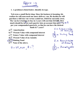


© Copyright 2026