
Acute glomerulonephritis BEST PRACTICE C S Vinen, D B G Oliveira
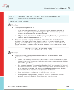
Downloaded from pmj.bmj.com on August 22, 2014 - Published by group.bmj.com
206
BEST PRACTICE
Acute glomerulonephritis
C S Vinen, D B G Oliveira
.............................................................................................................................
Postgrad Med J 2003;79:206–213
Glomerulonephritis is an important cause of renal failure
thought to be caused by autoimmune damage to the
kidney. While each type of glomerulonephritis begins
with a unique initiating stimulus, subsequent common
inflammatory and fibrotic events lead to a final pathway
of progressive renal damage. In this article the different
forms of inflammatory glomerulonephritis and their
diagnosis are discussed. In a review of therapy both
immediate life saving treatment given when
glomerulonephritis causes acute renal failure and more
specific treatments designed to modify the underlying
mechanisms of renal injury are considered.
..........................................................................
G
See end of article for
authors’ affiliations
.......................
Correspondence to:
Professor David Oliveira,
Department of Renal
Medicine, St George’s
Hospital Medical School,
Cranmer Terrace, London
SW17 0RE, UK;
[email protected]
Submitted 17 May 2002
Accepted
5 November 2002
.......................
www.postgradmedj.com
lomerulonephritis is an important cause
of renal impairment accounting for 10%–
15% of cases of end stage renal failure in
the USA, following only diabetes and hypertension in importance.1 In defining acute
glomerulonephritis, we have chosen to discuss
those glomerular diseases that may present with
a nephritic syndrome—that is with haematuria,
proteinuria, and impaired renal function together
with hypertension, fluid overload, and oedema.
Their pathology involves intraglomerular inflammation and cellular proliferation with secondary
renal impairment over days to weeks. This definition excludes glomerular diseases without cell
proliferation or nephritic presentations, such as
minimal change disease, membranous nephropathy, and focal segmental glomerulosclerosis that
can, none the less, chronically compromise renal
function. In primary glomerulonephritis, disease
is almost entirely restricted to the kidneys (as in
IgA
nephropathy
or
post-streptococcal
glomerulonephritis)
while
in
secondary
glomerulonephritis it occurs in association with
more diffuse inflammation (as in systemic lupus
erythematosus or systemic vasculitis). Prompt
diagnosis of glomerulonephritis is vital as patients with even mildly impaired renal function,
hypertension, and urinary abnormalities may
rapidly lose kidney function if not treated
urgently.
Although our understanding of the causes of
glomerulonephritis is still at a basic level, inflammation is thought to be autoimmune mediated
and involve both cellular and humoral immune
systems. In each case a unique initiating stimulus
(occurring by one of at least four different
mechanisms) is followed by a common pathway
of inflammatory and subsequently fibrotic events.
In antiglomerular basement membrane disease,
patients produce antibodies that react directly
with the specialised basement membranes of the
lung and glomerulus.2 In post-streptococcal
glomerulonephritis antibodies are formed not to
an endogenous antigen but to an exogenous
streptococcal antigen planted in the glomerulus
at the time of infection.3 In systemic lupus
erythematosus and IgA nephropathy, the antigen
antibody reaction occurs not only in situ in the
glomerulus but also systemically with subsequent
trapping of complexes in the kidney. Finally in the
glomerulonephritis seen in small vessel vasculitis,
cellular rather than humoral immune responses
are thought to be stimulated, with inflammation
often originating in organs distant to the kidney
with a subsequent renal influx of T-cells and macrophages as crescentic glomerulonephritis
evolves.
Whatever the initial events, common inflammatory pathways follow with activation of the
coagulation and complement cascades and production of proinflammatory cytokines.4 Activation of complement components leads to chemotaxis of inflammatory cells and cell lysis (via the
membrane attack complex). The coagulation cascade leads to fibrin deposition. Cellular proliferation of parietal epithelial cells in Bowman’s space
together with an influx of inflammatory cells
such as macrophages and neutrophils results in
acute glomerular crescent formation. Cytokine
release leads to activation of the glomerular cells
themselves and a change in endogenous cell phenotype results in cell proliferation, overproduction of proteases and oxidants, and laying down
of extracellular matrix with subsequent fibrosis,
perhaps stimulated by factors such as platelet
derived growth factor and transforming growth
factor beta. Failure of apoptosis (the normal
mechanism allowing resolution of inflammation)
is also important. Finally in a chronic phase of
damage, haemodynamic alterations lead to hyperfiltration and intraglomerular hypertension5
with subsequent development of glomerular sclerosis and chronic interstitial damage. Thus a
process that is initially inflammatory with the
potential to resolve may progress to fibrosis and
irreversible scarring. This dynamic picture may
partly explain why in post-streptococcal
glomerulonephritis where antigen is rapidly
cleared, even acute renal failure can be expected
to resolve spontaneously. By contrast in hepatitis
C associated mesangiocapillary glomerulonephritis (MCGN) where viral infection is
chronic, antigen cannot be cleared and renal
damage may chronically progress.
.................................................
Abbreviations: ACE, angiotensin converting enzyme;
ANCA, antineutrophil cytoplasmic antibodies; HSP,
Henoch-Schönlein purpura; MCGN, mesangiocapillary
glomerulonephritis; RPGN, rapidly progressive
glomerulonephritis; WHO, World Health Organisation
Downloaded from pmj.bmj.com on August 22, 2014 - Published by group.bmj.com
Acute glomerulonephritis
Figure 1 Section through a normal renal glomerulus. Blood is
carried in to the glomerulus by an afferent arteriole and leaves by
the efferent arteriole. Capillary loops that emerge from the vascular
pole are supported by stalks of mesangial cells. On entering the
lumen of a capillary loop, blood is filtered through a barrier
consisting of a fenestrated endothelial layer, the glomerular
basement membrane, and an epithelial layer. Urine emerges in to
the urinary space and passes in to the proximal tubule.
To understand the histology of glomerulonephritis, we need
to revisit the basic structure of the normal kidney (see fig 1).
Inflammatory, proliferative, and fibrotic changes may affect
specific cells of the kidney differently or may result in more
global changes with particular patterns resulting in a
spectrum of clinical presentations. In table 1, we have
attempted to summarise the complex nomenclature that surrounds glomerulonephritis by naming each disease, describing its common renal clinical presentation and explaining its
underlying histological lesion. In table 2 we have focused
purely on clinical aspects which may aid rapid diagnosis.
Renal biopsies are vital both in defining a diagnosis, and also
in offering prognostic information by differentiating acute
reversible damage from chronically scarred non-viable kidney
which does not justify the risks of potentially toxic therapy.
Although current treatments are, at best, crude, with greater
understanding of pathological events we hope to design more
specific therapy both to limit acute damage, and to prevent
progression to chronic scarring with its inevitable decline in
renal function.
POST-INFECTIOUS ENDOCAPILLARY
GLOMERULONEPHRITIS
Post-streptococcal glomerulonephritis is the best known
example of endocapillary glomerulonephritis, the most
common form of acute glomerulonephritis seen after some
bacterial, viral, fungal, and parasitic infections. Although this
pattern of glomerular injury after a streptococcal infection
remains an important cause of acute renal failure in the
developing world, in Europe and the USA this lesion is
increasingly seen in infections such as endocarditis after
intravenous drug abuse. In post-streptococcal glomerulonephritis, children are usually affected with a male
preponderance.6 It can follow pharyngitis (commonly in winter) or skin infections (commonly in summer) with a
β-haemolytic nephritogenic strain of streptococcus (often type
12) with the glomerulonephritis occurring one to 12 weeks
after initial infection. It affects up to 15% of those infected,
although many cases are subclinical and self resolving. In
207
children most severely affected, presentation is with the classic nephritic picture of puffy eyelids, facial oedema, hypertension, and dark scanty urine with microscopic haematuria
and proteinuria.
The pathology is that of a planted antigen where a streptococcal component is deposited in the glomerulus during
infection.3 Subsequent production of antibody by the host
produces in situ immune complex formation which alters the
permeability of the glomerular basement membrane and
allows subsequent deposition of further pre-formed immune
complexes. In addition streptococcal antigen may cross react
with glomerular structures or directly activate complement
with subsequent attraction of inflammatory cells.7 8 Immune
deposits initiate a diffuse proliferative glomerulonephritis
particularly affecting mesangial and endothelial cells.
Immunostaining shows C3 in the mesangium and along capillary walls with accompanying IgG.
Serology may show raised antistreptolysin antibody titres
but its absence does not exclude the diagnosis as many
nephritogenic strains do not produce streptolysin. Low C3 levels with normal C4 levels (due to alternative pathway activation) are seen acutely but should have returned to normal
within two months.
MESANGIOPROLIFERATIVE GLOMERULONEPHRITIS/
IGA NEPHROPATHY
IgA nephropathy is the commonest of all glomerulonephritides world wide. Thus although only 4%–13% of patients
present with acute nephritis (the commoner presentation
being with micro or macroscopic haematuria), this still represents a considerable number of cases.9 Peak presentation is
during the second and third decades showing a 2:1 male preponderance with attacks sometimes after infection (particularly pharyngitis10 11). The disease shows great geographic
variation and is more common in the Western Pacific rim and
in Asia (accounting for 50% of primary glomerular disease in
Japan) but is rare in black populations.4
IgA nephropathy is the classic mesangioproliferative
glomerulonephritis where cellular proliferation may be either
diffuse or focal but affects predominantly the mesangium.
Immunofluorescence shows paramesangial deposition of IgA
(with some IgG and IgM) together with alternative pathway
complement components, while electron microscopy shows
mesangial dense deposits. Polymeric IgA1 is deposited12 in the
kidney after overproduction of systemic IgA1 polymers
(possibly in response to infection) together with impaired
clearance through both the hepatic and the myeloid routes. In
addition abnormal glycosylation of IgA may make it more
prone to self aggregate and form immune complexes with
affinity for the mesangium.12 The disease is associated with a
raised serum concentrations of IgA in 50% of patients, but
serum complement levels are normal as complement activation is restricted to the kidneys alone.13
HENOCH-SCHÖNLEIN PURPURA
The renal lesion of Henoch-Schönlein purpura (HSP) is almost
identical to that of the more severe variants of IgA nephropathy. However, as a small vessel vasculitis, HSP also has the systemic features of a purpuric rash largely affecting the lower
limbs, arthritis or arthralgia, and abdominal pain sometimes
in association with rectal bleeding. The disease is most
commonly seen in those less than 20 years of age. Renal
involvement is not always present initially but its incidence
increases with time and is more common in older children
who have associated abdominal pain and a persisting rash.14
Renal involvement can also occur in adults where it is thought
to carry a worse prognosis. Although haematuria and
proteinuria are the most common renal presentations, 8% of
patients will have an acute nephritis and up to 29% may
present with a combined nephritic and nephrotic picture.9
www.postgradmedj.com
Downloaded from pmj.bmj.com on August 22, 2014 - Published by group.bmj.com
208
Vinen, Oliveira
Table 1 The classification of acute glomerulonephritis by disease, renal
presentation, and histological lesion (where the nephritic syndrome is a relatively rare
presentation, the more usual clinical presentation is given in bold type)
Disease
Possible clinical renal
presentations
Most common histological lesion
Postinfectious
Nephritic syndrome, haematuria,
glomerulonephritis
proteinuria
(Historically post-streptococcal but
also seen with other bacterial, viral
and parasitic infections)
Endocapillary glomerulonephritis—
a diffuse proliferative
glomerulonephritis especially
affecting mesangial and endothelial
cells possibly provoked by in situ
immune complex deposition due to
a planted streptococcal antigen
IgA nephropathy
Nephritic syndrome
Mesangioproliferative
Macroscopic/microscopic
glomerulonephritis—a focal or
haematuria
diffuse cellular proliferation
affecting predominantly the
mesangium possibly stimulated by
polymeric IgA deposition
Henoch-Schönlein purpura
Nephritic syndrome, haematuria,
Mesangial cell proliferation may be
proteinuria, nephrotic syndrome
associated with glomerular
crescents, capillary necrosis and
leucocytoclastic vasculitis possibly
due to subtle differences in size of
IgA deposits
Wegener’s granulomatosis,
Rapidly progressive
A focal or diffuse proliferative
microscopic polyangiitis,
glomerulonephritis, nephritic
glomerulonephritis with extensive
idiopathic crescentic
syndrome
crescent formation in greater than
glomerulonephritis
50% of glomeruli
A focal segmental
Antiglomerular basement
Rapidly progressive
glomerulonephritis with necrosis
membrane disease
glomerulonephritis, nephritic
which rapidly progresses to
syndrome
widespread crescent formation
caused by antibodies to type IV
collagen
Type I MCGN, idiopathic. In
Nephritic syndrome
A mesangiocapillary
association with infective
Nephrotic syndrome,
glomerulonephritis with intense
endocarditis, visceral abscesses, haematuria, proteinuria
cellular proliferation involving
infected arteriovenous shunts
mesangial expansion and
thickening of capillary walls due to
extension of proliferation into
capillary loops. Probably provoked
by subendothelial immune complex
deposition
Hepatitis C associated type I
May have additional intracapillary
MCGN
cryoglobulin deposition
Type II MCGN (sometimes seen
Intense mesangial cell proliferation
in association with partial
as above in the absence of immune
lipodystrophy)
complex deposition but in
association with dense
intramembranous deposits
Systemic lupus erythematosus
Nephritic syndrome, nephrotic
WHO type III
syndrome, haematuria, proteinuria A focal proliferative
glomerulonephritis involving cellular
proliferation in mesangial and
endocapillary areas affecting
<50% of glomeruli
WHO type IV
A diffuse proliferative
glomerulonephritis involving
mesangial and endocapillary
proliferation in >50% of glomeruli
sometimes in association with
necrosis and crescent formation
Patients with HSP also have systemic IgA containing
immune complexes, though their size is larger than those in
IgA disease. Mesangial deposition of IgA is usually seen but
capillary wall staining for IgA is also frequent. Glomerular
crescents and fibrin deposition are more common in HSP, as is
capillary necrosis and leucocytoclastic vasculitis.9 It is thought
that subtle differences in the IgA complexes in HSP lead to
greater leucocyte stimulation and thus to the small vessel vasculitis and extrarenal manifestations that define HSP.
RAPIDLY PROGRESSIVE GLOMERULONEPHRITIS
(RPGN)
The rapidly progressive glomerulonephritides are the most
serious of all glomerulonephritides with the potential to
www.postgradmedj.com
destroy renal function within days. Although causes are
heterogeneous, they are united by the histological finding of
extensive crescents (a proliferation of parietal epithelial cells
and mononuclear phagocytes with possible fibroblasts in
Bowman’s capsule) affecting more than 50% of glomeruli.
Causes fall into three broad categories with different
presentations, treatments, and prognoses.
Pauci-immune glomerulonephritis caused by small vessel
vasculitides accounts for about 50% of RPGN with an
incidence of approximately 2 per 100 000 per year and a peak
in the sixth decade with equal sex distribution. Disease may be
limited to the kidney (idiopathic crescentic glomerulonephritis) or be associated with widespread systemic inflammation (Wegener’s granulomatosis and microscopic polyangiitis). Overt presentation is often preceded by weight loss
Downloaded from pmj.bmj.com on August 22, 2014 - Published by group.bmj.com
Acute glomerulonephritis
Table 2
209
Clinical features of acute glomerulonephritides
Type of glomerulonephritis
Age and sex
Investigations
Extrarenal manifestations
Post-infectious glomerulonephritis
(post- streptococcal
glomerulonephritis )
IgA nephropathy
Most common between 2 and 12
years; boys > girls
Low C3 (alternative complement
pathway), antistreptolysin titre
Sore throat or skin infection 7 days to
12 weeks before presentation
Commonly presents in 20s and 30s;
men > women
<20 years of age
Raised serum IgA in 50% of cases,
complement normal
Complement normal
Wegener’s granulomatosis,
microscopic polyangiitis
50s/60s; men = women
ANCA, complement normal
Antiglomerular basement membrane
disease
MCGN:
Type I
Type II
Young men; 50s and 60s; both sexes Antiglomerular basement membrane
antibody, complement levels normal
Macroscopic haematuria may relate
to time of infections
Purpuric rash on legs, arthritis,
abdominal pain
Weight loss, malaise, upper and
lower respiratory tract symptoms,
arthritis, palpable purpura
Lung haemorrhage especially in
smokers
Henoch-Schönlein purpura
Type I with hepatitis C
Lupus nephritis
20s; women > men
teenage; women > men
Middle age
Young women in 20 and 30s
and general malaise with later features relating to individual
illnesses. Microscopic polyangiitis has cutaneous (palpable
purpura), neurological (mononeuritis multiplex) or gastrointestinal vasculitis as well as renal failure, with pulmonary
symptoms in only 50% of cases (due to non-granulomatous
arteriolar vasculitis and capillaritis). By contrast, Wegener’s
granulomatosis is dominated by pulmonary manifestations
with upper (deafness, nasal cartilage collapse, sinusitis), and
lower (pulmonary haemorrhage due to granulomatous vasculitis) respiratory tract involvement and cavitating lung lesions
seen on radiography.
Biopsy shows a focal or diffuse proliferative glomerulonephritis with extensive crescents. The pathogenesis of vasculitis remains the focus of much research but direct immunoglobulin deposition in the glomerulus is not thought to play
a significant part (hence the term pauci-immune). Serologically, however, these diseases are linked in about 90% of cases
by the finding of antineutrophil cytoplasmic antibodies
(ANCA). Antibody staining is usually directed against the
neutrophil cytoplasm in Wegener’s with an antigen specificity
for proteinase 3 on ELISA, whereas in microscopic polyangiitis it is generally perinucleur in pattern and is directed against
myeloperoxidase. A direct causative role for ANCA in small
vessel vasculitis remains controversial with experimental evidence pointing towards roles for neutrophils, macrophages,
and T-cells in its pathogenesis.15
Antiglomerular basement membrane disease accounts for
10%–20% of cases of RPGN with a frequency of 0.5 cases per
million per year in a European caucasoid population.2 The disease occurs in two peaks, one in the third decade with a male
preponderance and the second in the sixth and seventh
decades affecting both sexes equally.16 Associated lung
involvement is more common in young men (when the
disease is known as Goodpasture’s disease), while that
isolated to the kidneys is commoner in older patients. A
prodrome of weight loss and malaise is less common than in
the vasculitides and patients often present with either acute
renal failure or haemoptysis due to lung involvement.17
Haemoptysis is commoner in smokers and in those with fluid
overload or intercurrent infections (the later also making the
kidney damage more severe). Lung haemorrhage is the most
common cause of death during early disease and should be
suspected with haemoptysis or where a chest radiograph
shows alveolar shadowing without restriction by anatomical
fissures and with sparing of the upper zones. Acute
Low C4 (classical path activation)
Low C3 (alternative path activation),
C3 nephritic factor
Low C4, +ve hepatitis C serology,
hepatitis C RNA on polymerase chain
reaction, serum cryoglobulins, +ve
antinuclear antibody/ +ve Rh factor
Low C3, antinuclear antibody/
anti-ds DNA, anticardiolipin antibody
Gaunt face due to partial
lypodystrophy
Abnormal liver function tests (rarely
cirrhosis), pupuric rash, neuropathy,
polyarthralgia, leg ulcers
Arthralgia, photosensitive skin rash,
pleurisy, and pericarditis
haemorrhage may be confirmed by a transiently raised transfer factor on pulmonary function testing.
Antiglomerular basement membrane disease is caused by
antibodies that bind the apha 3 chain of type 4 collagen found
in the specialised basement membranes of the kidney and
lung.18 Initially histology may show a focal segmental
glomerulonephritis with necrosis and interstitial inflammation but will rapidly progress to show widespread crescent
formation with all crescents at the same stage of evolution (a
previously normal kidney can develop 100% crescents in as
little as five days). Immunofluorescence shows the linear
deposition of IgG antibodies (sometimes associated with C3)
along the glomerular basement membrane. Serology is
positive for antiglomerular basement membrane antibodies
but in 20%–30% of patients ANCA antibodies are also
detected.19 These latter patients behave clinically more like
those with vasculitis (with lethargy, malaise, weight loss) and
have a better renal prognosis than those with antiglomerular
basement membrane antibodies alone—this may be because
they are actually affected by vasculitis with the antiglomerular basement membrane antibodies being a secondary
response to the damaged basement membrane.
Some 30%–40% of RPGN is due to a group of heterogeneous
conditions where renal damage is associated with immune
complex deposition or other causes of basement membrane
damage such as accelerated hypertension. Pathology is
frequently an aggressive variant of a glomerulonephritis normally associated with a more benign course (such as
post-streptococcal glomerulonephritis, or IgA nephropathy),
with histology being complicated by extensive inflammation
and crescent formation. It is also seen after infections such as
endocarditis and shunt nephritis or in association with multisystem disease such as systemic lupus erythematosus.
MESANGIOCAPILLARY GLOMERULONEPHRITIS;
ALSO KNOWN AS MEMBRANOPROLIFERATIVE
GLOMERULONEPHRITIS
This rare form of glomerulonephritis has enjoyed renewed
interest after the discovery that a subtype of MCGN type I is
associated with chronic hepatitis C infection. MCGN commonly presents as a nephrotic syndrome but in 16%–30% of
patients the initial presentation is with acute nephritis. The
disease can be subdivided into types I and II, with its
idiopathic forms mostly seen in children and young adults
with cases presenting at a younger age in type II (15 years ±11
www.postgradmedj.com
Downloaded from pmj.bmj.com on August 22, 2014 - Published by group.bmj.com
210
years) than in type I (24 years ±16 years) disease, with a slight
female preponderance. Type I MCGN shares some features
with lupus nephritis, and a similar histological picture can
also be seen with endocarditis and infected arteriovenous
shunts. In type II MCGN, patients may have an associated
partial lypodystrophy giving them a very gaunt facial appearance.
It has recently been realised that a significant proportion of
cases of type I MCGN previously labelled as idiopathic in fact
occur in association with chronic hepatitis C infection20 of
which 20%–25% will present with acute nephritis.21 There is
geographical variation in this association and while hepatitis
C associated renal disease appears particularly common in
Japan (where the infection is found in up to 60% of cases of
membranoproliferative glomerulonephritis) and Italy, it is less
common in the USA (10%–20% of cases of membranoproliferative glomerulonephritis) and has been seen relatively little
in France.20 21 Patients present 10–15 years after infection in
middle age and have subclinical liver disease with mild
biochemical abnormalities. Renal disease is often seen in the
context of cryoglobulinaemia (cold precipitable mixed immunoglobulins composed of monoclonal IgM rheumatoid factor
and polyclonal IgG). Patients suffer malaise, anaemia, peripheral neuropathy, polyarthralgia, and a purpuric rash together
with lower limb ulceration and Raynaud’s disease. Rarely vasculitis also affects the gastrointestinal or cardiological
systems.22
Idiopathic type I MCGN is associated with activation of the
classical complement pathway (and therefore with low C4
concentrations) while in MCGN type II alternative pathway
activation is seen with low C3 and the presence of the C3
nephritic factor (an antibody leading to permanent activation
of the complement cascade). In hepatitis C associated MCGN
in addition to low classical complement component levels,
patients have positive antihepatitis C antibodies and hepatitis
C RNA on polymerase chain reaction. They may also have a
positive antinuclear antibody and rheumatoid factor tests
(70%) and positive cryoglobulins (75%).23
The pathogenesis of MCGN is obscure but probably involves
intense cellular proliferation particularly involving mesangial
cells. Histologically both types show mesangial expansion and
thickening of the capillary walls (with reduction in the capillary lumina), which in the case of MCGN type I is partly due
to cellular proliferation extending between the capillary basement membranes causing thickening and giving the classic
tramline effect. Mesangial and capillary loop deposition of C3
occurs in both forms of MCGN but is accompanied by immunoglobulin deposition only in type I MCGN. The distinction
between the different types is based on electron microscopy
findings: in type I subendothelial immune deposits are seen in
the glomerular basement membrane while in type II dense
intramembranous deposits are seen in glomerular, tubular,
and vascular basement membranes (the nature of these
deposits in type II disease remains unknown but does explain
its alternative name of dense deposit disease). In hepatitis C
associated type I MCGN intracapillary deposits are thought to
be due to precipitation of the cryoglobulins themselves.
LUPUS NEPHRITIS
Renal involvement in systemic lupus erythematosus can
present with proteinuria, haematuria, nephrotic syndrome, or
with an acute nephritis. It is rarely the first manifestation of
systemic lupus but usually occurs within five years and may be
the first presentation leading to a definitive diagnosis.24
Patients (most commonly women in their 20s and 30s with a
black preponderance) will frequently have suffered lethargy,
arthralgia or arthritis, skin rashes, and the symptoms of pleurisy and pericarditis in the months before presentation.25 More
than any other glomerulonephritis, lupus nephritis can
change and evolve over time so that in a patient with an
www.postgradmedj.com
Vinen, Oliveira
initially benign glomerular lesion, a new presentation with
acute glomerulonephritis should prompt repeat biopsy and if
needed more aggressive treatment. High titres of antinuclear
antibodies and antidouble stranded DNA antibodies together
with low complement levels are helpful in a nephritic flare,
although changes in such markers often precede the actual
glomerular inflammation, sometimes by months.26
The pathology is at least in part that of immune complex
deposition, with antigen antibody complexes forming systemically or in situ and subsequently activating the inflammatory cascade. Positively charged nuclear histone antigens can
also bind to the glomerular basement membrane altering
function and permeability and acting as planted antigens that
are then the target of anti-DNA antibodies.
Acute glomerulonephritis in lupus is seen in patients who
have focal and diffuse proliferative glomerulonephritis—that
is World Health Organisation (WHO) class III and IV lupus
nephritis27 (WHO class I (normal kidney) and WHO class II
(mesangial proliferation) lupus nephritis do not present as
acute glomerulonephritis). In class III lupus nephritis (focal
proliferative glomerulonephritis) there is proliferation in the
mesangial and endocapillary areas in less than 50% of
glomeruli. Such patients have haematuria and proteinuria and
are sometimes nephritic. More commonly nephritic syndrome
and renal impairment is associated with the more aggressive
class IV diffuse proliferative glomerulonephritis where mesangial and endocapillary hypercellularity affect more than
50% of glomeruli with additional necrosis and possibly
crescent formation. Subendothelial deposits give thickened
basement membrane with a wire loop appearance on light
microscopy. Immunofluorescence shows extensive granular
deposition of IgG, IgA, IgM, and complement in subendothelial and mesangial areas.
TREATMENT OF GLOMERULONEPHRITIS
The treatment of acute glomerulonephritis falls into two categories. Supportive treatment such as blood pressure control
and dialysis is immediate and frequently life saving, but does
not attempt to reverse the underlying pathology. Specific
treatments aim to prevent and reverse glomerular inflammation and ultimately to preserve renal function—such treatments are often highly toxic and rely on non-specific suppression of the entire immune system. They carry the immediate
risks of overwhelming infection and the later risk of
reproductive toxicity and malignancy. In choosing such therapies, we need to select patients in whom kidney recovery is
unlikely to occur spontaneously but where toxicity can be justified by the potential reversibility of the condition. On this
basis we discuss current therapies and where possible present
the rationale for their use (table 3). Many of these treatments
together with newer therapies are the subject of ongoing
clinical trials to determine optimum strategies.
The importance of supportive therapies in acute glomerulonephritis cannot be over emphasised. Tight blood pressure
control, appropriate use of diuretics, and control of hyperkalaemia, uraemia and fluid overload, if necessary by dialysis, are
quite literally life saving. Blood pressure control is vital not
just in the short term but also later for any patient left with
even mild renal impairment or proteinuria, with angiotensin
converting enzyme (ACE) inhibitors having a particular place
for their additional antiproteinuric and antifibrotic effects.28
In most cases of post-streptococcal glomerulonephritis
where inflammation does resolve spontaneously, supportive
therapies alone will be sufficient with improved renal function
being seen between four and 14 days after the initial acute
failure in 95% of patients.29 Serum creatinine generally returns
to baseline levels by four weeks but haematuria may persist for
six months and mild proteinuria may be present in a few
patients even at 10 years.30 Rarely haematuria and proteinuria
persist long term and are accompanied by hypertension and
Downloaded from pmj.bmj.com on August 22, 2014 - Published by group.bmj.com
Acute glomerulonephritis
211
Table 3 Treatment of glomerulonephritis (treatments used widely in clinical practice
are in bold type while newer therapies are in normal type)
Glomerulonephritis
Specific treatments used
Rationale for treatment
Endocapillary
glomerulonephritis
Mesangioproliferative
glomerulonephritis
None required
Inflammation generally self resolving
Antiglomerular basement
membrane disease
ANCA positive vasculitis
Immune complex-mediated
RPGN
MCGN type I: idiopathic
Type I: hepatitis C related
Type II
Lupus nephritis
Acute nephritic phase:
Blood pressure control with ACE
inhibitors
Pulsed intravenous steroids,
cyclophosphamide, mycophenolate
mofetil intravenous immunoglobulin
Pulsed intravenous steroids 1 g
for 3/7 followed by oral steroids
(60 mg/day)
Cyclophosphamide orally (2–3
mg/kg/day)
Plasma exchange (daily for 14
days or until no anti-GBM
antibody)
Pulsed intravenous steroids 1 g
for 3/7 + oral steroids (start 60
mg), cyclophosphamide (2
mg/kg/day orally or 0.5–1 g
monthly intravenous)
Plasma exchange ? for creatinine
>500 or pulmonary haemorrhage
Treat underlying histological
variant
If idiopathic as for ANCA positive
vasculitis
Steroids 40 mg/m2 alternate days
in children only
Aspirin (325 mg/day)
Dipyridamole (75–100 mg three
times a day) in adults only
Alpha-interferon/ribavirin
Steroids, cyclophosphamide
(plasma exchange)
No specific therapy shown to be
helpful
Intravenous steroids + oral
steroids
Intravenous/oral
cyclophosphamide
Mycophenolate mofetil, cyclosporin
declining renal function.31 For most other causes of glomerulonephritis however, if renal function is to be preserved, we must
aim to reverse the underlying events causing glomerular
inflammation.
The exact immunological events of IgA nephropathy are
unknown and therefore treatment of IgA nephropathy is
extremely difficult. For patients who present acutely with
macroscopic haematuria, but with normal renal function and
blood pressure, regular review alone may be all that is
required. For patients who follow a more accelerated clinical
course, once again control of blood pressure and careful fluid
management are vital. Acute inflammation on biopsy may
justify the use of immunosuppressives with anecdotal reports
of success with mycophenolate mofetil, cyclophosphamide
and pulsed steroids, and intravenous immunoglobulin.32–34 In
the more chronic phase, use of ACE inhibitors, both in hypertensive and in non-hypertensive patients who have proteinuria (>1 g/24 hours), is emerging as increasingly important.
These drugs both reduce the level of proteinuria and slow the
decline in glomerular filtration rate normally seen.35
Prognosis is difficult to estimate for those patients presenting acutely with IgA nephropathy. Certainly hypertension,
impaired renal function, and severe proteinuria at presentation are adverse prognostic features36 with one study suggesting that a combination of a raised creatinine (>150 µmol/l)
together with proteinuria (>1 g/24 hours) gave a patient only
a 20% chance of independent renal function seven years
Reduce inflammation especially
where renal function declining and
crescents present
To switch off antiglomerular
basement membrane antibody
production
To remove existing antiglomerular
basement membrane antibody while
immunosuppression takes effect
Suppression of antibody and cellular
immune arms
Removal of ANCA/immune
complexes?
Removal of proinflammatory
cytokines?
Suppression of antibody response
As antiplatelet agents to decrease
cellular proliferation
To lessen viral drive
To treat inflammatory component
To suppress antibody production and
reduce immune complexes
later.12 In HSP clinical presentation predicts prognosis with
15% of nephritic patients eventually reaching end stage renal
failure, but up to 50% of those with a combined nephritic and
nephrotic picture eventually needing renal replacement
therapy.37
Rapidly progressive glomerulonephritis can irreversibly
destroy renal function within days without treatment. Such
risks therefore justify the use of significantly toxic therapies in
an attempt to preserve independent renal function. In
antiglomerular basement membrane disease, high dose
steroids and cyclophosphamide are used to switch off B-cell
production of antiglomerular basement membrane antibody
with additional plasma exchange to remove existing antibody
during the two weeks before the effects of cyclophosphamide
as seen. In renal vasculitis (Wegener’s and microscopic
polyangiitis) much less is known of the pathogenesis but
similar regimens aim to switch off both T-cell and B-cell
function.38
Unless treatment is prompt, the renal prognosis in
antiglomerular basement membrane disease is poor with few
patients presenting with a serum creatinine greater than 600
µmol/l and requiring dialysis ever regaining independent renal
function.39 With plasma exchange and aggressive cytotoxic
treatment, 80% of patients with a creatinine less than 600
µmol/l can expect improvements in renal function,2 which are
generally seen within days of starting treatment. Plasma
exchange may be used even in those with irretrievably
www.postgradmedj.com
Downloaded from pmj.bmj.com on August 22, 2014 - Published by group.bmj.com
212
damaged kidneys in an attempt to treat pulmonary
haemorrhage.40
The prognosis in ANCA positive RPGN is better than that in
antiglomerular basement membrane disease. With aggressive
treatment using steroids, cyclophosphamide, and plasma
exchange at least five times if they are dialysis dependent,
even 75% of those patients initially requiring renal support
may recover renal function, with 80% of these remaining
dialysis independent at five years.41 Plasma exchange is also
used in ANCA positive vasculitis associated pulmonary
haemorrhage.42 Recent trial data have confirmed that, after
initial induction with steroids and cyclophosphamide for
three months, many of these patients may be safely converted
to an oral azathioprine regimen.43
The prognosis in immune complex RPGN is determined by
the level of glomerular inflammation and treatment is
directed at underlying pathology. The few cases of truly
idiopathic immune complex RPGN appear to take a similar
clinical course to pauci-immune RPGN and immunosuppressive regimens similar to those used in ANCA positive disease
may be appropriate.
The pathology of idiopathic MCGN remains obscure and
with little specific treatment of proven value, the importance
of blood pressure control increases. In an attempt to limit the
platelet activation associated with cellular proliferation,
aspirin and dipyridamole have been used with some success
and there may be a place for steroid treatment in children.44
Type II MCGN is rare and shows little response to conventional
therapies. Renal prognosis in truly idiopathic MCGN type I
gives a 60% renal survival at 10 years; this figure is probably
worse in MCGN type II.
Treatment of MCGN with hepatitis C infection is complex. If
the disease is thought to be driven by virus-containing
immune complexes, then control of viral load using alphainterferon and ribavirin should be most effective—although
this has shown some success with improvements in mild
MCGN, relapse of viral load after stopping treatment is often
seen.22 Alternatively, where renal damage is more severe, the
inflammatory component of the lesion might best be controlled with immunosuppressives albeit at a risk of viral
activation. Some nephrologists would therefore treat an
aggressive nephritic flare with pulsed methylprednisolone followed by 3–6 months of tapered oral steroids. Where disease is
particularly active oral cyclophosphamide for two months has
been used. With such treatments of cryoglobulinaemic vasculitis, extrarenal manifestations respond very quickly and in
more than 85% of patients the plasma creatinine falls within
a week, although proteinuria is much slower to respond. Long
term immunosuppression is not justified and plasma exchange remains controversial.22 Approximately 10% of patients with hepatitis C related kidney disease will develop end
stage renal failure.21
The treatment of lupus nephritis is also complex with only
part of its pathology being understood. As a disease that often
strikes young women, the risks of renal disease must be
weighed against possible infertility associated with immunosuppressive regimens. Renal biopsy is vital since, with an acute
nephritic flare, it is important to distinguish scarred and irreversibly damaged kidneys from those that might benefit from
aggressive immunosuppression. A recent study of patients
with type IV lupus nephritis suggested that a combination of
pulsed monthly methylprednisolone and intravenous cyclophosphamide resulted in a remission rate of approximately
85%.45 Further quarterly doses of pulsed cyclophosphamide
after the six months of monthly induction therapy also
reduced the subsequent relapse rate. There may also be a place
for intravenous immunoglobulin (working by solubilising
immune complexes or blocking Fc receptors to prevent the
inflammatory cascade) in refractory cases or mycophenolate
mofetil in acute flares.46 The most recent data suggest that
with current treatment 70%–85% of patients with type III and
www.postgradmedj.com
Vinen, Oliveira
Key references
• Hricik DE, Chung-Park M, Sedor JR. Glomerulonephritis. N
Engl J Med 1998;339:888–99.
• Couser WG. Glomerulonephritis. Lancet 1999;353:1509–
15.
• Madaio MP, Harrington JT. The diagnosis of glomerular
diseases: acute glomerulonephritis and the nephrotic
syndrome. Arch Intern Med 2001;161:25–34.
• Ruggenenti P, Schieppati A, Remuzzi G. Progression,
remission, regression of chronic renal diseases. Lancet
2001;357:1601–7.
Sources of further information
• National Kidney Federation at www.kidney.org.uk produces very helpful patient leaflets on glomerulonephritis,
IgA nephropathy, and mesangiocapillary glomerulonephritis.
• Arthritis Research Campaign at www.arc.org.uk produces
excellent patient leaflets on lupus and vasculitis.
IV lupus nephritis will retain independent renal function at
five years. Repeated acute nephritic flares are a poor prognostic indicator, as are hypertension and black race.
Glomerulonephritis is an important cause of renal failure
for which we currently have only non-specific and potentially
toxic therapies. With increasingly prompt diagnosis and
greater understanding of pathology, we must hope to improve
this situation. As knowledge grows, we may prevent some
glomerulonephritides altogether, for instance by successful
vaccination against hepatitis C for MCGN or by designing
therapies to reverse immune complex formation in systemic
lupus erythematosus. For patients in whom glomerulonephritis does occur, drugs may be designed which tackle
inflammation by interrupting the complement or cytokine
cascades or which target the cell signalling that leads to proliferation and subsequent fibrosis. Only then can we hope to
prevent the many cases of chronic renal failure caused by
these diseases.
SELF TEST QUESTIONS ON ACUTE
GLOMERULONEPHRITIS (ANSWERS AT END OF
REFERENCES)
Q1. What is the most likely diagnosis in a 15 year old boy presenting to casualty with a sore throat and macroscopic
haematuria?
Q2. Which of the following statements about lupus nephritis
are true?
(A) Patients with lupus nephritis may have a normal serum
creatinine value
(B) Patients with lupus nephritis may require multiple
sequential biopsies
(C) In lupus nephritis a fall in C3 levels and a rise in antidouble stranded DNA levels may precede actual glomerular
inflammation
(D) Patients with lupus nephritis often develop infertility as a
result of their treatment
Q3. Plasma exchange is a recognised treatment in which of the
following forms of glomerulonephritis?
(A) Lupus nephritis
(B) Antiglomerular basement membrane disease
(C) ANCA positive vasculitis
(D) IgA nephropathy
Downloaded from pmj.bmj.com on August 22, 2014 - Published by group.bmj.com
Acute glomerulonephritis
Q4. Immune complex deposition is thought to be important in
the pathogenesis of which of the following forms of
glomerulonephritis?
(A) Lupus nephritis
(B) IgA nephropathy
(C) ANCA positive vasculitis
(D) MCGN type II
Q5. Which of the following diseases, which can present as an
acute nephritic syndrome, also commonly present with a
nephrotic picture?
(A) Lupus nephritis
(B) IgA nephropathy
(C) Antiglomerular basement membrane disease
(D) Mesangioproliferative glomerulonephritis
(E) Post-streptococcal glomerulonephritis
.....................
Authors’ affiliations
C S Vinen, D B G Oliveira, Department of Renal Medicine, St. George’s
Hospital Medical School, London
REFERENCES
1 US Renal Data Systems. USRDS 1997 annual data report. Bethesda:
National Institute of Health, National Institute of Diabetes and Digestive
and Kidney Diseases, April 1997.
2 Kluth DC, Rees AJ. Anti-glomerular basement membrane disease. J Am
Soc Nephrol 1999;10:2446–53.
3 Oliveira DBG. Poststreptococcal glomerulonephritis: getting to know an
old enemy. Clin Exp Immunol 1997;107:8–10.
4 Hricik DE, Chung-Park M, Sedor JR. Glomerulonephritis. N Engl J Med
1998;339:888–99.
5 Couser WG. Glomerulonephritis. Lancet 1999;353:1509–15.
6 Tejani A, Ingulli E. Poststreptococcal glomerulonephritis: current clinical
and pathological concepts. Nephron 1990;55:1–5.
7 Peake PW, Pussel BA, Karpus TE, et al. Post-streptococcal
glomerulonephritis: studies on the interaction between nephritis
strain-associated protein (NSAP), complement and the glomerulus.
APMIS 1991;99:460–6.
8 Johnson RJ, Lovette D, Lehrer RJ, et al. Role of oxidants and proteases in
the glomerular injury. Kidney Int 1994;45:352–9.
9 Davin J-C, Ten Berge IJ, Weeing JJ. What is the difference between IgA
nephropathy and Henoch Schönlein purpura nephritis? Kidney Int
2001;59:823–34.
10 Emancipator SN. IgA nephropathy: morphological expression and
pathogenesis. Am J Kidney Dis 1994;23:451–62.
11 Galla JH. IgA nephropathy. Kidney Int 1995;47:377–87.
12 Floege J, Feehally J. IgA nepropathy: recent developments. J Am Soc
Nephrol 2000;11:2395–403.
13 D’Amico G. Clinical features and natural history in adults with IgA
nephropathy. Am J Kidney Dis 1988;12:353–7.
14 Kaku Y, Nohara K, Honda S. Renal involvement in Henoch Schönlein
purpura: a multivariate analysis of prognostic factors. Kidney Int
1998;53:1755–9.
15 Tipping PG, Kitching AR, Cunningham MA, et al. Immnuopathogenesis
of crescenteric glomerulonephritis. Curr Opin Nephrol Hypertens
1999;8:281–6.
16 Merkel F, Pullig O, Marx M, et al. Course and prognosis of
anti-basement membrane antibody mediated disease: report of 35 cases.
Nephrol Dial Transplant 1994;9:372–6.
17 Donaghy M, Rees AJ. Cigarette smoking and lung haemorrhage in
glomerulonephritis caused by autoantibodies to glomerular basement
membrane. Lancet 1983;ii:1390–3.
18 Kalluri R, Wilson CV, Weber M, et al. Identification of the α-3 chain of
type IV collagen as the common autoantigen in anti-glomerular basement
membrane disease and Goodpasture’s syndrome. J Am Soc Nephrol
1995;6:1178–84.
19 Jayne DR, Marshall PD, Jones SJ, et al. Autoantibodies to GBM and
neutrophil cytoplasm in rapidly progressive glomerulonephritis. Kidney Int
1990;37:965–70.
213
20 Johnson RJ, Wilson R, Yamabe H, et al. Renal manifestations of
hepatitis C virus infection. Kidney Int 1994;46:1255–63.
21 Daghestani L, Pomeroy C. Renal manifestations of hepatitis C infection.
Am J Med 1999;106:347–54.
22 Campise M, Tarantino A. Glomerulonephritis in mixed
cryoglobulineamia: what treatment? Nephrol Dial Transplant
1999;14:281–3.
23 D’Amico G, Fornasieri A. Cryoglobulinaemic glomerulonephritis: a
membranoproliferative glomerulonephritis induced by hepatitis C virus.
Am J Kidney Dis 1995;25:361–9.
24 Baldwin DS, Gluck MC, Lowenstein J, et al. Lupus nephritis: clinical
course as related to morphological forms and their transitions. Am J Med
1977;62:12–30.
25 Madaio MP, Harrington JT. The diagnosis of glomerular diseases: acute
glomerulonephritis and the nephrotic syndrome. Arch Intern Med
2001;161:25–34.
26 Boumpas DT, Austin HA, Fessler BJ, et al. Systemic lupus erythematosus:
emerging concepts. 1. Renal, neuropsychiatric, cardiovascular,
pulmonary, and haematological disease. Ann Intern Med
1995;122:940–50.
27 Kashgarian M. Lupus nephritis: lessons from the path lab. Kidney Int
1994;45:928–38.
28 Ruggenenti P, Schieppati A, Remuzzi G. Progression, remission,
regression of chronic renal diseases. Lancet 2001;357:1601–7.
29 Lewy JE, Salinas-Madrigal L, Herdson TB, et al. Clinico-pathological
correlations in acute post streptococcal glomerulonephritis: a correlation
between renal function, morphological damage and clinical course of 46
children with acute post-streptococcal glomerulonephritis. Medicine
1971;50:453–71.
30 Potter EV, Lipschultz SA, Abidh S, et al. Twelve to seventeen year follow
up of patients with post streptococcal acute glomerulonephritis in
Trinidad. N Engl J Med 1982;307:725–9.
31 Schact RG, Gluck MC, Gallo GR, et al. Progression to ureamia after
remission of acute post streptococcal glomerulonephritis. N Engl J Med
1976;295:977–81.
32 Nolin L, Corteau M. Management of IgA nephropathy: evidence-based
recommendations. Kidney Int 1999;55(suppl 70):S56–62.
33 Nowack R, Birck R, van der Woude FJ. Mycophenolate mofetil for
systemic vasculitis and IgA nephropathy. Lancet 1997;349:774.
34 McIntyre CW, Fluck RJ, Lambie SH. Steroid and cyclophosphamide
therapy for IgA nephropathy associated with crescenteric changes: an
effective treatment. Clin Nephrol 2001;56:193–8.
35 Cattran DC, Greenwood C, Ritchie S. Long term benefit of
angiotensin-converting enzyme inhibitor therapy in patients with severe
immunoglobulin A nephropathy: a comparison to patients receiving
treatment with other hypertensive agents and to patients receiving no
therapy. Am J Kidney Dis 1994;23:247–54.
36 D’Amico G. Natural history of idiopathic IgA nephropathy: role of
clinical and histological prognostic factors. Am J Kidney Dis
2000;36:227–37.
37 Goldstein AR, White RHR, Akuse R, et al. Long-term follow-up of
childhood Henoch Schönlein nephritis. Lancet 1992;339:280–2.
38 Jindal KK. Management of idiopathic crescenteric and diffuse
proliferative glomerulonephritis: evidence-based recommendations.
Kidney Int 1999;55(suppl 70):S33–40.
39 Mokrzycki MH, Kaplan AA. Therapeutic plasma exchange:
complications and management. Am J Kidney Dis 1994;23:817–27.
40 Levy JB, Pusey CD. Still a role for plasma exchange in rapidly
progressive glomerulonephritis. J Nephrol 1997;10:7–13.
41 Gaskin G, Pusey CD. Long term outcome after immunosuppression and
plasma exchange for severe vasculitis associated with glomerulonephritis.
J Am Soc Nephrol 1999;10:101A.
42 Levy J. New aspects in the management of ANCA-positive vasculitis.
Nephrol Dial Transplant 2001;16:1314–17.
43 Jayne D, Rasmussen N. European collaborative trials in vasculitis:
EUVAS update and latest results. Clin Exp Immunol 2000;20(suppl
1):13–15.
44 Levin A. Management of membranoproliferative glomerulonephritis:
evidence-based recommendations. Kidney Int 1999;55(suppl 70):41–6.
45 Gourley MF, Austin HA, Scott D, et al. Methylprednisolone and
cyclophosphamide, alone or in combination, in patients with lupus
nephritis. A randomised controlled trial. Ann Intern Med
1996;125:549–57.
46 Chan TM, Li FK, Tang CSO, et al. Efficacy of mycophenolate mofetil in
patients with diffuse proliferative lupus nephritis. N Engl J Med
2000;343:1156–62.
ANSWERS
Q1. IgA nephropathy. Q2. A, B, C. Q3. B, C. Q4. A, B. Q5. A, D.
www.postgradmedj.com
Downloaded from pmj.bmj.com on August 22, 2014 - Published by group.bmj.com
Acute glomerulonephritis
C S Vinen and D B G Oliveira
Postgrad Med J 2003 79: 206-213
doi: 10.1136/pmj.79.930.206
Updated information and services can be found at:
http://pmj.bmj.com/content/79/930/206.full.html
These include:
References
This article cites 43 articles, 6 of which can be accessed free at:
http://pmj.bmj.com/content/79/930/206.full.html#ref-list-1
Article cited in:
http://pmj.bmj.com/content/79/930/206.full.html#related-urls
Email alerting
service
Topic
Collections
Receive free email alerts when new articles cite this article. Sign up in
the box at the top right corner of the online article.
Articles on similar topics can be found in the following collections
Urology (98 articles)
Acute renal failure (7 articles)
Notes
To request permissions go to:
http://group.bmj.com/group/rights-licensing/permissions
To order reprints go to:
http://journals.bmj.com/cgi/reprintform
To subscribe to BMJ go to:
http://group.bmj.com/subscribe/


© Copyright 2026