
Full Text - Environmental and Experimental Biology
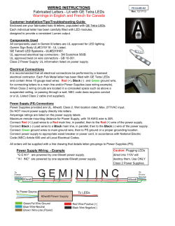
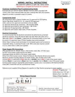
Environmental and Experimental Biology (2014) 12: 155–159
Brief Communication
The response of protocorm-like bodies of nine hybrid
Cymbidium cultivars to light-emitting diodes
Jaime A. Teixeira da Silva1,2*
Faculty of Agriculture and Graduate School of Agriculture, Kagawa University, Miki-cho, Kagawa, 761-0795, Japan
Current address: Miki-cho Post Office, Ikenobe 3011-2, P. O. Box 7, Kagawa-Ken, 761-0799, Japan
1
2
*Corresponding author, E-mail: [email protected]
Abstract
The objective of this study was to assess how the protocorm-like bodies (PLBs) of nine hybrid Cymbidium cultivars would respond to
light-emitting diodes (LEDs) at different ratios under in vitro conditions. The red (R) to blue (B) LED ratio (100% R; 60% R + 40% B; 50%
R + 50% B; 40% R + 60% B; 100% B; control = plant growth fluorescent light (PGF)) affected neo-PLB formation in a cultivar-dependent
way: 100% R or 100% B had a negative effect on neo-PLB production while 40% R + 60% B favored neo-PLB formation more than under
PGF, although the resulting PLBs were of poor quality, i.e., could not be used for clonal purposes. This study reinforces the fact that it
is essential to test optimal conditions for the in vitro culture of individual hybrid Cymbidium cultivars and that light conditions can
quantitatively influence the organogenic result. Furthermore, caution must be exercised when interpreting the results, as these are based
on the timing of sampling and on the developmental stage of the material.
Key words: light-emitting diode, protocorm-like body, Teixeira Cymbidium medium.
Abbreviations: B, blue; LED, light-emitting diode; PLB, protocorm-like body; R, red; TC, Teixeira Cymbidium medium.
Introduction
Lighting is one of the most important abiotic factors
affecting the success of an in vitro plant culture, and can
in some instances constitute a form of stress, either when
excessive and reaching the light saturation point (e.g.,
Rakić et al. 2015), or when too weak (e.g., Liu et al. 2014).
Consequently, the performance of a plant in vitro, which
depends on its physiology, can be influenced by the quality
of the light source. Of two light sources that have seen
expanded applications in plant research, light-emitting
diodes (LEDs; Dutta Gupta, Jatothu 2013) and coldcathode fluorescent lamps (e.g., Norikane et al. 2013), the
former is the focus in this study due to its reliability (Chang
et al. 2012).
The most common form of LEDs used in horticultural
experiments are blue (B) and red (R), since these correspond
to absorption peaks of chlorophyll b and chlorophyll a,
respectively, at 455 and 660 nm, respectively (Massa et al.
2008). LEDs have been used in experimental systems to
manipulate plant growth, usually under different ratios
(e.g., a recent case using papaya; Teixeira da Silva 2014a and
references therein). The use of R and B LEDs is not new
to Cymbidium research and has been shown to enhance
Cymbidium plantlet growth in vitro under CO2 enrichment
(Tanaka et al. 1998) and morphogenesis of Cymbidium
protocorm-like body (PLB) segments (Nhut et al. 2005).
Using half-PLB segments as explants and Cymbidium
Environmental and Experimental Biology ISSN 2255-9582
Twilight Moon cultivar ‘Day Light’, Huan and Tanaka
(2004) induced most callus under 75% R + 25% B, but most
new PLBs (i.e., neo-PLBs) with a 25% R + 75% B LED ratio.
Despite these studies, there has been no attempt to explore
whether the response to different PLB ratios is a standard
and cultivar-independent response. Thus, using this cultivar
as the basis for comparison, the effect of different ratios of
B : R LEDs was tested on nine hybrid Cymbidium cultivars.
The ability to manipulate organogenesis and morphogenesis
in in vitro conditions using methods – in this case LEDs
– other than chemical means, such as the conventional
application of plant growth regulators, is attractive, since
it allows the abiotic environment to be strictly controlled
(e.g., the R : B LED ratio). Such an experimental system is
attractive for studying light-induced stress mechanisms,
because the LED ratio can be fine-tuned to establish a
specific ratio (Fig. 1), while the light intensity can also be
perfectly programmed. Since no such developed systems
exist for orchids, this makes this study unique, allowing
for a similar experimental design to be established for any
orchid to conduct light-induced stress studies.
Materials and methods
Chemicals and reagents
All plant growth regulators were purchased from SigmaAldrich (St. Louis, USA) and were of tissue culture grade. All
other chemicals and reagents were of the highest analytical
155
J.A. Teixeira da Silva
A
D
B
E
C
F
G
H
Fig. 1. Distribution of red (R) and blue (B) LED bulbs used in the present study. A, R LEDs; B, R + B LEDs; C, B LEDS; D, 100% R; E, 60%
R + 40% B; F, 50% R + 50% B; G, 40% R + 60% B; H, 100% B. R and B LEDs were arranged on the ceiling of “LED PACKS”, 25 × 31 × 25
cm, described in Tanaka et al (1998). Each LED PACK contained 176 LED bulbs (5 mm diameter, Sharp Electric Ltd, Tokyo, Japan) and
the R + B LED ratio was adjusted based on a uniform division of R + B LED bulbs. LED PACKS were placed in culture rooms in which
CO2 concentration was maintained at 3000 ppm.
grade available and were purchased from Wako or Nacalai
Tesque (Osaka, Japan), unless specified otherwise.
Plant material: stocks, culture medium and culture
conditions
Stock cultures of PLBs of eight hybrid Cymbidium cultivars
(kindly donated by Bio-U, Tokushima, Japan; see Table 1)
were maintained on Teixeira Cymbidium medium 1 (TC1), whose mineral constituents can be found in Teixeira da
Silva (2012). Subcultures and in vitro culture conditions
are detailed in Teixeira da Silva (2014b), while details
on the theoretical aspects related to explant sizes and
timing of sampling are described in Teixeira da Silva and
Dobránszki (2014) and Teixeira da Silva and Dobránszki
(2013), respectively. More specifically, PLBs of all hybrids
originally developed from shoot-tip culture on Vacin
and Went (1949) agar medium without plant growth
regulators, were induced and subcultured (PLB induction
and proliferation medium) every two months on TC-1
medium (Teixeira da Silva 2012), which was supplemented
with 0.1 mg L–1 α-naphthaleneacetic acid (NAA) and 0.1
mg L–1 kinetin (Kin), 2 g L–1 tryptone and 20 g L–1 sucrose,
and solidified with 8 g L–1 Bacto agar (Difco Labs, Becton
Dickinson and Co., NJ, USA), following Teixeira da Silva
et al. (2005) and Teixeira da Silva and Tanaka (2006). All
media were adjusted to pH 5.3 with 1 N NaOH or HCl
prior to autoclaving at 100 KPa for 17 min. Cultures were
kept on 40 mL medium in 100-mL Erlenmeyer flasks,
156
double-capped with aluminium foil, at 25 °C, under a 16-h
photoperiod with a photosynthetic photon flux density of
45 µmol m–2 s–1 provided by 40-W plant growth fluorescent
lamps (Homo Lux, Matsushita Electric Industrial Co.,
Hiroshima, Japan). Longitudinally dissected two pieces of
PLB (3 to 4 mm in diameter) segments, 10 per flask, were
used as explants for neo-PLB induction and proliferation,
and as the experimental material.
The LED irradiation source
Red (wavelength, 660 nm; material, GaN; GL5UR3K1 = 3
cd, 5 mm in diameter; Sharp Electric Ltd., Tokyo, Japan) and
blue (wavelength, 450 nm; material, GaA1As, NLPB 500 =
1 cd, 5 mm in diameter, Nichia Chemicals Ltd., Tokushima,
Japan) LEDs were arranged on the ceiling of “LED PACKS”,
25 × 31 × 25 cm (Fig. 1), originally described in Tanaka
et al. (1998). Each LED PACK contained 176 LED bulbs
(5 mm in diameter, Sharp Electric Ltd.) and the R+B LED
ratio was adjusted based on a uniform division of R+B LED
bulbs, for example, 88 R : 88 B, evenly interspersed, for the
50% R : 50% B treatment. The following LED ratios were
examined: 100% R; 60% R + 40% B; 50% R + 50% B; 40%
R + 60% B; 100% B. LED PACKS were placed in culture
rooms in which CO2 concentration was maintained at 3000
ppm. The intensity of LEDs was maintained at 45 µmol
m–2 s–1 under a 16-h photoperiod. The R : B LED ratio as
well as the intensity of light were adjusted using a PA36-2A
Regulated DC Power Supply (Kenwood TMI Corp., Tokyo,
Response of hybrid Cymbidium protocorm-like bodies to light-emitting diodes
Table 1. Response (number of neo-PLBs) of nine hybrid Cymbidium cultivars’ half-PLBs to different combinations of red : blue LED
ratios (n = 30). Cultivar crosses (BioU classification): 91-8 (Aroma Candle ‘Hot Heart’) = Jenteel ‘Pair Look’ × Seaside ‘Crown Princess’;
167-1 (Pretty Poetry ‘Malachite’) = Mini Sarah ‘Artisan’ × Eastern Star ‘Green Fields’; 204-1 (Alice Beauty ‘No 1’) = Alice Luna × Sleeping
Beauty ‘Mistuko’; 246-2 (Twilight Moon ‘Day Light’) = Lovely Bunny ‘Romeo’ × Hiroshima Golden Cup ‘Sunny Moon’; 485-12 (Spring
Night ‘No 12’) = Tiny Sour × Twilight Moon ‘Day Light’; 536-1 (Dream City ‘No 1’) = Great Katy ‘Tender’ X Lucky Flower ‘Anmitsuhime’;
553-1 (Call Me Love ‘Snow Princess’) = Jenteel ‘Pair Look’ × Great Katy ‘Tender’; 649-4 (Energy Star ‘No 4’) = Morning Moon ‘Great
Tiger’ × Twilight Moon ‘Day Light’; 653-2 (Sweet Moon ‘No 2’) = Yellow Candy ‘Lemon Fresh’ × Twilight Moon ‘Day Light’. Results from
100 red and 100% blue are not presented since almost no PLBs formed in all nine cultivars. PGF values reported in Teixeira da Silva and
Dobránszki (2014) and cultivars listed based on their decreasing neo-PLB production under PGF. Different letters across all treatments
and for each cultivar indicate significant differences at P ≤ 0.05 (according to DMRT). LED, light-emitting diode; PGF, plant growth
fluorescent light; PLB, protocorm-like body
Cymbidium cultivar
246-2
653-2
649-4
485-12
167-1
553-1
91-8
536-1
204-1
PGF
8.3 b
7.2 b
6.4 b
4.6 b
3.2 b
3.1 b
2.6 a
2.2 a
1.8 a
Red (R) : blue (B) LED ratio
60% R : 40% B
50% R : 50% B
4.2 d
7.4 c
3.4 d
5.9 c
2.6 c
6.1 b
1.8 d
3.4 c
0.9 d
2.7 c
0.6 d
2.2 c
0.1 c
1.1 b
0 c
1.8 b
0 c
0.9 b
Japan) and a LI-250 Light Meter (LI-COR Inc., Nebraska,
USA). Plant growth fluorescent light (PGF; 40-W, Homo
Lux), at the same light intensity and photoperiod, served as
the control light treatment.
Growth analysis
Growth was quantified by the number of new PLBs that
formed (i.e., neo-PLBs).
Statistical analysis
Experiments were organized according to a randomized
complete block design (RCBD) with three blocks of 10
replicates per treatment. All experiments were repeated
in triplicate (n = 30, total sample size per treatment). Data
was subjected to analysis of variance (ANOVA) with mean
separation by Duncan’s multiple range test using SAS® vers.
6.12 (SAS Institute, Cary, NC, USA). Significant differences
between means were assumed at P ≤ 0.05.
Results and discussion
The objective of this study was to assess whether different
ratios of red and blue LEDs could increase neo-PLB
production in nine Cymbidium cultivars, and to assess if
the response to LED treatments was cultivar-dependent.
Only red (i.e., 100% R) or only blue (i.e., 100% B) LEDs had
a detrimental effect on neo-PLB production (Fig. 2D) for
all nine cultivars, while a lower R:B ratio favoured a greater
production of neo-PLBs relative to the control light source,
PGF (Table 1).
Huan and Tanaka (2004), using Twilight Moon ‘Day
Light’, one of the cultivars also used in this study, showed
40% R : 60% B
10.1 a
8.6 a
7.3 a
5.5 a
4.4 a
4.0 a
2.9 a
2.6 a
2.1 a
that callus was most prolific under a 75% R + 25% B LED
regime, but that most PLBs formed with a 25% R + 75% B
LED ratio. Using a different LED ratio (explained in more
detail in Teixeira da Silva 2014a and Fig. 1) in this study, it
was found that a 40% R + 60% B LED regime favoured neoPLB formation more than under PGF (control light) (Table
1) although the PLBs formed were of a more irregular
shape (Fig. 2A vs 2B), which would make them difficult to
use in clonal culture. Teixeira da Silva and Tanaka (2006)
demonstrated that the tiny PLBs that formed from PLB
explants were de facto somatic embryos and that the calli
that Huan and Tanaka (2004) were describing were in
fact undeveloped PLBs. This may explain why Huan and
Tanaka (2004) claim to have observed as many as 89.6
neo-PLBs per half-moon PLB segment under PGF and
73.6-98.0 neo-PLBs per half-moon PLB segment under
different R:B LED ratios. In contrast, Teixeira da Silva and
Tanaka (2006), Teixeira da Silva (2012) and the results of
this study showed an almost 10-fold lower number of neoPLBs per half-moon PLB segment under PGF and different
R : B LED ratios, most likely because tiny, underdeveloped
PLBs, which are extremely difficult to count, and in some
cases are fused to one another, were not considered. This is
because, in practical terms, they represented a PLB cluster
to be subcultured intact, and only a limited number of
fully developed neo-PLBs develop further into shoots. The
numbers thus reported by Huan and Tanaka (2004) may
have thus been grossly over exaggerated, although their
study indicated that the choice of red or blue LED, and
their ratio, could influence the morphogenic outcome, a
conclusion confirmed in this study, but for a wide range
of cultivars. The timing of sampling also strongly affects
157
J.A. Teixeira da Silva
A
B
C
D
Fig. 2. Growth of hybrid Cymbidium Twilight Moon ‘Day Light’ in response to different light conditions. A, PGF; B 40% R + 60% B LED
ratio; C, 60% R + 40% B LED ratio; D, 100% R.
the quantitative outcome, a very strong factor in hybrid
Cymbidium (Teixeira da Silva, Dobránszki 2013). Thus, if
the number of neo-PLBs were to be assessed at 20, 45, 60 or
90 days after plating on culture medium, the quantitative
results would be vastly different. In this study, the “optimal”
developmental stage of 45 days, in which the shoot apex
has barely developed, but where the neo-PLB has attained
maximum size optimal for sub-culture, was used.
There is an increase in endopolyploidy in response to
extreme light spectral ranges in Phalaenopsis PLBs (Park
et al. 2010), and in papaya seedling tissue (Teixeira da Silva
2014a), which is not uncommon for Cymbidium PLB tissues
exposed to different abiotic stresses (Teixeira da Silva,
Tanaka 2006). Follow-up experiments should determine
the stress-related events in neo-PLB formation, including
the expression of stress-related enzyme systems. There
are currently no studies in orchids that have made such
a detailed biochemical assessment, and this experimental
design demonstrates that an LED system (Fig. 1) can be
used as a powerful tool for examining such abiotic stressrelated responses.
Acknowledgements
Thanks to Prof. Michio Tanaka for providing LED photos and for
allowing the use of the lab equipment, including the LED boxes.
All Cymbidium cultivars were courtesy of BioU (Tokushima,
Japan).
References
Chang M-H., Das D., Varde P.V., Pecht M. 2012. Light emitting
diodes reliability review. Microelectronics Reliability 52: 762–
782.
Dutta Gupta S., Jatothu B. 2013. Fundamentals and applications
of light-emitting diodes (LEDs) in in vitro plant growth and
morphogenesis. Plant Biotechnol. Rep. 7: 211–220.
Huan L.V.T., Tanaka M. 2004. Effect of red and blue light-emitting
diodes (LEDs) on callus induction, callus proliferation, and
protocorm-like body formation from callus in Cymbidium
orchid. Environ. Control Biol. 42: 57–64.
Liu Q-H., Wu X., Chen B-C., Ma J-Q., Gao J. 2014. Effects of low
158
light on agronomic and physiological characteristics of rice
including grain yield and quality. Rice Sci. 21: 243–251.
Massa G.D., Kim H-H., Wheeler R.M., Mitchell C.A. 2008. Plant
productivity in response to LED lighting. HortScience 43,
1951–1956.
Nhut D.T., Tien T.N.T., Huong M.T.N., Hien N.T.H., Huyen P.X.,
Luan V.Q., Le B.V., Teixeira da Silva J.A. 2005. Artificial seeds
for preservation and propagation of Cymbidium spp. Prop.
Ornamental Plants 5: 67–73.
Norikane A., Teixeira da Silva J.A., Tanaka M. 2013. Growth of
in vitro Oncidesa plantlets cultured under cold cathode
fluorescent lamps (CCFLs) with super-elevated CO2
enrichment. AoB Plants 5: plt044.
Park S.Y., Yeung E.C., Paek K.Y. 2010. Endoreduplication in
Phalaenopsis is affected by light quality from light-emitting
diodes during somatic embryogenesis. Plant Biotechnol. Rep.
4: 303–309.
Rakić T., Gajić G., Lazarević M., Stevanović B. 2015. Effects of
different light intensities, CO2 concentrations, temperatures
and drought stress on photosynthetic activity in two
paleoendemic resurrection plant species Ramonda serbica
and R. nathaliae. Env. Exp. Bot. 109: 63–72.
Tanaka M., Takamura T., Watanabe H., Endo M., Yanagi T.,
Okamoto K. 1998. In vitro growth of Cymbidium plantlets
cultured under superbright red and blue light-emitting diodes
(LEDs). J. Hortic. Sci. Biotechnol. 73: 39–44.
Teixeira da Silva J.A. 2012. New basal media for protocorm-like
body and callus induction of hybrid Cymbidium. J. Fruit
Ornam. Plant Res. 20: 127–133.
Teixeira da Silva J.A. 2014a. Photoauto-, photohetero- and
photomixotrophic in vitro propagation of papaya (Carica
papaya L) and response of seed and seedlings to light-emitting
diodes. Thammasat Int. J. Sci. Technol. 19: 57–71.
Teixeira da Silva J.A. 2014b. Response of hybrid Cymbidium
(Orchidaceae) protocorm-like bodies to 26 plant growth
regulators. Bot. Lithuanica 20: 3–13.
Teixeira da Silva J.A., Dobránszki J. 2013. How timing of sampling
can affect the outcome of the quantitative assessment of plant
organogenesis. Sci. Hortic. 159: 59–66.
Teixeira da Silva J.A., Dobránszki J. 2014. Dissecting the concept of
the thin cell layer: theoretical basis and practical application
of the plant growth correction factor to apple, Cymbidium and
chrysanthemum. J. Plant Growth Reg. 33: 881–895.
Teixeira da Silva J.A., Tanaka M. 2006. Multiple regeneration
pathways via thin cell layers in hybrid Cymbidium
Response of hybrid Cymbidium protocorm-like bodies to light-emitting diodes
(Orchidaceae). J. Plant Growth Reg. 25: 203–210.
Teixeira da Silva J.A., Yam T., Fukai S., Nayak N., Tanaka M.
2005. Establishment of optimum nutrient media for in
vitro propagation of Cymbidium Sw. (Orchidaceae) using
protocorm-like body segments. Prop Ornamental Plants 5:
129–136.
Vacin E., Went F.W. 1949. Some pH changes in nutrient solutions.
Bot. Gaz. 110: 605–613.
Received 17 July 2014; received in revised form 14 September 2014; accepted 17 November 2014
159


© Copyright 2026