
Fever of Unknown Origin: Focused Diagnostic Approach Based on Clinical Physical Examination,
Infect Dis Clin N Am 21 (2007) 1137–1187
Fever of Unknown Origin: Focused
Diagnostic Approach Based on Clinical
Clues from the History,
Physical Examination,
and Laboratory Tests
Burke A. Cunha, MD, MACPa,b,*
a
Infectious Disease Division, Winthrop-University Hospital, 259 First Street,
Mineola, Long Island, NY 11501, USA
b
State University of New York School of Medicine, Stony Brook, NY, USA
Few clinical problems are as challenging and difficult as that presented by
the patient who has a febrile illness for more than 10 to 15 days, the origin
of which remains obscure. No type of illness puts to a stronger test the physician’s ability to approach a clinical problem effectively..
–Philip A. Tumulty, MD
Fever of unknown origin (FUO) was first and correctly termed ‘‘prolonged and perplexing fevers’’ by Kiefer and Leard [1]. Prolonged and perplexing fevers are difficult-to-diagnose febrile disorders aptly termed FUOs.
FUOs may be conveniently divided into four general categories based on the
etiology of the FUO: (1) infectious, (2) rheumatic-inflammatory, (3) neoplastic, or (4) miscellaneous disorders. Petersdorf and Beeson [2] in 1961
were the first to define FUO in terms of time-based diagnostic criteria. These
have since been termed ‘‘classic’’ FUOs and may be defined as disorders
with temperatures greater than or equal to 101 F that have persisted for
at least 3 weeks that were not diagnosed after a week of intensive in-hospital
testing. The classic and current causes of FUO have been modified, reflecting the changing spectrum of diseases and the availability of sophisticated
diagnostic tests in the outpatient setting [3,4].
* Infectious Disease Division, Winthrop-University Hospital, 259 First Street, Mineola,
Long Island, NY 11501.
0891-5520/07/$ - see front matter Ó 2007 Elsevier Inc. All rights reserved.
doi:10.1016/j.idc.2007.09.004
id.theclinics.com
1138
CUNHA
The main diagnostic difficulty with FUOs is an efficient and effective diagnostic approach. Clinicians are advised that to diagnose FUOs effectively,
they should be comprehensive. Unfortunately, often this has only resulted in
excessive diagnostic testing to rule out every disorder causing FUOs. A nonfocused approach has the effect of incurring unnecessary expense, inconveniencing patients, and delaying or obscuring the FUO diagnostic work-up.
The undesirable effect of the ‘‘shotgun approach’’ to diagnostic testing is
that it underuses the FUO tests appropriate for the most likely diagnostic
categories, and it overtests for unlikely diagnoses [5].
The diagnostic approach to the FUO patient should be focused and relevant to the clinical syndromic presentation. Because all patients with FUOs
by definition have fevers, the clinician should identify the predominant features of the clinical presentation to determine the general category of FUO
of the patient. With infectious FUOs, fevers are often accompanied by chills
or night sweats. Weight loss without loss of appetite is another potential indicator of an infectious disease etiology. The clinical presentation of patients
with FUOs caused by rheumatic-inflammatory disorders is dominated by
arthralgias, myalgias, or migratory chest or abdominal pain. The predominant symptoms of patients with neoplastic FUOs are fatigue and weight loss
with early or dramatic decrease in appetite. Night sweats may also be a feature
of neoplastic disorders. Patients presenting with FUOs whose symptoms do
not suggest an infectious, rheumatic-inflammatory, or neoplastic disorder
should be considered as having an FUO of miscellaneous causes [6–8].
It makes little sense to get thyroiditis tests for every FUO patient if there
is not an antecedent history of thyroid or autoimmune disease or physical
findings referable to subacute thyroiditis. Similarly, just because subacute
bacterial endocarditis (SBE) is a common cause of FUO, transthoracic
echocardiography (TTE)–transesophageal echocardiography (TEE) should
not be obtained on all FUO patients. In patients with FUOs, TTE-TEE
should be obtained only in those with heart murmurs. In FUO patients
with heart murmurs, vegetations seen on TTE-TEE can indicate SBE
(culture positive and culture negative); systemic lupus erythematous (SLE;
Libman-Sacks vegetations); or marantic (nonbacterial) endocarditis.
By using a focused approach the clinician can order tests that are more
relevant to the presenting clinical syndrome; such tests more efficiently
and effectively lead to a correct FUO diagnosis. The diagnostic approach
to FUOs may be considered as consisting of three phases. The initial phase
consists of the initial FUO history and physical examination and nonspecific
laboratory tests. This phase provides the clinician with a general sense of
whether the FUO is likely to be caused by an infection or by a rheumaticinflammatory or neoplastic disorder. Phase II involves re-evaluating the
patient using a focused FUO history and physical examination and additional nonspecific and specific laboratory tests. The focused FUO evaluation
has the effect of narrowing diagnostic possibilities and eliminating possibilities from further diagnostic consideration [1,5].
FUO: DIAGNOSTIC APPROACH
1139
Nonspecific laboratory tests included during the initial evaluation are
helpful to increase the diagnostic probability of some entities, whereas
decreasing or eliminating the diagnostic probability of others. Nonspecific
laboratory tests, as with other clinical findings, are more significant when
considered together rather than individually. For example, the combination
of the following nonspecific laboratory tests, which alone are unhelpful
diagnostically, should suggest a particular diagnosis (ie, increased lactate
dehydrogenase, atypical lymphocytes in the peripheral smear, and thrombocytopenia should suggest the possibility of malaria in patients with an
appropriate epidemiologic exposure). The patient exposed in malarious
areas is also exposed to typhoid fever. Both typhoid fever and malaria
have some clinical features in common and neither has localizing signs,
making these infections difficult to diagnose if the epidemiologic history is
not taken into account and the nonspecific laboratory clues are not appreciated for their diagnostic significance. The same three nonspecific laboratory tests argue strongly against typhoid fever and should point the
clinician to the possibility of malaria as the cause of the patient’s FUO [1,9].
Fever of unknown origin: classic and current causes
FUOs fall into four general categories. The relative frequency of the
causes of FUO in each category is the basis for a phased diagnostic approach. Phase I of an FUO evaluation consists of a FUO relevant history,
physical examination, and nonspecific laboratory tests. The phase I evaluation provides the basis for determining the course of the FUO work-up. Features in the history, physical findings, and laboratory abnormalities in the
initial FUO evaluation suggest which general category of disorder is responsible for the patient’s FUO. Although all FUOs, by definition, are associated with fever, the predominant symptoms usually suggest a particular
FUO category.
In general, infectious diseases may be associated with chills, night sweats,
myalgias, or weight loss with an intact appetite. Arthralgias or myalgias are
the predominant complaints of the patient presenting with rheumatic-inflammatory causes of FUO. These patients often have fatigue, but weight
loss or night sweats are unusual findings. Even with some overlap in symptoms, the clinician can usually determine from the dominant clinical features
whether the patient is likely to have an infectious, rheumatic-immunologic,
or neoplastic cause of their FUO. Typically, in addition to fever and fatigue,
neoplastic disorders have night sweats and weight loss accompanied by
a dramatic and profound loss of appetite. Patients that do not fit in any
of these categories have FUOs of a variety of miscellaneous causes. It is
sometimes difficult to differentiate between infectious and neoplastic or infectious and rheumatic disorders. In such situations, the next phase of
FUO investigation using a focused diagnostic approach provides additional
1140
CUNHA
information with a history and physical examination or additional laboratory tests, which clearly differentiate one group from another, and are
able to narrow differential diagnostic possibilities within a category. Classic
and current FUO causes have been reviewed (Table 1) [2,3,6,10–13].
Fever of unknown origin: focused diagnostic approach
Overview
After the initial FUO-relevant evaluation most of the common causes of
FUOs in each category may be readily diagnosed. Combining the relevant
FUO features on physical examination with selected nonspecific laboratory
test abnormalities limits diagnostic possibilities and eliminates other causes
from further diagnostic consideration. The diagnostic significance of selected nonspecific tests cannot be overemphasized. The clinical significance
of nonspecific laboratory abnormalities is enhanced when they are considered together. As with FUO-relevant historical facts or physical findings,
nonspecific laboratory abnormalities taken together increase diagnostic
specificity and significance.
The function of the initial phase of FUO evaluation is to diagnose disorders, which are most easily diagnosed among the FUOs, and to limit differential diagnostic possibilities that direct the second phase of focused FUO
evaluation. The focused FUO history of physical examination and laboratory tests has the purpose to refine further the differential diagnosis of disorders that have not been diagnosed during the initial evaluation [14,15].
Fever of unknown origin: initial evaluation
Although the initial direction of the diagnostic work-up is suggested by
FUO-relevant aspects of the history and physical examination, the basic
battery of nonspecific laboratory tests helps to define further differential diagnostic possibilities. Many disorders in all categories of FUO are accompanied by some nonspecific laboratory abnormalities. The diagnostic
significance of such findings alone and more critically taken together is often
overlooked as having no importance because the abnormalities are not of
sufficiently impressive magnitude or the abnormalities are associated with
many potential disorders. The basic nonspecific laboratory test battery includes the complete blood count, erythrocyte sedimentation rate (ESR),
C-reactive protein, and liver function tests. Imaging tests include a chest radiograph (if there are signs or symptoms referable to the chest) and CT and
MRI scans of the abdomen and pelvis (as dictated by clinical clues suggesting an intra-abdominal or pelvic pathology. Blood cultures are also included
as part of the initial diagnostic evaluation [1,16–19].
Blood cultures pick up common causes of SBE (bacteremia from an intra-abdominal or pelvic or renal-perinephric source), and intra-abdominal
FUO: DIAGNOSTIC APPROACH
1141
imaging provides important information for the focused phase of FUO evaluation. If the patient has a very highly elevated ESR (R100 mm/h) it suggests possible FUO etiologies including abscesses, osteomyelitis, SBE, and
adult Still’s disease. Among the rheumatic inflammatory causes of FUO
the ESR greater than or equal to 100 mm/h may point to adult Still’s disease, polymyalgia rheumetica/temporal arteritis, late-onset rheumatoid arthritis, SLE, periarteritis nodosa, Takayasu’s arteritis, Kikuchi’s disease,
or familial Mediterranean fever. Among neoplastic disorders an increased
ESR rate may be present with any of them but an unelevated ESR has no
differential diagnostic value and does not rule out neoplastic or other disorders. The high ESR rate may also point to drug fever and the miscellaneous
category and elevated ESR rate may indicate drug fever, regional enteritis,
subacute thyroiditis, deep vein thrombosis or small pulmonary emboli, and
so forth.
Imaging tests (ie, CT and MRI scanning of the chest, abdomen, and pelvis) may show otherwise unsuspected adenopathy, hepatomegaly or splenomegaly, abscesses, or masses. The initial phase FUO evaluation provides the
important diagnostic information that should guide the subsequent diagnostic process. The focused FUO diagnostic approach should be based on the
initial FUO evaluation. Findings of the initial FUO evaluation should be
based on this [5,10,16–20].
Fever of unknown origin: focused FUO evaluation
The focused FUO evaluation builds on the initial FUO diagnostic impression. During the second phase of FUO evaluation focused diagnostic
approach uses a more detailed history, physical examination, and additional
nonspecific laboratory tests not obtained during the initial evaluation. The
focused FUO evaluation confirms or eliminates any differential diagnostic
difficulties encountered during the initial evaluation and is designed to identify less common causes of FUO in each category. The laboratory tests included in the focused test battery include antinuclear antibodies, rheumatoid
factor, serum protein electrophoresis, serum ferritin, cold agglutinins, and
so forth. Also included is serology for Epstein-Barr virus, cytomegalovirus,
and Bartonella. If SLE is in the differential diagnosis, double-stranded DNA
and anti–Smith antibodies are included. If malignancies are likely diagnostic
possibilities, then additional nonspecific tests, such as uric acid, lactate dehydrogenase, and leukocyte alkaline phosphatase, are included. If diagnostic findings suggest the possibility of subacute thyroiditis, then tests for
thyroid antibodies and thyroid function tests should be included. FUO patients with a heart murmur should have a TTE-TEE as part of the work-up
for endocarditis. Patients with a heart murmur and a high-grade continuous
bacteremia (with an organism associated with endocarditis) with or without
peripheral manifestations are diagnosed with SBE. Patients with a heart
murmur and negative blood cultures without peripheral manifestations of
1142
Table 1
Classic causes of FUO
Most common
Common
Uncommon
Infectious diseases
Subacute bacterial endocarditis
Intra-abdominal abscesses
Pelvic abscesses
Renal-perinephric abscesses
Typhoid-enteric fevers
Miliary TB
Renal TB
TB meningitis
Epstein-Barr virus mononucleosis (elderly)
Cytomegalovirus
Cat-scratch disease
Visceral leishmaniasis (kala-azar)
Rheumatic-inflammatory
disorders
Adult Still’s disease (adult
juvenile rheumatoid arthritis)
Polymyalgia rheumatica/
temporal arteritis
Late-onset rheumatoid arthritis
Systemic lupus erythematosus
Periarteritis nodosa/microscopic polyangiitis
Toxoplasmosis
Brucellosis
Q fever
Leptospirosis
Histoplasmosis
Coccidioidomycosis
Trichinosis
Relapsing fever
Rat-bite fever
Lymphogranuloma venereum
Chronic sinusitis
Relapsing mastoiditis
Subacute vertebral osteomyelitis
Whipple’s disease
Takayasu’s arteritis
Kikuchi’s disease
Polyarticular gout
Pseudogout
Familial Mediterranean fever
Sarcoidosis
CUNHA
Category
Neoplastic disorders
Lymphomas (HL-NHL)
Hypernephromas
Miscellaneous disorders
Drug fever
Alcoholic cirrhosis
Hepatomas/liver metastases
Myeloproliferative disorders (CML-CLL)
Preleukemias (AML)
Colon carcinomas
Crohn’s disease (regional enteritis)
Subacute thyroiditis
Atrial myxomas
Primary-metastatic CNS tumors
Pancreatic carcinomas
Abbreviations: AML, acute myelogenous leukemia; CLL, chronic lymphatic leukemia; CML, chronic myelogenous leukemia; CNS, central nervous
system; DVT, deep vein thrombosis; HL, Hodgkin’s lymphoma; NHL, non-Hodgkin’s lymphoma; TA, temporal arteritis; TB, tuberculosis.
FUO: DIAGNOSTIC APPROACH
Cyclic neutropenia
DVT/pulmonary emboli (small
multiple/recurrent)
Hypothalamic dysfunction
Pseudolymphomas
Schnitzler’s syndrome
Hyper-IgD syndrome
Factitious fever
1143
1144
CUNHA
SBE have marantic endocarditis. Patients with atrial myxomas have a heart
murmur; vegetations on TTE-TEE with or without peripheral embolic
phenomenon ‘‘culture-negative endocarditis’’ is a frequently misapplied
diagnosis indicating heart murmur with negative blood cultures. True culture-negative endocarditis refers to patients with a heart murmur, vegetations on TTE-TEE (no evidence of an atrial myxoma), with peripheral
manifestations of SBE [1,10,13].
Some tests obtained during the initial FUO evaluation may be of assistance with some disorders in this category (ie, regional enteritis [Crohn’s disease]). Presenting as an FUO, Crohn’s disease may be a difficult diagnosis
when unaccompanied by abdominal complaints. If there are findings on
the abdominal CT or MRI suggesting terminal ilial abnormalities then a gallium-indium scan may be obtained, which should also show increased uptake in the ileum. FUO patients with regional enteritis may present only
with extraintestinal manifestations (eg, episcleritis). The diagnostic significance of episcleritis as an initial manifestation of Crohn’s disease is easily
overlooked in an FUO patient. The patient with regional enteritis may
also have an increased ESR and monocytosis, which together with other
findings suggests that Crohn’s disease is indeed the cause of the patient’s
FUO (see the article by Cunha elsewhere in this issue for further exploration
of this topic) [1,15–28].
Diagnostic significance of fever patterns
Morning temperature spikes
In obscure causes of FUO, fever curves are useful diagnostically and often provide the only clue to the diagnosis. The first step in evaluating fever
patterns is to determine the time of the peak period during a 24-hour period.
Most patients with fever have peak temperatures in the late afternoon or
early evening. This means that there are relatively few disorders associated
with morning temperature elevations. If not altered by antipyretic medications or devices, the periodicity of fever can be a useful diagnostic aid in
obscure cases of FUO. The causes of FUO associated with morning temperature elevations are typhoid fever; tuberculosis; and among the noninfectious disorders, periarteritis nodosa [29,30].
Relative bradycardia
A pulse-temperature deficit is termed ‘‘relative bradycardia’’ (Faget’s
sign). For a pulse temperature to be termed relative bradycardia there
must be a significant pulse temperature deficit relative to the degree of fever.
Relative bradycardia should not be applied to children or those with
temperatures of less than 102 F or adults with temperatures of less than
102 F or those on b-blockers, diltiazem, verapamil, or who have pacemaker-induced rhythms or arrhythmias. The pulse rate for any given degree
of temperature elevation is physiologic and predictable. For every degree of
1145
FUO: DIAGNOSTIC APPROACH
temperature elevation in degrees Fahrenheit there is a concomitant increase
in pulse rate of 10 beats per minute. In the absence of the exclusion criteria
mentioned, a temperature of 104 F should be accompanied by an appropriate pulse response of 130 beats per minute. This patient with relative bradycardia would have a pulse less than or equal to 120 beats per minute.
Appropriate pulse-temperature relationships are shown in Table 2. Applied
correctly in the appropriate clinical context in patients with FUO relative
bradycardia is an important diagnostic sign. In FUO patients, relative bradycardia may occur in association with malaria, typhoid fever, any central
nervous system disorder, some lymphomas, and drug fever. Simultaneous
pulses should be obtained in all patients with FUOs to determine if relative
bradycardia is present. Relative tachycardia refers to an inappropriately
rapid pulse for a given degree of temperature, and is only associated with
pulmonary emboli among the causes of FUO [29–31].
Double quotidian fevers
Double quotidian fevers refer to two temperature spikes occurring within
a 24-hour period. Although double quotidian fevers are not a common fever
pattern, they are most helpful when present in febrile patients presenting
with a differential diagnosis. Infectious causes of FUO associated with double quotidian fevers include miliary tuberculosis, visceral leishmaniasis, and
mixed malarial infections. In returning travelers from India, malaria and
typhoid fever are important differential diagnostic considerations. In such
a patient with a double quotidian fever, typhoid fever is immediately eliminated from further diagnostic consideration. Although malaria caused by
one Plasmodium species does not present with a double quotidian fever,
a mixed malarial infection may be accompanied by a double quotidian fever
pattern. For example, in a returning traveler from India, a double quotidian
fever eliminates a mixed malarial infection and typhoid fever from diagnostic consideration, and the astute clinician should then consider the possibility of visceral leishmaniasis.
Table 2
Physiologic pulse-temperature relationships
Appropriate temperature
106 F
105 F
104 F
103 F
102 F
(41.1 C)
(40.6 C)
(40.7 C)
(39.4 C)
(38.9 C)
Pulse rate (beats/min)
Pulse in relative bradycardiaa
150
140
130
120
110
!140
!130
!120
!110
!100
a
In adults with temperature O102 F and not on b-blockers, verapamil, diltiazem, or with
pacemaker pulses/second/third degree heart block.
Data from Cunha BA. Antibiotic essentials. 6th edition. Royal Oak (MI): Physicians Press;
2007.
1146
CUNHA
Among noninfectious causes of FUO, a double quotidian fever pattern is
a key diagnostic finding in adult Still’s disease. Patients with adult Still’s disease often present as an FUO without many multisystem symptoms or findings. If the clinical syndromic presentation includes adult Still’s disease then
a double quotidian fever pattern is a key diagnostic finding because no other
rheumatic-inflammatory disorder is associated with a double quotidian
fever.
In febrile patients, double quotidian fevers may be artificially induced by
intermittent antipyretic medications; devices (eg, hypothermia blankets); or
other body cooling mechanisms. Before using a double quotidian fever pattern as diagnostic sign the clinician must be sure that the patient has not
been subjected to antifever medications or maneuvers [29,30].
Camelback (dromedary) fevers
A camelback or dromedary fever curve is one that has a few days with
fever, separated by a decrease in fever between the febrile episodes over
the period of a week. Graphed on temperature chart the two periods of temperature prominence are separated by a period of decreased temperatures,
resembling a two-humped camel or dromedary silhouette. As with other unusual fever curves, camelback fever patterns are of most use when the differential diagnosis includes obscure otherwise difficult-to-diagnose infections
presenting as FUOs. A camelback fever curve may occur in leptospirosis,
brucellosis, and ehrlichiosis [29,30].
Relapsing fevers
Relapsing fevers refer to those that are recurring and separated by periods
with low-grade fever or no fever. Rat-bite fever, relapsing fever, Bartonella,
tuberculosis, and relapsing fever patterns are important in FUOs because,
by definition, the fever in patients with FUOs is of long duration (ie, R3
weeks). Inherent in the definition of a relapsing fever is the notion that the underlying disorder responsible for ongoing fever continues to be clinically active in terms of its febrile expression. In contrast, recurrent fevers recur
periodically and are associated with fever flares, which is an expression of
the flare of the underlying disorder (eg, SLE). A relapsing fever pattern
may be difficult to appreciate in acute fevers where the duration of the fever
may not permit an appreciation of the relapsing nature of the fever.
Among the infectious causes of FUO, relapsing fever pattern is classically
associated with relapsing fever (Borrelia recurrentis) but has also been associated with typhoid fever, malaria, brucellosis, and rat-bite fever [29,30].
Nonrelapsing fevers may also be caused by a variety of noninfectious etiologies. In the FUO patient, noninfectious causes of relapsing fever include cyclic
neutropenia, familial Mediterranean fever, SLE, vasculitis, hyperimmunoglobulinemia D syndrome, and Schnitzler’s syndrome. Relapsing fevers may be
mimicked by antipyretic interventions, and by inappropriately or partially
treated infectious diseases in FUO patients (Tables 3–21) [29,30,32].
FUO: DIAGNOSTIC APPROACH
1147
Table 3
Sequence of diagnostic approach to FUO
Initial FUO assessment
I. Initial FUO history
A. Initial FUO infectious disease history
PMH-FMH of infectious disease
Pet-animal contact
STD history
Travel
Heart murmur
Surgical-invasive procedures
B. Initial FUO rheumatic history
PMH-FMH of rheumatic disorder
SLE
RA
Gout
Sarcoidosis
HA, mental confusion
Eye symptoms
Neck or jaw pain
Sore throat
Mouth ulcers
Acalculous cholecystitis
Abdominal pain (intermittent, recurrent)
Heart murmur
Myalgias, arthralgias
Joint swelling, effusion
C. Initial FUO neoplastic history
PMH-FMH of malignancy
Night sweats
Decrease in appetite with weight loss
Fundi
D. Initial FUO miscellaneous history
Drug, medication, fume or exposure
Alcoholism
Thyroid, autoimmune disorders
IBD
II. Initial FUO physical examination
A. Infectious disease physical
examination
Fever pattern
Fundi
Nodes
Liver tenderness, hepatomegaly
Spleen tenderness, hepatomegaly
B. Rheumatic disease physical
examination
Focused FUO assessment (after initial FUO
evaluation)
I. ID etiology suspected based on focused
ID history and physical examination
A. See Tables 3–10
II. RD etiology suspected based on focused
RD history and physical examination
A. See Tables 11–14
III. ND etiology suspected based on focused
ND history and physical examination
A. See Tables 15,16
IV. Miscellaneous disorders suspected based
on a negative focused ID, RD, ND
history and physical examination
A. See Table 17
V. Definitive FUO laboratory tests
A. ID suspected
TTE-TEE (if heart murmur)
Naprosyn test (if DDx between ND and ID)
Special blood culture-media incubation
Specific relevant serology
Tissue biopsy of appropriate nodes, liver,
bone marrow, and so forth
B. Rheumatic disorder suspected
TTE-TEE (if heart murmur)
Specific relevant serology
Tissue biopsy of appropriate nodes, liver,
bone marrow, and so forth
C. Neoplastic disorder suspected
TTE-TEE (if heart murmur)
Tissue biopsy of appropriate nodes, liver,
bone marrow, and so forth
Naprosyn test (if DDx between neoplastic
and infectious disorders)
D. Miscellaneous disorder suspected
History, physical examination, and
laboratory tests negative for infectious,
rheumatic, or neoplastic disorders
Individualized tests for obscure causes
(continued on next page)
1148
CUNHA
Table 3 (continued )
Initial FUO assessment
Focused FUO assessment (after initial FUO
evaluation)
Fever pattern
Temporal artery tenderness
Fundi
Mouth ulcers
Nodes
Heart murmur
Epididymitis
Joint swelling, effusion
C. Neoplastic physical examination
Heart murmur
Sternal bone tenderness
D. Miscellaneous disease physical
examination
Fever pattern
Adenopathy
Splenomegaly
Signs of alcoholic cirrhosis
III. Initial infectious, rheumatic,
and neoplastic disorders; FUO
laboratory tests
A. CBC (manual differential count)
B. ANA
C. RF
D. SPEP
E. Cold agglutinins
F. ESR
G. Ferritin levels
H. CT or MRI of chest, abdomen, pelvis
(if suggested by history and clinical
findings)
Abbreviations: ANA, antinuclear antibodies; BCs, blood cultures; CBC, complete blood
count; CT, computed tomography scan; CXR, chest radiograph; ESR, erythrocyte sedimentation rate; ID, infectious disease; KUB, kidney, ureter, bladder film; LDH, lactate dehydrogenase; LFTs, liver function tests; MRI, magnetic resonance imaging; ND, neoplastic disorder;
PET, positron emission tomography; RD, rheumatic disorder; SPEP, serum protein electrophoresis; TTE-TEE, transthoracic-transesophageal echocardiogram; UA-UC, urinalysis and urine
culture.
Fever of unknown origin: diagnostic usefulness of the Naprosyn test
The Naprosyn test was first developed by Chang [32], an oncologist. Using Naprosyn (naproxen) over a 3-day period (375 mg orally twice daily) he
was able to differentiate neoplastic from infectious FUOs. The Naprosyn
test is considered positive when there is a rapid or sustained defervescence
during the 3 days of the test period. Fever in patients with neoplastic disorders recurs after cessation of the Naprosyn test. Those with infectious diseases undergo little or no drop in their temperatures during the test
Table 4
Common infectious disease causes of FUO: focused infectious disease history*
SBE
Abscess
CNS TB
Renal TB
Miliary TB
Typhoid
CMV
EBV
HIV
CSD
TOXO
Recent or close contact with
similar illness
Recent contaminated water or
food exposure
History of blood transfusion
HIV exposure
Recent insect, rodent, animal
exposure
Recent travel to Asia, Latin
America, Africa
Consumption of contaminated or
unpasteurized milk or cheese
History of TB or TB exposure
Eye pain, visual complaints
History of heart murmur
Night sweats
Weight loss
Headaches, mental status
changes
Arthralgias, arthritis
Prominent myalgias
New onset of back pain
–
–
–
–
–
–
þ
þ
–
–
–
–
–
–
–
þ
–
–
–
–
–
–
–
–
–
–
–
–
–
–
–
–
–
–
–
–
–
–
–
þ
þ
–
–
–
–
þ
þ
–
–
–
þ
–
–
–
–
–
–
–
–
–
–
–
–
–
–
–
–
–
–
–
–
–
–
–
–
–
þ
þ
þ
þ
–
–
–
þ
–
þ
–
–
–
þ
þ
–
–
–
þ
–
–
þ
þ
–
–
–
–
þ
–
þ
–
–
–
–
–
–
–
–
–
–
–
–
–
–
–
–
–
–
–
–
–
–
þ
–
–
–
–
–
–
þ
–
–
–
–
–
–
–
–
–
–
–
–
–
–
–
–
–
–
–
–
–
–
–
–
–
–
–
–
–
–
1149
Abbreviations: Abscess, intra-abdominal, pelvic; CMV, cytomegalovirus; CSD, cat-scratch disease; EBV, Epstein-Barr virus; HIV, human immunodeficiency virus; KA, kala-azar (visceral leishmaniasis); SBE, subacute bacterial endocarditis; TB, tuberculosis; TOXO, toxoplasmosis; Typhoid, typhoid, enteric
fever.
þ, Usually present; , may be present; –, Usually not present.
* When presenting as an FUO.
FUO: DIAGNOSTIC APPROACH
Historical clues
1150
Table 5
Uncommon and rare infectious disease causes of FUO: focused infectious disease history*
EHR/ANA
LEPTO
BRU
Q Fever
TRICH
Malaria
HISTO
COCCI
KA
RBF
RF
LGV
WD
Recent or close contact with
same illness
Recent contaminated water exposure
History of blood transfusion
Recent insect, rodent,
animal exposure
Recent travel to Asia, Latin
America, Africa
Consumption of contaminated
or unpasteurized milk or cheese
Sleep disturbances
Sore throat
Tongue tenderness
History of heart murmur
Night sweats
Weight loss
Headaches, mental status changes
Arthralgias, arthritis
Prominent myalgias, new-onset
back pain
Diarrhea
–
–
–
–
–
–
–
–
–
–
–
þ
–
–
–
þ
þ
–
þ
–
–
þ
–
–
þ
–
–
þ
–
þ
–
–
–
–
–
–
–
–
þ
–
–
þ
–
–
þ
–
–
–
–
–
–
–
þ
–
þ
–
–
þ
–
–
–
–
þ
–
–
–
–
–
–
–a
–
–
–
–
–
–
–
–
þ
–
þ
þ
þ
–
–
–
–
þ
–
þ
þ
þ
–
þb
–
þ
þ
þ
–
–
–
þ
þ
þ
þ
–
–
–
–
–
–
–
þ
–
þ
–
–
–
–
–
–
þ
–
–
–
–
–
þb
þ
þ
–
–
–
–
–
–
b
þ
þ
–
–
–
–
–
–
–
–
–
–
–
–
–
þ
þ
þ
þ
–
þ
–
–
–
þ
þ
þ
–
–
–
–
–
–
þ
þ
þc
–
–
–
þb
–
þ
þ
þ
–
–
–
–
–
–
–
–
–
–
–
–
–
þ
Abbreviations: BRU, brucellosis; COCCI, coccidiomycosis; EHR/ANA, ehrlichosis-anaplasmosis; HISTO, histoplasmosis; KA, kala-azar (visceral leishmaniasis); LEPTO, leptospirosis; LGV, lymphogranuloma venereum; RBF, rat-bite fever (Streptobacillus minus, S moniliformis); RF, relapsing fever (Borrelia
recurrentis); TRICH, trichinosis; WD, Whipple’s disease.
þ, Usually present; , may be present; –, usually not present.
* When presenting as an FUO.
a
Streptobacillus moniliformis.
b
If SBE, CNS coccidiomycosis may be only manifestation of disseminated coccidiomycosis.
c
In females.
CUNHA
Historical clues
Table 6
Common infectious disease causes of FUO: focused infectious disease physical examination*
Abscess
CNS TB
Renal TB
Miliary TB
Typhoid
CMV
EBV
HIV
CSD
TOXO
Morning temperature spikes
Relative bradycardiaa
Double quotidian fever
Vitritisb
Chorioretinitis
Lacrimal gland enlargement
Conjunctival suffusion
Roth’s spots
Optic neuritis (with macular star)
Cytoid bodies (cotton wool spots)
Retinal hemorrhages
Palatal petechiae
Tender fingertips
Trapezoid (upper border) muscle
tenderness
Spinal tenderness
Heart murmur
Regional or localized
adenopathy
–
–
–
–
–
–
–
þ
–
–
–
–
þ
–
–
–
–
–
–
–
–
–
–
–
–
–
–
þd
þ
–
–
–
–
–
–
þ
–
–
–
–
–
þ
–
–
–
–
–
–
–
–
–
–
–
–
–
þ
þ
–
–
–
–c
–
þ
–
–
–
–
–
þ
þ
–
–
–
–
–
–
–
–
–
–
–
–
–
–
–
þ
þ
–
–
–
–
þ
þ
þ
–
–
–
–
–
–
–
–
–
–
–
–
–
þ
–
–
–
–
–
–
–
–
–
–
–
þ
þ
–
–
–
–
–
–
–
þ
–
–
–
þ
þ
–
–
–
–
–
–
–
þ
þ
–
–
–
þ
–
–
–
–
þ
–
–
–
–
–
–
–
–
–
–
–
–
–
–
–
–
–
–
þ
–
–
–
–
–
þ
–
þe
þ
–
–
þ
(continued on next page)
1151
SBE
FUO: DIAGNOSTIC APPROACH
Physical clues
SBE
Abscess
CNS TB
Renal TB
Miliary TB
Typhoid
CMV
EBV
HIV
CSD
TOXO
Generalized adenopathy
Splenomegaly
Hepatomegaly
Thigh tenderness
Epididymo-orchitis,
epididymal nodule
Arthritis, joint effusion
Skin hyperpigmentation
–
þ
–
–
–
–
–
þf
–
–
–
–
–
–
–
–
–
–
–
þ
þ
þ
–
–
–
þ
–
–
–
–
–
þ
þ
–
–
þ
þ
–
–
–
–
–
–
–
–
–
–
–
–
–
–
–
–
–
–
þg
–
–
–
–
–
–
–
–
–
–
–
CUNHA
Abbreviations: Abscess, intra-abdominal, pelvic; CMV, cytomegalovirus; CSD, cat-scratch disease; EBV, Epstein-Barr virus; HIV, human immunodeficiency virus; KA, kala-azar (visceral leishmaniasis); SBE, subacute bacterial endocarditis; TB, tuberculosis; TOXO, toxoplasmosis; Typhoid, typhoid, enteric
fever.
þ, Usually present; , may be present; –, usually not present.
* When presenting as an FUO.
a
Pulse temperature deficit.
b
Vitreitis (‘‘headlight in the fog’’).
c
Phlyctenular keratoid conjunctivitis.
d
Subdiaphragmatic.
e
If SBE.
f
If liver abscess large.
g
If Addison’s disease.
1152
Table 6 (continued )
Physical clues
Table 7
Uncommon and rare infectious disease causes of FUO: focused infectious disease physical examination*
Physical clues
LEPTO
BRU
Q fever
TRICH
Malaria
HISTO
COCCI
KA
RBF
RF
LGV
WD
þ
–
–
–
–
–
–
–
–
–
–
þ
–
–
–
–
þ
–
–
–
–
þ
–
–
–
–
–
–
–
–
–
–
þ
–
–
–
–
–
–
–
–
–
–
–
–
–
–
–
þ
–
–
–
–
–
þ
þb
–
–
–
–
þe,f
–
–
–
–
–
–
–
–
þd
þ
–
þ
–
–
–
–
–
–
–
–
–
–
–
–
þ
þ
–
–c
–
þ
–
–
–
–
–
–
–
–
–
–
–
–
–
–
–
–
–
–
–
þ
–
–
–
–
–
–
–
–
–
–
–
þ
–
–
–
–
–
–
–
–
–
–
þ
–
–
–
–
–
–
–
–
–
þ
–
–
þ
–
g
–
–
–
þg
–
–
þ
–
–
–
–
–
–
–
–
g
–
–
–
–
–
–
–
þ
–
–
þg
þ
–
–
þh
–
–
–
–
–
–
þg
–
–
þ
–
þ
–
–
–
þ
þ
þ
–
–
þ
þ
–
þi
þ
þ
þ
–
þ
–
(continued on next page)
1153
Relative bradycardia
Double quotidian fever
Long eyelashes
Epistaxis
Conjunctivitis
Conjunctival suffusion
Uveitis
Chorioretinitis
Roth’s spots
Tongue ulcer
Trapezoid (upper border)
muscle tenderness
Spinal tenderness
Abdominal wall tenderness
Heart murmur
Regional, localized
adenopathy
Generalized adenopathy
Splenomegaly
EHR-ANA
FUO: DIAGNOSTIC APPROACH
a
Hepatomegaly
Thigh tenderness
Epididymo-orchitis,
epidymal nodules
Arthritis, joint effusion
Skin hyperpigmentation
EHR-ANA
LEPTO
BRU
Q fever
TRICH
Malaria
HISTO
COCCI
KA
RBF
RF
LGV
WD
i
–
–
–
þ
–
–
–
þ
–
–
–
–
–
–
–
–
–
þ
–
–
–
–
–
þ
–
–
þ
–
þj
þ
–
þ
–
–
–
–
–
–
–
–
–
–
þ
–
–
–
–
–
–
–
–
þk
þ
–
–
þ
þi
–
þ
–
–
–
þ
þ
CUNHA
Abbreviations: BRU, brucellosis; COCCI, coccidiomycosis; EHR-ANA, ehrlichosis, anaplasmosis; HISTO, histoplasmosis; KA, kala-azar (visceral leishmaniasis); LEPTO, leptospirosis; LGV, lymphogranuloma venereum; RBF, rat-bite fever (S minus, S moniliformis); RF, relapsing fever (Borellia recurrentis);
TRICH, trichinosis; WD, Whipple’s disease.
þ, Usually present; , may be present; –, usually not present.
* When presenting as an FUO.
a
Pulse temperature deficit.
b
If mixed malarial infection.
c
Conjunctival nodules.
d
Peripheral hypopigmented ‘‘histoplasmosis spots.’’
e
Retinal hemorrhages.
f
Cytoid bodies.
g
If SBE.
h
If myocarditis.
i
Streptobacillus moniliformis.
j
Streptobacillus minus.
k
If Addison’s disease.
1154
Table 7 (continued )
Physical clues
Table 8
Common focused infectious disease causes of FUO: clues from initial laboratory tests*
Laboratory tests
SBE
Abscess
CNS TB
Renal TB
Miliary TB
Typhoid
CMV
EBV
HIV
CSD
TOXO
þ
–
–
–
þ
þ
–
þ
–
–
–
–
þ
–
–
–
–
–
–
–
–
–
–
–
–
–
–
þ
þ
–
þ
–
þ
–
þ
þ
–
–
–
–
þ
þ
þ
–
–
þ
–
þ
þ
þ
–
–
þ
–
þ
þ
–
–
–
þ
–
–
–
–
–
–
–
–
–
þ
–
–
–
þ
þ
–
–
–
–
–
–
–
–
–
þ
–
–
–
–
–
–
–
–
–
–
þ
–
–
–
–
–
–
–
–
–
–
–
–
–
–
–
þ
–
–
–
–
–
þb
–
–
–
–
þ
þ
–
þ
–
þ
–
–
–
–
–
–
1155
Abbreviations: Abscess, intra-abdominal, pelvic; CBC, complete blood count; CMV, cytomegalovirus; CSD, cat-scratch disease (Bartonella); EBV, Epstein-Barr virus; ESR, erythrocyte sedimentation rate; HIV, human immunodeficiency virus; KA, kala-azar (visceral leishmaniasis); LFTs, liver function tests;
RF, rheumatoid factors; SBE, subacute bacterial endocarditis; SGOT, serum glutamic-oxaloacetic transaminase; SGPT, serum glutamic-pyruvic transaminase; SPEP, serum protein electrophoresis; TB, tuberculosis; TOXO, toxoplasmosis; Typhoid, typhoid, enteric fever.
þ, Usually present; , may be present; –, usually not present.
* When presenting as an FUO.
a
Reactive, not abnormal lymphocytes.
b
Liver abscess.
FUO: DIAGNOSTIC APPROACH
CBC
Leukocytosis
Leukopenia
Lymphopenia (relative)
Atypical lymphocytesa
Monocytosis
Thrombocytosis
Thrombocytopenia
ESR
Highly elevated
(O100 mm/h)
RF
[ RF
Cryoglobulins
þ Cryoglobulins
SPEP
Polyclonal gammopathy
LFTs
[ SGOT/SGPT
[ alkaline phosphatase
1156
Table 9
Uncommon and rare infectious disease causes of FUO: initial laboratory tests*
Laboratory tests
EHR-ANA
LEPTO
BRU
Q Fever
TRICH
Malaria
HISTO
COCCI
KA
RBF
RF
LGV
WD
–
þ
þ
þ
–
–
þ
þ
–
–
–
–
–
–
þ
–
–
–
þ
þ
–
–
–
–
–
–
–
–
–
–
–
–
–
–
–
–
þ
–
–
–
–
þ
þ
þ
–
–
þ
–
þ
–
–
–
–
þb
–
–
–
–
–
þ
–
–
–
–
–
–
þ
–
–
–
þ
–
–
–
–
–
–
–
þ
þ
–
–
–
–
–
–
–
–
–
–
–
–
–
–
–
–
þ
–
–
–
–
–
–
–
þc
þc
–
–
–
–
–
–
–
–
–
–
–
–
–
þ
–
–
–
–
–
–
–
–
–
–
–
–
–
–
–
–
–
–
–
–
–
–
–
–
–
–
þ
–
–
–
–
–
þ
–
–
–
–
þ
–
–
þ
þ
–
þ
–
CUNHA
CBC
Leukocytosis
Leukopenia
Lymphopenia (relative)
Atypical lymphocytesa
Monocytosis
Eosinophilia
Thrombocytosis
Thrombocytopenia
ESR
Highly elevated
(O100 mm/h)
Subnormal (w0 mm/h)
Rheumatoid factor
[ Rheumatoid factor
Cryoglobulins
þ Cryoglobulins
SPEP
Polyclonal gammopathy
LFTs
[ SGOT, SGPT
[ Alkaline phosphatase
LDH
[ LDH
CPK
[ CPK
–
þ
þ
þ
–
þ
–
–
–
þ
–
þ
þ
–
–
þ
–
–
–
þ
–
–
–
–
–
–
–
–
–
–
þ
–
–
–
–
–
–
–
–
þ
–
–
þ
–
–
–
–
–
–
–
–
FUO: DIAGNOSTIC APPROACH
Abbreviations: BRU, brucellosis; CBC, complete blood count; COCCI, coccidiomycosis; CPK, creatine phosphokinase; EHR-ANA, ehrlichosis, anaplasmosis; HISTO, histoplasmosis; ESR, erythrocyte sedimentation rate; KA, kala-azar (visceral leishmaniasis); LDH, lactate dehydrogenase; LEPTO, leptospirosis; LFTs, liver function tests; LGV, lymphogranuloma venereum; RBF, rat-bite fever (S minus, S moniliformis); RF, relapsing fever (B recurrentis); SGOT,
serum glutamic-oxaloacetic transaminase; SGPT, serum glutamic-pyruvic transaminase; SPEP, serum protein electrophoresis; TRICH, trichinosis; WD,
Whipple’s disease.
þ, Usually present; , may be present; –, usually not present.
* When presenting as an FUO.
a
Reactive, not abnormal lymphocytes.
b
If disseminated.
c
If PVE, SBE.
1157
1158
Table 10
Common infectious disease causes of FUO: clinical summaries*
Infectious disease
SBE
History
Physical findings
Abscess
Physical findings
Laboratory tests
History
Physical findings
Laboratory tests
Renal TB
History
Physical findings
Laboratory tests
Miliary TB
History
Physical findings
Laboratory test
Typhoid,
enteric fever
History
Physical findings
Laboratory tests
CUNHA
CNS TB
Laboratory tests
History
Night sweats; weight loss; arthralgias; heart murmur; recent dental or surgical (below waist) or urologic
procedure; recent or unexplained LUQ pain; back pain; recent or unexplained CVA
Roth’s spots, conjunctival hemorrhages, heart murmur, splinter hemorrhages, Osler’s nodes, Janeway
lesions, splenomegaly, spinal tenderness, joint pain or effusion, microscopic hematuria
Leukocytosis, monocytosis, thrombocytopenia, [ ESR, [ RF
Previous gastrointestinal or genitourinary or pelvic infection or invasive or surgical procedure, night
sweats, chills, weight loss
Trapezoidal tenderness (subdiaphragmatic abscess); hepatomegaly (if large liver abscess)
Leukocytosis, thrombocytosis, [ ESR, þ CT/MRI or gallium/indium scans, aspirate abscess
Previous TB, headache or mental status changes
Morning temperature spikes, relative bradycardia, abducens palsy (CN VI)
Cerebrospinal fluid: lymphocytic pleocytosis (!500 WBC/mm3); þ RBCs; [ protein; Y glucose;
[ lactic acid; þ AFB smear or culture
Previous TB, night sweats, weight loss
Morning temperature spikes, epididymo-orchitis or nodule
Microscopic hematuria (not gross hematuria); urine pH !5.5’ þ CT/MRI or gallium/indium scans’
þ PPD þ AFB smear or culture of urine
Previous TB or exposure, immunosuppressive disorder or drugs, night sweats, weight loss with intact
appetite
Morning temperature spikes, choroid tubercles, hepatomegaly, splenomegaly, generalized adenopathy
Leukopenia, lymphopenia, thrombocytopenia, [ LFTs, þ CT/MRI or gallium/indium scans, – PPD/
anergic, þ AFB smear or culture of liver or bone marrow
Recent contaminated food or water exposure, recent foreign travel, headache or mental status changes,
night sweats, weight loss
Morning temperature spikes, relative bradycardia, splenomegaly, hepatomegaly
Leukopenia, lymphopenia, eosinopenia, [ LFTs, þCT/MRI, or gallium-indium scans, þ IgM titers,
þ blood, urine, stool, or BM cultures
CMV
History
Physical findings
Laboratory tests
EBV
History
Physical findings
Laboratory tests
HIV
History
CSD
TOXO
History
Physical findings
Laboratory tests
History
Physical findings
Laboratory tests
FUO: DIAGNOSTIC APPROACH
Physical findings
Laboratory tests
Recent body secretion exposure, blood transfusions
Chorioretinitis; cytoid bodies (cotton wool spots); retinal hemorrhages; palatal petechiae; adenopathy;
splenomegaly
Leukopenia, lymphopenia, atypical lymphocytes, [ LFTs, þCT/MRI or gallium-indium scans, [ IgM
titers, þ PCR
Recent body secretion exposure
Palatal petechiae, adenopathy, splenomegaly
Leukopenia, lymphopenia, atypical lymphocytes, [ LFTs, þ PCR, þCT/MRI or gallium/indium scans,
[ IgM VCA titers
Recent body secretion contact, IVDA, blood transfusions, headache or mental status changes, weight loss,
night sweats, skin or nail changes, severe oral or rectal lesions
Cytoid bodies (cotton wool spots), adenopathy
Leukopenia, lymphopenia, thrombocytopenia, SPEP: polyclonal gammopathy, þ CT/MRI
or gallium-indium scans þHIV serology/[ viral load
Recent kitten, cat licking, or scratch exposure; headaches or mental status changes
Chorioretinitis; cytoid bodies (cotton wool spots); retinal hemorrhages; optic neuritis (with ‘‘macular
star’’) adenopathy; splenomegaly, Roth’s spots
þ CT/MRI or gallium-indium scans, [ IgM B henselae titers, þ BCs, node biopsy
Recent cat or undercooked meat exposure
Vitreitis (‘‘headlight in the fog’’); Roth’s spots; chorioretinitis (unilateral); adenopathy; splenomegaly
Atypical lymphocytes, [ LFTs, þ CT/MRI or gallium/indium scans, [ IgM titers, node biopsy
Abbreviations: Abscess, intra-abdominal, pelvic; CMV, cytomegalovirus; CSD, cat-scratch disease (Bartonella henselae); CT, computer tomography; CVA,
cerebrovascular accident; EBV, Epstein-Barr virus; ESR, erythrocyte sedimentation rate; HIV, human immunodeficiency virus; KA, kala-azar (visceral leishmaniasis); LFT, liver function tests; LUQ, left upper quadrant; MRI, magnetic resonance image; PCR, polymerase chain reaction; PPD, purified protein derivative (tuberculin); RF, rheumatoid factor; SBE, subacute bacterial endocarditis; SPEP, serum protein electrophoresis; TB, tuberculosis; TOXO,
toxoplasmosis; typhoid, typhoid/enteric fever.
þ, Usually present; , may be present; –, usually not present.
* When presenting as an FUO.
1159
1160
Table 11
Uncommon and rare infectious disease causes of FUO: clinical summaries*
Infectious disease
Ehrlichiosis/anaplasmosis
History
Physical findings
Laboratory tests
Leptospirosis
History
Brucellosis
Physical findings
Laboratory tests
History
Q fever
Trichinosis
Malaria
Histoplasmosis
Laboratory tests
History
Physical findings
Laboratory tests
History
Physical findings
Laboratory tests
History
Physical findings
Laboratory tests
History
Physical findings
Laboratory tests
CUNHA
Physical findings
Recent insect exposure, headache, myalgias
Relative bradycardia, camelback fever curve, splenomegaly
Leukopenia, lymphopenia, thrombocytopenia, [ LFTs, SPEP: polyclonal gammopathy,
[ IgM titers, WBC inclusions (morula)
Recent contaminated water or rodent exposure, sleep disturbances, headache or mental status
changes, sore throat, myalgias
Relative bradycardia, conjunctival suffusion, abdominal wall tenderness, hepatomegaly
Leukocytosis, thrombocytopenia, [ LFTs, [ CPK, [ IgM titers, abnormal UA, urine culture
Recent animal exposure, contaminated milk or cheese exposure, sleep disturbances, headache
or mental status changes, sore throat, myalgias, arthralgias, back pain
Relative bradycardia, adenopathy, splenomegaly, thigh tenderness, spinal tenderness, arthritis,
unusual affect
Atypical lymphocytes, [ LFTs, [ IgM titers, þ blood cultures
Recent parturient cat or animal exposure, night sweats, chills, headache or mental status changes,
heart murmur or prosthetic heart valve
Relative bradycardia; heart murmur (if SBE); splenomegaly
[ LFTs, [ phase I/II titers, þ PCR
Recent rare or raw meat ingestion or exposures, headache, myalgias
Conjunctival suffusion, abdominal wall muscle tenderness, muscle tenderness
[ CPK, eosinophilia, Y ESR, [ IgM BF titers
Recent or frequent foreign travel, mosquito exposure, blood transfusion, headache
Relative bradycardia H labialis, splenomegaly
Lymphopenia, eosinophilia, atypical lymphocytes, thrombocytopenia, [ LFTs, [ LDH,
SPEP: polyclonal gangliopathy, RBCs malarial inclusions
Histoplasmosis exposure, night sweats, weight loss
Uveitis, tongue ulcer, adenopathy, splenomegaly, hepatomegaly
Leukopenia, thrombocytopenia, [ LFTs, þ CT/MRI or gallium/indium scans, [ phase I/II titers,
þ fungal smear or culture of biopsy specimen
Coccidiomycosis
History
Physical findings
Laboratory tests
Kala-azar
History
Physical findings
Laboratory tests
History
RF
Physical findings
Laboratory tests
History
Physical findings
LGV
Whipple’s disease
Laboratory tests
History
Physical findings
Laboratory tests
History
Physical findings
Laboratory tests
1161
Abbreviations: BF, bentonite flocculation; CPK, creatine phosphokinase; CT, computer tomography; ESR, erythrocyte sedimentation rate; LDH, lactate
dehydrogenase; LFTs, liver function tests; MRI, magnetic resonance image; PAS, periodic acid–Schiff; PCR, polymerase chain reaction; RBC, red blood cells;
RBF, rat-bite fever; RF, rheumatoid factor; SBE, subacute bacterial endocarditis; SPEP, serum protein electrophoresis; STD, sexually transmitted disease;
VDRL, Venereal Disease Research Laboratories; WBC, white blood cell.
* When presenting as an FUO.
FUO: DIAGNOSTIC APPROACH
RBF
Coccidiomycosis exposure, headaches, arthralgias, night sweats, weight loss
Chorioretinitis, uveitis, spinal tenderness, arthritis, E nodosa
Eosinophilia, [ IgM titers, þ CT/MRI or gallium/indium scans, þ fungal smear or culture
of biopsy specimen
Foreign travel or insect exposure, night sweats, weight loss, skin darkening
Double quotidian fever, long eyelashes, adenopathy, hepatomegaly, splenomegaly, hyperpigmented
skin
[ LFTs, SPEP: polyclonal gammopathy, þ CT/MRI or gallium/indium scans, stained biopsy
specimen of liver, spleen, BM for intracellular amastigotes
Recent rodent exposure; contaminated or unpasteurized milk exposure (Haverhill fever);
headaches; arthralgias; rash
Conjunctival suffusion; heart murmur (if SBE); adenopathy; splenomegaly; hepatomegaly; arthritis
Stained blood smear (S minus); blood culture (S moniliformis); þ VDRL (S minus)
Recent rodent exposure, chills, rash, sleep disturbances, tongue tenderness, headaches or mental
confusion, arthralgias, myalgias
Conjunctival suffusion, adenopathy, hepatomegaly, splenomegaly, jaundice, epidiymo-orchitis
or nodule
B recurrentis in stained blood smears
STD exposure; headache; arthralgias; back pain (lower)
Uveitis, adenopathy
SPEP: polyclonal gammopathy; highly [ C trachomatis L1-3 titers; node biopsy (stellate necrosis)
Headache or mental status changes; diarrhea; arthralgias; weight loss; skin darkening
Heart murmur (if SBE); hyperpigmented skin; adenopathy; arthritis; edema
Lymphopenia; þ fecal occult blood; HLA B 27 þ; tissue biopsy (PAS þ stain of biopsy of small
intestine)
or PCR of heart (if SBE)
1162
Table 12
Common, uncommon, and rare rheumatic causes of FUO: focused rheumatic disorders history*
SLE
LORA
Adult Still’s
disease
PAN
TA
Takayasu’s
arteritis
Kikuchi’s
disease
FMF
Sarcoidosis
Headache
Dry eyes
Watery eyes
Eye pain or visual
disturbances
Transient facial edema
Neck pain
Jaw pain or claudication
Nasal stuffiness
Tongue tenderness
Dry cough or chest pain
Abdominal pain
Myalgias, arthralgias
–
–
–
a
–
–
–
–
–
–
–
þ
–
–
þ
–
þ
–
–
þ
þ
–
–
þa
–
–
–
–
–
–
–
–
þ
–
þ
–
–
–
–
–
þ
–
–
–
–
–
–
–
þ
–
–
–
–
–
–
þ
–
–
–
–
–
–
þ
–
–
–
þ
–
þ
–
–
þ
þ
–
–
–
–
–
–
–
–
–
–
–
–
–
–
–
–
–
–
–
–
þ
þ
–
–
–
þ
–
þ
–
Abbreviations: FMF, familial Mediterranean fever; LORA, late-onset rheumatoid arthritis; SLE, systemic lupus erythematous; PAN, periarteritis nodosa;
TA, temporal arteritis.
þ, Usually present; , may be present; –, usually not present.
* When presenting as an FUO.
a
Amaurosis fugax.
CUNHA
Historical clues
Table 13
Common, uncommon, and rare rheumatic causes of FUO: focused rheumatic disease physical examination*
LORA
Adult Still’s
disease
PAN
TA
Takayasu’s
arteritis
Kikuchi’s
disease
FMF
Sarcoidosis
–
–
þ
–
–
–
–
þ
–
þ
–
þ
–
–
–
–
–
–
–
–
–
–
–
–
–
–
þ
–
–
þ
–
þ
–
–
–
þ
–
–
–
–
–
–
–
þ
þ
þ
–
þ
þ
–
–
–
–
þ
–
–
–
–
þ
–
–
þ
–
–
–
–
–
–
–
–
–
–
–
–
–
–
–
–
–
–
–
–
–
–
–
–
–
–
–
–
–
–
–
–
–
–
þ
–
–
–
þ
þa
þ
–
þ
þ
þ
–
þ
þ
–
þ
–
–
–
–
–
–
–
–
þ
–
(continued on next page)
1163
Fever pattern
Morning temperature spike
Double quotidian fever
Cranial nerve palsies
(CN III, IV, VI)
External eyes
Lacrimal gland enlargement
Episcleritis
Scleritis
Iritis
Band keratopathy
Conjunctivitis
Dry eyes
Watery eyes
Argyll-Robertson
or Adie’s pupils
Fundi
Uveitis
Cytoid bodies
SLE
FUO: DIAGNOSTIC APPROACH
Physical examination clues
Physical examination clues
SLE
LORA
Adult Still’s
disease
PAN
TA
Takayasu’s
arteritis
Kikuchi’s
disease
FMF
Sarcoidosis
–
þ
–
–
–
–
–
–
–
–
–
–
–
þ
–
–
–
þ
þ
b
þ
–
–
–
–
–
–
–
–
þ
þ
–
–
–
–
–
–
–
þ
–
–
–
–
–
–
–
–
–
–
–
–
–
þ
–
–
–
–
–
–
–
–
–
þ
þ
–
þ
–
–
þ
þ
–
þ
–
þ
–
þ
–
–
–
–
–
–
–
–
Abbreviations: FMF, familial Mediterranean fever; LORA, late-onset rheumatoid arthritis; PAN, periarteritis nodosa, microscopic polyangiitis; SLE,
systemic lupus erythematous; TA, temporal arteritis.
þ, Usually present; , may be present; –, usually not present
* When presenting as an FUO.
a
Conjunctival nodules.
b
If Libman-Sacks vegetations present.
CUNHA
‘‘Candlewax drippings’’
Roth’s spots
Adenopathy
Parotid enlargement
Regional or localized
Generalized lymphadenopathy
Splenomegaly
Heart murmur
Epididymo-orchitis,
epididymal nodule
Tender fingertips
Arthritis, joint effusion
1164
Table 13 (continued )
Table 14
Common, uncommon, and rare rheumatic causes of FUO: rheumatic disorders laboratory tests*
Rheumatic tests
LORA
Adult Still’s
disease
PAN
TA
Takayasu’s
arteritis
Kikuchi’s
disease
FMF
Sarcoidosis
–
þ
þ
þ
–
–
–
–
–
–
þ
–
–
þ
–
–
–
–
–
–
þ
–
–
–
þ
þ
–
–
–
–
þ
þ
–
–
–
–
–
–
–
–
–
–
–
–
–
–
–
þ
–
–
–
–
þ
–
–
þ
þ
–
–
þ
þ
þ
þ
þ
þ
þ
–
–
–
–
–
þ
þ
þ
–
þ
–
–
–
–
–
–
–
þ
þ
–
–
–
–
–
–
–
–
–
–
–
–
þ
–
–
–
–
–
–
–
–
–
–
–
–
–
–
–
–
–
þ
þ
–
þ
–
–
–
–
–
–
1165
Abbreviations: ACE, angiotensin-converting enzyme; ANA, antinuclear antibodies; CBC, complete blood count; ESR, erythrocyte sedimentation rate;
FMF, familial Mediterranean fever; LFTs, liver function tests; LORA, late-onset rheumatoid arthritis; PAN, periarteritis nodosa, microscopic polyangiitis;
SGOT, serum glutamic-oxaloacetic transaminase; SGPT, serum glutamic-pyruvic transaminase; SLE, systemic lupus erythematous; SPEP, serum protein
electrophoresis; TA, temporal arteritis.
þ, Usually present; , may be present; –, usually not present.
* When presenting as an FUO.
FUO: DIAGNOSTIC APPROACH
CBC
Leukocytosis
Leukopenia
Lymphopenia (relative)
Monocytosis
Eosinophilia
Thrombocytosis
Thrombocytopenia
ESR
Highly elevated (O100 mm/h)
LFTs
[ SGOT, SGPT
[ Alkaline phosphatase
SPEP
Polyclonal gammopathy
[ ANA
Cryoglobulins
þ Cryoglobulins
[ ACE
Ferritin
[ Ferritin
SLE
1166
Table 15
Common, uncommon, and rare rheumatic causes of FUO: clinical summaries*
Clinical summaries
Other tests
SLE
History clues: photosensitivity, alopecia, eye
symptoms, seizures, headache or mental
confusion, sore throat, arthralgias, chest or
abdominal pain, tender fingertips, rash,
testicular pain, acalculous cholecystitis
Physical clues: alopecia; oral ulcers; scleritis;
iritis; uveitis; Roth’s spots; cytoid bodies
(cotton wool spots); heart murmur (if
Libman-Sacks endocarditis); Osler’s
nodes adenopathy; splenomegaly;
epididymo-orchitis
Laboratory clues: leukopenia, relative
lymphopenia, monocytosis, [ ferritin,
[ ANA, cryoglobulins, Y complement,
thrombocytopenia. SPEP: polyclonal
gammopathy, proteinuria
DsDNA
Anti-SM
APA
CT/MRI scans of
organ involved
Tissue biopsy
Chest, abdomen
Lymph node
CUNHA
Rheumatic disorder
Adult Still’s disease
PAN
NA
Abdomen
NA
NA
Intra-abdominal
angiography
Involved artery
Sural nerve
(continued on next page)
FUO: DIAGNOSTIC APPROACH
History clues: eye symptoms; sore throat;
truncal rash (evanescent); arthralgias
Physical clues: conjunctival suffusion; double
quotidian fever; uveitis; arthritis (late);
if rash, dermatographia (Köbner’s
phenomenon), adenopathy, splenomegaly
Laboratory clues: marked [ WBC count,
[ ESR, [ alkaline phosphatase, [ ferritin
History clues: hearing loss, watery eyes,
acalculous cholecystitis, hypertension
Physical clues: morning temperature spikes;
watery eyes; episcleritis; cytoid bodies
(cotton wool spots); optic neuritis (with
‘‘macular star’’); Roth’s spots; cranial nerve
palsies; mononeuritis multiplex
Laboratory clues: eosinophilia, [ ESR,
[ alkaline phosphatase. SPEP: polyclonal
gammopathy
1167
1168
Table 15 (continued )
Rheumatic disorder
Clinical summaries
Other tests
CT/MRI scans of
organ involved
TA
History clues: depression, amaurosis fugax,
headaches, eye pain, myalgias, jaw pain
Physical clues: scalp nodules; temporal artery
tenderness; episcleritis; optic disc pallor;
cytoid bodies (cotton wool spots); cranial
nerve palsies
Laboratory clues: monocytosis; [ ESR
(PMR-TA); [ alkaline phosphatase (TA)
History clues: headaches; amaurosis fugax
early or severe HT; pain with arm
movements over head; TIAs; arthralgias;
night sweats; weight loss; rash; facial edema
(transient); claudication
Physical clues: unequal pulses; bruits over
affected arteries (subclavian, carotid, aorta,
renal)
Laboratory clues: [ ESR
History clues: neck swelling
Physical clues: cervical adenopathy
Laboratory clues: [ ESR
History clues: þ FMH, abdominal-joint pain,
rash, testicular pain
Physical clues: serositis; peritonitis with
attacks; hepatomegaly (with amyloidosis);
rash; arthritis; epididymo-orchitis or nodule
Laboratory clues: leukocytosis; [ fibrinogen;
proteinuria (with renal amyloidosis)
NA
Temporal arteries
Temporal artery
Bone marrow
NA
Aorta, aortic arch,
great vessels
Involved artery
NA
Cervical nodes
Colchicine response
þ MEFV gene
Chest, abdomen, pelvis
(to exclude other disorders)
Lymph node
(eosinophilic
adenopathy)
NA
Takayasu’s arteritis
FMF
CUNHA
Kikuchi’s disease
Tissue biopsy
Sarcoidosis
[ ACE
PFTs
Y DLco
Gallium-indium scan
(head-neck: ‘‘panda sign’’)
Conjunctival nodule
Lymph node
Lung
Abbreviations: ACE, angiotensin-converting enzyme; Anti-SM, anti-Smith autoantibodies; APA, antiphospholipid antibody; BM, bone marrow; CCP,
cyclic citrillated peptide; Dlco, carbon monoxide diffusing capacity; DsDNA, double stranding; ESR, erythrocyte sedimentation rate; FMF, familial Mediterranean fever; LB, liver biopsy; LORA, late-onset rheumatoid arthritis; N, lymph node; NA, not applicable; PAN, periarteritis nodosa/microscopic polyangiitis; PFTs, pulmonary function tests; PMR, polymyalgia rheumatica; RF, rheumatoid factor; SLE, systemic lupus erythematous; SOB, shortness of
breath; SPEP, serum protein electrophoresis; TA, temporal arteritis; TIA, transient ischemic attack; WBC, white blood count.
þ, Usually present; , may be present; –, usually not present.
* When presenting as an FUO.
FUO: DIAGNOSTIC APPROACH
History clues: headache or mental status
changes, eye symptoms, stuffy nose,
polyphagia, deafness, skin lesions or rash,
SOB, [ central-peripheral neuropathy,
arthritis, [ urinary output
Physical clues: violaceous skin plagues (lupus
pernio) on face; facial nerve palsy (CN
VII); deafness; basal keratopathy;
Argyll-Robertson or Adie’s pupils; iritis
with keratitic precipitates (‘‘mutton fat’’
deposits); lacrimal gland enlargement;
keratoconjunctivitis sicca; conjunctival
nodules; ‘‘candlewax drippings’’; parotid
enlargement; hepatomegaly; E nodosum
Laboratory clues: leukopenia, relative
lymphopenia, monocytosis, eosinophilia,
RF, hypercalciuria, hypercalcemia. SPEP:
polyclonal gammopathy, anergic
1169
1170
Table 16
Common, uncommon, and rare neoplastic causes of FUO: focused neoplastic disorders historical clues, physical examination, and laboratory tests*
Diagnostic clues
Renal cell
cancer
Hepatoma/liver
metastases
CNS
neoplasms/metastases
Preleukemias
(AML)
CMLa
CLLb
Atrial
myxomas
þ
–
–
þ
þ
–
þ
þ
–
–
–
–
–
þ
þ
þ
þ
–
–
–
–
þ
–
þ
–
–
–
þ
–
–
–
–
þ
þ
–
–
–
–
–
–
–
–
–
–
–
–
–
–
–
–
–
–
–
–
–
–
–
–
–
c
d
–
þ
–
–
–
–
–
–
–
–
–
–
–
–
þ
–
–
–
–
–
–
–
–
–
–
–
–
–
–
–
–
–
–
–
–
–
þ
–
–
–
–
–
–
þ
þ
–
–
þ
–
–
–
–
–
–
–
–
–
–
–
–
þ
CUNHA
Historical clues
Decreased appetite,
weight loss
Night sweats
Pruritus (post hot shower
or bath)
Headache or mental
status changes
Early satiety
Abdominal fullness pain
Physical examination clues
Relative bradycardia
Cranial nerve palsies
Sternal tenderness
Fundi
Roth’s spots
Cytoid bodies
Heart murmur
Localized adenopathy
Generalized adenopathy
Splenomegaly
Hepatic bruit
Epididymo-orchitis or
epididymal nodule
Splinter hemorrhages
Lymphomas
(HL-NHL)
þ
þ
þ
þ
þ
–
þ
þ
–
–
–
–
–
þ
þ
–
–
–
–
–
þ
–
–
–
–
–
–
–
–
–
–
–
–
–
þ
þ
þ
–
þ
þ
þ
–
þ
þ
–
–
–
–
–
–
–
þ
þ
þ
þ
–
–
–
–
þ
–
–
–
–
–
–
–
–
–
–
–
–
–
–
–
–
þ
–
þ
–
þ
þ
þ
–
–
–
–
–
1171
Abbreviations: AML, acute myelogenous leukemia; CLL, chronic lymphocytic leukemia; CML, chronic myelogenous leukemia; ESR, erythrocyte sedimentation rate; GGT, g-glutamyltransferase; HL, Hodgkin’s lymphoma; LDH, lactate dehydrogenase; LFTs, liver function tests; N, normal; NHL, non-Hodgkin’s lymphoma; SPEP, serum protein electrophoresis.
þ, Usually present; , may be present; –, usually not present.
* When presenting as an FUO.
a
With blast transformation.
b
With Richter’s transformation.
c
HL.
d
NHL.
FUO: DIAGNOSTIC APPROACH
Laboratory tests
ESR
Highly elevated
(O100 mm/h)
GGT
[ GGT
LFTs
[ Alkaline phosphatase
SPEP
[ a1/a2 globulins
Polyclonal gammopathy
Ferritin
Highly elevated
(R 2 x N)
LDH
[ LDH
þ Coombs test
Cold agglutinins
[ Cold agglutinins
þ Cryoglobulins
[ B12
Y B12/folate
1172
Table 17
Common, uncommon, and rare neoplastic causes of FUO: clinical summaries*
Neoplastic FUO
causes
Lymphomas
(HL-NHL)
Clinical summaries
Special tests
CT/MRI/PET scans
History clues: treatment for HL;
primary immune deficiencies;
posttransplant immunosuppressive;
HIV; high hectic or septic fever
(Pel-Ebstein in some); night sweats;
weight loss; pruritus; malabsorption
symptoms (NHL); bone pain (NHL)
Physical clues: regional adenopathy
(HL); hepatomegaly; splenomegaly
Laboratory clues: relative
lymphopenia; monocytosis;
eosinophilia; basophilia;
thrombocytosis; thrombocytopenia
(if ITP); [ alkaline phosphatase.
SPEP: [ a1a2 globulins or
hypogammaglobulinemia, [ ferritin,
þ cryoglobulins
History clues: von Hippel-Lindau
disease, adult polycystic kidney
disease, excessive phenacetin use,
flank pain, hematuria
Physical clues: flank mass, left
hydrocele
Laboratory clues: gross/microscopic
hematuria, [ alkaline phosphatase
[
[
[
[
cold agglutinins
LAP
haptoglobin
B12 level
microglobulin
[ a1-antitrypsin
þ Coombs test
Y B12 levela
Y folate
[ uric acid
[ LDH
Chest/abdomen/
pelvis
þ CT/MRI or
gallium-indium
scans: localized or
contiguous nodes
with HL;
extranodal disease
(ie, lung, liver, BM
with NHL)
þ
Tissue biopsy
Lymph node
Bone marrow
NHL: (cytogenetic/
phenotyping); HL:
(Reed Sternberg
cells [ie, large
binucleated
B-lymphocytes
with clear halo
‘‘owl eyes’’])
þ Urine cytology
[ GGT
[ calcium
Abdomen
þ
Renal biopsy
CUNHA
Hypernephroma
(renal cell
carcinoma)
Gallium,
indium, or
PET scans
History clues: a1-antitrypsin
deficiency, cirrhosis
Physical clues: [ ESR, hepatomegaly,
liver bruit
Laboratory clues: polycythemia,
[ alkaline phosphatase
CNS neoplasms,
metastases
History clues: headache or mental
status changes, seizures
Physical clues: cranial nerve
abnormalities, papilledema
Laboratory clues: CSF: highly [
protein, þ RBCs
History clues: night sweats, weight loss
Physical clues: sternal tenderness
Laboratory clues: metamyelocytes,
nucleated or teardrop RBCs, [
ESR, [ LDH, [ ferritin
History clues: night sweats, weight
loss, pruritus, bleeding, bone pain,
abdominal fullness
Physical clues: retinal hemorrhage,
adenopathy, splenomegaly, sternal
tenderness
Laboratory clues: leukocytosis,
eosinophilia, basophilia
thrombocytopenia (if ITP)b,
thrombocytosis, [ ESR, [ LDH,
[ ferritin, [ cold agglutinins
Preleukemia
(AML)
CML (with blast
transformation)
Polycythemia
[ alphafetoprotein
[ GGT
þ HBV/HCV
serology
[ calcium
Y FBS
Y folate levels
CSF cytology
Abdomen
þ
Liver biopsy
Head
NA
Brain lesion
[ uric acid
NA
–
Bone marrow
[ uric acid
Y LAP
þ Philadelphia
chromosome
[ B12 level
[ LDH
NA
þ
Bone marrow
1173
(continued on next page)
FUO: DIAGNOSTIC APPROACH
Hepatoma, liver
metastases
1174
Table 17 (continued )
Neoplastic FUO
causes
CLL (with Richter’s
transformation)
Atrial myxomas
Special tests
CT/MRI/PET scans
History clues: night sweats, weight loss
Physical clues: adenopathy,
splenomegaly
Laboratory clues: eosinophilia,
basophilia, ‘‘smudge cells’’ in
peripheral smear, [ ESR, SPEP:
hypogammaglobulinemia, þ
Coombs test, cryoglobulins,
History clues: heart murmur, weight
loss
Physical clues: cytoid bodies (cotton
wool spots), Roth’s spots, heart
murmur, splinter hemorrhages
Laboratory clues: [ ESR, SPEP:
polyclonal gammopathy
(vegetations on TTE/TEE with
negative blood cultures)
þ Coombs test
(AIHA)
[ B12 level
Urine
immunoglobulins
[ LDH
NA
þ
Bone marrow
TTE/TEE
NA
NA
Myocardial biopsy
Tissue biopsy
Abbreviations: AML, acute myelogenous leukemia; CEA, carcinoembryonic antigen; CLL, chronic lymphocytic leukemia; CML, chronic myelogenous
leukemia; CNS, central nervous system; CSF, cerebrospinal fluid; ESR, erythrocyte sedimentation rate; GGT, glucose tolerance test; HL, Hodgkin’s lymphoma; LAP, leukocyte alkaline phosphatase; LDH, lactate dehydrogenase; NHL, non-Hodgkin’s lymphoma; RBCs, red blood cells; SPEP, serum protein
electrophoresis; TTE-TEE, transthoracic-transesophageal echocardiogram.
þ, Usually present; , may be present; –, usually not present.
* When presenting as an FUO.
a
b12 is normal in HL, but may be Y in NHL; RF, rheumatoid factor.
b
May be the first sign of blast transformation in CML; AIHA, autoimmune hemolytic anemia.
CUNHA
Clinical summaries
Gallium,
indium, or
PET scans
Table 18
Common, uncommon, and rare miscellaneous disorders causing FUO: focused miscellaneous disorders history, physical examination, and laboratory tests*
Diagnostic clues
Drug
fever
Subacute
thyroiditis
Cyclic
neutropenia
DVTs
Hyper-IgD
syndrome
Factitious
fever
Schnitzler’s
syndrome
–
–
–
–
þ
–
–
–
–
–
–
þ
–
–
–
–
–
–
–
–
–
þ
–
–
–
–
–
–
–
–
–
–
–
–
–
–
–
–
–
þ
–
–
–
–
þ
–
–
–
–
–
–
–
–
–
–
–
–
–
þ
þ
–
–
þ
–
þ
–
–
–
–
–
–
–
þ
–
–
–
þ
þ
–
–
–
–
–
þ
þ
–
þ
–
þb
–
–
–
þb
þ
þ
–
þb
–
–
–
–
þ
þ
–
–
–
–
–
–
–
–
–
þ
–
–
–
–
–
–
–
–
–
–
þ
–
–
–
–
–
–
–
–
–
–
–
–
–
–
–
þd
–
–
(continued on next page)
1175
Crohn’s
disease
FUO: DIAGNOSTIC APPROACH
Historical clues
On sensitizing medication
Hypercoagulable state/
venous stasis
Arthralgias/joint pain
Sore throat
Thyroid/autoimmune disease
Chronic alcoholism
Intermittent urticaria
Abdominal pain
Diarrhea
Physical examination clues
Relative bradycardiaa
Clinically well appearing
Fundi
Episcleritisc
Choroiditis
Oral ulcers
Neck or angle of jaw
tenderness
Adenopathy
Alcoholic
cirrhosis
1176
Table 18 (continued )
Diagnostic clues
Crohn’s
disease
Drug
fever
Subacute
thyroiditis
Cyclic
neutropenia
DVTs
Hyper-IgD
syndrome
Factitious
fever
Schnitzler’s
syndrome
–c
–
–
–
–
–
–
–
–
–
þ
þ
–
–
–
–
–
–
–
þ
–
–
–
þ
–
–
–
–
–
–
–
þ
–
þ
–
þ
–
–
–
–
–
–
–
–
–
–
þb
–
–
b
–
–
–
–
–
–
–
–
–
–
–
–
–
–
–
–
–
–
–
–
–
–
–
–
–
–
–
–
–
–
–
–
–
–
þ
–
þ
–
–
þ
–
–
–
–
–
–
–
–
–
–
–
þ
þ
þ
–
þ
–
þ
–
–
–
–
–
þ
–
–
–
–
þ
–
–
–
–
–
–
–
–
–
–
þe
–
–
–
þ
–
CUNHA
Splenomegaly
Hepatomegaly
Laboratory clues
CBC
Leukocytosis
Leukopenia
Lymphocytosis
Lymphopenia (relative)
Eosinophilia
Atypical lymphocytes
Thrombocytosis
Thrombocytopenia
LFTs
[ SGOT/SGPT
[ Alkaline phosphatase
ESR
Elevated (R2 x n)
FSP
Highly elevated
SPEP
Monoclonal IgM/IgD spike
Polyclonal gammopathy
Alcoholic
cirrhosis
–
–
–
þ
–
–
–
–
–
–
–
–
–
–
–
–
þ
–
–
–
–
–
–
–
–
þ
–
–
–
–
þ
þ
–
–
–
–
–
–
–
–
–
–
–
–
–
–
–
þ
–
–
–
–
–
–
–
–
–
–
–
–
Abbreviations: ATG, antithyroglobulins; CBC, complete blood count; C3/CH50, serum complement; DVTs, deep vein thrombosis; ESR, erythrocyte sedimentation rate; FSP, fibrin split products; LE, lower extremity; LFTs, liver function tests; n, ¼normal; SGOT, serum glutamic-oxaloacetic transaminase;
SGPT, serum glutamic-pyruvic transaminase; SPEP, serum protein electrophoresis; TPO, antithyroid peroxidase.
þ, Usually present; , may be present; –, usually not present
* When presenting as an FUO.
a
Pulse-temperature deficit.
b
May be persistent during fever, attacks.
c
Presents with Crohn’s disease, but not ulcerative colitis.
d
Cervical adenopathy.
e
[ IgD.
FUO: DIAGNOSTIC APPROACH
Thyroiditis tests
[ TPO titers
[ ATG titers
Urine
Temperature less than rectal
temperature
[ urine mevalonic acid,
neoptermin levels
Tissue biopsy
Thyroid
Ileum
Liver
þ Doppler ultrasound (LE)
1177
1178
Table 19
Common infectious disease causes of FUO: focused infectious disease diagnostic tests*
Stained
blood
smears
Special blood
cultures
([CO2/6 weeks)
SBE
Abscess
CNS TB
Renal TB
Miliary TB
Typhoid
CMV
EBV
HIV
CSD
TOXO
–
–
–
–
–
–
–
–
–
–
–
þ
–
–
–
–
–
–
–
–
þ
–
TTE/TEE
Intra-abdominal
pelvic CT/MRIa
Gallium/Indium
scansa
Specific
[IgM
titers
CSF
AFB
culture
DX
aspirate-biopsy
þ
–
–
–
–
–
–
–
–
þf
–
–
þ
–
þ
þ
þ
þ
–
þ
þ
–
þ
–
þ
þ
þ
þ
–
þ
þ
þb
–
–
–
–
þ
þ
þ
þe
þ
þ
–
–
þ
–
–
–
–
–
–
–
–
–
–
þ
þ
þc
–
–
–
–
–
–
–
þ
–
–
þc
þd
þ
þ
–
þ
þ
Abbreviations: Abscess, intra-abdominal or pelvic; CMV, cytomegalovirus; CNS, central nervous system; CSD, cat-scratch disease (B henselae); CSF, cerebrospinal fluid; CT, computer tomography; EBV, Epstein-Barr virus; HIV, human immunodeficiency virus; KA, kala-azar (visceral leishmaniasis); MRI,
magnetic resonance imaging; SBE, subacute bacterial endocarditis; TB, tuberculosis; TTE-TEE, transthoracic-transesophageal echocardiogram; TOXO, toxoplasmosis; Typhoid, typhoid or enteric fever.
þ, Usually present; , may be present; –, usually not present.
* When presenting as an FUO.
a
If not already done as part of initial FUO diagnostic tests.
b
For culture-negative SBE pathogens (Q fever, brucellosis, and so forth).
c
AFB smear or culture of liver, lymph nodes, or bone marrow.
d
Blood, urine, stool, liver, or bone marrow.
e
HIV serology or viral load.
f
If SBE.
CUNHA
Infectious
disease
Table 20
Uncommon and rare infectious disease causes of FUO: further focused infectious disease diagnostic tests*
Stained blood
smears
Special blood cultures
([CO2/6 wk)
TTE/TEE
Intra-ABD/
pelvic CT/MRIa
Gallium/indium
scansa
Specific
[IgM titers
CSF
AFB
culture
Dx
aspirate-biopsy
EHR-ANA
LEPTO
BRU
Q Fever
TRICH
Malaria
HISTO
COCCI
KA
RBF
RF
LGV
WD
þ
–
–
–
–
þ
þd
–
–
þg
þ
–
–
–
–
þb
–
–
–
þb
–
–
–
–
–
þb
–
–
þb
þb
–
–
þb
–
–
–
–
–
þb
–
þ
–
þ
–
þ
þ
þ
þ
–
–
þ
þ
–
þ
–
þ
–
þ
þ
þ
þ
–
–
þ
þ
þ
þ
þ
þc
þ
–
þc
þ
þ
–
–
þi
–
–
–
–
–
–
–
–
–f
–
–
–
–
þk
–
–
–
–
–
–
–
–
–
–
–
–
–
–
–
þ
–
þ
–
þe
þe
þe
þh
þe
þj
þk
1179
Abbreviations: BRU, brucellosis; COCCI, coccidiomycosis; CT, computer tomography; EHR-ANA, ehrlichiosis-anaplasmosis; HISTO, histoplasmosis;
KA, kala-azar (visceral leishmaniasis); LEPTO, leptospirosis; LGV, lymphogranuloma venereum; MRI, magnetic resonance tomography; RBF, rat-bite fever
(S moniliformis, S minus); RF, relapsing fever (B recurrentis); TRICH, trichinosis; WD, Whipple’s disease.
þ, Usually present; , may be present; –, usually not present.
* When presenting as an FUO.
a
If not already done as part of initial FUO diagnostic tests.
b
If heart murmur or signs of SBE.
c
[ phase I-II titers.
d
With HIV only.
e
Liver, spleen, node, muscle, or bone marrow.
f
With CNS abnormalities.
g
S moniliformis.
h
S minus.
i
Highly [ IgG C trachomatis L1-3 titers diagnostic.
j
Lymph node.
k
PCR or PAS þ stain of small intestine, nodes, or heart valve.
FUO: DIAGNOSTIC APPROACH
Infectious
disease
1180
Table 21
Diagnostic tests for selected uncommon infectious disease causes of FUO*
Obscure causes of FUOs
Tests that may provide clues
Diagnostic findings
Infected pacemaker wire or generator
Indium scan or PET scan
Intermittently or persistently þ BCs
PET scan
Indium scan or PET scan
[ Uptake of wire, generator
Aortitis (infective)
Infected aortic aneurysm or graft or
infected AV graft
CT/MRI scan of aneurysm/graft
Subacute vertebral osteomyelitis
Chikungunya fever
Head CT/MRI
Head CT/MRI
Panorex radiograph of jaws
Gallium scan of jaws
Bone scan
CT/MRI of spine
CT/MRI of hands and feet
Gallium/indium scan
Graft, aneurysm periluminal thickening or
collection
Mucosal thickening, air fluid level
Abnormal mastoid
Periapical collection
[ Periapical uptake
[ Vertebral uptake
Vertebral osteomyelitis
Small joint arthritis
[ Uptake posterior cervical nodes
Abbreviations: AV, arteriovenous; CT, computer tomography; MRI, magnetic resonance imaging; PET, positron emission tomography.
þ, Usually present; , may be present; –, usually not present.
* When presenting as an FUO.
CUNHA
Chronic sinusitis
Relapsing mastoiditis
Periapical dental abscess
[ Uptake in aorta
[ Uptake in graft, aneurysm
FUO: DIAGNOSTIC APPROACH
1181
period. The Naprosyn test may use other nonsteroidal anti-inflammatory
drugs, but experience with these drugs is limited. The Naprosyn test is
very useful diagnostically, but if the differential diagnosis of an FUO is between a neoplastic and infectious disorder the Naprosyn test is unhelpful in
differentiating neoplastic from noninfectious disorders (ie, rheumatic, inflammatory, or miscellaneous disorders) [32,33].
Fever of unknown origin: definitive evaluation
Definitive diagnostic testing is done in the third or final phase of diagnostic FUO evaluation. In patients with an appropriate epidemiologic history,
serologic tests for visceral leishmaniasis should be obtained. Most infectious, rheumatic-inflammatory, neoplastic, and miscellaneous disorders
should be diagnosed after an initial and focused diagnostic FUO evaluation.
The disorders not diagnosed to this point are uncommon causes of FUO
and require special testing or tissue biopsy for diagnosis as guided by nonspecific laboratory test abnormalities and pertinent features of the focused
FUO history and physical examination [1,14,15,34–38].
Invasive diagnostic tests
Liver biopsy
If there are signs and symptoms in a presenting FUO syndrome complex
that suggest liver involvement, then liver biopsy may be diagnostically helpful.
Liver biopsy is most useful in granulomatous hepatitis where the differential
diagnosis may be useful in differentiating granulomas caused by infections,
rheumatic-inflammatory disorders, or neoplastic causes. A liver biopsy may
be useful in diagnosing suspected miliary tuberculosis as a cause of FUO [4,39].
Lymph node biopsy
Lymph node biopsy is most useful to diagnose lymphomas. Anterior cervical, axillary, or inguinal nodes should not be biopsied if at all possible because the pathology is invariably reported as ‘‘nonspecific/reactive cannot
rule out lymphoma.’’ The preferred nodes to biopsy are the posterior cervical,
epitrochlear, or supraclavicular nodes. Lymph node pathology is diagnostic
with lymphoma, lymphogranuloma venereum, toxoplasmosis, and Kikuchi’s
arteritis. Granulomas in lymph node biopsies may represent a granulomatous
disorder (eg, tuberculosis, sarcoidosis) and lymphoma [40,41].
Bone marrow biopsy
Bone marrow biopsy, as with liver biopsy, may be helpful diagnostically
with disorders that are associated with bone marrow abnormalities. Bone
marrow biopsy is of importance in diagnosing various neoplastic disorders
1182
CUNHA
(eg, preleukemia, multiple myeloma when other tests are negative). Bone
marrow biopsy is also useful in detecting intracellular infectious pathogens
associated with FUO (eg, disseminated histoplasmosis). Bone marrow biopsy is also useful diagnostically in cases of suspected miliary tuberculosis.
Bone marrow biopsy is also helpful in a variety of miscellaneous disorders
not usually associated with abnormal bone marrow findings (eg, temporal
arteritis) [42–44].
Exploratory laparotomy
With the advent of sophisticated serologic tests and imaging and various
imaging modalities, the necessity for exploratory laparotomy has been
largely eliminated. Because the initial work-up of the FUO patient includes
abdominal and pelvic CT and MRI scanning and total body gallium-indium
scanning, exploratory laparotomy is useful primarily to obtain lymph node
or organ biopsies that are otherwise unobtainable. Blind exploratory laparotomy has a low diagnostic yield. The clinical syndromic presentation
and the pattern of physical and laboratory abnormalities determines the pattern of organ involvement, which should guide the surgeon to the appropriate tissue when doing an exploratory laparotomy [45,46].
Fever of unknown origin: approach to undiagnosed and recurrent disorders
after a focused evaluation
Even some rare disorders may be potentially diagnosed during the initial
and focused FUO evaluation. The serum protein electrophoresis may suggest otherwise unsuspected sarcoidosis, hyperimmunoglobulinemia D syndrome, or Schnitzler’s syndrome. The serum protein electrophoresis with
increase in IgD accompanied by a decrease in IgA should suggest hyperimmunoglobulinemia D syndrome. Schnitzler’s syndrome is suggested by
a monoclonal increase in IgM antibodies. Polyclonal gammopathy seen
on the serum protein electrophoresis should suggest previously undiagnosed
causes of FUO including sarcoidosis, lymphogranuloma venereum, or atrial
myxoma (if heart murmur was missed). The ESR may be helpful in indicating trichinosis. Patients with trichinosis presenting as an FUO may no longer have eosinophilia. There are no causes of FUO with polymyositis that
are associated with a subnormal ESR rate approaching 0 mm/h.
Highly elevated ESR rate accompanied by elevated fibrin split products
should prompt further specific testing for deep vein thrombosis and small
pulmonary emboli. Isolated cervical adenopathy not previously diagnosed
during the initial or focused phases of FUO evaluation should raise the possibility of toxoplasmosis, Kikuchi’s disease, or pseudolymphoma. Further
specific diagnostic testing and tissue biopsy can be done to diagnose definitively each of these entities. In patients with an appropriate reason and zoonotic contact history, serologic tests may be sent for brucellosis, Q fever,
FUO: DIAGNOSTIC APPROACH
1183
and leptospirosis. When presenting as FUOs, both Q fever and to a lesser
extent brucellosis may present as ‘‘culture-negative endocarditis.’’ If rodent
or rat bite exposure is present, then relapsing fever and rat-bite fever may
be diagnosed by blood smear or culture. Symptoms referable to the great
vessels in a patient with vasculitis should suggest the possibility of Takayasu’s arteritis for testing with a positron emission tomography scan evaluation. Familial Mediterranean fever is suggested by migratory recurrence
serositis in patients of Mediterranean descent. Appropriate genetic studies
may be sent to confirm the diagnosis of familial Mediterranean fever
[47,48].
A head CT reveals obscure chronic sinusitis; relapsing mastoiditis; apical
dental abscesses; and primary, metastatic, and central nervous system neoplasms. There may be abnormalities on the head CT and MRI to suggest
tuberculous meningitis or hypothalamic abnormalities. Serologic tests for
histoplasmosis and coccidiomycosis should be obtained if the presenting
syndrome complex and exposure history to these endemic mycoses indicates
the diagnostic possibility.
Two diagnoses that are not readily diagnosable during the initial and focused FUO evaluation are Whipple’s disease and factitious fever. Whipple’s
disease is a particularly difficult diagnosis because intestinal biopsy is required for confirmation for a tissue diagnosis and specific serologic tests
must be ordered to confirm the diagnosis. Whipple’s disease should be considered as a cause of FUO in patients with prolonged fever, mental status
changes, arthritis symptoms, and diarrhea or malabsorption. Whipple’s disease may also present as true culture-negative endocarditis. Whipple’s disease may be diagnosed by demonstrating periodic acid–Schiff positive
material in macrophages in small intestinal biopsies. Polymerase chain
reaction may be used to diagnose Whipple’s disease in tissue samples of
the small intestine or heart valve (if the valve was replaced because of
endocarditis).
Even a focused diagnostic FUO work-up can miss certain rare causes of
FUO. Cervical carcinoma and colon carcinoma may be missed during the
FUO evaluation. If FUO patients have persistent fever and no other diagnosis, then these diagnoses should be considered in the appropriate patient
setting and diagnosis confirmed by endoscopy or biopsy of the cervix.
Factitious fever is rare cause of FUO. It occurs most commonly in young
adults, usually in medical personnel. Factitious fever may be suspected during the initial or focused phases of FUO evaluation on the basis of negative
history, physical, and nonspecific laboratory findings. If these FUO evaluations are a noncontribution, then factitious fever should be suspected in the
proper clinical context. Factitious fever may be diagnosed by comparing
rectal and oral temperatures with temperature of urine. Urine reflects the
core temperature of the individual and in patients with factitious fever
should be normal, or the temperature obtained from other sites is elevated
by one means or another. Patients with factitious fever are inventive and
1184
CUNHA
have many ways to alter temperature recordings. An obvious clue to the factitious fever is a pulse-temperature deficit or relative bradycardia. In such
patients, the rectal temperature should be taken under direct observation
as should the urinary specimen collection to avoid maneuvers that alter temperatures [1,10,14,15,48].
The three-tiered focused diagnostic approach diagnoses all but the most
unusual causes of FUO. The only disorders missed by focused FUO evaluation are very rare entities. After a focused FUO diagnostic approach, however, there are no more than half a dozen obscure disorders that may be
pursued depending on the patient’s age, ethnicity, and so forth, or for example the periodic fever syndromes. The phased diagnostic approach eliminates ‘‘shotgun testing’’ and undue reliance on laboratory testing at the
expense of an FUO-relevant and detailed history and physical examination.
Clinicians are often unaware of the diagnostic significance of certain physical findings in evaluating FUO patients. The significance of nonspecific laboratory tests is enhanced when considered together to increase diagnostic
specificity. Although abnormalities are nonspecific, most entities responsible
for FUO have several abnormalities, the significance of which taken together may point to the diagnosis. Laboratory tests should not be excessive;
rather, they should be focused and comprehensive in the category dealing
with differential diagnostic possibilities. A complete and detailed history
and physical examination not relevant to FUO evaluation is unhelpful. Clinicians should endeavor to become more familiar with the causes of FUO
that are not readily diagnosable by tests and that have subtle or uncommon
findings that may be the only clues to diagnosis [47–50].
Summary
FUOs usually are limited by their progression and are self-terminating or
are terminated with effective therapy. Some causes of FUO are prone to recurrence. In the main, recurrent FUOs are most often caused by rheumaticinflammatory etiologies. Patients with infectious FUOs usually resolve with
or without therapy in less than a year. Neoplastic disorders usually present
themselves in less than 1 year but some disorders may recur episodically
over a prolonged period of time (eg, preleukemias, myeloproliferative disorders). Some infectious diseases are prone to recur (eg, relapsing fever). As
a general rule, the longer that an FUO remains undiagnosed, the less likely
it is caused by an infectious etiology [49,50].
Using a focused diagnostic approach a three-tiered system leaves very few
disorders undiagnosed. Most of the common causes of FUO are diagnosed
during the initial FUO evaluation. The focused FUO evaluation should be
able to diagnose less common and obscure disorders associated with prolonged and perplexing fevers. The objective of the focused diagnostic evaluation is to prompt the clinician to order specific diagnostic tests to rule out or
FUO: DIAGNOSTIC APPROACH
1185
confirm various causes of FUO in the differential diagnosis based on the clinical syndromic presentation. Definitive FUO evaluation should be diagnostic
for nearly all infectious, rheumatic-inflammatory, neoplastic, and miscellaneous causes of FUO. Some causes of FUO remain obscure even after such
a focused and relevant FUO work-up. Clinicians faced with obscure causes
of FUO that remain undiagnosed should consult the FUO literature to evaluate systemically each of these very rare diagnostic possibilities [14,15,28].
References
[1] Keefer CS, Leard SE. Prolonged and perplexing fevers. Boston: Little, Brown; 1955.
[2] Petersdorf RG, Beeson PB. Fever of unexplained origin: report on 100 cases. Medicine
(Baltimore) 1961;40:1–30.
[3] Petersdorf RF. Fever of unknown origin: an old friend revisited. Arch Intern Med 1992;152:
21–2.
[4] Knockaert DC, Vanneste LJ, Vannester SB, et al. Fever of unknown origin in the 1980s: an
update of the diagnostic spectrum. Arch Intern Med 1992;152:51–5.
[5] Tumulty PA. Topics in clinical medicine. The patient with fever of undetermined origin.
Johns Hopkins Med J 1967;120:95–106.
[6] Jacoby GA, Swartz MN. Fever of undetermined origin. N Engl J Med 1973;289:1407–10.
[7] Ergonul O, Willke A, Azap A, et al. Revised definition of fever of unknown origin: limitations and opportunities. J Infect 2005;50:1–5.
[8] Brusch JL, Weinstein L. Fever of unknown origin. Med Clin North Am 1988;72:1247–61.
[9] Cunha BA. Diagnostic significance of nonspecific laboratory abnormalities in infectious diseases. In: Gorbach SL, Bartlett JG, Blacklow NE, editors. Infectious diseases. 3rd edition.
Philadelphia: Lippincott, Williams and Wilkins; 2004. p. 158–65.
[10] Murray HW, editor. Fever of unknown origin: fever of undetermined origin. Mount Kisco
(NY): Informa Healthcare; 1983.
[11] Cunha BA. Fever of unknown origin. Infect Dis Clin North Am 1996;10:111–28.
[12] Cunha BA. Fever of unknown origin. In: Gorbach SL, Bartlett JG, Blacklow NE, editors.
Infectious diseases. 3rd edition. Philadelphia: Lippincott, Williams & Wilkins; 2005.
p. 1568–77.
[13] Cunha BA, editor. FUO: fever of unknown origin. New York: Informa Healthcare; 2007.
[14] Esposito AL. Planning and proceeding with the diagnostic evaluation. In: Murray HW, editor. Fever of undetermined origin. Mount Kisco (NY): Future Publishing; 1983. p. 141–55.
[15] Cunha BA. Fever of unknown origin: a focused diagnostic approach. In: Cunha BA, editor.
Fever of unknown origin. New York: Informa Healthcare; 2007. p. 9–16.
[16] Tumulty PA. Obtaining the history: the effective clinician. Philadelphia: WB Saunders; 1973.
p. 17–28.
[17] Tumulty PA. The physical examination: the effective clinician. Philadelphia: WB Saunders;
1973. p. 51–98.
[18] Orient JM. Sapira’s art & science of bedside diagnosis. 3rd edition. Philadelphia: Lippincott,
Williams & Wilkins; 2005.
[19] Tumulty PA. The history and physical examination. In: Murray HW, editor. FUO of undetermined origin. Mount Kisco (NY): Futura Publishing; 1983. p. 141–55.
[20] Tumulty PA, editor. The patient with fever of unknown origin (FUO): the effective clinician.
Philadelphia: WB Saunders; 1973. p. 137–70.
[21] Gorbach SL, Bartlett JG, Blacklow NE, editors. Infectious diseases. 3rd edition. Philadelphia: Lippincott Williams & Wilkins; 2005.
[22] Kasper DL, Fauci AS, Longo DL, et al, editors. Harrison’s principles of internal medicine.
16th edition. New York: Mc-Graw-Hill; 2005.
1186
CUNHA
[23] Gold DH, Weingeist TA, editors. Color atlas of the eye in systemic disease. Philadelphia:
Lippincott Williams & Wilkins; 2001.
[24] Quillen DA, Blodi BA. Clinical retina. Chicago: AMA Press; 2002.
[25] Schneiderman PI, Grossman ME. A clinician’s guide to dermatologic differential diagnosis.
London: Informa Healthcare; 2006.
[26] Wallach J. Interpretation of diagnostic tests. 7th edition. Philadelphia: Lippincott Williams
& Wilkins; 2000.
[27] Cunha BA. Nonspecific tests in the diagnosis of fever of unknown origin. In: Cunha BA,
editor. Fever of unknown origin. New York: Informa Healthcare; 2007.
[28] Bleeker-Rovers CP, Vos FJ, de Kleijn EM, et al. A prospective multicenter study on fever of
unknown origin: the yield of a structured diagnostic protocol. Medicine (Baltimore) 2007;86:
26–38.
[29] Woodward TE. The fever pattern as diagnostic aid. In: Mackowiak PA, editor. Fever: basic
mechanisms and management. 2nd edition. Philadelphia: Lippincott-Raven; 1997. p. 215–35.
[30] Cunha BA. The diagnostic significance of fever curves. Infect Dis Clin North Am 1996;10:
33–44.
[31] Cunha BA. Diagnostic significance of relative bradycardia. Infectious Disease Practice 1997;
21:38–40.
[32] Chang JC. How to differentiate neoplastic fever from infectious fever in patients with cancer:
usefulness of the Naproxen test. Heart Lung 1987;16:122–7.
[33] Reme P, Cunha BA. NSAIDs and the Naprosyn test in fever of unknown origins. Infectious
Disease Practice 2000;24:32.
[34] Purnendu S, Louria DB. Non-invasive and invasive diagnostic procedures and laboratory
methods. In: Henry W Murray, editor. FUO of undetermined origin. Mount Kisco (NY):
Futura Publishing Company; 1983.
[35] Kosmin AR, Lorber B. Specific tests in the diagnosis of fever of unknown origin. In:
Cunha BA, editor. Fever of unknown origin. New York: Informa Healthcare; 2007.
p. 159–208.
[36] Trivedi Y, Yung E, Katz DS. Imaging in fever of unknown origin. In: Cunha BA, editor.
Fever of unknown origin. New York: Informa Healthcare; 2007. p. 209–28.
[37] Meller J, Sahlmann CO, Scheel AK. 18F-FDG PET and PET/CT in fever of unknown origin. J Nucl Med 2007;48:35–45.
[38] Rijinders AJ, Bleeker-Rovers CP, Vos FJ, et al. A prospective multi-center study of the value
of FDG-PET as part of a structured diagnostic protocol in patients with fever of unknown
origin. Eur J Nucl Med Mol Imaging 2007;34:694–703.
[39] Holtz T, Moseley RH, Scheiman JM. Liver biopsy in fever of unknown origin: a reappraisal.
J Clin Gastroenterol 1993;17:29–32.
[40] Sinclair S, Beckman E, Ellman L. Biopsy of enlarged superficial lymph nodes. JAMA 1974;
228:602–3.
[41] Dorfman RF, Remington JS. Value of lymph node biopsy in the diagnosis of acute acquired
toxoplasmosis. N Engl J Med 1973;289:878–81.
[42] Pease GL. Granulomatous lesions in bone marrow. Blood 1956;11:720–34.
[43] Ellman L. Bone marrow biopsy in the evaluation of lymphoma, carcinoma and granulomatous disorders. Am J Med 1976;60:1–7.
[44] Enos WF, Pierre RV, Rosenblatt JE. Giant cell arteritis detected by bone marrow biopsy.
Mayo Clin Proc 1981;56:381–3.
[45] Arch-Ferrer JE, Velazquez-Fernandez D, Sierra-Madero J, et al. Laparoscopic approach to
fever of unknown origin. Surg Endosc 2003;17:494–7.
[46] Tanaka PY, Hadad DJ, Barletti SC, et al. Bone marrow biopsy in the diagnoses of infectious and non-infectious causes in patients with advanced HIV infection. J Infect 2007;54:
362–6.
[47] Wolff SM, Fauci AS, Dale DC. Unusual etiologies of fever and their evaluation. Annu Rev
Med 1975;26:277–81.
FUO: DIAGNOSTIC APPROACH
1187
[48] Molavi A, Weinstein L. Persistent perplexing pyrexia: some comments on etiology and diagnosis. Med Clin North Am 1970;54:379–96.
[49] Knockaert DC, Dujardin KS, Bobbaers HJ. Long-term follow up of patients with undiagnosed fever of unknown origin. Arch Intern Med 1996;156:618–20.
[50] Knockaert DC. Recurrent fever of unknown origin. In: Cunha BA, editor. Fever of unknown origin. New York: Informa Healthcare; 2007. p. 133–50.


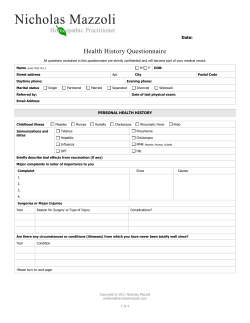


© Copyright 2026