
IN A PIEDIATRIC UNIT - Postgraduate Medical Journal
Downloaded from http://pmj.bmj.com/ on December 29, 2014 - Published by group.bmj.com
POSTGRAD. MED. J. (1964), 40, 590
THE INVESTIGATION OF AN OUTBREAK OF
PSEUDOMONAS PYOCYANEA INFECTION
IN A PIEDIATRIC UNIT
J. JACOBS, M.D., M.R.C.P., D.C.H.
Visiting Professor of Paediatrics, and Head of Department of Paediatrics, Hadassah University Hospital,
University of Jerusalem, Israel.
Consultant Padiatrician, Welsh Regional Hospital Board and United Cardiff Hospitals, Lecturer in
Paediatrics, Welsh National School of Medicine.
THE INTRODUCTrION of potent antimicrobial
agents has resulted in the lesser frequency of
potentially fatal infections caused by 8-haemolytic streptococci and pneumococci, but the
greater frequency of staphylococcal, fungal and
other bacterial infections of the coliform and
related gram-negative species. Pseudomonas
pyocyanea or Ps. aeruginosa owing to its notorious resistance to therapy occupies a unique
position.
Charrin (1890) first recognised pseudomonas
as a pathogen in man and recently many
descriptions of the disease have been given.
Curtin (1957), a leader in the Lancet (1961),
Rogers (1960), Markley, Audmendi, Charez
and Bazan, (1957), Sussman and Stevens (1960),
Williams (1960), Lubsen (1961), Curtin, Petersdros and Sennott (1961), and McCabe and Gee
Jackson (1962), all warn of the frequency
of the infection in young and debilitated
patients, in burns, in orthopaedic cases, in
urinary problems, in gram negative septicemia,
in geriatric patients, in premature infants and
those with congenital abnormalities. Deaths
were frequent in most of the series.
Clinical Material
Between July, 1960 and March, 1964 some
73 cases of Ps. pyocyanea infection have been
seen in paediatric units in various hospitals in
Cardiff. The patients could be placed in the
following groups:
1. Associated with prematurity-49 patients
with nine deaths including two infants with congenital abnormality of the urinary tract and one
cleft pa'late with other congenital abnormalities,
(trisomy 18). These occurred in a premature
unit (A).
2. Associated with congenital abnormalities16 patients. (a) 12 meningomyeloceles (7 died)
in a pxediatric unit (B) (b) one child with cleft
lip and palate (trisomy 15-17) (who died later
but not of the disease) in a paediatric unit (C)
(c) one child with hydroureter and hydronephrosis (alive) in a paediatric unit (C) (d)
two children with abnormal hearts (large
patent ductus; pulmonary atresia) (both died)
in a paediatric unit (D) of a chest hospital.
3. Associated with leukwemia- two patients
(who died later but not of the disease) in a
unit (C).
paediatric
4. Associated with chronic disease-two
patients with fibrocystic disease (both died
later but not of the disease) in a pwediatric
unit (C).
5. Chance findings- four patients: - two
with infected urine (in a pxediatric unit), one
neonatal infant with no obvious abnormality
who survived (in a paediatric unit) and one
neonatal infant with no obvious underlying
abnormality (transferred from a neonatal paediatric unit with a chest infection to a chest unit)
who died of pyocyaneas infection of lungs.
In these various infants, positive swabs were
obtained from umbilicus (4), urine (29--including two at post mortem), lung (10-all at post
mortem), rectal swabs and stools (51), skin (3),
ear (3), eye (3), nose and throat (34), meninges
(two-at post mortem) and pericardial fluid
(one-at post mortem).
Classical Symptoms and Signs
Of the premature infants in group 1, 30 were
admitted from home or from outside maternity
units, whilst 19 were born in the hospital
containing the premature unit. The weights lay
between 2 lbs. 4 ozs. (1,023 g.) and 5 lbs. 5 ozs.
(2,360 g.). The disease commenced at the age of
one to fifteen days and deaths occurred from
one to twenty-three days after birth. 19 infants
had signs of respiratory distress syndrome
within eight hours of birth and it was difficult
to recognise when the respiratory problem had
altered to one of a generalised infection. Later
cases showed a fall in temperature (from
95°F to 870°F), a gradual increase in cyanotic
attacks and, in some cases a peculiar type of
diarrhoea with a brownish-black stool, semi
solid in character, sometimes with slime or
blood. Some infants had an odd membrane on
the mouth, starting with thrush-like white
patches, but proceeding to small brown
Downloaded from http://pmj.bmj.com/ on December 29, 2014 - Published by group.bmj.com
October, 1964 JACOBS: Investigation of Outbreak of Pseudomonas Pyocyanea Infection
lesions with black centres. These lesions spread
·*·····1·
.-··
to cheek and oropharynx and later to the
.:: .·:.·~ ::·:·::·: ·· :;.::
anus, the rectal margin often being everted.
Occasional infants had convulsions (only one
:·:
li
:*:
··i·:.::
.:
···:
·······
meningitis). The infants
being associated with
became
anorexic
and
often
regurgitated easily,
died after vomiting blood. The abdomen
showed the picture of ileus with distension
:··K·.;L.
···.
·''iiiiii~:iii.::iii·:':
:·:::*
and ladder patterning. Oedema was a prominent
feature.
Various skin lesions were seen, a toxic
erythema (like erythema multiforme); "ecthyma
gangrenosum" with a characteristic macule
changing to an indurated area with a black
necrotic centre; generalised purpura (in one
to ecchymoses); pustular lesions
proceeding
(being clusters of vesicles growing Ps. pyocyanea) and small pink nodules.
The blood picture showed little. Anaemia and
leucocytosis were rarely seen, the common finding being the presence of toxic granules with
neutrophilic shift to the left. Thrombopenia
was noted in two cases.
Sixteen cases had had previous antibiotic
therapy (13 methicillin or cloxacillin and three
ampicillin) and of these four died, whereas
five died of the remaining cases not given antia
biotics previously.
In the 16 patients in Group 2, the infection
at the operation sites which became
developed
brown-black and necrotic. Meningitis appeared
with the usual physical signs (pyrexia, neck
stiffness, bulging of fontanelle, lethargy, vomiting, cyanotic attacks and convulsions.
Occasionally
patients seemed to ,live in symbiosis with the organism. Death was from septicaemia and meningitis. Antibiotics had been
used. The patients in the chest unit
previously
were considered to be cardiac problems, in
one instance with a superadded pulmonary
infection. Skin lesions developed in one just
before death, whilst in both the organism was
only found at death (again antibiotics had been
used).
In groups 3 and 4, infective episodes were
shown to be due to Ps,pyocyanea infection, but
the patients survived. All patients had been on
antibiotics, the leukaemia' patients also having
had steroids and methotrexate.
In group 5 one mature infant died at the
age of five weeks in a chest unit after transfer
from a neonatal unit. Multiple antibiotics had
been given for a chest infection, but ventilation
become inadequate even after tracheostomy.
Post Mortem Findings
The characteristic lesion on the tongue
.·n·.:· :
591
·a
Z%
'"""::j..:
:· ··:·:p ··.,:
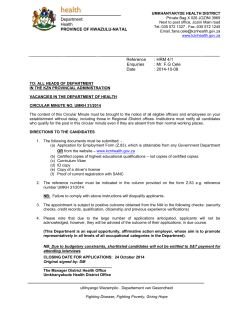
FIG. 1.-Skin, showing arteritis of vessel in dermis.
H& E X 80.
showed an area of muscle necrosis and infiltra-
tion.
Gram stains showed numerous gram
negative bacilli in the necrotic areas. The lungs
showed intra-alveolar and intra-bronchial polymorphonuclear exudates and haemorrhage. In
places there was necrosis and abscess formation
with gram negative bacilli. The walls of small
blood vessels showed acute inflammatory
arteritis (Fig. 1) the thrombosis secondary to
this leading to arterial occlusion and small
infarct-like areas (Fraenkel, 1917). The affinity
of the organism for the wal,ls of small vessels
was seen in all tissues showing lesions, i.e.,
skin, lung, brain and kidney. (Fig. 1).
In the gut necrotic non-exudative lesions were
seen with a yellowish opaque centre and deep
red margins.
Investigation of Epidemics
In the premature unit involved, cases have
occurred over a period of more than threeand-a-half years. Conditions in such a unit are
conducive to epidemics in that infants are
in debilitating situations, often being very
small, occasionally having congenital abnormalities liable to high levels of bilirubin with
resultant lethargy and difficulty in feeding,
while the unit is liable to sudden increases in
numbers in the ward with overcrowding
of nurse/patient ratio. Such patients are prone
to respiratory difficulties and tend to be treated
with an "antibiotic cover" to prevent possible
infection or deal with apparent infection. Again
being small and in poor condition they tend to
be nursed in an incubator with a relatively
high humidity atmosphere. (Hoffmann &
Finberg, 1955). The ambient temperature and
humidity in a premature unit tends to be higher
than average. All these conditions produce
a
situation in which infections like Monilia
Downloaded from http://pmj.bmj.com/ on December 29, 2014 - Published by group.bmj.com
POSTGRADUATE MEDICAL JOURNAL
592
TABLE I
MODES OF SPREAD
Previous
Dust
Dust
1
Airborne
No positive swabs were found initially in
any part of the unit except in the incubators
which had held a case. Later swabs were
cases
positive in various sinks.
A general investigation in various wards in
the hospital (including the labour ward), and
was no
nearby hospitals,inshowed that there
uniform policy
any section with regard
to sterilization. For thermometers sterilization
Methods of cleaning
Incubators, AirHand driers
conditioning
Humidifiers
particles
Nose
Hands
Contact
Procedures
October, 1964
Dressings
Suction tubes, oxygen supply
"Sterilising" materials (quarternary
pounds, etc.).
Injections
Lumbar
com-
puncture
Intravenous tubes
albicans and Ps. pyocyanea tend to flourish.
All the cases concerned in the unit had been
in incubators. Those admitted from other units
could not have been initially affected in this
way. All methods of spread were considered
(Table 1).
Swabs were taken (1) from skin, nose and
throat of all personnel (doctors, nurses, tech-
nicians, cleaners, mothers), (2) piped oxygen
sets. (3) Floors, wal,ls, basins and sinks (4)
incubators-from the water in the humidity
tank as well as walls and floor of the apparatus. Plates were put out in the unit.
All chemical antiseptic procedures were
reviewed-the fluid in the tube holding thermometers, for keeping polythene tubes for feeding,
for cleaning tubing, for cleaning incubators,
etc.
Drying procedures, the use of hot air hand
looked at as a possible sourse of infection.
Techniques used were found to be
inadequate.
Quaternary compounds were
used in many places (incubator,
being
thermometer tubes, Woolf bottles, for damping
dust and cleaning tubing). Rubber tubing
was left attached to suction apparatus and
found to contain moisture.
driers, towels, both ordinary and paper, were
(Table 2) involved the use of some six different
materials-some of which looked nice but were
of no value from a sterilization point of view.
Brushes were cleaned in a variety of substances;
rubber tubing was treated with one of seven
different media. In general the antiseptic used
related to the material which the sister in
charge of a ward had been accustomed to
using in her training hospital. With regard to
strength of solution used, it could be taken
as a general rule that wherever solutions were
made up on the ward from a stock solution
which required diluting, the final solutior was
usually dependent on how the person making
up the solution felt about the colour rather
than on suggested dilutions needed.
In the chest unit where the two children
were infected, about eight adult chest cases
were also infected. Here positive swabs were
obtained from the noses of radiographer, physio-
therapist, nurses, orderlies, cleaner, laboratory
technician and one doctor, as well as in the
rubber tubing attached to anaesthetic apparatus, sucker, nozzle, a brush from a cleaning
bottle, the outside of a pulmoflater and also
on plates put in the post-operative ward.
Positive swabs were obtained from a sink
and from a wall.
In the unit with the meningomyeloceles, the
positive swabs were obtained from the sink
disposal unit, but not from the atmosphere or
any personnel.
In the units concerned an attempt was first
made to assess the value of antiseptics in
common use. Dilutions of antiseptic in terms
of the active ingredient 'after a 21-minute
TABLE 2
THERMOMETERS
Hospital
'A'
Hospital
'B'
Hospital
'C'
Hospital
'D'
Hospital
'E'
Roccal (5)
Phenol (1)
Glycothymoline (13) Savlon (1)
Glycothymoline (7)
(3)
Glycothymoline
Glycothymoline
(7)
Glycothymoline
(3)
Savlon (2)
Hibitane (1)
Hibitane (5)
Bradosol (4)
Dettol (1)
Hibitane (2)
(The number represent the numbers of wards using material)
Downloaded from http://pmj.bmj.com/ on December 29, 2014 - Published by group.bmj.com
593
October, 1964 JACOBS: Investigation of Outbreak of Pseudomonas Pyocyanea Infection
antiseptic and bacteria were
noted (Table 3), Table 4 shows the ratio
between dilution for complete kill and
recommended strength.
A change was made in the premature unit
to the use of hibitane, C.T.A.B. and hexaas well as savlon (a mixture of
chlorophene,
chlorhexidine 1.5 per cent. w/v, -and centrimide 15 per cent). Ps. pyocyanea has been
shown to survive in cetrimide alone
contact test of
('Cetavlon'), (cetyltrimethylammonium cromide) (Robinson 1957) especially when bark
corks were used in the bottles. Anderson
(1959) grew the organism in the humidifying
water in an operating theatre ventilation plant.
Coliform bacteria have
also been found at the
same site, also in incubators and where lumbar
puncture needles have been inadequately
cleansed. McLeod and Mason (1963) have
shown how topical antibiotics ('polybactrin'
polymyxin 0.4 per cent., neomycin 1 per cent
and bacitracin 1 per cent) are superior to
chlorhexidine or domiphen as local methods
of killing bacteria.
Treatment
The tendency of Ps. pyocyanea infections to
occur in damaged ischaemic and fibrotic or
debilitated
patients (premature infants, congenital abnormality, "poor genetic material"trisomy 15-17, 18 etc.") (Smith 1963) makes
difficult. The most important part of
therapy
treatment is prevention of the entry of the
organism into the patient, but as the organism
is ubiquitous this means keeping a careful
check on the conditions of susceptible patients
and their surroundings. (Table 5). Regular
surveys must be taken of personnel, apparatus,
procedures and antiseptics in use. Great
thought must be taken before antibiotics are
prescribed and their use in prophylaxis abandoned. "Chemotherapy without bacteriology
is guesswork." Serious considerations should
be given to the use of disposable apparatus
of all kinds-brushes, tubing, syringes, needles
and so on, so that the need for the use of
antiseptics will diminish.
A relief can be obtained by closing down a
particular unit and fumigating with formaldehyde vapour. In the premature unit involved
this procedure has had to be repeated for only
temporary relief has been obtained, and the
infection has continued for almost four years.
The sinks have given an intermittent
positive finding of organisms to a degree that
one wondered whether the U-tube part of the
TABLE 3
2m-MINUTE
CONTACT TESTS OF ANTISEPTICS WITH
BACTERIA IN WATER, BROTH AND
BLOOD BROTH
25%
Ps.pyocyanea
Water
25%
Broth Blood
Hibitane (Chlorhexidine)
80,000 20,000 2,000
Domiphen bromide ,(Bradosol) 48,000 2,000 400
Phenoctide
10,000 400 100
p,Chlor-m-xylenol
3,330 312 104
4.8 % solution
Cetavlon (Cetrimide)
8,000 1,000 400
Benzalkonium chloride (Roccal) 32,000 1,500 500
Sodium Hypochlorite
10,000 200
The
figure show
terms
the final dilution of antiseptic in
of the active ingredient
TABLE 4
RATIO BETWEEN DILUTION FOR COMPLETE KILL AND
RECOMMENDED USER STRENGTH-22 MINUTE CONTACT
Recommended Ps.pyocyanea
dilution for
25 %
midwifery
lotion
Broth blood
Hibitane 2%
10
1
1/40
1
0.2
Domiphen bromide 5% 1/100
0.6
0.2
p-Chlor-m-xylenol
1/25
4.8% solution
Cetavlon 1%
1
0.4
1/10
Benzalkonium chloride 1/10-1/20 1.5-7.5 0.4-0.2
1%
Sodium hypochlorite 1% 1/20
0.025
TABLE 5
PREVENTION
1. Review of all chemical antiseptic procedures
2. Review use of antibiotics
3. Review water-borne carriage of disease (also airborne)
4. Bacteriological supervision of staff, patients
5. Isolation of cases
6. Adequate treatment
7. Closure of unit
drainage might represent a constant source of
re-infection. An attempt to sterilize this area
by use of Phenoxetol (phenoxyethanol) in
strength above one per cent or Wescodyne
Fluid (Iodine-detergent complex (idophor)
from 1/160 (one ounce to a gallon) to 1/20
(eight ounces to the gallon)), Dettol and Lysol
have had only a temporary effect on the
positive swabs from the washbasin and sinks.
Treatment of the actual disease in a patient
has varied from the use of Phenoxetol one per
cent as a local application to a skin lesion or
to a discharging ear, to streptomycin by intramuscular injections alone (dosage 20-40
Downloaded from http://pmj.bmj.com/ on December 29, 2014 - Published by group.bmj.com
594
POSTGRADUATE MEDICAL JOURNAL
or with polymyxin B. Colistin
mg./kg./day)
has been used in the majority and thiosporin
in about three cases. Of the nine deaths in
the premature group, three were virtually dead
were the two
started
before treatment was
(as
cases in the chest unit), two more were treated
early in the experience of polymyxin, the
remaining four being two with colistin and two
with thiosporin.
The experience of finding infants apparently
having respiratory distress with positive swabs
for Ps. pyocyanea is becoming increasingly
common (personal communications-other
units). There is now a tendency to treat any
episode of pyrexia changing to hypothermia
of odd ileus or skin rash or sudden anorexia
in the premature babies, by giving one of the
polymyxin series.
The polymyxin preparations used have been
B. sulphate, sulphomethylpolymyxin
polymyxin
(both in dosages of 2,000 units/kg. 4 hourly),
colistin sulphate and colistin methanesulphonate
(both in dosages of 50-100,000 units/kg. 6-8
third
hourly). It would seem that the first andfourth
are virtually the same as the second and
No evidence of toxicity has been
preparations.
seen in the dosage used.
No more than impressions of the value of
can be given, for the disease does
therapy
seem to vary from the patient living in symbiosis with the organism, to a severe septicaemia with rapid death. To evaluate any agent,
the stage of the disease at which diagnosis is
made and treatment started has to be judged.
one of the polymyxins, certainly patients
Using
with generalised cedema and cyanotic attacks
have been seen to recover.
October, 1964
No toxic reactions to polymyxin B in the
form of renal damage (other than that due to
the disease) or fever were seen (the other
described effects of dizziness, ataxia, peri-
pheral neuritis, parasthesia, especially itching
and facial flushing, not being recognised). With
colistin no fever, apparent pain at site of
injection or nausea was seen, nor leucopenia or
azotaemia.
The other infants in the series (meningomyeloceles), were treated by intramuscular,
intravenous and intraventricular colistin and
seven died, so that only two with meningitis
tended to recover from the disease whilst the
other three seemed to confine their disease to
the local area with little generalised spread.
The leukaemia patients had negative swabs
after the use of colomycin. The children with
fibrocystic disease did not get disseminated
disease when given colomycin, although becoming intermittently positive. The other cases
responded to systemic colomycin; Zynotracin
powder (xanthocillin, zinc bacitracin, hydrocortisone) was used locally on the umbilicus.
An attempt was made to isolate each case
as it developed and "prevention of cross
infection" techniques carried out.
Summary
A total of 73 cases of pyocyanea infection
have occurred, in 49 premature infants, 16 with
congenital a'bnormalities, two with leukzemia,
two with fibrocystic disease and four other
children with infection.
The available means of treating the infection
and endeavouring to prevent further cases have
been described.
RIR?FRFRNCF.
ANDERSON, K. (1959): Pseudomonas Pyocyanea disseminated from the Air Cooling Apparatus, Med. J. Aust.,
1, 529.
CHARRIN, A. (1890): Maladie Pyoconique chez 1'Homme, C. R. Soc. Biol. (Paris), 9, 496.
CURTIN, J. A., PETERSDORF, R. G., and BENNETT, I. L. Jr. (1957): Acquired Arteriovenous Fistula complicated
by Ps. aeruginosa Endarteritis and Endocarditis, Bull. Johns Hopk. Hosp., 101, 140.
- (1961): Pseudomonas Bacteremia, Review of 91 Cases, Ann. intern. Med., 54, 1077.
FRAENKEL, E. 1(1917): Weiters Untersuchungen uber die Menschen-pathogenitat des Bacillus Pyocyaneus, Z.
Hyg. Infekt. -Kr., 84, 369.
HOFFMAN, M. A., and FINBERG, I. (1955): Pseudomonas Infection in Infants associated with High Humidity
Environments, Pediatrics, 46, 624.
LUBSEN, N., BUISSEVAIN, W., and FASS, H. (1961): Chlorhexidine as the Probable Cause of an Increase in
Proteus Rettgeri Infections of the Urinary Tract. Lancet, i, 921.
MCCABE, W. R. and JACKSON, G. G. (1962): Gram-negative Bacteremia, Arch. intern. Med., 110, 847,
856.
ROBINSON, G. L. (1957): Pscudomonas Pyocyanea in Cetrimide (correspondence), Brit. med. J., i, 1242.
MARKLEY, K., GURMENDI, G., CHAREZ, P. M., and BAZAN, A. (1957): Fatal Pseudomonas Septicaemia in Burned
Patients, Ant;. Surg. 145, 175.
LANCET (1960): 'Green for Danger', ii, 352.
MCLEOD, J. W., MASON, J. M., and PILLEY, A. (1963): Prophylactic Control of Infection of the Urinary Tract
Consequent on Catheterisation, Lancet, i, 292.
ROGERS, K. B. (1960): Pseudomonas Infections in a Children's Hospital, J. Appl. Bact., 23, 3.
SMITH, D. W. (1963): The No. 18 Trisomy and D, Trisomy Syndromes, Ped. Clin. N. Amer., 10, 389.
SUSSMAN, M., and STEVENS, J. (1960): Pseudomonas Pyocyanea Wound Infection, Lancet, ii, 734.
WILLIAMS, R., WILLIAMS, E. D., and ADAMS, D. E. (1960): Cross Infection with Pseudomonas Pyocynanea,
Lancet, i, 376.
Downloaded from http://pmj.bmj.com/ on December 29, 2014 - Published by group.bmj.com
The Investigation of an
Outbreak of Pseudomonas
Pyocyanea Infection in a
Paediatric Unit
J. Jacobs
Postgrad Med J 1964 40: 590-594
doi: 10.1136/pgmj.40.468.590
Updated information and services can be found
at:
http://pmj.bmj.com/content/40/468/590.citatio
n
These include:
Email alerting
service
Receive free email alerts when new articles cite
this article. Sign up in the box at the top right
corner of the online article.
Notes
To request permissions go to:
http://group.bmj.com/group/rights-licensing/permissions
To order reprints go to:
http://journals.bmj.com/cgi/reprintform
To subscribe to BMJ go to:
http://group.bmj.com/subscribe/


© Copyright 2026