
Matrix Ellison Institute poised for preeminence Also in this issue:
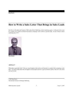
Matrix
Volume 4 • Number 5
UC Davis
School of Medicine
and Medical Center
August 1997
Also in this issue:
Ellison Institute poised
for preeminence
In the Silicon Valley, Lawrence J. Ellison found
everything he needed to build a new software idea
into a Fortune 500 company. But when the selfmade computer billionaire shattered his left elbow in
a 1992 bicycle accident, he had to go to the Central
Valley to find a surgeon with the skill to repair it.
At UC Davis Medical Center, Department of
Orthopaedics Chair Michael W. Chapman reconstructed Ellison’s elbow in a series of two operations,
restoring the badly fractured joint to full function.
When Ellison, 52, recently challenged world-class
triathlete Peter Kain, 32, to a “bar dip” contest to
benefit a charity, Ellison won. He did 49 of the
pushup-like exercises to Kain’s 40.
“All the king’s horses and all the king’s men
couldn’t put Humpty together again,” a grateful
Ellison said after his final operation. “But Mike
Chapman could. And did.”
Ellison, co-founder and chief executive officer of
Oracle Corporation, expressed his thanks with a $5
million gift to support musculoskeletal programs at
the UC Davis School of Medicine and Medical Center.
The gift, matched by a $1.3 million UC Davis Health
System contribution, represents the largest single private bequest in the institution’s history.
“I wanted to make the art and science of Mike
Chapman and his colleagues generally available, instead of available only to the few,” Ellison said in
announcing his 1994 bequest. “I wanted more people
to be able to benefit.”
They soon will. The Lawrence J. Ellison
Neuromusculoskeletal Institute, a newly designated
UC Davis Health System Center of Excellence, is evolving into one of the preeminent clinical and research
centers for connective tissue disorders in the world.
The Institute brings together under a single umbrella
patient care and research programs within several
departments, including orthopaedics, physical medicine and rehabilitation, neurology and neurosurgery,
radiology and nuclear medicine. In addition, Institute
After a full recovery from a shattered elbow, computer billionaire
Larry Ellison (far right) beat triathlete Peter Kain (middle) and Jim
MacLaren in a charity weight lifting contest.
Page 3
One-stop shopping
Page 5
The mysteries of tissue
regeneration
clinicians and investigators will coordinate their
efforts with other departments that pursue musculoskeletal research, including the Veterinary
Orthopaedic Research Laboratory, the Otolaryngology Research Laboratory, the Division of Materials
Science, the Biomedical Engineering Graduate
Group and the Departments of Physical Education,
Civil Engineering and Mechanical and Aeronautical
Engineering.
“Combined with the resources of the Medical
Center, the new Shriners Hospital, the School of
Medicine, other centers on the Davis campus and
the UC Davis Orthopaedic Research Laboratories,
(continued on page 2)
Page 6
Cartilage implants for
knee injuries
Page 7
Cartilage transplants
Page 8
New neurosurgery chair
Page 10
Implantable pump for
chronic spasticity
UC Davis Matrix
Connective tissue research, repair a focus
New clinical
programs
TheNeuromusculoskeletal
Center of Excellence will
encompass at least 16
separate clinical programs.
The programs will be
housed together in the
Lawrence J. Ellison
Ambulatory Care Center, due
to open next summer. Their
aims will be to increase
communication and
collaboration among the
varied specialists who treat a
particular disease or disorder;
to streamline patient
referrals and care; and to
facilitate comprehensive,
prospective outcomes studies.
“Patients now have to
go to one place for one thing
and another place for another
thing, requiring multiple
appointments and inhibiting
interdisciplinary communication among treating
physicians,” said center chair
Ralph Johnson.
“This can be resolved by
having a program with a
coordinator who can bring it
all together, and by housing
services in a single building
so that the patient comes to
one place for a thorough
evaluation and treatment.”
The Spine Care Program,
which opened May 1, was
the first to begin seeing
patients under the aegis of
the new Center of Excellence.
Southern California spine
surgeon Munish Gupta has
been recruited to head the
program. According to
Johnson, the program will
bring together surgical and
nonsurgical health professionals skilled in the
diagnosis and treatment of a
broad spectrum of spine
2
(from page 1)
the Institute should eventually
“Thanks to this gift,
funds will elevate the lab “to the
provide us with the biggest
first rank of such laboratories in
critical mass of investigators in
we have the potential
the world.” The lab’s primary
the molecular and cellular biology
focus has been trying to
of connective tissues anywhere in
within 10 years of
understand how bone, cartilage,
the world,” Chapman said.
tendon and ligament cells
The Institute will specialize not
having a research
continually renew and repair
only in orthopaedic problems like
themselves as they adapt to the
Ellison’s shattered elbow, but also
program in musculochanging demands of daily
in muscular and neurologic disoractivities.
ders stemming from such diseases
skeletal and neuroOnce this fundamental
as cerebral palsy, multiple sclerosis
mystery has been solved,
and stroke.
muscular biology here
investigators will have a means of
“We’re excited about it,” said
healing and preventing connecRalph Johnson, acting chair of the
at Davis that will be
tive tissue diseases, from arthritis
new Center of Excellence. “It will
to osteoporosis. “Ultimately we
bring together the competence of
unmatched. It will
may learn to reconstruct the
those departments and specialists
skeleton biologically rather than
who care for neuromuscular and
make available to the
with carpentry,” Martin said.
musculoskeletal disease, provide
It took master carpentry to
comprehensive care to patients and
citizens of our region a
repair Ellison’s bicycle injury.
offer both new research opportuniThe software magnate suffered an
ties and new opportunities for evalulevel of care that is
intraarticular T-type supracondyation of treatment outcomes.”
lar fracture of the humerus,
Three new endowed chairs conunparalleled and
complicated by loss of motion in
stitute the centerpiece of the Instithe elbow.
tute. The Lawrence J. Ellison Chair
provide them with
Ellison’s Bay Area orthopaein Musculoskeletal Molecular
dist, a member of a prominent
Biology, assumed April 1 by A. Hari
access to the most
sports medicine practice that
Reddi, is endowed with $2.7
provides team physicians for the
million from the Ellison bequest
recent advances as they
San Francisco 49ers, promptly
and $500,000 in UC Davis Health
referred the complicated injury
System matching funds.
come along.”
to Chapman, an internationally
The other chairs will be
renowned traumatologist.
activated when the ribbon is cut
In one operation, Chapman
next summer on The Lawrence J.
fixed the fracture with plates and
Ellison Ambulatory Care Center. The
screws. In a second surgery, he
David Linn Chair in Orthopaedic
removed the hardware and
Surgery, named for Ellisons brother-in-law,
performed a soft tissue release that restored the
will support the Chair of the Department of Orthojoint’s function.
paedics. Its endowment includes $1.3 million from
“He got essentially a perfect result — an almost
the Ellison gift and $300,000 in Health System
unheard-of outcome,” Chapman said, attributing
matching funds. The Doris Ellison Linn Chair in
Ellison’s spectacular recovery in large measure to
Bone Biology, named for Ellisons sister, will support
Ellison himself. The same drive and determination
research at the Orthopaedic Research Laboratories. It
that took Ellison to the top of the software business
carries a $1 million endowment from the Ellison gift.
carried him through the painful, months’-long
Rather than paying the salaries of incumbent
rehabilitation process. During that ordeal, Ellison
faculty who assume the chairs, the David Linn and
first proposed making a gift to the Department of
Doris Ellison Linn endowments will be used to
Orthopaedics.
support additional post-doctoral fellows, graduate
Ellison has a history of backing winning ideas.
students, technicians, equipment and supplies in
Born in New York City and raised in a tenement on
orthopaedic research.
Chicago’s South Side, the son of Russian Jewish
R. Bruce Martin, director of the Orthopaedic
Research Laboratories, predicts the endowment
(continued on page 9)
August 1997
New building centralizes multispecialty care
house special procedures and pain clinics; obstetFor patients seeking care at UC Davis Medical
rics and gynecology; ophthalmology; dermatology;
Center, the Lawrence J. Ellison Ambulatory Care
urology; plastic surgery; and academic offices for
Center will mean one-stop shopping for services that
the departments of obstetrics and gynecology,
now require visits to multiple office sites along
ophthalmology and family practice. The top floor
Stockton Boulevard. For physicians and researchers,
will be home to an Alzheimer’s disease clinic and
the shared space will mean new opportunities for
academic offices for faculty in orthopaedics,
interdisciplinary collaboration. And for the UC
physical medicine and rehabilitation, neurology and
Davis Health System, the building will mean a more
neurosurgery, pain management, urology, dermacost-effective alternative to expensive leased office space.
tology and radiology.
The four-story, 375,000-square-foot building is
The building also will feature an outdoor
rising along 48th Street on the eastern edge of the
wheelchair PAR course designed to teach patients
medical center’s 140-acre Sacramento campus.
in wheelchairs to negotiate sand, gravel, small
When completed next July, the $81.2 million
bridges and other everyday obstacles.
building and its 124 exam rooms will accommodate
Named for Oracle Corporation chairman and
up to 806 patients a day — about 70 percent of all
chief executive officer Lawrence J. Ellison, a former
ambulatory patients seen at the medical center.
patient and major benefactor, the building is part of
The expansive facility will also house 250 faculty
a long-range development plan adopted by the UC
offices, a visitors cafeteria and an employee food service
Regents in 1989.
operation. A new freestanding parking structure, con“The Ellison Ambulatory Care Center is going
nected to the building by a landscaped breezeway, will
to change the way we do business,” said Ray
provide convenient parking for 800 cars.
Groom, manager of medical sciences planning. “A
“The Lawrence J. Ellison Ambulatory Care
patient will be able to see a physician, get an X-ray
Center is the culmination of the Neuroor other radiographic diagnostic procedure, fill a
musculoskeletal Center of Excellence,” said Center
prescription and have lunch without ever leaving
Director Ralph Johnson. “It will bring together all
the building. Literally everything they need will be
those departments that deal with neuromuscular
right there.”UCD
and musculoskeletal problems in the same building
for clinical evaluation, imaging studies and
treatment.”
Occupying the basement floor will be the neurology and neurosurgery clinics; the cardiac rehabilitation and cardiology clinics; and an
imaging center. On the
first floor will
be
orthopaedics;
physical
medicine and
rehabilitation;
occupational
and physical
therapy; indoor therapy
pools;
an
orthotics laboratory; family
practice; the
remainder of
the imaging
center and a
pharmacy.
The secThe Lawrence J. Ellison Institute will open in July 1998.
ond floor will
problems, from fibromyalgia
to workers’ compensation
injuries.
An arthritis and total
joint replacement program will
be the Center’s second major
priority, Johnson said. Other
top priorities are programs in
stroke treatment and sports
medicine.
A trauma/vascular
surgery program will be
headed by Paul Muizelaar,
new chair of the Department
of Neurological Surgery and a
nationally recognized vascular
surgeon. Muizelaar is bringing
UC Davis to the forefront of
research into ways to improve
the outcomes of people with
traumatic brain injury.
Plans are also underway
for an anesthesiology/chronic
pain management program
and a spasticity management
program for patients who
suffer from chronic, debilitating muscle spasms due to
stroke, cerebral palsy,
multiple sclerosis and a wide
range of other injuries and
disorders. Available therapies
for these patients include
tendon-lengthening surgery
and use of an implantable
pump to deliver baclofen, a
drug that inhibits ungoverned
muscle reflexes.
Johnson is meeting with
a committee to identify other
clinical programs that may be
developed within the Center
of Excellence. Ultimately, the
Center may encompass as
many as 30 programs.
3
UC Davis Matrix
New Ellison Chair studies the
mysteries of tissue regeneration
This story begins with a mystery. In 1965, UCLA
surgeon Marshall Urist ground up rabbit bones and
demineralized them in acid so that only powder
remained. When Urist sprinkled the powder into the
muscle of a living rabbit, the unexpected happend:
new bone started to grow in the muscle tissue.
What was it about the bone powder that could
cause muscle tissue to become new bone? For
science to be able to take practical advantage of this
wonder, someone would have to isolate the specific
factor or factors that caused bone to grow.
Finding the answer would take nearly twenty
years. And it would deliver into the hands of science
an unprecedented power: to regenerate bone and
learn the secrets of tissue formation.
The solution of this mystery story has A. Hari
Reddi at its center. Reddi, the holder of the new
Lawrence J. Ellison Chair in Molecular Biology at
the UC Davis School of Medicine and Medical
Center, is not the only one who helped solve
the mystery. Science is additive and collaborative.
But the contributions of Reddi, who comes to UC
Davis from an endowed chair at Johns Hopkins,
were pivotal.
In his laboratory at Johns Hopkins, and before
that at the National Institutes of Health, Reddi
struggled with the implications of Urist’s results.
Reddi persisted at a time in the 1970s and early
1980s when most other scientists had lost interest in
what they considered a hopeless quest.
“He kept the field alive,” said Vicky Rosen, a
biochemist at Genetics Institute, a biotechnology
4
A. Hari Reddi, I
company based in Cambridge , Mass. Ultimately,
the solution would come from the biotechnology
industry, but not before Reddi and his colleagues had
squeezed clues from the dry bones — clues that
would later allow him to embark on an ambitious
program of tissue engineering.
In any mystery story, the person at the center
has to find his way to the action . In Reddi’s case, the
journey was particularly long. It began in Madras, in
the dusty southern part of India, far to the south of
Delhi. Already as a student at Annamalai University
in the isolated town of Annamalainagar, 100 miles
south of Madras, Reddi knew that he wanted to do
research in the United States.
“There was very good research in Madras at
the time,” Reddi said. “But it wasn’t enough. So I
decided to travel 1,000 miles to pursue scientific
studies at the graduate level in Delhi. Such things
were almost never done,” he said.
Reddi advanced by learning from a series of
successful mentors. His graduate advisor, M.R.N.
Prasad, had done research in Wisconsin. “He
encouraged me to work independently,” in contrast
to the more hierarchical style then in vogue in Indian
universities.
Reddi’s independent thinking paid off. One of
the readers of Reddi’s dissertation, H.G. WilliamsAshman of Johns Hopkins University,
recommended Reddi for the distinction of magna cum
laude. Soon Reddi was on the way to WilliamsAshman’s laboratory for post-doctoral work.
What came next was the move that put Reddi in
the right place at the right time for addressing the
mystery. In 1969, Williams-Ashman moved to the
University of Chicago and Reddi joined him. Down
the hall from their lab was Charles Huggins,
a powerhouse of biochemistry, who was to win
the 1966 Nobel Prize for his work in understanding
the endocrine regulation of prostate cancer. Trained as
a surgeon, Huggin’s rapacious intellect kept bringing
new questions and new collaborators into his
laboratory. Urist had been a student of Huggins’, back
in the 1940s, and Reddi would join him.
“But I already have a firm offer to go to Harvard
and to return to India after that,” Reddi recalled
telling him. But Huggins immediately picked up the
phone and dialed Reddi’s erstwhile mentor at
Harvard, saying, “He’s not coming!”
“It was a decision that changed my life and my
focus of research,” he said.
Huggins had planted the seed for Urist’s work
many years earlier, when he implanted a canine tooth
fragment into the abdomen of a dog and showed that
August 1997
The experience of
isolating bone induction
normal environment. But success
factors gave Reddi
new bone formed there. But the
in bone tissue culture meant that
mystery was even more baffling
bone cells would begin to calcify
unique insight into
then than it would become thirty
to the bottom of the dish,
years later.
limiting their own growth. That
tissue formation. In
Years passed. Reddi became a
made it even more difficult to
professor at University of Chicago,
determine what had caused the
some ways, bone is an
then accepted an offer to become a
cells to grow.
visiting scientist at NIH in
Another obstacle, one that
unusual tissue but in
Bethesda. Reddi had successes in
had stymied Urist, was that he
other areas, especially in characterdid not have a reliable assay for
other ways it is typical.
izing the roles of specific proteins
quantifying bone growth. The
in expanding bones that already
key to “grind and find” lay
existed.
in knowing what one has found,
But all along, he remembered
said Reddi. But the assay
Huggins’ challenge: What
problem seemed insurmountable.
property of the bone powder —
When Kuber Sampath told Reddi he wanted to
or the dog’s tooth — was acting like an inducer,
work on finding “bone induction factor,” Reddi tried
causing bone to grow in muscle or abdomen?
to dissuade him. “Maybe you’d like to think it over,”
Night after night, month after month, Reddi
Reddi said. “You might not get any publications.”
went after the elusive “bone induction factor.” His
But Sampath persisted and ultimately succeeded at
former post-doctoral fellow Kuber Sampath
winning Reddi’s support.
remembers seeing all the notebooks in Reddi’s NIH
Sampath’s presence reinvigorated the search for
laboratory. “Reddi had spent a lot of time looking
the mystery factor. He also brought new methods
for these factors,” recalled Sampath, now a senior
and new ideas from his background as a protein
research director at Creative Biomolecules, a
biochemist. It was a true collaboration, Sampath
biotechnology company in
recalled, free of the usual hierarchy. “Hari never
Hopkinton, Massachusetts.
acted as a senior person,” a superior, said Sampath.
Part of the difficulty was in not knowing if a
Instead, Sampath and Reddi would talk on the
single factor was there to be found. Looking for a
verandah outside Reddi’s NIH office.
bone induction factor was “an orphan project” at
Over the next five years, “we developed a bona
that time, said Rosen. “No one even believed that
fide assay that reflects bone formation,” recalled
there was a factor. They thought it was a mixture of
Reddi with pride. The assay, which is still in wide
things.”
use, involves implanting a pellet with the mixture to
Another problem lay in the nature of bone.
be tested beneath the skin of a rat. In a few days, if
Reddi’s approach is what he calls classic “grind and
the test mixture is active, progenitor cells migrate to
find” biochemistry. It involved taking the tissue in
the area and bone begins to form. Twelve days later,
question , grinding it up and then sifting through it
the implant is removed and the effect of the test
looking for the activity, in this case, bone inducmixture is quantified — the key to a scientific assay
tion. In the days before cloning and sequencing of
— by two different measures.
human genes, grind and find was about the only
With the assay problem solved, it was time to
option available.
find the bone induction factor or factors. UnfortuBut it did not work very well on bones. It was
nately, the amounts of the active substances were so
difficult both to find potential factors and then to
small that they could not be used to characterize the
see what effects they had. Finding factors was hard
molecule. “It would have cost too much money,”
because the scientists had to be so harsh in
said Sampath. “We would have had to extract the
extracting them — how could they be sure that the
substances from twenty kilograms of bone every
acid bath that degradated the bone was not also
week, and the chemicals we used were
irreversibly altering the factor?
expensive.” In the end, he recalled, it cost tens of
Observing the effects was difficult because of
millions of dollars to isolate the gene.
the challenge involved in putting bone cells in
That’s where biotechnology companies stepped
tissue culture. Most cells could be induced to grow
in a somewhat normal manner in a tissue culture
(continued on page 12)
dish, even though they were outside of their
A. Hari Reddi was recently
awarded the Urist award
from the ....(more TK).
5
UC Davis Matrix
Cartilage implants offer treatment
for arthritic knee injuries
6
The talk is generally called “The middle-aged
arthritic knee.” It ‘s not exactly an award-winning
title, but year after year, overflow crowds pack a
lecture on this topic at the American Academy of
Orthopedic Surgeons’ annual meeting to hear about
the latest treatment options.
“The problem is that we see arthritic knees
every day, and there is no single treatment that is as
good as we would like it to be,” said Juan J. Rodrigo,
professor of orthopaedics at UC Davis School of
Medicine and Medical Center.
Knee problems in the United States are reaching
epidemic proportions. In 1994, the last year that
statistics are available, orthopedic surgeons in the
United States performed 311,000 knee surgeries, an
increase of nearly 50 percent from four years earlier.
Hips and shoulders present similar problems and are
responsible for increases in the number of surgeries
on the three joints, which totaled 649,000 in 1994.
Given that there is no “one-size-fits-all” treatment, a medical center like UC Davis does the best
for its patients by offering every available option, said
Rodrigo. “We treat patients who are experiencing the
early stages of arthritis as well as 90-year-olds who
need end-stage treatment revisions.”
Most of the treatments involve cartilage because
while bone heals, cartilage doesn’t. Made up of long
strands of water-cushioned collagen protein,
cartilage is a slippery shock absorber for the bangs
and bumps of life. But sometimes it is torn off, as by
an injury. Or it can rub away due to arthritis or
everyday wear and tear. A long, painful slide into
immobility often results.
In an ideal world, physicians would augment
worn or missing cartilage with more cartilage
through transplantation or regrowth. And that is
just the way the medical profession is moving. The
trend is toward “true biological replacements,” said
Rodrigo. “They offer the potential for remodeling
and healing.”
But until recently, the treatment of choice for
many patients — especially those 45 years old and
up — has been to replace the entire joint with a
metal or plastic prosthesis. With the appropriate
rehabilitation program, recovery time is quick, said
Rodrigo — in as little as three months, patients can
resume many of their previous activities. The
prostheses can last as long as 20 years.
However, knee and hip replacements have their
drawbacks as well. The joints can fail, they can wear
out and they can wiggle loose from their moorings.
Worse still, said Rodrigo, replacement joints almost
inevitably destroy bone and often cause infection. A
second replacement has to be larger for surgeons to
be able to fuse it to healthy bone. And the larger
implant then reduces the patient’s mobility even
further.
When patients are younger than 45, and
especially if the problem is caused by a recent injury,
Rodrigo typically turns to one of the “biological”
treatments — a procedure known as debridement
and microfracture (D&M), which could just as well
be called “scrape and tap.”
The advantage of D&M is that it provides an
opportunity for the patient’s own cartilage to fill in
the gaps. With the aid of a viewing device inserted
through a periscope-like arthroscope, Rodrigo
removes any remnants of cartilage and debrides the
bone with a sharp blade. He then uses a second
arthroscope to insert needle-like awls with a
diameter no thicker than the width of a dime. He
uses the awls, which look like tiny ice picks, to poke
holes into the bone down to the marrow. After
punching 20 to 30 holes, he closes the incision and
the real work begins — by the patient through
rehabilitation.
But where does the cartilage come from? It
grows by itself, said Rodrigo, from precursor cells
that ooze from the bone marrow onto the scraped
bone surface. Over time, the precursor cells turn
into “fibrocartilage,” a replacement tissue that is
“not exactly the same as native cartilage but serves
the same purpose,” he said.
The advantages of the procedure are simple.
“We are just regrowing a tissue that would otherwise not grow,” he said. The success rate for this
procedure, which is performed around the country,
is about 80 percent. Success is defined as a return of
function to within 10 percent to 20 percent of what
it was before injury. Compared to the typical 50
percent reduction of function in patients when they
come in for treatment, it is a significant improvement.
But D&M has some drawbacks too. The failure
rate for regrown cartilage is high — 20 percent to 25
percent in 10 years.
Consequently, many groups are attempting to
improve on cartilage regrowth. One of the most
advanced efforts, which is being performed on an
experimental basis at UC Davis, takes the patient’s
own cartilage cells and multiplies them in a
laboratory before re-implanting as many as thirty
million cells several weeks later.
The treatment, which was first used in Sweden
10 years ago, has a reported success rate of 88
percent in treating knee injuries involving cartilage
August 1997
damage. It is used only for injuries, not arthritis.
Genzyme Corporation of Cambridge, Massachusetts,
licensed the technique from its Swedish inventor
and is in the process of training 1,500 U.S. surgeons
— including Rodrigo — to do the procedure.
Despite the high cost — $30,000, which is more
than the price for knee replacement or for D&M —
Rodrigo sees the procedure as “a major step
forward” that could ultimately replace D&M in
certain patients. Currently, cartilage regrowth and
replacement are offered only if other treatments fail.
But Rodrigo is involved in studies to see if it could
become the treatment of choice.
Despite 10 years of promising data, the
Genzyme procedure remains controversial. Physicians at the last Academy meeting wondered
whether cell reimplantation would survive the test
of time. They wanted to see more evidence from
controlled studies demonstrating the procedure’s
usefulness. The Academy itself stated that the
technique remains unproven and long-term results
are unknown.
Still, Rodrigo is optimistic that the procedure
will find its place among the treatments UC Davis
(continued on page 12)
Cartilage transplants remain a focus of research
Ever since 1915, cartilage transplantation has been
a tantalizing — and ultimately elusive — goal.
“Success rates have improved steadily,” said
orthopaedic surgeon Juan J. Rodrigo, “but the
ability to take cartilage from a recently deceased
young person and transplant it into a recipient —
usually a trauma victim — is far from a standard
procedure. The current success rate is 75 percent to
80 percent; but surgeons like to see 90 percent.”
Rodrigo has performed more than 20 cartilage
transplants since coming to UC Davis in 19XX.
This is more than most of his colleagues at other
universities. But the real promise of his program
lies in his research lab. There, Rodrigo and his
colleagues have been working on breaking down
the barriers to routine transplantation and overcoming them one by one.
The initial hurdle facing cartilage transplant
was simple: a lack of antibiotics, which kept the
failure rate as high as 50 percent, primarily due to
infection. After World War II, antibiotic drugs
appeared as did a reliable tissue bank — courtesy
of the U.S. Navy. Success rates shot up close to
where they are now.
Since then, Rodrigo and his colleagues have
been struggling with what may be the final barrier:
an aggressive immune response. As happens when
other organs are implanted, the recipient’s
immune system often puts up a spirited fight
against the donated tissue, which it perceives as an
“intruder.” In animal experiments, Rodrigo
attempted to reduce this with immune-suppressant drugs. “They eliminated rejection, all right,”
said Rodrigo. The problem was, they were also
toxic, sometimes fatally so. “You wouldn’t want to
do a knee joint implantation and then have your
patients die from complications from a drug.”
So he began working on other methods.
Suspecting that cartilage itself is not the perceived
“intruder,” but rather the bits of bone tissue that
cling to it, he began looking for ways to make the
donated tissue “invisible” to the recipient’s immune
system.
Strangely enough, a cousin of ordinary household detergent may be the answer. Using a laboratory detergent called “Triton-X,” Rodrigo has
succeeded at shrinking the immune response of the
recipient animals. The detergent partially digests the
bone cells and makes them seem less of a threat;
detergent damage. “The cartilage cells are hidden to
a certain extent,” he said.
It will take a good two years before human trials
can begin. Before that, Rodrigo and his colleagues
will have to make sure that any detergent that clings
to the cartilage implants is not toxic to human
patients. Furthermore, he will have to be careful that
the implanted mixture of cartilage and partly
digested bone cells retains its desirable properties,
such as their ability to bond to the existing bone and
cartilage. “ Adding detergent might change the
healing properties of the implant,” said Rodrigo. Just
like toxic immune-suppressant drugs, “You might
get rid of one problem and give yourself another.”
Rodrigo likens his cartilage transplant project to
a quest for a magic elixir that will help him create
the ideal transplant material.
“I’ve been looking for 25 years,” he adds with a
laugh, “and I haven’t found it yet.” UCD
7
UC Davis Matrix
About the source
Jan Paul Muizelaar earned his
medical and doctorate degrees
at the University of Amsterdam
School of Medicine. In 1974 he
completed a fellowship in
neurophysiology at the
Netherlands Central Institute of
Brain Research. He completed
his residency at the Military
Hospital in Utrecht and the
University of Nymegen and the
University of Amsterdam. In
1982 he completed a
fellowship in neurosurgery at
the Medical College of Virginia
in Richmond joining the staff as
assistant professor of
neurosurgery in 1982. He was
appointed the Lind Lawrence
Chair in 1987 becoming vice
chair of the Division of
Neurosurgery in 1991. Three
years later he accepted an
appointment as professor of
neurosurgery and director of
the Detroit Neurotrauma
Institute at Wayne State
University. He joined the
faculty at UC Davis as
department chair in May
1997. He can be reached
at (916) 734-3658.
Neurosurgery chair specializes in
treating traumatic brain injury
Jan Paul Muizelaar is infectiously enthusiastic. He
speaks quickly and decisively about his plans for the
Department of Neurological Surgery while sprinkling
his conversation with a generous amount of easy-going
humor.
A nationally recognized expert in vascular
surgery, head injury and trauma, he was appointed
professor and chair in May. He was attracted to UC
Davis because the teaching hospital has such a high
volume of trauma patients.
Muizelaar has already initiated important
changes. The ICU is being equipped with special
computers to monitor head injury patients and a
new digital holography machine — one of only six
in the nation — is on order to help surgeons image
procedures before operating. To improve the
residency training program, he plans to combine
neurosurgery conferences with those at University of
California, San Francisco and Stanford, thereby
increasing the residents’ exposure to cases and
treatment plans and broadening their areas of
discussion.
“Resident training is my first priority,”
Muizelaar said. “In many ways the combined
conferences will serve as mock oral boards. For
instance, UCSF’s center would present their cases to
Vascular surgeon Jan Paul Muizelaar joins medical center as new chair.
8
our residents who would then decide what kind of
X-rays should be made. X-rays taken previously
would be shown and the residents would have to
explain what they see and then come up with a
treatment plan and
an alternative plan. The process will help them
think through cases in an organized fashion under
the guidance of experienced people who will try to
trip them up,” he quipped.
Under Muizelaar’s leadership, residents also will
be exposed to a number of clinical trials. He is on
the forefront of new treatments for stroke and head
injury.
He is the principal investigator for an international phase III clinical trial of a pre-synaptic
calcium channel blocker in severe head injury
patients. The drug has shown promising preliminary
results, appearing to protect against insufficient
blood flow while normalizing metabolism in the
injured brain.
“I am working on a paper showing that the
neuropsychological outcome after severe head injury
in rats is much improved with this drug,” Muizelaar
said. “Motor function, memory and learning
capabilities are quite severely disturbed for 1 to 8
weeks after experimental head injury in rats. But this
drug improves all of these
problems considerably, and it is
the only drug that can be given
fairly late, up to 3 hours in rats,
which equates to 20 hours after
injury in humans.”
Muizelaar is planning a
multicenter, placebo-controlled
clinical trial of the drug in
comatose patients with severe
trauma to determine whether
CI1009 will improve outcomes.
He also is scientific advisor
to a large, multicenter clinical
trial of hypothermia in patients
with severe head injuries. Led
nationally by the University of
Texas, Houston, UC Davis will
be one of only seven research
centers participating once the
study criteria gets approved by
the state senate.
“California law prohibits
enrolling patients in clinical
trials in which the body temperature is
changed without informed con-
August 1997
“California law
prohibits enrolling
patients in clinical trials
sent,” Muizelaar said. “But the law
is contrary to federal law and those
in other states. Hopefully, we can
change it because many patients
with head injury who are eligible
for clinical trials are deeply comatose. Treatment needs to be started
right away, and the family is not
always available.”
He has already sent a letter to
Gov. Pete Wilson outlining his concerns.
Muizelaar’s new plans for patient care include some of the most
technically advanced equipment in
the world.
A new holographic digital imaging device, which will convert
CT or MRI scans, or even combine
the two, into holographic images,
will enable surgeons to see the injury and everything around it in
space. “You can inject dye and see
exactly where all of the blood vessels are in the head and whether
there is an aneurysm in any of
them,” Muizelaar said.
“The device has special advantages for stroke patients because we
can image blood vessels without
doing angiography. Today strokes
are being treated with clot busting
drugs, but therapy must be admin-
in which the body
temperature is changed
without informed
consent. But the law is
contrary to federal law
and those in other
states. Hopefully, we
can change it because
many patients with
head injury who are
eligible for clinical trials
are deeply comatose.
Treatment needs to be
started right away, and
the family is not always available.”
istered within three hours to be
effective. It would be ideal to know
whether a patient has an occluded
blood vessel before you give the
medication because the drug has
quite severe side effects.”
In the ICU, Muizelaar is installing new computerized monitoring equipment to record all of
the patient’s physiological data,
such as blood pressure, oxygenation of arterial blood, oxygenation
of blood in the brain, cerebral blood
flow and intracranial pressure. The
monitoring system is so advanced
that UC Davis will be one of only a
few major centers in the world with
this capability. Such systems are
only available at the Medical College of Virginia, Wayne State University and Baylor College in the
United States.
With so many new training,
research and patient care efforts
already under way, Muizelaar
speaks rather modestly about
his role.
“I feel UC Davis is a perfect
fit for me,” he said. “Here the
emphasis is on trauma, and
there is a real need for a
vascular surgeon who performs
acute neurosurgery.” UCD
Connective tissue
(from page 9)
immigrants drifted into the computer business just
out of college in the 1960s, according to a 1993
Fortune magazine profile. He was working at Amdahl
when he struck out on his own with an idea for a
software database that could enable big companies to
manage corporate records on large mainframe
computers.
Oracle, founded in 1977, soon catapulted Ellison
to Forbes Magazine’s list of America’s 400 richest
people. The company grew to become California’s
largest software firm and the third largest independent global supplier of database software. Today it
employs more than 23,000 people in more than 100
countries and generates annual revenues of more
than $4.2 billion.
With his $5 million gift to UC Davis, the man
behind Oracle is investing in a new vision: an
Institute that will benefit patients with nerve, muscle
and bone disorders for decades to come.
Said Chapman: “Thanks to this gift, we have the
potential within 10 years of having a research
program in musculoskeletal and neuromuscular
biology here at Davis that will be unmatched. It will
make available to the citizens of our region a level of
care that is unparalleled and provide them with
access to the most recent advances as they come
along.”UCD
9
UC Davis Matrix
Implantable pump delivers optimal
dosages to quell chronic spasticity
Jennifer Bandalan’s cheerful disposition seems
incompatible with her painful muscle spasms and
repetitive muscle contractions and relaxations. The
11-year-old is a reminder to her parents, doctors and
therapists of cerebral palsy’s cruel oppression.
There is no cure for cerebral palsy, which
afflicts one of every 400 to 800 children born in this
country. About 75 percent of the 750,000 U.S.
residents with cerebral palsy suffer from chronic
spasticity, which interferes with everything from
standing to smiling.
Spasticity originates when areas of the brain or
spinal cord that produce descending inhibitory
impulses are damaged. Sensory signals from
receptors escalate uncontrollably while stretch
reflexes are deprived of normal negative modulation.
Chronic spasticity contorts young bodies. It can
Six-year old Vincent Gonzales can walk, but his dream is to run.
10
lead to musculoskeletal deformities. To relieve the
symptoms, surgeons sometimes cut nerve fibers near
the spinal cord that are involved in hyperexcitable reflex pathways. However, muscle flacidity
occasionally produced by such rhizotomies can be as
debilitating as the spasticity they are intended
to relieve.
Baclofen and other muscle relaxants blunt some
of the symptoms. Baclofen, a structural analog of the
inhibitory neurotransmitter gamma-aminobutyric
acid (GABA), binds to GABA receptors, which are
present primarily in the spinal cord and thalamus.
The drug inhibits monosynaptic and polysynaptic
excitatory transmissions, which thereby dampens
ungoverned reflexes. Unfortunately, baclofen doesn’t
diffuse into the area where it is needed the most —
cerebrospinal fluid — so patients must be given
large doses orally to try to increase cerebrospinal
concentrations. The side effects can be sickening.
Children often suffer nausea, headache and
other side effects, and many also are too drowsy to
read or listen attentively to a Dr. Seuss story.
In June 1996, Jennifer and hundreds other
children with cerebral palsy in Craig McDonald’s
practice at UC Davis Medical Center could begin to
dream of swinging on a swing, eating with a fork or
sitting up on the sofa with a book. That was when
the Food and Drug Administration approved
intrathecal (within the spinal sheath) baclofen
therapy to treat severe spasticity in children
and adults with cerebral palsy, spinal cord injuries
and multiple sclerosis. Jennifer and 6-year-old
Vincent Gonzales were among the first of
McDonald’s patients to receive intrathecal baclofen
therapy.
”Before, Vincent could stand on one leg,” said
his mother, Shawn Reynolds. The baclofen therapy
relaxed his left leg enough so that Vincent
could stand on both legs. “Now, he can walk,” his
mother said. “His dream is to run.”
McDonald hopes Vincent’s dream will come true.
McDonald’s intrathecal baclofen therapy program
involves implanting a programmable infusion system,
made by Medtronic Inc., of Minneapolis, Minnesota,
under the abdominal skin of patients. The device
continually pumps baclofen into
patients’ cerebrospinal fluid. (It can be programmed
to increase the dosage at night to help patients sleep
and lower the dosage during the day to
improve muscle strength.) UC Davis neurosurgeon
James Boggan has implanted the system in 20 of
McDonald’s patients, including Jennifer and Vincent:
That’s more than any medical center in the West.
August 1997
Prospective intrathecal baclofen therapy patients
must meet age, weight and other patient-selection
criteria. They also must show a significant reduction
in their spasticity symptoms after they are given one
injection of baclofen by lumbar puncture. While a
small number of expected problems have cropped
up with the baclofen pumps, the overall results have
been stunning, McDonald said.
“The beauty of this treatment is we can bring
these children in and do a single test dose with a
bolus intrathecal injection and find out if
the children respond without subjecting them to a
surgery,” McDonald said. The patients who receive
the pump implants receive roughly one-100th of the
daily oral dose of baclofen. “It certainly doesn’t cure
the cerebral palsy of patients who get the pump,” he
said, “but the functional results are pretty
staggering at times.”
In the surgery, Boggan inserts the tip of a
silicone catheter into the fluid cavity of the anesthetized patient’s spine and sutures it in place. He
threads the remainder of the tube under the patient’s
skin, around their waist, to their abdominal area.
Next, Boggan attaches the free end of the tube to the
titanium implant device, a programmable infusion
system. The device, about the size and shape of a
hockey puck, contains a collapsible 10- or 18milliliter reservoir with a refill port, lithium battery,
peristaltic pump and antenna.
The FDA cleared the infusion system for
intrathecal baclofen treatment of severe spasticity
due to multiple sclerosis and spinal cord injury in
1992, and for intrathecal delivery of morphine for
pain in 1991. More than 30,000 Medtronic pumps
have been implanted worldwide, according to the
company.
An integral part of the Medtronic pumps is an
external device that looks like a laptop computer.
Doctors use it to externally adjust the infusion rate
of the pump with a radio telemetry signal. Within
days of the operation, parents are dumfounded.
“I couldn’t believe it,” said Barbara Keeler, who
with her husband Dave Keeler have been Jennifer’s
guardians since 10 days after the girl’s birth.
Jennifer, who previously couldn’t sit up on a sofa,
suddenly could sit up, unaided, on a bar stool and
spin on it. She also could now draw a straight line
with a pen and could easily transfer herself from her
wheelchair to a chair or the toilet and into the
seat of a swing in the back yard. Jennifer was
functionally transformed.
“All I wanted to do was sit in my chair and
watch her,” said Barbara Keeler. “Even our care of
her is 100 percent easier. Before, both of us had to
hold her arms up to shave her underarms. Now, she
holds her own arms up... Her voice is softer... Now,
she is completely continent — we didn’t even dream
that would happen.”
Parents like the Keelers predict that the baclofen
pump will transform the lives and futures of other
children as well. But there also are risks.
About 250 medical centers have installed
Medtronic’s baclofen pumps. Some of their patients
have developed infections after the surgery, and in
those cases surgeons removed the pumps. Also, the
device’s silicone tubes have kinked and pulled away
from the spinal column in some cases, which
requires a follow-up surgery to repair the problem.
The pump must be replaced surgically every five
years or so when the battery wears down.
Most patients and their parents willingly accept
the risks. “For a while, people would tease Jennifer:
‘Can I have your pump?’” said Keeler. “She’d say,
‘No way.’”
McDonald currently is “titrating” Jennifer’s
baclofen dosage high enough to control her
spasticity and low enough to avoid drowsiness and
muscle weakness. Studies have shown that the
intrathecal dosage of baclofen must be increased in
most adults and children during the first year or
two. Those studies have shown that only 5 percent
of intrathecal baclofen patients become refractory to
increased doses over time.
Because of the program’s success, corrective
orthopedic surgeries are no longer out of the
question for McDonald’s patients.
“Before intrathecal baclofen, the deformities
would likely recur after the surgical procedures,” he
said. He recommended surgery for one of
his patients, a 6-year-old girl with cerebral palsy,
who couldn’t stand because her spasticity had
caused permanent leg and hip muscle contractures.
“We’ve been able to control the spasticity with the
baclofen pump, and she has had successful tendonlengthening surgery to correct the deformities,” he
said. ”She is standing in therapy now with braces on
her legs.”
Doctors and their patients’ families interested in
getting more information about Dr. Craig McDonald’s
Intrathecal Baclofen may call (916) 734-5293. UCD
11
UC Davis Matrix
Ellison Chair
Matrix is published for physicians
and medical science researchers by
the Office of Medical Sciences Public
Affairs, (916) 734-2784.
Joseph Silva
Dean
School of Medicine
Frank J. Loge
Director
Hospital and Clinics
Bonnie Hyatt
Manager
Medical Sciences Public Affairs
Carole Gan
Matrix Editor
Claudia Morain
Steven Dickman
Janet Dolan
Rex Graham
Matrix Writers
Pat Grind
Matrix Artist
Marsha Mustain
Matrix Editorial Assistant
Editorial Advisory Board
Kent Erickson, Sarah Gray, Fred
Meyers, Thomas Nesbitt, Edith
Perez, Fredrick Troy,
Donal Walsh.
(from page 3)
in. By 1987, Genetics Institute and Creative
Biomolecules had found the identity of the active
substances, which most scientists started to call
“bone morphogenic proteins.” Both companies
have BMP-based compounds in clinical trials to
repair and heal bone. With the help of his assay, the
biotech companies “bottled the genie” of bone
induction factor, said Reddi. But even though
biotech companies took the prize, Rosen of
Genetics Institute gives Reddi “enormous credit for
having the patience and fortitude to stick with a
purification project for 20 years.”
The experience of isolating bone induction
factors gave Reddi unique insight into tissue
formation. In some ways, bone is an unusual tissue
but in other ways it is typical. Reddi began to
formulate some general principles that have
turned out to be pivotal for other tissues as well:
Three elements are necessary for successful engineering of human tissues: inductive signals, like
BMPs; responding cells; and the appropriate
scaffolding or microenvironment.
These principles are just as useful in cartilage as
they are in bone. Articular cartilage regeneration has
been a difficult problem for science, Reddi is looking
for ways to regenerate cartilage in patients who have
had an injury or severe arthritis. ”The structure
where cartilage is replaced by bone is known as the
growth plate,” Reddi explained. At the growth plate,
cartilage cells divide, then they die off and calcify,
blood vessels come in and the cartilage is replaced by
bone. “We’re asking what is responsible for the
organization of the growth plate. I’d like to learn
how this happens on a molecular level.”
But Reddi doesn’t want to stop at cartilage and
bone. He is extending his work to ligaments as well,
and like cartilage, no one knows how to regrow so
that it is just like new. He is even looking at kidney
regeneration, since kidney is a tissue that cannot be
formed without the help of one of the BMPs.
Scientists at Harvard University have shown that
eliminating one of these BMPs in mice “completely
knocks out kidney development,” he said. BioGen,
another biotech company, has put over $100 million
into using BMP-7 to treat acute kidney disease.
“I want to let these factors lead me to other
organ systems,” Reddi said, where he can apply
principles he has learned so well from bone.
Ultimately Reddi’s work is leading him back to
the study of the developing embryo, where most
mysteries of the human body begin.
“Cartilage begins to form in the embryo and is likely
to be maintained throughout one’s lifetime by the
same molecules. We need to isolate, maintain and
purify these factors,” he said. UCD
In accordance with applicable State and Federal laws and
University policy, the University of California does not
discriminate in any of its policies, procedures, or practices
on the basis of race, color, national origin, religion, sex,
sexual orientation, handicap, age, veterans status,
medical condition (cancer-related) as defined in Section
12926 of the California Government Code, ancestry, or
marital status; nor does the University discriminate on the
basis of citizenship, within the limits imposed by law or
University policy. In conformance with applicable law and
University policy, the University of California is an
affirmative action/equal opportunity employer. Inquiries
regarding the University’s equal opportunity policies may
be directed to the Vice Chancellor of Academic Affairs —
Affirmative Action Officer and Title IX Coordinator, 525
Mrak Hall, (916) 752-2070. Speech or hearing impaired
persons may dial 752-7320 (TDD).
Address correction requested
UC DAVIS
SCHOOL OF MEDICINE
MEDICAL CENTER
2315 Stockton Boulevard
Sacramento, CA 95817
12
Non-Profit Org.
U.S. Postage
PAID
UC Davis


© Copyright 2026