
Transfusion guidelines for neonates and older children
guideline
Transfusion guidelines for neonates and older children
This document updates the ‘Guideline for the Administration
of Blood Products: Transfusion of Infants and Neonates’,
published in 1994. In doing so it acknowledges changes in
transfusion practice during the past decade, particularly in
respect of safety issues and further published transfusionrelated guidelines. The transfusion requirements of the neonate
are recognized as unique, but there are other groups of children
who are regularly transfused and who have very specific
transfusion needs. There remains a lack of evidence for many
transfusion practices in the neonatal period and childhood,
making recommendations difficult in a number of areas.
The British Committee for Standards in Haematology
published its last Guideline for the Administration of Blood
Products regarding the Transfusion of Infants and Neonates in
1994 (British Committee for Standards in Haematology, 1994).
This highlighted the lack of scientific evidence for many of the
then widely accepted practices, which were often based on
outdated information, particularly in neonatal transfusion. It
sought to replace these with recommendations for which there
was some scientific support or, at a minimum, defendable
broad agreement. It influenced practice positively, but a
number of transfusion guideline documents have been published in the last few years incorporating recommendations for
transfusion practice in neonates and children. However, in the
absence of controlled evaluation, many areas of uncertainty
still remain. In addition, the National Health Service Executive
(2002), entitled ‘Better blood transfusion: appropriate use of
blood’ is as applicable to children as it is to adults.
Transfusion practice has advanced since 1994, particularly
with respect to safety issues regarding the risk of transfusiontransmitted variant Creutzfeldt-Jacob disease (vCJD). (See the
Guidelines for the use of fresh frozen plasma (FFP), cryoprecipitate and cryosupernatant (www.bcshguidelines.com) and
the vCJD position statement in the document library of the UK
Blood Services; http://www.transfusionguidelines.org.uk.)
Although there is no current alternative to red cells and platelets
from UK donors if the UK demand for these products is to be
satisfied, sourcing FFP from donors residing in areas where
bovine spongiform encephalopathy (BSE) and vCJD have never
been endemic is more feasible. However, this may introduce
other risks (e.g. if the prevalence of transfusion-transmissible
diseases caused by known organisms is relatively high in such
Correspondence: Dr F. Boulton, National Blood Service, Southampton
Centre, Oxford Road, Southampton SO16 5AF, UK.
E-mail: [email protected]
areas), but most of these diseases can be effectively eliminated
from plasma by virus inactivation procedures. Although these
procedures do not inactivate prions, by applying them to
imported plasma the overall risks of transmitting infection
(including vCJD) from treated products will be reduced.
Important changes in transfusion practice include:
d
d
d
d
d
Leucocyte depletion (LD) of blood components, operative
throughout the UK from 1 November 1999. This Guideline
assumes that all cellular blood components, except granulocyte concentrates, are leucocyte depleted at the point of
manufacture to comply with recent specifications (The
Stationary Office, 2002) (<5 · 106 white blood cells per
component in at least 99% of components with 95%
confidence). This is monitored by a statistical control process
as the residual leucocyte content is not ascertained in all
components issued (3Æ52 million in the UK in 2000/2001).
The manufacture of fractionated pooled products from
non-UK sourced plasma from November 1999.
Although single donor plasma products (FFP, cryoprecipitate and cryosupernatant) are currently still prepared from
UK-sourced plasma, FFP subjected to virus inactivating
procedures (‘virus inactivated plasma’, VIP), such as photoinactivation in the presence of methylene blue (MB FFP) or
treatment with solvent detergent (SD FFP), has been
available in limited quantities since 2002. However, virus
inactivated cryoprecipitate is not yet available. Recipients of
SD FFP have been infected with parvovirus B19 (a non-lipid
enveloped virus which is less susceptible to inactivation)
(Koenigbauer et al, 2000).
The SD FFP is sourced from the USA where neither BSE nor
vCJD are endemic. MB FFP, sourced from the USA, will
become available from 2004. These are suited to children
born after January 1996 who have therefore not been
exposed to BSE in the food chain.
Plasma treated with psoralen S-59 and UVA light has
undergone clinical trials in the USA in patients with liver
disease and those with rare single clotting factor deficiencies. No significant differences from other VIP have been
noted. S-59-UVA-treated FFP is produced from single units
of plasma and may become available in the UK soon.
Clinical advances have proceeded at an even greater pace.
Progress in neonatal intensive care, extra corporeal membrane
oxygenation (ECMO), cardiac bypass surgery, bone marrow and
solid organ transplantation, and the management of haemoglobinopathies and malignancy means that any neonate or child
requiring transfusion will be among the most intensively
ª 2004 Blackwell Publishing Ltd, British Journal of Haematology, 124, 433–453
doi:10.1111/j.1365-2141.2004.04815.x
Guideline
transfused of all hospital patients. Furthermore, they are likely to
need highly specified products; the intensity of their transfusion,
their age and potential life expectancy makes safety paramount.
This Guideline re-evaluates current transfusion practices,
particularly evidence-based practices where they exist, and
updates recommendations in existing guidelines in the light of
developments in transfusion and clinical practice. Indications
for transfusion, product selection, compatibility testing and
administration of blood products, will be considered (see
Appendix 1 for detailed recommendations).
1. Blood and blood component specification
1.1. General recommendations (fetuses, neonates, infants
and children)
More precise product specifications for cellular and plasma
components, including cryoprecipitate, are given in the
‘Guidelines of the UK Blood Transfusion Services’ (The
Stationary Office, 2002). More details on granulocyte preparations are given in Section 3.2.4 of this Guideline.
1.1.1. Donors
Components for transfusion in utero or to children under 1 year
of age must be prepared from blood donated by donors who
have given at least one previous donation within the past 2 years,
which was negative for all mandatory microbiological markers.
1.1.2. Leucocyte depletion
All components other than granulocytes should be leucocyte
depleted (not more than 5 · 106 leucocytes per unit) at the time
of manufacture (level IV evidence, grade C recommendation).
1.1.3. Cytomegalovirus
The ‘Guidelines of the UK Transfusion Services’ (The Stationary Office 2002) state that blood transfused in the first year of
life should be cytomegalovirus (CMV) seronegative. The
evidence for this is still under review, so this advice holds for
the present. Other authorities state that components that have
been leucodepleted to <5 · 106/unit have a significant reduction in risk of CMV transmission (American Association of
Blood Banks, 2000; Council of Europe, 2002: level IIb evidence,
grade A recommendation). Those at greatest risk of transfusion
transmitted CMV are fetuses and infants weighing under 1Æ5 kg,
immunodeficient patients and stem cell transplant recipients.
Some clinicians may prefer CMV seronegative components for
recipients of haematopoietic stem cell transplants and patients
with cellular immunodeficiency who are considered to be
particularly susceptible to severe CMV infection.
Although the efficiency with which blood products in the
UK are depleted of leucocytes is high, only a few products are
directly tested for compliance with the specification. This
means that there is no guarantee that an individual product
has been sufficiently depleted, so that the use of products that
are CMV seronegative is still recommended where CMV-free
434
products are indicated. However, in an emergency and where
seronegative blood components are not available, transfusion
of leucodepleted components is an acceptable, although less
desirable, alternative (American Association of Blood Banks,
2000; Ronghe et al, 2002; The Stationary Office, 2002).
1.1.4. Irradiation
Blood components should be irradiated prior to transfusion
in line with the Guidelines published by the British Committee for Standards in Haematology (1996a) (see also
Appendix 2).
It is essential to irradiate all red cell and platelet components
(with the exception of frozen red cells) for:
1 Intrauterine transfusion (IUT) (level III evidence, grade B
recommendation).
2 Exchange transfusion (ET) of red cells after IUT (level III
evidence, grade B recommendation).
3 Top-up transfusion after IUT (level III evidence, grade B
recommendation).
4 When the donation is from a first- or second-degree relative
or a human leucocyte antigen (HLA)-selected donor
(level III evidence, grade B recommendation).
5 When the child has proven or suspected immunodeficiency
(level III evidence, grade B recommendation).
6 Other indications as listed in the above Guidelines.
The component must be irradiated to a minimum dose of
25 Gy. For IUT and large volume transfusion (e.g. ET), the
component should be used within 24 h of irradiation and within
5 d of donation (level IV evidence, grade C recommendation).
Red cells for top-up transfusion may be irradiated at any time up
to 14 d after collection, and thereafter stored for a further 14 d
from irradiation (level IV evidence, grade C recommendation).
Platelets transfused in utero to treat alloimmune thrombocytopenia and platelet transfusions given after birth to infants
who have received either red cells or platelets in utero should
be irradiated. However, there is no need to irradiate other
platelet transfusions in preterm or term infants, unless they are
from first- or second-degree relatives (level III evidence,
grade B recommendation).
All granulocytes should be irradiated for patients of any age
and transfused as soon as possible after irradiation (level III
evidence, grade B recommendation).
1.1.5. Plasma and platelet compatibility
Platelets should be ABO and RhD identical with the recipient. If
this cannot be ensured, then compatible components lacking
high titre anti-A or anti-B should be transfused to group A or B
recipients. Group AB FFP, specifically for transfusion in the first
year of life, may be given. For platelet and FFP transfusions,
plasma compatibility should be ensured whenever possible.
Both products contain enough red cell stroma to stimulate Rh
immunization (level IIb evidence, grade B recommendation and
level IV evidence, grade B recommendation). Therefore, RhDnegative girls for whom only RhD positive products are
ª 2004 Blackwell Publishing Ltd, British Journal of Haematology, 124, 433–453
Guideline
available should receive anti-D immunoglobulin. The dose
should be 50 IU anti-D per unit of FFP (200–300 ml) or per
500 ml of platelets transfused, or 250 IU per adult therapeutic
dose of platelets (c. 250–350 ml, whether from a single
aphaeresis donation or from a pack derived from a buffy coat
pool from four donations). Components must not contain
other clinically significant red cell antibodies.
1.1.6. Administration
All components should be transfused through a standard blood
giving set with a screen filter (170–200 l) or an alternative system incorporating the same filtration. Where small volumes are
drawn into a syringe an appropriate filter must be used. Microaggregate filters (40 l) are not required for LD components.
1.2. Pretransfusion testing for neonates and infants within
the first four postnatal months
Wherever possible, samples from both mother and infant should
be obtained for initial ABO and RhD group determination.
Investigations on the maternal sample:
d
d
ABO and RhD group.
Screen for the presence of atypical red cell antibodies.
Investigations on the infant sample:
d
d
d
ABO and RhD. ABO by cell group only, repeated on same
sample if no historical result (a reverse group would detect
passive maternal antibodies).
Direct antiglobulin test (DAT) performed on the neonate’s
red cells.
In the absence of maternal serum, screen infant’s serum for
atypical antibodies by an indirect antiglobulin technique
(IAT).
A positive DAT on the neonate’s red cells or an atypical red cell
antibody in maternal or neonatal serum suggests possible
haemolytic disease of the newborn (HDN). In such cases, special
serological procedures will be necessary to allow selection of
appropriate blood (level IV evidence, grade C recommendation).
1.2.1. Selection of blood component
Components should be
d
d
d
Of the neonate’s own ABO and RhD group, or an
alternative compatible ABO and RhD group.
Compatible with any ABO or atypical red cell antibody
present in the maternal or neonatal plasma.
An electronic cross-match may not select blood that is
compatible with maternally derived ABO antibodies in the
neonate’s plasma. Therefore, it may not be appropriate to
include neonatal samples in electronic cross-match protocols unless an appropriate algorithm has been created. ABO
identical adult blood transfused to an infant with maternal
anti-A or anti-B may haemolyse even if the pretransfusion
DAT is negative, due to stronger ABO antigen expression
Table I. Choice of ABO group for blood products for administration
to children.
ABO group of blood product to be transfused
Red cells
Platelets
FFP*
First choice
Second choice
O
–
O
A
O
A or B or AB
First choice
Second choice
A
O
A
O
A or AB
–
First choice
Second choice
AB
First choice
Second choice
Third choice
B
O
B
A or O
B or AB
–
AB
A, B
O
AB
A
AB
A
Patient’s ABO group
O
A
B
*Group O fresh frozen plasma (FFP) should only be given to patients
of group O. Although group AB FFP can be given to people of any
ABO blood group, supplies are usually limited.
Group O components which test negatively for ‘high titre’ anti-A and
anti-B should be selected.
Platelet concentrates of group B or of group AB may not be available.
d
d
on adult cells (see Section 3.1.3; level IV evidence, grade C
recommendation).
Small volume transfusions can be given repeatedly over the
first 4 months of life without further serological testing,
provided that there are no atypical maternal red cell
antibodies in the maternal/infant serum, and the infant’s
DAT is negative when first tested.
If either the antibody screen and the DAT (or both) are
positive, serological investigation or full compatibility
testing will be necessary.
Infants rarely produce atypical red cell antibodies other than
following repeated large volume transfusion and (possibly) the
use of blood from donations collected up to 5 d before
transfusion. It is only under these circumstances that repeat
antibody screening of the recipient is advised (level IIb
evidence, grade B recommendation). After the postnatal age of
4 months, compatibility tests should be conducted in accordance with national guidelines for pretransfusion testing in
adult practice (British Committee for Standards in Haematology, 1996b, 2003a) (see Table I).
2. Intrauterine transfusion
2.1. Indications and aims
Intrauterine transfusions are usually administered only on
specialized units. Intrauterine red cell transfusion is indicated
to correct fetal anaemia caused by red cell alloimmunization
(most important antigen-RhD followed by Rhc and K) or, less
ª 2004 Blackwell Publishing Ltd, British Journal of Haematology, 124, 433–453
435
Guideline
commonly, for fetal parvovirus infection. Intrauterine platelet
transfusions are indicated to correct fetal thrombocytopenia
caused by platelet alloimmunization. The aims of IUT are (i)
to prevent or treat fetal hydrops before the fetus can be
delivered and (ii) to enable the pregnancy to advance to a
gestational age that will ensure survival of the neonate (in
practice, up to 36–37 weeks) with as few invasive procedures as
possible (because of the risk of fetal loss). This is achieved by
(i) starting the transfusion programme as late as safely possible
but before hydrops develops and (ii) maximizing the intervals
between transfusions, by transfusing as large a volume of red
cells as is considered safe. Cell counting should be available
close to fetal sampling or transfusion to provide an immediate
haematocrit/haemoglobin or platelet count.
d
Desired PCV Fetal PCV
Fetoplacental BV;
Donor PCV Desired PCV
where BV is blood volume;
d
d
d
d
d
d
d
d
d
d
be group O (low titre haemolysin) or ABO identical with
the fetus (if known) and RhD negative. K-negative blood is
recommended to reduce additional maternal alloimmunization risks. In exceptional cases, e.g. for haemolysis because
of maternal anti-c, it may be necessary to give RhD positive,
c-negative blood;
be IAT-cross-match compatible with maternal serum and
negative for the relevant antigen(s) determined by maternal
antibody status.
be <5 d old and in citrate phosphate dextrose (CPD)
anticoagulant;
be CMV seronegative;
be irradiated as above (see Section 1.1.4);
be have a haematocrit (packed cell volume, PCV) of up to
but not more than 0Æ75;
not be transfused straight from 4C storage. As no
specifically designed warming systems exist for the small
volume of blood used for IUT, any active warming must be
Table II. Component volumes to be transfused to children and neonates.
Component
Red cell concentrates
A. Exchange transfusion
For a term infant
For a preterm infant
B. Top-up transfusion
Platelet concentrates
Children weighing <15 kg
Children weighing >15 kg
Fresh frozen plasma
Cryoprecipitate
436
Volume
80–160 ml/kg
100–200 ml/kg
Desired Hb (g/dl) ) actual
Hb · weight (kg) · 3
(usually 10–20 ml/kg)
be transfused at a rate of 5–10 ml/min.
2.2.2. Platelet preparations
Platelet preparations for IUT should
2.2. Component and procedure specification (see Table II)
2.2.1. Red cells preparations
Red cells preparations for IUT should
carried out with great care and the blood product not
exposed to temperatures higher than 30C. Active warming
may not be necessary if the infusion is conducted carefully
and at an appropriate rate (see below);
be in a volume calculated from the formula of Rodeck and
Deans (1999):
d
d
d
d
d
be group O RhD negative and test negatively for high-titre
anti-A or anti-B (i.e. have a low titre haemolysin) or group
specific/compatible with maternal antibody;
be human platelet-specific alloantigen (HPA) compatible
with maternal antibody;
preferably be collected by aphaeresis. A platelet concentrate
derived from whole blood donations is less preferred;
be irradiated as above (see Section 1.1.4);
be concentrated to a platelet count of at least 2000 · 109/l;
be warmed, if warmed at all, with extreme care. As the
ambient temperature for storing platelet concentrates is
22C, and as the recommended rate of infusion (see below) is
slower than that for red cells, active warming may not be
needed. If it is conducted, it should not be beyond 30C;
be in a volume calculated from the formula
Desired platelet increment
Feto-placental BV
Platelet count of concentrate
d
be transfused at a rate of 1–5 ml/min (transfused more
slowly than red cells because of the increased risk of fetal
circulatory stasis and asystole).
Compatible platelets should be available at the time of
diagnostic fetal sampling for alloimmune thrombocytopenia,
even if the primary purpose is not that of transfusion, because in
the presence of severe fetal thrombocytopenia, fetal haemorrhage can be prevented by platelet transfusion.
Teflon-coated needles should be used because they are
considered to allow samples of fetal blood which give more
accurate cell counts (Welch et al, 1995: level IIb evidence,
grade B recommendation).
3. Neonatal transfusion
10–20 ml/kg
Single aphaeresis unit/standard pool
10–20 ml/kg
5 ml/kg or 15–30 kg ¼ 5 units,
>30 kg ¼ 10 units
3.1. Exchange transfusion
3.1.1. Indication and aims
Exchange transfusion may be used to manage severe anaemia
at birth, particularly in the presence of heart failure, and to
treat severe hyperbilirubinaemia, usually caused by HDN. In
ª 2004 Blackwell Publishing Ltd, British Journal of Haematology, 124, 433–453
Guideline
the treatment of HDN, the aim is to remove both the
antibody-coated red cells and the excess bilirubin. Controversial indications such as metabolic disease, septicaemia and
disseminated intravascular coagulation (DIC) have not been
subjected to adequate clinical evaluation.
Exchange transfusion is a specialist procedure associated with
a potential for serious adverse events. As such, it should be
undertaken only by staff who are experienced in the procedure.
3.1.2. Principles
While there is, as yet, no consensus amongst neonatologists,
plasma-reduced red cells with a haematocrit of 0Æ50–0Æ60
should be suitable for ET for both hyper-bilirubinaemia and
severe anaemia (level IV evidence, grade C recommendation).
Whole blood, with a haematocrit of 0Æ35–0Æ45 may result in a
postexchange Hb of <12 g/dl in a severely anaemic baby and
thus increase subsequent donor exposure. Packed red cells may
have a haematocrit of up to 0Æ75, leading to an unacceptably
high postexchange haematocrit.
Exchanging the estimated volume of the baby’s blood in a
‘single-volume exchange’ will remove 75% of red cells, while a
double-volume exchange (160–200 ml/kg, depending on gestation) removes 90% of the initial red cells. A double-volume
exchange can remove 50% of available intravascular bilirubin.
The pH of a unit of whole blood or plasma-reduced red cells
is around 7Æ0. This does not contribute to acidosis in the infant.
Acidosis is more likely to be a result of underlying hypovolaemia, sepsis or hypoxia. ‘Correction’ of pH to physiological
levels by the addition of buffer solutions is not indicated.
3.1.2.1. Component and procedure specifications. Red cells for
ET should
d
d
d
d
d
d
d
d
d
be group O or ABO compatible with maternal and neonatal
plasma, RhD negative (or RhD identical with neonate);
be negative for any red cell antigens to which the mother
has antibodies;
be IAT-cross-match compatible with maternal plasma;
be 5 d old or less (to ensure optimal red cell function and
low supernatant potassium levels);
be collected into CPD anticoagulant;
be CMV seronegative;
be irradiated and transfused within 24 h of irradiation.
Irradiation is essential if the infant has had a previous IUT
and is recommended for all ETs (see Section 1.1.4 and
Appendix 2). Irradiation for ET in absence of IUT is not
essential if this would lead to clinically significant delay;
have a haematocrit of 0Æ50–0Æ60;
not be transfused straight from 4C storage. If it is decided
to warm the product prior to transfusion, extreme care
must be taken to avoid over-heating. There is no easy way
of achieving this for babies as the equipment designed to
warm whole packs of blood warms it immediately prior to
infusion; this arrangement is not suited to the intermittent
bolus nature of ET procedures. Most clinical units allow
d
the infusate to approximate the ambient temperature while
the blood is flowing from the primary pack through the
syringes and filters before finally entering the patient’s
blood circulation;
volume transfused is usually 80–160 ml/kg for a term infant
and 100–200 ml/kg for a preterm infant (i.e. 1–2 · blood
volume) depending on the clinical indication (see Table I;
all level IV evidence, grade C recommendation).
3.1.3. ABO haemolytic disease of the newborn
Haemolysis may develop in fetuses and neonates who are
ABO incompatible with their mother. Clinically significant
haemolysis generally occurs only if the mother is group O
and the infant group A (occasionally in group B babies). The
haemolysis is due to the IgG anti-A or anti-B crossing the
placenta and binding to the fetal red cells. Group A babies of
group O mothers have a lower mean Hb and a higher mean
cord bilirubin than in ABO compatible pairs. Nevertheless,
clinically significant haemolysis is uncommon. The expression
of A and B antigens on neonatal red cells is much weaker
than on adult red cells which reduces the number of
molecules of IgG which can bind, thus reducing or preventing haemolysis.
The diagnosis of HDN is complicated. Mothers with a high
titre of IgG anti-A or anti-B are more likely to have affected
babies but there is no direct relationship with the antibody titre.
In addition, although severely affected babies will almost always
have a positive DAT, this is not always the case. The preparation
of eluates from DAT negative cells has been recommended but a
positive DAT and positive eluate can be found in infants who
have no evidence of haemolysis. Thus, at times the diagnosis of
ABO HDN must be a diagnosis of exclusion: a relatively low
cord blood Hb which continues to fall, a raised bilirubin level,
ABO incompatibility with the mother and a positive DAT in the
absence of any other alloantibodies. Spherocytes are a prominent feature on the blood smear. A high titre IgG anti-A or
anti-B in the mother is supportive evidence but a low titre does
not exclude the diagnosis.
If transfused with blood of their own group, group A or B
babies who have maternal anti-A or anti-B in their plasma may
convert to DAT positivity and develop haemolysis. This is due
to the increased expression of A and B antigens on adult cells
of those groups. Group O blood, compatible with the maternal
plasma, should be used for transfusion (level IV evidence,
grade C recommendation).
If an ET is required in ABO HDN, this should be with
group O red cells with low titre plasma anti-A and anti-B, or
with group O red cells suspended in AB plasma (level IV
evidence, grade C recommendation).
3.2. Small volume transfusion
Most neonatal transfusions are small volumes (10–20 ml/kg),
given to replace phlebotomy losses (see Tables II and III).
ª 2004 Blackwell Publishing Ltd, British Journal of Haematology, 124, 433–453
437
Guideline
Table III. Suggested
4 months of age.
transfusion
thresholds
Transfusion of red blood cells
Anaemia in the first 24 h
Cumulative blood loss in 1 week,
neonate requiring intensive care
Neonate receiving intensive care
Acute blood loss
Chronic oxygen dependency
Late anaemia, stable patient
Administration of platelets
Preterm or term neonate, with bleeding
Sick preterm or term infant, not bleeding
Stable preterm or term infant, not bleeding
for
infants
under
Hb 12 g/dl
(Hct c. 0.36)
10% blood
volume
Hb 12 g/dl
10%
Hb 11 g/dl
Hb 7 g/dl
50 · 109/l
30 · 109/l
20 · 109/l
Most departments have local guidelines with a range of
haemoglobin values, depending on clinical status, at which to
initiate transfusion.
Dedicating aliquots from a single donation of red cells (or
aphaeresis platelets) to allow sequential transfusions from the
same donor for neonates and small children who are likely to
be repeatedly transfused is considered good practice. These
must be transfused within the normal shelf-life (currently 35 d
for red cells in additive solution, 5 d for platelets).
3.2.1. Guidelines for administration of red cells
It is impossible to produce clear evidence-based criteria for the
administration of red cells in the neonatal period. However,
clinicians who transfuse according to agreed local guidelines
give fewer transfusions and it is recommended that local
transfusion protocols be established in all neonatal units (Ross
et al, 1989: level Ib evidence, grade A recommendation). Furthermore, there is no difference in outcome as determined by
mortality or duration of hospital stay by transfusion approach.
Table II gives proposals for neonatal red cell audit criteria.
These are not ‘transfusion triggers’ per se, but represent
standards against which individual nurseries can assess the
appropriateness of their local transfusion policies (level IV
evidence, grade C recommendation).
Surrogate markers of anaemia include respiratory irregularity, tachycardia, poor weight gain, lethargy, poor suck and
increased blood lactate levels. All of these are susceptible to
influence from confounding factors. Patients with a higher
oxygen extraction ratio (>40%), a measure of adequacy of
oxygen delivery, seem more likely to benefit from transfusion
(Ross et al, 1989).
Although red cell transfusions may improve these
parameters, there is no clear evidence of an associated
improved outcome, such as reduced mortality or hospital
stay. Furthermore, similar benefits may be obtained
simply by volume expansion, implying that some of these
surrogate markers may reflect a hypovolaemic state (Alverson
et al, 1988).
438
3.2.1.1. Anaemia of prematurity. The aim of a top-up
transfusion is to restore or maintain adequate tissue
oxygen delivery without a marked increase in oxygen
consumption (Alverson et al, 1988; Maier et al, 2000).
3.2.1.2. Oxygen dependency. Neonates with severe pulmonary
disease are thought to benefit from a higher haemoglobin or
haematocrit (0Æ40), which allows oxygen delivery to be optimized
in the presence of underlying respiratory insufficiency. There
is now some evidence that systemic oxygen delivery is improved
and oxygen consumption decreased in infants with oxygendependent bronchopulmonary dysplasia by maintaining a
haematocrit more than 0Æ40 (Alverson et al, 1988: level Ib
evidence, grade A recommendation).
3.2.1.3. Erythropoietin. Recombinant human erythropoietin
(EPO) may reduce red cell transfusion requirements in
neonates. However, its effect appears to be relatively modest
and does not reduce transfusion requirements within the first
2 weeks of life, when sick neonates are most transfusion
dependent because of frequent blood sampling. The optimal
dose, timing and nutritional support required during EPO
therapy has yet to be defined and currently the routine use of
EPO in this patient group is not recommended as similar
reductions in blood use can probably be achieved by
institution of appropriate transfusion protocols (Maier et al,
1994, 1998; Shannon et al, 1995; Franz & Pohlandt, 2001:
level IIb evidence, grade B recommendation).
3.2.2. Fresh frozen plasma
Fresh frozen plasma should never be used as a simple volume
replacement and it is not clearly superior to crystalloids or
colloids in the management of neonatal hypotension. Routine
administration to preterm infants to try to prevent periventricular haemorrhage (PVH) has been shown to confer no
benefit and should therefore be avoided (Northern Neonatal
Nursing Initiative Trial Group, 1996: level IIb evidence, grade A
recommendation).
The clotting times of normal infant blood may be longer
than those of adults, and those of premature infants (with
reduced protein synthesis by the liver) may be even longer,
even in the absence of further pathology (Male et al, 1999).
Neonates with a significant coagulopathy [e.g. prothrombin
time (PT) or activated partial thromboplastin time (APTT)
ratio >1Æ5] and significant risk of bleeding (e.g. preterm and/or
intubated, previous PVH) or who are about to undergo an
invasive procedure should receive FFP at a dose of c. 15 ml/kg
(level IV evidence, grade C recommendation). (Note, polycythaemia may lead the plasma of a citrated sample to be overcitrated and dilute.) Correction of the prolonged coagulation
screen is unpredictable and this should therefore be rechecked
following administration.
Fresh frozen plasma should not be used to treat
polycythaemia unless there is a co-existent coagulopathy.
FFP has not been proven to have clinical benefit when given
ª 2004 Blackwell Publishing Ltd, British Journal of Haematology, 124, 433–453
Guideline
to septic patients in an attempt to improve immune
function. Indeed the use of this component in sepsis may
increase mortality, although the reason for this is not clear
(Busund et al, 1993).
3.2.3. Platelets
Thrombocytopenia is common in sick preterm infants and is
associated with an increased risk of severe periventricular
bleeding (Andrew et al, 1987). However, the administration of
platelets to manage moderate thrombocytopenia (platelets 50–
100 · 109/l) did not appear to reduce the severity of bleeding
(Andrew et al, 1993). In the absence of randomized, controlled
trials in this patient group, recommendations for platelet
transfusion must be made on the basis of clinical experience.
Term infants are unlikely to bleed if the platelet count is
maintained above 20 · 109/l but in small, preterm babies a
higher threshold is generally recommended, particularly
during the first few days when the risk of PVH is highest or
if there is a co-existent coagulopathy (level IV evidence,
grade C recommendation). In neonatal alloimmune thrombocytopenia, HPA-compatible platelets will be required, in
addition to high dose intravenous immunoglobulin. In these
patients, a minimum platelet count of 30 · 109/l is recommended because the HPA antibody can impair platelet
function (level IV evidence, grade C recommendation) (see also
British Committee for Standards in Haematology, 2003b;
Table III).
3.2.4. Granuloctye concentrate
benefit from granulocyte transfusion. However, these patients
may also respond to the administration of G-CSF and
currently it is not clear which of these approaches is more
effective.
3.3. Component specification and procedure
3.3.1 Red cells for small volume transfusion
Red cells for small volume transfusion should
d
d
d
d
d
d
d
3.3.2. Platelets for neonatal transfusion
Platelets for neonatal transfusion should
d
3.2.4.1. Production and storage. Granulocyte concentrates
obtained by centrifugation of refrigerated whole blood units
are of poor function and generally yield inadequate doses. They
should be obtained by centrifugation leucapheresis. If the
donor is not preconditioned, this product is referred to as
unstimulated granulocytes. However, it is generally impossible
to obtain an adequate dose without the use of steroids
and/or granulocyte-colony stimulating factor (G-CSF) to
precondition the donor. In some UK centres, family members
and friends who volunteer may, having given informed consent,
be pretreated with G-CSF and dexamethasone to increase
the yield (mobilized or stimulated granulocytes) (Engelfriet
et al, 2000; Murphy et al, 2000).
Granulocytes should be stored in the same donor’s citrateanticoagulated plasma at room temperature and kept unagitated. They should be administered within 12 h of preparation.
Storage for more than 8–12 h is associated with marked loss of
function. Close liaison with the blood transfusion centre is
essential to ensure that mandatory virology testing can be
completed in time to allow infusion of a potentially effective
component.
3.2.4.2. Indications for granulocyte transfusion. Neonates
with severe sepsis, who are deteriorating despite antibiotics
and who have severe neutropenia for more than 24 h may
be ABO compatible with mother and infant, and infant’s
RhD group (or RhD negative) (see Table I for ABO group
selection of all components);
be IAT compatible with maternal plasma (if available) or
neonate’s plasma for first transfusion (and subsequent
transfusions up to four postnatal months if atypical
maternal antibodies present);
be 35 d old or less (if in SAG-M or similar additive system)
or 28 d old or less (if in CPD) (level Ia evidence, grade A
recommendation);
have a haematocrit of 0Æ50–0Æ70;
be irradiated if appropriate (see Section 1.1.4);
usually be infused in a volume of 10–20 ml/kg;
be aliquotted donations (pedipack) from a single unit
dedicated to one infant (level Ib evidence, grade B recommendation).
d
d
d
d
be ABO identical or compatible (Table I): RhD identical or
compatible;
be HPA compatible in infants with alloimmune thrombocytopenia;
be produced by standard techniques without further
concentration;
be irradiated if appropriate;
usually be infused in a volume of 10–20 ml/kg (see
Table II).
3.3.3. Fresh frozen plasma for neonatal transfusion
Fresh frozen plasma for neonatal transfusion should
d
d
be group AB, or compatible with recipient’s ABO red cell
antigens (see Table I);
usually be infused in a volume of 10–20 ml/kg (see
Table II).
Virus inactivated plasma should be used for the treatment of
patients with inherited coagulation deficiencies where no
pathogen-inactivated (PI) factor concentrate is available
(United Kingdom Haemophilia Centre Directors’ Organisation, 1997). In other children the decision to use a PI-FFP rests
with individual clinicians. Coagulation factor levels are lower
in PI-FFP than untreated FFP. In MB-FFP, fibrinogen and
factor VIII (FVIII) levels can be as low as 65% and 67%
respectively. Other coagulation factors are generally present at
ª 2004 Blackwell Publishing Ltd, British Journal of Haematology, 124, 433–453
439
Guideline
>75% normal activity. In SD FFP and S-59-UVA-FFP,
coagulation factor levels are >75% and usually in the range
of 80–95%.
3.3.4. Granulocytes: dose and duration of therapy
The suggested dose is 1–2 · 109 granulocytes/kg (Englefriet
et al, 2000: level IIa evidence, grade B recommendation). The
component must be ABO compatible with the recipient (as it is
heavily contaminated with red cells), RhD compatible (RhD
negative for RhD negative females) and irradiated to a minimum
dose of 25 Gy prior to administration. It should also be CMV
seronegative if appropriate (see Sections 1.1.3 and 5.1). The
optimal duration of therapy is unclear but two or more daily
infusions of an appropriate dose have been associated with
improved outcome (Englefriet et al, 2000).
3.4. Special indications for blood products
3.4.1. Partial exchange transfusion for polycythaemia
In the newborn, the whole blood viscosity increases exponentially above a haematocrit of 0Æ65 and is particularly
marked as the haematocrit exceeds 0Æ68. Hyperviscosity is
associated with an increased risk of thrombosis and cardiac
failure. Reduction of the haematocrit with partial ET does
not appear to correlate directly with a reduction in
morbidity. However, in the presence of symptomatic
hyperviscosity, partial ET to reduce the haematocrit to
0Æ55 or below may be beneficial (level IV evidence, grade C
recommendation). Crystalloid is an effective exchange fluid
and controlled studies show no additional benefit when FFP
or albumin is employed (level Ib evidence, grade A recommendation). However, if the baby is hypoalbuminaemic then
dilutional exchange performed with 4Æ5% albumin will
benefit the hypoalbuminaemia. The formula for calculating
the volume (in ml) is:
Blood volume Observed PCV Desired PCV
:
Observed PCV
3.4.2. Use of albumin, synthetic colloids and crystalloids
Albumin administration may be associated with an excess
mortality in adult patients (Cochrane Injuries Group Albumin
Reviewers, 1998). A similar analysis of paediatric practice is
not available. Albumin is not clearly superior to crystalloids in
the management of hypovolaemic hypotension and does not
significantly alter the respiratory status of hypoalbuminaemic
sick preterm infants (So et al, 1997). Low molecular weight
hydroxyethyl starch (hetastarch) appears as effective as albumin for volume replacement in neonates undergoing cardiopulmonary bypass, but when given at volumes more than
20 ml/kg may lead to a prolongation of the PT (not evidently
associated with clinical bleeding), and close laboratory
and clinical monitoring is then advised. Gelatin solution
(Haemaccel, Beacon Pharmaceuticals, Tunbridge Wells, UK)
has been shown to maintain the colloid osmotic pressure and
the albumin level less effectively than 4Æ5% albumin in
440
neonates undergoing major surgery, but without an evident
increase in morbidity or mortality.
Severe hypoalbuminaemia may be associated with marked
peripheral oedema and respiratory distress and hypoalbuminaemic infants have an increased mortality. However, it is not
clear that this relationship is causal, and there is no evidence
that simply increasing the albumin level by albumin infusion
positively affects the outcome.
3.4.3. Transfusion in necrotizing enterocolitis
Infants with necrotizing enterocolitis (NEC) may occasionally
be systemically infected with neuraminidase-producing organisms, such as Clostridium sp. Neuraminidase can strip sialic
acid residues from red cell sialoglycoproteins exposing the
T-crypto antigen; a state commonly known as ‘T-activation’.
T-activation can be detected simply and rapidly using a
commercial lectin panel. Adult (but not neonatal plasma)
almost invariably contains anti-T, a potentially haemolytic IgM
antibody. There is currently no consensus either with respect
to the frequency of T-activation or the clinical significance of
this finding in infants with NEC (Eder & Manno, 2001;
Ramasethu & Luban, 2001).
It is recommended that patients with NEC be transfused
with red cells in SAG-M as this is relatively plasma-free.
Platelets, FFP and/or cryoprecipitate should only be administered when clearly indicated. Any patient with NEC who
develops haemolysis, should be investigated to determine the
cause of this. This should include a lectin test to look for
T-activation. Where it is felt that T-activation is the likely
cause, then an ET may be necessary. There is support but no
consensus for routine provision of ‘low-titre anti-T’ plasma
and platelet product for patients with T-activation. Access to
these rare products is limited.
4. Transfusion support for children with
haemoglobinopathies
4.1. General considerations
4.1.1. Children with haemoglobinopathies
These children are not just frequently transfused, but are
possible future candidates for haemopoietic stem cell transplantation (SCT). Although some clinicians consider blood
products that have been depleted to <5 · 106 leucocytes per
unit to be CMV-safe (see Section 1.1.3), others consider that
more data are needed to demonstrate whether leucodepleted or
CMV-seronegative components are the best option for minimizing transfusion-transmitted CMV after SCT (see Table IV;
Section 5.1).
All children on regular transfusions should be vaccinated
against hepatitis B as early as possible. Those on chronic
transfusion therapy, particularly those with haemoglobinopathies, but also those with congenital dyserythropoietic
anaemia, aplastic anaemia and other bone marrow failure
syndromes, should have an extended red cell phenotype [Rh
ª 2004 Blackwell Publishing Ltd, British Journal of Haematology, 124, 433–453
Guideline
Table IV. Indications for transfusion in children with sickle cell disease.
Top-up
Splenic sequestration*
Hepatic sequestration*
Aplastic crises*
Exchange transfusion
Chest syndrome*
Stroke*
Priapism*
Hepatic failure*
Mesenteric syndrome
Hypertransfusion
Stroke (to prevent recurrence)*
Renal failure (to prevent/delay deterioration)
Chronic sickle lung disease
Osteonecrosis
Leg ulcers
Surgery§
Selected patients pre-operatively (e.g. joint replacement)
Using data from Davies and Roberts-Harewood (1997).
*Proven value.
May help.
No proof of value shown yet.
§See Section 4.2.2.
and Kell; see also Section 4.3 for sickle cell disease (SCD)]
performed prior to, or as soon as possible after, commencing
regular transfusions. Reviews of the literature addressing
allogeneic red cell and plasma transfusions in children have
been published recently (Hume, 1996; Hume et al, 1997:
level Ib evidence, grade A recommendation).
recommendation) and the new Thalassaemia International
Federation guidelines (Olivieri, 1999: level IIa evidence, grade B
recommendation) recommend:
d
d
d
d
d
4.2.2. Sickle cell disease
Red cell transfusion in children with SCD (Ohene-Frempong,
2001; Telen, 2001) should not be routine but reserved for
specific indications (level Ib evidence, grade A recommendation;
see Table IV).
When to use simple additive or top-up transfusion in SCD:
d
d
d
d
The recommended rate of transfusion of red cell products is
c. 5 ml/kg/h.
4.2. Indications and aims
4.2.1. Thalassaemia major
By definition all patients with thalassaemia major are transfusion dependent. Transfusion therapy is determined by the
degree of anaemia and evidence of failure to thrive. Most
children start transfusion when their haemoglobin concentration falls below 6 g/dl.
Aim: current guidelines (Cazzola et al, 1997: level IIb,
grade B recommendation; Prati, 2000: level IV evidence, grade C
acute chest syndrome (level IV evidence, grade C recommendation). The aim is to reduce sickling and increase
oxygen carriage with out an increase in viscosity;
stroke; priapism (see Table II).
When to use hypertransfusion in SCD:
Desired Hb (g/dl) Actual Hb Weight (kg) 3:
4.1.3. Acceptable ABO group
Acceptable ABO blood groups for red cell transfusion (see
Table I).
splenic or hepatic sequestration;
aplastic crisis.
Aim: To raise the haemoglobin concentration to the child’s
normal steady state (the haemoglobin should never be raised
acutely to >10 g/dl, as this is likely to cause an increase in
blood viscosity).
When to use ET in SCD (Schmalzer et al, 1987; Emre et al,
1995):
d
4.1.2. Volume of blood for top-up (standard) transfusion
A commonly used formula for determining the volume of
packed red cells for top-up (standard) transfusion in infants
and children is:
maintaining an average Hb of 12 g/dl;
maintaining a pretransfusion Hb of 9–10 g/dl;
that transfusion should prevent marrow hyperplasia, skeletal changes and organomegaly;
red cell requirements should be adjusted to accommodate
growth and hypersplenism considered if red cell requirements increase unexpectedly;
iron chelation therapy should be considered after 10
transfusions and started once the ferritin is more than
1000 lg/l (if possible starting after 2 years of age) (Olivieri,
1999: level IIa evidence, grade B recommendation).
d
d
patients on regular transfusions to prevent recurrence of
stroke (Pegelow et al, 1995: level IIa evidence, grade B
recommendation);
of probable value to delay or prevent deterioration in end
organ failure (e.g. chronic sickle lung);
to prevent the development of stroke in children with SCD
with Doppler and/or magnetic resonance imaging evidence
of cerebro-vascular infarction/haemorrhage in the absence
of clinical evidence of stroke (Miller et al, 1992: level III
evidence, grade C recommendation; Adams et al, 1998:
level Ib evidence, grade A recommendation).
Aim: To maintain the percentage of sickle haemoglobin
(HbS) below 25% and the Hb between 10Æ0 and 14Æ5 g/dl.
After 3 years a less intensive regimen maintaining the HbS at
£50% may be sufficient for stroke prevention (Adams et al,
1998: level Ib evidence, grade A recommendation; Cohen et al,
1992: level Ib evidence, grade B recommendation).
ª 2004 Blackwell Publishing Ltd, British Journal of Haematology, 124, 433–453
441
Guideline
Transfusion and surgery in SCD (Riddington & Williamson,
2001). It is standard practice in Europe and North America to
transfuse children with SCD preoperatively despite lack of
evidence. Based on observational studies (Koshy et al, 1995:
level Ib evidence, grade A recommendation; Griffin & Buchanan,
1993: level III evidence, grade B recommendation) and one large
randomized controlled study (Vichinsky et al, 1995: level IIb
evidence, grade B recommendation),
d
d
d
top-up transfusion aiming for Hb 8–10 g/dl is as effective as
ET and may be safer (Vichinsky et al, 1995: level IIb
evidence, grade B recommendation);
minor and straightforward procedures (e.g. tonsillectomy,
possibly cholecystectomy) can be safely undertaken without
transfusion in most patients (Roberts-Harewood et al, 1997:
level III evidence, grade B recommendation; Hatley et al,
1995: level IV evidence, grade C recommendation; Haberkern
et al, 1997: level Ib evidence, grade A recommendation);
transfusion should be performed preoperatively for major
procedures (e.g. hip or knee replacement, organ transplantation, eye surgery and considered for major abdominal
surgery).
Exchange transfusion in SCD. Reducing the percentage of HbS
in the blood of children in the acute situation to 20% or less
requires a total exchange of 1Æ5 to twice their blood volume.
When conducted manually this generally requires two to three
procedures; but automated cell separation enables the
exchange to be completed in one procedure.
Normal saline (not FFP or albumin) should be used as
volume replacement at the beginning of the exchange prior to
starting venesection to avoid dropping the circulating blood
volume. ET may also be used to minimize iron overload in
patients on regular transfusions (Cohen et al, 1992: level IIb
evidence, grade B recommendation; Kim et al, 1994: level IIb
evidence, grade B recommendation).
4.3. Red cell specification for transfusion in thalassaemia
and SCD (see also Table IV)
Such patients should be extensively phenotyped for red cell
antigens (Rh, K in thalasaemia; Rh K, Fy, Jk and MNS in SCD)
before the first transfusion. This is to facilitate selection of
appropriate products should they become necessary, and to
minimize alloimmunization (Singer et al, 2000: level IIb
evidence, grade B recommendation; Olujohungbe et al, 2001:
level III evidence, grade B recommendation; Davies & RobertsHarewood, 1997: level IIa evidence, grade B recommendation;
Vichinsky et al, 2001: level IIb evidence, grade B recommendation). All S) and s) patients should be typed for U.
Red cell preparations for thalassaemia and SCD should
d
d
be ABO compatible (see Table I);
be matched for Rh and K antigens (two-third of antibodies
are in the Rh or K system and may be transient leading to a
risk of delayed haemolytic transfusion reaction). The Ro
442
d
d
d
(cDe) genotype is common in people of Afro-Caribbean
origin: all individuals phenotypically Ro must be transfused
with C-negative and E-negative blood. This can be provided
from rr or Ro red cells; Ro is to be preferred if available as rr
blood should, whenever possible, be reserved for D-negative
patients;
be 35 d old or less (if collected into SAG-M or similar
additive system) or 28 d old or less (if collected into CPD);
there is no overall advantage in using ‘neocytes’ for top-up
transfusion (Collins et al, 1994; Spanos et al, 1996: level IIb
evidence, grade B recommendation).
be tested for HbS prior to transfusion, as sickle-trait
positive red cells should not be transfused;
be CMV negative if appropriate (see Section 1.1.3).
5. Transfusion support for haemopoietic SCT,
aplastic anaemia and malignancies
5.1. General points
All children with aplastic anaemia, or who are being treated
with high-dose chemotherapy and/or radiotherapy may
become candidates for SCT. While some clinicians consider
components that have been depleted to <5 · 106 leucocytes
per unit to be CMV-safe (see Section 1.1.3), not all SCT
centres agree (see Section 4.1.1).
Irradiation of blood products is not necessary in children
receiving chemotherapy for leukaemia or solid tumours with
the exceptions listed in Section 5.3.1.
5.2. Indications for transfusion
5.2.1. Red cells
There are no controlled trials upon which to base decisions
about red cell transfusions in this group of children. The decision
therefore depends on clinical judgement, taking into account the
child’s general condition, the presence or absence of bleeding
and whether or not there are signs of haematological recovery.
For children with aplasia, red cell transfusions are usually
reserved for symptomatic patients with Hb values <7 g/dl, as
sensitization to large numbers of transfusions reduces the chance
of a successful outcome. The introduction of universal LD in the
UK appears likely to reduce this risk (Saarinen et al, 1993;
Williamson, 2000: level III evidence, grade B recommendation;
level Ib evidence, grade A recommendation).
5.2.2. Platelets
In the absence of evidence-based guidelines for children,
Table V reflects current recommended practice in children
(Hume, 1996: level IIb evidence, grade B recommendation;
Cahill & Lilleyman, 1998: level IV evidence, grade C recommendation; Ancliff & Machin, 1998: level IV evidence, grade C
recommendation; Howard et al, 2000: level III evidence, grade B
recommendation) and in adults (National Institutes of Health,
ª 2004 Blackwell Publishing Ltd, British Journal of Haematology, 124, 433–453
Guideline
Table V. Indications for prophylactic platelet transfusion in children
with thrombocytopenia as a result of reduced production.
Platelet count <10 · 109/l
Platelet count <20 · 109/l and one or more of the following
Severe mucositis
Disseminated intravascular coagulation (DIC)
Anticoagulant therapy
Platelets likely to fall <10 · 109/l before next evaluation
Risk of bleeding due to a local tumour infiltration
Platelet count 20–40 · 109/l and one or more of the following
DIC in association with induction therapy for leukaemia
Extreme hyperleucocytosis
Prior to lumbar puncture or central venous line insertion
efficacy of granulocytes collected from G-CSF-stimulated
donors may be superior and is currently being evaluated
(Price et al, 2000; Hubel et al, 2001: level IV evidence, grade C
recommendations).
5.3. Component specification
5.3.1. Irradiation of blood products
Irradiation of blood products (see Appendix 2)
d
d
d
Using data from Hume (1996).
1987; Norfolk et al, 1998: level IV evidence, grade C recommendations; Wandt et al, 1998: level IIa, grade B recommendation), as well as the recent evidence-based guidelines
produced by the American Society of Clinical Oncology which
almost exclusively refers to studies in adults (Schiffer et al,
2001: level Ib evidence, grade A recommendation). In children
with aplasia, a restrictive policy with platelet transfusion is safe
for long-term management (Sagmeister et al, 1999: level IV
evidence, grade C recommendation). However, children with
aplastic anaemia during and following treatment with ALG in
particular may require intensive platelet support. In contrast,
some paediatricians are prepared to conduct follow-up lumbar
punctures on children with counts as low as 20 · 109/l, having
not experienced unduly high adverse effects. (Note, this
recommendation differs from that in the recent Guidelines
for the transfusion of platelets (British Committee for Standards in Haematology, 2003b), where the recommended
threshold value is 50 · 109/l.)
5.2.3. Granulocytes
There is no evidence to support the use of prophylactic
granulocyte transfusions (Engelfriet et al, 2000: level IV evidence, grade C recommendation). Empirical data from some
but not all studies (level Ib evidence, grade A recommendation)
support their use in the setting of severe bacterial or fungal
infection in neutropenic children (Englefriet et al, 2000:
level IV evidence, grade C recommendation; Price et al, 2000:
level IV evidence, grade C recommendation; Bhatia et al, 1994:
level III evidence, grade B recommendation) and, after SCT, to
reduce the incidence of infection (Hubel et al, 2001: level III
evidence, grade B evidence), but they increase the risk of platelet
refractoriness, and few SCT centres use them. Therapeutic
granulocyte transfusions may have a role in patients with
congenital neutrophil dysfunction or severe neutropenia who
are suffering from severe bacterial infection, are clinically
deteriorating and unlikely to recover in a week despite
maximal supportive care, including cytokines (Price et al,
2000: level IV evidence, grade C recommendation). Patients who
are likely to receive a sibling/parent allograft should not receive
granulocytes from family donors (see Section 3.2.4.1). The
d
d
d
d
d
for 2 weeks before all types of SCT and during conditioning
for all types of SCT whichever is longer;
in allogeneic SCT, irradiation should continue indefinitely;
in autologous SCT, irradiation should continue for
3 months post-SCT (6 months if total body irradiation
(total body irradiation (TBI) given);
for SCT in children with severe combined immunodeficiency (SCID), irradiation should continue for at least a
year following SCT or until normal immune function has
been achieved;
for 7 d prior to harvesting of autologous bone marrow or
peripheral blood stem cells (PBSCs);
for children with Hodgkin’s disease during treatment and
thereafter the susceptibility to transfusion-associated graft
versus host disease (GvHD) is now considered to be lifelong (Williamson, 1998: level IV evidence, grade C recommendation);
during treatment with fludarabine and for at least 2 years or
until full recovery of cellular immune function (Williamson
et al, 1996; Williamson, 1998: level IV evidence, grade C
recommendation);
where blood products from relatives are being used.
5.3.2. Red cell transfusion in SCT: specification
For patients who have received an ABO compatible SCT red
cell components for transfusion should
d
d
d
d
d
be ABO group compatible (see Table I);
be RhD compatible (N.B. After SCT, RhD negative red cells
are given if the patient is RhD negative and/or the donor is
RhD negative.);
be leucocyte depleted (<5 · 106/unit) at the time of
manufacture;
CMV negative if appropriate (see Section 1.1.3);
be irradiated to a minimum of 25 Gy if SCT imminent (see
Section 5.3.1).
For patients who have received an ABO incompatible SCT, red
cell components for transfusion should
d
be group O (irrespective of the ABO group of SCT donor)
until ABO antibodies to the donor ABO type are undetectable and the DAT is negative; thereafter red cells of the
donor group are given.
ABO incompatibility between the patient and SCT donor
may be major, minor or both. In major incompatibility, the
ª 2004 Blackwell Publishing Ltd, British Journal of Haematology, 124, 433–453
443
Guideline
recipient has antibodies to the SCT donor red cells; in minor
incompatibility, the SCT preparation from the donor has
antibodies to recipient cells; in both major and minor
incompatibility, the recipient’s plasma contains antibodies to
the donor’s cells and the donor plasma contains antibodies to
the recipient’s cells (e.g. recipient group B and SCT donor
group A). However, selection of group O red cells for transfusion following an ABO incompatible SCT (SCT donor
group A or B; patient group O) is straightforward, as O red
cells in SAGM contain only small quantities of plasma.
However, if a group A or B SCT shows relatively slow
engraftment of red cells and anti-A or anti-B antibodies are
slow to disappear, group O preparations from donors who are
negative for high-titre anti-A,B or suspended in saline, may be
preferred (see Section 3.1.3).
>8 years old) autologous blood donation should be considered around 2 weeks prior to marrow/PBSC donation.
Allogeneic blood transfused to the donor during the
bone marrow harvest should be extensively phenotyped
(Rh, K, Fy, Jk and MNS), irradiated and CMV-safe (see
Section 4.1.1).
6. Transfusion support for cardiac surgery,
ECMO and acquired coagulopathies
6.1. Cardiac surgery
Each year in the UK c. 3Æ5 thousand children undergo cardiac
surgery. Of these, 72% are open heart or bypass operations.
Many children are iron deficient; pre-operative assessment
should therefore include iron status.
5.3.3. Platelets: specification
d
d
d
d
d
d
d
ABO compatible where possible (see Table I): in view of the
risk of haemolysis where there is major ABO incompatibility (Duguid et al, 1999: level IV evidence, grade C recommendation).
>After an ABO incompatible SCT; platelets of the recipient’s ABO group should be given until there is conversion to the donor ABO group and ABO antibodies to the
donor ABO group are undetectable. Thereafter give donor
group.
Rh-D compatible: RhD negative girls must receive RhD-negative platelets in view of the risk of sensitization by contaminating red cells; RhD-negative platelets are also
recommended for RhD-negative boys wherever possible.
After SCT, RhD negative platelets are given if the patient is
RhD negative and/or the donor is RhD negative.
CMV negative if appropriate (see Section 5.1).
Irradiated to a minimum of 25 Gy if SCT imminent (see
Section 5.3.1).
Recommended volume of platelet concentrate is 10–20 ml/
kg for children under 15 kg and an aphaeresis unit for
children over 15 kg.
6.1.1. Red cells for cardiac surgery
A number of factors influence practice.
d
d
d
d
5.3.4. Granulocytes
d
d
d
d
ABO compatible
RhD compatible (RhD negative girls must receive RhD
negative granulocytes).
CMV negative if appropriate (see Section 1.1.3).
Irradiated to a minimum of 25 Gy for all recipients.
5.3.5. Fresh frozen plasma after ABO incompatible SCT
After SCT from a major or a minor ABO mismatch, FFP of
group AB should be given.
5.3.6. Components for bone marrow donors
Healthy children who act as bone marrow donors for their
sibling(s) usually require blood transfusion to cover blood
lost during the procedure. In older children (over 25 kg and
444
d
There are some evidence that blood losses may be less
when fresh blood (<48 h old) is transfused (Mohr et al,
1988; Manno et al, 1991; Chambers et al, 1996), but only
in very small children (under 2 years old) undergoing
complex procedures. The benefit of fresh whole blood in
cardiac surgery cannot be considered proven (Hershey &
Glas, 1992).
Infants having bypass surgery are effectively undergoing ET.
For infants, it is reasonable to apply the same specifications
as would be used in ET, i.e. red cells <5 d of age and not
collected into optimal additive solutions, because of
theoretical concerns about toxicity of the additive solution
(grade C recommendation).
There is no evidence to suggest that the transfusion of
blood collected in additive solutions is associated with
detriment in children older than 6 months (grade C
recommendation).
Older blood can be used for those older than 1 year,
although units <10 d old should be provided whenever
possible to cover the intraoperative and immediate postoperative periods when large volumes may be given quickly
(grade C recommendation).
The choice of fluid for bypass circuit priming (colloid
and red cells, whole blood, crystalloid) is partly determined by the size of the patient, the volume of the
extracorporeal circuit and the starting haemoglobin concentration.
6.1.2. Pharmacological agents to reduce blood requirements
d
d
Desmopressin (DDAVP) in children undergoing cardiac
surgery (Reynolds et al, 1993). This has been shown to be of
no benefit in reducing blood loss (level Ib evidence).
High dose aprotinin appears to be of value in reducing
blood loss only in patients undergoing complex primary
procedures (e.g. transposition of the great arteries) or in
ª 2004 Blackwell Publishing Ltd, British Journal of Haematology, 124, 433–453
Guideline
d
d
d
re-do procedures (Boldt et al, 1993; Carrel et al, 1998;
Miller et al, 1998: level II evidence, grade B recommendation).
Low dose aprotinin (e.g. 500 000 units in pump prime
only) is ineffective.
Tranexamic acid has been shown to reduce blood loss in
children with cyanosis undergoing cardiac surgery and in
those undergoing repeat procedures. A variety of dose
regimes have been used, but a dose of 10 mg/kg followed by
an infusion of 1 mg/kg/h in adults produces an appropriate
inhibitory level of tranexamic acid throughout the procedure (Fiechtner et al, 2001: level III evidence, grade B
recommendation).
Vitamin K deficiency is common in cyanotic infants
preoperatively and should be corrected (Urban et al, 1984:
level IIb evidence, grade B recommendation).
d
d
d
d
6.1.3. Cell salvage and ‘bloodless’ surgery
d
d
d
Cell salvage procedures should be encouraged. Red cells
salvaged from the extracorporeal circuit at the end of bypass
are safe and effective in reducing homologous transfusion
(Friesen et al, 1993: level III evidence, grade B recommendation).
Bloodless cardiac surgery using isovolaemic haemodilution
and bloodless priming of the extracorporeal circuit has been
carried out successfully in the children of Jehovah’s
Witnesses (Stein et al, 1991: level III evidence, grade B
recommendation).
Evidence is available from adult practice (Spence et al,
1992) to support acceptance of a lower postoperative Hb
level of 7 g/dl (level III evidence), which should also be
appropriate in children with good postoperative cardiac
function. There is no evidence to suggest any benefit from
attempting to maintain a postoperative Hb concentration
within the normal range (grade B recommendation).
6.1.4. Cold-reacting antibodies
Cold-reacting antibodies are of no clinical significance, even in
patients who will be rendered hypothermic, and therefore do
not require to be detected on antibody screens.
6.1.5. Coagulation components for cardiac surgery
Bypass procedures induce a complex haemostatic defect, which
has been well reviewed (Bevan, 1999: level IV evidence). Blood
loss is higher in complex and ‘re-do’ procedures and in
children <1 year of age. Reduction in the size of the bypass
circuit can significantly reduce FFP and platelet requirements (De Somer et al, 1996: level III evidence, grade B
recommendation).
d
The routine use of FFP is of no proven benefit in cardiac
surgery. It offers no proven advantage unless there are
documented derangements of coagulation after correction
of excess heparinization. There is no place for ‘formula’ use
of FFP (British Committee for Standards in Haematology,
1992; unpublished observations: level IV evidence, grade C
recommendation).
Neonates in particular may have significantly low coagulation factors prior to bypass, which are then lowered further
by dilution (Kern et al, 1992; Chan et al, 1997: level IV
evidence).
Excess protamine has been identified as an important and
controllable cause of excessive bleeding (DeLaria et al, 1994:
level IIa evidence).
Platelet transfusions may be useful for thrombocytopenic
bleeding or where platelet function is thought to be
impaired.
Topical thrombin/fibrin glues are effective in reducing
suture line bleeding. If products incorporating aprotinin are
used then it should be borne in mind that these patients
may mount an immune response similar to those receiving
intravenous aprotinin, which may cause reactions at the
time of subsequent exposure.
6.1.6. Irradiation for Di George’s syndrome (see Appendix 2)
It is increasingly recognized that infants with a variety of
congenital cardiac lesions have lesions of chromosome 22,
i.e. are variants of Di George’s syndrome. Dysmorphic
infants with truncus or interrupted aortic arch who do not
have all the features of Di George’s syndrome and who need
cardiac surgery should have irradiated cellular procedures
until the syndrome has been excluded (grade C recommendation).
6.2. Extra corporeal membrane oxygenation
During this highly specialized respiratory support, children are
anticoagulated with heparin and require regular monitoring of
coagulation parameters and platelet count.
d
d
d
d
d
d
The combination of coagulopathy from the primary illness,
the haemostatic defects associated with ECMO and haemodilution contribute to a high risk of intracranial
haemorrhage.
Following the initial ‘coating’ prime with albumin, priming
with whole blood, packed red cells or packed cells and FFP
may be indicated, particularly in very small babies and in
those with a pre-existing coagulopathy.
Blood should be as fresh as possible, and not more than 5 d
old, in order to minimize the risk of hyperkalaemia.
Whole blood or semi-packed red cells will contain a
significant amount of relatively fresh plasma containing
useful levels of all factors other than FVIII and FV.
Red cells in additive solution are not advised for priming in
view of the concerns about the possible toxicity of the
constituents.
Platelet transfusions should be given to maintain the
platelet count above 100 · 109/l and FFP given to manage
excessive bleeding caused by documented coagulation factor
deficiency.
ª 2004 Blackwell Publishing Ltd, British Journal of Haematology, 124, 433–453
445
Guideline
d
d
d
The fibrinogen level should be maintained above 0Æ8–1Æ0 g/l
with cryoprecipitate 5 ml/kg.
Antithrombin levels may be very low, and at least one group
recommend antithrombin infusion to keep the levels
adequate for heparin function (Urlesberger et al, 1996).
A normal haematocrit (of around 0Æ45) has been associated
with increased risk of clotting in the circuit and increased
donor exposure, which may be reduced by lowering the
haematocrit to c. 0Æ35 (Griffin et al, 1992: level Ib evidence).
However, the optimal haematocrit has not been determined.
6.3. Congenital and acquired coagulopathies
d
d
6.3.2.2. Disseminated intravascular coagulation.
d
d
6.3.1. Congenital coagulopathies
Congenital bleeding disorders are rare, but important to
recognize in the bleeding infant.
d
d
d
Where an infant presents unexpectedly with a bleeding
diatheses requiring urgent treatment an adequate blood
sample must be obtained for immediate testing prior to
infusion of any blood product.
If treatment cannot be delayed until the results of specific
tests are available, VIP sourced from non-UK plasma may
be given. A dose of 20 ml/kg should result in a rise of c. 20%
in coagulation factor levels.
FFP is not optimal therapy for the more common severe
coagulopathies, and is not sufficient for a baby with severe
haemophilia A or B.
6.3.2. Acquired coagulopathies
The important acquired coagulopathies in infants and small
children are:
d
d
d
d
vitamin K deficiency;
disseminated intravascular coagulation;
liver disease – liver failure;
anticoagulant reversal.
d
d
d
d
d
d
d
d
d
d
In the child with a coagulopathy caused by vitamin K
deficiency without bleeding, intravenous vitamin K treatment is sufficient.
The response to systemic vitamin K is rapid (within
30–120 min).
In the presence of bleeding it is advisable to give, along with
vitamin K, either FFP 10–20 ml/kg (preferably a VIP and
sourced from non-UK plasma if age appropriate), or an
intermediate purity FIX concentrate (‘prothrombin complex concentrate’, PCC), which contains factors II, IX and
446
The neonate is particularly vulnerable to the onset of DIC,
perhaps because of the relative immaturity of the liver.
While the primary aim should be to correct the underlying
cause, FFP at a dose of 10–15 ml/kg, preferably pathogeninactivated and sourced from non-UK plasma if the patient
is of appropriate age, is indicated unless the coagulopathy is
mild (coagulation times <1Æ5 · control) and the child is
haemostatic.
Cryoprecipitate at a dose of 5 ml/kg is indicated if the
fibrinogen falls acutely to less than 0Æ8–1Æ0 g/l.
Heat-treated pooled fibrinogen concentrates are at present
unlicensed and not available in doses suitable for neonates.
Platelet concentrates are indicated for significant thrombocytopenia (see Table III).
DIC needs to be monitored frequently to guide appropriate
blood product therapy.
6.3.2.3. Liver disease.
d
6.3.2.1. Vitamin K deficiency (Baglin, 1998; Sutor et al, 1999;
unpublished observations). Vitamin K is required for normal
function of factors of II, VII, IX and X. Regimens for
prevention and treatment of vitamin K deficiency have
recently been published with the evidence base (level IV
evidence).
X. If such a concentrate is used, consideration should be
given to vaccinating the child/baby against hepatitis B.
FIX concentrate (‘PCC’) used in this way has been shown to
be effective for bleeding because of warfarin excess in adults,
but there are no data in children with vitamin K deficiency
to guide dosage (level IV evidence, grade C recommendation).
It is important to repeat coagulation tests regularly over
24–48 h to ensure correction is complete.
d
d
d
Severe liver failure is usually accompanied by profound
coagulation derangements, including hypo-fibrinogenaemia.
These children will need blood product support with cryoprecipitate (if the fibrinogen is less than 0Æ8–1Æ0 g/l) and FFP,
until the liver recovers or the child has a liver transplant.
Lesser degrees of coagulation derangement in hepatic
dysfunction may require no coagulation support unless
invasive procedures are required.
Liver units tend to be guided by the international normalized ratio (INR) and consider liver biopsy to be safe if the
INR is <1Æ4 or the PT up to 4 s longer than the upper limit
of the normal range. APTT and thrombin time are not
normally relevant for decision making (McGill et al, 1990:
level IV evidence, grade C recommendation).
The response to FFP in liver disease is unpredictable and
repeat coagulation testing should be carried out immediately following completion of the infusion. The merits of
continuous FFP administration (e.g. 5 ml/kg/h) versus
intermittent boluses have not been addressed.
A platelet count of at least 50 · 109/l is recommended for
liver biopsy (Grant & Neuberger, 1999), although a count of
at least 70 · 109/l may be preferable, particularly in the
presence of an underlying coagulopathy.
An important factor in bleeding risk may be the experience
of the operator (Gilmore et al, 1995: level III evidence).
ª 2004 Blackwell Publishing Ltd, British Journal of Haematology, 124, 433–453
Guideline
6.3.2.4. Anticoagulation in children, and its reversal.
d
d
d
d
d
d
There are few published data on anticoagulation in
children.
A single centre review of 319 children (Streif et al, 1999)
includes useful guidelines for dosing strategies, noting that
infants who have had a Fontan procedure require a smaller
dose of warfarin to achieve the target INR than other
children (level III evidence, grade C recommendation).
Guidelines on oral anticoagulation produced by the British
Committee for Standards in Haematology are based entirely
on adult data, and there are no trials demonstrating that
these guidelines are optimal for children (British Committee for Standards in Haematology, 1998: level IV evidence).
The principles of anticoagulant reversal in children are the
same as for adults: for children with an INR >8Æ0 without
bleeding, satisfactory partial reversal is likely to be obtained
with low dose vitamin K (at one-tenth of the therapeutic
dose) given parenterally (Bolton-Maggs & Brook, 2002) or
orally, although the data for this route are known only for
adults (Crowther et al, 1998).
The INR should be checked after 2–6 h, and further doses
given as required.
If a high INR is associated with bleeding immediate reversal
can be obtained with FFP (pathogen inactivated) or
theoretically with a FIX concentrate (‘PCC’) containing
factors II, IX and X (FVII may be required in addition).
However, there are no published data in children.
Children on oral anticoagulants may require dental extractions. Evidence in adults demonstrates that extractions may be
safely carried out without stopping the anticoagulation providing the INR is within the therapeutic range and there is no
gross gum pathology (Devani et al, 1998: level IIa evidence).
Good local haemostatic modalities are sufficient under these
circumstances (Saour et al, 1994; Blinder et al, 1999: level IIa
evidence, grade B recommendation).
effects of these procedures should be taken into account in
discussing the options with the child and/or parents. Patients
who predonate autologous blood are more likely than others to
receive a transfusion as they are more likely to be anaemic at
the time of surgery and tend to be transfused with their
autologous units at a higher haematocrit.
The child must understand the nature of the procedure and
be willing to co-operate. Informed consent must be obtained
from the parents.
7.2. Autologous Predeposit
d
d
d
d
d
d
d
7. Autologous transfusion in children
7.1. Indications and aims
As in adult practice, autologous transfusion techniques are
employed primarily with the intention of reducing allogeneic
donor exposure.
Autologous predeposit should be considered for children
undergoing elective surgical procedures, including bone
marrow harvest, in which there is a reasonable expectation
that blood will be transfused.
Normovolaemic haemodilution and red cell salvage may be
useful as an alternative or an adjunct to autologous predonation to minimize red cell losses during surgery. These
techniques are not addressed further here but details can be
found in the Guidelines of the British Committee for
Standards in Haematology (1993). The potential adverse
This should be considered in children over 25 kg but is
technically difficult below this weight.
The iron status of the child should be considered.
Children with no unstable cardiovascular or pulmonary
problems and a Hb concentration of >11 g/dl can be
considered for predeposit.
The maximum volume drawn at each donation is 12% of
the estimated blood volume. The volume of citrate anticoagulant in the pack should be adjusted as required to
maintain the appropriate ratio of blood to anticoagulant.
Packs for paediatric use, which contain 35 ml of anticoagulant for the withdrawal of 250 ml of blood, are available
and should be used wherever possible. Packs with small
gauge needles suitable for phlebotomy in children should be
used, when available.
In some children a ‘leap-frog’ technique has been used to
ensure a more adequate collection of blood. In this, the
oldest donation that has been collected is re-infused during
the collection of a ‘double-volume’ unit to avoid excessive
volume depletion and acute anaemia.
It should be borne in mind that this exposes the child both
to the risks of the donation and to the risk of transfusion.
The transfusion of an autologous unit, while not carrying a
risk of viral transmission (unless units have been mixed up)
may still result in a potentially fatal septic transfusion
reaction (Popovsky et al, 1995). If the predeposited blood is
not used, it may be appropriate to give supplemental iron
for a few weeks.
8. Blood handling and administration
The serious hazards of transfusion reporting scheme (Love et al,
2001; Stainsby et al, 2003) has shown that children as well as
adults may be affected by transfusion errors, may suffer from
immunological transfusion reactions and may develop transfusion-transmitted infections. There are a number of circumstances that may place infants and children at particular risk.
d
Confusion of maternal and baby (or placental) samples at
time of birth, perhaps because of prelabelling of sample
tubes or failure to label a sample from the mother before
drawing the placental sample.
ª 2004 Blackwell Publishing Ltd, British Journal of Haematology, 124, 433–453
447
Guideline
d
d
d
Newborn multiple births. Mistakes may occur due to
transposition of samples, for example, due to placental
sampling with allocation of the wrong placenta to a
particular baby or due to confusion arising between
laboratory and neonatal unit when the infants are finally
named.
Failure to apply wristbands, particularly in children who are
too young to state their identity and date of birth.
failure to communicate special transfusion needs during
shared care. The particular risks facing patients who
require irradiated products may be minimized by the
issue of a special card recently developed by the British
Committee for Standards in Haematology in collaboration with the National Blood Service Clinical Policies
Group.
For these reasons, attention to the correct identification of
the patient and product at all stages of the transfusion process
is essential. Monitoring during transfusion is equally necessary
in paediatric patients as in adults and perhaps more so in
younger children who may be less able to communicate
discomfort or anxiety (British Committee for Standards in
Haematology, 1999).
Disclaimer
Although the advice and information contained in these
guidelines is believed to be true and accurate at the time of
going to press, neither the authors nor the publishers can
accept any legal responsibility for any errors or omissions that
may have been made.
Appendix 1
Key to evidence statements and grades of recommendations
The definitions of the types of evidence and the grading of
recommendations used in this guideline originate and derived
from the US Agency for Health Care Policy and Research.
Statements of evidence
Ia Evidence obtained from meta-analysis of randomized
controlled trials.
Ib Evidence obtained from at least one randomized controlled
trial.
IIa Evidence obtained from at least one well-designed controlled study without randomization.
IIb Evidence obtained from at least one other type of welldesigned quasi-experimental study.
III Evidence obtained from well-designed non-experimental
descriptive studies, such as comparative studies, correlation studies and case studies.
IV Evidence obtained from expert committee reports or
opinions and/or clinical experiences of respected authorities.
448
Grades of recommendations
A Requires at least one randomized controlled trial as part of a
body of literature of overall good quality and consistency
addressing the specific recommendation (evidence levels Ia,
Ib).
B Requires the availability of well conducted clinical studies
but no randomized clinical trials on the topic of recommendation (evidence levels IIa, IIb, III).
C Requires evidence obtained from the expert committee
reports or opinions and/or clinical experiences of respected
authorities. Indicates an absence of directly applicable
clinical studies of good quality (evidence level IV).
Appendix 2
Summary of British Committee for Standards in
Haematology Guidelines on recommendations for the
irradiation of blood and blood products for transfusion as
applied to neonates and children (British Committee for
Standards in Haematology, 1996a)
IUT and ET
All blood for IUT should be irradiated. It is essential to
irradiate blood for ET if there has been a previous IUT, or if
the donation is from a first- or second-degree relative. For
other ET cases irradiation is recommended provided that it
does not unduly delay transfusion. For IUT and ET, blood
should be transfused within 24 h of irradiation, and in any case
at 5 d or less from collection.
Small volume transfusion
There is no necessity to irradiate blood for routine top-up
transfusions of premature or term infants unless either
there has been a previous IUT or the blood is from a firstor second-degree relative, in which case the blood should be
irradiated.
Platelet transfusions
Irradiation should be performed on platelets transfused in
utero to treat alloimmune thrombocytopenia, and on platelet
transfusions given after birth to infants who have received
either red cells or platelets in utero. However, there is no need
to irradiate other platelet transfusions for preterm or term
infants, unless they are from first- or second-degree relatives.
Granulocytes
All granulocytes should be irradiated for babies of any age, and
transfused as soon as possible after irradiation.
Cardiac surgery
There is no need to irradiate red cells or platelets for infants
undergoing cardiac surgery unless clinical or laboratory
features suggest co-existing immunodeficiency. There needs
to be a high index of suspicion. If in doubt, blood should be
ª 2004 Blackwell Publishing Ltd, British Journal of Haematology, 124, 433–453
Guideline
irradiated until a definitive diagnosis is made. If Di George
syndrome is confirmed, then irradiated products are essential.
Congenital and acquired immunodeficiency
All immunological deficiency states outlined, with the exception of chronic mucocutaneous candidiasis, should be considered as indications for irradiation of cellular blood products.
Once a diagnosis of immunodeficiency has been suspected,
irradiated products should be given while further diagnostic
tests are being undertaken. There is no indication for the
irradiation of cellular blood components for infants or children
who are human immunodeficiency virus (HIV) antibody
positive, or who have acquired immunodeficiency syndrome.
Acute leukaemia and bone marrow transplantation
It is not necessary to irradiate red cells or platelets for children
with acute leukaemia, except for children receiving fludarabine
and children receiving HLA-matched platelets or donations
from first- or second-degree relatives. All recipients of
allogeneic bone marrow or PBSC transplantation should
receive gamma-irradiated blood products from the time of
initiation of conditioning chemo/radiotherapy and this should
be continued while the patient remains on GvHD prophylaxis,
i.e. usually 6 months, or until the lymphocyte count is more
than 1 · 109/l. It may be necessary to irradiate blood products
for SCID for considerably longer (up to 2 years), and for
patients with chronic GvHD, if there is evidence of immunosuppression. Blood transfused to bone marrow donors prior to
or during the harvest should be irradiated.
Patients undergoing bone marrow or PBSC harvesting for
future autologous re-infusion should only receive gammairradiated cellular blood products during and for 7 d before
the bone marrow/stem cell harvest, to prevent the collection of
viable allogeneic T lymphocytes that could withstand cryopreservation. All patients undergoing autologous bone marrow
transplantation or PBSCT should then receive gamma-irradiated cellular blood products from the initiation of conditioning chemo/radiotherapy until 3 months post-transplant
(6 months if TBI used).
All children with Hodgkin’s disease (but not non-Hodgkin’s
lymphoma) and those receiving purine analogue drugs (fludarabine, cladribine and deoxycoforycin). It is not necessary to
irradiate blood components for children with solid tumours,
organ transplants, HIV or aplastic anaemia. However, the
effects of new regimes of chemo- and immunotherapy must be
monitored.
Components
Gamma irradiation is currently the only recommended method
for transfusion-associated GvHD prevention. Leucodepletion
by current filtration technology is inadequate for this purpose.
For at-risk patients, all red cells, platelet and granulocyte
transfusions should be irradiated, except for cryopreserved red
cells after deglycerolization. It is not necessary to irradiate
FFP, cryoprecipitate or fractionated plasma products. All
transfusions from first- or second-degree relatives should be
irradiated, even if the patient is immunocompetent. Likewise,
all HLA-selected platelets should be irradiated, even if the
patient is immunocompetent.
Red cells may be irradiated at any time up to 14 d after
collection, and thereafter stored for a further 14 d from
irradiation. Where the patient is at particular risk from
hyperkalaemia, e.g. IUT or ET, it is recommended that red cells
be transfused within 24 h of irradiation.
Platelets can be irradiated at any stage in their 5-d storage
period and can thereafter be stored up to their normal shelf life
of 5 d after collection. Granulocytes for all recipients should be
irradiated as soon as possible after production and thereafter
transfused with minimum delay.
British Committee for Standards in Haematology Transfusion
Task Force: Writing group; Dr Brenda E. S. Gibson (Chair),
Dr Audrey Todd, Prof. Irene Roberts, Dr Derwood Pamphilon,
Prof. CharlesRodeck (representing RCOG), Dr Paula BoltonMaggs, Dr Geoff Durbin (Royal College of Paediatrics and
Child Health). Task Force Members; Dr J. Duguid (Chair),
Dr F. Boulton, Dr H. Cohen, Dr N. Smith, Dr D. B. L.
McClelland, Dr M. Rowley, Dr G. Turner.
References
Adams, R.J., McVie, V.C., Hsu, L., Files, B., Vichinsky, E., Pegelow, C.,
Abboud, M., Gallagher, D., Kutlar, A., Nichols, F.T., Bonds, D.R. &
Brambilla, D. (1998) Prevention of a first stroke by transfusions in
children with sickle cell anemia and abnormal results on transcranial
Doppler ultrasonography. New England Journal of Medicine, 339,
5–11.
Alverson, D.C., Isken, V.H. & Cohen, R.S. (1988) Effect of booster
blood transfusions on oxygen utilisation in infants with bronchopulmonary dysplasia. Journal of Pediatrics, 113, 722–726.
American Association of Blood Banks (2000) Standards for Blood Banks
and Transfusion Services, 20th edn. AABB, Bethesda.
Ancliff, P.J. & Machin, S.J. (1998) Trigger factors for prophylactic
platelet transfusion. Blood Reviews, 12, 234–238.
Andrew, M., Castle, V., Saigal, S., Caster, C. & Kelton, J.G. (1987)
Clinical impact of neonatal thrombocytopenia. Journal of Pediatrics,
110, 457–464.
Andrew, M., Vegh, P., Caco, C., Kirpalani, H., Jeffries, A., Ohlsson, A.,
Watts, J., Sagial, S., Milner, R. & Wang, E.A. (1993) Randomised
controlled trial of platelet transfusions in thrombocytopenic premature infants. Journal of Pediatrics, 123, 285–291.
Baglin, T. (1998) Management of warfarin (Coumarin) overdose.
Blood Reviews, 12, 91–98.
Bevan, D.H. (1999) Cardiac bypass haemostasis: putting blood through
the mill. British Journal of Haematology, 104, 208–219.
Bhatia, S., McCullough, J., Perry, E.J., Clay, M., Ramsay, N.K.C. &
Neglia, J.P. (1994) Granulocyte transfusions: efficacy in treating
fungal infection in neutropenic patients following bone marrow
transplantation. Transfusion, 34, 226–232.
Blinder, D., Manor, Y., Martinowitz, U., Taicher, S. & Hashomer, T.
(1999) Dental extractions in patients maintained on continued oral
anticoagulant: comparison of local hemostatic modalities. Oral
ª 2004 Blackwell Publishing Ltd, British Journal of Haematology, 124, 433–453
449
Guideline
Surgery, Oral Medicine, Oral Pathology, Oral Radiology, & Endodontics, 88, 137–140.
Boldt, J., Knothe, C., Zickmann, B., Wege, N., Dapper, F. & Hempelmann, G. (1993) Comparison of two aprotinin dosage regimens
in pediatric patients having cardiac operations. Influence on platelet
function and blood loss (see comments). Journal of Thoracic and
Cardiovascular Surgery, 105, 705–711.
Bolton-Maggs, P. & Brook, L. (2002) The use of vitamin K for reversal
of over-warfarinization in children. British Journal of Haematology,
118, 924.
British Committee for Standards in Haematology (1992) Working
Party of the Blood Transfusion Task Force Guidelines for the use of
fresh frozen plasma. Transfusion Medicine, 2, 57–63.
British Committee for Standards in Haematology (1993) Blood Transfusion Task Force. Guidelines for autologous transfusion. I. Preoperative autologous donation. Transfusion Medicine, 3, 307–316.
British Committee for Standards in Haematology (1994) Blood
Transfusion Task Force. Guidelines for administration of blood
products: transfusion of infants and neonates. Transfusion Medicine,
4, 63–69.
British Committee for Standards in Haematology (1996a) Guidelines
on gamma-irradiation of blood components for the prevention of
transfusion-associated graft-versus-host disease. Transfusion Medicine, 6, 261–271.
British Committee for Standards in Haematology (1996b) Guidelines
for pre-transfusion compatibility procedures in blood transfusion
laboratories. Transfusion Medicine, 6, 273–283.
British Committee for Standards in Haematology (1998) Guidelines on
oral anticoagulation: third edition. British Journal of Haematology,
101, 374–387.
British Committee for Standards in Haematology (1999) Blood
Transfusion Task Force. The administration of blood and components and the management of transfused patients. Transfusion
Medicine, 9, 227–238.
British Committee for Standards in Haematology (2003a) Guidelines
for compatibility in blood transfusion laboratories. Transfusion
Medicine, in press.
British Committee for Standards in Haematology (2003b) Guidelines
for the use of platelet transfusions. British Journal of Haematology,
122, 10–23.
Busund, R., Straume, B. & Revhaug, A. (1993) Fatal course of severe
meningococcemia: clinical predictors and effect of transfusion
therapy. Critical Care Medicine, 21, 1699–1705.
Cahill, M.R. & Lilleyman, J.S. (1998) The rational use of platelet
transfusions in children. Seminars in Thrombosis and Hemostasis, 24,
567–575.
Carrel, T.P., Schwanda, M., Vogt, P.R. & Turina, M.I. (1998) Aprotinin
in pediatric cardiac operations: a benefit in complex malformations
and with high dose regimen only. Annals of Thoracic Surgery, 66,
153–158.
Cazzola, M., Borgna-Pignatti, C., Locatelli, F., Ponchio, L., Beguin, Y.
& De Stefano, P. (1997) A moderate transfusion regimen may
reduce iron loading in b-thalassaemia major without producing
excessive expansion of erythropoiesis. Transfusion, 37, 135–140.
Chambers, L.A., Cohen, D.M. & Davis, J.T. (1996) Transfusion patterns in pediatric open heart surgery. Transfusion, 36, 150–154.
Chan, A.K., Leaker, M., Burrows, F.A., Williams, W.G., Gruenwald,
C.E., Whyte, L., Adams, M., Brooker, L.A., Adams, H., Mitchell, L. &
Andrew, M. (1997) Coagulation and fibrinolytic profile of paediatric
450
patients undergoing cardiopulmonary bypass. Thrombosis and
Haemostasis, 77, 270–277 (published erratum appears in Thrombosis
and Haemostasis 1997; 77: 1047).
Cochrane Injuries Group Albumin Reviewers (1998) Human albumin
administration in critically ill patients: systematic review of randomised controlled trials. British Medical Journal, 317, 235–240.
Cohen, A.R., Martin, M.B., Silber, J.H., Kim, H.C., Ohene-Frempong,
K. & Schwartz, E. (1992) A modified transfusion program for prevention of stroke in sickle cell disease. Blood, 79, 1657–1661.
Collins, A.F., Goncalves-Dias, C., Haddad, S., Talbot, R., Herst, R.,
Tyler, B.J., Zuber, E., Blanchette, V.S. & Olivieri, N.F. (1994)
Comparison of a transfusion preparation of newly formed red cells
and standard transfusions in patients with homozygous betathalassemia. Transfusion, 34, 517–520.
Council of Europe (2002) Guide to the Preparation, Use and Quality
Assurance of Blood Components, 8th edn. Council of Europe Publishing, Strasbourg.
Crowther, M., Donovan, D., Harrison, L., McGinnis, J. & Ginsberg, J.
(1998) Low dose oral vitamin K reliably reverses overanticoagulation
due to warfarin. Thrombosis and Haemostasis, 79, 1116–1118.
Davies, S.C. & Roberts-Harewood, M. (1997) Blood transfusion in
sickle cell disease. Blood Reviews, 11, 57–71.
De Somer, F., Foubert, L., Poelaert, J., Dujardin, D., Van Nooten, G. &
Francois, K. (1996) Low extracorporeal priming volumes for infants:
a benefit? Perfusion, 11, 455–460.
DeLaria, G.A., Tyner, J.J., Hayes, C.L. & Armstrong, B.W. (1994)
Heparin-protamine mismatch. A controllable factor in bleeding after
open heart surgery. Archives of Surgery, 129, 944–951.
Devani, P., Lavery, K.M. & Howell, C.J. (1998) Dental extractions in
patients on warfarin: is alteration of anticoagulant regime necessary?
(see comments). British Journal of Oral and Maxillofacial Surgery, 36,
107–111.
Duguid, J.K., Minards, J. & Bolton-Maggs, P.H. (1999) Lesson of the
week: incompatible plasma transfusions and haemolysis in children.
British Medical Journal, 318, 176–177.
Eder, A.F. & Manno, C.S. (2001) Does red cell T activation matter?
British Journal of Haematology, 114, 25–30.
Emre, U., Miller, S.T., Gutierez, M., Steiner, P., Rao, S.P. & Rao, M.
(1995) Effect of transfusion in acute chest syndrome of sickle cell
disease. Journal of Pediatrics, 127, 901–904.
Engelfriet, C.P., Reesink, H.W. & Klein, H.G. (2000) International
forum: granulocyte transfusions. Vox Sanguinis, 79, 59–66.
Fiechtner, B.K., Nuttall, G.A., Johnson, M.E., Dong, Y.,
Sujirattanawimol, N., Oliver, Jr, W.C., Sarpal, R.S., Oyen, L.J. &
Ereth, M.H. (2001) Plasma tranexamic acid concentrations during
cardiopulmonary bypass. Anesthesia & Analgesia, 92, 1131–1136.
Franz, A.R. & Pohlandt, F. (2001) Red blood cell transfusion in very
and extremely low birthweight infants under restrictive transfusion
guidelines: is exogenous erythropoietin necessary? Archives of Disease
in Childhood Fetal & Neonatal Edition, 84, F96–F100.
Friesen, R.H., Tornabene, M.A. & Coleman, S.P. (1993) Blood conservation during pediatric cardiac surgery: ultrafiltration of the extracorporeal circuit volume after cardiopulmonary bypass.
Anesthesia & Analgesia, 77, 702–707.
Gilmore, I., Burroughs, A., Murray-Lyon, I., Williams, R., Jenkins, D.
& Hopkins, A. (1995) Indications, methods and outcomes of percutaneous liver biopsy in England and Wales: an audit by the British
Society of Gastroenterology and the Royal College of Physicians of
London. Gut, 36, 437–441.
ª 2004 Blackwell Publishing Ltd, British Journal of Haematology, 124, 433–453
Guideline
Grant, A. & Neuberger, J. (1999) Guidelines on the use of liver biopsy
in clinical practice, British Society of Gastroenterology. Gut,
45(Suppl. 4), IV1–IV11.
Griffin, T.C. & Buchanan, G.R. (1993) Elective surgery in children with
sickle cell disease without preoperative blood transfusion. Journal of
Pediatric Surgery, 28, 681–685.
Griffin, M.P., Minifee, P.K., Daeschner III, C.W. & Zwischenberger,
J.B. (1992) Benefits of a lower haematocrit during extracorporeal
membrane oxygenation? American Journal of Diseases of Children,
146, 373–374.
Haberkern, C.M., Neumayr, L.D., Orringer, E.P., Earles, A.N.,
Robertson, S.M., Black, D., Abboud, M.R., Koshy, M., Idowu, O. &
Vichinsky, E.P. (1997) Cholecystectomy in sickle cell anemia
patients: perioperative outcome of 364 cases from the National
Preoperative Transfusion Study. Blood, 89, 1533–1542.
Hatley, R.M., Christ, D., Howell, C.F., Herline, A.J. & Gadacz, T.R.
(1995) Laparoscopic cholecystectomy in children with sickle cell
disease. American Surgeon, 61, 169–171.
Hershey, M.D. & Glas, D.D. (1992) Con: whole blood transfusions are
not useful in patients undergoing cardiac surgery. Journal of
Cardiothoracic and Vascular Anesthesia, 6, 761–763.
Howard, S.C., Gajjar, A. & Ribeiro, R.C. (2000) Safety of lumbar
puncture for children with acute lymphoblastic leukemia and
thrombocytopenia. Journal of the American Medical Association, 284,
2222–2224.
Hubel, K., Dale, D.C., Engert, A. & Liles, W.C. (2001) Current status of
granulocyte (neutrophil) transfusion therapy for infectious diseases.
Journal of Infectious Diseases, 183, 321–328.
Hume, H.A. (1996) Transfusion Support of children with hematologic
and oncologic disorders in clinical practice of transfusion medicine.
In: Clinical Practice of Transfusion Medicine, 3rd edn (ed. by L.D.
Petz, S.N., Swisher, S. Kleinman, R.K. Spence & R.G. Strauss),
pp. 705–732. Churchill Livingstone, New York.
Hume, H.A., Kronick, J.B. & Blanchette, V.B. (1997) Review of the
literature on allogeneic red blood cells and plasma transfusion in
children. Canadian Medical Association Journal, 156, S41–S49.
Kern, F.H., Morana, N.J., Sears, J.J. & Hickey, P.R. (1992) Coagulation
defects in neonates during cardiopulmonary bypass (see comments).
Annals of Thoracic Surgery, 54, 541–546.
Kim, H.C., Dugan, N.P., Silber, J.H., Martin, M.B., Schwartz, E.,
Ohene-Frempong, K. & Cohen, A.R. (1994) Erythrocytapheresis
therapy to reduce iron overload in chronically transfused patients
with sickle cell disease. Blood, 83, 1136–1142.
Koenigbauer, U.F., Eastlund, T. & Day, J.W. (2000) Clinical illness due
to parvovirus B19 infection after infusion of solvent/detergenttreated pooled plasma. Transfusion, 40, 1203–1206.
Koshy, M., Weiner, S.J., Miller, S.T., Sleeper, L.A., Vichinsky, E.,
Brown, A.K., Khakoo, Y. & Kinney, T.R. (1995) Surgery and
anesthesia in sickle cell disease. Blood, 86, 3676–3684.
Love, E.M., Jones, H., Williamson, L.M., Cohen, H., Todd, A., Soldan,
K., Revill, J., Norfolk, D.R., Barbara, J., Atterbury, C.L.J. & Asher, D.
(2001) Serious Hazards of Transfusion Annual Report 1999–2000.
Serious Hazards of Transfusion Steering Group, Manchester, UK
(ISBN 0 9532 789 3 X).
McGill, D., Rakela, J., Zinzsmeister, A. & Ott, B. (1990) A 21 year
experience with major hemorrhage after percutaneous liver biopsy.
Gastroenterology, 99, 1396–1400.
Maier, R.F., Obladen, M., Scigalla, P., Linderkamp, O., Duc, G.,
Heironimi, G., Halliday, H.L., Versmold, H.T., Moriette, G., Jorch,
G., Verellwen, G., Semmekrot, B.A., Grauel, E.L., Holland, B.M. &
Wardrop, C.A.J. (1994) The effect of epoietin beta (recombinant
human erythropoietin) on the need for transfusion in verylow-birth-weight infants. European Multicentre Erythropoietin
Study Group. New England Journal of Medicine, 330, 1173–
1178.
Maier, R.F., Obladen, M., Kattner, E., Natzschka, J., Messer, J.,
Regazzoni, B.M., Speer, C.P., Fellman, V., Grauel, E.L., Groneck, P.,
Wagner, M., Moriette, G., Salle, B.L., Verellen, G. & Scigalla, P.
(1998) High- versus low-dose erythropoietin in extremely low birth
weight infants. The European Multicenter rhEPO Study Group.
Journal of Pediatrics, 132, 866–870.
Maier, R.F., Sonntag, F., Walka, M., Liu, G., Metze, B.C. & Obladan,
M. (2000) Changing practices of red blood cell transfusion in infants
with birth weights less than 1000 g. Journal of Pediatrics, 136,
220–224.
Male, C., Johnston, M., Sparling, C., Brooker, L., Andrew, M. &
Massicotte, P. (1999) The influence of developmental haemostasis
on the laboratory diagnosis and management of haemostatic
disorders during infancy and childhood. Clinics in Laboratory
Medicine, 19, 39–69.
Manno, C.S., Hedberg, K.W., Kim, H.C., Bunin, G.R., Nicolson, S.,
Jobes, D., Schwartz, E. & Norwood, W.I. (1991) Comparison of the
hemostatic effects of fresh whole blood, stored whole blood
and components after open heart surgery in children. Blood, 77,
930–936.
Miller, S.T, Jensen, D. & Rao, S.P. (1992) Less intensive long-term
transfusion therapy for sickle cell anemia and cerebrovascular
accident. Journal of Pediatrics, 120, 54–57.
Miller, B.E., Tosone, S.R., Tam, V.K., Kanter, K.R., Guzzetta, N.A.,
Bailey, J.M. & Levy, J.H. (1998) Haematologic and economic impact
of aprotinin in reoperative cardiac operations. Annals of Thoracic
Surgery, 66, 535–540.
Mohr, R., Martinowitz, U., Lavee, J., Amroch, D., Ramot, B. & Goor,
D.A. (1988) The hemostatic effect of transfusing fresh whole blood
versus platelet concentrates after cardiac operations. Journal of
Thoracic and Cardiovascular Surgery, 96, 530–534.
Murphy, M.F., Pamphilon, D. & Devereux, S. (2000) International
forum: granulocyte transfusions. Vox Sanguinis, 79, 61–62.
National Health Service Executive (2002) Health Service Circular HSC
2002/009. Better Blood Transfusion: Appropriate Use of Blood.
Department of Health, UK. WWW document. URL. http://
www.doh.gov.uk/bbt2/009hsc2002.pdf.
National Institutes of Health (1987) Consensus conference; platelet
transfusion therapy. Transfusion Medicine Review, 1, 195–200.
Norfolk, D.R., Ancliff, P.J., Contreras, M., Hunt, B.J., Machin, S.J.,
Murphy, W.G. & Williamson, L.M. (1998) Consensus conference on
platelet transfusion. Royal College of Physicians of Edinburgh,
27–28 November 1997. British Journal of Haematology, 101,
609–617.
Northern Neonatal Nursing Initiative Trial Group (1996) Randomised
trial of prophylactic early fresh-frozen plasma, gelatin or glucose in
pre-term babies: outcome at 2 years. Lancet, 348, 229–232.
Ohene-Frempong, K. (2001) Indications for red cell transfusion in
sickle cell disease. Seminars in Hematology, 38, 5–13.
Olivieri, N.F. (1999) The b-thalassemias. New England Journal of
Medicine, 341, 99–109.
Olujohungbe, A., Hambleton, E., Stephens, L., Serjeant, B. & Serjeant,
G. (2001) Red cell antibodies in patients with homozygous sickle cell
ª 2004 Blackwell Publishing Ltd, British Journal of Haematology, 124, 433–453
451
Guideline
disease: a comparison of patients in Jamaica and the United Kingdom. British Journal of Haematology, 113, 661–665.
Pegelow, C.H., Adams, R.J., McKie, V., Abboud, M., Berman, B.,
Miller, S.T., Olivieri, N., Vichinsky, E., Wang, W. & Brambilla, D.
(1995) Risk of recurrent stroke in patients with sickle cell disease
treated with erythrocyte transfusions. Journal of Pediatrics, 126,
896–899.
Popovsky, M.A., Whitaker, B. & Arnold, N.L. (1995) Severe outcomes
of allogeneic and autologous blood donations: frequency and
characterisation. Transfusion, 35, 734–737.
Prati, D. (2000) Benefits and complications of regular blood transfusion in patients with beta-thalassaemia major. Vox Sanguinis, 79,
129–137.
Price, T.H., Bowden, R.A., Boeckh, M., Bux, J., Nelson, K., Liles, W.C.
& Dale, D.C. (2000) Phase I/II trial of neutrophil transfusions from
donors stimulated with G-CSF and dexamethasone for treatment of
patients with infections in haemapoietic stem cell transplantation.
Blood, 95, 3302–3309.
Ramasethu, J. & Luban, N.L.C. (2001) T activation. British Journal of
Haematology, 112, 259–263.
Reynolds, L.M., Nicolson, S.C., Jobes, D.R., Steven, J.M., Norwood,
W.I., McGonigle, M.E. & Manno, C.S. (1993) Desmopressin does
not decrease bleeding after cardiac operation in young children.
Journal of Thoracic and Cardiovascular Surgery, 106, 954–958.
Riddington, C. & Williamson, L. (2001) Preoperative blood transfusions for sickle cell disease (Cochrane review). Cochrane Database of
Systematic Review, 3, CD003149.
Roberts-Harewood, M., Nokes, T.J.C., Taylor, P.C. & Davies, S.C.
(1997) Adenotonsillectomy in sickle cell disease: is routine transfusion necessary? British Journal Haematology, 97(Suppl. 1), 75.
Rodeck, C.H. & Deans, A. (1999) Red cell allo-immunisation. In:
Fetal Medicine: Basic Science and Clinical Practice (ed. by C.H.
Rodeck & M.J. Whittle), pp. P785–P804. Churchill Livingstone,
London.
Ronghe, M.D., Foot, A.B., Cornish, J.M., Steward, C.G., Carrington,
D., Goulden, N., Marks, D.I. & Oakhill, A. (2002) The impact of
transfusion of leucodepleted platelet concentrates on cytomegalovirus disease after allogeneic stem cell transplantation. British
Journal of Haematology, 118, 1124–1127.
Ross, M.P., Christensen, R.D., Rothstein, G., Koenig, J.M., Simmons,
M.A., Noble, N.A. & Kimura, R.E. (1989) A randomized trial to
develop criteria for administering erythrocyte transfusions to anemic
preterm infants 1 to 3 months of age. Journal of Perinatology, 9,
246–253.
Saarinen, U.M., Koskimies, S. & Myllyla, G. (1993) Systematic use of
leukocyte-free blood components to prevent alloimmunization and
platelet refractoriness in multitransfused children with cancer. Vox
Sanguinis, 65, 286–292.
Sagmeister, M., Oec, L. & Gmur, J. (1999) A restrictive platelet
transfusion policy allowing long-term support of outpatients with
severe aplastic anemia. Blood, 93, 3124–3126.
Saour, J.N., Ali, H.A., Mammo, L.A. & Sieck, J.O. (1994) Dental
procedures in patients receiving oral anticoagulation therapy.
Journal of Heart Valve Disease, 3, 315–317.
Schiffer, C.A., Anderson, K.C., Bennett, C.L., Bernstein, S., Elting, L.S.,
Goldsmith, M., Goldstein, M., Hume, H., McCullough, J.J., McIntyre, R.E., Powell, B.L., Rainey, J.M., Rowley, S.D., Rebulla, P.,
Troner, M.B. & Wagnon, A.H. for the American Society of Clinical
Oncology (2001) Platelet transfusion for patients with cancer:
452
clinical practice guidelines of the American Society of Clinical
Oncology. Journal of Clinical Oncology, 19, 1519–1538.
Schmalzer, E.A., Lee, J.O., Brown, A.K., Usami, S. & Chien, S. (1987)
Viscosity of mixtures of sickle and normal red cells at varying
hematocrit levels: implications for transfusion. Transfusion, 27, 228–
233.
Shannon, K.M., Keith III, J.F., Mentzer, W.C., Ehrenkranz, R.A.,
Brown, M.S., Widness, J.A., Gleason, C.A., Bifano, E.M., Millard,
D.D. & Davis, C.B. (1995) Recombinant human erythropoietin
stimulates erythropoiesis and reduces erythrocyte transfusions in
very low birth weight preterm infants. Pediatrics, 95, 1–8.
Singer, S.T., Wu, V., Mignacca, R., Kuypers, F.A., Morel, P. & Vichinsky, E.P. (2000) Alloimmunization and erythrocyte autoimmunization in transfusion-dependent thalassemia patients of
predominantly Asian descent. Blood, 96, 3369–3373.
So, K.W., Fok, T.F., Ng, P.C., Wong, W.W. & Cheung, K.L. (1997)
Randomised controlled trial of colloid or crystalloid in hypotensive
preterm infants. Archives of Diseases of Children, 76, F43–F46.
Spanos, T., Ladis, V., Palamidou, F., Papassotiriou, I., Banagi, A.,
Premetis, E. & Kattamis, C. (1996) The impact of neocyte transfusion in the management of thalassemia. Vox Sanguinis, 70, 217–
223.
Spence, R.K., Alexander, J.B., DelRossi, A.J., Cernaianu, A.D., Cilley, Jr,
J., Pello, M.J., Atabek, U., Camishion, R.C. & Vertrees, R.A. (1992)
Transfusion guidelines for cardiovascular surgery: lessons learned
from operations in Jehovah’s Witnesses. Journal of Vascular Surgery,
16, 825–831.
Stainsby, D., Jones, H., Knowles, S., Beatty, C., Revill, J., Cohen, H.,
Todd, A., Taylor, C., Davison, K. & Norfolk, D.R. (2003) Serious
Hazards of Transfusion Annual Report 2001–2002. Serious
Hazards of Transfusion Steering Group, Manchester, UK (ISBN
0 9532 789 5 6).
Stein, J.I., Gombotz, H., Rigler, B., Metzler, H., Suppan, C. & Beitzke,
A. (1991) Open heart surgery in children of Jehovah’s Witnesses:
extreme hemodilution on cardiopulmonary bypass. Pediatric Cardiology, 12, 170–174.
Streif, W., Andrew, M., Marzinotto, V., Massicotte, P., Chan, A.K.,
Julian, J.A. & Mitchell, L. (1999) Analysis of warfarin therapy in
pediatric patients: a prospective cohort study of 319 patients. Blood,
94, 3007–3014.
Sutor, A.H., von Kries, R., Cornelissen, E.A., McNinch, A.W. & Andrew, M. (1999) Vitamin K deficiency bleeding (VKDB) in infancy.
ISTH Pediatric/Perinatal Subcommittee. International Society on
Thrombosis and Haemostasis. Thrombosis and Haemostasis, 81,
456–461.
Telen, M.J. (2001) Principles and problems of transfusion in sickle cell
disease. Seminars in Hematology, 38, 315–323.
The Stationary Office (2002) Guidelines for the Blood Transfusion Services in the United Kingdom, 6th edn. The Stationary Office, UK.
United Kingdom Haemophilia Centre Directors’ Organisation (1997)
Therapeutic Products to Treat Haemophilia and Other Hereditary
Coagulation Disorders. Available at http://www.medicine.ox.ac.uk/
ohc/ukhcdo.htm.
Urban, A.E., Popov-Cenic, S., Noe, G. & Kulzer, R. (1984) Aprotinin in
open-heart surgery of infants and children using the heart-lung
machine. Clinical Therapeutics, 6, 425–433.
Urlesberger, B., Zobel, G., Zenz, W., Kuttnig-Haim, M., Maurer, U.,
Reiterer, F., Riccabona, M., Dacar, D., Gallisti, S., Leschnik, B. &
Muntean, W. (1996) Activation of the clotting system during
ª 2004 Blackwell Publishing Ltd, British Journal of Haematology, 124, 433–453
Guideline
extracorporeal membrane oxygenation in term newborn infants.
Journal of Pediatrics, 129, 264–268.
Vichinsky, E., Haberkern, C.M., Neumayr, L., Earles, A.N., Black, D.,
Koshy, M., Pegelow, C., Abboud, M. & Ohene-Frempong, K. (1995)
A comparison of conservative and aggressive transfusion regimens in
the perioperative mangement of sickle cell disease. New England
Journal of Medicine, 333, 206–213.
Vichinsky, E.P., Luban, N.L., Wright, E., Olivieri, N., Driscoll, C.,
Pegelow, C.H. & Adams, R.J. (2001) Stroke prevention trial in sickle
cell anemia: prospective RBC phenotype matching in a stroke-prevention trial in sickle cell anemia: a multicenter transfusion trial.
Transfusion, 9, 1086–1092.
Wandt, H., Frank, M., Ehninger, G., Schneider, C., Brack, N., Daoud,
A., Fackler-Schwalbe, I., Fischer, J., Gackle, R., Geer, T., Harms, P.,
Loffler, B., Ohl, S., Otremba, B., Raab, M., Schonrock-Nabulsi, P.,
Strobel, G., Winter, R. & Link, H. (1998) Safety and cost effectiveness of a 10 · 10(9)/L trigger for prophylactic platelet transfusions
compared with the traditional 20 · 109/L trigger: a prospective
comparative trial in 105 patients with acute myeloid leukemia.
Blood, 91, 3601–3606.
Welch, C.R., Talbert, D.G., Warwick, R.M., Letsky, E.A. & Rodeck,
C.H. (1995) Needle modifications for invasive fetal procedures.
Obstetrics and Gynecology, 85, 113–117.
Williamson, L.M. (1998) Transfusion-associated graft-versus-hostdisease: new insights and a route towards therapy. Transfusion
Medicine, 8, 169–172.
Williamson, L.M. (2000) Leucocyte depletion and the blood supply –
how will patients benefit? British Journal of Haematology, 110,
256–272.
Williamson, L.M., Wimperis, J.Z., Wood, M.E. & Woodcock, B. (1996)
Fludarabine treatment and transfusion-associated graft-versus-hostdisease. Lancet, 348, 472–473.
Keywords: guidelines, transfusion, neonates, older children.
ª 2004 Blackwell Publishing Ltd, British Journal of Haematology, 124, 433–453
453


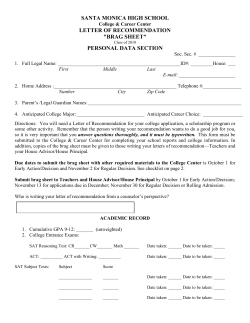
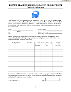


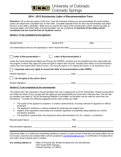
© Copyright 2026