
Circadian expression of clock genes and angiotensin II type 1... in suprachiasmatic nuclei of sinoaortic-denervated rats
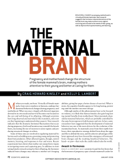
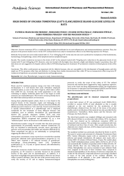
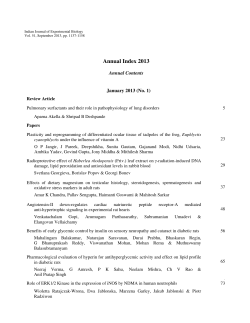
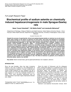
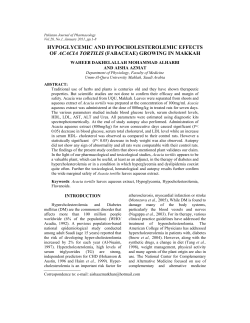
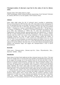
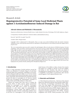
Acta Pharmacol Sin 2007 Apr; 28 (4): 484–492
Full-length article
Circadian expression of clock genes and angiotensin II type 1 receptors
in suprachiasmatic nuclei of sinoaortic-denervated rats1
Hui LI2, Ning-ling SUN2,4, Jin WANG3, Ai-jun LIU3, Ding-feng SU3,4
2
Department of Cardiology, Peking University People’s Hospital, Beijing 100044, China; 3Department of Pharmacology, Second Military
Medical University, Shanghai 200433, China
Key words
Abstract
circadian rhythm; arterial baroreflex; clock
g e n e s; si n o a o r t i c d e n e r v a t i o n ; r e n i n angiotensin system
Aim: To investigate whether the circadian expression of central clock genes and
angiotensin II type 1 (AT1) receptors was altered in sinoaortic-denervated (SAD)
rats. Methods: Male Sprague-Dawley rats underwent sinoaortic denervation or a
sham operation at the age of 12 weeks. Four weeks after the operation, blood
pressure and heart period were measured in the conscious state in a group of
sham-operated (n=10) and SAD rats (n=9). Rest SAD and sham-operated rats
were divided into 6 groups (n=6 in each group). The suprachiasmatic nuclei (SCN)
tissues were taken every 4 h throughout the day from each group for the determination of the mRNA expression of clock genes (Per2 and Bmal1) and the AT1
receptor by RT-PCR; the protein expression of Per2 and Bmal1 was determined by
Western blotting. Results: Blood pressure levels in the SAD rats were similar to
those of the sham-operated rats. However, blood pressure variabilities significantly increased in the SAD rats compared with the sham-operated rats. The
circadian variation of clock genes in the SCN of the sham-operated rats was characterized by a marked increase in the mRNA and protein expression during dark
periods. Per2 and Bmal1 mRNA levels were significantly lower in the SAD rats,
especially during dark periods. Western blot analysis confirmed an attenuation of
the circadian rhythm of the 2 clock proteins in the SCN of the SAD rats. AT1
receptor mRNA expressions in the SCN were abnormally upregulated in the light
phase, changed to a 12-h cycle in the SAD rats. Conclusion: The circadian variation of the 2 central clock genes was attenuated in the SAD rats. Arterial baroreflex
dysfunction also induced a disturbance in the expression of AT1 receptors in the
SCN.
1
Project wa s pa rtia l ly su pported by the
Nationa l Na tu ra l Science Fou nda tion of
China (No 30330650).
4
Correspondence to Prof Ning-ling SUN and
Prof Ding-feng SU.
Phn & Fax 86-10-8836-1687.
E-mail [email protected] (Ning-ling SUN)
P h n 86 -2 1-2 50 7-0 32 3.
Fax 86 -2 1-6 54 9-3 95 1.
E-mail [email protected] (Ding-feng SU)
Received 20 06 -0 9-20
Accepted 20 06 -1 2-11
doi: 10.1111/j.1745-7254.2007.00543.x
Introduction
The circadian clock system is responsible for the daily
timing of behavior and physiological processes [1]. In
mammals, the master circadian pacemaker controlling most
of these rhythms is located in the suprachiasmatic nuclei
(SCN) of the anterior hypothalamus, whose phase is mainly
light-entrained directly through the retinohypothalamic
tract[2]. Recent experiments have demonstrated an oscillatory system including a transcriptional-translational feedback loop of clock genes. These include 3 homologues of
the Drosophila gene period (Per1, Per2, and Per3), 2
cryptochrome genes (Cry1 and Cry2), and the transcriptional
484
activator genes: clock (the mammalian counterpart of the
Drosophila cycle) and Bmal1 (a member of the basic helixloop-helix/PER-ARNTSIM domain family of transcription
factors)[3,4]. These clock genes are expressed in a pattern
with circadian oscillations close to 24 h in the SCN. Among
these clock genes, Per2 and Bmal1 are most important and
were studied in the present work.
Intra-arterial blood pressure monitoring and non-invasive ambulatory blood pressure monitoring had shown that
blood pressure exhibited variations consistent with circadian rhythm[5,6]. Most normotensive and essential hypertensive individuals show a diurnal pattern of blood pressure
with markedly higher values during the active phase and
©2007 CPS and SIMM
Http://www.chinaphar.com
lower values during the rest phase (dippers); however, a
number of subjects display abnormal circadian blood pressure profiles characterized by an absence or even reversal in
a nocturnal fall in blood pressure (non-dippers). Crosssectional studies have indicated that target-organ damage is
more pronounced in “non-dipper” than in “dipper” patients
with comparable clinical blood pressure levels[7]. The
transgenic hypertensive TGR(mREN2)27 rats harboring the
murine Ren2 gene manifest an inverted circadian rhythm of
blood pressure similar to “non-dippers”, while 24 h profiles
of motor activity and heart rate remain undisturbed[8].
Moreover, the circadian rhythm of clock gene mRNA expression in the SCN of TGR(mRen2)27 rats was abolished[9].
Arterial baroreflex (ABR) is one of the most important
mechanisms in the regulation of cardiovascular activities,
especially in the stabilization of blood pressure. ABR function is impaired in hypertension and closely related to
hypertensive organ damage[10]. In addition, the interruption
of ABR by sinoaortic denervation could lead to irreversible
organ damage such as cardiac lesion, vascular remodeling,
and renal damage in rats[11]. Furthermore, it was found that
sinoaortic denervation changed the rhythmicity of blood pressure from a 24 h period to a 12-h period[12]. However, it is not
clear whether the function of baroreflex influences the circadian expression of clock genes in the central nervous system.
The present work was therefore designed to observe the
possible effects of sinoaortic denervation on the mRNA and
protein expression of 2 main clock genes: Per2 and Bmal1 in
the rat SCN.
Angiotensin II (Ang II), a key molecule for cardiovascular regulation, influences the activity of brain areas involved
in the determination of circadian-dependent variations of
blood pressure[13]. Therefore, the expression of Ang II type 1
(AT1) receptors in the same brain area of rats was also studied.
Materials and methods
Preparation of sinoaortic-denervated (SAD) rats Male
Sprague-Dawley (SD) rats were provided by Sino-British
SIPPR/BK Lab Animal Ltd (Shanghai, China). At the age of
12 weeks, sinoaortic denervation was performed with a previously used method[14,15]. Briefly, the rats were anesthetized with a mixture of ketamine (50 mg/kg) and diazepam
(5 mg/kg), intraperitoneally, and were then medicated with
atropine sulfate (0.5 mg/kg, intraperitoneally) and procaine
benzylpenicillin (60000 U, intramuscularly). After a midline
cervical incision and bilateral isolation of the neck muscles,
aortic baroreceptor denervation was carried out bilaterally
by cutting the superior laryngeal nerves near the vagi,
Li H et al
removing the superior cervical ganglia, and sectioning the
aortic depressor nerves. The carotid sinus baroreceptors
were denervated bilaterally by stripping the carotid bifurcation and its branches followed by the application of 10%
phenol (in 95% ethanol) to the external, internal, and common carotid arteries and the occipital artery. In the control
rats, the sham operation was performed under the same
conditions, without cutting and removing the nerves and
carotid adventitia. The animals were allowed to recover spontaneously after the denervation. All procedures were in
accordance with institutional animal care guidelines, and
approved by the local institutional committee.
Blood pressure measurement Blood pressure and heart
period were continuously recorded using previously
described techniques[16,17]. Briefly, the rats were anesthetized with a combination of ketamine (50 mg/kg) and diazepam (5 mg/kg). A floating polyethylene catheter was
inserted into the lower abdominal aorta via the left femoral
artery for blood pressure measurement. The catheters were
exteriorized through the interscapular skin. After a 2 d
recovery period, the animals were placed for blood pressure
recording in individual cylindrical cages containing food and
water. The aortic catheter was connected to a blood pressure transducer via a rotating swivel that allowed the animals to move freely in the cage. After about 4 h of habituation,
the blood pressure signal was digitized by a microcomputer.
Blood pressure and heart period values from every heartbeat were determined online. The mean values of these
parameters during a period of 4 h were calculated. The standard deviation of the mean values was also calculated and
served as the variabilities of blood pressure or heart period.
Experimental protocol Four weeks after the operation,
blood pressure and heart period were measured in the conscious state in a group of sham-operated (n=10) and SAD
rats (n=9). The remaining 72 (36 SAD rats and 36 shamoperated) rats were brought up in a separate, environmentally-controlled room (temperature 23–25 °C) with ventilated
chambers under a 12/12 h light/dark cycle (8:00−20:00 light,
20:00−8:00 dark) for at least 10 d with food and water available ad libitum. The light was provided by white fluorescent bulbs (220 mW/cm2). Under entrained conditions, lights
on was defined as Zeitgeber time (ZT) 0. Six SAD rats and 6
sham-operated rats were killed every 4 h from ZT0 to ZT20
by ether anesthesia. The mRNA and protein expression of
the clock genes (Per2, Bmal1) and AT1 receptors were examined by RT-PCR and Western blotting, respectively. The
brain was removed from the skull and snap-frozen in liquid
nitrogen until use. The SCN region was punched out with a
2 mm diameter needle that was inserted to a 1 mm depth into
485
Li H et al
Acta Pharmacologica Sinica ISSN 1671-4083
the surface of the coronal plane, an area that included the
SCN[17,18]. The animals were killed at each time point on 3
separate days to ensure reproducibility of the gene expression cycle.
RNA extraction and RT-PCR Total RNA was extracted
from the SCN using TRIzol reagent (Invitrogen, Carlsbad,
CA, USA) according to the manufacturer’s instructions.
Total RNA concentrations were determined by spectrophotometry at 260 nm, and total RNA quality was assessed by
electrophoresis on 1.3% denaturing formaldehyde agarose
gels. Total RNA (<1 µg) was reverse transcribed using an RT
system kit from Promega (Promega Corporation, Madison,
WI, USA) according to the manufacturer’s protocols. Primers were designed according to cDNA sequences reported
in the GenBank database and by computer analysis using
the Primer premier 5.0. (PREMIER Biosoft International, Palo
Alto, CA, USA) and synthesized at the SBS Genetech Co
(Beijing, China). The sequences of primers are described in
Table 1. PCR was performed with the 2×Taq PCR MasterMix
(Tianwei Biotech, Beijing, China) using a Peltier Thermal Cycler 200 (MJ Research Inc, Watertown, MA, USA). The PCR
protocol for Per2 was as follows: initial incubation at 94 °C
for 2 min, 35 cycles of denaturation at 94 °C for 40 s, annealing at 54 °C for 1 min, and extension 72 °C for 1 min, and
finally a prolonged extension step at 72 °C for 10 min. The
PCR protocol for Bmal1 and AT1 differed slightly from that
of Per2; annealing was performed at 58 °C or 56 °C, respectively. The relative amount of each mRNA was normalized to
the housekeeping gene, β-actin, mRNA that the expression
levels remained constant throughout the day. The PCR products were resolved by electrophoresis on a 2.0 % agarose gel,
stained with ethidium bromide (EB), and their relative quantities were determined using the Gel-pro Analyzer 4.5 soft-
ware (Media Cybernetics, Silver Spring, MD, USA).
Western blotting Microdissected SCN tissue was homogenized in lysis buffer [50 mmol/L Tris-HCl (pH 7.5)], 150
mmol/L NaCl, 5 mmol/L EDTA, 1% NP-40, 1 mmol/L PMSF,
plus a protease inhibitor cocktail (pH 7.4) for 30 min on ice.
After removal of tissue debris by centrifugation (12 000×g, 5
min), the protein concentration of each lysate sample was
determined using the BCA-100 protein assay kit (Boshide
Biotech, Wuhan, China) according to the manufacturer’s
instructions. The samples were boiled in ×2 SDS sample
buffer and loaded with 50 µg proteins per lane onto 8% SDSpolyacrylamide gels. Following separation at 125 V for 120
min, the proteins were transferred onto nitrocellulose
membranes, which were then blocked with 5% nonfat dried
milk in TBS-Tween (10 mmol/L Tris-HCl, pH 7.5, 150 mmol/L
NaCl, 0.25% Tween-20) for 4 h at room temperature. After
brief washes, the membranes were incubated with polyclonal
anti-mouse Per2 (1:100) and Bmal1 (1:200) antibodies (Abcam
Inc, Cambridge, MA, USA) at 4 °C overnight. Immunoreactive bands were visualized using anti-rabbit IgG-HRP antibody and an ECL chemiluminescence system (Santa Cruz
Biotech. Inc, CA, USA). The exact quantities of Per2 and
Bmal1 were normalized against actin as a constitutively
expressed internal control using optical density determined
by the Gel-pro Analyzer 4.5 software (Media Cybernetics,
USA).
Statistical analysis The statistical analyses were conducted directly with SPSS11.5 software. All data are expressed as mean±SD. Multi-group mean values were compared
by using ANOVA followed by Student Newman-Keuls test,
and two-group mean values were compared by using an unpaired Student’s t-test. A significant difference among mean
values was assigned at P<0.05.
Table 1. Primer sequences used in RT-PCR.
Name
Per2
Bmal1
AT 1
β-a c tin
486
Primer sequence
Forward: 5'-GACCGATGCCATCAGACTAGC-3'
Reverse: 5'-GGAAGCATGTCGGAACAATCT-3'
Forward: 5'-GATGCGAAGACTGGACTT-3'
Reverse: 5'-TGTGGAACCATGTGTGAG-3'
Forward: 5'-CTGGGCATTATCCGTGAC-3'
Reverse: 5'-TATCTGAAGGGCGGTAGG-3'
Forward:
5'-TGGAATCCTGTGGCATCCATGAAC-3'
Reverse:
5'-TAAACGCAGCTCAGTAACAGTCCG-3'
Primer site
45 26– 45 46
49 50– 49 70
681–698
983–1000
940–957
11 51– 11 68
887–910
12 11– 12 34
Product
length (bp)
GenBank
Accession No
46 5
NM03 16 78
31 9
NM02 43 62
22 9
NM03 10 09
34 8
NM03 11 44
Http://www.chinaphar.com
Li H et al
Results
Blood pressure and heart period in SAD rats It was
found that blood pressure levels in the SAD rats were similar
to those of the sham-operated rats. However, blood pressure variabilities significantly increased in the SAD rats compared with the sham-operated rats (Figure 1). Heart period
and heart period variability were not modified by sinoaortic
denervation operation.
Figure 1. Blood pressure and blood pressure variability in shamopera ted (Sha m, n=10 ) and SAD (n=9) rats. SBP, systolic blood
pressure; DBP, diastolic blood pressure; HP, heart period; SBPV, SBP
variability; DBPV, DBP variability; HPV, heart period variability.
b
P<0.05 vs sham-operated rats.
Circadian expression of Per2 mRNA in the SCN of SAD
rats The Per2 gene exhibited a robust circadian expression
pattern in the SCN of the sham-operated SD rats, as previously reported in Wistar rats[18]. By visual inspection of gel
electropherogram, high intensity DNA bands were observed
at ZT12, ZT16, and ZT20, and weak, but still recognized bands,
at ZT0, ZT4, and ZT8 (Figure 2A). Clear circadian transcription profiles of Per2 mRNA were detected by the relative
quantitative analyses, with higher transcription levels around
the light-to-dark transition (ZT12) and lower levels during
the early daytime (ZT0-4). The peak expression was observed at ZT12 and was 2.19-fold stronger than the trough
value at ZT0 (Figure 2B).
In the SAD rats, the Per2 gene showed a similar, but
attenuated, circadian oscillation. The intensity of the DNA
bands was weaker throughout the day than in the shamoperated rats (Figure 2A). The Per2 mRNA levels in the SCN
Figure 2. Circadian expression of Per2 mRNA in the SCN of SAD
and sham-operated (Sham) rats under a 12 h light/12 h dark cycle.
(A) representative gel electrophoresis of PCR products stained with
EB, showing Per2 and β-actin mRNA in the SCN at 6 time points.
Molecular sizes of the respective specific PCR products in relation to
a molecular size standard are indicated. (B) relative quantitative analysis of Per2 mRNA in the SCN at 6 time points. All values are normalized against the housekeeping gene, β-actin. Data represent mean±SD
values for 6 independent experiments (n=6) in SAD (●) and shamoperated rats (□). b P<0 .0 5, c P<0 .0 1 vs sha m-operated rats. M,
marker; ZT, Zeitgeber time.
were significantly lower in the SAD than in the sham-operated rats at most time points, except for ZT4 and ZT8 (Figure
2B).
Circadian expression of Bmal1 mRNA in the SCN of
SAD rats A clear circadian expression of the Bmal1 gene
was observed in the SCN of the sham-operated rats, which is
in accordance with the finding in Wistar rats[19]. By visual
inspection of gel electropherogram, high intensity DNA bands
were detected at ZT16, whereas moderate intensity bands
were detected at ZT0, ZT4, and ZT20. The intensity of the
bands was faint at ZT8 and ZT12 (Figure 3A). The relative
quantitative analyses displaying Bmal1 mRNA expression
reached its peak early at night at ZT16 and descended to a
487
Li H et al
Acta Pharmacologica Sinica ISSN 1671-4083
of -130 kDa in the sham-operated and SAD rats, in accordance with the predicted mass of 136 kDa (Figure 4A). The
intensity of the Per2-immunoreactivity (ir) bands exhibited a
robust circadian oscillation in the sham-operated groups,
with high expression during the early dark phase (ZT12−16),
and low levels at the early light phase (ZT0−4), as previously reported in mice[20]. In the SAD rats, the intensity of
the Per2-ir bands was significantly reduced at every time
point compared with the controls, but a circadian pattern
persisted. The quantitative analysis of normalizing the Per2ir bands against actin-ir bands demonstrated that levels
peaked at ZT16 and declined to a trough at ZT4 in both
groups. There was a trend for decreased Per2 levels in the
SCN of the SAD rats at most time points; the significant
difference was only observed at ZT12 (Figure 4B).
Figure 3. Circadian expression of Bmal1 mRNA in the SCN of SAD
and sham-operated (Sham) rats under a 12 h light/12 h dark cycle.
(A) representative gel electrophoresis of PCR products stained with
EB, showing Bmal1 and β-actin mRNA in the SCN at 6 time points.
Molecular sizes of the respective specific PCR products in relation to
a molecular size standard are indicated. (B) relative quantitative analysis of Bma l1 mRNA in the SCN at 6 time points. All valu es a re
normalized against the housekeeping gene, β-actin. Data represent
mean±SD values for 6 independent experiments (n=6) in SAD (●)
and sham-operated rats (□). b P<0.05, c P<0.01 vs sham-operated
rats. M, marker; ZT, Zeitgeber time.
trough late in the day at ZT8. The expression pattern was
anti-phase to that observed for Per2 mRNA (Figure 3B).
In the SAD group, the Bmal1 gene also presented synchronous, but subdued, circadian oscillation. There was
scarcely any difference among the intensity of DNA bands
at most time points by visual inspection of electropherogram (Figure 3A). Bmal1 mRNA expression in SAD rats significantly decreased throughout the day compared with the
sham-operated rats, except at ZT8 (Figure 3B).
Circadian expression of the Per2 and Bmal1 proteins in
the SCN of SAD rats The proteins extracted from the
microdissected SCN were probed to detect Per2 and Bmal1.
The anti-Per2 antibody consistently identified a major band
488
Figure 4. Circadian expression of the Per2 protein in the SCN of
SAD and sham-operated (Sham) rats under a 12 h light/12 h dark
cycle. (A) Western blotting of Per2-ir bands in the SCN at 6 time
points. (B) relative density of Per2-ir bands was normalized against
actin-ir bands. Data represent mean±SD values for 6 independent
experiments (n=6) in SAD (●) and sham-operated rats (□). bP<0.05
vs sham-operated rats. OD, optical density; ZT, Zeitgeber time.
The anti-Bmal1 antibody identified a band around 68 kDa
in the same SCN samples of the sham-operated and SAD
rats (Figure 5A). The intensity of the Bmal1-ir band varied
with a circadian pattern synchronized in both groups, with
peak expression during late night at ZT16−20 and a nadir
early in the subjective day at ZT0−4, accorded with the findings in Wistar rats[21]. The quantitative analysis of normalizing the Bmal1-ir bands against actin-ir bands showed that
Bmal1 expression reached its peak at ZT20 and descended
Http://www.chinaphar.com
Li H et al
Figure 5. Circadian expression of the Bmal1 protein in the SCN of
SAD and sham-operated (Sham) rats under a 12 h light/12 h dark
cycle. (A) Western blotting of Bmal1-ir bands in the SCN at 6 time
points. (B) relative density of Bmal1-ir bands was normalized against
actin-ir bands. Data represent mean±SD values for 6 independent
experiments (n=6) in SAD (●) and sham-operated rats (□). b P<0.05
vs sham-operated rats. OD, optical density; ZT, Zeitgeber time.
to a trough at ZT4. The Bmal1 levels significantly decreased
at ZT8 and ZT20 in the SCN of the SAD rats compared with
the sham-operated groups (Figure 5B).
These results reinforced the findings that the circadian
rhythm of some clock gene cycles in the SCN were attenuated after interruption of the arterial baroreflex.
Circadian expression of AT1 receptor mRNA in the SCN
of SAD rats The circadian expression pattern of AT1 receptor mRNA expression in the SCN of the sham-operated rats
was obvious. High-intensity bands were detected during
the dark phase (ZT12−20), while weak intensity bands were
detected in the light phase (ZT0−8, Figure 6A). AT1 receptor expression peaked at ZT16 and descended to a trough at
ZT8 in the sham-operated rats (Figure 6B).
In contrast, the circadian expression profile of AT1
receptor mRNA changed to a bimodal pattern in the SCN of
the SAD rats. High-intensity bands were detected abnormally at ZT4 (Figure 6A). AT1 receptor mRNA levels in the
SAD group significantly increased in the light phase, greater
than in the sham-operated group (Figure 6B).
Discussion
The present work shows, for the first time, that arterial
baroreflex dysfunction may dramatically attenuate the circadian variation of clock gene expression in the SCN. The
main findings of this study are as follows: (i) although Per2
Figure 6. Circadian expression of AT1 receptor mRNA in the SCN
of SAD and sham-operated (Sham) rats under a 12 h light/12 h dark
cycle. (A) representative gel electrophoresis of PCR products stained
with EB showing AT1 and β-actin mRNA in the SCN at 6 time points.
Molecular sizes of the respective specific PCR products in relation to
a molecular size standard are indicated. (B) relative quantitative analysis of AT1 mRNA in the SCN at 6 time points. All values are normalized against the housekeeping gene, β-actin. Data represent mean±SD
values for 6 independent experiments (n=6) in SAD (●) and shamoperated rats (□). b P<0 .0 5, c P<0 .0 1 vs sham-operated rats. M,
marker; ZT, Zeitgeber time.
and Bmal1 mRNA oscillated synchronously in the SCN of
the SAD and sham-operated rats, the expression of mRNA
levels and amplitude of oscillation were remarkably depressed
in the SAD rats; (ii) the contents of the Per2 and Bmal1
proteins and the extent of the circadian variations were distinctly weakened in the SCN of the SAD rats compared with
the controls; and (iii) AT1 receptor mRNA expressions in the
SCN were abnormally upregulated in the light phase and
altered the circadian rhythm of AT1 receptors in the SAD
rats.
Circadian clock is normally entrained by periodic environmental cues, with the daily light-dark cycle being the most
489
Li H et al
potent entraining signal in mammals. The clock showed phase
delay and phase advance shifts in response to light exposure in early and late night, respectively. Per1 and Per2
have been demonstrated to be light-inducible in different
rodent species[22,23]. In the present study, Per2 and Bmal1
genes oscillated synchronously in the SCN of the SAD and
sham-operated rats. The peak level of Per2 mRNA was observed at ZT12 and at the trough level at ZT0, while Bmal1
mRNA transcription reached a peak at ZT16 and a nadir at
ZT8. However, the Per2 and Bmal1mRNA levels and the
amplitude of oscillation were dramatically reduced in the SCN
of the SAD rats. The phenomena were confirmed by a Western blot analysis of Per2 and Bmal1 proteins in the same
samples. Circadian profiles of the Per2 protein exhibited evident oscillation with a peak at ZT16; the Bmal1 protein
showed a nocturnal peak at ZT20, indicating that the protein
cycles were delayed by about 4–6 h, relative to the mRNA
cycles under the light-dark conditions in the SCN. Similarly,
the contents of Per2 and Bmal1 proteins at certain time points
and the extent of circadian variations were distinctly weakened in the SCN of the SAD rats compared with the controls.
The post-transcriptional regulation mechanisms may implicate the difference between the levels of mRNA and proteins
of the clock genes. Our findings suggested that interruption
of the arterial baroreflex could attenuate the circadian
expression and rhythmicity of some central clock genes.
The ABR is most important in the regulation of heart rate
and the stabilization of blood pressure. When ABR function
is destroyed by sinoaortic denervation operation, blood pressure becomes unstable and blood pressure variability is great.
However, the blood pressure level remains unchanged[14,15].
ABR function is impaired in hypertension, heart failure, and
diabetes and has been found to be a crucial determinant of
sudden death after acute myocardial infarction[24]. In hypertensive rats, the target organ damage correlates negatively
with ABR function[10,25]. Interruption of ABR by sinoaortic
denervation induced aortic and cardiac hypertrophy independent of the mean level of blood pressure[26–28]. In the
present study, it was found that dysfunction of the ABR
attenuated the circadian expression of clock genes in the
SCN. It has been reported that mean blood pressure was
significantly elevated during the light period in the SAD
rats, thus the 24 h rhythmicity in mean blood pressure was
suppressed and a bimodal pattern was observed under light–
dark cycles, whereas the heart rate and locomotor activity
were not dramatically affected in the same rats[12]. Although
the precise mechanisms responsible for the selective elimination of the circadian rhythm of mean blood pressure in the
SAD rats have not yet been satisfactorily clarified, the alter490
Acta Pharmacologica Sinica ISSN 1671-4083
ation of central clock genes induced by the disruption of
ABR may be related with the phenomenon. These results
further support the finding that ABR is not only important in
maintaining the stability of blood pressure in the short term,
but also in regulating the diurnal variation in blood pressure
during a 24 h cycle.
It is well established that the central nervous system plays
an important role in controlling ABR function. The major
baroreceptor reflex pathway exists in the brain stem, such as
the nucleus tractus solitarii and the caudal and rostral ventrolateral medulla[29]. Although these brain regions do not
project directly to the SCN, the multisynaptic connections
between them are much more extensive. Using the retrograde transneuronal tracer, the pseudorabies virus (PRV),
which localizes the afferent inputs to the SCN in SD rats, has
demonstrated that cells in the nucleus tractus solitarii, C3
catecholamine region, rostral ventrolateral medulla, periaqueductal gray matter, and lamina I and dorsomedial area of
the spinal trigeminal nucleus are higher order sites in pathways which affect the SCN[30]. The presence of PRV-infected
neurons in the brain stem indicates a pathway associated
with regulatory mechanisms of arterial pressure which can
influence SCN function and even clock gene expression.
However, there is insufficient evidence showing a direct
action of ABR on gene expression in SCN.
The renin-angiotensin system (RAS) is one of the major
humoral mechanisms regulating cardiovascular function.
Ang II plays a major role in the modulation of blood pressure,
stimulation of vasopressin and aldosterone release, sodium
appetite, and sympathetic facilitation. Most of these effects
show a day–night variation and are mediated via AT1
receptors. Furthermore, the SCN contains not only Ang IIimmunoreactive cells and fibers, but also a high density of
Ang II receptors[31]. It was recently described that Ang II
induces oscillatory expression of clock genes in vascular
smooth muscle cells through the AT1 receptor subtype[32]
and alters the ABR through peripheral (vascular and renal)
and central effects[33]. Impaired ABR function is usually
accompanied by overactivation of tissue RAS. Previous studies have confirmed that plasma Ang II levels did not increase
in the chronic phase of sinoaortic denervation; in contrast,
cardiac, aortic, and renal Ang II contents increased and AT1
receptor mRNA expression in the left ventricle and aorta was
upregulated after sinoaortic denervation[34,35]. In the present
work, the SCN AT1 receptor mRNA expression was abnormally upregulated in the light phase, resulting in the circadian rhythm of AT1 receptors from a 24 h cycle to a 12 h cycle
in the SAD rats. This is in accordance with the changes of
blood pressure rhythm induced by the interruption of ABR.
Http://www.chinaphar.com
It is reasonable to hypothesize that ABR dysfunction
may activate tissue RAS, including the SCN. Ang II acts as
a candidate entraining signal, altering circadian expression
of central clock genes through AT1 receptors. However, the
cause–effect relationship between the changes in clock genes
and RAS is not clear at this stage of study.
In conclusion, the circadian variation of the 2 central clock
genes was attenuated in the SAD rats. ABR dysfunction
also induced a disturbance in the expression of AT1 receptors in the SCN. The precise mechanisms underlying ABR
dysfunction inducing an attenuated expression of the 2 clock
genes remain to be elucidated in the future.
References
1
2
3
4
5
6
7
8
9
10
11
12
13
Whitmore D, Cermakian N, Crosio C, Foulkes NS, Pando MP,
Travnickova Z, et al. A clock organ. Biol Chem 2000; 381:
7 96 –8 .
Reppert SM, Weaver DR. Coordination of circadian timing in
mammals. Nature 2002; 418: 935–41.
Shearman LP, Sriram S, Weaver DR, Maywood ES, Chaves I,
Zheng BH, et al. Interacting molecular loops in the mammalian
circadian clock. Science 2000; 288: 1013–9.
Reppert SM, Weaver DR. Molecular analysis of mammalian
circadian rhythms. Annu Rev Physiol 2001; 63: 647–76.
Basset A, Laude D, Laurent S, Elghozi JL. Contrasting circadian
rhythms of blood pressure among inbred rat strains: recognition
of dipper and non-dipper patterns. J Hypertens 2004; 22: 727–37.
Finta E, Laude D, Alfoldi S, Farsang C, Elghozi JL. Effects of
rilmenidine on 24-h rhythmicity of blood pressure and spontaneous baroreflex sensitivity in essential hypertensive subjects. J
Hypertens 2006; 24: 1619–25.
Cuspidi C, Meani S, Salerno M, Valerio C, Fusi V, Severgnini B, et
al. Cardiovascular target organ damage in essential hypertensives
with or without reproducible nocturnal fall in blood pressure. J
Hypertens 2004; 22: 273–80.
Lemmer B, Ma ttes A, Böhm M, Ganten D. Circadian blood
pressure variation in transgenic hypertensive rats. Hypertension 1993; 22: 97–101.
Lemmer B, Witte K, Enzminger H, Schiffer S, Hauptfleisch S.
Transgenic TGR(mREN2)27 rats as a model for disturbed circadian organization at the level of the brain, the heart, and the
kidneys. Chronobiol Int 2003; 20: 711–38.
Xie HH, Shen FM, Miao CY, Su DF. Blood pressure, baroreflex
sensitivity, and end organ damage in hybrid offspring of spontaneously hypertensive ra ts a nd Spra gu e-Da wley ra ts. Acta
Pharmacol Sin 2005; 26: 1049–56.
Shan ZZ, Dai SM, Su DF. Arterial baroreflex deficit induced
orga n da mage in sinoaortic denerva ted rats. J Ca rdiovasc
Pharmacol 2001; 38: 427–37.
Makino M, Hayashi H, Takezawa H, Hirai M, Saito H, Ebihara S.
Circadian rhythms of cardiovascular functions are modulated by
the baroreflex and the autonomic nervou s system in the ra t.
Circulation 1997; 96: 1667–74.
Braga ANG, Lemos MS, Silva JR, Fontes WRP, Santos RAS.
Effects of angiotensins on da y-night flu ctu ations a nd stress-
Li H et al
14
15
16
17
18
19
20
21
22
23
24
25
26
27
28
29
induced changes in blood pressure. Am J Physiol Regul Integr
Comp Physiol 2002; 282: R1663–71.
Gu XW, Xie HH, Wang J, Shen FM, Su DF. Arterial baroreflex is
not involved in salt preference in ra ts. Clin Exp Pha rmacol
Physiol 2006; 33: 607–11.
Wang J, Shen FM, Wamg MW, Su DF. Effects of nine antihypertensive drugs on blood pressure variability in sinoaortic-denervated rats. Acta Pharmacol Sin 2006; 27: 1013–7.
Zhan LS, Guan YF, Su DF, Miao CY. Blood pressure variability
and baroreflex sensitivity are not different in spontaneously hypertensive rats and stroke-prone spontaneously hypertensive rats.
Acta Pharmacol Sin 2005; 26: 959–62.
Han P, Chu ZX, Shen FM, Xie HH, Su DF. Synergism of hydrochlorothiazide and nitrendipine on the reduction of blood pressure and blood pressure variability in spontaneously hypertensive rats. Acta Pharmacol Sin 2006; 27: 1575–9.
Yan L, Takekida S, Shigeyoshi Y, Okamura H. Per1 and per2
gene expression in the rat suprachiasmatic nucleus circadian profile and the compartment-specific response to light. Neuroscience 1999; 94: 141–50.
Oishi K, Sakamoto K, Okada T, Nagase T, Ishida N. Antiphase
circadia n expression between BMAL1 and period homologue
mRNA in the suprachiasmatic nucleus and peripheral tissues of
rats. Biochem Biophys Res Commun 1998; 253: 199–203.
Zheng BH, Albrecht U, Kaasik K, Sage M, Lu WQ, Vaishnav S, et
al. Nonredundant roles of the mper1 and mper2 genes in the
mammalian circadian clock. Cell 2001; 105: 683–94.
Tamaru T, Isojima Y, Yamada T, Okada M, Nagai K, Takamatsu
K. Light and glutamate-induced degradation of the circadian
oscillating protein BMAL1 during the mammalian clock resetting.
J Neurosci 2000; 20: 7525–30.
Albrecht U, Sun ZS, Eichele G, Lee CC. A differential response of
two putative mammalian circadian regulators, mper1 and mper2
to light. Cell 1997; 91: 1055–64.
Dardente H, Poirel VJ, Klosen P, Pevet P, Masson-Pevet M. Per
and neuropeptide expression in the rat suprachiasmatic nuclei:
compartmentalization and differential cellular induction by light.
Brain Res 2002; 958: 261–71.
La Rovere MT, Pinna GD, Hohnloser SH, Marcus FI, Mortara A,
Nohara R, et al. Baroreflex sensitivity and heart rate variability
in the identifica tion of pa tients a t risk for life-threatening
arrhythmias: implications for clinical trials. Circulation 2001;
103: 2072–7.
Wang DS, Xie HH, Shen FM, Cai GJ, Su DF. Blood pressure
variability, cardiac baroreflex sensitivity and organ damage in
experimentally hypertensive rats. Clin Exp Pharmacol Physiol
2005; 32: 545-52.
Shen FM, Zhang SH, Xie HH, Jing Q, Su DF. Early structural
changes of aortic wall in sinoaortic denervated rats. Clin Exp
Pharmacol Physiol 2006; 33: 358–63.
Miao CY, Su DF. The importance of blood pressure variability in
rat aortic and left ventricular hypertrophy produced by sinoaortic
denervation. J Hypertens 2002; 20: 1865–72.
Miao CY, Xie HH, Zhan LS, Su DF. Blood pressure variability is
more important than blood pressure level in determination of
end-organ damage in rats. J Hypertens 2006; 24: 1125–35.
Dampney RAL. Functional orga nization of central pathways
regulating the cardiovascular system. Physiol Rev 1994; 74:
491
Li H et al
Acta Pharmacologica Sinica ISSN 1671-4083
32 3– 64 .
3 0 Krout KE, Kawano J, Mettenleiter TC, Loewy AD. CNS inputs
to the suprachiasmatic nucleus of the rat. Neuroscience 2002;
110: 73–92.
3 1 Thomasa MA, Fleissnerb G, Stohrb M, Hauptfleischa S, Lemmer
B. Localization of components of the renin-angiotensin system
in the suprachiasmatic nucleus of normotensive Sprague-Dawley
rats. Brain Res 2004; 1008: 212–35.
3 2 Nonaka H, Emoto N, Ikeda K, Fukuya H, Rohman MS, Raharjo
SB, et al. Angiotensin II induces circadian gene expression of
clock genes in cultured vascular smooth muscle cells. Circulation
2001; 104: 1746–8.
3 3 Sanderford MG, Bishop VS. Central mechanisms of acute ANG II
modulation of arterial baroreflex control of renal sympathetic
nerve a ctivity. Am J Physiol Heart Circ Physiol 200 2; 2 82:
H15 92–6 02.
3 4 Shan ZZ, Dai SM, Fang F, Su DF. Changes of central norepinephrin e, β-en dorphi n, leu -enk ep ha lin, perip hera l a rgini nevasopressin, and angiotensin II levels in acute and chronic phases
of sino-aortic denervation in rats. J Cardiovasc Pharmacol 2004;
43: 234–41.
35 Miao CY, Zhang LM, Yuan WJ, Su DF. Angiotensin II and AT1
receptor in hypertrophied ventricles and aorta s of sinoaorticdenervated rats. Acta Pharmacol Sin 2003; 24: 812–8.
~~~~~~~~~~~~~~~~~~~~~~~~~~~~~~~~~~~~~~~~~~~~~~~~~~~~~~~~~~~~~~~~~~~~~~~~~~~~~~~~~~~~~~~~~~~~~
World Congress of Pharmacy and Pharmaceutical Sciences 2007
67th International Congress of FIP
Beijing, China
1-6, September 2007
For further information:
FIP Congresses & Conferences
Andries Bickerweg 5 P.O. Box 84200 2508 AE The Hague
The Netherlands
Phn: +31-(0)70-302 1982/1981
Fax: +31-(0)70-302 1998/1999
Email: [email protected]
Website: http://www.fip.org/beijing2007
492


© Copyright 2026