
E-book
African Journal of Biotechnology Volume 14 Number 1, 7 January, 2015 ISSN 1684-5315 ABOUT AJB The African Journal of Biotechnology (AJB) (ISSN 1684-5315) is published weekly (one volume per year) by Academic Journals. African Journal of Biotechnology (AJB), a new broad-based journal, is an open access journal that was founded on two key tenets: To publish the most exciting research in all areas of applied biochemistry, industrial microbiology, molecular biology, genomics and proteomics, food and agricultural technologies, and metabolic engineering. Secondly, to provide the most rapid turn-around time possible for reviewing and publishing, and to disseminate the articles freely for teaching and reference purposes. All articles published in AJB are peerreviewed. Submission of Manuscript Please read the Instructions for Authors before submitting your manuscript. The manuscript files should be given the last name of the first author Click here to Submit manuscripts online If you have any difficulty using the online submission system, kindly submit via this email [email protected]. With questions or concerns, please contact the Editorial Office at [email protected]. Editor-In-Chief Associate Editors George Nkem Ude, Ph.D Plant Breeder & Molecular Biologist Department of Natural Sciences Crawford Building, Rm 003A Bowie State University 14000 Jericho Park Road Bowie, MD 20715, USA Prof. Dr. AE Aboulata Plant Path. Res. Inst., ARC, POBox 12619, Giza, Egypt 30 D, El-Karama St., Alf Maskan, P.O. Box 1567, Ain Shams, Cairo, Egypt Editor N. John Tonukari, Ph.D Department of Biochemistry Delta State University PMB 1 Abraka, Nigeria Dr. S.K Das Department of Applied Chemistry and Biotechnology, University of Fukui, Japan Prof. Okoh, A. I. Applied and Environmental Microbiology Research Group (AEMREG), Department of Biochemistry and Microbiology, University of Fort Hare. P/Bag X1314 Alice 5700, South Africa Dr. Ismail TURKOGLU Department of Biology Education, Education Faculty, Fırat University, Elazığ, Turkey Prof T.K.Raja, PhD FRSC (UK) Department of Biotechnology PSG COLLEGE OF TECHNOLOGY (Autonomous) (Affiliated to Anna University) Coimbatore-641004, Tamilnadu, INDIA. Dr. George Edward Mamati Horticulture Department, Jomo Kenyatta University of Agriculture and Technology, P. O. Box 62000-00200, Nairobi, Kenya. Dr. Gitonga Kenya Agricultural Research Institute, National Horticultural Research Center, P.O Box 220, Thika, Kenya. Editorial Board Prof. Sagadevan G. Mundree Department of Molecular and Cell Biology University of Cape Town Private Bag Rondebosch 7701 South Africa Dr. Martin Fregene Centro Internacional de Agricultura Tropical (CIAT) Km 17 Cali-Palmira Recta AA6713, Cali, Colombia Prof. O. A. Ogunseitan Laboratory for Molecular Ecology Department of Environmental Analysis and Design University of California, Irvine, CA 92697-7070. USA Dr. Ibrahima Ndoye UCAD, Faculte des Sciences et Techniques Departement de Biologie Vegetale BP 5005, Dakar, Senegal. Laboratoire Commun de Microbiologie IRD/ISRA/UCAD BP 1386, Dakar Dr. Bamidele A. Iwalokun Biochemistry Department Lagos State University P.M.B. 1087. Apapa – Lagos, Nigeria Dr. Jacob Hodeba Mignouna Associate Professor, Biotechnology Virginia State University Agricultural Research Station Box 9061 Petersburg, VA 23806, USA Dr. Bright Ogheneovo Agindotan Plant, Soil and Entomological Sciences Dept University of Idaho, Moscow ID 83843, USA Dr. A.P. Njukeng Département de Biologie Végétale Faculté des Sciences B.P. 67 Dschang Université de Dschang Rep. du CAMEROUN Dr. E. Olatunde Farombi Drug Metabolism and Toxicology Unit Department of Biochemistry University of Ibadan, Ibadan, Nigeria Dr. Stephen Bakiamoh Michigan Biotechnology Institute International 3900 Collins Road Lansing, MI 48909, USA Dr. N. A. Amusa Institute of Agricultural Research and Training Obafemi Awolowo University Moor Plantation, P.M.B 5029, Ibadan, Nigeria Dr. Desouky Abd-El-Haleem Environmental Biotechnology Department & Bioprocess Development Department, Genetic Engineering and Biotechnology Research Institute (GEBRI), Mubarak City for Scientific Research and Technology Applications, New Burg-Elarab City, Alexandria, Egypt. Dr. Simeon Oloni Kotchoni Department of Plant Molecular Biology Institute of Botany, Kirschallee 1, University of Bonn, D-53115 Germany. Dr. Eriola Betiku German Research Centre for Biotechnology, Biochemical Engineering Division, Mascheroder Weg 1, D-38124, Braunschweig, Germany Dr. Daniel Masiga International Centre of Insect Physiology and Ecology, Nairobi, Kenya Dr. Essam A. Zaki Genetic Engineering and Biotechnology Research Institute, GEBRI, Research Area, Borg El Arab, Post Code 21934, Alexandria Egypt Dr. Alfred Dixon International Institute of Tropical Agriculture (IITA) PMB 5320, Ibadan Oyo State, Nigeria Dr. Sankale Shompole Dept. of Microbiology, Molecular Biology and Biochemisty, University of Idaho, Moscow, ID 83844, USA. Dr. Mathew M. Abang Germplasm Program International Center for Agricultural Research in the Dry Areas (ICARDA) P.O. Box 5466, Aleppo, SYRIA. Dr. Solomon Olawale Odemuyiwa Pulmonary Research Group Department of Medicine 550 Heritage Medical Research Centre University of Alberta Edmonton Canada T6G 2S2 Prof. Anna-Maria Botha-Oberholster Plant Molecular Genetics Department of Genetics Forestry and Agricultural Biotechnology Institute Faculty of Agricultural and Natural Sciences University of Pretoria ZA-0002 Pretoria, South Africa Dr. O. U. Ezeronye Department of Biological Science Michael Okpara University of Agriculture Umudike, Abia State, Nigeria. Dr. Joseph Hounhouigan Maître de Conférence Sciences et technologies des aliments Faculté des Sciences Agronomiques Université d'Abomey-Calavi 01 BP 526 Cotonou République du Bénin Prof. Christine Rey Dept. of Molecular and Cell Biology, University of the Witwatersand, Private Bag 3, WITS 2050, Johannesburg, South Africa Dr. Kamel Ahmed Abd-Elsalam Molecular Markers Lab. (MML) Plant Pathology Research Institute (PPathRI) Agricultural Research Center, 9-Gamma St., Orman, 12619, Giza, Egypt Dr. Jones Lemchi International Institute of Tropical Agriculture (IITA) Onne, Nigeria Prof. Greg Blatch Head of Biochemistry & Senior Wellcome Trust Fellow Department of Biochemistry, Microbiology & Biotechnology Rhodes University Grahamstown 6140 South Africa Dr. Beatrice Kilel P.O Box 1413 Manassas, VA 20108 USA Dr. Jackie Hughes Research-for-Development International Institute of Tropical Agriculture (IITA) Ibadan, Nigeria Dr. Robert L. Brown Southern Regional Research Center, U.S. Department of Agriculture, Agricultural Research Service, New Orleans, LA 70179. Dr. Deborah Rayfield Physiology and Anatomy Bowie State University Department of Natural Sciences Crawford Building, Room 003C Bowie MD 20715,USA Dr. Marlene Shehata University of Ottawa Heart Institute Genetics of Cardiovascular Diseases 40 Ruskin Street K1Y-4W7, Ottawa, ON, CANADA Dr. Hany Sayed Hafez The American University in Cairo, Egypt Dr. Clement O. Adebooye Department of Plant Science Obafemi Awolowo University, Ile-Ife Nigeria Dr. Ali Demir Sezer Marmara Üniversitesi Eczacilik Fakültesi, Tibbiye cad. No: 49, 34668, Haydarpasa, Istanbul, Turkey Dr. Ali Gazanchain P.O. Box: 91735-1148, Mashhad, Iran. Dr. Anant B. Patel Centre for Cellular and Molecular Biology Uppal Road, Hyderabad 500007 India Dr. Yee-Joo TAN Department of Microbiology Yong Loo Lin School of Medicine, National University Health System (NUHS), National University of Singapore MD4, 5 Science Drive 2, Singapore 117597 Singapore Prof. Hidetaka Hori Laboratories of Food and Life Science, Graduate School of Science and Technology, Niigata University. Niigata 950-2181, Japan Prof. Thomas R. DeGregori University of Houston, Texas 77204 5019, USA Dr. Wolfgang Ernst Bernhard Jelkmann Medical Faculty, University of Lübeck, Germany Prof. Arne Elofsson Department of Biophysics and Biochemistry Bioinformatics at Stockholm University, Sweden Dr. Moktar Hamdi Department of Biochemical Engineering, Laboratory of Ecology and Microbial Technology National Institute of Applied Sciences and Technology. BP: 676. 1080, Tunisia Prof. Bahram Goliaei Departments of Biophysics and Bioinformatics Laboratory of Biophysics and Molecular Biology University of Tehran, Institute of Biochemistry and Biophysics Iran Dr. Salvador Ventura Department de Bioquímica i Biologia Molecular Institut de Biotecnologia i de Biomedicina Universitat Autònoma de Barcelona Bellaterra-08193 Spain Dr. Nora Babudri Dipartimento di Biologia cellulare e ambientale Università di Perugia Via Pascoli Italy Dr. Claudio A. Hetz Faculty of Medicine, University of Chile Independencia 1027 Santiago, Chile Dr. S. Adesola Ajayi Seed Science Laboratory Department of Plant Science Faculty of Agriculture Obafemi Awolowo University Ile-Ife 220005, Nigeria Prof. Felix Dapare Dakora Research Development and Technology Promotion Cape Peninsula University of Technology, Room 2.8 Admin. Bldg. Keizersgracht, P.O. 652, Cape Town 8000, South Africa Dr. Geremew Bultosa Department of Food Science and Post harvest Technology Haramaya University Personal Box 22, Haramaya University Campus Dire Dawa, Ethiopia Dr. José Eduardo Garcia Londrina State University Brazil Prof. Nirbhay Kumar Malaria Research Institute Department of Molecular Microbiology and Immunology Johns Hopkins Bloomberg School of Public Health E5144, 615 N. Wolfe Street Baltimore, MD 21205 Prof. M. A. Awal Department of Anatomy and Histplogy, Bangladesh Agricultural University, Mymensingh-2202, Bangladesh Prof. Christian Zwieb Department of Molecular Biology University of Texas Health Science Center at Tyler 11937 US Highway 271 Tyler, Texas 75708-3154 USA Prof. Danilo López-Hernández Instituto de Zoología Tropical, Facultad de Ciencias, Universidad Central de Venezuela. Institute of Research for the Development (IRD), Montpellier, France Prof. Donald Arthur Cowan Department of Biotechnology, University of the Western Cape Bellville 7535 Cape Town, South Africa Dr. Ekhaise Osaro Frederick University Of Benin, Faculty of Life Science Department of Microbiology P. M. B. 1154, Benin City, Edo State, Nigeria. Dr. Luísa Maria de Sousa Mesquita Pereira IPATIMUP R. Dr. Roberto Frias, s/n 4200-465 Porto Portugal Dr. Min Lin Animal Diseases Research Institute Canadian Food Inspection Agency Ottawa, Ontario, Canada K2H 8P9 Prof. Nobuyoshi Shimizu Department of Molecular Biology, Center for Genomic Medicine Keio University School of Medicine, 35 Shinanomachi, Shinjuku-ku Tokyo 160-8582, Japan Dr. Adewunmi Babatunde Idowu Department of Biological Sciences University of Agriculture Abia Abia State, Nigeria Dr. Yifan Dai Associate Director of Research Revivicor Inc. 100 Technology Drive, Suite 414 Pittsburgh, PA 15219 USA Dr. Zhongming Zhao Department of Psychiatry, PO Box 980126, Virginia Commonwealth University School of Medicine, Richmond, VA 23298-0126, USA Prof. Giuseppe Novelli Human Genetics, Department of Biopathology, Tor Vergata University, Rome, Italy Dr. Moji Mohammadi 402-28 Upper Canada Drive Toronto, ON, M2P 1R9 (416) 512-7795 Canada Prof. Jean-Marc Sabatier Directeur de Recherche Laboratoire ERT-62 Ingénierie des Peptides à Visée Thérapeutique, Université de la Méditerranée-Ambrilia Biopharma inc., Faculté de Médecine Nord, Bd Pierre Dramard, 13916, Marseille cédex 20. France Dr. Fabian Hoti PneumoCarr Project Department of Vaccines National Public Health Institute Finland Prof. Irina-Draga Caruntu Department of Histology Gr. T. Popa University of Medicine and Pharmacy 16, Universitatii Street, Iasi, Romania Dr. Dieudonné Nwaga Soil Microbiology Laboratory, Biotechnology Center. PO Box 812, Plant Biology Department, University of Yaoundé I, Yaoundé, Cameroon Dr. Gerardo Armando Aguado-Santacruz Biotechnology CINVESTAV-Unidad Irapuato Departamento Biotecnología Km 9.6 Libramiento norte Carretera IrapuatoLeón Irapuato, Guanajuato 36500 Mexico Dr. Abdolkaim H. Chehregani Department of Biology Faculty of Science Bu-Ali Sina University Hamedan, Iran Dr. Abir Adel Saad Molecular oncology Department of Biotechnology Institute of graduate Studies and Research Alexandria University, Egypt Dr. Azizul Baten Department of Statistics Shah Jalal University of Science and Technology Sylhet-3114, Bangladesh Dr. Bayden R. Wood Australian Synchrotron Program Research Fellow and Monash Synchrotron Research Fellow Centre for Biospectroscopy School of Chemistry Monash University Wellington Rd. Clayton, 3800 Victoria, Australia Dr. G. Reza Balali Molecular Mycology and Plant Pthology Department of Biology University of Isfahan Isfahan Iran Dr. Beatrice Kilel P.O Box 1413 Manassas, VA 20108 USA Prof. H. Sunny Sun Institute of Molecular Medicine National Cheng Kung University Medical College 1 University road Tainan 70101, Taiwan Prof. Ima Nirwana Soelaiman Department of Pharmacology Faculty of Medicine Universiti Kebangsaan Malaysia Jalan Raja Muda Abdul Aziz 50300 Kuala Lumpur, Malaysia Prof. Tunde Ogunsanwo Faculty of Science, Olabisi Onabanjo University, Ago-Iwoye. Nigeria Dr. Evans C. Egwim Federal Polytechnic, Bida Science Laboratory Technology Department, PMB 55, Bida, Niger State, Nigeria Prof. George N. Goulielmos Medical School, University of Crete Voutes, 715 00 Heraklion, Crete, Greece Dr. Uttam Krishna Cadila Pharmaceuticals limited , India 1389, Tarsad Road, Dholka, Dist: Ahmedabad, Gujarat, India Prof. Mohamed Attia El-Tayeb Ibrahim Botany Department, Faculty of Science at Qena, South Valley University, Qena 83523, Egypt Dr. Nelson K. Ojijo Olang’o Department of Food Science & Technology, JKUAT P. O. Box 62000, 00200, Nairobi, Kenya Dr. Pablo Marco Veras Peixoto University of New York NYU College of Dentistry 345 E. 24th Street, New York, NY 10010 USA Prof. T E Cloete University of Pretoria Department of Microbiology and Plant Pathology, University of Pretoria, Pretoria, South Africa Prof. Djamel Saidi Laboratoire de Physiologie de la Nutrition et de Sécurité Alimentaire Département de Biologie, Faculté des Sciences, Université d’Oran, 31000 - Algérie Algeria Dr. Tomohide Uno Department of Biofunctional chemistry, Faculty of Agriculture Nada-ku, Kobe., Hyogo, 657-8501, Japan Dr. Ulises Urzúa Faculty of Medicine, University of Chile Independencia 1027, Santiago, Chile Dr. Aritua Valentine National Agricultural Biotechnology Center, Kawanda Agricultural Research Institute (KARI) P.O. Box, 7065, Kampala, Uganda Prof. Yee-Joo Tan Institute of Molecular and Cell Biology 61 Biopolis Drive, Proteos, Singapore 138673 Singapore Prof. Viroj Wiwanitkit Department of Laboratory Medicine, Faculty of Medicine, Chulalongkorn University, Bangkok Thailand Dr. Thomas Silou Universit of Brazzaville BP 389 Congo Prof. Burtram Clinton Fielding University of the Western Cape Western Cape, South Africa Dr. Brnčić (Brncic) Mladen Faculty of Food Technology and Biotechnology, Pierottijeva 6, 10000 Zagreb, Croatia. Dr. Meltem Sesli College of Tobacco Expertise, Turkish Republic, Celal Bayar University 45210, Akhisar, Manisa, Turkey. Dr. Idress Hamad Attitalla Omar El-Mukhtar University, Faculty of Science, Botany Department, El-Beida, Libya. Dr. Linga R. Gutha Washington State University at Prosser, 24106 N Bunn Road, Prosser WA 99350-8694. Dr Helal Ragab Moussa Bahnay, Al-bagour, Menoufia, Egypt. Dr VIPUL GOHEL DuPont Industrial Biosciences Danisco (India) Pvt Ltd 5th Floor, Block 4B, DLF Corporate Park DLF Phase III Gurgaon 122 002 Haryana (INDIA) Dr Takuji Ohyama Faculty of Agriculture, Niigata University Dr Mehdi Vasfi Marandi University of Tehran Dr FÜgen DURLU-ÖZKAYA Gazi Üniversity, Tourism Faculty, Dept. of Gastronomy and Culinary Art Dr. Reza Yari Islamic Azad University, Boroujerd Branch Dr. Sang-Han Lee Department of Food Science & Biotechnology, Kyungpook National University Daegu 702-701, Korea. Dr Zahra Tahmasebi Fard Roudehen branche, Islamic Azad University Dr. Bhaskar Dutta DoD Biotechnology High Performance Computing Software Applications Institute (BHSAI) U.S. Army Medical Research and Materiel Command 2405 Whittier Drive Frederick, MD 21702 Dr Ping ZHENG Zhejiang University, Hangzhou, China Dr. Muhammad Akram Faculty of Eastern Medicine and Surgery, Hamdard Al-Majeed College of Eastern Medicine, Hamdard University, Karachi. Prof. Pilar Morata University of Malaga Dr. M. Muruganandam Departtment of Biotechnology St. Michael College of Engineering & Technology, Kalayarkoil, India. Dr Hsiu-Chi Cheng National Cheng Kung University and Hospital. Dr. Gökhan Aydin Suleyman Demirel University, Atabey Vocational School, Isparta-Türkiye, Dr Kürsat Korkmaz Ordu University, Faculty of Agriculture, Department of Soil Science and Plant Nutrition Dr. Rajib Roychowdhury Centre for Biotechnology (CBT), Visva Bharati, West-Bengal, India. Dr Albert Magrí Giro Technological Centre Dr. Kgomotso P. Sibeko University of Pretoria Dr Greg Spear Rush University Medical Center Dr Jian Wu Harbin medical university , China Prof. Pavel Kalac University of South Bohemia, Czech Republic Dr. Shuyang Yu Department of Microbiology, University of Iowa Address: 51 newton road, 3-730B BSB bldg. Iowa City, IA, 52246, USA Dr. Binxing Li Dr. Mousavi Khaneghah College of Applied Science and TechnologyApplied Food Science, Tehran, Iran. Dr. Qing Zhou Department of Biochemistry and Molecular Biology, Oregon Health and Sciences University Portland. Dr Legesse Adane Bahiru Department of Chemistry, Jimma University, Ethiopia. Dr James John School Of Life Sciences, Pondicherry University, Kalapet, Pondicherry Instructions for Author Electronic submission of manuscripts is strongly encouraged, provided that the text, tables, and figures are included in a single Microsoft Word file (preferably in Arial font). The cover letter should include the corresponding author's full address and telephone/fax numbers and should be in an e-mail message sent to the Editor, with the file, whose name should begin with the first author's surname, as an attachment. Article Types Three types of manuscripts may be submitted: Regular articles: These should describe new and carefully confirmed findings, and experimental procedures should be given in sufficient detail for others to verify the work. The length of a full paper should be the minimum required to describe and interpret the work clearly. Short Communications: A Short Communication is suitable for recording the results of complete small investigations or giving details of new models or hypotheses, innovative methods, techniques or apparatus. The style of main sections need not conform to that of full-length papers. Short communications are 2 to 4 printed pages (about 6 to 12 manuscript pages) in length. Reviews: Submissions of reviews and perspectives covering topics of current interest are welcome and encouraged. Reviews should be concise and no longer than 4-6 printed pages (about 12 to 18 manuscript pages). Reviews are also peer-reviewed. Review Process All manuscripts are reviewed by an editor and members of the Editorial Board or qualified outside reviewers. Authors cannot nominate reviewers. Only reviewers randomly selected from our database with specialization in the subject area will be contacted to evaluate the manuscripts. The process will be blind review. Decisions will be made as rapidly as possible, and the journal strives to return reviewers’ comments to authors as fast as possible. The editorial board will re-review manuscripts that are accepted pending revision. It is the goal of the AJFS to publish manuscripts within weeks after submission. Regular articles All portions of the manuscript must be typed doublespaced and all pages numbered starting from the title page. The Title should be a brief phrase describing the contents of the paper. The Title Page should include the authors' full names and affiliations, the name of the corresponding author along with phone, fax and E-mail information. Present addresses of authors should appear as a footnote. The Abstract should be informative and completely selfexplanatory, briefly present the topic, state the scope of the experiments, indicate significant data, and point out major findings and conclusions. The Abstract should be 100 to 200 words in length.. Complete sentences, active verbs, and the third person should be used, and the abstract should be written in the past tense. Standard nomenclature should be used and abbreviations should be avoided. No literature should be cited. Following the abstract, about 3 to 10 key words that will provide indexing references should be listed. A list of non-standard Abbreviations should be added. In general, non-standard abbreviations should be used only when the full term is very long and used often. Each abbreviation should be spelled out and introduced in parentheses the first time it is used in the text. Only recommended SI units should be used. Authors should use the solidus presentation (mg/ml). Standard abbreviations (such as ATP and DNA) need not be defined. The Introduction should provide a clear statement of the problem, the relevant literature on the subject, and the proposed approach or solution. It should be understandable to colleagues from a broad range of scientific disciplines. Materials and methods should be complete enough to allow experiments to be reproduced. However, only truly new procedures should be described in detail; previously published procedures should be cited, and important modifications of published procedures should be mentioned briefly. Capitalize trade names and include the manufacturer's name and address. Subheadings should be used. Methods in general use need not be described in detail. Results should be presented with clarity and precision. The results should be written in the past tense when describing findings in the authors' experiments. Previously published findings should be written in the present tense. Results should be explained, but largely without referring to the literature. Discussion, speculation and detailed interpretation of data should not be included in the Results but should be put into the Discussion section. The Discussion should interpret the findings in view of the results obtained in this and in past studies on this topic. State the conclusions in a few sentences at the end of the paper. The Results and Discussion sections can include subheadings, and when appropriate, both sections can be combined. The Acknowledgments of people, grants, funds, etc should be brief. Tables should be kept to a minimum and be designed to be as simple as possible. Tables are to be typed doublespaced throughout, including headings and footnotes. Each table should be on a separate page, numbered consecutively in Arabic numerals and supplied with a heading and a legend. Tables should be self-explanatory without reference to the text. The details of the methods used in the experiments should preferably be described in the legend instead of in the text. The same data should not be presented in both table and graph form or repeated in the text. Figure legends should be typed in numerical order on a separate sheet. Graphics should be prepared using applications capable of generating high resolution GIF, TIFF, JPEG or Powerpoint before pasting in the Microsoft Word manuscript file. Tables should be prepared in Microsoft Word. Use Arabic numerals to designate figures and upper case letters for their parts (Figure 1). Begin each legend with a title and include sufficient description so that the figure is understandable without reading the text of the manuscript. Information given in legends should not be repeated in the text. References: In the text, a reference identified by means of an author‘s name should be followed by the date of the reference in parentheses. When there are more than two authors, only the first author‘s name should be mentioned, followed by ’et al‘. In the event that an author cited has had two or more works published during the same year, the reference, both in the text and in the reference list, should be identified by a lower case letter like ’a‘ and ’b‘ after the date to distinguish the works. Examples: Abayomi (2000), Agindotan et al. (2003), (Kelebeni, 1983), (Usman and Smith, 1992), (Chege, 1998; 1987a,b; Tijani, 1993,1995), (Kumasi et al., 2001) References should be listed at the end of the paper in alphabetical order. Articles in preparation or articles submitted for publication, unpublished observations, personal communications, etc. should not be included in the reference list but should only be mentioned in the article text (e.g., A. Kingori, University of Nairobi, Kenya, personal communication). Journal names are abbreviated according to Chemical Abstracts. Authors are fully responsible for the accuracy of the references. Examples: Chikere CB, Omoni VT and Chikere BO (2008). Distribution of potential nosocomial pathogens in a hospital environment. Afr. J. Biotechnol. 7: 3535-3539. Moran GJ, Amii RN, Abrahamian FM, Talan DA (2005). Methicillinresistant Staphylococcus aureus in community-acquired skin infections. Emerg. Infect. Dis. 11: 928-930. Pitout JDD, Church DL, Gregson DB, Chow BL, McCracken M, Mulvey M, Laupland KB (2007). Molecular epidemiology of CTXM-producing Escherichia coli in the Calgary Health Region: emergence of CTX-M-15-producing isolates. Antimicrob. Agents Chemother. 51: 1281-1286. Pelczar JR, Harley JP, Klein DA (1993). Microbiology: Concepts and Applications. McGraw-Hill Inc., New York, pp. 591-603. Short Communications Short Communications are limited to a maximum of two figures and one table. They should present a complete study that is more limited in scope than is found in full-length papers. The items of manuscript preparation listed above apply to Short Communications with the following differences: (1) Abstracts are limited to 100 words; (2) instead of a separate Materials and Methods section, experimental procedures may be incorporated into Figure Legends and Table footnotes; (3) Results and Discussion should be combined into a single section. Proofs and Reprints: Electronic proofs will be sent (email attachment) to the corresponding author as a PDF file. Page proofs are considered to be the final version of the manuscript. With the exception of typographical or minor clerical errors, no changes will be made in the manuscript at the proof stage. Fees and Charges: Authors are required to pay a $650 handling fee. Publication of an article in the African Journal of Biotechnology is not contingent upon the author's ability to pay the charges. Neither is acceptance to pay the handling fee a guarantee that the paper will be accepted for publication. Authors may still request (in advance) that the editorial office waive some of the handling fee under special circumstances Copyright: © 2015, Academic Journals. All rights Reserved. In accessing this journal, you agree that you will access the contents for your own personal use but not for any commercial use. Any use and or copies of this Journal in whole or in part must include the customary bibliographic citation, including author attribution, date and article title. Submission of a manuscript implies: that the work described has not been published before (except in the form of an abstract or as part of a published lecture, or thesis) that it is not under consideration for publication elsewhere; that if and when the manuscript is accepted for publication, the authors agree to automatic transfer of the copyright to the publisher. Disclaimer of Warranties In no event shall Academic Journals be liable for any special, incidental, indirect, or consequential damages of any kind arising out of or in connection with the use of the articles or other material derived from the AJB, whether or not advised of the possibility of damage, and on any theory of liability. This publication is provided "as is" without warranty of any kind, either expressed or implied, including, but not limited to, the implied warranties of merchantability, fitness for a particular purpose, or non-infringement. Descriptions of, or references to, products or publications does not imply endorsement of that product or publication. While every effort is made by Academic Journals to see that no inaccurate or misleading data, opinion or statements appear in this publication, they wish to make it clear that the data and opinions appearing in the articles and advertisements herein are the responsibility of the contributor or advertiser concerned. Academic Journals makes no warranty of any kind, either express or implied, regarding the quality, accuracy, availability, or validity of the data or information in this publication or of any other publication to which it may be linked. African Journal of Biotechnology International Journal of Medicine and Medical Sciences Table of Contents: Volume 14 Number 1, 7 January, 2015 ARTICLES Characterization Of Microbial Degradation Of Oxytetracycline In River Water And Sediment Using Reversed Phase High Performance Liquid Chromatography Dzomba, P., Kugara, J. and Zaranyika, M. F. Comparison Of Fatty Acid Profile Of Wild And Farm Reared Freshwater Prawn Macrobrachium Rosenbergii (De Man) Brooders For Broodstock Diet Formulation Paramaraj Balamurugan, Pitchaimuthu Mariappan and Chellam Balasundaram Utilization Of Sweet Potato Starches And Flours As Composites With Wheat Flours In The Preparation Of Confectioneries Hussein Adinoyi Etudaiye, Emmanuel Oti, Chinyere Aniedu and Majekodunmi Rachael Omodamiro Screening Model For Indolocarbazole-Producing Microorganisms By Molecular Probe Combined With High Performance Liquid Chromatography (HPLC) Method Zhenping Shi, Jiaojiao Dong, Wenhan Lin, Aigang Yang, Jiuming Zhang and Li Tian Identification And Characterization Of Putative Conserved IAM-Hydrolases In Developing Rice Grains Yousef M. Abu-Zaitoon The Effects Of Storage Conditions On The Viability Of Enteropathogenics Bacteria In Biobanking Of Human Stools: Cases Of Yersinia Enterocolitica, Salmonella Enterica Typhimurium And Vibrio Cholerae O: 1 SARAKA N. Daniel, MONEY Marcelle, DOUMBIA Simini, CISSE Souleymane, ODI Nazaire, FAYE Kette Hortence, and DOSSO M. Table of Contents: Volume 14 Number 1, 7 January, 2015 Application Of Molecular Markers To Find Out Classificatory Determinants Of Rice (Oryza Sativa L.) D. Singh, Anil Sirohi, Pooran Chand, Preeti Sirohi, Parul Sirohi, Suresh Chand , Ravinder Kumar, S.K. Mishra, Navneet Kumar, Anuj Kumar, Rajender Kumar, Atar Singh, L.K. Gangwar and Shalini Gupta Oyster Mushrooms (Pleurotus) Are Useful For Utilizing Lignocellulosic Biomass E. A. Adebayo, and D. Martínez-Carrera Vol. 14(1), pp. 1-9, 7 January, 2015 DOI: 10.5897/AJB2014.14091 Article Number: 0252AB249422 ISSN 1684-5315 Copyright © 2015 Author(s) retain the copyright of this article http://www.academicjournals.org/AJB African Journal of Biotechnology Full Length Research Paper Characterization of microbial degradation of oxytetracycline in river water and sediment using reversed phase high performance liquid chromatography Dzomba, P.*, Kugara, J. and Zaranyika, M. F. Chemistry Department, University of Zimbabwe, P. O. Box MP167, Mount pleasant, Harare, Zimbabwe. Received 7 August, 2014; Accepted 12 December, 2014 The fate of oxytetracycline (OTC) in river water and sediment and control experiments was investigated. A high-performance liquid chromatography (HPLC) separation and identification method was used to separate, identify and quantify OTC and its major degradation products. Minimum degradation of OTC was observed in control experiments. Non microbial degradation observed up to day 26 contributed slightly above 20% of the degradation in exposed distilled water experiment. Increase in degradation of OTC after day 26 for both the covered and exposed distilled water experiments were attributed to microbial degradation due to contamination through the openings left in the set ups. Microbial degradation was observed in the river water and sediment experiment and two major degradation products were identified: 4-epi-oxytetracycline (4-epi-OTC) and β-apo-oxytetracycline (β-apo-OTC). βapo-OTC was found to be the most stable degradation product as compared to the other main degradation products 4-epi-oxytetracycline (4-epi-OTC) and α-apo-oxytetracycline, (α-apo-OTC). The present results have shown that microbial degradation plays a major role in the removal of OTC in natural environments. Key words: Oxytetracycline, microbial degradation, rephrased phase HPLC, river water and sediment. INTRODUCTION Contamination of aquatic environment with antibacterial agents has been a subject of discussion by many authors (Winckler and Grafe, 2001; Zhou et al., 2011, 2013). Tetracycline antibacterials are the widely applied antibiotics worldwide. More than 2500 tonnes are used annually in Europe and 21000 tonnes in China (Zhou et al., 2013) with oxytetracycline being the most used antibiotic. In Africa, tetracycline antibacterials are the widely applied antibiotics because they can be used to treat diseases caused by both Gram positive and Gram negative bacteria. They are also cheap. Oxytetracycline (OTC) has been detected widely in surface waters and soil with concen-1 tration reaching up to 2 mg L (Ooishi and Tosa, 2010). Recently, Zhou et al. (2013) and Yang et al. (2011) *Corresponding author. E-mail: [email protected], [email protected]. Author(s) agree that this article remains permanently open access under the terms of the Creative Commons Attribution License 4.0 International License 2 Afr. J. Biotechnol. monitored different types of environmental samples and detected frequently tetracyclines than any other antibiotics. Oxytetracycline has been reported to degrade abiotically in solution therefore many studies that have been done so far concentrated on photodegradation and hydrolysis of oxytetracycline in buffered and humic acid added distilled water or filtered surface waters (Xuan et al., 2010; Harring-SØrensen et al., 2003). Photodegradation products either in distilled water or filtered environmental waters that have been identified include, 4-epi oxytetracycline (4-epi-OTC), α-apo-oxytetracycline, (αapo-OTC), β-apo-oxytetracycline (β-apo-OTC) (Xuan et al., 2010; Kuhne et al., 2001) and in addition 4-epi-Ndesmethyl-OTC, N-desmethyl-OTC, N-didesmethyl-OTC and 4-epi- N-didesmethyl-OTC (Halling-SØrensen et al., 2003). Insights emanating from these studies are now being applied to treat drinking water of pharmaceuticals. Effectiveness of UV, ozone and other advanced oxidation techniques to degrade tetracyclines have been reported in literature (Wu and Chen, 2010; Chung et al., 2009). These techniques have some limitations because they can only be applied to treat clear water and they are expensive. If effluents from farms, hospitals and municipal sewage works are to be rendered free of pharmaceuticals more cost effective strategies still need to be sort. Pharmaceuticals have been assumed not to degrade microbiologically because they are bacteriostatic (Halling-SØrensen et al., 2003). Recent development has shown that micro-organisms in the environment have the potential to degrade tetracyclines and many other antibiotic types (Maki et al., 2006). A review of literature show limited information on the microbiological degradation of tetracyclines in the aquatic environment where photolysis is expected to be hampered by presence of radiation attenuating materials (Xuan et al., 2010). Therefore this study was aimed at studying microbial degradation of oxytetracycline in river water and sediment. Microcosm experiments were setup so as to resemble the real aquatic environment. High performance liquid chromatography has been the widely chosen method for analysis of environmental samples (HallingSØrensen et al., 2003; Xuan et al., 2010). This is because there is a need to separate the parent compound from its degradation products. A previously reported high-performance liquid chromatography (HPLC) method (Xuan et al., 2010; Chinese pharmacopeia, 2005) was modified and used to separate oxytetracycline and its degradation products from river water and sediment. MATERIALS AND METHODS Chemicals Oxytetracycline hydrochloride Mw, 496.9 g (OTC), 4-epi-oxytetracycline Mw, 460, 4 g (4-apo-OTC) α-apo-oxytetracycline Mw, 442.4 g (α-apo-OTC), β-apo-oxytetracycline Mw, 442.4 g (β-apo-OTC) were purchased from Sigma Aldrich, Darmstadt, Germany. All were of 95-98% purity. Methanol (HPLC grade), primary and secondary amine sorbent material (57738-U-SUPELCO supelclean PSA), acetonitrile (HPLC grade) and nylon disposable filter units (MILLPORE 0.45 µm) were also obtained from Sigma Aldrich. Oxalic acid, orthophosphoric acid, nitric acid, sodium hydrogen phosphate, citric acid and disodium ethylenediamine tetraacetate (Na2EDTA) were of analytical grade and were obtained from SKYLABS, Gauteng, South Africa. River water (80 L) and sediment (2 kg) was collected from Wayerera River, Bindura, Zimbabwe (19o 19' 52'' South, 42o 21' 52'' East). High-performance liquid chromatography (HPLC) system OTC and its metabolites were analyzed according to a procedure reported in the Chinese pharmacopeia (2005) and by Xuan et al. (2010). A Varian HPLC UV Prostar 325 equipped with a Rodyne manual injector, a 20 mL loop and a Prostar 325 UV detector was used to analyze OTC and its metabolites. The detector was controlled remotely by the Varian Star/ Galaxie Chromatography Workstation software version 6. All HPLC separations were carried out using C18 (Varian Microsorb MV 1005 packed columns (250 × 4.6 mm id, 5 µm SPELCO). A mixture of methanol, acetonitrile and 0.01 M aqueous oxalic acid in the ratio of 1:1.5:7.5, pH 3.0 offered the best resolution of parent compound and its metabolites Figure 1. The flow rate was maintained at 1.0 mlmin-1 in the isocratic mode, at ambient temperature. A sonicator was used to mix and remove air bubbles from the mobile phase prior to HPLC analysis. The detector was set at 360 nm (the absorbance at maximum wavelength was determined using a UV-Vis instrument, GENESYS 10S UV-Vis v4.003 2L9Q129001, Thermofisher). Sample injection volume was 10 µL. Preparation of standard samples Stock solutions of OTC, 4-apo-OTC, α-apo-OTC and β-apo-OTC of concentration 1×10⁻³ g mL-1 were prepared in methanol and kept in refrigerator in amber bottles. Duplicate calibration curves were made by diluting the stock solutions in the range 0.01-1 µg mL-1. Microbial degradation experiments Microcosm experiments were set up following a previous method described by Zaranyika and Nyoni (2013) with some modifications. Volumes of 1 × 80 L of river and 2 × 80 L each of distilled water were added into separate 80 L white plastic tanks (Mega Pak Zimbabwe (Pvt), Harare), and levels marked. Two kilograms of sediment was added in the vessel with river water. Control experiments consisting of distilled water and heat sterilized river water and sediment were also set as follows: one vessel containing distilled water was covered with aluminium foil to prevent light penetration, but making sure that air was not excluded. The other tank with distilled water was left exposed to sunlight. The last tank consisted of heat sterilized river water and sediment. Heat was applied cautiously so that only microbes would be destroyed without destroying humus. The tanks were spiked with standard oxytetracycline dissolved in methanol so as to obtain a final concentration of 1.2 µg mL-1. The contents were then mixed thoroughly. The system was left to settle for 1 h and samples taken immediately thereafter. Containers were covered with perforated transparent polythene and left outside in a safe place near the Bindura University laboratory. Thereafter samples were collected periodically for a period of 90 days, each time compensating for evaporation by adding distilled water 24 h prior to sampling. Sediment samples were collected from the bottom of tanks with minimum agitation using a stainless scoop. The temperature and Dzomba et al. 3 Figure 1. Selected chromatograms for river water, (a) blank river water sample, (b) blank sediment sample, (c) chromatogram for day 34, (d) 64, (e) and (f) 90. pH of each tank were recorded before each sampling. The following data were obtained: temperature of 27 ± 5°C; pH, 7.2 ± 0.4 (river water); temperature 28 ± 3°C and 5.3 ± 0.8 (distilled water). Once collected, all samples were stored in a freezer in plastic bottles with screw caps until required for analysis. Sample extraction, clean up and concentration OTC was extracted from water and sediment samples using ultrasonic assisted dispersive solid phase extraction (UA-DSPE). Dispersive solid phase extraction was previously used to extract pharmaceuticals from food samples (Cruz-Vera et al., 2011). Water samples Water samples were analyzed in triplicate. One hundred milliliters of water samples were centrifuged at 3000 rpm. The supernatant was collected and vigorously shaken with 10 mL of acetonitrile in a separating funnel. Five milliliters of 0.1 M Na 2EDTA, and 10 mL of McIlvaine buffer (pH 4) were also added to chelate any metals present. Magnesium sulphate and sodium chloride 0.5 g each were then added to displace the extraction equilibrium towards the 4 Afr. J. Biotechnol. Figure 1. Contd organic phase. The contents were centrifuged at 3000 rpm for 10 min and the organic supernatants were transferred to a conical flask followed by addition of 40 mg of primary secondary amine sorbent material (57738-U-SUPELCO Supelclean PSA) to remove interferences such as humic acid (Cruz-Vera et al., 2011). The analyte of interest remained in the organic phase. The mixture was ultrasonicated for 15 min and centrifuged at 3000 rpm for 10 min. The supernatants were collected and evaporated to almost dryness under vacuum and then redissolved in 500 μL of methanol. The contents were filtered through a 0.45 µm glass Millipore filters to remove any particulate matter and then placed into amber vials and stored in a fridge until HPLC-UV analysis (Zhou et al., 2011). Sediment samples Two grams of sediment samples were extracted after removing excess water by centrifugation. Ten milliliters of McIlvaine buffer (pH 4) were added into each glass tube and mixed for 1 min, then centrifuged at 3000 rpm for 10 min. The supernatants from each Dzomba et al. 5 Figure 2. Sediment sample selected chromatograms for days: (a) 26, (b) 34, (c) 64, (d) 72, (e) 90. tube were placed into 250 mL flasks. The extraction process was repeated twice and the supernatants from the two extractions were combined and diluted to 100 mL with ultrapure water and vigorously shaken with 10 mL of acetonitrile in a separating funnel. Five milliliters of 0.1 M Na2EDTA, and 10 mL of McIlvaine buffer (pH 4) were also added to chelate any remaining metals, after which the extraction was carried out as described above for water samples. RESULTS AND DISCUSSION Degradation products In the present study degradation products peaks (Figures 1 and 2) tR(4-apo-OTC) = 2.38 ± 0.34 min and (β-apoOTC) = 3.26 ± 0.32 min were only visible when the concentration of the parent peak had reduced to 0.06 µg/mL such that they were not visible in the covered and exposed distilled water experiments since the concentration did not drop to such levels. Degradation product α-apo-OTC) was not detected. The reasons might be that it was in very small quantities or it degraded quickly as it was formed. In an almost similar study that was conducted by Halling-SØrensen et al. (2003) α-apoOTC was found to degrade faster in light than its counterpart epimer β-apo-OTC. Degradation products 6 Afr. J. Biotechnol. Figure 2. Contd such as 4-epi-N-desmethyl-OTC, N-desmethyl-OTC, Ndidesmethyl-OTC and 4-epi- N-didesmethyl-OTC that were reported in Halling-SØrensen et al. (2003) study was not detected in this study. The major reason might be the different spiking concentrations that were applied. For this study very low concentrations were chosen so as to mirror as close as possible concentrations in the real environment while Halling-SØrensen and coworkers used a higher spiking level that ensured detection of very small fractions of degradation products as compared to parent compound. Degradation of OTC in microcosm and control experiments Degradation experiments were performed over 90 days and followed the transformation of OTC in distilled water both exposed to light and in the dark, river water and sediment sterilized and unsterilized. Figures 3 and 4 show the concentration of OTC and its metabolites respectively versus time over the period of degradation study. Slow degradation of OTC (Figure 3a) was initially observed in the covered distilled water experiment however slight increase in degradation was observed from day 26. This was attributed to microbial degradation since the experiment consisted of openings to allow exchange with the environment. Furthermore no significant difference was observed between the results obtained from the covered distilled water set up and sterilized river water and sediment showing that no other transformation process other than hydrolysis was significant. Tetracyclines have been reported to degrade 2+ abiotically by Fenton reactions involving Fe and by oxidation in the presence of metal oxides (Chen and Huang, 2011). Also traces of algae were observed at the end of the experiment. Xuan et al. (2010) performed similar experiments with modified distilled water in the dark and also observed slow degradation of OTC. Similar profiles were observed by Doi and Stoskopf (2000) in their study of the degradation of OTC in deionized water however they did not discuss the issue. Much faster degradation as compared to the covered distilled water experiment was observed in the exposed distilled water setup Figure 3(b). This can be attributed to photodegradation. OTC has been found to degrade readily in distilled water under UV light irradiation (Doi and Stoskopf, 2000). A change in degradation speed was also observed from day 26 and this was attributed to microbial degradation. Contamination by micro- organisms in the atmosphere Dzomba et al. 7 Figure 3. Concentration of oxytetracycline (OTC) in river water, covered distilled water and distilled water exposed to sunlight, river sediment. may have occurred through the transparent plastic perforations. Abiotic degradation under natural conditions contributed to slightly above 20% degradation in the exposed distilled water experi-ment over the 90 day period showing that OTC can persist under natural conditions if microbes that degrade the antibiotic are absent. In filtered and autoclaved soil, interstitial water Halling-SØrensen and coworkers observed a general stability of OTC molecules with half-lives up to 270 days. Significant degradation was observed in the river water and sediment Figure 3(c) and (d) and this can be attributed to microbial degradation. Photodegradation was not expected to be significant in the study by looking at results from the control experiments. Photodegradation is only significant in shallow clear water and is expected to be hampered by presence of soil and organic particulate matter in the naturally relevant conditions as employed in this study (López-Peñalver et al., 2010). Traces of algae were also observed in the microcosm experiments. It has been reported in previous studies that algae can degrade organic molecules such as tetracyclines by using them as a source of carbon by releasing enzymes into the solution (Migliore et al., 2012). Figure 4 a-d shows the disappearance of the main degradation products in river water and sediment spiked with OTC. The Figures depict β-apo-OTC to be the domi- nant degradation product whereas in similar studies αapo-OTC was the dominant product (Loke et al. (2003). Halling-SØrensen et al. (2003) also observed β-apo-OTC to be the predominant degradation product. 4-epi-OTC reached the highest concentration in this study as compared to the other degradation products. Formation of 4epi-OTC reached a mole fraction of 0.2-0.3 to the parent compound at day 64 in river water Figure 1 (d). These results are almost in agreement with what was reported by Halling-SØrensen et al. (2003). In their study more than 60% of OTC was converted to 4-epi-OTC after 100 days. Conversion from OTC to β-apo-OTC Figure 5 involves loss of a water molecule. The degradation therefore most likely involves dehydrolase enzymes released from micro-organisms and algae (Nnenna et al., 2011). These enzymes can be extracted, incubated, optimized and applied in the remediation of contaminated sites and to treat effluents from farms, pharmaceutical industry, hospital and municipal effluents before they can be released into the environment. Very few studies have been devoted to this regard. Meyers and Smith (1962) investigated the application of Xylaria digitata, a fungi to degrade tetracycline antibiotics and found out that they were effective. Other investigations involving the use of microbial degradation are reported by Maki et al. (2006). 8 Afr. J. Biotechnol. Figure 4. Concentration of, 4-epi-oxytetracycline (OTC), β-apo- oxytetracycline (OTC) in river sediment and water. H HO CH3 OH H N(CH 3)2 H H3C H H N(CH 3)2 OH OH -H 2O O NH2 NH2 OH OH O OH O OH O OH OH O O O β-apo-OTC OTC Figure 5. Hydrolysis of oxytetracycline (OTC) to β-apo-oxytetracycline (OTC). and Wen et al. (2009). Conflict of Interests The author(s) have not declared any conflict of interests. Conclusion REFERENCES Results of the present study show that microbial degradation plays an important role in the removal of OTC in the aquatic environment. All degradation products were present in trace levels. The study found out that in river water and sediment β-apo-OTC is the most stable degradation product. Microbial degradation of OTC to 4epi-OTC reached a higher mole ratio, 0.2:0.3 as compared to the parent compound as other compounds. Chen WR, Huang CH (2011). Transformation kinetics and pathways of tetracycline antibiotics with manganese oxide. Environ. Pollut. 159:1092-1100. China Pharmacopoeia Commission (2005). Pharmacopoeia of the people ‘s republic of China (Part one). Beijing. Chemical Industry Press. 1:17-18 Chung BY, Lee S, Cho (2009). Advanced Oxidation Pr ocess of Veterinary Antibiotic Tetracycline by Electron Beam Irradiation. J. Korean Soc. Appl. Biol. Chem. 52(6):675-680. Dzomba et al. Cruz-Vera M, Lucena R, Cárdenas S, Valcárcel M (2011). Sample treatments based on dispersive (micro) extraction. Anal. Methods 3:1719. Doi AM, Stoskopf M.K (2000). The kinetics of oxytetracycline degradation in deionized water under varying temperature, pH, light, substrate, and organic matter. J. of Aquatic Anim. Health. 12:246253. Halling-Sørensen B, Lykkeberg A, Ingerslev F, Blackwell P, Tjørnelund J (2003). Characterisation of the abiotic degradation pathways of oxytetracyclines in soil interstitial water using LC–MS–MS. Chemosphere 50:1331-1342 Kühne M, D. Ihnen, G. Möller, O. Agthe (2001). Stability of Tetracycline in Water and Liquid Manure, J. of Veterinary Medicine Series A. 47 (6):379-384 Loke ML, Jespersen S, Vreeken R, Halling-Sørensen B,Tjørnelund J (2003). Determination of oxytetracycline and its degradation products by high-performance liquid chromatography tandem-mass spectroscopy in manure containing anaerobic test systems. J. Chromatogr. B 48:351-361. Lopez Penaver JJ, Polo Sanchez M, Carla V, Pacheco, Utrilla JR (2010). Photodegradtion of tetracyclines in aqueous solution by using UV and UV/H2O2 oxidation processes. J. Chem. Technol. Biotechnol. 85: 1325-1333. Maki JH, Hasegawa H, Kitami K, Fumoto Y, Munekage, Ueda K (2006). Bacterial degradation of antibiotic residues in marine fish farm sediments of Uranouchi Bay and phylogenetic analysis of antibiotic degrading bacteria using 16S rDNA sequences. Fish. Sci. 72:811820. Meyers E, Smith DA (1962). Microbiological degradation of tetracyclines. J. Bacteriol. 84:797-802. Migliore L, Fiori M, Spadoni A, Galli E (2012). Biodegradation of Oxytetracycline by Pleurotus ostreatus mycelium: a mycoremediation technique. J. Hazard. Mater. 215–216:227-232. Nnenna FP, Lekiah P, Obemeata O (2011). Degradation of antibiotics by bacteria and fungi from the aquatic environment. J. Toxicol. Environ. Health 3:275-285. 9 Ooishi T, Tosa K (2010). Occurrence of Tetracycline-Resistant and Tetracycline Degrading Bacteria in Wastewater Treatment Plant Effluent and Environmental Water Systems. J. Water Environ. Technol. 8(4):321-327. Wen X, Jia Y, Li J (2009). Degradation of Tetracycline and Oxytetracycline by Crude Lignin Peroxidase Prepared from Phanerochaete chrysosporium. A White Rot Fungus. Chemosphere 75(8):1003-1007. Winckler C, Grafe A (2001). Use of veterinary drugs in intensive animal production-evidence for persistence of tetracycline in pig slurry. J. Soils Sediments 1:66-70. Wu Y, Chen DH (2010). Aqueous Photodegradation of Selected Antibiotics under Different Conditions. Intern. Conference on Biology, Environment and Chemistry IPCBEE. IACSIT Press, Singapore. 1:191-194 Xuan R, Arisi L, Wang Q, Yates SR, Biswas KC (2010). Hydrolysis and photolysis of oxytetracycline in aqueous solution. J. Environ. Sci. Health Part B 45:73–81 Yang JF, Ying GG, Zhao JL, Tao R, Su HC, Liu YS (2011). Spatial and seasonal trends of selected antibiotics in Pearl Rivers, South China. J. Environ. Sci. Health Part B. 46:272-280. Zaranyika MF, Nyoni S (2013). Degradation of paraquat in the aquatic environment: A proposed enzymatic kinetic model that takes into account adsorption/desorption of the herbicide by colloidal and sediment particles. Int. J. Res. Chem. Environ. 3:26-35. Zhou Li-Jun, Guang-Guo Ying, Jian-Liang Zhao, Ji-Feng Yang, Li Wang, Bin Yang, Shan Liu (2011). Trends in the occurrence of human and veterinary antibiotics in the sediments of the Yellow River, Hai River and Liao River in northern China. Environ. Pollut. 159:1877-1885 Zhou Li-Jun, Guang-Guo Ying, Shan Liu, Jian-Liang Zhao, Bin Yang, Zhi-Feng Chen, Hua-Jie L (2013). Occurrence and fate of eleven classes of antibiotics in two typical wastewater treatment plants in South China. Sci. Total Environ. 452(453):365-376. Vol. 14(1), pp. 10-16, 7 January, 2015 DOI: 10.5897/AJB09.30 Article Number: 597B9AD49425 ISSN 1684-5315 Copyright © 2015 Author(s) retain the copyright of this article http://www.academicjournals.org/AJB African Journal of Biotechnology Full Length Research Paper Comparison of fatty acid profile of wild and farm reared freshwater prawn Macrobrachium rosenbergii (De Man) brooders for broodstock diet formulation Paramaraj Balamurugan1*, Pitchaimuthu Mariappan2 and Chellam Balasundaram3 1 Department of Biotechnology, St. Michael college of Engineering and Technology, Kalayarkoil-630551, Sivagangai District, Tamilnadu, India. 2 Department of Zoology, Rajah Serfoji Government College, Thanjavur 613005, India. 3 Department of Animal Science, Bharathidasan University, Thiruchirapalli 620024, India. Received 5 March, 2009; Accepted 12 December, 2014 Fatty acid profiles of midgut gland (MG), ovary and eggs of Macrobrachium rosenbergii of wild and farm reared brooders indicate a significant variation of their components during the sexual maturation and spawning. In both groups, major fatty acids found in the chosen tissues were 14:0, 16:0, 18:0, 18:1n-9, 18:2n-6, 18:3n-3, 20:4n-6, 20:5n-3 and 22:6n-3. Of these, saturated fatty acids dominate over the monounsaturated (MUFA), polyunsaturated (PUFA) and highly unsaturated fatty acids (HUFA). Though all the four groups of fatty acids are found in both groups except n-3 HUFA (P>0.05) other categories are found in higher wild brooders (P<0.05). There was a significant difference in total weight, total length and clutch weight (P<0.05) between farm and wild brooders, but the gonadosomatic index (GSI) and midgut gland somatic index (MSI) did not vary significantly (P>0.05). Key words: Fatty acids profile, morphometry, Macrobrachium rosenbergii, wild and farm brooders. INTRODUCTION A thorough knowledge on the physiology, metabolism and biochemistry of commercially important species during maturation is essential for a complete understanding of its reproductive processes for hatchery operation (Mourente et al., 1994); indeed reproductive control of a cultured species is importance only next to its consumer demand (Bardach et al., 1972). Reproduction in crustacean entails maternal mobilization, biosynthesis and bioaccumulation of materials for export as self sufficient capsules; the unfertilized eggs (Harrison, 1990). The eggs are rich in yolk substances that are used as nutrient for embryonic development processes. Usually protein, one of the main components of yolk, plays an important role in both morphogenesis and energy supply in embryos (Holland, 1978; Luo et al., 2004). In decapod eggs, lipid content is relatively high and constitutes one of the major energy sources. Lipids play an important role in embryonic metabolism as they are the most important energy source and provide at least 60% of the total energy expended by the developing crustacean embryo; during the stages of *Corresponding author. E-mail: [email protected] Author(s) agree that this article remains permanently open access under the terms of the Creative Commons Attribution License 4.0 International License Balamurugan et al. embryonic development, lipids also serve as the components of biological membranes and pigments of compound eyes (Wehrtmann and Graeve, 1998). In the early 1990s, commercial stocks of giant prawn have experienced productivity decline due to inbreeding depression; this is believed to have resulted from brood stock sourced from grow-out ponds (New, 2000). The fecundity and brood quality of wild M. rosenbergii are far better than the farm reared females (Wilder et al., 1999). But wild stocks of M. rosenbergii have also declined in recent years, as a result of over-harvesting, habitat loss and increased pollution; this is further compounded by the seasonal availability of wild brooders (Hien et al., 1998). Decline in quality and quantity of wild population has been reported in Bangladesh, India, Indonesia, Malaysia, the Philippines and Thailand (New et al., 2000). Hence M. rosenbergii hatcheries are constrained to depend on brooders procured from farms (Hien et al. 1998). Understanding the interaction between nutrition and reproduction, quantification of the nutrient requirement is essential to promote successful maturation and spawning which in turn enable year round hatchery seed production (Harrison, 1997). In this regard, fatty acids of n-6 and n-3 series play a vital role in the reproductive performance of crustaceans. Hence a through evaluation of fatty acid profile during maturation and spawning is essential for sustainable aquaculture. In India, freshwater prawn culture industry declines in terms of growth and production due to poor seed quality, random selection of brooders by hatchery, improper management practices in culture and imbalanced feeds. In this study area also for the past one decade a vicious cycle of supply of brooders from farms to hatcheries and the resultant juveniles from hatcheries to farms (unpublished data). In this scenario, the present study aims to find out the variations in the chosen morphometry and biochemical profiles of brooders collected from two different farms are the main suppliers for hatcheries with the wild brooders to find out the differences in brood quality between wild and farm reared M. rosenbergii as a measure of brood quality assessment. 11 b) incubating females with bright yellow colour newly spawned eggs (Pandian and Balasundaram, 1982; Chang and Shih, 1995). In the first group (n=3) individuals were sacrificed and tissues were sampled from midgut gland and ovary. The gonado-somatic index (GSI) and midgut gland somatic index (MSI) were calculated as the percentage weight of the gonad and midgut gland (MG) to the total body weight respectively. Clutch weight was determined separately after removing excess water by blotting with filter paper; care was taken to sample eggs of the same stage since there is a significant variation in biochemical parameters during different developmental stages of the egg (Cavalli et al., 2001). All the samples were stored at -70°C until further analysis. Biochemical analysis Total lipid (Folch et al., 1957) and fatty acid methyl esters (FAME) (Miller and Berger, 1985) content of midgut gland, ovary and eggs were quantified adopting standard analytical procedures. FAME was analyzed using a Hewlett-Packard 5890 gas liquid chromatograph equipped with Diethylene Glycol Succinate (DEGS) column and flame ionization detector (FID) using nitrogen as the carrier gas. The column temperature was 180°C and injection temperature was 200°C. The detector response was recorded. The FAME were identified and quantified by comparison of peak area and retention time of standard fatty acids. Statistical analysis The biometric variables such as total weight, total length, gonadosomatic index (GSI), midgut gland somatic index (MSI), clutch weight and selected tissue biochemical composition of M. rosenbergii brooders of wild and farms are analyzed through oneway analysis of variance (ANOVA). Subsequently the significant means are ranked through multiple mean comparison tests with LSD. RESULTS Morphometry The biometric variables of M. rosenbergii females are summarized in Table 1. There were no significant differences found in GSI and MSI of the brooders between wild and farms but total wet weight, total length and clutch weight varied significantly (P<0.05) between wild and farm brooders. MATERIALS AND METHODS Brooder collection Fatty acid profile M. rosenbergii females captured from Vembanad lake (Lat 9º 28' & 10º 10' N and long 76º 13' & 31' E) Arookkutty Tool, Cochin, Kerala, India were transported to the laboratory in polyethylene bag filled with aerated freshwater with least disturbance. Farm reared brooders purchased from Kottaipatinum (9º 57' N, 79º 30' E, Puddukotai District, Farm I) and Srikandapuram, (10º 59' N, 79º 35'E, Nagapatinum District, Farm II) Tamilnadu, India were also transported under above condition to the laboratory. The prawns were measured individually for wet weight (± 0.1 g) and total length (±0.1 cm) (from tip of rostrum to the end of the telson). The prawns were then sorted into two groups: a) females with ripe ovaries extending beneath the third rostral spine of the cephalothorax and The fatty acid profile of midgut gland, ovary and eggs of brooders collected from wild and farms are compared. The MG of wild brooder have maximum total lipid (34.3%) content than the farm reared brooders (Table 2) and the difference is statistically significant (P<0.05). The total lipid content of farm I (29.5%) and farm II (28.5%) also differed significantly (P<0.05). Fatty acids found predominantly in all these organs are saturated fatty acids like myristic (14:0), palmitic (16:0), stearic (18:0) and monounsaturated fatty acid such as oleic (18:1n-9) acid. 12 Afr. J. Biotechnol. Table 1. Total weight, total length, GSI, MSI and clutch weight of wild and farm brooders of M. rosenbergii. Superscript letters within rows indicate significant differences (P<0.05). Values are mean ± SD (n=10). Variable Total weight (g) Total length (cm) GSI (%) MSI (%) Clutch weight(g) Wild b 44.5±13.4 c 17.4±2.9 5.1±0.9 4.0±0.6 b 4.1±0.8 Farm I ab 42.4±12.7 a 15.5±2.5 4.9±0.8 3.8±0.6 a 3.1±0.6 Farm II a 40.3±12.0 b 16.8±2.7 4.8±0.8 3.9±0.5 a 3.2±0.1 Table 2. Selected principal fatty acid content (mg g-1 dry weight) of the MG of wild and farm broodersof M. rosenbergii. Each value is a mean ± S.D (n=3). Different superscripts within rows represent significant differences (P<0.05). Fatty acid Total lipids 14:0 16:0 18:0 18:1n-9 18:2n-6 18:3n-3 20:4n-6 20:5n-3 22:6n-3 Σ Saturates* Σ Mono-unsaturated** Σ n-6 PUFA Σ n-3 HUFA DHA/EPA ratio n-6/n-3 ratio Wild c 34.3±2.4 c 38.04±2.3 c 32.03±2.2 c 28.83±1.7 c 33.82±2.4 c 28.73±2.0 c 7.46±0.52 c 5.51±0.44 a 5.02±0.40 a 4.59±0.41 b 98.89±4.94 c 33.82±2.4 c 34.24±2.74 a 17.06±1.36 0.913 2.01 Farm I a 28.5±1.9 b 11.61±1.2 a 5.53±0.61 a 5.89±0.64 a 4.54±0.59 a 10.71±1.07 a 4.47±0.63 a 2.04±0.76 a 5.82±0.75 a 5.31±0.48 a 23.04±2.3 a 4.54±0.59 a 12.75±1.40 a 15.59±1.24 0.912 0.817 Farm II b 29.5±2.1 a 5.50±0.68 b 7.96±0.95 b 6.44±0.77 b 6.76±0.87 b 11.89±1.30 b 5.57±0.72 b 3.29±0.46 b 6.96±0.90 b 6.59±0.72 a 20.89±2.29 b 6.76±0.87 b 15.19±1.36 b 19.12±1.72 0.947 0.794 *Saturates: 14:0; 16:0; 18:0. **Monounsaturates: 18:1n-9, n-6 PUFA: 18:2n-6; 20:4n-6. n-3 HUFA: 18:3n-3; 20:5n-3; 22:6n-6. The n-6 polyunsaturated fatty acids (PUFA) such as linoleic (18:2n-6) and arachidonic acid (20:4n-6) and n-3 highly unsaturated fatty acids such as eicosapentaenoic (20:5n-3) and docosahexaenoic acid (22:6n-3) are present at an intermediate levels in all the tissues studied. The comparison of fatty acids of MG indicate that the -1 wild brooders (98.8 mg g ) have maximum saturated fatty acids which is significantly (P<0.01) different from farm reared brooders. But there was no such significant -1 difference (P>0.05) between farm I (23.0 mg g ) and -1 farm II (20.8 mg g ) brooders. There was significant difference in the amount of monounsaturated fatty acid -1 (18:1n-9) (P<0.01) in the wild brooders (33.8 mg g ) -1 when compared with Farm II (6.8 mg g ) and Farm I (4.5 -1 mg g ) brooders. The content of n-6 PUFA were also -1 significantly higher (P<0.05) in wild (34.2 mg g ) than the farm reared brooders. Interestingly in MG, the n-3 HUFA level showed a significant variation between farm and wild brooders. The total lipid and fatty acid profile of ovary of wild and farm reared brooders are presented in Table 3. The total ovarian lipid content of wild brooders was 37.4%, which is differed significantly (P<0.05) when compared to farm I (33.5%) and Farm II (33.7%). Though there is a variation in farm and wild among the farm reared brooders there is no such difference. Saturated fatty acids are found higher -1 in the ovary of wild brooder (52.8 mg g ) than the farm reared brooders (P<0.05). Monounsaturated fatty acids -1 18:1n-9 was found rich in wild (35.3 mg g ; P<0.01) -1 -1 followed by farm II (7.6 mg g ) and farm I (6.3 mg g ) (P<0.05) reared brooders. The n-6 series of polyun-1 saturated fatty acids of wild brooder (35.5 mg g ) was -1 significantly higher (P<0.05) than Farm I (16.4 mg g ) -1 and Farm II brooders (15.4 mg g ). There was no significant difference in n-3HUFA level among brooders Balamurugan et al. 13 Table 3. Selected principal fatty acid content (mg g-1 dry weight) of the ovary of wild and farm brooders of M. rosenbergii. Each value is a mean ± S.D (n=3). Different superscripts within rows represent significant differences (P<0.05). Fatty acid Total lipids 14:0 16:0 18:0 18:1 n-9 18:2 n-6 18:3 n-3 20:4 n-6 20:5 n-3 22:6 n-3 Σ Saturates Σ Mono-unsaturated Σ n-6 PUFA Σ n-3 HUFA DHA/EPA ratio n-6/n-3 ratio Wild b 37.4±2.99 c 14.59±1.60 b 23.89±2.62 c 14.41±1.58 c 35.39±3.89 c 30.57±3.36 a 4.53±0.45 b 4.95±0.44 a 6.59±0.59 a 6.77±0.61 b 52.89±5.82 c 35.39±3.89 b 35.51±3.91 a 17.89±1.96 1.050 2.053 Farm I a 33.5±3.02 a 4.53±0.41 a 9.76±0.87 b 9.93±0.89 a 6.39±0.61 b 14.45±1.58 b 6.26±0.55 a 1.98±0.39 a 6.98±0.62 a 5.28±0.61 a 24.23±2.66 a 6.39±0.61 a 16.43±1.81 a 19.53±2.14 0.848 0.846 Farm II a 33.7±3.03 b 5.97±0.55 a 9.68±0.77 a 8.03±0.72 b 7.67±0.69 a 13.03±1.43 ab 5.59±0.63 a 2.43±0.52 b 7.78±0.70 a 6.05±0.54 a 23.68±2.84 b 7.67±0.69 a 15.46±1.70 a 19.44±2.13 0.788 0.804 Table 4. The selected principal fatty acid content (mg g-1 dry weight) of eggs of wild and farm brooders of M. rosenbergii. Each values is a mean ± S.D (n=3). Different superscripts within rows represent significant differences (P<0.05). Fatty acid Total lipids 14:0 16:0 18:0 18:1n-9 18:2n-6 18:3n-3 20:4n-6 20:5n-3 22:6n-3 Σ Saturates Σ Mono-unsaturated Σ n-6 PUFA Σ n-3 HUFA DHA/EPA ratio n-6/n-3 ratio Wild b 34.7±3.81 c 10.58±1.37 c 14.66±1.61 b 13.09±1.44 b 30.59±3.36 c 36.33±3.99 a 3.91±0.35 b 3.83±0.66 ab 5.36±0.63 a 4.66±0.73 b 38.33±4.98 b 30.59±3.36 c 40.16±4.82 a 13.94±1.67 0.848 3.05 of wild and farms (P>0.05). Total lipid and fatty acids of eggs in wild and farm brooders show a similar trend like ovary (Table 4). DISCUSSION In India, commercial farming of M. rosenbergii culture Farm I a 33.4±4.00 b 7.19±0.64 a 4.98±0.44 a 6.68±0.59 a 5.74±0.47 b 13.09±1.57 ab 4.41±0.62 a 1.57±0.63 a 4.71±0.53 a 4.83±0.53 a 18.85±2.26 a 5.74±0.47 b 14.66±1.76 a 13.95±1.68 1.026 1.081 Farm II a 33.6±4.03 a 3.53±0.63 b 6.95±0.62 a 5.55±0.75 a 6.15±0.52 a 9.79±0.87 b 5.16±0.46 a 2.03±0.24 b 6.06±0.54 a 5.11±0.45 a 16.54±2.12 a 6.15±0.52 a 11.82±1.42 a 16.33±1.95 0.833 0.723 face productivity declined in recent years. Poor quality of seeds and low feed conversion efficiency affect the growing industry. Previous studies on reproductive performance explained inbreeding depression as a major cause which is as a result of brood stock sourced from the grow-out ponds rather than from the wild (New, 2000; Mather and Bruyn, 2003). In Macrobrachium culture, brooders are chosen for seed production not on the basis 14 Afr. J. Biotechnol. of size, but on the availability and ready to spawn. This results in selection of smallest brooders that in turn lead to a substantial reduction of mean size across generations and loss of performance (New, 1995). In the wild, M. rosenbergii female first mature at a size of 20-40 g; eggs obtained from these females are of good quality and their larvae show high percent of survival. However, females of hatchery origin, which are cultured as broodstock often, mature at a size of 7-10 g. Selection of such a precociously mature females for seed production results in eggs and larvae of poor quality; offspring of these females may mature even more precociously (Wilder et al., 1999). Among the various factors govern to the broodstock quality the prime factors is the nutritional status of the brooder (Harrison, 1990). The total lipid content of the tissues of wild brooders is reflected by the habit of the organism, that is omnivorous food habit in a natural ecosystem where it has ready access to variety of organisms such as worms, insect larvae, small mollusks, aquatic insects, fish and other crustaceans (Ling, 1969). This enhances the constant growth under natural conditions and suggests the organism’s ability to meet out the seasonal food shortage (Sahavacharin and Pongsuwan, 1974). Studies in crustacean brooders, indicate that there is an accumulation of lipid content in ovary when compare to the eggs and midgut gland. Such an increased in lipid content suggests that the dietary lipids stored in the MG are transferred to the ovary for the maturation (Harrison, 1990; Cavalli et al., 1999). As indicated above, the nutrition status of the organism determines the accumulation of nutrient reserves. In this study also there is a significant difference in total lipid content of wild brooders when compare to the farm reared ones. The fatty acid profiles of MG, ovary and eggs of wild and farm brooders in this study showed that the proportion of saturated and monounsaturated fatty acids was higher than that of n-6PUFA and n-3HUFA series. Previous studies also support the present findings in M. rosenbergii eggs (Tidwell et al., 1998; Cavalli et al., 2000), newly hatched larvae (Roustaian et al., 1999), Juveniles (Chanmugam et al., 1983) and midgut gland and ovary of mature females (Cavalli et al., 2000). The wild brooder possesses highest level of saturated and mono-unsaturated fatty acids than the farm brooders in all the organs. The highest level of saturated and monounsaturated fatty acids accumulation in the tissue shows these organs are the major energy source for embryonic (Clarke et al., 1990) and early larval development (Roustaian et al., 1999). The comparison of n-6PUFA and n-3HUFA content in the tissues show much difference in wild and farm brooders. The n-6PUFA fatty acids are found higher in all the organs compare to n-3HUFA. For Penaeids n3HUFA, particularly EPA and DHA are considered as essential fatty acids (Kanazawa et al., 1979a; 1979). The maturation performance and offspring quality of Penaeids are linked to dietary n-3HUFA level (Middleditch et al., 1980; Teshima et al., 1998; Teshima et al., 1989; Xu et al., 1994). In the case of freshwater prawn, n-6PUFA series of fatty acids predominate over n-3HUFA fatty acids (Chanmugam et al., 1983). The essential role of n6PUFA especially linoleic acid (18:2n-6) has been demonstrated for juveniles (Reigh and Stickney, 1989; D’Abramo and Sheen, 1993). Increase in the amount of ovarian linoleic acid during maturation, indicates its importance in metabolism or its response. This increase influences the fecundity of M. rosenbergii (Cavalli et al., 1999). In the present study n6PUFA fatty acids were abundantly present in wild brooders compare to farms; this may be true for the wild caught brooders of Vembanad lake where they have recorded a high fecundity of 227,161 eggs per female with a total length of 258 mm and weight of 208 g; the smallest female measuring a total length of 158 mm and a total weight of 33.7 g had a clutch size of 30,666 eggs (Sureshkumar and Kurup, 1998). The small sized farm reared brooder measured a total length of 155 mm and weight of 42.4 g had a clutch size 34,020 (Balamurugan, 2006). This gives a strong support on the role of richness in n-6PUFA in the wild when compared to farm brooders. Moreover, the high level of n-6 fatty acids, in wild brooders is not surprising because of its natural diets (Chanmugam et al., 1983). Hence the evaluation of broodstock prawns captured at different times and different habitat is essential for quality seed production. Variation between these individuals and their nutritional condition play a vital role on brood quality (Marsden et al., 1997). The n-6/n-3 ratio of the farm brooder is higher than the wild brooders is due to the diet available in their environment. Increase in n-3 HUFA series of farm reared brooders of M. rosenbergii is as a result of formulated grow out diet which are rich in animal proteins of marine origin that are rich source of n-3 series of HUFA. In wild, shrimp maturation and reproduction are greatly influenced by environmental factors (Bray and Lawrence, 1992) and the type of autothonous food which includes a variety of food items (Ling, 1969). Changes in the abundance and distribution of these food determine the preference of n-3 and n-6 series of fatty acids and perform the pattern of reproductive activity (Crocos and Coman, 1997). The n-3 HUFA content of ovary and eggs of farm I and farm II brooders are more than in the wild brooders. For instance the amount of docosahexaenoic and eicosapentanoic acid are more and these acids play an important role in hatching, larval survival and structural component of cell membranes and formation of central nervous system in embryo (Cavalli et al., 1999). In M. rosenbergii bioconversion ability of EPA to DHA is lacking (Teshima et al., 1992). A well-balanced feed enriched by the required fatty acids will enhance the synthesis of EPA to DHA (D' Abramo and Sheen, 1993). Balamurugan et al. Comparatively low level of n-3 HUFA series especially docosahexaenoic acid in tissues of wild brooders would adversely affect the hatchability which is evident from the brood stock collected from the wild (unpublished data). Hence while selecting the brood stock from the wild, the hatcheries should take care of the nutritional level especially with reference to the eicosapentanoic and docosahexaenoic acid levels since they determine the hatchability and larval survival. Conclusion The present study clearly explained the importance of n-6 PUFA and n-3 HUFA content in the brooders for fecundity, hatchability and larval survival of M. rosenbergii for sustainable culture. The wild brooders lipid and fatty acids analysis show more amount of n-6 PUFA content which increased their fecundity and their low level of n-3 HUFA content reduced the hatchability and larval survival. In such a way that the farm reared brooders lipid and fatty acid analysis show less amount of n-6 PUFA and high level n-3 HUFA content reflects reduced fecundity and better larval survive supplied to culture. So, the present study strongly recommend that maturation diet fortified with ingredients should have a rich sources of n-6PUFA and n-3 HUFA for enhanced fecundity, hatchability and larval survival for sustainable M. rosenbergii culture. Conflict of Interests The author(s) have not declared any conflict of interests. ACKNOWLEDGEMENTS The authors are grateful to UGC-New Delhi for the financial assistance in the form of Major Research Project to CB. The authors are indebted to Mr. Igbal, for liberal supply of brooders for this research. A grant awarded to the department thorough FIST, Department of Science and Technology is profoundly acknowledged. REFERENCES Balamurugan P (2006). Biochemical analysis of brooders for the formulation of broodstock diet to augment production of freshwater prawn Macrobrachium rosenbergii. Ph.D dissertation, Bharathidasan University, Trichirapalli, India. pp. 1- 72. Bardach JE, Ryther JH, McLarney WO (1972). Aquaculture: The farming and husbandry of freshwater and marine organisms. Wiley Interscience, New York. p. 868. Bray WA, Lawrence AL (1992). Effects of ingestion rate on dietary protein and energy requirements of Penaeus vannamei and the optimal protein to energy ratio. Memorias 2 Simposio en Nutricion Acuicola. Monterrey, Mexico. pp. 1-19. Bray WA, Lawrence AL, (1992). Reproduction of Penaeus species in captivity. In: Fast, A., Lester, L.J. Ž. Eds. , Marine Shrimp Culture: 15 Principles and Practices. Elsevier, Amsterdam, The Netherlands, pp. 93-170. Cavalli RO, Lavens P, Sorgeloos P (1999). Performance of Macrobrachium rosenbergii broodstock fed diets with different fatty acid composition. Aquaculture 179:387-402. Cavalli RO, Menschaert G, Lavens P, Sorgeloos P (2000). Maturation performance, offspring quality and lipid composition of Macrobrachium rosenbergii females fed increasing levels of dietary phospholipids. Aquac. Int. 8:41-58. Cavalli RO, Tamtin M, Lavens P, Sorgeloos P (2001). Variations in lipid classes and fatty acid content in tissues of wild Macrobrachium rosenbergii (de Man) females during maturation. Aquaculture 193: 311-324. Chang CF, Shih TW (1995). Reproductive cycle of ovarian development and vitellogenin profiles in the freshwater prawns, Macrobrachium rosenbergii. Invertebr. Reprod. Dev. 27(1):11-20. Chanmugam P, Donovan J, Wheeler CJ, Hwang DH (1983). Difference in the lipid composition of Fresh water prawn (Macrobrachium rosenbergii) and Marine Shrimp. J. Food Sci. 48:1440-1443. Clarke A, Brown JH, Holmes LJ (1990). The biochemical composition of eggs from Macrobrachium rosenbergii in relation to embryonic development. Comp. Biochem. Physiol. 96B (3):505-511. Crocos PJ, Coman GJ (1997). Seasonal and age variability in the reproductive performance of Penaeus semisulcatus broodstock: optimising broodstock selection. Aquaculture 155:55-67. D’Abramo LR, Sheen SS (1993). Polyunsaturated fatty acid nutrition in juvenile freshwater prawn Macrobrachium rosenbergii. Aquaculture 115:63-86. Folch J, Lees M, Stanley GHS (1957). A simple method for the isolation and purification of total lipids from animal tissues. J. Biol. Chem. 266:497-509. Harrison KE (1990). The role of nutrition in maturation, reproduction and embryonic development of decapod crustaceans: A Review. J. Shellfish Res. 9:1-28. Harrison KE (1997). Broodstock nutrition and maturation diets. In: D'Abramo, L.R. Conklin, D.E. Akiyama D.M. (Eds), Crustacean nutrition. Advances in World Aquaculture, The World Aquaculture Society, Vol 6, pp. 390-408. Hien TTT, Minh TH, Phuong NT, Wilder MN (1998). Current status of freshwater prawn culture in the Mekong River Delta of Vietnam. JIRCAS J. 6:89-100. Holland DL (1978). Lipid reserves and energy metabolism in the larvae of benthic marince invertebrates. Malins,D.C. and S.R. Sargent eds. Biochemical and biophysical perspectives in marine biology. New york, Academic Press. 3:85-123. Kanazawa AS, Teshima, Ono K (1979). Relationship between essential fatty acid requirements of aquatic animals and the capacity for bioconversion of linolenic to highly unsaturated fatty acids. Comp. Biochem. Physiol. 63b:295-298. Ling SW (1969). Methods of rearing and culturing Macrobrachium rosenbergii. FAO Fisheries Reports No 57 3:607-619. Luo Z, Liu YJ, Mai KS, Tian LX, Liu DH, Tan XY (2004). Optimal dietary protein requirement of grouper Epinephelus coioides juveniles fed isoenergetic diets in floating net cages. Aquac. Nutr. 10(4):247-252. Marsden GE, McGuren JJ, Hansford SW, Burke MJ (1997). A moist artificial diet for prawn broodstock. Its effect on the variable reproductive performance of wild caught Penaeus monodon. Aquaculture 149:145-156. Mather PB, de Bruyn M (2003). Genetic diversity in wild stocks of the giant freshwater prawn Macrobrachium rosenbergii: Implications for aquaculture and conservation. Naga, World Fish Centre Quarterly 26:4 -7. Middleditch BS, Missler SR, Hines HB, McVey JP, Brown A, Ward DG, Lawrence AL (1980). Metabolic profiles of Penaeid shrimp dietary lipids and ovarian maturation. J. Chromatogr. 195:359-368. Miller L, Berger T (1985). Bacteria identification by GC of whole cell fatty acids. GC Hewlett Packard Appl. Note. pp. 228-241. Mourente G, Medina A, Gonzalez S, Rodriguez A (1994). Changes in the lipid class and fatty acid contents in the ovary and midgut gland of the female fiddler crab, Uca tangeri (Decapoda, Ocypodiadae) during maturation. Mar. Biol. 121:187-197. New MB (1995) Status of freshwater prawn farming: a review. Aquac. 16 Afr. J. Biotechnol. Res. 26:l-54. New MB (2000). History and global status of freshwater prawn farming. In:MB. New and WC. Valenti (Eds). Freshwater prawn culture: the farming of Macrobrachium rosenbergii. Blackwell Science, Oxford. pp. 1-11. New MB (2002). Farming freshwater prawns. A manual for the culture of the giant river prawn (Macrobrachium rosenbergii). FAO Fisheries Technical Paper 428:85-125. New MB, D'Abramo LR, Valenti WC, Singholka S (2000). Sustainability of freshwater prawn culture. In: New, M. B., Valenti, W.C. (Eds), Freshwater prawn culture: The farming of Macrobrachium rosenbergii. Blackwell, Oxford, pp. 429-443. Pandian TJ, Balasundaram C (1982). Molting and spawning cycles in Macrobrachium nobilii (Henderson and Mathai). Int. J. Invertebr. Reprod. 5:21-30. Reigh RC, Stickney RR (1989). Effect of purified dietary fatty acids on the fatty acid composition of freshwater shrimp, Macrobrachium rosenbergii. Aquaculture 77:157-174. Roustaian P, Kamarudin MS, Omar H, Saad CR, Ahmad MH (1999). Changes in fatty acid profile during larval development of freshwater prawn Macrobrachium rosenbergii (de Man). Aquacult. Res. 30:815824. Sahavacharin S, Pongsuwan P (1974). A study on certain biological aspects of Macrobrachium rosenbergii in lake Songkhla by tagging. Technical report of Songkhla Marine Fishery station 1973-1974. Department of Fisheries Bangkok. pp. 55-60. Sureshkumar S, Kurup MB (1998). Fecundity indices of giant freshwater prawn Macrobrachium rosenbergii (de Man). J. Aquac. Trop 13(3): 181-188. Teshima S, Kanazawa A, Koshio S, Horinouchi K (1989). Lipid metabolism of the prawn Penaeus japonicus during maturation: variation in lipid profiles of the ovary and hepatopancreas. Comp. Biochem. Physiol. 92(B):45-49. Tidwell JH, Webster SD. Coyle WH, Daniels, D' Abramo LR (1998). Fatty acid and amino acid composition of eggs, muscle and midgut glands of freshwater prawns, Macrobrachium rosenbergii (de Man), raised in fertilized ponds, unfertilized ponds or fed prepared diets. Aquac. Res. 29:37-45. Wehrtmann IS, Graeve M (1998) Lipid composition and utilization in developing eggs of two tropical marine caridean shrimps (Decapoda: Caridea:Alpheidae, Palaemonidae). Comp. Biochem. Physiol. 121: 457-463. Wilder MN, Wei-Jun Y, Do Thi Thanh H, Masachika M (1999). Reproductive mechanisms in the giant freshwater prawn, Macrobrachium rosenbergii and cooperative research to improve seed production technology in the Mekong Delta Region of Vietnam. UJNR Technical reports No.28, pp. 149-156. Xu XL, Li XL, Castell JD, Dor RK (1994). Essential fatty acid requirement of the Chinese prawn, Penaeus chinensis. Aquaculture 127:29-40. Vol. 14(1), pp. 17-22, 7 January, 2015 DOI: 10.5897/AJB12.2651 Article Number: F0C5B5E49427 ISSN 1684-5315 Copyright © 2015 Author(s) retain the copyright of this article http://www.academicjournals.org/AJB African Journal of Biotechnology Full Length Research Paper Utilization of sweet potato starches and flours as composites with wheat flours in the preparation of confectioneries Hussein Adinoyi Etudaiye*, Emmanuel Oti, Chinyere Aniedu and Majekodunmi Rachael Omodamiro National Root Crops Research Institute (NRCRI), Umudike, PMB 7006, Umuahia, Abia State, Nigeria. Received 4 September, 2012; Accepted 12 December, 2014 Freshly harvested roots of sweet potato variety TIS 87/0087 were processed into flour and starch using standard methods. Blends of Wheat and sweet potato composite flour was developed in the ratios 80:20, 70:30, 60:40 and 50:50. Also, blends of wheat and sweet potato starch were developed in the ratios 80:20, 70:30, 60:40 and 50:50. Whole sweet potato flour and starch were also included where 100% wheat flour was used as control or standard. Functional properties of wheat: sweet potato composite flour showed that water absorption capacity ranged from 2.0-2.5 g/ml, oil absorption capacity: 1.5-2.5 g/ml and bulk density: 0.68-0.82 g/ml. Gelatinization temperature ranged from 49.0070.25°C. Wheat: sweet potato composite starch showed that water absorption capacity ranged from 1.52.0 g/ml, oil absorption capacity: 1.0-2.0 g/ml and bulk density: 0.74-0.78 g/ml. Gelatinization temperature ranged from 48.0-65.5°C. Sensory evaluation scores showed that up to 40% inclusion of sweet potato starch gave acceptable bread. Up to 40 and 50% inclusion of starch or flour to wheat gave acceptable cakes with desired colours. Also, 50% inclusion of starch or flour to wheat gave acceptable chinchin for colour and general acceptability. Key words: Sweet potato, flour, starch, confectionery, functional properties, sensory evaluation. INTRODUCTION Sweet potato is an important food security crop that feeds millions of people in the developing world. The crop is popular among farmers with limited resources and produces more biomass and nutrient per hectar than any other food crop in the world (Parkash, 1994). Sweet potato as food product is a source of energy, proteins, pro-vitamin A (βcarotene), vitamin C and iron (Dufour et al., 1996). In Nigeria, the roots are cooked and prepared in limited number of ways; most commonly boiled. They are also cooked with beans and other foods, and sometimes fried as chips (Woolfe, 1992). Sweet potato roots possess a variety of chemical compound relevant to human health. About 80-90% of sweet potato dry matter is made up of carbohydrate consisting mainly of starch and sugar with *Corresponding author. E-mail: [email protected]. Tel: +234 803 775 5503. Author(s) agree that this article remains permanently open access under the terms of the Creative Commons Attribution License 4.0 International License 18 Afr. J. Biotechnol. lesser amount of pectin and hemi-cellulose and cellulose. Sweet potato also contains protein (0.46-2.93%), dietary fiber (0.49-471%), lipids (0.06-0.48%) and ash (0.311.06%). It also contains essential minerals such as zinc, phosphorus, magnesium, potassium and iron. Sweet potato is also an important source of pro-vitamin A, thiamine, riboflavin, niacin, ascorbic acid and many other functional compounds (Woolfe, 1992). Sweet potato tuber and leaves also contain anti-nutrients such as phytate, oxalate and tanins (Fleming, 1981; Udoessien and Ifon, 1990; Osagie, 1990). Sweeetpotato is now consumed mainly for its nutrients rather than for its energy (Jack et al., 1992). The crop has high Beta-carotene than any other root and tuber crops (Suda et al., 1999). Orange fleshed sweet potato is very rich in pro-vitamin A (Jack et al., 1992). Sweet potato starch is isolated same way like other starchy roots except that the solution is kept alkaline (pH 8) using lime which help to dissolve the pigment (Akoroda and Egeonu, 2009). Unlike cassava and maize starches, sweet potato starch does not have high viscosity values on pasting and gelatinization temperature (Garcia, 1993). Sweet potato flour can serve as a source of energy and nutrients: carbohydrate, beta-carotene (pro-vitamin A), minerals and can add natural sweetness, colour flavour and dietary fiber to processed food products (Woolfe, 1992; Ulm, 1988). Bread may be described as a fermented confectionary product produced mainly from wheat flour, water, yeast and salt by a series of process involving mixing, kneading, proofing, shaping and baking (Dewettinck et al., 2008). Bread has been a major component of human diet dating back to pre-historic man. This has made the baking of yeast leavened and sour dough bread one of the oldest biotechnological process (Christine et al., 2012). Bread is an important staple food, the consumption of which is steady and increasing in Nigeria. It is however relatively expensive, being made from imported wheat that is not cultivated in the tropics for climate reasons (Edema et al., 2005). Cake is a baked batter made from sugar, egg, shortenings, milk and leavening mixed together in such a way as to produce a fluffy fined grained baked product (Victor et al., 1995). It is described as a desirable delicate tender, highly sweetened, non-yeast baked product (Okaka, 2005). Cake has become a constant food in our diet for a long time and their continual popularity has encouraged the development of newer and more attracttive product that is available in the market today (Eke et al., 2009). They can be served alone, packed with lunch, taken on a picnic or traditional favourite as refreshment for guests (Signori, 2004). Chinchin is a fried snack popular in West Africa. It is a sweet, hard, donut-like baked or fried dough of wheat flour, and other customary baking items. Chinchin may also contain cowpeas (Akubor, 2004). Many people also bake it with ground nutmeg for flavor. It is usually kneaded and cut into small squares of 1 square inch or so, about a quarter of an inch thick, before frying (Mepha, 2007). Wheat as the chief raw material in the production of wheat flour cannot thrive well in Nigerian soils; therefore wheat flour has to be imported. This leads to relatively high price of bakery goods. The availability of adequate supply of flour has been a major economic and political issue in Nigeria. The objective of this work is to utilize sweet potato flour and starch as substitutes to wheat in bread making and confectionery as this may help to improve nutrition of consumers of root crops in Nigeria and also ensure national food security. MATERIALS AND METHODS Source of materials Fresh roots of sweet potato variety TIS 87/0087 were harvested at about 8 weeks old after planting from the experimental trial of sweet potato Programme, National Root Crops Research Institute (NRCRI), Umudike, Nigeria. Processing of sweet potato roots to flour The method described by Etudaiye et al. (2008) with some modifycations was employed. This involved washing of fresh roots, peeling, soaking (24 h to leach out sugars), decanting of water, sun-drying of chips, milling, sieving (with muslin cloth) to obtain fine flour and packaging. Starch isolation from sweet potato roots The method of Sanchez et al. (2005) with some modifications was used. This involved washing of fresh roots, peeling, grating, continuous washing with water ( about 6 times),sedimentation of starch (6 h), decanting, sun-drying, milling and sieving (with muslin cloth) to obtained fine starch flour and packaging. Development of wheat: sweet potato composites The method of Etudaiye et al. (2008) was employed. Wheat flour was blended with sweet potato flour/starch as composites in the ratios: 80:20, 70:30, 60:40 and 50:50. Whole wheat flour, whole sweet potato flour and whole sweet potato starch were used as controls. Preparation of recipes The method of Aniedu and Oti (2008) was employed (Table 1). Production of bread The method recommended by Edema et al. (2005) with some modification was used. Whole wheat, sweet potato flour/ starch, and 80:20, 70:30, 60:40 and 50:50 blends of wheat: sweet potato flour/ starch were used. Samples were weighed based on 100 g standard. All ingredients as written in Table 1 were taken in a bowl and mixed at high speed with the aid of Philips hand mixer Type HR1453 for 10 min. The mixture was allowed to stand for 1 to 4 h at Etudaiye et al. 19 Table 1. Preparation of recipes based on 100% wheat: sweet potato composite flours. Ingredient Sugar Margarine Yeast Salt Water Egg slurry Baking powder Grated nut meg Vanilla essence Bread 1 table spoon 1 table spoon ½ table spoon A pinch As required - Cake 2 table spoon 3 table spoon 4 table spoon ½ table spoon ½ table spoon 1 ml Chinchin 1 table spoon ½ table spoon 2 table spoon 1 table spoon ½ table spoon - Aniedu and Oti (2008). room temperature for batter development. This was followed by gentle mixing for 5 min after which the batter was scaled into greased baking pans. Loaves were baked at 160 to 180°C for 35 min in an LG Gas cooker OMEGA 4B oven. After baking, the loaves were left for about 10 min in the oven. They were then quickly removed from the pans, arranged in trays and returned to the oven for 1 to 2 h or until required for analysis. Analyses were carried out after the baked loaves had attained room temperature or internal crumb temperature of about 35±2°C. Determination of oil absorption (OAC) and water absorption capacities (WAC) About 1 g of dry starch sample was weighed into 15 ml centrifuge tube and 10 ml of distilled water or oil was added. The sample was mixed thoroughly and allowed to stand for 30 min at room temperature and centrifuged at 2000-5000 rpm for 30 min. The volume of free water or oil (the supernatant) was read directly from the calibrated centrifuge tube. The amount of water or oil retained by the starch sample was from the difference in volume of the initial amount of water or oil added to that decanted after centrifugation. Production of cake The method recommended by Okorie and Oyeneke (2012) with some modifications was used. Whole wheat, sweet potato flour/ starch, and 80:20, 70:30, 60:40 and 50:50 blends of wheat: sweet potato flour/ starch were used. The baking fat and granulated sugar were creamed together with the Kenwood mixer for 20 min until light. The eggs were beaten for 5 min with the homogenizer. This was done to prevent the curdling of batter. After batter development of a soft velvety feel, the vanilla essence (flavoring) was added. The mixed batter, each was mixed with milk and water to proportion and poured into greased cake pans. These were put in the oven and baked at temperature of 190°C for 15 min. The cakes were cooled and removed from the pan after 1 h. The cooled cake were packaged in aluminum foils and kept in shelf until required for sensory evaluation. Determination packed bulk density (BD) About 50 g of dry starch sample was weighed into a granulated measuring cylinder and its volume was recorded. Next, the measuring cylinder was tapped constantly on the Table (10-15 times) until there was no further change in volume. Bulk density was calculated as the weight of the flour sample in grams per volume (g/ml). Determination of gelatinization Temperature About 10% of the starch sample was prepared in a test tube and the mixture was boiled with continuous stirring. The temperature was recorded 30 s after gelatinization was visually observed. Gelatinization temperature was taken with a thermometer and recorded in degree centigrade. Production of chinchin The method of Aniedu and Omodamiro (2012) with some modifications was used. Whole wheat, sweet potato flour/ starch, and 80:20, 70:30, 60:40 and 50:50 blends of wheat: sweet potato flour/ starch were used for the preparation of chinchin. Samples were weighed based on 100 g standard. Ingredients with the right measurements as reported on Table 1 were added with required water to form pastry samples. The pastry samples were spread on a board and cut into bits and the bits were cooked in deep hot oil until attractively brown in colour. Sensory evaluation Sensory evaluation was carried out with the method of Iwe (2002). Seven (7) points Hedonic scale was used where 7 = like extremely, 4 = neither like nor dislike and 1 = dislike extremely. Fourty (40) panelists comprising males and females who are regular consumers of confectioneries participated in the experiment. Bread, cakes and chinchin produced from 100% wheat flour were used as control. Statistical analysis Functional properties and gelatinization temperature Functional properties (water absorption capacity, oil absorption capacity, bulk density) and gelatinization temperature were determined by the methods of Etudaiye et al. (2008). Statistical analytical system (SAS) software, version 8, (2009) was used for data analysis. Analysis of variances was carried out and means separation was done using Fischer LSD to determine significant differences (P at 0.05%). 20 Afr. J. Biotechnol. Tables 2. Functional properties of wheat : sweet potato and wheat: starch composite flour. Wheat : sweet potato Sample 100% wheat 80:20 70:30 60:40 50:50 100% Sweet potato LSD (0.5%) Water absorption capacity (g/ml) b 2.0 b 2.0 b 2.0 b 2.0 a 2.5 a 2.5 0.02 Oil absorption capacity(g/ml) a 2.0 a 2.0 a 2.0 b 1.5 a 2.0 a 2.0 0.03 Gelatinization temperature (ºC) d 58.00 d 60.25 a 70.25 c 60.25 c 60.45 e 49.00 1.54 Wheat : starch Bulk density(g/ml) b 0.78 a 0.82 c 0.73 d 0.68 d 0.68 d 0.69 0.01 Water absorption capacity (g/ml) a 2.0 a 2.0 b 1.5 b 1.5 b 1.5 b 1.5 0.02 Oil absorption capacity(g/ml) a 2.0 b 1.5 a 2.0 b 1.5 b 1.5 c 1.0 0.02 Gelatinization temperature (ºC) b 58.0 c 54.3 b 58.0 a 65.3 a 65.5 d 48.0 0.87 Bulk Density (g/ml) a 0.78 a 0.78 b 0.74 b 0.74 a 0.77 a 0.77 0.01 Mean values down the columns with the same alphabet are not significantly different (P>0.05). 80:20, 70:30, 60:40 and 50:50= wheat: sweet potato blends. RESULTS AND DISCUSSION Table 2 shows the functional properties of sweet potato flour and starch and their composites with wheat flour. Values of water absorption capacity, oil absorption capacity, gelatinization temperature and bulk density were within the range of previous values reported by Etudaiye et al. (2008) in a work titled functional properties of wheat: sweet potato composite flour and sensory qualities of confectioneries produced from the composites. Also, values of water absorption capacity were in line with that of Ojinaka et al. (2009) in cocoyam starch modification effects on functional, sensory and cookies qualities which ranged from 0.83-1.67 g/ml. Gelatinization temperature (GT) of the flours and starches and their blends with wheat flour were within the ranges of values reported by Woolfe (1992) where GT ranged from 58-69°C, 58-75°C and 65-80°C. GT is a measure of consistency of a starch suspension when it is heated at a certain temperature for a given period of time. It enhances the body and texture of a product (Onimawo et al., 1998). High gelatinization gives a good and easier cooking quality than low gelatinization. However, high gelatinization temperature may require more heat energy and costs. This suggests that flours and starches of the sweet potato variety should not be added to a formula where gelling is required below 58ºC. Bulk density (BD) of wheat: sweet potato starch composites gave higher values than the previous report made by Etudaiye et al. (2008) but was within the range of values 0.62-0.75 g/ml of Ojinaka et al. (2009). BD is an indication of porosity of a product which influences package design. It is affected by moisture content and particle size distribution of the flour (Onimawo et al., 1998). High bulk density of the of flours and starches indicate that they would serve as good thickeners in food products (Adebowale et al., 2005), while the low bulk density of flours and starch samples will be suitable for the formulation of high nutrient density weaning food (Mepba et al., 2007). Tables 3 shows that up to 40% inclusion of sweet potato starch gave acceptable bread. This is an advantage over the recommended 10% inclusion of cassava flour with wheat in bread making in Nigeria. The process modification improved the colours of the cake sample (Though, not a primary focus in the study) (Table 4). Acceptable cakes up to 60:40 wheat:sweet potato starch composite and 50:50 wheat:sweet potato composites flour were achieved as compared with cakes prepared from 80:20 wheat:sweet potato composite flour in the work reported by Etudaiye et al. (2008). Table 5 shows that general acceptability of chinchin prepared from 80:20 and 50:50 wheat: sweet potato starch composites were acceptable by the panelists. However, chinchin produced from wheat: sweet potato composite flour (Table 5) showed general acceptability up to 50:50 ratios. Colours of products from composites up to 50:50 ratios were also acceptable and showed no significant difference (P>0.05). Conclusion The study shows that sweet potato flour and starch gave acceptable products: cake, chinchin and bread. They also showed appealing, desirable Etudaiye et al. Tables 3. Sensory evaluation of Bread produced from wheat: sweet potato starch and flour composite. Starch Sample 100% Wheat 80:20 70:30 60:40 50:50 100% Sweet potato LSD (0.5%) Colour a 6.0 a 5.9 ab 5.4 b 5.0 c 4.0 d 2.8 0.99 Taste Texture a Aroma a 6.4 b 5.4a bc 4.9 bc 4.7 e 4.0 d 2.4 1.1 Flour General acceptability a 6.2 ab 5.6 ab 5.4 ab 5.6 b 4.6 c 2.9 1.2 Colour a 6.1 ab 5.3 ab 5.1 ab 5.2 bc 4.4 c 3.3 1.1 Taste a 6.1 ab 5.3 ab 5.1 b 5.0 b 4.3 c 2.9 1.1 Texture a 5.9 a 5.5 ab 4.9 bc 4.1 cd 3.4 d 2.6 1.2 Aroma a 5.6 ab 4.8 bc 4.0 cd 3.3 d 2.5 d 2.4 1.0 a 6.1 a 5.4 b 4.3 d 3.8 b 3.4 c 2.1 1.1 General acceptability a 5.9 ab 5.1 bc 4.3 cd 3.6 de 2.9 e 2.3 1.1 6.0 ab 5.0 bc 4.0 cd 3.3 cd 3.1 d 2.3 1.0 Mean values down the columns with the same alphabet are not significantly different (P>0.05). 80:20, 70:30, 60:40 and 50:50= wheat: sweet potato blend. Tables 4. Sensory evaluation of Cakes produced from wheat: sweet potato starch and flour composite Starch Sample 100% wheat 80:20 70:30 60:40 50:50 100% Sweet potato LSD (0.5%) Colour Taste a Texture a 6.0 a 5.4 a 5.3 a 5.3 b 4.3 b 3.9 0.8 ab 5.9 ab 5.6 ab 5.6 ab 5.5 b 5.1 b 5.0 0.7 5.5 a 5.6 a 5.7 a 5.7 ab 5.0 b 4.8 0.7 Flour Aroma a General acceptability Colour a 5.7 ab 5.4 a 5.7 abc 5.3 c 4.8 bc 5.0 0.6 Taste a 5.8 a 5.8 ab 5.5 a 5.8 ab 5.2 b 4.9 0.6 a 6.3 b 5.5 ab 5.8 bc 5.1 ab 5.6 c 4.4 0.8 5.7 a 5.7 ab 5.6 ab 5.2 ab 5.3 b 4.9 0.7 Texture a 5.7 b 5.0 ab 5.2 bc 4.9 bc 4.9 c 4.2 0.7 Aroma a 5.8 ab 5.5 ab 5.5 abc 5.1 bc 4.9 c 4.7 0.7 General acceptability a 6.1 b 5.4 ab 5.8 bc 5.1 b 5.4 c 4.7 0.7 Mean values down the columns with the same alphabet are not significantly different (P>0.05). 80:20, 70:30, 60:40 and 50:50= wheat: sweet potato blends. Tables 5. Sensory evaluation of Chin-Chin produced from wheat: sweet potato starch and flour composite. Starch Sample 100% wheat 80:20 70:30 Colour a 5.96 a 5.7 ab 5.2 Taste a 5.76 ab 5.2 bc 4.4 Texture a 5.7 ab 5.2 bc 4.4 Flour Aroma a 5.8 ab 5.1 a 5.2 General acceptability a 5.5 abc 5.1 bcd 4.4 Colour a 5.5 a 5.1 a 5.2 Taste a 5.6 a 5.1 a 4.9 Texture a 5.5 a 5.2 a 5.1 Crispiness a 5.4 a 5.2 a 4.9 Aroma a 5.3 ab 4.8 ab 4.7 General acceptability a 5.9 ab 4.9 a 5.1 21 22 Afr. J. Biotechnol. Table 5.Contd 60:40 50:50 100% Sweet potato LSD (0.5%) bc 4.3 a 5.5 c 3.7 1.0 bc 4.3 ab 5.2 c 3.8 0.92 c 4.2 a 5.4 c 3.6 0.9 bc 4.2 ab 5.1 c 4.0 1.98 cd 4.2 ab 5.2 d 3.5 1.0 a 5.6 a 5.0 b 3.6 1.0 a 5.6 ab 4.9 b 4.0 0.9 a 5.2 ab 4.9 b 4.0 1.0 a 5.2 ab 4.7 b 3.7 1.0 a 5.3 ab 4.9 b 4.0 1.0 a 5.3 a 5.1 b 4.0 1.0 Mean values down the columns with the same alphabet are not significantly different (P>0.05). 80:20, 70:30, 60:40 and 50:50= wheat: sweet potato blends. and acceptable colours except in products from 100% sweet potato flour and starch. Flours and starches blended with wheat flour at different ratios gave good functional properties which enhanced their utilization potentials. This work recommends the promotion and utilization of sweet potato flour and starch in bread making, preparation of cake and chinchin in Nigeria, that is known to fully depend on imported wheat for bread and other confectionery production. Furthermore, the utilization of sweet potato will help to improve nutrition of consumers of root crops in Nigeria and also ensure national food security. Conflict of Interests The author(s) have not declared any conflict of interests. REFERENCES Adebowale YA, Adeyemi IA, Oshodi, AA (2005).Functional and physicochemical properties of flours of six Mucuna species. Afr. J. Biotechnol. 4(12):1461-1468. Akoroda M, Egeonu I (2009). Sweet potato in Nigeria: Proceedings of the First National Sweet potato Conference held during 16-18 September 2008. At the First Bank Hall,Faculty of Agriculture and Forestry, University of Ibadan,Nigeria. Under the Auspices of the Sweet potaton Promotion Group. p. 9. Akubor PI (2004). Protein contents, physical and sensory properties of Nigerian snack foods (cake, chin-chin and puff- puff) prepared from cowpea - wheat flour blends. Int. J. Food Sci. Technol. 39 (4): 419–424. Aniedu C, Omodamir, RM (2012) Use of Newly Bred βCarotene Cassava in Production of Value Added Products: Implication for Food Security in Nigeria. Glob. J. Sci. Front. Res. Agric. Vet. Sci. 12(10) Version 1.0. :0975-5896 Aniedu C, Ot E (2008). Cassava based recipes. Extension bulletin National Root Crop Root Institute, Umudike. p.10. Christine EI, Christopher E, Godwin I (2012). Nutritional and Organoleptic properties of Wheat (Triticumaestiuum) Beniseed (Sesame indicum)Composite Flour Baked Foods. J. Food Res. 1:3. Dewettinck K, Van Bockstaele F, Kuhne B, Van de valle, Courtens T, Gellynck X (2008). Nutritional Value of Bread: Influence of processing Food interaction and consumer perception. Rev. J. Cereal Sci. 48: 243-257 Dufour D, O’Brien GM, Rupert B (1996). Cassava Flour and starch: Progress in research and development. Centro International de Agriculture Tropical. Apartado Aereo. 6713, Cali, Colombia. p.72. Edema MS, Sanni, LO, Sanni AI (2005).Evaulation of Maizesoyabean flour blends for soya maize bread production in Nigeria. Afr. J. Biotechnol. 4(9):911-918. Eke J, Achinewhu SC, Sanni L (2008). Nutritional and Sensory Qualities of some Nigeria Cakes. Niger. Food J. 26(2):1217. Etudaiye H, Oti E, Aniedu C (2008). Functional properties of wheat:sweet potato composite flour and sensory qualities of confectioneries produced from the composites. Nig. J. Nutr. Sci. 29(2):139-146. Fleming SF (1981). A Case Study of Relationship between Flatus Potential and Carbohydrate distribution in legume seed. J. Food Sci. 106:779-803. Garcia HAM (1993) Physico-chemical Characterisation of Sweet potato Starch. M.Sc Thesis, North California State University, USA. Iwe MO (2002). A handbook of sensory methods and Analysis. First edition. Rejoint Communication Service Ltd. 65, Adelabu St. Uwani Enugu. p.71. Jack SJT, Mei-chin CC, Chiu OC (1992). Promoting Sweet potato through recipes. In Sweet potato Technology for the 21stCentury . W. A. Hill, C.K. Bonsi and P. A. Loretan (Eds.) 1992. Tuskegee University, AL. p. 473. Mepba HD, Achinewhu SC, Aso SN, Wachukwu CK (2007). Microbiological Quality of Selected Street Foods In Port Harcourt, Nigeria. J. Food Saf. 27 (2):208-18. Ojinaka M, Akobundu ENT, Iwe MO (2009). Cocoyam Starch Modification Effects on Functional, Sensory and Cookies Qualities. Pak. J. Nutr. 8 (5): 558-567. Onimawo AI, Egbekun KM (1998). Comprehensive food science and nutrition.Revised edition.Published by AMDIK Press Ltd. Bennin City, Edo State. pp. 193 – 201 and 206 207. Parkash CS (1994). Sweet potato Biotechnology: Progress and potential. Biotechnology and Development Monitor, No.18, mp.1819/22. http:/www.biotech-monitor.nl/1811.htm. Accessed September 25, 2008. Signori P (2004). How to become a mass marketer from a local market. Br. Food J. 106(10-11):714-821. Statistical Analytical System (2009). copyright (C) SAS Institute Inc.; Cary NC, USA. Suda I, Yoshimoto M, Yamakaw O (1999).Sweet potato Potentiality. Prevention of Life style-related disease induced by recent food habits in Japan. Food Food Ingredients J. Jpn 181:59-68. Udoessien E, Ifon, ET (1990). Chemical Evaluation of some Malnutrition Constituents in Species of Yam. Trop. Sci. 32: 115-119. Ulm SG (1988) The effect of Storage Condition on Selected Quality, Attributes of Sweet potato Flour, Thesis of Tennessee, Knoxville. pp. 7-26. Victor C, Ronald K, David F (1995). Practical cookery, (Cakes, small cakes; basic mixtures): 8th Rev. edition, Holder and Stoughton Education, Euston Road, London. pp. 706-707. Woolfe JA (1992) Sweet potato. An untapped Food Resource, Cambridge University Press, Cambridge, U.K. pp. 118-187. Vol. 14(1), pp. 23-28, 7 January, 2015 DOI: 10.5897/AJB2013.12028 Article Number: 930A7CA49429 ISSN 1684-5315 Copyright © 2015 Author(s) retain the copyright of this article http://www.academicjournals.org/AJB African Journal of Biotechnology Full Length Research Paper Screening model for indolocarbazole-producing microorganisms by molecular probe combined with high performance liquid chromatography (HPLC) method Zhenping Shi1,2, Jiaojiao Dong1,2, Wenhan Lin3, Aigang Yang1, Jiuming Zhang1 and Li Tian1,2* 1 Qingdao University of Science and Technology, Qingdao 266042, China. First Institute of Oceanography, State Oceanic Administration, Qingdao 266061, China. 3 State Key Laboratory of Natural and Biomimetic Drugs, Peking University, Beijing100083, China. 2 Received 14 June, 2013; Accepted 23 July, 2013 Indolocarbazole (ICZ) alkaloid constitutes a group of natural products which manifested powerful biological activity, especially anticancer. However, it was laborious to find ICZs-yield organism when anticancer activity was adopted as selection target. The specificity of 6 primer pairs was detected and the results revealed that primer rebBN1/rebBC1 possessed preferable specificity because indolocarbazole-producers DNA was exclusively amplified and no target fragment was amplified from any of indolocarbazole nonproducers. Therefore, primer rebBN1/rebBC1 could specifically identify indolocarbazole-producers from microorganism strains. Key words: Indolocarbazole, rebBN1/rebBC1, ICZs-producing microorganism screening. INTRODUCTION The indolocarbazole (ICZ) family is an important class of natural products isolated from bacteria, actinomycetes, cyanobacteria, fungi, slime moulds and marine invertebrates (Sanchez et al., 2006). Since the discovery of staurosporine as the first ICZs in 1977, a variety of ICZs (more than 120) have been found and attracted great attention of chemists, biologists, physicians and pharmaceutical for their biological activities and the prospective number of derivatives from the basic back- bone alone. Indolocarbazole (ICZs) compounds are structurally characterized by possessing an indolo[2,3a]pyrrolo[3,4-c ]carbazole core derived from two units of tryptophan, with sugars attached derived from glucose and methionine. According to their structural features and mechanisms of actions, ICZs are divided into two major classes (Gribble and Berthel, 2001). The first class was characterized by a sugar moiety linked to both indole nitrogen atoms of the indolocarbozole core to form a *Corresponding author. E-mail: [email protected]. Tel: 86-532-88967423. Fax: 86-532-88963253. Abbreviations: ICZ, Indolocarbazole; REB, rebeccamycin; STA, staurosporine; CTAB, cetyl trimethyl ammonium bromide; PCR, polymerase chain reaction; DMSO, dimethyl sulfoxide ; BSA, bovine serum albumin; EtOAc, ethyl acetate; HPLC, high performance liquid chromatography; ORFs, open reading frames. 24 Afr. J. Biotechnol. “closed” indol-carbazole and showed inhibition to protein kinase C, such as K-252a and staurosporine (STA). The second class contains a sugar moiety attached only to one of the indole nitrogen atoms of the aglycone by a βglycosidic linage to form an “open” bisindolymaleimide and exhibit inhibition to DNA topoisomerase I, such as rebeccamycin (REB) and AT2433. Despite the differences, it is assumed that the presence of the sugars in both classes appears to be essential for the biological activity of ICZ compounds (Bailly et al., 1999). ICZs display a wide range of biological activities including antibacterial, antifungal, antiviral, hypotensive, and neuroprotective activities. However, the most attracting point is their therapeutic potential as anti-cancer drugs. Recently, their strong effects to various harmful insects were discovered by our team (related article under review). Although several ICZs derivatives have already entered clinical trials for the treatment of cancer or other diseases, no analog has yet been launched onto the market and great efforts have been made to find or generate ICZs derivatives with improved properties for drugs. Despite the abundant diversity of ICZs-origin organisms, microbes were the main origin of most known ICZs. During the search for novel ICZs-producing organisms and the isolation of ICZs, blindness and repetitiveness have been the formidable obstacles with great loss of labor and time. In order to solve the problem, the feasibility of using molecular probes combined with HPLC to detect ICZs-producing microorganisms was investigated in this article. MATERIALS AND METHODS Strains in this research Totally, 56 strains with potential pharmaceutical activity, including bacteria 10, actinomycetes 36, and fungi 10 deposited in China Pharmaceutical Marine Microbiological Databank (which was established by our team) were selected as test microbes. Among them, Streptomyces nitrosporus CQT14-24 (T14-24) was the staurosporine-producing strain which was under intensive investigation in our lab. This strain was isolated from sediment of Chukchi Sea (the North Pole) and were deposited both in China Pharmaceutical Marine Microbiological Databank (No.HTTMS-F04008) and China General Microbiological Culture Collection Center (No.CGMCC 4607), and recorded at GenBank with the accession number FJ821473. S. nitrosporus CQT5-L25-1 (5-L25-1) was a mutant of T14-24 with increased yield of staurosporine and increased kinds of ICZs homolog. T14-24 and 5-L25-1 were used as positive control. Culture conditions and DNA isolation For ICZs extraction, bacteria, actinomycete and fungi strains were shaking cultured by using MYPD medium, Gauze No.1 medium, and MPDA medium at 25°C for 7 days, 12 days and 10 days respectively. For DNA extraction, strains were cultured by using the above mentioned medium for 2 days. Bacteria and actinomycete DNA were isolated by standard cetyl trimethyl ammonium bromide (CTAB) method while fungi DNA was isolated through glass beads method (Alessandro et al., 2001). Molecular probes adopted in this article Different conservative sequences of ICZs gene cluster were sought among known literatures and selected primers were listed as Table 1. Primers rebBN1 / rebBC1 were initially designed to amplify rebD of rebeccamycin gene cluster from Streptomyces sp. TP-A0274 (Onaka et al., 2002). Primers CS035 / CS036 were designed to carry out in situ colony hybridization of a constructed cosmid library of Streptomyces longisporoflavus DSM10189 which was highly similar to rebD in Lechevalieria aerocolonigenes (Salas et al, 2005). Primers StaCN / StaCH and StaPN /StaPH were designed to amplify staC and staP in L. aerocolonigenes (Howard-Jones et al., 2006). Primers RebD1 / RebD2 were designed to amplify rebO in L. aerocolonigenes ATCC 39243 (Howard-Jones et al., 2005). Primers NGT1 / NGT2 were designed to amplify ngt (now named rebG) in L. aerocolonigenes ATCC 39243 (Onaka et al., 2003). Polymerase chain reaction (PCR) amplification PCR amplifications were performed in 50 μl containing 1 μl template, 1 μl of each primer (10 μmol/L), 1 μl deoxynucleotide triphosphates (2.5 mmol/L), 5 μl 10× polymerase chain reaction (PCR) buffer, 1.5 μL MgCl2 (1.5 mmol/L), 1.5 μL dimethyl sulfoxide (DMSO), 1.5μL bovine serum albumin (BSA) (0.1%), 0.25μL TaqDNA polymerase (2U). The cycling parameters were: initial denaturation for 5 min at 95°C followed by 35 cycles of denaturation at 94°C for 1 min, annealing at 55°C for 1 min, and extension at 72°C for 2 min, and a terminal extension step of 72°C for 15 min. Isolation of ICZs After fermentation, the culture broth of different strains was filtered through cheesecloth to separate it into supernatant and mycelia; the former was extracted with equal volume ethyl acetate (EtOAc) for two times while the later was extracted with EtoAc (200ml) by supersonic method; both of the EtOAc solutions were concentrated under reduced pressure to afford an MeOH solutions (5 ml). The concentration of indolecarbazole alkaloids in MeOH solutions was determined by high performance liquid chromatography (HPLC) method. The analytical HPLC system was composed of Waters Acquity UPLC and BEH c18 column (1.7 μm, 2.1 × 5 0mm); chromatographic condition: (0 to 9 min, 5%-100% MeOH, 9-11 min, 100% min, 0.5 ml/min). RESULTS ICZs producing microorganisms Among 54 test strains, four strains (all actinomycetes) including Streptomyces sp. T9-33, S. nitrosporeus T1312, Streptomyces sp. YH3-2, and Streptomyces sp. YT128 were detected to yield ICZs compounds while other strain showed no production of ICZs. Therefore, there were totally 6 strains (T14-24 and 5-L25-1 as positive control) which produced ICZ compounds. The HPLC spectrum of the six strains was similar with difference of Shi et al. 25 Table 1. Primers used in this study. Primer name Primer sequence 5'-GAAGAATTCGTSATGCTSCAGTACCTSTA-3' 5'-CGAAAGCTTSAGGAASAGGTGGTGCTCSCC-3' 5'-ATATAAGCTTGATGGCCCAGCACTTCGG-3' 5'-TATCTAGACGGCGGGCGGAAGCGGTC-3' 5'-GGAGAGCATATGAGCGTCTTCGACCTG-3' 5'-GTCAAGCTTTCGCGGTCCTTCCGTTGC-3' 5'-GGAGAGCATATGCCATCCGCGACGCTGC-3' 5'-GTCAAGCTTGGGGTGGCTGGCCGAGGG-3' 5'-GGAGAGCATATGACGCATTCCGGTGAGCGGACC-3' 5'-GTCAAGCTTGCCCCGCGGCTCACGGGGCGCGGC-3' 5'-TCGGAATTCATGGGGGCACGAGTGCTG-3' 5'-AGGAAGCTTGAACGGGCCGACGAACCT-3' rebBN1 rebBC1 CS035-StaD CS036-StaD RebD1 RebD2 StaPN StaPH StaCN StaCH NGT1 NGT2 Reference Onaka et al. (2002) Salas et al. (2005) Howard-Jones et al. (2005) Howard-Jones et al. (2006) Howard-Jones et al. (2006) Onaka et al. (2003) mAU Extract-207nm,4nm (1.00) 1500 1000 500 0 0.0 5.0 10.0 15.0 20.0 25.0 30.0 35.0 40.0 45.0 min Figure 1. High performance liquid chromatography (HPLC) chromatograph of ICZs producing microorganisms 5-L25-1. peak area. Among them, strain 5-L25-1 yielded the maximum amount of ICZ (peak area 17089081) while strain YH 3-2 yielded the minimum of ICZ (peak area 1311480) (Figure 1). The ultraviolet absorption spectrum of the 5 strain except 5-L25-1 was similar with the main component of staurosporine. As for 5-L25-1, the ultraviolet absorption spectrum showed two main components staurosporine and K252c (Figure 2). The specificity of different primers Different open reading frames (ORFs) fragments were amplified using specific primers and the results showed that for almost primers except StaPH/StaPN visible fragments were amplified only when chromosomal DNAs from indolocarbazole producers were used as templates. However, there was no regularity and specific indicator band(s) among the fragments bands except primers rebBN1/rebBC1 (Figure 3, A-E lane 1-6). The specificity of primers rebBN1/rebBC1 The primers, rebBN1/rebBC1, were synthesized using the highly conserved regions corresponding to amino acids from 611 to 618 and from 780 to 786 in RebD amino acid sequence and were supposed to produce fragment of 0.5 to 0.7 kb. As shown in Figure 2F, a 0.5 kb and/or 0.75 kb fragments were amplified only when chromosomal DNAs from indolocarbazole producers were used as templates (Figure 3F; lane 1-6). On the other hand, any DNA fragment with the size of neither 0.5 kb nor 0.75 kb was amplified with chromosomal DNAs from indolocarbazole nonproducers. The results demonstrate that this primers pairs possessed preferable specificity because indolocarbazole-producers DNA was exclusively amplified. Since no target fragment was amplified from any of indolocarbazole nonproducers, this primer pair could specifically identify indolocarbazoleproducers from strains. 26 Afr. J. Biotechnol. 6.214 ? ? ? 5.641 ? ? ? 194.1 0.045 0.70 0.040 0.035 0.60 288.8 0.030 0.025 0.50 0.40 AU AU 0.020 291.2 241.3 0.015 0.010 0.30 334.2 0.005 0.000 0.20 -0.005 242.5 0.10 -0.010 333.0 369.6 -0.015 -0.020 0.00 200.00 A 300.00 400.00 500.00 600.00 nm ? ? 200.00 250.00 300.00 ? ? nm 350.00 400.00 B Figure 2. UV spectrum of ICZs producing microorganisms 5-L25-1.A, Staurosporine; B, K252c. DISCUSSION ICZs have attracted the attention of many researchers from different disciplines for their biological activities especially anticancer property. This property may be due to different mechanisms of action, including inhibition of protein kinases, DNA intercalation, and inhibition of DNA topoisomerases. Protein kinases and DNA topoisomerases are important in cell proliferation so specific inhibitors are promising candidates for antitumor or antifungi drugs and pesticide to kill harmful insects. Despite the high inhibition activities, certain known ICZs also exhibited high toxicity with low specificity which limited their direct clinical application. Therefore, great efforts have been made to search or generate novel ICZs derivatives with higher inhibitory specificity. Although organic chemistry methods are routinely used to synthesize and modify natural products, harvesting the product (or a modifiable precursor) from the natural source is often the only cost-effective way of production. Despite the abundant diversity of ICZs-origin organisms, most ICZs are isolated from actinomycetes: rebeccamycin is from L. aerocolonigenes ATCC 39243 (Nettleton et al., 1985), staurosporine is from L. aerocolonigenes subsp. staurosporeus and Streptomyces sp. strain TP-A0274 (Omura et al., 1977), K252a is from Nocardiopsis sp. strain K252 (Kase et al., 1986). Herein, actinomycetes were considered as the main test strains in this research along with certain bacteria and fungi strains and our results also verified this phenomenon. Among 54 test strains, 4 ICZs-yielding strains were all actinomycetes while no bacteria and fungus strains was detected to produce ICZs. Although the searching scope has been narrowed mainly to actinomycetes during the search for natural novel ICZs, it was still laborious to find ICZs-yield organism when anticancer activity was adopted as selection target. Therefore, in order to reduce the blindness, specific molecular probes might be feasible in effectively narrowing screening strains scope considering the fact that ICZs were biosynthesized through similar pathways and encoded by corresponding biosynthesis gene cluster. Up to now, although the basic biosynthetic pathway for the indolocarbazole aglycone was revealed in the analysis of rebeccamycin and staurosporine biosynthesis, modified pathways for the individual indolocarbazole compounds remain unidentified. Studies on the biosynthesis of ICZs have shown that the indolocarbazole core is formed by decarboxylative fusion of two tryptophanderived units, whereas the sugar moiety is derived from glucose (Pearce et al., 1988; Meksuriyen and Cordell, 1988). Biosynthetic studies through isotope-labeled precursors indicate that the rebeccamycin and staurosporine indolocarbazole cores are derived from L-tryptophan (LTrp) via a series of oxidative transformations. The initial pathways to the two aglycones follow very similar routes, differing only by the oxidation state at the C-7 position of aglycone and the pattern of connectivity between the deoxysugar and aglycone. Subsequent N-glycosylation and tailoring modifications follow divergent pathways toward rebeccamycin and staurosporine (Howard-Jones et al., 2006). Biosynthesis of ICZs can be divided into five stages catalyzed by different sets of enzymes, which are(i) tryptophan modification (halogenation) by RebH; Shi et al. 25 Fig 3 M 1 2 3 4 5 6 7 8 9 C AM 1 2 3 4 5 3 4 5 6 6 3 4 5 6 7 8 9 7 8 9 B AM M 1 2 3 4 5 6 7 8 9 7 8 9 D AM 1 2 3 4 5 6 7 8 9 A M 1 2 M 1 2 F E A Figure 3. Sensitivity of different primers for detectionAof ICZs producing microorganisms. A, Primers of RebD1/RebD2; B, primers of StaPH/StaPN; C, primers of StaCN/StaCH; D, primers of CS035/CS036; E, primers of NGT1/NGT2; F, primers of rebBN1/rebBC1. Lane M, DNA Marker 3 ; lane 1-9, different strains of T14-24, 5-L25-1, 3-2, T13-12, T1-28, T9-33, Fc02, T9-37, G19-24, respectively. (ii) dimerization by RebO/RebD or StaO/StaD; (iii) decarboxylative ring closure by RebC/RebP or StaC/StaP; (iv) glycosylation by RebG or StaG; and (v) sugar modification by RebM or several enzymes for staurosporine before and/or after glycosylation (refs. 10– 13 and this article). Stages ii and iii constitute the central reactions in the pathway, whereas the other stages can be considered as accessories for „„tailoring‟‟ the alkaloid skeleton (Sanchez et al., 2005). In this sense, RebO/RebD (or StaO/StaD) and RebC/RebP (or StaC/StaP) play crucial role in the formation of the ICZs molecule backbone. On the other hand, some researchers consider glycosyl in ICZs is essential for its biological activities (Sanchez et al., 2005). Therefore, we focus our intention also on genes encoded these two enzymes. In recent years, an increasing amount of information has become available on the molecular genetics of indolocarbazole biosynthesis. Until now, four kinds of ICZs biosynthetic gene clusters including rebeccamycin, staurosporine, AT2433, and K252a were cloned. The genetic organization of these four indolocarbazole gene clusters is quite similar and all of them contain genes required for the biosynthesis of the aglycone, biosynthesis and transfer of the deoxysugar and regulation. The rebeccamycin biosynthesis gene cluster was taken as an example. On the basis of sequence analysis and database searches about rebeccamycin gene cluster from L. aerovolonigenes ATCC 39243, Sanchez et al. (2002) proposed that there are 11 ORFs including four for ICZ-core biosynthesis (rebO, rebD, rebC and rebP), two for halogenation (rebH and rebF), glycosylation (rebG, the name was changed from ngt), and sugar methylation (rebM), as well as a regulatory gene (rebR) and two resistance and secretion genes (rebU and rebT). There were approximately counterparts in other three gene clusters. Considering the conservation in related gene clusters, gene fragment of different ORFs were adopted as molecular probe to detect ICZs producing microorganisms among bacteria and fungi strains which was deposited in our lab. To be specific, primers rebBN1/rebBC1 and CS035/CS036 were designated to amplify rebD/StaD gene with about 28 Afr. J. Biotechnol. 0.3 kb/0.5 kb fragment. In the biosynthesis of indolocarbazoles, chromopyrrolic acid or 11,11'-dichlorochromopyrrolic acid is presumed to be an intermediate which might be directly biosynthesized by rebD which encodes a putative chromopyrrolic acid synthase (Onaka et al., 2002; Salas et al., 2005). Primers RebD1/RebD2 were designed to amplify rebO fragment which was predicted to be a flavoprotein encode gene (HowardJones et al., 2005). Primers StaPN/StaPH and StaCN/StaCH were designed to amplify staP and staC, respectively (Howard-Jones et al., 2006). RebP and RebC are responsible for the oxidative decarboxylation and ring fusion reactions that create the six-ring indolopyrrolocarbazole rebeccamycin aglycone (Sanchez et al., 2005). The RebG (or StaG, once named Ngt) protein converted an indolocarbazole to its N-glucoside indicating that RebG (or StaG, once named ngt ) is responsible for N-glycosylation in rebeccamycin biosynthesis (Onaka et al., 2003). An internal fragment of ngt was usually adopted to clone rebeccamycin biosynthetic genes as a probe. Based on these information, 6 primer pairs were selected to amplify different ORF fragment for detection of ICZs-yield microorganisms. The results reveal that there were varied conservation degree among different ORFs, to be specific, staD (or rebD) possesses the maximum of conservation which may be owe to chromopyrrolic acid synthase which is the key enzyme in the indolocarbazole aglycone synthesis and thus its existence is expected to be a useful indicator of indolocarbazole-producing strains. However, the conservation degree of different fragments in this ORF varied according to our results. Primer pair CS035/CS036 and rebBN1/rebBC1 were both designed to amplify DNA fragment in StaD (or rebD), with designated region of 1 to 323 and 611 to 1110, respectively. The results reveal that fragment amplified by primer pair rebBN1/rebBC1 showed preferable conservation among ICZs-yielding microorganisms. In addition, since chromopyrrolic acid (or 11,11'-dichlorochromopyrrolic acid) might be the common intermediate of different kinds of ICZs, the primer pair could theoretically detect microorganism producing each kind ICZs. On the other hand, although glycosylation is very important because biological activity of ICZs requires the presence of the sugar, the primer pair to amplify gene encode for glycosyltransferase RebG (or StaG, once named ngt) showed low specificity to detect ICZs-yielding microorganisms. This might be because of the major structural difference between the two kinds of ICZs: either through a single nitrogen (in the case of rebeccamycin) or through two nitrogen and this property might lead to the flexibility of glycosyltransferase which makes RebG (or StaG) improper probe region. ACKNOWLEDGMENTS This study was supported by the National High Technology Research and Development Program (863 Program) of China (2011AA10A202-2), National Key Technologies R&D Program (2011BAE06B04) and the National Natural Science Foundation of China (40976104). REFERENCES Bailly C, Qu X, Graves DE, Prudhomme M, Chaires JB (1999). Calories from carbohydrates: energetic contribution of the carbohydrate moiety of rebeccamycin to DNA binding and the effect of its orientation on topoisomerase I inhibition. Chem Biol. 6:277-286. Gribble GW, Berthel SJ (2001). A survey of indolo[2,3-a]carbazoles and related natural products. In: Rahman AU Studies in natural products chemistry. 12:365-409. Howard-Jones AR, Walsh CT (2005). Enzymatic generation of the chromopyrrolic acid scaffold of rebeccamycin by the tandem action of RebO and RebD. Biochemistry. 44(48):15652-15663. Howard-Jones AR, Walsh CT (2006). Staurosporine and rebeccamycin aglycones are assembled by the oxidative action of StaP, StaC, and RebC on chromopyrrolic acid. J Am Chem Soc. 128(37):12289-98. Kase H, Iwahashi K, Ymatsuda T (1986). K-252a, a potent inhibitor of protein kinase C from microbial origin. J. Antibiotics. 39:1059-1065. Meksuriyen D, Cordell GA. Biosynthesis of staurosporine (1988). 2. Incorporation of tryptophan. J. Nat. Prod. 51:893-899. Nettleton DE, Doyle TW, Krishnan B, Matsumoto GK, Clardy J (1985). Isolation and structure of rebeccamycin-a new antitumor antibiotic from Nocardia aerocoligenes. Tetrahedron lett. 26:4011-4014. Omura S, Iwai Y, Hirano A, Nakagawa A, Awaya J, Tsuchiya H, Takahashi Y, Masuma R (1977). A new alkaloid AM-2282 of Streptomyces origin taxonomy, fermentation, isolation and preliminary characterization. J. Antibiot. 30(4):275-282. http://en.wikipedia.org/wiki/PubMed_Identifier Onaka H, Tanifguchiy SI, Igarashi A, Furumai T (2002). Cloning of the staurosporine biosynthetic gene cluster from streptomyces sp. TPA0274 and its heterologous expression in Streptomyces lividans. J. Antibiotics. 55(12):1063-1071. Onaka H, Taniguchi S, Igarashi Y, Furumai T (2003). Characterization of the biosynthetic gene cluster of rebeccamycin from Lechevalieria aerocolonigenes ATCC 39243. Biosci. Biotechnol. Biochem. 67(1):127-138. Pearce CJ, Doyle TW, Forenza S, Lam KS, Schroeder DR (1988). The biosynthetic origins of rebeccamycin. J. Nat. Prod. 51: 937-940. Salas AP, Zhu L, Sánchez C, Braña AF, Rohr J, Méndez C, Salas JA (2005). Deciphering the late steps in the biosynthesis of the antitumour indolocarbazole staurosporine: sugar donor substrate flexibility of the StaG glycosyltransferase. Mol Microbiol. 58(1):17-27. Sanchez C, Butovich AI, Brana FA, Rohr J, Mendez C, Salas AJ (2002). The biosynthetic gene cluster for the antitumor rebeccamycin: characterization and generation of indolocarbazole devivatives. Chem Biol. 9:519-531. Sanchez C, Mendez C, Salas JA (2006). Indolocarbazole natural products: occurrence, biosynthesis, and biological activity. Nat Prod Rep. 23:1007-1045. Sanchez C, Zhu L, Brana F A, Salas AP, Rohr J, Mendez C, Salas JA (2005). Combinatorial biosynthesis of antitumor indolocarbazole compounds. Proc Natl Acad Sci USA. 102:461-466. Vol. 14(1), pp. 29-37, 7 January, 2015 DOI: 10.5897/AJB2014.13730 Article Number: AAF03FC49431 ISSN 1684-5315 Copyright © 2015 Author(s) retain the copyright of this article http://www.academicjournals.org/AJB African Journal of Biotechnology Full Length Research Paper Identification and characterization of putative conserved IAM-hydrolases in developing rice grains Yousef M. Abu-Zaitoon Department of Biology, Faculty of science, Al-Ghad College in Madinah, Saudi Arabia. Received 16 February, 2014; Accepted 28 November, 2014 Indole-3-acetamide (IAM) pathway was proposed to produce indole-3-acetic acid (IAA). This pathway was proven to be functional in many species of bacteria and expected to operate in plants based on the identification of IAM and IAM-hydrolase (AMI) activity in certain plant species. Available putative AMI sequences from a wide array of monocot and dicot plants were identified and the phylogenetic tree was constructed and analyzed. We identified in this tree, a clade that contained sequences from species across the plant kingdom suggesting that AMI is conserved and may have a primary role in plant growth and development. This clade contained the isolated and well characterized Arabidopsis and Nicotiana AMIs. The preliminary reverse transcriptase polymerase chain reaction (RT-PCR) results for the conserved rice AMI (Os04G02780) showed a transcript for the gene encoded by this enzyme at 1, 7 and 21 days after anthesis (DAF) during the active period of IAA accumulation in developing grains. Based on the accumulated data, we suggested that sequences in this clade are conserved and have an important role in IAA synthesis or other primary processes in plant development. Key words: Auxin, indole-3-acetic acid, indole-3-acetamide, tryptophan, IAM-hydrolase. INTRODUCTION Auxin, from the Greek word "auxein" which means to grow, is found in animals, plants, fungi, and bacteria but having growth regulation effects only in plants. Auxin is an essential plant hormone affecting virtually all aspects of plant growth and development. It affects cell division, elongation, embryogenesis, fruit development, organ patterning, root growth, lateral root initiation, leaf expansion, shoot architecture, and vascular development. Despite the century-long history of auxin research, the complete picture of how plants synthesize IAA is far from complete understanding. Two major pathways for IAA production have been proposed: the tryptophan (Trp)-dependent pathways and a Trp-independent one. Tryptamine pathway, indole-3-pyruvic acid pathway (IPA), indole-3-acetaldoxime (IAOx) pathway and IAM pathway were proposed to convert Trp to IAA. Plant genes implicated in IAA biosynthesis as well as the reactions catalyzed by the encoded enzymes are illustrated in Figure 1. The IPA pathway has recently fully elucidated and proven functional in Arabidopsis (Mashiguchi et al., 2011; Won et al., 2011). In this pathway, a tryptophan aminotransferase (TAA) converts Trp to IPA whereas; YUCCA, A flavin monooxygenase, catalyzes the conversion of E-mail: [email protected]. Tel: 00966581492450. Author(s) agree that this article remains permanently open access under the terms of the Creative Commons Attribution License 4.0 International License 30 Afr. J. Biotechnol. O O N OH IAOx OH CYP79B2/3 cyp79b2/3 Trp N H NH NH2 OH wei8/sav3/tir2 TAA1TAR1/2 IPA N H TDC O NH2 HTAM TAM N H Cyp71A13 pad4 N IAN O IAAld IAM NH NH2 YU CC A N H NIT nit1 1/2/3 N H O AO AMI OH IAA N H O Figure 1. Proposed routes from tryptophan to IAA in plants. Bold lines refer to the first complete dissected pathway of IAA synthesis in plants. Dashed lines indicate that neither a gene nor enzyme activity has been identified in any member of plants. Trp: tryptophan; IAOx: Indole-3-acetaldoxime; IAM: indole-3-acetamide; IAA: indole-3-acetic acid; IPA: indole-3-pyruvic acid; TAM: tryptamine; HTAM: N-hydroxyl tryptamine; IAAld: indole-3-acetaldehyde; IAN: indole-3-acetonitrile; TAA: tryptophan aminotransferase; AMI: amidase; NIT: nitrilase; AO: aldehyde oxidase; YUCCA: a flavin monooxygenase. References for mutant phenotypes are described in Stepanova et al., 2008 (wei8), Tao et al., 2008 (sav3), Yamada et al., 2009 (tir2), Zhao et al., 2002 (cytp79b2/3); Nafisi et al., 2007 (pad4) and Normanly et al., 1997) (nit1). Gene abbreviations are given in upper case italics. Mutant alleles are given in lower case italics. IPA to IAA. IAA produced via IPA pathway was found to have a critical role in various developmental processes including embryogenesis, flower development, seedling growth, vascular patterning, lateral root formation, tropism, shade avoidance, and temperature-dependent hypocotyl elongation (Dai and Zhao, 2006, 2007; Stepanova et al., 2008; Tao et al., 2008). The mutants in TAA or YUCCA of Arabidopsis are not lethal suggesting that other pathways together with the IPA are expected. The mutant in Vanishing tassel2 (Vt2), which encode grass-specific TAA, was reported to account for 60% reduction in IAA level (Phillips et al., 2011). This hormone in plant is crucial and two pathways of IAA production or more may be needed to ensure the efflux of enough amount of IAA. Moreover, multiple IAA biosynthetic pathways may contribute to regulation of IAA production. Moreover, two pathways of IAA synthesis in bacteria have been fully elucidated. Indole-3-acetamide has also been proposed to be an IAA precursor in higher plants. The occurrence of IAM as an endogenous compound was reported in young fruits of Citrus unshiu (Takahashi et al., 1975), hypocotyls of Japanese cherry (Saotome et al., 1993), seedlings of A. thaliana, coleoptiles of rice and maize as well as apexes of tobacco (Sugawara et al., 2009). IAM was also detected in a sterile-grown Arabidopsis thaliana and the Abu-Zaitoon microbial contributions were found to be insignificant (Pollmann et al., 2002). Pollmann et al. (2009) suggested the role of IAM as an intermediate in IAA synthesis based on the observation that Trp is converted to IAA via IAM in the cell-free Arabidopsis extract. The enzyme that catalyze the conversion of Trp to IAM is unknown, but the gene encodes IAM-hydrolase which converts IAM to IAA, has been initially cloned and characterized in A. thaliana (Pollmann et al., 2003), and Nicotiana tabacum (Nemoto et al., 2009). IAM-hydrolyzing activity was also observed in wild and cultivated rice as well as cell-free extracts from various tissues of trifoliata orange (Poncirus trifoliata Rafin) (Kawaguchi et al., 1993; Kawaguchi et al., 1991). AMI was partially purified from rice cells and found to hydrolyze IAM, IAA ethyl ester in addition to the IAM homologue, 1-naphthalene-acetamide (Arai et al., 2004). The indole-3-acetamide pathway of auxin biosynthesis has been worked definitely in bacteria. Tryptophan-2monooxygenase, encoded by the iaam gene, catalyses the first step in this reaction converting Trp to IAM. The second step (IAM to IAA) is catalyzed by IAM-hydrolase that encoded by the iaaH gene. This pathway was found to be operated in a number of bacterial species including Agrobacterium (Inze et al., 1984), Azospirillum (Bar and Okon, 1993), Streptomyces (Manulis et al., 1994), methylobacteria, Bradyrhizobium (Sekine et al., 1989), Rhizobium (Theunis et al., 2004), Pantoea agglomerans (Manulis et al., 1998), and Pseudomonas (Magie et al., 1963). A high degree of similarity between IAA biosynthesis pathways in plants and bacteria was observed. Tryptophan has been identified as the main precursor for IAA biosynthesis pathways in bacteria and plants. More than one pathway of IAA synthesis was found to be operated in a single strain of bacteria (Patten and Glick, 1996) and the same thing is expected in plants. IPA was found to be an important intermediate in IAA synthesis in bacteria and a crucial role for this intermediate has been recently confirmed in plants. IAM was also proved functional in bacteria and several lines of evidence for its role in IAA synthesis were found in plants. Differences in IAM reactions between bacterial and plant pathways are also expected. Because auxin is almost implicated in every step in plant development, enzymes involved in IAA synthesis were expected to be conserved across the plant kingdom. Based on this hypothesis it was expected to find a conserved clade for IAM- hydrolase enzyme proposed to have a role in IAM pathway of IAA synthesis from all studied plant species. Abu-Zaitoon et al. (2012) quantified the free level of IAA in developing rice grains at 1, 4, 7, 10, 14, and 21 DAF using liquid chromatography electrospray ionization / mass spectrometry (LC-ESI-MS). The amount of IAA was found to increase from below the detection limit at 1 DAF to 1.5 µg/gfw at 21 DAF. Rice kernels were therefore used as an experimental material 31 and 1,7,21 DAF were selected to detect the expression profile of AMI. The major aim of the research described in this work was to discuss in the light of phylogenetic analysis whether the AMI enzyme is conserved across the plant kingdom and therefore its potential importance as an important enzyme in IAA synthesis. Additionally, the involvement of the indole-3-acetamide pathway in IAA production in plants and rice, in particular, will be discussed based on the expression profile of the conserved AMI gene as well as the recent available publications. MATERIALS AND METHODS Bioinformatics Bioinformatics was used to investigate the phylogenetic relationships of AMI enzymes proposed to be involved in the IAM pathway of IAA production. IAM-hydrolase from Arabidopsis was used as query sequences. Homologues of the targeted proteins from diverse taxonomic groups of moss, non-seed vascular plants, monocots, and dicots were identified. Constructed phylogenetic tree for IAM-hydrolyase enzymes were then investigated to identify a clade, if any, that are conserved across the plant kingdom. Homologues of AMI1 were identified following BlastP searches (Altschul et al., 1997) against proteomes of Arabidopsis thaliana, Physcomitrella patens, Selaginella moellendorffii, Oryza sativa subsp. Japonica, Zea mays, Sorghum bicolor, Populus trichocarpa, Vitis vinifera, Glycine max, Fragaria vesca, Medicago truncatula, Ricinus communis, Theobroma cacao, Carica papaya, Nicotiana tabacum, Hordeum vulgare subsp. Vulgare, Brachypodium distachyon, and Picea sitchensis (Plaza 2.5; http://bioinformatics.psb.ugent.be/plaza/ (Bel et al., 2012; Proost et al., 2009), as well as Solanum lycopersicum (The Sol Genomics Network (SGN; http://solgenomics.net/) (Bombarely, et al., 2011). The GenBank database (Non-redundant protein sequences) was screened to search for sequences from other plants. Phylogeny.fr free web service was used to construct and analyze phylogenetic relationships between AMI sequences (Dereeper et al., 2010; Dereeper et al., 2008). Alignment of amino acids rather than nucleotide sequences was used in this study because it is easier to find unauthentic alignment of DNA sequence which consists of only 4 nucleotides comparing to 20 different amino acids for protein sequences. DNA mutates quickly as a 24% of single base changes produce the same amino acid. Sequences were aligned with MUSCLE (v3.7 with default settings) configured for highest accuracy. Edgar (2004) reported that MUSCLE can achieve both better average accuracy and better speed than CLUSTALW or T-Coffee, depending on the chosen options. Default parameters were claimed to give the best average accuracy. Distance-matrix (Fitch, 1981), maximum parsimony (Fitch, 1971), and maximum likelihood (Felsenstein, 1981) are the three major methods used to construct phylogenetic trees. Maximum likelihood method implemented in the PhyML program (v3.0 aLRT) (Guindon and Gascuel, 2003) was used to reconstruct the tree. Due to simplicity as well as accuracy and speed, PhyML has been widely used (>2500 citations in ISI Web of Science) (Guindon et al., 2010). Approximate Likelihood-Ratio Test (aLRT) rather than Bootstrapping procedure was used to statistically test branches support. SH-like algorithm was used to estimate the confidence of the aLRT statistics. Default substitution model was used. 32 Afr. J. Biotechnol. Table 1. Forward and reverse primer sequences used to detect the expression of the rice AMI as well as the reference rice genes (act1, and ubc) in developing rice grains. Gene Os04g02780 Os05g01600 Os02g42314 Name ami act1 ubc Forward Primer (5` 3`) CATCAAGGACATCTTCGACATC CTTCATAGGAATGGAAGCTGCGGGTA CCGTTTGTAGAGCCATAATTGCA MUSCLE (Edgar, 2004) was initially used to perform multiple sequence alignments. The fractional identity between each pair of sequences is computed using k-mer counting. A tree is then constructed from the triangular distance matrix computed from the pairwise similarities using UPGMA and a root is identified. The resulting tree is improved by building a new progressive alignment and a new tree is constructed by computing a Kimura distance matrix. SH-like is an algorithm used to estimate the confidence of the aLRT statistics. This method of branch test is considered to be a very fast, alternative and complement to the standard bootstrap analysis. An appropriate array is stored from simply drawn and summed values. Moreover, the nonparametric SH-like support is closely related to P values of statistical tests. Experiments with simulated data indicate that the new SH-like interpretation of the aLRT statistic should be preferred to the parametric chi-square– based interpretation due to unavoidable simplifications of substitution models when analyzing real data. As the standard bootstrap, aLRT with SH-like interpretation is conservative (Guindon et al., 2010). Reverse Primer(5` 3`) ATCTCGTCCATGATGGTGGT CGACCACCTTGATCTTCATGCTGCTA AGGTTGCCTGAGTCACAGTTAAGTG Product size (bp) 156 196 76 for the cDNAs of all targeted genes. Primer sequences are listed in Table 1. Gene-specific primers were designed so that either the left or right primer was complementary to an exon-exon boundary to exclude amplification of genomic DNA. Primers were dissolved in fresh sterile milli-Q water to prepare 50 µM stock solution; 10 µM working solutions were prepared and frozen at -20 °C. The reverse transcriptase polymerase chain reaction with genespecific primers was run according to the instructions of the manufacturer (QIAGEN®, OneStep RT-PCR Kit, Lot No. 133206209). Thermal cycler (PTC-100 TM Programmable Thermal Controller, MJ Research, Inc., USA) conditions are listed in Table 1. The same amount of RNA extracts (1.2 µg) was used to perform all RT-PCR reactions. 3 µL each of 10 µM primer solution was added to a final concentration of 0.6 µM. RT-PCR was attempted using 30 cycles. To estimate the size of each RT-PCR product, BenchTop 100 bp DNA Ladder was used (Promega). The intensity of the 500 band is three times higher than that of the other equal intensity bands and as a result can be used as a reference band. Purification and sequencing Plant materials Rice plants, Oryza sativa (c.v. Jarrah) were grown in a glasshouse in flooded soil-filled plastic pots. Pots were filled with black soil containing Aquasol fertilizers (N: P: K 23:4:18). Temperature inside the green house was 30°C during the day and 18°C during the night. Each branch of rice plants was marked when the top half of the panicle had reached anthesis. Superior caryopses were harvested 1, 7, and 21 DAF. Microfuge tubes containing 60 to 70 mg of rice seed (10, 3, and 2 seeds from 1, 7, and 21 day samples respectively) were then weighed and frozen in liquid nitrogen for total RNA extraction. Samples not used immediately were stored in a freezer at -70°C for later use. RNA Isolation Total RNA from rice grains was extracted following the instructions for RNeasy® Mini Kit (Qiagen, Cat. No. 74903). The absorbance of extracted RNA was measured at 260 nm (A 260). The concentration of RNA in µg/mL was calculated as follows: Concentration= 40* A 260* Dilution Factor (QIAGEN, 2002). A nano-drop spectrophotometer (NanoDrop® ND-1000 Spectro-photometer, NanoDrop Technologies, Inc., USA) was also used to measure RNA concentration, A260/A280 and A260/A230 ratios. RNA extracts with high purity, A260/A280 and A260/A230 > 1.8 and 2 respectively, were used in all experiments (QIAGEN manual). RNA quality was checked by electrophoresis in 1.2% agarose run at 100V for 45 min after heating at 80 ؛C for 10 min and cooling on ice (Nolan et al., 2006). Polymerase chain reaction The program, primer3 (Rozen, 1998) was used to design primers For PCR, the amplification protocol was the same as indicated for RT-PCR but with 35 cycles and without the reverse transcriptase reaction. cDNA products were purified according to the Wizard SV Gel and PCR Clean-Up System (Promega, Cat. # A9281). Concentration of cDNA samples was calculated according to the following equation Concentration= 50* A 260* Dilution Factor (Sambrook and Russell, 2001). Full-length cDNA of all genes were sequenced by Prince Alfred Molecular Analysis Centre, Sydney University. RESULTS AND DISCUSSION Phylogenetic relationships of IAM-hydrolase from the available sequenced plant genomes were investigated. Arabidopsis AMI1 (AtAMI1) was used as a query sequence to search for all putative AMI in plants. As a result of BlastP search using the available plant proteomic data bases, 154 putative AMI sequences were found. The number of sequences ranges from 1 for P. sitchensis to 21 for P. trichocarpa. To simplify the phylogenetic tree, sequences from the same species that are much more similar to each other rather than to sequences from other plants were omitted. Therefore, the number of sequences was reduced to 138. The phylogenetic tree for the 138 IAM hydrolase sequences (figure not shown due to the large number of sequences) showed that all sequences are classified into two major branches. The majority of plant sequences are found in the first major branch that is supported at 88% by statistical analysis. For example O. sativa, Z. mays Abu-Zaitoon and S. bicolor have 9, 6 and 7 sequences, respectively in this branch. The Arabidopsis sequence (At5g64440) that was not found to hydrolyze indole-3-acetamide detectably is classified in the first branch suggesting that sequences in this branch may not be involved in IAA synthesis. At5g64440, a fatty acid amide hydrolase (Shrestha et al., 2003), has the ability to convert N-acylethanolamine to ethanolamine and the corresponding free fatty acids. Large amount of ester IAA for plants including rice may explain the large number of putative amidases in this branch. Enzyme kinetics and substrate specificity of the other two Arabidopsis sequences (At5G07360, At3G25660) in this clade needs to be worked out. However, these two sequences have the essential residues of the supposed amidase activity. To get a more precise picture, phylogenetic tree for all sequences in the second major branch was reconstructed at high stringent selection (Figure 2). Unlike the first major branch, all studied plants were found to have representative sequences in this branch. The second major branch that is supported at 100% by statistical analysis is divided into two minor branches. The first minor one, supported at 94% by statistical analysis, contains the Arabidopsis sequence (At5G09420) that does not found to have amidase activity suggesting that representatives of this branch may not be involved in IAA synthesis. At5G09420 is localized in the outer mitochondrial membrane in complex with a protein translocase (Chew et al., 2004). The second minor branch, supported at 94% by statistical analysis, is also divided into two clades. The well characterized Arabidopsis amidase 1(AMI1) and N. tabacum 1 (NtAMI) as well as the only two sequences of H. vulgare are found in the first clade that was supported at 100% by statistical analysis. The only sequence of P. sitchensis is also found in this clade. No any sequences for P. patens and S. moellendorffii were identified in this clade. Unlike other Arabidopsis putative amidases that was not found to be involved in IAA synthesis and found in mitochondria or chloroplast, the intracellular location of the AtAMI1 was identified in the cytoplasm where IAA synthesis occurs (Pollmann et al., 2006). All sequences except Pt13G02310 in this clade contains the essential residues of the supposed amidase catalytic triad (LyscisSer-Ser) (Shin et al., 2003) (Figure 3). The first clade is conserved and expected to have a role in IAA synthesis or other primary role in plant growth and development. Characterization of amidases in this clade from different plants especially those of agricultural importance that are generally used in IAA studies; i.e rice, maize, and tomato, will gather pieces of knowledge to form a complete picture of IAM in plants. The second clade, supported at 74% by statistical analysis, contains the Arabidopsis sequence (At3G17970) which is not expected to have amidase activity. At3G17970, a part of the preprotein translocon of the outer envelope of chloroplasts (Toc64-III), shows some structural differences 33 from AtMI1. Toc64-III contains tetratricopeptide repeat motifs in its C-terminal domain that are found in the ancient moss P. patens (Hofmann and Theg, 2003) but not the essential residues of the supposed amidase catalytic triad. IAM hydrolase was partially purified from rice cells and reported to hydrolyze IAA ethyl ester as well as IAM and its homologue 1-naphthalene acetamide but not indole-3acetonitrile (Arai et al., 2004). Although the molecular mass of the enzyme was very similar to that of the Agrobacterium tumefaciens, a significant role for this enzyme in IAA synthesis was excluded due to the considerably high km value. AMI generally has dual functions as amidase and esterase. Together with the observation that rice ears contain a large amount of esterified IAA at the stage of anthesis (Kobayashi et al., 1989). Arai et al. (2004) suggested that this enzyme may serve to control IAA accumulation via the hydrolysis of IAA. In this study we reported that rice have 15 putative IAM hydrolase enzymes, one of them (Os04G02780) is in the conserved clade. As a preliminary result, a transcript for Os04G02780 was detected in rice grain at 1, 7 and 21 days after anthesis where large amount of IAA is accumulated during this period of growth (Figure 4). Therefore, it is expected for Os04G02780 to have a significant role in IAA synthesis via the IAM intermediate. Pollmann et al. (2009) isolated IAA synthase, an enzyme complex that can convert Trp to IAA, from 14 different plant species. They suggested that IAM is an intermediate in IAA synthesis by this complex due to the finding that IAM interferes with the conversion of 1mM 2 2 [ H]5-Trp to [ H]5- IAA. The reduction in IAA conversion was reported to b 66% and 80% in case of 1 mM and 0.1 mM IAM respectively. IAA synthase was also reported to convert labeled IAM to IAA and increasing concentrations of Trp interfered with this conversion. The amount of IAM in Arabidopsis seedlings was found at levels similar to that of free IAA (Pollmann et al., 2002). Based on the 2 higher conversion rate of L-Trp and [ H]5-IAM to IAA comparing to excess L-Trp, Pollmann et al. (2009) concluded that the formation of IAM rather than the conversion of IAM to IAA is the rate limiting step in IAM pathway of IAA synthesis. The physiological importance of the IAM pathway of IAA synthesis came from the observation that transgenic BY-2 cells overexpressing N. tabacum AMI1 could grow in medium containing low concentrations of IAM, whereas suppression of the NtAMI1 gene by RNA interference caused severe growth inhibition in medium containing IAM. These results clearly show that the NtAMI1 gene is expressed in plant cells and is required for the conversion of IAM to the auxin IAA that involved in plant cell division (Nemoto et al., 2009). The same scenario as IPA pathway in bacteria and plants, the possibility for the direct conversion of Trp to IAM and therefore the 2-step IAM pathway of IAA synthesis in plant as bacteria is not ruled out. Generating 34 Afr. J. Biotechnol. PP00100G00530 VV00G00290 TC02G010910 GM05G24400 Solyc10g047300.1.1 AT5G09420 RC29806G00470 PT01G20490 OS02G51810 BD3G58760 gi 326497425 dbj BAK05802.1 ZM05G40870 SB04G027530 MT1G082750 GM10G29640 Solyc10g086170.1.1 gi 215272285 dbj BAG84637.1 AT1G08980 BD5G27490 OS04G02780 ZM02G39120 SB02G039510 TC05G029530 FV6G06000 PT13G02310 RC30128G01520 VV14G00270 CP00048G00340 gi 224285989 gb ACN40706.1 SM00081G00340 Solyc03g118690.1.1 AT3G17970 RC29651G00110 VV17G07180 PT15G03630 FV5G16590 CP00003G03930 MT8G107280 GM08G05870 SB01G010650 OS03G50010 BD1G11110 gi 326531444 dbj BAJ97726.1 0.1 Figure 2. The tree was constructed using MUSCLE with default settings (v3.7) to align sequences (Edgar, 2004). Maximum likelihood method implemented in the PhyML program (v3.0 aLRT) (Guindon and Gascuel, 2003) was used to reconstruct the tree. Reliability for internal branch was assessed using the aLRT test (SH-Like). TreeDyn (v198.3) (Chevenet et al., 2006). Bold line branches indicate that they have 95% statistical support or more. The phylogenetic tree was edited and represented using TreeView (Page, 1996). The tree analysis was performed on the Phylogeny.fr platform (http://www.phylogeny.fr/version2_cgi/index.cgi). The scale bar represents 0.1 substitutions per amino acid. A 43 taxa with average sequence lengths of 524 amino acids (between 352 and 637) from the second branch of the constructed tree in figure 1. This tree was constructed at high stringent selection. Abu-Zaitoon 35 Figure 3. Triad of the amidase activity domain in all sequences of the clade represented by asterisk. Locus number of each sequence is shown on the left side whereas the perspective amino acid position is shown on the right side. a mutant with a decreased level of IAM will help to find out the enzyme that catalyses the rate limiting step of this pathway. Making a microarray screens for the expression level of plant genes before and after silencing IAM 36 Afr. J. Biotechnol. DAY1 DAY7 DAY21 OsUBC OsACT11 OsAMI Figure 4. RT-PCR analysis of the rice AMI gene. The rice act1 and ubc were used as reference genes. Reactions used 1.2 g of RNA extracted from developing rice grains at 1, 7 and 21 days after anthesis (DAF). RT-PCR conditions are outlined in table 1. 30 cycles of amplification and 54°C annealing temperature were used in this experiment. Other genes appear in this figure is related to other study. hydrolase may give us some indications about the change in the gene level for the enzyme catalyze the missed step in the IAM pathway. Conflict of Interests The author(s) have not declared any conflict of interests. REFERENCES Abu-Zaitoon YM, Bennett K, Normanly J, Nonhebel HM (2012). A large increase in IAA during development of rice grains correlates with the expression of tryptophan aminotransferase OsTAR1 and a grainspecific YUCCA. Physiol. Plant. 146(4):487-499. Altschul SF, Madden TL, Schaffer AA, Zhang J, Zhang Z, Miller W, Lipman DJ (1997). Gapped BLAST and PSI-BLAST: a new generation of protein database search programs. Nucleic Acids Res. 25:3389-3402. Arai Y, Kawaguchi M, Syono K, Ikuta A (2004). Partial purification of an enzyme hydrolyzing indole-3-acetamide from rice cells. J. Plant Res. 117:191-198. Bar T, Okon Y (1993). Tryptophan conversion to indole-3-acetic-acid via indole-3-acetamide in Azospirillum-brasilense Sp7. Can. J. Microbiol. 39(1):81-86. Bel MV, Proost S, Wischnitzki E, Movahedi S, Scheerlinck C, Peer YVd, Vandepoele K (2012). Dissecting plant genomes with the PLAZA comparative genomics platform. Plant Physiol. 158:590-600. Bombarely A, Menda N, Tecle I, Buels R, Strickler S, Fischer-York T, et al. (2011). The Sol Genomics Network (solgenomics.net): growing tomatoes using Perl. Nucleic Acids Res. 39:D1149-D1155 Chevenet F, Brun C, Banuls AL, Jacq B, Chisten R (2006). TreeDyn: Towards dynamic graphics and annotations for analyses of trees. BMC Bioinformatics 7:439. Chew O, Lister R, Qbadou S, Heazlewood J, Soll J, Schleiff E, et al. (2004). A plant outer mitochondrial membrane protein with high amino acid sequence identity to a chloroplast protein import receptor. FEBS Lett. 557(1-3):109-114. Dereeper A, Audic S, Claverie JM, GB (2010). BLAST-EXPLORER helps you building datasets for phylogenetic analysis. BMC Evol. Biol. 10:8. Dereeper A, Guignon V, Blanc G, Audic S, Buffet S, Chevenet F, et al. (2008). Phylogeny.fr: robust phylogenetic analysis for the nonspecialist. Nucleic Acids Res. 36(Web Server issue):W465-469. Edgar RC (2004). MUSCLE: a multiple sequence alignment method with reduced time and space complexity. BMC Bioinformatics 5:113. Edgar RC (2004). MUSCLE: multiple sequence alignment with high accuracy and high throughput. Nucleic Acids Res 32(5):1792-1797. Felsenstein J (1981). Evolutionary trees from DNA sequences: a maximum likelihood approach. J. Mol. Evol. 17(6):368-376. Fitch WM (1971). Toward defining the course of evolution - minimum change for a specific tree topology. Syst. Zool. (20):406-416. Fitch WM (1981). A non-sequential method for constructing trees and hierarchical classifications. J. Mol. Evol. 18(1):30-37. Guindon S, Dufayard J-F, Lefort V, Anisimova M, Hordijk W, Gascuel O (2010). New Algorithms and Methods to Estimate MaximumLikelihood Phylogenies: Assessing the Performance of PhyML 3.0. Syst. Biol. 59(3):307-321. Guindon S, Gascuel OA (2003). simple, fast, and accurate algorithm to estimate large phylogenies by maximum likelihood. Syst. Biol. 52(5):696-704. Hofmann NR, Theg SM (2003). Physcomitrella patens as a model for the study of chloroplast protein transport: conserved machineries between vascular and non-vascular plants. Plant Mol. Biol. 53(5): 643-654 Inze D, Follin A, Van Lijsebettens M, Simoens C, Genetello C, Van Montagu M, Schell J (1984). Genetic analysis of the individual T-DNA genes of Agrobacterium tumefaciens; further evidence that two genes are involved in indole-3-acetic acid synthesis. Mol. Gen. Genet. 194(1):265-274. Kawaguchi M, Fujioka S, Sakurai A, Yamaki YT, Syono K (1993). Presence of a Pathway for the Biosynthesis of Auxin via Indole-3Acetamide in Trifoliata Orange. Plant Cell Physiol. 34(1):121-128. . Kawaguchi M, Kobayashi M, Sakurai A, Syono K (1991). The Presence of an Enzyme that Converts IndoIe-3-acetamide into IAA in Wild and Cultivated Rice. Plant Cell Physiol. 32(2):143-149. Kobayashi M, Sakurai A, Saka H, Takahashi N (1989). Fluctuation of the endogenous IAA level in rice during its life cycle. Agric. Biol. Chem. 53:1089-1094. Magie AR, Wilson EE, Kosuge T (1963). Indoleacetamide as an Intermediate in the Synthesis of Indoleacetic Acid in Pseudomonas savastanoi. Science 141(3587): 1281-1282. Manulis S, Haviv-Chesner A, Brandl M, Lindow S, Barash I (1998). Differential involvement of indole-3-acetic acid biosynthetic pathways in pathogenicity and epiphytic fitness of Erwinia herbicola pv. gypsophilae. Mol. Plant Microbe Interact. 11:634-642. Manulis S, Shafrir H, Epstein E, Lichter A, Barash I (1994). Biosynthesis of indole-3-acetic acid via the indole-3-acetamide pathway in Streptomyces spp. Microbiology 140(5):1045-1050. Mashiguchi K, Tanaka K, Sakai T, Sugawara S, Kawaide H, Natsume M, et al. (2011). The main auxin biosynthesis pathway in Arabidopsis. PNAS 108(45):18512-18517. Nafisi M, Goregaoker S, Botanga CJ, Glawischnig E, Olsen CE, Halkier BA, Glazebrook J (2007). Arabidopsis cytochrome P450 monooxygenase 71A13 catalyzes the conversion of indole-3acetaldoxime in camalexin synthesis. Plant Cell 19(6):2039-2052. Nemoto K, Hara M, Suzuki M, Seki H, Muranaka T, Mano Y (2009). The NtAMI1 gene functions in cell division of tobacco BY-2 cells in the presence of indole-3-acetamide. FEBS Lett. 583:487-492. Abu-Zaitoon Nolan T, Hands RE, Bustin SA (2006). Quantification of mRNA using real-time RT-PCR. Nat. Protoc. 1(3):1559-1582. Normanly J, Grisafi P, Fink G, Bartel B (1997). Arabidopsis mutants resistant to the auxin effects of indole-3-acetonitrile are defective in the nitrilase encoded by the NIT1 gene. Plant Cell 9(10):1781-1790. Page RDM (1996). TREEVIEW: An application to display phylogenetic trees on personal computers. Comput. Appl. Biosci. 12:357-358. Patten CL, Glick BR (1996). Bacterial biosynthesis of indole-3-acetic acid. Can. J. Microbiol. 42(3):207-220. Phillips KA, Skirpan AL, Liu X, Christensen A, Slewinski TL, Hudson C (2011). Vanishing tassel2 Encodes a Grass-Specific Tryptophan Aminotransferase Required for Vegetative and Reproductive Development in Maize. Plant Cell 23:550-566 Pollmann S, Düchting P, Weiler E (2009). Tryptophan-dependent indole-3-acetic acid biosynthesis by IAA-synthase proceeds via indole-3-acetamide. Phytochemistry 70(4):523-531. Pollmann S, Muller A, Piotrowski M, Weiler EW (2002). Occurrence and formation of indole-3-acetamide in Arabidopsis thaliana. Planta 216(1):155-161. Pollmann S, Neu D, Lehmann T, Berkowitz O, Schafer T, Weiler EW (2006). Subcellular localization and tissue specific expression of amidase 1 from Arabidopsis thaliana. Planta 224:1241-1253. Pollmann S, Neu D, Weiler E (2003 ). Molecular cloning and characterization of an amidase from Arabidopsis thaliana capable of converting indole-3-acetamide into the plant growth hormone, indole3-acetic acid. Phytochemistry 62(3):293-300. Proost S, Van Bel M, Sterck L, Billiau K, Van Parys T, Van de Peer Y, Vandepoele K (2009). PLAZA: a comparative genomics resource to study gene and genome evolution in plants. Plant Cell 21: 37183731. QIAGEN (2002). QIAGEN OneStep RT-PCR Kit Handbook. Rozen SaS HJ (1998). Primer3, from http://www.genome.wi.mit.edu/genome_software/other/primer3.html Sambrook J, Russell DW (2001). Preparation and Analysis of Eukaryotic Genomic DNA (third edition ed. Vol. 1). New York: Cold Spring Harbor Laboratory Press, Cold Spring Harbor. Saotome M, Shirahata K, Nishimura R, Yahaba M, Kawaguchi M, Syono K (1993). The Identification of Indole-3-Acetic Acid and Indole3-Acetamide in the Hypocotyls of Japanese Cherry. Plant Cell Physiol. 34(1):157-159. Sekine M, Watanabe K, Syono K (1989). Molecular Cloning of a Gene for Indole-3-Acetamide Hydrolase from Bradyrhizobium japonicum. J. Bacteriol. 171(3):1718-1724. 37 Shin S, Yun Y, Koo H, Kim Y, Choi K, Oh B (2003). Characterization of a novel Ser-cisSer-Lys catalytic triad in comparison with the classical Ser-His-Asp triad. J. Biol. Chem. 278(27):24937-24943. Shrestha R, Dixon RA, Chapman KD (2003). Molecular Identification of a Functional Homologue of the Mammalian Fatty Acid Amide Hydrolase in Arabidopsis thaliana. J. Biol. Chem. 278: 34990-34997. Stepanova A, Robertson-Hoyt J, Yun J, Benavente L, Xie D, Dolezal K (2008). TAA1-mediated auxin biosynthesis is essential for hormone crosstalk and plant development. Cell 133(1):177-191. Sugawara S, Hishiyama S, Jikumaru Y, Hanada A, Nishimura T, Koshiba T(2009). Biochemical analyses of indole-3acetaldoximedependent auxin biosynthesis in Arabidopsis. P. Natl. A. Acad. Sci. USA. 106(13):5430-5435. Takahashi N, Yamaguchi I, Kono T, Igoshil M, Hirose K, Suzuki K (1975). Characterization of plant growth substances in Citrus unshiu and their change in fruit development. Plant Cell Physiol. 16:11011111. Tao Y, Ferrer J, Ljung K, Pojer F, Hong F, Long J (2008). Rapid synthesis of auxin via a new tryptophan-dependent pathway is required for shade avoidance in plants. Cell 133(1):164-176. Theunis M, Kobayashi H, Broughton WJ, Prinsen E (2004). Flavonoids, NodD1, NodD2, and Nod-Box NB15 Modulate Expression of the y4wEFG Locus That Is Required for Indole-3-Acetic Acid Synthesis in Rhizobium sp. strain NGR234. Mol. Plant Microbe Interact. 17(10): 1153-1161. Won C, Shen X, Mashiguchi K, Zheng Z, Daia X, Cheng Y (2011). Conversion of tryptophan to indole-3-acetic acid by tryptophan aminotransferases of Arabidopsis and YUCCAs in Arabidopsis. PNAS 108(45):18518-18523. Yamada M, Greenham K, Prigge MJ, Jensen PJ, Estelle M (2009). The TRANSPORT INHIBITOR RESPONSE2 Gene Is Required for Auxin Synthesis and Diverse Aspects of Plant Development. Plant Physiol. 151:168-179. Zhao YD, Hull AK, Gupta NR, Goss KA, Alonso J, Ecker JR (2002). Trpdependent auxin biosynthesis in Arabidopsis: involvement of cytochrome P450s CYP79B2 and CYP79B3. Genes Dev. 16(23):3100-3112. Vol. 14(1), pp. 38-41, 7 January, 2015 DOI: 10.5897/AJB2014.13668 Article Number: BD4114149433 ISSN 1684-5315 Copyright © 2015 Author(s) retain the copyright of this article http://www.academicjournals.org/AJB African Journal of Biotechnology Full Length Research Paper The effects of storage conditions on the viability of enteropathogenics bacteria in biobanking of human stools: Cases of Yersinia enterocolitica, Salmonella enterica Typhimurium and Vibrio cholerae O: 1 SARAKA N. Daniel1*, MONEY Marcelle2, DOUMBIA Simini2, CISSE Souleymane², ODI Nazaire² FAYE Kette Hortence1,2,3 and DOSSO M.1,2,3 1 Université Félix Houphouët Boigny/Département de Biologie Humaine Tropicale, 01 BP V 34 ABIDJAN 01, Côte d'Ivoire. 2 Institut Pasteur de Côte d'Ivoire/Département Biobanque/ Unité de Conservation de la Biodiversité, 01 BP V 34 ABIDJAN 01, Côte d'Ivoire. 3 Institut Pasteur de Côte d'Ivoire/Département de Bactériologie and Virologie, 01 BP V 34 ABIDJAN 01, Côte d'Ivoire. Received 28 January, 2014; Accepted 20 June, 2014 Long-terms recoverability of enteropathogens is necessary for future epidemiological studies to screen stool samples when conditions do not permit immediate processing. The aim of this study was to determine the viability and the recoverability of three enteropathogens bacteria (Yersinia enterocolitica, 7 Vibrio cholerae O: 1 and Salmonella enterica Typhimurium) artificially contaminated at 10 CFU/ml in human stool samples after our storage conditions. Preservations media used were saline, phosphate buffered saline (PBS) and Modified PBS without glycerol and containing 10 and 20% glycerol at three temperatures (4, -20 and -80°C). The viabilities and the recoverabilities of these strains were determined (weekly and monthly) respectively by plating onto tryptic soy agar and detection from artificial inoculated stools samples onto specifics agars. Bacteria populations decreased by 1-5 log10 CFU/ml depending on the strains tested, on the preservation media, on the glycerol concentration and the storage condition. The greatest population decrease was observed in the first week of storage at +4°C and freezing at -20°C and a slow decline in survival occurred thereafter. No organisms were recovered after one month from samples at these temperatures. When samples were subjected to storage at -80°C, all enteropathogenics bacterial were recovered after nine months storage. Keys words: Biobanking, enteropathogenics bacteria, storage conditions, preservation media, Côte d’Ivoire. INTRODUCTION In developing countries, diarrheal diseases are a major public health problem that particularly affects children. *Corresponding author. E-mail: [email protected], [email protected]. Tel: 225 07 24 02 18. Author(s) agree that this article remain permanently open access under the terms of the Creative Commons Attribution License 4.0 International License Saraka et al. Due to the very high infant mortality in the world more often in African countries (Boschi-Pinto et al., 2008), it is necessary to acquire knowledge on the etiological and epidemiological characteristics of diarrhea (Randremanana et al., 2012) and to have a clear view of the situation in order to design specific therapeutic measures and control strategies. However, the ability to detect an etiologic agent is lack in many parts in remote regions of the world, especially where diarrheal disease is the most common and where most infant mortality occurred (Petti et al., 2006). Transportation of clinical specimens to specialized centers and long-term recoverability of enteropathogens are also a major problem in bacterial diagnosis, particulary when the conditions do not permit the immediate processing (Acha et al., 2005). The long-term storage of stools samples or rectal swabs is therefore useful to carry out studies in several research field. That can also provide an opportunity to reassess later old specimens to screener emerging or remerging pathogens. Methods for the long-term storage of bacterial strains were well established, and several methods are available. Previous studies on the effect of storage conditions are laid mainly on the survival of bacteria (Yamamoto and Harris, 2001; Gorman and Adley, 2004; Ternent et al., 2004), but very few experimental studies have been conducted on the storage of biological polymicrobial specimens especially stools samples. Freezing is the preferred method for storage of specimens of faeces but the problem with this method is the harm caused by the freezing and thawing according to several authors (Freeman and Wilcox, 2003; Acha et al., 2005). In Côte d'Ivoire with limited financial resources, the freezing at -20°C is the most storage widely used in many health centers. Also the use of non expensive preservation media is suitable for storage of stools samples. The aim this study was to determine a pretreatment method and storage conditions of stools samples effects on the viability of enteropathogenics bacterial strains during their biobanking. MATERIALS AND METHODS Sampling and bacteria strains This study was carried out from February to November 2013 at the Department of biological ressources (Biobank) of the Institut Pasteur in Côte d'Ivoire. It consisted of four human stools freshly issued and three reference strains of Yersinia enterocolitica IP383, Vibrio cholerae O:1 Salmonella enterica Typhimurium from biocollection of the Institut Pasteur in Côte d'Ivoire and the Pasteur Institute in Paris. The fecal samples included in this study were free for these enteric bacteria and were collected from healthy people. 39 Pretreatment of fecal samples Thiel and Blaut (2005) described the pretreatment method with some modifications. Briefly, 2 g of fresh stools were transferred into a sterile falcon tube (15 ml) and diluted in 10 ml of different preservation media which was to be tested. These preservation media were: Phosphate Buffered Saline (containing 150 mM NaCl, 10 mM NaH2PO4, 20 mM NaH2PO4, pH 7.4), the saline (0.9% NaCl), and modified PBS (0.1% peptone, 0.25% yeast extract). These preservation media were prepared without glycerol or with glycerol at 10 and 20% in final concentration. The samples were homogenized by adding ten sterile glass beads (3 mm of diameter) and stirring for 3 min using vortex mixer (Sterilin ® STUART LTD). Thereafter, the fecal samples were centrifuged at 3000 rpm for 5 min to remove the glass beads and larger solid particles (debris) contained in the stool. Quantification of bacterial From a pure culture of reference strains of 18 to 24 h onto nonselective agar, a bacterial suspension was prepared in 5 ml of suspension medium (Biomerieux ®) at 0.5 Mac-Farhland equivalent to 108 CFU/ml using a DENSIMAT (VITEK, Biomerieux ®). The fecal samples were pretreated and the control media were contaminated with bacteria suspensions in a 1: 10 proportion (1 ml of bacterial suspension + 9 ml of stool). This gave an initial concentration of 107 CFU/ml in the samples. Several aliquots (1 ml) were then made and packaged in cryovials and Eppendorf tubes. Each stool sample and his control were stored at different storage temperatures (+4, -20 and -80°C) for 9 months. Viability tests The viability test was consisted of a simultaneous detection and counting of bacteria, respectively, in contaminated storage media and stool samples. Before any weekly and monthly analysis, all samples were placed at room temperature for thawing. The numbering was performed after a serial of decimal dilution, and 0.1 ml was spread onto tryptic soy agar TSA (Biorad). For bacterial detection a loopful of test contaminated stool samples was streaked on culture medium appropriate to the strain to be recovered:TCBS agar (Scharlau) for Vibrio cholerae, Hecktoen agar (Lyofilchem) for Salmonella enterica and CIN agar (Cefsulodin Irgasan Novobiocin, Yersinia Selective Agar Base and Yersinia Supplement, Liofilchem) for Yersinia enterocolitica. All agar plates were incubated at 37°C except CIN agar which was incubated at 30°C. The number of colony forming unit (CFU) of each preservation medium was determined by the following formula according the French Nor (AFNOR, 2002): N CFU / ml C V n1 0,1n2 d Where, ΣC is the The total of all colonies counted on the agar plate in two serial decimal dilutions; V is the volume of the inoculum applied on agar plate; n1 is the the number of plate retained for the first considered dilution; n2 is the the number of plate retained for the second considered dilution and d is the the first dilution considered Statistical analysis To compare the results of all assays, statistical analysis was carried Afr. J. Biotechnol. A1 C1 B1 8 8 8 S a lin e S a lin e PBS S t o r a g e t im e s ( w e e k s ) 6 3 S 2 S 1 S 0 S S S S 4 0 3 0 2 2 S t o r a g e t im e s ( w e e k s ) M o d ifie d P B S 4 2 1 4 S 3 S 2 S 1 S S 0 0 4 S 2 M o d ifie d P B S 0 4 6 S M o d ifie d P B S lo g 1 0 C F U /m l PBS 6 lo g 1 0 C F U /m l lo g 1 0 C F U /m l PBS 4 S a lin e S 40 S t o r a g e t im e s ( w e e k s ) Figure 1. Recovery of enteropathogenics bacteria stored in continuous cyclic frozen without thawing in the preservation media at -20°C. A1,Yersinia enterocolitica IP383. B1, Salmonella enterica Typhimitium. C1:Vibrio cholera O:1. Figure 1. Recovery of enteropathogenics bacteria stored in continuous cyclic frozen without thawing in the preservation media at -20°C ( A1:Yersinia enterocolitica out using a GraphPad Prism version 5 software. The values reported define the boundaries of an interval with 95% certainty. The results were only considered statistically different at P < 0.05. RESULTS AND DISCUSSION To date, there is no ideal medium that preserves the viability of pathogens and flora without allowing the growth or inhibition of some species (Wasfy et al., 1995). The preservation of pathogen viability in biological specimens is also essential for their recovery. This is especially true for faecal specimens; overgrowth of normal faecal flora can mask enteropathogenics and impair isolation. In our study, after storage at 4°C, most strains were not viable for more than one week. Exceptionally Yersinia enterocolitica was viable and recoverable in all preservation media more than one month. These results are comparable to those of Dan et al. (1989) when a transport medium was not used. The ability of Y. enterocolitica to grow at low temperature due to its psychrotrophic character but the other bacteria strains are mesophilic. In our study, the recoverability and viabibity of enteropathogenics species at -20°C were ambiguous. The directly frozen of stools samples without cryoprotective agent (glycerol) in the preservation media at -20°C was catastrophic for all enteropathogenics bacteria. Poor results were obtained when stools samples were frozen for one week at -20°C. With the greatest glycerol concentration (20%) Vibrio cholerae, Y. enterocolitica and Salmonella enterica Typhimirium underwent globally 3 to 4,7 log10 CFU/ml population decline after one week storage in the different preservation media (Figure 1). Non-significant result was obtained between all the preservation media. The different bacteria species may vary in their susceptibility to damage by freezing and thawing. At this low temperature, the pH and biochemical activities of stool microbiota (aerotolerant anaerobes) have been reported to be detrimental to the viabilities of pathogenic organisms in fecal samples (Lennette et al., 1985). All the enteropathogenics species tested were preserved at -80°C for the entire period of study and quantitative recovery was very satisfactory whereas there was a difference between the results obtained with the various preservation media, and between storage with the different glycerol concentration in the preservation media. All preservation media with 20% glycerol gave also better results. The loss of viability was also strain-dependent (p<0.05). In this study, Salmonella enterica was better recovered than Y. enterocolitica followed by Vibrio cholerae. V. cholerae was the most sensitive strains exhibiting 3,8 to 4,9 log10 CFU/ml decrease comparatively to Yersinia and Salmonella strains whose showed respectively 2,9 to 4 log10 CFU/ml and 2,3 to 2.9 log10 CFU/ml decrease during the study period. The comparison of bacterial strains sustainability, viability and forever recoverabilty during this storage method in preservation media showed that there was not a difference between saline and PBS and between PBS and modified PBS for all bacterial strains. The difference was observed only between saline and modified PBS. A significant difference (p<0.05) was observed at the seventh month storage for Y. enterocolitica strain and this difference was extremely significant (P<0.01) during the two last months (Figure 2). Right from the fourth month storage, difference was observed for Salmonella and after six (6) months storage, the decrease was similar in the all preservation media. That also has been observed Figure 1. Recovery of enteropathogenics bacteria stored in continuous cyclic frozen without thawing in the preservation media at -20°C ( A1:Yersinia enterocolitica IP383, B1 Salmonella enterica Typhimitium, C1:Vibrio cholera O:1) Saraka et al. B2 A2 C2 S a lin e 8 8 4 2 4 2 S t o r a g e t im e s ( M o n t h s ) 8 7 6 5 4 3 9 M M M M M M 2 M 1 8 9 M M 6 5 4 3 7 M M M M 1 2 M M 0 0 M 8 9 M 7 6 S to r a g e t im e s (M o n t h s ) M M 5 M 4 M M 2 1 3 M M M M 0 0 M o d ifie d P B S M 0 PBS 0 2 6 S a lin e M o d ifie d P B S 6 lo g 1 0 C F U /m l 4 lo g 1 0 C F U /m l M o d ifie d P B S 6 M lo g 1 0 C F U /m l PBS PBS M S a lin e M 8 41 S to r a g e t im e s (M o n t h s ) Figure 2. Enteropathogenics bacteria recovered in continuous cyclic frozen in the preservation media with 20% glycerol at 80°C. A2,Yersinia enterocolitica IP383. B2. Salmonella enterica Typhimitium. C2, Vibrio cholera O:1. for V. cholerea strain but from the eighth month storage this decrease was more significant. This observation is valid for all bacteria studied but strains of V. cholerae seems to lose viability quickly, and the long-term cultivability. Conversely, when stool samples were stored at -80°C in buffered media the viability and recoverability of enteropathogenics bacteria were better compared to those obtained without storage medium and physiological saline. The sharpest decline in viability was observed in saline and PBS but better recoverability seems to be obtaining with modified PBS media. This suggestion may disagree with the view that the low nutrient content of Carry Blair and utilization of phosphate as a buffering agent may provide suitable sample viability and control of contaminants that may mask the recovery of true pathogens. We conclude that most enteropathogens can survive in faecal samples for as long as nine months when stored only at lowest temperature -80°C. These bacteria can also be preserved, at least partially, for the same period, if preservation media are used in conjunction with low temperatures. Conflict of Interests The author(s) have not declared any conflict of interests. ACKNOWLEDGEMENT We thank Dr. Elisabeth CARNIEL (Centre National des Yersinia, Institut Pasteur, Paris, France) for the reference strains. REFERENCES Acha SJI, Kühn G, Mbazima P, Colque-Navarro RM (2005). Changes of viability and composition of the Escherichia coli flora in faecal samples during long time storage. J. Microbiol. Methods 63:229-238 AFNOR (2002). Microbiologie alimentaire, 8e édition. Saint-Denis LaPlaine Cedex. Tome 1:676p. Boschi-Pinto C, Velebit L, Shibuya K (2008) Estimating child mortality due to diarrhoea in developing countries. Bull World Health Organ 86:710-717. Dan M, Richardson J, Miliotis MD, Koornhof JH (1989). Comparison of preservation media and freezing conditions for storage of specimens of faeces. J. Med. Microbiol. 28:151-154. Freeman J, Wilcox MH (2003). The effects of storage conditions on viability of Clostridium difficile vegetative cells and spores and toxin activity in human faeces. J. Clin. Pathol. 56(2):126-128. Gorman R, Adley CC (2004). An evaluation of five preservation techniques and conventional freezing temperatures of -20 and -85°C for long-term preservation of Campylobacter jejuni. Lett. Appl. Microbiol. 38:306-310. Lennette EH, Balows A, Hausler WJ, Shadomy HJ (1985) .Manual of clinical microbiology, 4th edn. American Society for Microbiology , Washington D. C. Petti CA, Polage CR, Quinn TC, Ronald AR, Sande MA (2006). Laboratory medicine in Africa: a barrier to effective health care. Clin. Infect. Dis. 42:377-382. doi: 10.1086/499363. Randremanana R, Randrianirina F, Gousseff M, Dubois N, Razafindratsimandresy R, et al. (2012) Case-Control Study of the Etiology of Infant Diarrheal Disease in 14 Districts in Madagascar. PLoS ONE 7(9):e44533. doi:10.1371/journal.pone.0044533 Ternent HE, Innocent GT, Filshie LM, Taylor DJ, Steele WB, McEwen SA, Reilly GJ, Reid SW, Mellor DJ (2004). Frozen storage of Escherichia coli O157 in buffered peptone water and its detection on bovine carcasses. J. Food Prot. 67 (1):40- 45. Thiel R, Blaut M (2005). An improved method for the automated enumeration of fluorescenty labeled bacteria in human feaces. J. Microbiol. Methods. 61:369-379 Wasfy M, Buhari O, Atef E, Albert C (1995). Comparison of Preservation Media for Storage of Stool Samples J. Clin. Microbial. 33:2176-2178 Yamamoto SA, Harris LJ (2001). The effects of freezing and thawing on the survival of Escherichia coli O157:H7 in apple juice. Int. J. Food Microbiol. 67(2):89-96. Vol. 14(1), pp. 42-51, 7 January, 2015 DOI: 10.5897/AJB2013.13101 Article Number: 27F891C49435 ISSN 1684-5315 Copyright © 2015 Author(s) retain the copyright of this article http://www.academicjournals.org/AJB African Journal of Biotechnology Full Length Research Paper Application of molecular markers to find out classificatory determinants of rice (Oryza sativa L.) D. Singh1*, Anil Sirohi2, Pooran Chand1, Preeti Sirohi1, Parul Sirohi1, Suresh Chand1 , Ravinder Kumar1, S.K. Mishra1, Navneet Kumar1, Anuj Kumar1, Rajender Kumar3, Atar Singh1, L.K. Gangwar1 and Shalini Gupta2 1 Molecular Biology Laboratory, Department of Genetics and Plant Breeding, SVP University of Agriculture and Technology, Meerut, India-250 110. 2 College of Biotechnology, SVP University of Agriculture and Technology, Meerut, India-250 110. 3 Department of Agriculture Biotechnology, SVP University of Agriculture and Technology, Meerut, India-250 110. Received 31 July, 2013; Accepted 8 December, 2014 The present communication is aimed to find out determinants of molecular marker based classification of rice (Oryza sativa L) germplasm using the available data from an experiment conducted for development of molecular fingerprints of diverse varieties of Basmati and non Basmati rice adapted to irrigated and aerobic agro-eco systems. The distance-matrix based dendrogram was developed following UPGMA method that grouped all the 44 varieties agro-eco systems wise. We examined the clustering of Basmati and non- Basmati varieties in detail. The results suggest that the easily distinguishable simple characters viz. plant height, photosensitivity, leaf color, awn’s color, awns’ number etc., governed by major genes did not contribute significantly towards classification of rice genotypes used. The complex characters such as adaptation to agro-eco systems and rice quality may play vital role in determination of molecular marker based classification of rice genotypes. Key word: Basmati rice, molecular markers, genetic diversity, agro-eco systems, rice quality. INTRODUCTION The traditional varieties of Basmati rice are tall, long duration, photosensitive and contained minimum genetic diversity. Therefore, this crop could not be genetically benefitted even during green revolution era. However, low yielding, disease susceptible traditional varieties of Basmati rice still remained in cultivation due to their high quality that fetch high premium price in the local as well as international market. Lodging and disease appearance in such basmati rice varieties results into poor productivity and inferior quality (DelaCruz and Khush, 2002) which in turn results into less remuneration to the farmers in high input agriculture (Singh and Gupta, 1985). Furthermore, short duration and input efficient rice varieties of inferior quality like Sarbati resembling with basmati in appearance became a challenge for Basmati trade. Due to high price difference such varieties are used for blending in export consignments that loses national *Corresponding author. E-mail: [email protected]. Tel: +919358918146. Fax: +91 121 2888505. Author(s) agree that this article remains permanently open access under the terms of the Creative Commons Attribution License 4.0 International License Singh et al. credibility in the global market. Triggering efforts, to promote production and maintaining purity of Basmati rice, Government of India has declared this traditional basmati growing area as Agri Export Zone for Basmati rice (Singh et al., 2011; Singh and Sirohi, 2005). Traditionally used morphological and physico-chemical parameter does not provide more precise techniques for maintenance of purity of the commodity (Nagaraju et al., 2002). Therefore, foolproof instant safeguards are required to be developed to detect adulteration of any type of rice blended in the export consignments In parallel, reliability and precision of such discriminatory functions will apparently depend upon adequacy of molecular markers used and characters contributing towards estimates of genetic diversity. Once adequate molecular markers and efficient determinants are identified, further studies will be intensified to get information at finer level which could be potent to precisely detect admixture in commercial varieties (Botstein et al., 1980; Vos et al., 1995). Alternatively, it is needed to find out characteristics/determinants contributing to genetic diversity (Singh, 2010) which is the basis of classification/clustering of genotypes. So, the present study was undertaken with the objective to fingerprint/characterize different varieties of Basmati and non-Basmati rice adapted to different agroecological systems by using SSR and its alternative approach inter-SSR assays (Zietkiewicz et al., 1994, Singh et al., 2000a, 2000b) and to examine the results in order to work out efficient determinants for molecular marker based classification. MATERIALS AND METHODS Plant materials and molecular markers assay Forty four varieties of Basmati and non-Basmati rice along with their variety wise place of development/ origin and visibly distinguishable characteristics are listed in Table 1. The genomic DNA of 44 rice varieties was isolated using CTAB method (Moller et al., 1992). 10 ISSR and 28 SSR molecular markers (Table 4) were used for genetic analysis. The ISSR-PCR technique (Zietkiewicz et al., 1994) was used to enhance the speed of sensitivity of detection of molecular markers. The 5’, 3’ anchored ISSR, and SSR primers synthesized by Bangalore Genei were used. Each reaction mixture (20 ul) that used ISSR and SSR primers for amplification consisted of 10x assay (10 mM Tris HCl, pH 8.0, 50 mM KCl with 3.0 mM MgCl2): 1 U of Red Taq DNA polymerase, 1.0 mM each of dATP, dTTP, dCTP and dGTP, 10 μm of primer (Bangalore Genei) and approximately 25 ng of genomic DNA for Inter-SSR and SSR, respectively. The PCR amplification conditions for ISSR analysis were as follows: initial extended step of denaturation at 94°C for 4 min, followed by 45 cycles of denaturation at 94°C for 1 min, primer annealing at 50°C for 1 min, elongation at 72°C for 2 min, followed by extension step at 72°C for 7 min. For SSR analysis, the number of cycles was 35 and annealing temperature was 55°C, respectively. PCR products were mixed with 5 μl of gel loading dye (1x buffer, Bromophenol blue, 0.1%; xylene cyanol 0.1%; and glycerol in water, 50%). The amplification products were electrophoresed on 1.5 to 3% SFR agrose gel at 3 to 5 V/cm in 1x 43 TAE buffer. Genomic DNA was quantified by UV absorbance at 260 nm and 280 nm, using UV Bio-Rad spectrophotometer. Data analysis and detection of genetic diversity Forty-four rice varieties were used to estimate genetic diversity. Polymorphic products from ISSR-PCR and SSR-PCR assays were calculated qualitatively for presence (1) or absence (0). The proportion of bands that have been shared between any of the two varieties averaged over loci ISSR and SSR primers were used as the measure of similarity. Genetic diversity was calculated using the following formula (Botstein et al., 1980): Pij is the frequency of the jth allele for marker i and the summation extends over n alleles. The calculation was based on the number of bands in ISSR and the number of bands per primer for SSR. Cluster pattern was based on distance matrices by using the Unweighted pair group method analysis (UPGMA) program in WINBOOT software (Yap and Nelson, 1996). The diversity or similarities between varieties were given in the form of dendrogram (Figure 1). For the sake of brevity, dendrogram for combined ISSRSSR assays is given in Figure 1. Evaluation of grain and quality For determination of the length breadth (L/B) ratio, kernel length, breadth of milled and cooked rice, aroma and alkali spreading value (ASV) (Dela Cruz and Khush, 2000), 20 fully developed wholesome milled rice kernels were measured. The kernel elongation ratio (ER) was estimated as ratio of length of the cooked kernels to that of uncooked kernel. Determination of aroma was based on panel reports. Traditional Basmati varieties Taroari Basmati and evolved basmati variety Pusa Basmati-1 were used as standards. The strength of the aroma was scored sample wise (Sood and Siddiq, 1978) as Strongly Scented (SS) and Mildly Scented (MS). ASV was estimated based on visual rating of starchy endosperm (Little et al., 1978). Twenty rice kernels were incubated in 1.7% potassium hydroxide solution in a Petri dish for 23 h at 30°C. In addition, evaluation of quality was also made by a panel of experts for overall acceptability ratings on account of its acceptability for appearance, cohesiveness, tenderness on touching and chewing and, aroma, taste, elongation and flaky texture on cooking (Anonymous, 2007). RESULTS AND DISCUSSION The molecular marker based available techniques are used to estimate genetic diversity that may correspond to contribution of different characteristics in the categoryzation of germplasm. Development of molecular markers was cost effective by using RFLPs, AFLP, microsatellites, inter-SSR and RAPDs. Primary applications of molecular markers are: molecular characterization, classification of germplasm and mapping of the genes. Therefore, these markers are available in public domain. A variety of SSR markers, the most robust molecular markers is potent enough to tag the genes with small differences of 2 to 3 nucleotides for development of genetic maps and 44 Afr. J. Biotechnol. Table 1. Rice varieties (Oryza sativa) consisted of Basmati, scented non-Basmati, non-scented fine and coarse rice groups adapted to Irrigated and aerobic agro-eco systems across Asian countries. Variety Name Pusa -2511 Type-3 Ranbir Basmati Taroari Basmati Haryana Basmati-1 Pusa Basmati-1 CSR-30 Basmati-370 Pusa-1121 Vallabh Basmati-21 MAUB-57 Super Basmati N-22 Govind NDR-118 HKR-1 Sarbati-B Sarbati-A Pusa Sungandha-2 Pant Dhan-10 Pant Dhan-12 Vallabh Bangani Sathi-Safed Sathi-Black IR-74371-4-6-1-1 IR-71604-1-4-1 IR-71527-44-1-1 IR-74371-70-1-2 IR-74371-70-1-1 IR-80919 IR-75298-59-3-1-3 IR-73888-1-2-7 IR-75298-59-8-1-3 IR-71700-247-1-1-2 BG-352 BG-358 BG-379-2 DGI-380 DGI-155 DGI-152 DGI-296 DGI-154 BG-94-1 BG-359 Origin IARI, New Delhi, India Nagina, UP, India J & K, India CCS HAU, Kaul, India CCS HAU, Kabul, India IARI, New Delhi, India CSSRI, Karnal, India PAU, Ludhiana, India IARI, New Delhi, India SVPUAT, Meerut, India SVPUAT, Meerut, India PAU, Ludhiana, India Nagina, UP, India GBPUAT, UK, India NDAUT, UP, India CCS HAU, Kaul, India Farmers’ variety UP, India Farmers' variety UP IARI, New Delhi, India GBPUAT, UK, India GBPUAT, UK, India SVPUAT, Meerut, India Farmers’ Variety UP Farmers’ Variety UP IRRI, the Philippines IRRI, the Philippines IRRI, the Philippines IRRI, the Philippines IRRI, the Philippines IRRI, the Philippines IRRI, the Philippines IRRI, the Philippines IRRI, the Philippines IRRI, the Philippines Chhattisgarh, India Chhattisgarh, India Chhattisgarh, India Srilanka Srilanka Srilanka Srilanka Srilanka Chhattisgarh, India Chhattisgarh, India Characteristic features Irri, dwarf, medium duration, super fine, disease, LS, scented Irri, tall, long duration, photosensitive, disease, TB Irri, tall, long duration, photosensitive, disease , TB Irri, tall, long duration, photosensitive, disease, TB Irri, semi dwarf, super fine, disease, L S, Basmati, EB Irri, semi dwarf, long duration, large awns, disease, EB Irri, tall, long duration, photosensitive, disease, EB Irri, tall, long duration, photosensitive, disease, TB Irri, semi dwarf, medium duration, disease, EB Irri and aerobic, semi dwarf, medium duration, EB Irri, semi dwarf, long duration, photosensitive, EB Irri, tall, long duration, photo sensitive, disease, EB Irri, semi dwarf, drought resistant, disease, non -scented, Irri, dwarf, non-scented, short duration, coarse, L S, non-scented Irri, dwarf, medium duration, coarse, L S, non- scented Irri, semi dwarf, medium duration, disease, LS, scented Irri, semi dwarf, fine, short duration, lodging, disease, non-scented Irri, tall, fine, short duration, lodging, disease, non- scented Irri, fine, semi dwarf, medium duration, L S, scented Irri, semi dwarf, medium duration, L S, non- scented Irri, semi dwarf, medium duration, L S, non- scented Irri, dwarf, medium duration, violet color foliage, non-scented Aerobic, semi dwarf, short duration, disease, coarse, non -scented Aerobic, semi dwarf, short duration, disease, coarse, non- scented Aerobic, semi dwarf, mid- duration, L S, non -scented Aerobic, semi dwarf, mid- duration, LS, non- scented Aerobic, semi dwarf, mid- duration, LS, non- scented Aerobic, semi dwarf, mid- duration, LS, non -scented Aerobic, semi dwarf, mid- duration, LS, non -scented Aerobic, semi dwarf, mid- duration, LS, non- scented Aerobic, semi dwarf, mid- duration, non -scented Aerobic, semi dwarf, mid- duration, non- scented Aerobic, semi dwarf, mid- duration, non- scented Aerobic, semi dwarf, mid- duration, LS, non- scented Aerobic, semi dwarf, mid- duration, broader leaves, non- scented Aerobic, semi dwarf, mid- duration, broader leaves, non- scented Aerobic, semi dwarf, mid- duration, parrot green leaves, non-scented Aerobic, semi dwarf, mid- duration, non- scented Aerobic, semi dwarf, mid- duration, non -scented Aerobic, semi dwarf, mid- duration, non -scented Aerobic, semi dwarf, mid- duration, non- scented Aerobic, semi dwarf, mid- duration, non -scented Aerobic, semi dwarf, mid- duration, dark green, non -scented Aerobic, semi dwarf, mid- duration, light green, non -scented Irri: Adapted to Irrigated agro-eco system, Aerobic: Adapted to aerobic agro-eco system, LS: Long slender grain, Disease: Variety susceptible to diseases, TB: Traditional Basmati variety, EB: Evolved Basmati variety. molecular characterization of rice germplasm (Wu and Tanksley, 1993; Yang et al., 1994). SSR markers are simple, PCR based, locus specific, more reliable, reproducible and typically co-dominant markers. Singh et al. 45 Figure 1. Dendrogram showing clustering of 44 rice varieties constructed using UPGMA based on Jacquard’s similarity coefficient obtained from ISSR and SSR joint analysis. Ranbir B, Ranbir Basmati; Bas. 370, Basmati 370; PB-1, Pusa Basmati 1; HB-1, Haryana Basmati-1; VB-21, Vallabh Basmati-21; CSR-30, Basmati CSR-30; N-22, Nagina-22; PD-10, Pant Dhan-10; PD-12, Pant Dhan-12; V. Bagni, Vallabh Bangani; MAUB-57, Vallabh Basmati 23; Super Bas, Super Basmati. The present data used for characterization of rice genotypes was generated involving ten ISSR, 20 eight SSR molecular markers and 44 Basmati and nonBasmati rice varieties. The 22 varieties of Basmati and non-Basmati rice adapted to Irrigated agro ecosystem considered presently were developed in Indo-Gangetic plains of Northern India (Singh et al., 2000a) where rice is cultivated mainly in Irrigated puddled conditions (puddle 46 Afr. J. Biotechnol. that is, anaerobic conditions, the traditional method of rice cultivation in Northern India). The rest 22 rice genotypes collected from four different sources (1) IRRI, the Philippines (2) Sri Lanka (3) Central India (4) IndoGangetic plains of Northern India, were adapted to aerobic agro-ecosystem that is, direct seeding rice (unpuddled). Out of 22 varieties adapted to aerobic conditions, two varieties Sathi white and Sathi black were Farmers’ varieties collected from Indo-Gangetic plains of Northern India. Both the Sathi strains/ farmers’ varieties mature in 75 days (seed to seed) with minimal water requirement and remained in cultivation in aerobic conditions over decades in the area of their adaptation before deployment of irrigation facilities. Also, Vallabh Basmati-21, the variety recently released for Irrigated agro-eco system (Singh et al., 2011) has expressed excellent harvests in aerobic conditions (Singh, 2008). In ISSR-PCR assay, all the 44 rice varieties were classified agro-ecosystem wise into two major distinct clusters. Twenty two (22) varieties of irrigated agro ecosystem were further grouped into 5 clusters based on variations in quality. Out of these, 11 varieties of super fine quality were distributed over 3 clusters depending upon minor variations in quality. Four traditional Basmati varieties Ranbir Basmati, Type 3, Basmati 370 and Taroari Basmati along with four evolved basmati varieties Pusa-1121, HB-1, CSR-30 and PB-1 were clustered together as also reported earlier by Nagaraju et al. (2002). None of the variety was clustered with Vallabh Basmati 21 and therefore, it was put in a separate cluster. The improved dwarf genotype MAUB 57 recently identified as an elite genotype of Basmati rice for UP and Haryana by AICRP (Anonymous, 2007) and Super Basmati popular in Punjab were clustered together in a separate cluster consistently in all the three assays. The rest 11 varieties of non-basmati rice adapted to irrigated agro-eco system were grouped into two different clusters. Furthermore, all the 22 varieties adapted to aerobic conditions were further grouped into two sub clusters. Three varieties BG 352, BG 358 and Sathi Black were grouped into one sub cluster. The rest 19 genotypes were distributed over five sub clusters. Within agro-eco systems, the distribution of varieties in the main as well as in sub clusters reflected independence of their geographical origin. The varieties of aerobic rice despite having their origin in the Philippines, Sri Lanka, Northern India (Sathi strains) and Central India were grouped in only one major cluster indicating presence of common base sequence repeats in the genomic DNA corresponding to ISSR markers used. Such DNA sequences could perhaps have evolved, due to different kinds of selection pressures required to acquire fitness well for survival, in due course of time. The explanation could also be forwarded that all the genotypes of common gene pool would have exchanged over the locations collected there from not long before. In other words, the time passed after shifting of such germplasm was too short to attain adaptation as a consequence of a new genetic mechanism which could have evolved in due course of time. Such sequences were detected by the molecular markers used herein. Therefore, it might be concluded that the base sequences which determined ISSR markers were corresponding to the gene complexes responsible for quality and adaptation reactions. Examination of SSR-PCR assay reflected, minor variations in clustering pattern. However, clustering of the 8 varieties of basmati rice involving traditional and evolved groups remained the same. Likewise, both the evolved varieties of basmati rice MAUB-57 and Super Basmati were consistently grouped in a separate cluster. PS-2511 and Vallabh Basmati-21 adapted to irrigated agro ecosystem were clustered with varieties of aerobic rice, that is, Sathi Black, BG-375-2, and BG-358. Vallabh Basmati-21 being adapted to aerobic conditions might have genes common to those adapted to aerobic conditions. However, it could not be detected by ISSR markers. It suggested that PS-2511 requires further evaluation in aerobic agro ecosystem for adaptation reactions. Additionally, it might be concluded that SSR markers were, in general, more robust and therefore, could detect even the minor variations more efficiently in the genomic DNA. Such variations are taken as a basis of molecular characterization of the genotypes (Wu and Tanksley, 1993; Yang et al., 1994). Furthermore, the clustering pattern of joint assay was more similar to that of ISSR clustering pattern. However, it varied slightly when joint assay was taken into consideration for DNA profiling of these varieties (Figure 1). PS-2511, a super fine non Basmati variety was also included into the cluster containing traditional as well as evolved varieties of basmati rice. PS-2511 expressed quality characteristics of Basmati of inferior quality (Table 2) reflecting its limited closeness to Basmati group. This is why; despite continues efforts, PS- 2511 could not be popularized as Basmati among consumers. It also holds true in the present context as reflected by its inconsistent clustering with Basmati group. However, its molecular lineage with Basmati was detected only by ISSR+SSR joint assay (Figure 1). However, impact of adaptation and quality on classification of varieties remained distinct. In parallel, all the genotypes suitable for aerobic conditions were clustered in a distinct major cluster almost consistently in all the three assays. More variation was noticed in clustering pattern, in general, among the varieties adapted to irrigated agro-eco system over all the three analyses. The varied minor genetic changes which have occurred spontaneously during course of evolution to acquire fitness in a particular agro-eco system seem to be the only cause. It could be an evolutionary adaptation that serves to discriminate between genotypes on the basis of their quality. Examination of dendrogram (Figure 1) and the results discussed above suggested that Singh et al. 47 Table 2. The component characteristics of rice quality (approved by Government of India and acceptable in the global trade) were scored at quality laboratories, DRR, Hyderabad (AP) and CRRI Cuttack (Orissa). For the sake of brevity, results only of some of basmati varieties Vallabh Basmati-21, MAUB-57, Taroari Basmati and Pusa Basmati-1 obtained at DRR, Hyderabad are given below. Quality parameter Milling % Head rice recovery % Kernel length (mm) Kernel breadth (mm) Length breadth ratio Volume expansion ratio Water uptake (ml) Kernel length after cooking (KLAC) mm Elongation ratio (ER) Alkali Spreading Value (ASV) Amylose content % Gel Consistency (GC) Aroma score (1-3) Quality standards approved by DAC, GOI and expressed by Basmati varieties Quality parameters expressed by commercially popular and recently Approved by DAC, GOI released/identified varieties of Basmati rice and also acceptable in P SHaryana Taroari Vallabh Vallabh global trade PB-1 2511 Basmati Basmati Basmati-21 Basmati-22 65.0 (minimum) 67.0 71.0 70.4 69.2 66.2 69.37 45.0 (minimum) 52.1 58.9 44.5 52.2 51.0 52.01 6.61 (minimum) 6.98 7.95 6.62 6.94 7.64 7.45 2.00 (maximum) 1.78 1.89 1.72 1.79 1.79 1.72 3.5 (minimum) 3.88 4.21 3.85 3.88 4.27 4.30 3.0 (minimum) 4.85 3.75 5.50 4.63 4.7 4.74 250 (minimum) 362 315 285 268 310 205.50 12.0 (minimum) 15.5 14.4 11.0 13.3 13.3 14.80 1.70 (minimum) 2.22 1.8 1.51 1.92 1.73 1.98 4-7 7.0 7.0 7.0 5.0 7.0 5.08 20-25 24.81 24.60 24.12 24.44 24.48 23.47 >61 466.6 47.0 47.0 49.66 47.00 54.66 3.0 3.0 3.0 3.0 3.0 3.0 3.0 AICRP- All India Coordinated Research Project, DRR- Directorate of Rice Research, Hyderabad, India. CRRI- Central Rice Research Institute, Cuttack (Orissa), India. DAC- Department of Agriculture and Co-operation, GOI- Government of India. clustering of 44 rice genotypes was mainly based on two factors that is, adaptation and quality (Table 1 and 2). Both adaptation and quality of rice are complex traits. Adaptation is governed by several gene complexes. Adaptation is a function of the genes possessed by the variety for the regulation of biochemical and physiological processes during growth and development and how well these are matched with the available environmental resources and possible hazards. The adaptation results from a genetic mechanism which influences the matching of their growth and development processes within the environment (Fisher and Scott, 1993). Thus, process of adaptation is viewed as changes in the genetic constitution of genotypes as they accumulate genes or a change in gene frequencies within population which better match growth and development within the environment (Byth, 1981; Clements et al., 1983; Cooper and Hammer, 1996). Adaptation is more complicated but its elegant genetic studies are lacking. Quality of rice is the resultant effect of a number of component traits. Contrarily, quality of Basmati rice is a complex character consisting of 13 physicochemical component characteristics (Table 2). In addition, finally quality is judged/ examined by a panel of experts. Each component character is governed by different genetic mechanisms. Comprehensive information about genetic mechanism of quality components is available in the literature and some of such studies are listed in the Table 3. Though, aroma is an essential component of Basmati. However, among non-Basmati rice genotypes such as PS- 2511 also carried aroma. The study reveals that visibly distinguishable qualitative characters like plant height, photosensitivity, disease resistance, colour, size and number of awns, and sensory characters such as aroma etc. could not significantly influence clustering of genotypes. Therefore, the varieties differ for such characters could not be identified by using such molecular tools. For example, tall and dwarf collections of farmers’ variety Sarbati could not be identified by the markers presently used and, therefore, were clustered together in a single cluster in all the three cases. Likewise, morphologically distinct genotypes MAUB-57 and Super Basmati were also clustered together. Such results indicated limited utility of molecular markers to the breeders and traders. It appears selfexplanatory that small size of DNA sequences of genes responsible for such characters does not have significant bearing on the clustering of these varieties. DNA sequences of small size representing genes responsible for such characters could not be detected by the markers used. However, trait specific markers could be used to detect such characters. In such cases it seems easier to classify the varieties on the basis of morpho-physicochemical characteristics and application of molecular markers seems of limited value. In the study, impact of any individual component character of quality on classification could not be noticed. None of the variety was classified on the basis of aroma alone. DNA 48 Afr. J. Biotechnol. Table3. Studies describing genetic mechanisms of quality components of rice. Characters QTLs/Chrom. arm Aroma Aro3-1/3S, aro4-/4L, aro8-1/8L Alkali spreading value Asv6-1/6S Amylose content Amy6-1/6S Kernal elongation ratio Elr11-1/11L Length/breadth ratio Lbr7-1/7L, Lbr7-2/7L Grain breadth Grd7-1/7L, gbr7-2/7L Grain length Grl11-1/1L, grl7-1/7Lgrl7-2/7L Gel consistency qGC-4,qGC-6,qGC-4a,qGC-4b,qGC-5, qGC-8a, qGC-8b, qGC-12a, qGC-12b References Ahn et al.1992; Petrov et al., 1996; Lorieux et al., 1996; Wanchana et al., 2005; Chen et al., 2006; Amarawathi et al., 2008 McKenzie and Rutger, 1983; He et al., 1999; Amarawathi et al., 2008 McKenzie and Rutger, 1983; He et al., 1999; Kumar and Khush, 1996; Amarawathi et al., 2008 Ahn et al., 1993; Amarawathi et al., 2008 Redona and Mackill, 1998; Rabiei et al., 2004; Amarawathi et al., 2008 Lin et al., 1995; Redona and Mackill, 1998; Sarkar et al., 1994; Amarawathi et al., 2008 Lin et al., 1995; Redona and Mackill, 1998; Aluko et al., 2004; Wan et al., 2006; Amarawathi et al., 2008 Tan et al., 1999; He et al., 1999; Lanceras et al., 2000; Umemoto et al., 2002; Tian et al., 2005; Sabouri et al., 2009 Table 4. Primers sequence (SSR and ISSR). Primer code Forward RM 4 Reverse Forward RM 5 Reverse Forward RM11 Reverse Forward RM17 Reverse Forward RM30 Reverse Forward RM38 Reverse Forward RM42 Reverse Forward RM44 Reverse Forward RM51 Reverse Forward RM55 Reverse Forward RM84 Reverse Forward RM85 Reverse Forward RM104 Reverse Forward RM107 Reverse Forward RM127 Reverse Sequence TTGACGAGGTCAGCACTGAC AGGGTGTATCCGACTCATCG TGCAACTTCTAGCTGCTCGA GCATCCGATCTTGATGGG TCTCCTCTTCCCCCGATC ATAGCGGGCGAGGCTTAG TGCCCTGTTATTTTCTTCTCTC GGTGATCCTTTCCCATTTCA GGTTAGGCATCGTCACGG TCACCTACCACACGACACG ACGAGCTCTCGATCAGCCTA TCGGTCTCCATGTCCCAC ATCCTACCGCTGACCATGAG TTTGCTCTAAGTGGCGTACA TCGGGAAAACCTACCCTACC ACGGGCAATCCGCCCAACC TCTCGATTCAATGTCCTCGG CTACGTCATCATCGTCTTCCC CCGTCGCCGTAGTAGAGAAG TCCCGGTTATTTTAAGGCG TAAGGGTCCATCCACAAGATG TTGCAAATGCAGCTAGAGTAC CCAAAGATGAAACCTGGATTG GCACAAGGTGAGCAGTCC GGAAGAGGAGAGAAAGATGTGTGTCG TCAACAGACACACCGCCACCGC AGATCGAAGCATCGCGCCCGAG ACTGCGTCCTCTGGGTTCCCGG GTGGGATAGCTGCGTCGCGTCG AGGCCAGGGTGTTGGCATGCTG Maker IDT IDT IDT IDT IDT IDT IDT IDT IDT IDT IDT IDT IDT IDT IDT Singh et al. 49 Table 4. Contd. RM128 RM131 RM136 RM141 RM154 RM160 RM161 RM163 RM235 RM252 RM273 RM302 RM330 Forward Reverse Forward Reverse Forward Reverse Forward Reverse Forward Reverse Forward Reverse Forward Reverse Forward Reverse Forward Reverse Forward Reverse Forward Reverse Forward Reverse Forward Reverse AGCTTGGGTGATTTCTTGGAAGCG ACGACGAGGAGTCGCCGTGCAG TCCTCCCTCCCTTCGCCCACTG CGATGTTCGCCATGGCTGCTCC GAGAGCTCAGCTGCTGCCTCTAGC GAGGAGCGCCACGGTGTACGCC CACCACCACCACCACGCCTCTC TCTTGGAGAGGAGGAGGCGCGG ACCCTCTCCGCCTCGCCTCCTC CTCCTCCTCCTGCGACCGCTCC AGCTAGCAGCTATAGCTTAGCTGGAGATCG TCTCATCGCCATGCGAGGCCTC TGCAGATGAGAAGCGGCGCCTC TGTGTCATCAGACGGCGCTCCG ATCCATGTGCGCCTTTATGAGGA CGCTACCTCCTTCACTTACTAGT AGAAGCTAGGGCTAACGAAC TCACCTGGTCAGCCTCTTTC TTCGCTGACGTGATAGGTTG ATGACTTGATCCCGAGAACG GAAGCCGTCGTGAAGTTACC GTTTCCTACCTGATCGCGAC TCATGTCATCTACCATCACAC ATGGAGAAGATGGAATACTTGC CAATGAAGTGGATCTCGGAG CATCAATCAGCGAAGGTCC IDT IDT IDT IDT IDT IDT IDT IDT IDT IDT IDT IDT IDT Table 4. Contd. Primer code ISSR-1F ISSR-2F ISSR-3F ISSR-4F ISSR-5C ISSR-6F ISSR-7F ISSR-8F ISSR-9F ISSR-10F Sequence 5’GGCGGCGGCGGCGGCAT3’ 5’AAGAAGAAGAAGAAGGC3’ 5’AAGAAGAAGAAGAAGTG3’ 5’AAGAAGAAGAAGAAGCC3’ 5’AGCAGCAGCAGCAGCCA3’ 5’AGCAGCAGCAGCAGCCG3’ 5’GGCGGCGGCGGCGGCTA3’ 5’AGCAGCAGCAGCAGCGA3’ 5’AAGAAGAAGAAGAAGCG3’ 5’CCAGTGGTGGTGGTG3’ sequences accounted for by QTLs responsible for several component characters of adaption as well as quality were spread over the whole genome and were detected by the corresponding molecular markers used. Therefore, respective genes responsible for some crucial components of quality and adaptation which greatly influenced clustering of varieties of both the agro-eco systems seem to be largely represented by ISSR and SSR markers’ DNA sequences. Clustering of the genotypes used herein greatly depended upon adap- Maker IDT IDT IDT IDT IDT IDT IDT IDT IDT IDT tation to IRRI gated and aerobic agro-eco systems within limits of regulations of the genetic mechanism evolved for adaptation, represented by ISSR and SSR sequences. Likewise, within irrigated agro-eco system quality determined classification of varieties. Conflict of Interests The author(s) have not declared any conflict of interests. 50 Afr. J. Biotechnol. ACKNOWLEDGEMENTS The senior author is greatly thankful to Prof. H.S. Gaur, Hon’ble Vice Chancellor, for providing funds generously for strengthening Molecular Biology Laboratory where present study was undertaken. We are also thankful to scientists of DRR, Hyderabad and AICRP centers on rice involved in conduct of trials and management of data used herein. REFERENCES Ahn SN, Bollich CN, McClung AM, Tanksley SD (1992) RFLP tagging of a gene for aroma in rice. Theor. Appl. Genet. 84:825-828. Ahn SN, Bollich CN, McClung AM, Tanksley SD (1993) RFLP analysis of a genomic region associated with cook kernel elongation in rice, Theor. Appl. Genet. 87:27-32. Aluko G, Martinez C, Tohme J, Castano C, Bergman CJ, Oard JH (2004) QTL mapping of grain quality traits form the interspecific cross Oryza sativa x O. glaberrima. Theor. Appl. Genet. 109:630-639. Amarawathi Yellari, Singh Rakesh, Singh Ashok K, Singh Vijai P, Mohapatra Trilochan, Sharma TR, Singh NK (2008). Mapping of quantitative trait loci for basmati quality traits in rice (Oryza sativa L.). Mol. Breed. 21:49-65. Anonymous (2007) Varietal Improvement, All India Coordinated Rice Improvement programme. Progress Report, 43rd Annual Rice Group Meeting, Raipur, April 15-18, 2007. pp.1.310. Botstein DR, White RL, Skolnick M, Davis RW (1980) Construction of a genetic linkage map in man using restriction fragment length polymorphisms. Am. J. Hum. Genet. 32(3):314-331. Byth DE (1998). Genotype x environment interaction and environment adaptation in plant improvement - an overview. In: Byth, D.E. and Mungomery, V.E. (eds.), Interpretation of Plant Response and Adapataion to Agricultural Environments, Australian Institute of Agricultural Science, Queensland Branch. pp. 2-10. Chen S, Wu J, Yang Y, Shi W, Xu M (2006). The fgr gene responsible for rice fragrance was restricted with in 69 kb. Plant Sci. 171:505-514. Clements RJ, Hayward MD, Byth DE (1983). Genetic adaptation in pasture plants. In: McIvor, J.G. and Bray, R.A. (eds.) Genetic Resources of Forage Plants, CSIRO, East Melbourne. pp. 101-115. Cooper M, Hammer GL (1996). Plant adaptation and crop improvement (Ed.), Published by CAB International in association with the International Rice Research Institute, Manila, Philippines and International Crops Research Institute for Semi-Arid Tropics, Patancheru, Andhra Pradesh. p. 623. DelaCruz NM, Khush GS. (2000) Rice grain quality evaluation procedures, In: Aromatic rices (Singh RK, Singh US, and Khush GS, Eds), Oxford & IBH publishing Co, New Delhi.15-28 Fisher JA, Scott BJ (1993). Are we justified in breeding for tolerance to acid soils in southern New South Wales? In: Randall, P.J. Delhaize, E., Richards, R.A. and Munns, R. (eds) Genetic Aspects of Plant Mineral Nutrition, Kluwer Acadmic Publishers, The Netherlands. pp. 1-8. He P, Li SG, Qian Q, Ma YQ, Li JZ, Wang WM (1999). Genetic analysis of rice grain quality. Theor. Appl. Genet. 98:502-508. Khush GS and Dela Cruz NM (2002). Developing basmati rices with high yield potential, In: Duffy R(ed.) Specialty rices of the world: breeding, production and marketing, Science pub, Inc, Enfield, USA.15-18. Kumar I, Khush GS (1996). Inheritance of amylase content in rice (Oryza sativa L.). Euphytica 38: 261-269. Lanceras J, Huang ZL, Naivikul O, Vanavichit A, Ruanjaichon V, Tragoonrung S (2000). Mapping of genes for cooking and eating qualities in Thai jasmine rice (KDML105). DNA Res. 7:93-101. Lin HX, Min-Shao K, Xiong ZM, Qian HR, Zhuang JY, Lu J, Huang N, Zheng K, Lin HX, Min SK, Xiong ZM, Qian HR, JY Zhuang, Lu J, Huang N, Zheng KL (1995). RFLP mapping of QTLs for grain shape traits in indica rice (Oryza sativa L. subsp. indica). Sci. Agric. Sin. 28:1-7. Little RR, Hilder GB, Dawson EH (1978) Differentiation effect of dilute alkali on 25 varieties of milled whole rice. Cereal Chem. 35: 111-126. Lorieux M, Pertrov M, Huang N, Guiderdoni E and Ghesquiere A (1996). Aroma in rice: genetic analysis of quantitative of trait. Theor. Appl. Genet. 93:1145-1151. McKenzie KS, Rutger JN (1983). Genetic analysis of amylose content, alkali spreading score, and grain dismensions in rice. Crop Sci. 23:1145-1151. Moller EM, Bahnweg G, Sandermann H, Geiger HH (1992). A simple and efficient protocol for isolation of high molecular weight DNA from filament fungi, fruit bodies and infected plant tissue. Nucleic Acids Res. 20:6115-6116. Nagaraju J, Kathirvel M, Kumar RR, Siddiq EA, Hasnain SE (2002). Genetic analysis of traditional and evolved Basmati and non-Basmati rice varieties by using fluorescence-based ISSR-PCR and SSR markers. Proc. Natl. Acad. Sci. USA. 99: 5836-5841. Petrov M, Dnazart M, Giampaoli P, Faure J, Richard H (1996). Rice aroma analysis: discrimination between a scented and a non-scented rice. Sci. Aliments 16:347-360. Rabiei B, Valizadeh M, Ghareyazie B, Moghaddam M, Ali AJ (2004). Identification of QTLs for rice grain size and shape of Iranian cultivars using SSR markers. Euphytica 137:325-332. Redona ED, Mackill DJ (1998). Quantitative trait locus analysis for rice panicle and grain characteristics. Theor. Appl. Genet. 96:957-963. Sabouri H (2009). QTL detection of rice grain quality traits by microsatellite markers using an indica rice (Oryza Sativa L.) combination. J. Genet. 88(1):81-85. Sarkar RK, Nanda BB, Dash AB, Lodh SB (1994). Grain characteristics and cooking quality of aromatic and non-aromatic, long and slender varieties of rice (Oryza sativa L.). Indian J. Agric. Sci. 64: 305-309. Singh D (2010) On the use of Mahalanobis’ Generalized Distance D2: proposed measures to avoid its misleading applications, Vegetos 23 (2):197-203. Singh D and Gupta PK (1985). Selection of diverse genotypes for heterosis in yield and reponse in toria (Brassica campestris L.), Theor. Appl. Genet. 69:515-517. Singh D, Sirohi A (2005). Basmati rice Research and Status of Agri. Export Zone for Basmati rice at Sardar Vallabh Bhai Patel University of Agriculture & Technology, Meerut, In: National Symposium on “Basmati Rice Research: Current Trends and Future Prospects”, held on 6-8 Sept. 2005 at SVBP University 0f Ag. &Technology, Meerut, pp. 101-118. Singh D, Kumar A, Sirohi A, Kumar P, Singh J, Kumar V, Jindal A, Kumar S, Kumar N, Kumar V, Sharma V, Gupta S, Chand S (2011). Improvement of Basmati rice (Oryza sativa L.) using traditional breeding technology supplemented with molecular markers. Afr. J. Biotechnol. 10 (4): 499-506. Singh D (2008). Development of Vallabh Basmati-21 variety of quality rice for export through traditional and molecular breeding, In: souvenir and abstracts national symposium on "Advances in biotechnology Research for Crop Improvement and Food Security, 67 March 2008, SVBPUA &T Meerut. pp. 33-45. Singh RK, Singh US, Khush GS (2000a). Breeding aromatic rice for high yield and improved grain quality, In: Aromatic Rices. Oxford & IBH Publishing Co. Pvt Ltd, New Delhi. pp. 71-105 Singh RK, Singh US and Khush GS (2000b). Genetics and biotechnology of quality traits, In: Aromatic Rices, Oxford & IBH Publishing Co. Pvt. Ltd. New Delhi. Sood BC, Siddiq EA (1978). A rapid technique for scent determination in rice. Indian J. Genet. Plant Breed. 38: 268-271. Tan YF, Li JX, Yu SB, Xing YZ, Xu CG, Zhang QF (1999) The three important traits for cooking and eating quality of rice grains are controlled by a single locus in an elite rice hybrid, Shanyou 63. Theor. Appl. Genet. 99: 642-648. Tian R, Jiang G H, Shen LH, Wang LQ, He YQ (2005). Mapping quantitative trait loci underlying the cooking and eating quality of rice using a DH population. Mol. Breed. 15:117-124. Umemoto T, Yano M, Satoh H, Shomura A, Nakamura Y (2002). Mapping of a gene responsible for the difference in amylopectinstructure between japonica-type and indica-type rice varieties. Theor. Appl. Genet. 104: 1-8. Vos P, Hogers R, Bleekr M, Reijans M, Zabeau M (1995). AFLP: a new Singh et al. technique for DNA fingerprinting. Nucleic Acids Res. 23:4407-4414. Wan XY, Wan JM, Jiang L,Wang JK, Zhai HQ, Weng JF, Wang HL, Lei CL, Wang JL, Zhang X, Cheng ZJ, Guo P (2006) QTL analysis for rice grain length and fine mapping of an identified QTL with stable and major effects. Theor. Appl. Genet. 112:1258-1270. Wanchana S, Kanmolsukyunyong W, Ruengphayak S, Toojinda T, Tragoonrung S, Vanavichiy A (2005). A rapid construction of a physical contig across a 4.5cM region for rice grain aroma facilitate marker enrichment for positional cloning. Sci. Asia 31: 299-306. Wu KS, Tanksley SD (1993) Abundance, polymorphism and genetic mapping of micro satellites in rice. Mol. Gen. Genet. 241:225-235. Yang GP, Saghai Maroof MA, Xu CG, Zhang Q, Biyashev RM (1994). Comparative analysis of microsatellite DNA polymorphism in landraces and cultivars of rice. Mol. Gen. Genet. 245:187-194. 51 Yap I, Nelson RJ (1996). WINBOOT: A program for performing bootstrap analysis of binary data to determine the confidence limits of UPGMA-based dendrograms, IRRI Discussion Paper Series No. 14, International Rice Research Institute, Manila, Philippines. Zietkiewicz E, Rafalski A, Labuda D (1994). Genome fingerprinting by simple sequence repeat (SSR)-anchored polymerase chain reaction amplification. Genomics 20:176-183. Vol. 14(1), pp. 52-67, 7 January, 2015 DOI: 10.5897/AJB2014.14249 Article Number: AED32D349437 ISSN 1684-5315 Copyright © 2015 Author(s) retain the copyright of this article http://www.academicjournals.org/AJB African Journal of Biotechnology Review Oyster mushrooms (Pleurotus) are useful for utilizing lignocellulosic biomass E. A. Adebayo1,2* and D. Martínez-Carrera2 1 Department of Pure and Applied Biology, Ladoke Akintola University of Technology, P.M.B. 4000, Ogbomoso, Nigeria. Biotechnology of Edible, Functional and Medicinal Mushrooms, Colegio de Postgraduados, Apartado Postal 129, Puela 72001, Puebla, Mexico. 2 Received 16 October, 2014; Accepted 12 December, 2014 This review shows the biotechnological potential of oyster mushrooms with lignocellulosic biomass. The bioprocessing of plant byproducts using Pleurotus species provides numerous value-added products, such as basidiocarps, animal feed, enzymes, and other useful materials. The biodegradation and bioconversion of agro wastes (lignin, cellulose and hemicellulose) could have vital implication in cleaning our environment. The bioprocessing of lignin depends on the potent lignocellulolytic enzymes such as phenol oxidases (laccase) or heme peroxidases (lignin peroxidase (LiP), manganese peroxidase (MnP) and versatile peroxidase) produced by the organism. The cellulose-hydrolysing enzymes (that is, cellulases) basically divided into endo-β-1,4-glucanase , exo-β-1,4-glucanase I and II, and β-glucosidase, they attack cellulose to release glucose, a monomers units from the cellobiose, while several enzymes acted on hemicellulose to give D-xylose from xylobiose. These enzymes have been produced by species of Pleurotus from lignocellulose and can also be used in several biotechnological applications, including detoxification, bioconversion, and bioremediation of resistant pollutants. Key words: Oyster mushroom, lignin, cellulose, hemicellulose. INTRODUCTION Lignocellulosic materials are the most promising feedstock as natural and renewable resource essential to the functioning of modern industrial societies (Anwer et al., 2014). A large amount of such materials as waste byproducts are being generated through agricultural practices mainly from various agro based industries (Pe´rez et al., 2002). Large amounts of lignocellulosic waste generated through forestry and agricultural practices, paper-pulp industries, timber industries and many agro-industries, and they are posing serious environmental pollution problems (Howard et al., 2003). Accidentally, much of the lignocellulosic biomass is often disposed of by burning or just lying values without any important used attached to them. Recently, lignocellulosic biomasses have gained increasing research interests and special importance because of their renewable nature (Asgher et al., 2013; Ofori-Boateng and Lee, 2013). This has attracted the interest of many researchers in the utilization of lignocel- *Corresponding author. E-mail: [email protected], [email protected]. Tel: +2348038099092, +5212221903846. Author(s) agree that this article remains permanently open access under the terms of the Creative Commons Attribution License 4.0 International License Adebayo and Martínez-Carrera lulosic wastes. Pleurotus are characterized by a white spore print, attached to the gills, often with an essentric stip, or no stip at all, and they are commonly known as Oyster mushrooms (Miles and Chang, 1997). Growing oyster mushroom is becoming more popular throughout the world because of their abilities to grow at a wide range of climate conditions and utilizing various lignocelluloses. In nature, Pleurotus species live on parts of plants which are generally poor in nutrients and vitamins. For spawn running and fruit body development, lignin and cellulose materials such as corn cobs, all cereal straws, paper, wood shavings, sawdust, nutshells and vegetable wastes as well as food industry wastes are sufficient (Baysal et al., 2003). Species of the genus Pleurotus (Fr.) P. Kumm known as oyster mushrooms, rank third place in worldwide production of edible mushrooms after Agaricus bisporus and Lentinula edodes, and comprises various edible species with medical, biotechnological and environmental applications (Cohen et al., 2002). Pleurotus species present high adaptability to produce basidiomata within a wide variety of agroindustrial lignocellulosic wastes due to their production of ligninolytic and hydrolytic enzymes (Mikiashvili et al., 2006). Agro-residues contain three major structural polymers, cellulose, hemicellulose and lignin, which can be easily utilized or broken down by the lignocellulotic enzymes. Pleurotus species are the most efficient lignin-degrading organisms, with the ability to produce mainly laccases (EC 1.10.3.2), lignin peroxidase (EC 1.11. 10.14) and manganese peroxidase (EC1.11.1.13) (Adebayo et al., 2012a). These enzymes present a non-specific biocatalyst mechanism and have been used for bioremediation process due to their ability to degrade azo, heterocyclic, reactive and polymeric dyes (Baldrian and Snajdr, 2006; Forgacs et al., 2004). White-rot basidiomycetes are among the most potent organisms to biodegrade and detoxify a wide range of wastes and pollutants. These fungi selectively attack lignin and related compounds by producing one or more of phenol-targeting redox enzymes, namely the peroxidases and laccases/phenoloxidases (Ntougias et al., 2012). Prospection for fungi is the ability to secret high levels of lignin-degrading enzymes and novel enzyme variants, with desirable properties for biotechnological applications (Adebayo et al., 2012a). Therefore, the huge amounts of lignocellulosic biomass can be potentially bioconverted into different high value raw materials and products such as bio-ethanol, enriched animal feed, cheap energy sources for microbial cultivation (mushrooms included) and enzyme production, biodegradation and bioremediation of toxic organic compounds (Koutrotsios et al., 2014; Anwar et al., 2014; Asgher et al., 2013; Irshad et al., 2013; Ntougias et al., 2012). The objectives of this review are the compilation of the newer achievements in the technologies developed between oyster mushrooms and 53 lignocellulosic materials. CHEMICAL COMPOSITIONS OF LIGNOCELLULOSE Lignocellulosic biomass are the most abundant and less utilized biomass, they includes agricultural wastes, forestry residues, grasses and woody materials, which are good source of substrates for oyster mushroom and in turn resulted into valuable products. Agricultural lignocellulosic biomass basically consists of 40 to 50% cellulose, 25 to 30% hemicellulose and 15 to 20% lignin (Malherbe and Cloete, 2002; Iqbal, et al., 2011; Menon and Rao, 2012). Major structural component of plant cell walls is cellulose, which is responsible for its mechanical strength. Hemicelluloses are macromolecules with repeated polymers of pentoses and hexoses, while lignin contains three aromatic alcohols (coniferyl alcohol, sinapyl alcohol and pcoumaryl alcohol) produced through a biosynthetic process and forms a protective seal around the other two components i.e., cellulose and hemicelluloses (Figure 1) (Sanchez, 2009; Menon and Rao, 2012; Anwar et al., 2014). Composition of lignocellulose is generally depends on its source; whether it is derived from the hardwood, softwood, or grasses. Table 1 shows the typical chemical compositions in cellulose, hemicellulose and lignin from various lignocellulosic materials. The variation in chemical compositions may be due to the genetic variability among different sources (Iqbal et al., 2011; Menon and Rao, 2012: Kumar et al., 2009; Malherbe and Cloete, 2002). The chemical formula of cellulose is (C6H10O5)n, with structure of single chain polymer shown in Figure 1. Cellulose is a highly stable polymer consisting of glucose and attached with linear chains up to 12,000 residues. It is majorly com-posed of (1,4)-Dglucopyranose units, which are attached by β-1,4 linkages with an average molecular weight of around 100,000 (Himmel et al., 2007). Cellulose held together by intermolecular hydro-gen bonds in native state, but they have a strong tendency to form intra-molecular and intermolecular hydrogen bonds and this tendency increases the rigidity of cellulose and makes it highly insoluble and highly resistant to most organic solvents. Naturally, cellulose molecules exist as bundles which aggregated together in the form of micro-fibrils order that is, crystalline and amorphous regions (Iqbal et al., 2011; Taherzadeh and Karimi, 2008). Hemicellulose mainly consists of glucuronoxylan, glucomannan and trace amounts of other polysaccharides. They are majorly found in grasses and straws contain arabinan, galactan and xylan, while mannan is a component of hardwood and softwood (Brigham et al., 1996). Hemicelluloses have sugar backbone which composed xylans, mannans and glucans, with xylans and mannans being the most common (Wyman et al., 2005). Galactans, arabinans and arabinogalactans are also 54 Afr. J. Biotechnol. Figure 1. Diagrammatic illustration of the framework of lignocellulose; lignin, cellulose and hemicellulose. included in the hemicellulose group; however, they do not share the equatorial β-1,4 linked backbone structure. In hardwoods, glucuronoxylan (O-acetyl-4-O-methylglucurono-b-Dxylan) is the predominant component, xylospyranose is the backbone of the polymer and connected with β-1,4 linkages. Hemicellulose has average molecular weight of <30,000. Cellulose and hemicellulose binds tightly with non-covalent attractions Adebayo and Martínez-Carrera 55 Table 1. Composition of representative lignocellulosic feed stocks. Feedstock Barley hull Barley straw Bamboo Banana waste Corn cob Corn stover Cotton Cotton stalk Coffee pulp Douglas fir Eucalyptus Hardwood stems Rice straw Rice husk Wheat straw Wheat bran Grasses Newspaper Sugarcane bagasse Olive tree biomass Sugarcane tops Pine Poplar wood Jutefibres Switchgrass Grasses Winter rye Oilseed rape Softwood stem Oat straw Nut shells Sorghum straw Tamarind kernel powder Water hyacinth Carbohydrate composition (% dry wt) Cellulose Hemicellulose Lignin 34 36 19 36-43 24-33 6.3-9.8 49-50 18-20 23 13 15 14 32.3-45.6 39.8 6.7-13.9 35.1-39.5 20.7-24.6 11.0-19.1 85-95 5-15 0 31 11 30 33.7-36.9 44.2-47.5 15.6-19.1 35-48 20-22 15-21 45-51 11-18 29 40-55 24-40 18-25 29.2-34.7 23-25.9 17-19 28.7-35.6 11.96-29.3 15.4-20 35-39 22-30 12-16 10.5-14.8 35.5-39.2 8.3-12.5 25-40 25-50 10-30 40-55 24-39 18-30 25-45 28-32 15-25 25.2 15.8 19.1 35 32 14 42-49 13-25 3-29 45-51 25-28 10-21 45-53 18-21 21-26 35-40 25-30 15-20 25-40 25-50 10-30 29-30 22-26 16.1 27.3 20.5 14.2 45-50 24-40 18-25 31-35 20-26 10-15 25-30 22-28 30-40 32-35 24-27 15-21 10-15 55-65 ND 18.2-22.1 48.7-50.1 3.5-5.4 References a Kim et al., 2008 Garda-Aparricio et al., 2006 Alves et al., 2010 Monsalve et al., 2006 Mckendry, 2002 Mosier et al., 2005 Kadolph and Langford, 1998 Rubio et al., 1998 Perez-Diaz et al., 2005 Schell et al., 1999 Alves et al., 2010 Howard et al., 2003 Prassad et al., 2007 Abbas and Ansumali, 2010 Abbas and Ansumali, 2010 Miron et al., 2001 Hon, 2000 Howard et al., 2003 Singh et al., 2009 Cara et al., 2008 Jeon et al., 2010 Pereira, 1998 Torget and Hsu, 1994 Mosihuzzaman et al., 1982 Howard et al., 2003 Malherbe and Cloete, 2002 Petersson et al., 2007 Petersson et al., 2007 Howard et al., 2003 Rowell, 1992 Sinner et al., 1979 Vazquez et al., 2007 Menon et al., 2010 Aswathy et al., 2010 ND = Not determined, aFor detailed references: Menon and Rao (2012). Source: Menon and Rao (2012). to the surface of each cellulose micro-fibril. Hemicelluloses were originally believed to be intermediates in the biosynthesis of cellulose (Vercoe et al., 2005). Lignin has a long-chain, heterogeneous polymer composed largely of phenyl-propane units, most commonly linked by ether bonds. Lignin acts like a glue by filling the gap between and around the cellulose and hemicellulose complexion with the polymers. It is present in all plant biomass and comprised of complex and large polymer of phenyl-propane, methoxy groups and noncarbohydrate poly phenolic substance, which bind cell walls component together (Hamelinck et al., 2005). OYSTER MUSHROOMS Oyster mushrooms are commercially important in the world mushroom market, and several species are grown commercially on a large and small scale in many countries (Adebayo et al., 2012c). Pleurotus species have been recognized as mushroom with dual functions to humans; both as food and medicine (Chang and Buswell, 2003). They are nutritive with good quantity of proteins, vitamins and minerals. Medicinally, they are been recommended for obese persons and diabetes patients because of low calorie value (Chang and Buswell, 2003) and very low sugar without starch. 56 Afr. J. Biotechnol. Traditionally, extracts from Pleurotus species have been reported to be used in treating some ailments (Osemwegie et al., 2010; Idu et al., 2007). Pleurotus are preeminent wood decomposers; they grow on a wider array of forest and agricultural wastes than species of any other group. They thrive on most all hardwoods, on wood by-products (sawdust, paper, pulp sludge), all the cereal straws, corn and corn cobs, on sugar cane bagasse, coffee residues (coffee grounds, hulls, stalks and leaves), banana fronds, cottonseed hulls, agave waste, soy pulp and on other materials too numerous to mention and difficult to imagine possible. In cultivating oyster mushrooms, several valuable by-products are generated. After the crop cycle is complete, the remaining substrate is rendered into a form animal feeds such as cattle, chickens and pigs. Using the spent straw as a nutritious food source could help replace the wasteful practice of feeding grain in the dairy and cattle industry (Stamets, 2000). Taxonomy of genus Pleurotus Oyster mushrooms are cosmopolitan, and belong to the genus Pleurotus (Fungi: Basidiomycetes). Their cap is normally shell-like (about 5 to 20 cm in diameter; 1.9 to 7.8 inches), fleshy, with eccentric or lateral stipe; and their color can be white, cream, yellow, pink, brownish, or dark gray (Martínez-Carrera, 1999). Oyster mushroom was first cultivated by 1917 in Germany by Flank. Evolutionary connection among species in the genus Pleurotus is still not clear and many taxonomic issues remain controversial. The genus Pleurotus is one of the most diverse groups of cultivated mushrooms. Fungal populations are established and developed through sexual and asexual reproduction (Cohen et al., 2002). Conventional methods for classification (fruit body morphology, microscopic observations, mating studies between populations, biochemical analyses) have not provided clear-cut results (Martínez-Carrera, 1999). This taxonomic confusion has always been associated with the genus Pleurotus, especially species belonging to the Pleurotus ostreatus complex due to morphological variations of different specimens and similarity of isolates belonging to different species (Asef, 2012). Molecular studies have shown to be more informative; intra and interspecific heterogeneity was determined using ribosomal and mitochondrial DNA analyses, and phylogenetic studies of ribosomal DNA sequences indicated geographic speciation in several groups. Hilber (1982) reported results of mating reactions as well as the microscopic and macroscopic characteristics of many Pleurotus isolates and concluded that Pleurotus pulmonarius and P. ostreatus were different species, Pleurotus sapidus is more likely to correspond to Pleurotus cornucopiae, and Pleurotus columbinus is simply a variety of P. ostreatus. There are other disagree- ments concerning the exact taxonomic position of the cultivar Pleurotus florida (considered either as a P. pulmonarius or a P. ostreatus strain) and whether Pleurotus cystidiosus and Pleurotus abalonus belong to separate (Han et al., 1974; Bresinsky et al., 1987) or the same species (Jong and Peng, 1975; Moore, 1985). Zervakis and Labarer (1992) classified 23 isolates of genus Pleurotus with dendrogram of taxonomic distances demonstrated the suitability of enzyme isoelectric focusing, as it clearly distinguished the four well-defined species P. cornucopiae, P. cystidiosus, Pleurotus eryngii and Pleurotus flabellatus. The P. ostreatus, whose taxonomy is controversial, was separated from P. pulmonarius, and P. columbinus was classified as a distinct taxon. The delimitation of Pleurotus sajor-caju and P. sapidus strains appeared to be more difficult as they seemed to be closely related, the former with P. pulmonarius and the latter with P. ostreatus. Vilgalys et al. (1993) identified three intersterile groups, P. ostreatus, P. pulmonarius and a new species limited to North America: Pleurotus populinus. Vilgalys et al. (1996) identified 15 intersterility groups of Pleurotus (ostreatus, pulmonarius, populinus, cornucopiae, djamor, eryngii, cystidiosus, levis, dryinus, tuberregium, agaves, abieticola, brazil, australis, purpureo-olivaceus) of which some groups have been added in the later time. Oyster mushroom are generally classified as follow; Scientific Name: Pleurotus spp.; Phylum: Basidiomycotina; Class: Basidiomycetes; Subclass: Holobasidiomycetidae; Family: Polyporaceae; Genus: Pleurotus; Species: sajor caju, sapidus, ostreatus, eous, membranaceous, florida, citrinopileatus, flabellatus, pulmonarius etc. Life cycle of Pleurotus (Oyster mushroom) Pleurotus mushrooms show the typical life cycle of Basidiomycetes, a major fungal group (Figure 2). It begins with the germination of a basidiospore in a suitable substrate, which gives rise to a monokaryotic mycelium containing genetically identical nuclei (n) and capable of indefinite independent growth. When two compatible monokaryotic mycelia are in close contact, they are able to establish a fertile dikaryon by hyphal fusion or plasmogamy. This dikaryon (n+n), having clamp connexions and binucleate in each hyphal compartment, contains two genetically different nuclei (one from each monokaryon) throughout the mycelium. When environmental conditions are appropriate (temperature, light, relative humidity), the dikaryotic mycelium will differentiate into fruit bodies having specialized structures called basidia. In these club-shaped, binucleate cells, which are formed in the lamellae (hymenium) of each fruit body, karyogamy (fusion of the paired nuclei; 2n) and meiosis (recombination and segregation) take place. The four resulting haploid nuclei move to the sterigmata on the basidium, to form four new basidiospores. When the Adebayo and Martínez-Carrera 57 Figure 2. Life cycle of the oyster mushroom Pleurotus ostreatus. Source: Martínez-Carrera, 1999. fruit bodies are mature, basidiospores are discharged, starting the sexual life cycle again. Lignocellulose biomass as substrate for Pleurotus cultivation In commercial cultivation of mushroom, the availability of good substrates is major limiting factor, which determine mushrooms growth rate and quality of yield produced. The cost of mushroom is directly dependent on the substrate availability and utilizable potential of mushroom species. Pleurotus species have been known to grow on a wider array of forest and agricultural wastes than any other mushroom species. Cultivation of edible mushroom with agricultural residues is a value- added process to convert these materials, which otherwise considered to be a wastes, into human and animal food (Zhang et al., 2002). It represents one of most efficient biological ways by which these residues can be recycled (Madan et al., 1987). Oyster mushrooms (Pleurotus spp.) are in the third place after the white button and shiitake among the world mushroom production (Gyorfi and Hajdu, 2007). Due to a large variety of non-specific lignocellulosic enzymes produced by them, they can be cultivated on a number of agricultural wastes (Zhang et al., 2002). Varieties of agrowastes have been used in cultivating oyster mushroom, with most of them proved promising and enhanced the yield performance (Table 2). Amongst various cereal straws, paddy straw was reported to be the best substrate for the cultivation of oyster mushroom (Bano and Srivastava, 1962; Jandaik and Kapoor, 1974; Khanna and Garcha, 1982), whereas, next to the paddy straw, wheat straw proved to be the best substrate for the cultivation of Pleurotus species (Bano and Rajarathnam, 1982; Bhatti et al., 1987; Thampi et al., 1996; Bonatti et al., 2004). Sorghum straw was also effectively used to cultivate P. sajor-caju (Bahukhandi and Munjal, 1989; Patil et al., 1989). Similarly, Garcha et al. (1984) and Diwakar et al. (1989) reported the utility of pearl millet stalks in the cultivation of P. sajor-caju. Rye straw waste (Pal and Thapa, 1979), lawn grass (Yamashita et al., 1983), maize cobs (Bhatti et al., 1987), banana waste (Bonatti et al., 2004) and maize straw (Bahukhandi and Munjal, 1989) were reported as suitable substrates for cultivations of different Pleurotus species. Bhandari et al. (1991) successfully cultivated P. sajor-caju on straws of millets. Many other types of substrates were also reported to be useful for the cultivation of various species of Pleurotus species as shown in Table 2. 58 Afr. J. Biotechnol. Table 2. Yield performance of Pleurotus spp. on various agro-residues Substrate Paddy straw Wheat straw Saw dust Banana leaves Guniea grass Coconut leaves Sorghum stalks Sugarcane bagasse Newspaper Maize stalks Maize stover Coir Pith Guar straw Bajra straw Jowar straw Bagasse Sarkanda leaves Mango leaves Coffee pulp Palm oil husk Cotton Waste PP (day) 29 22 48 32 36 31 29 30 20 26 29 27 ND ND ND ND ND ND ND ND ND Total yield (g) 1,093 341 205 770 405 585 885 545 1010 725 ND ND 108 100 108 97 73 61 999 190 ND BE (%) 109.30 68.2 86.85 134.60 121.04 107.25 122.45 125.80 193.65 139.20 89.97 94.42 11.66 7.21 7.66 8.33 7.15 5.96 159.9 63.3 56.41 References Adebayo et al., 2013 Siddhant et al., 2013 Poppe, 1995 Bhavani and Nair, 1989 Kiran and Jandaik, 1989 Theradi, 1992 Patil et al., 2008 Khan and Ali, 1982 Poppe, 1995 Ruhul Amin et al., 2007 Ragunathan et al., 1996 Ragunathan et al., 1996 Ragini et al., 1987 Ragini et al., 1987 Ragini et al., 1987 Ragini et al., 1987 Ragini et al., 1987 Ragini et al., 1987 Martínez-Carrera, 2000 Adebayo et al., 2014 Martínez-Carrera, 2000 ND = Not determined. BIOCONVERSION OF AGRO- WASTE INTO USEFUL PRODUCTS Conversion of lignocellulose into food and feed rich in protein by fungi offers an alternative for developing unconventional source of proteins as food/feed (Mane et al., 2007). Large quantity of lignocellulose materials are produced as by-products and its accumulation in the environment could result to environmental problem. The utilization of waste (reuse or recycle) is very important, in order to keep our environment free of any obnoxious condition. Alternative methods of the utilizing these agricultural wastes are needed to mitigate the environment pollution problems associated with current disposal methods, such as open-field burning and soil incorporation. Pleurotus species as primary wood rot fungi are able to colonize different types of agricultural wastes as substrates. Thus, they are cultivated on different lignocellulose wastes. Exploitation of the substrate varies with the species, strain and cultivation technology (Zadrazil and Dube, 1992). In fact, the prospect of utilization of such largely unexploited materials is significant, especially with the cultivation of mushroom to generate new value-added products. Rumen microorganisms convert cellulose and other plant carbohydrates in large amounts to acetic, propionic and butyric acids, which ruminant animals can use as energy and carbon sources (Ezeji et al., 2006; Martin et al., 2006; Albores et al., 2006); these microbes also have potential for commercial bioprocessing of lignocellulosic wastes anaerobically in liquid digesters. Studies on combination of an integrated system of composting, with bio-inoculants (strains of P. sajor-caju, Trichoderma harzianum, Aspergillus niger and Azotobacter chroococcum) and subsequent vermicomposting showed an accelerated composting process of wheat straw, besides producing a nutrientenriched compost (Singh and Sharma, 2002). Koutrotsios et al. (2014) reported a high reduction of both fibre and the content of hemicelluloses and cellulose of spent cultivation substrate of P. ostreatus, an increase in crude protein of spent substrate is also reported compared to initial substrate (Table 3). ENZYMES PRODUCED BY PLEUROTUS SPECIES IN SEVERAL LIGNOCELLULOSE BIOMASS Basidiomycetes fungi especially Pleurotus species are the most efficient lignin-degrading organisms that produce mainly laccases (EC 1.10.3.2), lignin peroxidase (EC 1.11.10.14) and manganese peroxidase (EC1.11.1.13) (Adebayo et al., 2012a). These enzymes present a non-specific biocatalyst mechanism and have been used for bioremediation process due to their ability to degrade azo, heterocyclic, reactive and polymeric dyes Adebayo and Martínez-Carrera 59 Table 3. Selected constituents of nine (spent) cultivation substrates by Pleurotus ostreatus. Parameter N K Na P Ash Crude protein Crude fat Total carbohydrate Crude fibre Hemicellulose Lignin Cellulose Residual biomass AN 0.5 72 ND 46.8 1.7 3.1 3.7 91.6 41.0 18.4 16.6 28.8 148.3 BS 0.0 ND 2.1 39.5 1.8 0.3 0.3 97.7 50.9 22.1 11.0 48.7 159 CC 0.5 424 12.8 62.3 3.7 3.0 0.3 93.3 36.7 20.7 6.1 46.1 157.4 GM 2.7 1057 43.6 193.3 12.5 17.1 3.2 75.2 23.2 4.1 39.8 11.3 86.9 OL 1.8 758 27.0 90.9 14.7 11.6 2.1 74.6 25.2 8.8 18.3 12.7 103.3 OS 1.1 1164 21.4 74.1 4.1 6.6 0.8 78.9 49.5 15.8 21.0 20.4 93.1 PL 1.2 1328 52.6 121.8 15.0 7.5 0.2 77.7 31.5 14.3 14.7 26.9 140 PN 2.3 307 8.5 288.8 28.0 14.2 2.02 68.4 11.6 1.1 23.8 3.9 114.9 WS 0.5 364 14.2 66.9 13.0 2.8 0.5 68.6 32.9 12.9 7.4 39.3 136.9 ND= Not determined; Source: Koutrotsios et al. (2014). AN, almond and walnut shells 1:1 w/w; BS, beech sawdust; CC, corn cobs; GM, grape marc plus cotton gin trash 1:1 w/w; OL, olive mill by-products (leaves and two phase olive mill waste 1:1 w/w); OS, extracted olive-press cake; PN, pine needles; PL, date palm tree leaves and WS: wheat straw after the production. Values (% d.w., except of the first three parameters) are expressed as means. (Baldrian and Snajdr, 2006; Forgacs et al., 2004). Fungi prospection is the ability to secret high levels of lignindegrading enzymes and novel enzyme variants, with desirable properties for biotechnological applications. On the other hand, alternative low cost substrates like agricultural residues for enzyme production using solid state fermentation (SSF) offer economic and environmental advantages. Different types of agro-industrial wastes have been reported from literature as raw materials for diverse value-added products. The cost of raw materials in enzymes production, which translates into 40 to 60% has make enzymes production generally expensive (Hacking, 1987). In this perspective, the utilization of wastes as growth substrates for the production of enzymes is more economical and profitable. Agro-industrial wastes were omnipresence, high biodegradability, and rich in carbon with optimum potential to serve as substrate for enzymes production. Several species of Pleurotus have been isolated and identified as producer of lignocellulotic enzymes with better performance on agro waste as substrate (Table 4). Biodegradation of lignocellulosic biomass Lignocellulose degrading mushroom species are used in various SSF applications such as biodegradation of hazardous compounds and biological detoxification of toxic agro-industrial wastes (Soccol and Vandenberghe, 2003; Philippoussis and Diamantopoulou, 2011), biotransformation of agro-industrial residues to mushroom food and animal feed (Philippoussis, 2009), compost and product developments such as biologically active metabolites, enzymes, food, flavour compounds and other added value compounds (Philippoussis and Diamantopoulou, 2011). It has been reported that some species of Pleurotus are able to colonize different types of vegetable wastes, increasing their digestibility (VillasBoas et al., 2002; Zhang et al., 2002; Mukherjee and Nandi, 2004). Salmones et al. (2005) reported that three species of Pleurotus consumed lignocellulose with varied degradation percentage from each strain. In particular P. ostreatus efficiently degraded hemicellulose on coffee pulp and wheat straw and lignin on wheat straw, whereas Pleurotus djamor showed a preference for the cellulose of coffee pulp. P. pulmonarius was distinguished by a high consumption of lignin when grown on coffee pulp. Generally, greater decrease in lignocellulosic compounds was observed for wheat straw samples than for coffee pulp (Salmones et al., 2005). The cellulose has been reported as the most biodegraded component of the lignocellulose by fungi due to its content of the substrate in the production of fruiting bodies (Geetha and Sivaprakasam, 1998; Thomas et al., 1998), followed by the hemicellulose which is less abundant than the cellulose and least is the lignin (Salmones et al., 2005). Generally, Pleurotus species follow the mechanism employed by white-rot fungi in degrading lignocellulose waste. White-rot fungi degrade lignin by secreting enzymes collectively termed “ligninases”. Ligninases can be classified as either phenol oxidases (laccase) or heme peroxidases [lignin peroxidase (LiP), manganese peroxidase (MnP) and versatile peroxidase (VP) (Martinez et al., 2005). Production of lignin peroxidases (EC1.11.1.14) (LiP), manganese peroxidases (EC 1.11.1.13) (MnP) and laccases (EC 1.10.3.2) from P. ostreatus and P. pulmonarius have been studied (Okamoto 60 Afr. J. Biotechnol. Table 4. Enzymes produced by Pleurotus species in several agricultural wastes. Pleurotus species Substrates Enzyme Xylanases, cellulase, Laccase, MnP, LiP. References Marquez at al. (2007) Okamoto et al. (2002) P. ostreatus Bagasse of cane maize straw P. ostreatus, P. pulmonarius Coffee pulp, used nappy, grass residues, wheat straw, Industrial cotton fiber. Endoglucanase, cellobiohydrolase, Laccase, MnP, LiP. Marnyye et al. (2002) Okamoto et al. (2002) P. ostreatus, P. citrinopileatus Wheat straw Laccase, MnP, Endo- β -1,4-glucanase Endo- β -1,4-xylanase β-Glucosidase, β -xylosidase Carabajal et al. (2012) P. tuber-regium Cotton waste, rice straw, cocoyam peels, corncob, groundnut shell, sawdusts of Khaya ivorensis, Mansonia altissima and Boscia angustifolia coffee husks, eucalyptus sawdust, eucalyptus bark, with or without 20 % rice bran Cellulase, Proteinase, amylase, α-Amylase, Carboxymethylcellulase, Lipase, Peroxidase, Catalase, Polyphenol oxidase, Glucose-6-phosphatase. Kuforiji and Fasidi (2008) MnP, laccase, LiP, cellulases, xylanases, tanases. Luz et al. (2012). P. ostreatus P. eryngii Liquid substrate Versatile peroxidase Lip, MnP Camarero et al. (1999) P. ostreatus, P. eryngii Liquid substrate versatile peroxidase Aryl-alcohol oxidase Ruiz-Duenas et al. (1999) Cohen et al. (2002) P. sajor-caju, P. florida Used tea leaves Aryl-alcohol dehydrogenases Laccase, Lip. Guillen et al. (1992) P. ostreatus, P. sajor-caju saw dust, paddy straw, sugarcane bagasse Cellobiohydrolase β-glucosidase, CMCase. Rana and Rana (2011) P. florida, P. sajor- sajor Sugarcane leaves, paddy and Wheat straw Endo-β-1; 4-glucanase, Exo-β-1; 4-glucanase, β-glucosidase, cellulases Khalil et al. (2011) Ortega et al. (1993) et al., 2002; Carabajal et al., 2012; Luz et al., 2012), while VPs have been reported to be produced by P. eryngii (Camarero et al., 1999; Ruiz-Duenas et al., 1999) and P. ostreatus (Cohen et al., 2002) (Table 4). Lignin biodegradation Laccase Laccases are glycosylated blue multi-copper oxidoreductases (BMCO) that use molecular oxygen to oxidize various aromatic and nonaromatic compounds through a radical catalyzed reaction mechanism (Claus, 2004; Baldrian, 2006). Laccases couple the electron reduction of dioxygen into two molecules of water with the oxidation of a vast variety of substrates, such as phenols, arylamines, anilines, thiols and lignins (Figure 3a) (Thurston, 1994). Four copper ions in their catalytic center mediate the redox process. These are classified as being type-1 (T1), type-2 (T2) and two type-3 (T3 and T3’), based on the copper’s coordination and spectroscopic properties (Messerschmidt and Huber, 1990). The oxidation reactions catalyzed by laccases lead to the formation of free radicals which act as intermediate substrates for the enzymes (Figure 3b) (Ferraroni et al., 2007). These mediators can leave the enzyme site and react with a broad range of high-redox potential substrates and thus create non-enzymatic routes of oxidative polymerizing or depolymerizing reactions (Figure 1c) (Dashtban et al., 2010). Ultimately, laccase-mediator system (LMS) becomes involved in a range of physiological functions such as lignolysis (Figure 3d), lignin synthesis, morphogenesis, pathogenesis and Adebayo and Martínez-Carrera 61 Figure 3. Schematic diagram of lignin degradation by basidiomycetes white-rot fungi: the major steps and enzymes involved (refer to text). Lac: laccase-mediator system, LiP: lignin peroxidase, MnP: manganese peroxidase, VP: versatile peroxidase, H2O2-GO: H2O2generating oxidase, AAO: aryl-alcohol oxidase, GLOX: glyoxal oxidase, H2O2: hydrogen peroxide, AAD: aryl-alcohol dehydrogenases, QR: quinone reductases and OH: free hydroxyl radicals. Source: Dashtban et al. (2010). detoxification (Mayer and Staples, 2002). Heme peroxidase Lignin peroxidases (LiP): Lignin peroxidase is one of the heme-containing glycoproteins and plays a major role in the biodegradation of lignin cell wall constituent (Piontek, 2001). LiPs catalyze the H2O2-dependent oxidative depolymerization of a variety of non-phenolic lignin compounds (diarylpropane), β-O-4 non-phenolic lignin model compounds and a wide range of phenolic compounds (e.g. guaiacol, vanillyl alcohol, catechol, syringic acid, acteosyringone) (Wong, 2009). LiPs oxidize the substrates in multi-step electron transfers and form intermediate radicals, such as phenoxy radicals and veratryl alcohol radical cations (Figure 3e). These intermediate radicals undergo non-enzymatic reactions such as radical coupling and polymerization, side-chain cleavage, demethylation and intramolecular addition and rearrangement (Figure 3f) (Wong, 2009). Unlike the other peroxidises, LiP is able to oxidize non-phenolic aromatic 62 Afr. J. Biotechnol. substrates and does not require the participation of mediators due to its unusually high redox potential (Figure 3g) (Wong, 2009; Wang et al., 2008). The crystal structure of the first LiP has shown that the heme group is buried in the interior of the protein and has access to the outer medium through a channel (Dashtban et al., 2010). Although, the size of the channel is not sufficient to allow the large polymer lignin to access the heme group, small molecule substrates can find a suitable binding site (Piontek, 2001). Manganese peroxidases (MnP): Manganese Peroxidases are extracellular glycoproteins and are secreted in multiple isoforms which contain one molecule of heme as iron protoporphyrin IX (Asgher et al., 2008). MnP catalyzes the peroxide dependent oxidation of Mn (II) to Mn (III) (Figure 3h), which is then released from the enzyme surface in complex with oxalate or with other chelators (Figure 3i). Chelated Mn (III) complex acts as a reactive low molecular weight, diffusible redox-mediator (Figure 3, j) of phenolic substrates including simple phenols, amines, dyes, phenolic lignin substructures and dimers (Wong, 2009; Wesenberg et al., 2003; Asgher et al., 2008). The oxidation potential of Mn (III) chelator is only limited to phenolic lignin structures. However, for the oxidation of non-phenolic substrates by Mn (III), reactive radicals must be formed in the presence of a second mediator (Figure 3k). Organic acids, such as oxalate and malonate are the primary compounds that act as second mediators in the production of reactive radicals like carbon centered radicals (acetic acid radicals, COOHC•H2), peroxyl radicals (COOH-CH2OO•), superoxide (O2•-) and formate radicals (CO2•-) (Wong, 2009; Wesenberg et al., 2003; Asgher et al., 2008). In the absence of H2O2 (e.g. in fungi lacking H2O2-generating oxidases), these radicals can be used by MnP as a source of peroxides and increase the lignin-degrading efficiency of the fungi (Wong, 2009; Wesenberg et al., 2003; Asgher et al., 2008). Versatile peroxidases (VP): Versatile Peroxidases are glycoproteins with combine properties capable of oxidizing typical substrates of other basidiomycetes peroxidases including Mn (II) and also veratryl alcohol (VA), MnP and the typical LiP substrate, respectively (Figure 3) (Wesenberg et al., 2003; Asgher et al., 2008; Ruiz-Duenas et al., 1999). VPs form an attractive ligninolytic enzyme group due to their dual oxidative ability to oxidize Mn (II) and also phenolic and nonphenolic aromatic compounds (Wesenberg et al., 2003). It has been found that VPs can also efficiently oxidize high redox-potential compounds such as dye Reactive Black 5 (RB5) as well as a wide variety of phenols, including hydroquinones (Gomez-Toribio et al., 2001). It has been suggested that VPs can oxidize substrates spanning a wide range of potentials, including low- and high-redox potentials. This is a result of their hybrid molecular structures which provide multiple binding sites for the substrates (Camarero et al., 1999). This makes VPs superior to both LiPs and MnPs, which are not able to efficiently oxidize phenolic compounds in the absence of veratryl alcohol or oxidize phenols in the absence of Mn (II), respectively (Ruiz-Duenas et al., 1999). Similar to the MnP mechanism, Mn (III) is released from VPs and acts as a diffusible oxidizer of phenolic lignin and free phenol substrates (Figure 3h, i and j). Like other members of heme peroxidases, heme is buried in the interior of the protein and has access to the outer medium through two channels (Camarero et al., 1999). The function of the first channel is similar to that described for LiP and is conserved among all heme peroxidases. Conversely, the second channel is found to be specific to VP and MnP and is where the oxidation of Mn (II) to Mn (III) takes place (Ruiz-Duenas et al.,1999). Other enzymes and mechanisms involved in lignin degradation In addition to ligninases, other fungal extracellular enzymes which act as accessory enzymes have been found to be involved in lignin degradation. These include oxidases generating H2O2, which provide the hydrogen peroxide required by peroxidases, and myceliumassociated dehydrogenases, which reduce lignin-derived compounds (Figure 3l) (Martinez et al., 2005). Oxidases generating H2O2 include aryl-alcohol oxidase (AAO) (EC 1.1.3.7) found in P. eryngii (Guillen et al., 1992). In addition, aryl-alcohol dehydrogenases (AAD) (a flavoprotein) and quinone reductases (QR) are also involved in lignin degradation by P. eryngii (Figure 3, m) (Guillen et al., 1992). Moreover, it has been shown that cellobiose dehydrogenase (CDH), which is produced by many different fungi under cellulolytic conditions, is also involved in lignin degradation in the presence of H 2O2 and chelated Fe ions (Henriksson et al., 2000). It is proposed that the effect of CDH on lignin degradation is through the reduction of quinones, which can be used by ligninolytic enzymes or the support of a Mn-peroxidase reaction (Figure 3n) (Henriksson et al., 2000). Previous studies have shown the involvement of non-enzymatic mechanisms in plant cell-wall polysaccharide degradation. Mechanisms are mostly assisted by oxidation through the production of free hydroxyl radicals (•OH). Many white and brown-rot fungi have been shown to produce hydrogen peroxide (H2O2) which enters the Fenton reaction and results in release of •OH (Figure 3o) (Guillen et al., 1992; Suzuki et al., 2006). By attacking polysaccharides and lignin in plant cell walls in a nonspecific manner, these radicals create a number of cleavages which facilitate the penetration of the cell wall by lignocellulolytic enzymes (Call and Mücke, 1997; Wang Adebayo and Martínez-Carrera et al., 2006). Pathways by which fungi generate free •OH radicals are: cellobiose dehydrogenase (CDH) catalyzed reactions, low molecular weight peptides/quinone redox cycling and glycopeptide-catalyzed Fenton reactions (Renganathan et al., 1990). Cellulose biodegradation Cellulose is a homopolysaccharide composed of β-Dglucopyranose units, linked by β-(1→4)-glycosidic bonds. Cellulose contains both nonreducing (NR) and reducing (R) ends. The smallest repetitive unit of cellulose is cellobiose, which can be converted into glucose residues. The cellulose-hydrolysing enzymes (that is, cellulases) have been reported to be produced by species of Pleurotus from lignocellulose (Kuforiji and Fasidi, 2008; Carabajal et al., 2012; Luz et al., 2012) (Table 4). The enzyme (cellulases) divided into three major groups: endo-β-1,4-glucanase, exo-β-1,4-glucanase I and II, and β-glucosidase. The endo-β-1,4-glucanase catalyse random cleavage of internal bonds of the cellulose chain, while exo-β-1,4-glucanase I attack the chain at reducing ends to release cellobiose, and exo-β-1,4-glucanase II attack cellulose at nonreducing end chain. βglucosidases are only active on cellobiose, and release glucose monomers units from the cellobiose (Figure 4A). Hemicellulose biodegradation The second most abundant renewable biomass which accounts for 25 to 35% of lignocellulosic biomass is hemicellulose (Saha, 2000). They are heterogeneous polymers built up by pentoses (D-xylose, L-arabinose), hexoses (D-glucose, D-galactose), sugar acids (Ferulic acid and 4-O-methyl-D-glucuronic acid and acetyl group. Many enzymes are responsible for the degradation of hemicellulose. The α-D-glucuronidase attack 4-O-methylD-glucuronic acid, while endo-β-1,4- xylanase break the xylan chains and α-L- arabinofuranosidase attack end chain of L-arabinose. The further reactions are cutting off ferulic acid and removing of the acetyl groups by feruloyl esterase and acetylxylan esterase, respectively. Reduction of xylan to xylobiose is done by α-Dgalactosidase. The β-D-xylosidase release D-xylose, a monomers unit from xylobiose (Figure 4B). Like cellulose, hemicellulose is also an important source of fermentable sugars for biorefining applications (Figure 5). Xylanases are being produced and used as additives in feed for poultry and as additives to wheat flour for improving the quality of baked products at the industrial scale. CONCLUSION The challenges facing the whole world about the increased 63 production of lignocelluloloses materials from agricultural and forestry practices without efficient utilisation, has informed the alternative uses for this natural and renewable resources through a biotechnological manipulation of oyster mushroom. Majority of developing countries are still grappling with socio economic issues including food security, developing technological solutions in the agriculture, agro-processing and other related issues. Lignocellulolytic fungi, especially Pleurotus species, have attracted a great deal of interest as potential biomass degraders for large-scale applications due to their ability to produce vast amounts of valuable products and extracellular lignocellulolytic enzymes. Agro-waste materials which are majorly composed of lignin, cellulose and hemicellulose serves as major source of carbon and energy for Pleurotus species cultivation. Oyster mushroom and their enzymes serve as an efficient alternative for the biodegradation and bioconversion of lignocelluloses and other resistant pollutants. Lignocellulose biotechnology by oyster mushroom could produce numerous value-added products such as fruit body, extracellular enzymes, animal feed and other products. The bioprocessing of lignin depends on the potent lignocellulolytic enzymes such as phenol oxidases (laccase) or heme peroxidases (lignin peroxidase (LiP), manganese peroxidase (MnP) and versatile peroxidase) produced by the organism. The cellulose-hydrolysing enzymes (that is, cellulases) basically divided into endo-β-1,4-glucanase, exo-β-1,4glucanase I and II, and β-glucosidase, they attack cellulose to release glucose, a monomers units from the cellobiose, while several enzymes (α-D-glucuronidase, , endo-β-1,4- xylanase, α-L- arabinofuranosidase, feruloyl esterase, acetylxylan esterase, α-D-galactosidase and βD-xylosidase) acted on hemicellulose to give D-xylose from xylobiose. The bioconversion of lignocelluloses majorly involves transformation of lignin, hydrolysis of cellulose and hemicellulose to simple sugar, which can then play vital role in fermentation process. This bioprocess sometime refers to as fractionation. In fact, the prospect of utilisation of such largely unexploited materials is significant, especially when accomplished by combining environmentally sound management with the generation of new value-added products. An indicative case complying with such prerequisites is the controlled solid state fermentation of various plant residues leading to the production of edible mushrooms. The productions of the protein rich foods at low cost could solve the problem of insufficient supply of quantity and quality of the bodybuilding material (protein) in feeding man. Beside, that the enzymes produced serves in biodegradation or bioconversion of agro-waste, they can also be used in several biotechnological applications, including detoxification, bioconversion and bioremedia-tion of resistant pollutants. Several of these enzymes have previously reported of having the ability to degrade and 60 Afr. J. Biotechnol. Figure 4. Model of enzymatic degradation with site of action in cellulose and hemicellulose. Figure 5. Different chemical reaction in bioconversion of lignocellolosic biomass. Adebayo and Martínez-Carrera mineralize toxic chemicals, such as polycyclic aromatic hydrocarbons (PAHs), atrazine, organophosphorus and wastewaters. Conflict of Interests The author(s) have not declared any conflict of interests. REFERENCES Abbas A, Ansumali S (2010). Global potential of rice husk as a renewable feedstock for ethanol biofuel production. Bioenergy Res. 3:328-334. Adebayo EA, Oloke JK, Yadav A, Barooah M, Bora TC (2013). Improving yield performance of Pleurotus pulmonarius through hyphal anastomosis fusion of dikaryons. World J. Microbiol. Biotechnol. 29:1029-1037. Adebayo EA, Alao MB, Olatunbosun OO, Omoleye EO, Omisakin OB (2014). Yield evaluation of Pleurotus pulmonarius (Oyster mushroom) on different Agricultural wastes and various grains for spawn production. Ife J. Sci. 16(3):475-480. Adebayo EA, Oloke JK, Achana Y, Bora TC (2012a). Improvement of Laccase Production in Pleurotus pulmonarius-LAU 09 by Mutation. J. Microbiol. Res. 2(1):11-17. Adebayo EA, Oloke JK, Majolagbe ON, Ajani RA, Bora TC (2012c). Antimicrobial and anti-inflammatory potential of polysaccharide from Pleurotus pulmonarius LAU 09. Afr. J. Microbiol. Res. 6(13):33153323. Alborés S, Pianzzola MJ, Soubes M, Pía Cerdeiras M (2006). Biodegradation of agroindustrial wastes by Pleurotus spp for its use as ruminant feed. Electronic J. Biotechnol. 9(3):0717-3458. Alves FF, Bose SK, Francis RC, Colodette JL, Iakovlev M, Heiningen AV (2010). Carbohydrate composition of eucalyptus, bagasse and bamboo by a combination of methods. Carbohydr. Polym. 82:109710101. Anwar Z, Muhammad G, Muhammad I (2014). Agro-industrial lignocellulosic biomass a key to unlock the future bio-energy: A brief review. J. Rad. Res. Appl. Sci. 7: 163-173. Asef MR (2012). Intersterility groups of Pleurotus ostreatus complex in Iran. Mycology 3(2):147-152. Asgher M, Ahmad Z, Iqbal HMN (2013). Alkali and enzymatic delignification of sugarcane bagasse to expose cellulose polymers for saccharification and bio-ethanol production. Ind. Crops Prod. 44:488495. Asgher M, Bhatti HN, Ashraf M, Legge RL (2008). Recent developments in biodegradation of industrial pollutants by white rot fungi and their enzyme system. Biodegradation 19:771-783. Aswathy US, Sukumaran RK, Devi GL, Rajasree KP, Singhania RR, Pandey A (2010). Bio-ethanol from water hyacinth biomass: an evaluation of enzymatic saccharification strategy. Bioresour. Technol. 101:925-930. Bahukhandi D, Munjal RL (1989). Cultivation of Pleurotus sp. on different agricultural residues. Indian Phytopathol. 42(4): 492-495. Baldrian P (2006). Fungal laccases - occurrence and properties. FEMS. Microbiol. Rev. 30:215-242. Baldrian P, Snajdr J (2006). Production of ligninolytic enzymes by litterdecomposing fungi and their ability to decolorize synthetic dyes. Enzyme Microbiol. Technol. 39: 1023-1029. Bano Z, Rajarathnam S (1982). Studies on cultivation of Pleurotus sajor-caju. Mushroom J. 115:243-245. Bano Z, Srivastava HC (1962). Studies in the cultivation of Pleurotus sp. on paddy straw. Food Sci. 12: 363-368. Baysal E, Peker H, Mustafa KY, Ali T (2003). Cultivation of oyster mushroom on waste paper with some added supplementary materials. Bioresour. Technol. 89:95-97. Bhandari TPS, Singh RN, Verma BL (1991). Cultivation of oyster mushroom on different substrates. Indian Phytopathol. 44(4):555557. 65 Bhatti M, Mir FA, Siddiq M (1987). Effect of different bedding material on relative yield of oyster mushroom in successive flushes. Pak. J. Agric. Res. 8:256-259. Bhavani D, Nair M (1989). Observation of the biology and cultivation of Volvariella volvacea. Mushroom Sci. 12(2): 517-531. Bonatti M, Karnopp P, Soares HM, Furlan SA (2004). Evaluation of Pleurotus ostreatus and P. sajor-caju nutritional characteristics when cultivated on different lignocellulosic wastes. Food Chem. 88: 425428. Bresinsky A, Fischer M, Mbixner B, Paulus W (1987). Speciation in Pleurotus. Mycology 79: 234-245. Brigham JS, Adney WS, Himmel ME (1996). Hemicellulases: diversity and applications. In C. E. Wyman (Ed.), Bioethanol: Production and utilization. pp. 119-141. Call HP, Mücke I (1997). History, overview and applications of mediated lignolytic systems, especially laccase-mediator-systems (Lignozyme®-process). J. Biotechnol. 53: 163-202. Camarero S, Sarkar S, Ruiz-Duenas FJ, Martinez MJ, Martinez AT (1999). Description of a versatile peroxidase involved in the natural degradation of lignin that has both manganese peroxidase and lignin peroxidase substrate interaction sites. J. Biol. Chem. 274: 1032410330. Cara C, Ruiz C, Ballesteros M, Manzanares P, Negro MJ, Castro E (2008). Production of fuel ethanol from steam-explosion pretreated olive tree pruning. Fuel 87: 692-700. Carabajal M, Levin L, Albertó E, Lechner B. (2012). Effect of cocultivation of two Pleurotus species on lignocellulolytic enzyme production and mushroom fructification. Int. Biodeterior. Biodegrad. 66: 71-76. Chang ST, Buswell JA (2003). Medicinal mushrooms—as prominent source of nutriceuticals for the 21st century Curr. Top. Nutraceutical Res. 1: 257-280. Claus H (2004). Laccases: structure, reactions, distribution. Micron 35: 93-96. Cohen R, Persky L, Hadar Y (2002). Biotechnological applications and potential of wood-degrading mushrooms of the genus Pleurotus. Appl. Microbiol. Biotechnol. 58:582-594. Dashtban M, Heidi S, Tarannum AS, Wensheng Q (2010). Fungal biodegradation and enzymatic modification of lignin. Int. J. Biochem. Mol. Biol. 1(1): 36-50. Ezeji TC, Qureshi N, Karcher PM, Blaschek HP (2006). Production of butanol from corn. In: Alcoholic Fuels for today and tomorrow, edited by S.D. Minteer, New York: Marcel Dekker. pp. 99-122. Ferraroni M, Myasoedova NM, Schmatchenko V, Leontievsky AA, Golovleva LA, Scozzafava A, Briganti F (2007). Crystal structure of a blue laccase from Lentinus tigrinus: evidences for intermediates in the molecular oxygen reductive splitting by multicopper oxidases. BMC Struct. Biol. 7: 60. Forgacs E, Cserháti T, Oros G (2004). Removal of synthetic dyes from wastewaters: A review. Environ. Int. 30: 953-971. Garda-Aparicio MAP, Ballesteros I, Gonzalez A, Oliva JWM, Ballesteros M, Negro MAJ (2006). Effect of inhibitors released during steamexplosion pretreatment of barley straw on enzymatic hydrolysis. Appl. Biochem. Biotechnol. 129(32): 278-288. Geetha D, Sivaprakasam K (1998). Enzyme and sporophore production potential of oyster mushroom (Pleurotus spp.). Mushroom Res. 7: 3942. Gomez-Toribio V, Martinez AT, Martinez MJ, Guillen F (2001). Oxidation of hydroquinones by the versatile ligninolytic peroxidase from Pleurotus eryngii, H2O2 generation and the influence of Mn2+. Eur. J. Biochem. 268: 4787-4793. Guillen F, Martinez AT, Martinez MJ (1992). Substrate specificity and properties of the arylalcohol oxidase from the ligninolytic fungus Pleurotus eryngii. Eur. J. Biochem. 209: 603-611. Gyorfi J, Hajdu CS (2007). Casing-material experiments with P. eryngii. Int. J. Hortic. Sci. 13: 33-36. Hacking AJ (1987). Economic Aspects of Biotechnology, Cambridge University Press, Cambridge. p. 317. Hamelinck CN, Hooijdonk V, Faaij APC (2005). Ethanol from lignocellulosic biomass: techno-economic performance in short, middle and long-term. Biomass Bioenergy 28: 384-410. Han Yh, Chbn KM, Cheno S (1974). Characteristics and cultivation of a 66 Afr. J. Biotechnol. new Pleurotus in Taiwan. Mushroom Sci. 9: 167-174. Henriksson G, Johansson G, Pettersson G (2000). A critical review of cellobiose dehydrogenases. J. Biotechnol. 78: 93-113. Hilber O (1982). Die Gattung Pleurotus. Biblioth. Mycol. 87: 448. Himmel ME, Ding SY, Johnson DK, Adney WS, Nimlos MR, Brady JW (2007). Biomass recalcitrance: engineering plants and enzymes for biofuel production. Science 315: 804-807. Hon DNS (2000). In: Natural polymers and agrofibers composites. New York: Marcel Dekker Inc. Howard RL, Abotsi E, Van Rensburg EJ, Howard S (2003). Lignocellulose biotechnology: issues of bioconversion and enzyme production. Afr. J. Biotechnol. 2(12): 602-619. Idu M, Osemwegie O, Timothy O, Onyibe HI (2007). A survey of plants used in traditional healthcare by Waja tribe Bauchi State, Nigeria. Plant Arch. 7(2): 535- 538. Iqbal HMN, Asgher M, Bhatti HN (2011). Optimization of physical and nutritional factors for synthesis of lignin degrading enzymes by a novel strain of Trametes versicolor. BioResources 6: 1273-1278. Irshad M, Anwar Z, But HI, Afroz A, Ikram N, Rashid U (2013). The industrial applicability of purified cellulose complex indigenously produced by Trichoderma viride through solid-state bio-processing of agro-industrial and municipal paper wastes. BioResources 8(1): 145157. Jandaik CL, Kapoor JN (1974). Studies on cultivation of Pleurotus sajorcaju (Fr.) Singer. Mushroom Sci. 9(1): 667-672. Jeon YJ, Xun Z, Rogers PL (2010). Comparative evaluations of cellulosic raw materials for second generation bioethanol production. Lett. Appl. Microbiol. 51: 518-24. Jong SC, Peng JT (1975). Identity and cultivation of a new commercial mushroom in Taiwan. Mycology 67: 1235-1238. Kadolph SJ, Langford AL (1998). Textiles. 8th ed. Upper Saddle River, N.J: Prentice Hall, Inc. Khalil MI, Hoque MM, Basunia MA, Alam N, Khan MA (2011). Production of cellulase by Pleurotus ostreatus and Pleurotus sajorcaju in solid state fermentation of lignocellulosic biomass. Turk. J. Agric. 35: 333-341. Khan S, Ali M (1981). Cultivation of Oyster mushroom Pleurotus on ball locules. Mushroom Sci. 11(1): 691-695. Khanna P, Garcha HS (1982). Utilization of paddy straw for cultivation of Pleurotus species. Mushroom Newsl. Trop. 2(1): 5-9. Kim TH, Taylor F, Hicks KB (2008). Bioethanol production from barley hull using SAA (soaking in aqueous ammonia) pretreatment. Bioresour. Technol. 99: 5694-702. Kiran BM, Jandaik CL (1989). Cultivation of Pleurotus sapidus in India. Mushroom Sci. 12(2):179-185. Koutrotsios G, Konstantinos CM, Iordanis C, Georgios IZ (2014). Bioconversion of lignocellulosic residues by Agrocybe cylindracea and Pleurotus ostreatus mushroom fungi- Assessment of their effect on the final product and spent substrate properties. Food Chem. 161: 127-135. Kuforiji OO, Fasidi IO (2008). Enzyme activities of Pleurotus tuberregium (Fries)Singer, cultivated on selected agricultural wastes. Bioresour. Technol. 99: 4275-4278. Kumar P, Barrett DM, Delwiche MJ, Stroeve P (2009). Methods for pretreatment of lignocellulosic biomass for efficient hydrolysis and biofuel production. Ind. Eng. Chem. Res. 48: 3713-3729. Luz JMR, Nunes MD, Paes SA, Torres DP, Silva MCS (2012). Lignocellulolytic enzyme production of Pleurotus ostreatus growth in agroindustrial wastes. Braz. J. Microbiol. 43: 1508-1515. Madan M, Vasudevan P, Sharma S (1987). Cultivation of Pleurotus sajor-caju on different wastes. Biol. Waste. 22: 241-250. Malherbe S, Cloete TE (2002). Lignocellulose biodegradation: fundamentals and applications. Rev. Environ. Sci. Biotechnol. 1: 105114. Mane VP, Patil SS, Syed AA. Baig MMV (2007). Bioconversion of low quality lignocellulosic agricultural waste into edible protein by Pleurotus sajor-caju (Fr.) Singer. J. Zhejiang Univ. Sci. B 8: 745751. Marnyye A, Velásquez C, Mata G, Michel SJ (2002). Waste-reducing cultivation of Pleurotus ostreatus and Pleurotus pulmonarius on coffee pulp: changes in the production of some lignocellulolytic enzymes. World. J. Microb. Biotechnol. 18: 201-207. Márquez ATA, Mendoza MGD, González MSS (2007). Actividad fibrolitica de enzimas producidas por Trametes sp. EUM1, Pleurotus ostreatus IE8 y Aspergillus niger AD96.4 en fermentación sólida. Interciencia 32: 780-785. Martin C, López Y, Plasencia Y, Hernández E (2006). Characterization of agricultural and agroindustrial residues as raw materials for ethanol production. Chem. Biochem. Eng. 20: 443-447. Martinez AT, Speranza M, Ruiz-Duenas FJ, Ferreira P, Camarero S, Guillen F, Martinez MJ, Gutierrez A, del-Rio JC (2005).Biodegradation of lignocellulosics: microbial, chemical, and enzymatic aspects of the fungal attack of lignin. Int. Microbiol. 8: 195204. Martínez-Carrera D (1999). Oyster mushrooms. McGraw-Hill Yearbook of Science and Technology. Ed.: M. D. Licker. McGraw-Hill, Inc.,New York. ISBN 0-07-052625-7 (447 pp.) [http://books.mcgraw-hill.com]. pp. 242-245. Martínez-Carrera D (2000). Mushroom biotechnology in tropical America. Int. J. Mushroom Sci. 3: 9-20. Mayer AM, Staples RC (2002). Laccase: new functions for an old enzyme. Phytochemistry 60: 551-565. McKendry P (2002). Energy production from biomass (part 1): overview of biomass. Bioresour. Technol. 83: 37-43. Menon V, Prakash G, Rao M (2010). Enzymatic hydrolysis and ethanol production using xyloglucanase and Debaromyces hansenii from tamarind kernel powder: galactoxyloglucan predominant hemicellulose. J. Biotechnol. 148: 233-239. Menon V, Rao M (2012). Trends in bioconversion of lignocellulose: biofuels, platform chemicals and biorefinery concept. Prog. Energy Combust. Sci. 38(4): 522-550. Mikiashvili N, Wasser S, Nevo E, Elisashvili V (2006). Effects of carbon and nitrogen sources onPleurotus ostreatus ligninolytic enzyme activity. World J. Microbiol. Biotechnol. 22: 999-1002. Miles PG, Chang ST (1997). Mushroom Biology: Concise Basics and Current Developments. World scientific. Singapore. 194p. Miron J, Yosef E, Ben-Ghedalia D (2001). Composition and in vitro digestibility of monosaccharide constituents of selected byproduct feeds. J. Agric. Food Chem. 49: 2322-2326. Monsalve G, John F, Medina PIV, Ruiz CAA, Adriana A (2006). Ethanol production of banana shell and cassava starch. Dyna, Universidad Nacional de Colombia 73:21-27. Moore RT (1985). Mating type factors in Pleurotus cystidiosus. Trans. Br. Mycol. Soc. 85: 354-358. Mosier NS, Wyman C, Dale B, Elander R, Lee YY, Holtzapple M (2005). Features of promising technologies for pretreatment of lignocellulosic biomass. Bioresour. Technol. 96: 673-686. Mosihuzzaman M, Theander O, Aman P (1982). Comparative study of carbohydrates in the two major species of jute (Corchorus capsularis and Corchorus olitorius). J. Sci. Food Agric. 33: 1207-1212. Mukherjee R, Nandi B (2004). Improvement of in vitro digestibility through biological treatment of water hyacinth biomass by two Pleurotus species. Int. Biodeterior. Biodegrad. 53(1): 7-12. Ntougias S, Baldrian P, Ehaliotis C, Nerud F, Antoniou T, Merhautova V (2012). Biodegradation and detoxification of olive mill wastewater by selected strains of the mushroom genera Ganoderma and Pleurotus. Chemosphere 88: 620-626. Ofori-Boateng C, Lee KT (2013). Sustainable utilization of oil palm wastes for bioactive phytochemicals for the benefit of the oil palm and nutraceutical industries. Phytochem. Rev. 1-18. Okamoto K, Narayama S, Katsuo A, Shigematsui I, Yanase H (2002). Biosynthesis of p-anisaldehyde by the white-rot basidiomycete Pleurotus ostreatus. J. Biosci. Bioeng. 93: 207-210. Ortega GM, Marttinez PC, Betancourt, Ortega MA (1993). Enzyme activities and substrate degradation during white rot fungi growth on sugarcane straw in a solid state fermentation. World J. Microb. Biotechnol. 9: 210-212. Osemwegie OO, Okhuoya JA, Akpaja EO, Oghenekaro AO, Ihayere CA (2010). Nigeria Mushrooms:Underutilized Non- Wood Forest Resources. J. Appl. Sci. Environ. Manage.14 (1): 43-54. Patil BD, Jadhav SW, Kakade SS (1989). Studies on cultivation of Pleurotus sajor-caju on different substrates. J. Maharashtra Agric. Univ. 14(1):156-158. Patill SS, Kadam RM, Shinde SI, Deshmukh SA (2008). Effect of different Adebayo and Martínez-Carrera substrates on productivity and proximate composition of P.florida. lnt. J. Plant Sci. 3(1):151-153. Pe´rez J, Mun ˜oz-Dorado de la Rubia T, Martı´nez J (2002). Biodegradation and biological treatments of cellulose, hemicellulose and lignin: an overview. Int. Microbiol. 5:53-63. Pereira H (1988). Variability in the chemical composition of plantation eucalyptus. Wood Fiber Sci. 20: 82-90. Pérez-Díaz N, Márquez-Montesinos F, Autíe PM (2005). Pinar del Río: CIGET, www. ciget.pinar.cu/No.2005-2/beneficio.htm. Petersson A, Thomsen MH, Hauggaard-Nielsen H, Thomsen A-B (2007). Potential bioethanol and biogas production using lignocellulosic biomass from winter rye, oilseed rape and faba bean. Biomass Bioenergy 31:812-819. Philippoussis A (2009). Production of mushrooms using agro-industrial residues as substrates. In: Biotechnology for Agro-industrial Residues Processing. P. Sing Nigam and Pandey A. Eds. Springer. pp.163-196. Philippoussis A, Diamantopoulou P (2011). Agro-Food Industry Wastes and Agricultural Residues Conversion into High Value Products by Mushroom Cultivation. Proceedings of the 7th International Conference on Mushroom Biology and Mushroom Products (ICMBMP7 ). pp. 339-351. Piontek K, Smith AT, Blodig W (2001). Lignin peroxidase structure and function. Biochem. Soc. Trans. 29: 111-116. Poppe J (1995). Cultivation of Edible Mushrooms on tropical agricultural wastes. Biennial Training course. ABOS & VLIR. University Gent. Prassad S, Singh A, Joshi HC (2007). Ethanol as an alternative fuel from agricultural, industrial and urban residues. Resour. Conserv. Recycl. 50: 1-39. Ragini B, Madan M, Bisaria VS (1987). Biological Efficiency and Nutritive Value of Pleurotus sajor-caju Cultivated on Different Agro-wastes. Biol. Waste. 19:239-255 Ragunathan R, Gurusamy R, Palaniswamy M, Swaminathan K (1996). Cultivation of Pleurotus spp. on various agro-residues. Food Chem. 55:139-144. Rana IS, Rana AS (2011). Lignocellulolytic Enzyme Profile of Agaricus and Pleurotus Species Cultured on used Tea Leaves Substrate. Adv. Biotechnol. 11(6):10-14 Renganathan V, Usha SN, Lindenburg F (1990). Cellobiose oxidizing enzymes from the lignocellulose-degrading basidiomycete Phanerochaete chrysosporium - interaction with microcrystalline cellulose. Appl. Microbiol. Biotechnol.32: 609-613. Rowell MR (1992). Emerging technologies for material and chemicals from biomass. Proceedings of symposium. Washington, DC: American Chemical Society. pp. 26-31. Rubio M, Tortosa JF, Quesada J, Gomez D (1998). Fractionation of lignocellulosics: solubilization of corn stalk hemicelluloses by autohydrolysis in aqueous medium. Biomass Bioenergy 15:483-491. Ruhul Amin SM, Rahman MM, Hossain MM (2007). Effect of Different Substrates on the Growth and Yield of Five Selected Oyster Mushrooms. Banglad. J. Mushroom 1(2):21-25. Ruiz-Duenas FJ, Guillen F, Camarero S, PerezBoada M, Martinez MJ, Martinez AT (1999). Regulation of peroxidase transcript levels in liquid cultures of the ligninolytic fungus Pleurotus eryngii. Appl Environ. Microbiol. 65:4458-4463. Saha BC (2000). Alpha-L-arabinofuranosidases: Biochemistry, Molecular Biology and Application in Biotechnology. Biotechnol. Adv. 18:403-423. Salmones D, Mata G, Waliszewski KN (2005). Comparative culturing of Pleurotus spp. on coffee pulp and wheat straw: biomass production and substrate biodegradation. Bioresour. Technol. 96(5):537-544. Sanchez C (2009). Lignocellulosic residues: Biodegradation and bioconversion by fungi. Biotechnol. Adv. 27:185-194. Schell DJ, Ruth MF, Tucker MP (1999). Modeling the enzymatic hydrolysis of dilute acid pretreated douglasfir. Appl. Biochem. Biotechnol. 77-79:67-81. Siddhant, Swapanil Y, Aquil A, Singh CS (2013). Effect of wheat straw components on the yield of Pleurotus eous. Int. J. Curr. Microbiol. Appl. Sci. 2(8):221-225. Singh A, Sharma S (2002). Composting of a crop residue through treatment with microorganisms and subsequent vermicomposting. Bioresour. Technol. 85:107-111. 67 Singh R, Varma AJ, Laxman RS, Rao M (2009). Hydrolysis of cellulose derived from steam exploded bagasse by Penicillium cellulases: comparison with commercial cellulase. Bioresour. Technol. 100:66796681. Soccol CR, Vandenberghe LPS (2003). Overview of applied solid-state fermentation in Brazil. Biochem. Eng. J. 13: 205-218. Stamets P (2000). Chapter 21: Growth Parameters for Gourmet and Medicinal Mushroom Species". Growing gourmet and medicinal mushrooms (3rd ed.). Berkeley, California, USA: Ten Speed Press. pp. 316-320. ISBN 978-1-58008-175-177. Suzuki MR, Hunt CG, Houtman CJ, Dalebroux ZD, Hammel KE (2006). Fungal hydroquinones contribute to brown rot of wood. Environ. Microbiol. 8:2214-2223. Taherzadeh MJ, Karimi K (2008). Pretreatment of lignocellulosic wastes to improve ethanol and biogas production: a review. Int. J. Mol. Sci. 9:1621-1651. Thampi SC, Wani PV, Bachchhav MB, Sawant DM (1996). Studies on Different Pleurotus Species: Spawn Production and Morphological Features. Nat. Symp. On Integrated Disease Manag. Indian Soc. Plant Pathol. Rahuri, India. pp. 53-54. Theradi M (1992). Cultivation of Pleurotus and Volvariella on coconut waste in India. Mushroom Res. 27-31. Thomas GV, Prabhu SR, Reeny MZ, Bopaiah BM (1998). Evaluation of lignocellulosic biomass from coconut palm as substrate for cultivation of Pleurotus sajor-caju (Fr.) Singer. World J. Microbiol. Biotechnol. 14: 879-882. Thurston CF (1994). The structure and function of fungal laccases. Microbiol (UK) 140: 19-26. Torget R, Hsu TA (1994). Two temperature dilute-acid prehydrolysis of hardwood xylan using a percolation process. Appl. Biochem. Biotechnol. 45-46: 5-22. Vázquez M, Oliva M, Téllez-Luis SJ, Ramírez JA (2007). Hydrolysis of sorghum straw using phosphoric acid: evaluation of furfural production. Bioresour. Technol. 98: 3053-3060. Vercoe D, Stack K, Blackman A, Richardson D (2005). A study of the interactions leading to wood pitch deposition. In 59th Appita annual conference and exhibition: Incorporating the 13th ISWFPC (international symposium on wood, fibre and pulping chemistry), Auckland, New Zealand, 16-19: Proceedings. p. 123. Vilgalys R, Moncalvo JM, Liou SR, Volovcek M (1996). Recent advances in molecular systematics of the genus Pleurotus. In Mushroom Biology and Mushroom Products, Edited by D. J. Royse. Penn State: Penn State University. pp. 91-102. Vilgalys R, Smith A, Sun BL, Miller OK (1993). Intersterility groups in the Pleurotus ostreatus complex from the continental United States and adjacent Canada. Can. J. Bot. 71: 113-128. Villas-Boas SG, Esposito E, Mitchell DA (2002). Microbial conversion of lignocellulosic residues for production of animal feeds. Anim. Feed Sci. Technol. 98: (1-2):1-12. Wang L, Yan W, Chen J, Huang F, Gao P (2008). Function of the ironbinding chelator produced by Coriolus versicolor in lignin biodegradation. Sci. China. C. Life Sci. 51: 214-221. Wang W, Huang F, Mei Lu X, Ji Gao P (2006). Lignin degradation by a novel peptide, Gt factor, frombrown rot fungus Gloeophyllum trabeum. Biotechnol. J. 1: 447-453. Wesenberg D, Kyriakides I, Agathos SN (2003). White-rot fungi and their enzymes for the treatment of industrial dye effluents. Biotechnol. Adv. 22: 161-187. Wong DW (2009). Structure and action mechanism of ligninolytic enzymes. Appl. Biochem. Biotechnol. 157: 174-209. Wyman CE, Dale BE, Elander RT, Holtzapple M, Ladisch MR, Lee YY (2005). Coordinated development of leading biomass pretreatment technologies. Bioresour. Technol. 96: 1959-1966. Zadrazil F, Dube HC (1992). The Oyster mushroom. Importance and prospects. Mushroom Res. 1: 25-32. Zervakis G, Labarer J (1992). Taxonomic relationships within the fungal genus Pleurotus as determined by isoelectric focusing analysis of enzyme patterns. J. Gen. Microbiol. 138: 635-645 Zhang R, Li X, Fadel JG (2002). Oyster mushroom cultivation with rice and wheat straw. Bioresour. Technol. 82: 277-284. African Journal of Biotechnology Related Journals Published by Academic Journals Biotechnology and Molecular Biology Reviews African Journal of Microbiology Research African Journal of Biochemistry Research African Journal of Environmental Science and Technology African Journal of Food Science African Journal of Plant Science Journal of Bioinformatics and Sequence Analysis International Journal of Biodiversity and Conservation

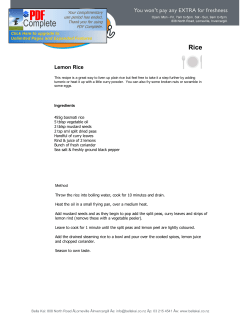


© Copyright 2026