
Side-on binding of p-sulphonatocalix[4]arene to the
ARTICLE IN PRESS
Journal of Inorganic Biochemistry xxx (2009) xxx–xxx
Contents lists available at ScienceDirect
Journal of Inorganic Biochemistry
journal homepage: www.elsevier.com/locate/jinorgbio
Side-on binding of p-sulphonatocalix[4]arene to the dinuclear platinum complex
trans-[{PtCl(NH3)2}2l-dpzm]2+ and its implications for anticancer drug delivery
Nial J. Wheate a,*, Grainne M. Abbott b, Rothwelle J. Tate a, Carol J. Clements b, RuAngelie Edrada-Ebel a,
Blair F. Johnston a
a
b
Strathclyde Institute of Pharmacy and Biomedical Sciences, University of Strathclyde, John Arbuthnott Building, 27 Taylor Street, Glasgow G4 0NR, United Kingdom
Strathclyde Innovations in Drug Research, University of Strathclyde, John Arbuthnott Building, 27 Taylor Street, Glasgow G4 0NR, United Kingdom
a r t i c l e
i n f o
Article history:
Received 8 September 2008
Received in revised form 12 December 2008
Accepted 18 December 2008
Available online xxxx
Keywords:
p-sulphonatocalix[4]arene
Platinum
Alamar blue
MTT
Drug delivery
Multinuclear
Cytotoxicity
Ovarian cancer
a b s t r a c t
The utility of p-sulphonatocalix[4]arene (s-CX[4]) as a drug delivery vehicle for multinuclear platinum
anticancer agents, using trans-[{PtCl(NH3)2}2l-dpzm]2+ (di-Pt; where dpzm = 4,40 -dipyrazolylmethane)
as a model complex, has been examined using 1H nuclear magnetic resonance, electrospray ionisation
mass spectrometry, molecular modelling and in vitro growth inhibition assays. s-CX[4] binds di-Pt in a
side-on fashion in a ratio of 1:1, with the dpzm ligand of the metal complex located within the sCX[4] cavity with binding further stabilised by ion–ion interactions and hydrogen bonding between
the metal complex am(m)ine groups and the s-CX[4] sulphate groups. Partial encapsulation of di-Pt
within the cavity does not prevent binding of 50 -guanosine monophosphate to the metal complex. When
bound to two individual guanosine molecules, di-Pt also remains partially bound by s-CX[4]. The cytotoxicity of free di-Pt and s-CX[4] and their host guest complex was examined using in vitro growth inhibition
assays in the A2780 and A2780cis human ovarian cancer cell lines. Free di-Pt has an IC50 of 100 and
60 lM, respectively, in the cell lines, which is significantly less active than cisplatin (1.9 and 8.1 lM,
respectively). s-CX[4] displays no cytotoxicity at concentrations up to 1.5 mM and does not affect the
cytotoxicity of di-Pt, probably because its low binding constant to the metal complex (6.8 104 M1)
means the host–guest complex is mostly disassociated at biologically relevant concentrations.
Ó 2008 Elsevier Inc. All rights reserved.
1. Introduction
In the 40 years since the approval of cisplatin as an anticancer
agent just two other platinum-based drugs have received worldwide approval [1]. New families of platinum drugs continue to be
synthesised and tested, including: platinum(IV) complexes [1,2],
sterically hindered complexes [1–3], DNA intercalators [4,5] and
multinuclear drugs [6,7]. We hypothesise, however, that the biggest break-through in the next decade for platinum-based chemotherapy will come from improved chemical delivery of already
approved drugs. Better drug delivery can be achieved through
the use of encapsulating agents, either using polymers to form
liposome/micelle formulations or using macrocycles to encapsulate single drug molecules. The drugs Prolindac and Aroplatin are
examples of successful formulations of oxaliplatin using liposomes
and micelles [8–12].
Our group is investigating the targeted delivery of mono- and
multinuclear platinum drugs using small macrocycles functionalised with targeting agents. In the first phase of our research we
are examining a range of macrocycles that fully or partially encap* Corresponding author. Tel.: +44 141 548 4962; fax: +44 141 552 2562.
E-mail address: [email protected] (N.J. Wheate).
sulate single molecules of drug, in order to select the best vehicle
for further development. Previously we have examined cucurbit[n]urils in detail [5,13–17], and cyclodextrins and calix[n]arenes
to a lesser extent [18].
Calix[n]arenes are a family of macrocycles made from the
hydroxyalkylation of a phenol and an aldehyde and are bowl or
cone shaped molecules [19,20]. p-Sulphonatocalix[4]arene (s-CX[4];
Fig. 1) is a particularly interesting member of the calix[n]arene
family and has potential as a drug delivery vehicle. The four
sulphate groups impart high water solubility on the molecule, it
is able to bind a range of small molecules and proteins with high
affinity, it has demonstrated zero haemolytic toxicity in vitro at
concentrations up to 5 mM, and it is non-toxic in vivo at doses
up to 100 mg/kg [21]. Previously we have examined the encapsulation of a family of platinum(II)-based DNA intercalators by
s-CX[4] [18]. The host–guest chemistry of the resultant complexes
was unusual, with a 2:2 complex being formed where two intercalator molecules were stacked head-to-tail and capped on either
end by s-CX[4] [18].
In this paper we report the side-on binding of s-CX[4] to a dinuclear platinum complex, trans-[{PtCl(NH3)2}2l-dpzm]2+ (di-Pt;
Fig. 1), where dpzm = 4,40 -dipyrazolylmethane, by 1H nuclear magnetic resonance (NMR) spectroscopy, electrospray ionisation mass
0162-0134/$ - see front matter Ó 2008 Elsevier Inc. All rights reserved.
doi:10.1016/j.jinorgbio.2008.12.011
Please cite this article in press as: N.J. Wheate et al., J. Inorg. Biochem. (2009), doi:10.1016/j.jinorgbio.2008.12.011
ARTICLE IN PRESS
2
N.J. Wheate et al. / Journal of Inorganic Biochemistry xxx (2009) xxx–xxx
2+
H3N
5
NH3
Cl
Pt
NH3
N
N
NH
3
N
H
Pt
Cl
NH3
2.3. Electrospray ionisation mass spectrometry
Negative ion and positive ion electrospray ionisation (ESI) mass
spectra were recorded on a Finnigan LTQ Orbitrap. Samples were
dissolved in H2O to a concentration of 100 lM and injected into
the instrument in 90% CH3CN/10% H2O at a flow rate of
400 lL min1. The capillary temperature and voltage were 200 °C
and 40 V, respectively, with a source voltage of 4000 V.
2.4. Molecular modelling
4-
O
O
S
O
Calculations were performed on a dual-Xeon processor, Dell
workstation using the GAUSSIAN 03 program [23]. Individual
geometry optimisations for both di-Pt (C1) and s-CX[4] (C4) were
undertaken at the HF/ LanL2DZ level [24–26] in order to locate
structurally stable conformers. Further geometry optimisation of
the di-Pt/s-CX[4] complex, using the same level of theory and basis
set, was then undertaken using the previously optimised structures as a starting point for the calculation. Vibrational frequencies
were calculated from analytic second derivatives to confirm the
structure as a local minimum on the potential energy surface.
2.5. Growth inhibition assays
OH
4
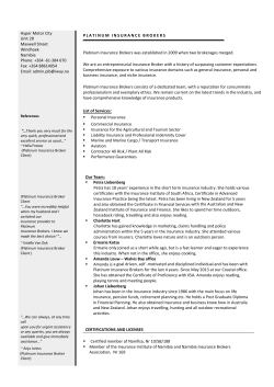
Fig. 1. The chemical structures of di-Pt and s-CX[4], showing the numbering
scheme of the pyrazole protons. Counter ions have been removed for clarity.
spectrometry and molecular modelling. The ability of the macrocycle to slow down the metal complex binding to guanosine and the
affect of s-CX[4] on the cytotoxicity of di-Pt in the A2780 and
A2780cis ovarian cancer cell lines are also reported.
The human ovarian carcinoma cell lines A2780 and A2780cis
were grown in either RPMI medium or DMEM with added hydrocortisone, insulin, gentamicin, glutamine and amphotericin B, at
37 °C and with humidified a 5% CO2 atmosphere. Cells were plated
in 96-well plates (100 lL per well) at a concentration of 5 104
cells/well in complete medium and incubated for 24 h before they
were treated with a range of cisplatin, di-Pt and s-CX[4] saline
solutions diluted with medium to 100 lL (final drug/calixarene
concentrations 0.1 lM to 1.5 mM). The cells were incubated for
between 24 and 72 h under standard conditions before drug
cytotoxicity was determined using either an Alamar BlueTM – or
MTT-based growth inhibition assay [27]. Data given are averages
derived from four independent experiments.
2. Materials and methods
2.6. Cell line DNA profiling
2.1. Materials
Di-Pt was made as previously described [22]. p-Sulphonatocalix[4]arene sodium salt and 50 -guanosine monophosphate disodium salt were purchased from Sigma–Aldrich. D2O (99.9%) was
purchased from Cambridge Isotope Laboratories. Insulin, hydrocortisone, amphotericin B, gentamicin, (3-(4,5-dimethylthiazol-2-yl)2,5-diphenyltetrazolium bromide (MTT) and RPMI-1640 medium
were purchased from Sigma. Dulbecco’s Modified Eagle’s Medium
(DMEM), fetal bovine serum, and L-glutamine were purchased from
Invitrogen. Alamar BlueTM was purchased from Serotec. The A2780
and A2780cis cell lines were purchased from the European
Collection of Cell Cultures (ECCC).
Genomic DNA was extracted from A2780 and A2780cis cell pellets using a GenElute Mammalian Genomic DNA miniprep kit (Sigma–Aldrich). The cell line DNA was used in reactions with the Cell
ID STR System (Promega) to compare the allele determinations at
10 loci, consisting of nine STR loci and amelogenin. The PowerPlex
16 STR system (Promega), which allows detection of 16 loci,
including the D8S1179 loci found in the SGM system, was also
used. The products were subjected to capillary electrophoresis
and fluorescent detection on an Applied Biosystems 3100-Avant
Genetic Analyzer (Applied Biosystems) and the results analysed
using GeneMapper ID v3.2 software (Applied Biosystems).
2.7. Calculation of binding constant
2.2. Nuclear magnetic resonance
NMR spectra were recorded using a JEOL JNM-LA400 spectrometer operating at 400 MHz. Samples were dissolved in 600 lL of
D2O (2–20 mM) and referenced internally to the solvent
(4.78 ppm at 25 °C). One-dimensional spectra were recorded using
16–256 scans and a relaxation delay of 2–4 s. Visualisation of the
platinum ammine resonances was made by dissolving the metal
complex in solvent just before the NMR spectra were recorded.
Two-dimensional rotating frame Overhauser effect spectroscopy
(ROESY) were conducted using 32 scans, a relaxation delay of
1.5 s, a mix time of 500 ms, with 2048 increments in the t1 dimension and 256 increments in the t2 dimension.
The binding constant (Kb, M1) of s-CX[4] to di-Pt was calculated using the following equation:
Kb ¼
½host=guest complex
½host½guest
ð1Þ
The concentrations of free and bound host and guest were
determined using the following equation:
dobs ¼ df vf þ db vb
ð2Þ
where dobs is the chemical shift of the di-Pt H5 proton resonance at
any titration point, df is the chemical shift of the same resonance in
Please cite this article in press as: N.J. Wheate et al., J. Inorg. Biochem. (2009), doi:10.1016/j.jinorgbio.2008.12.011
ARTICLE IN PRESS
3
N.J. Wheate et al. / Journal of Inorganic Biochemistry xxx (2009) xxx–xxx
the absence of s-CX[4], db is the chemical shift of the resonance after
the addition of 2 mole equivalents of s-CX[4] and vf and vb are the
mole fractions of free and bound di-Pt, respectively.
2.8. Guanosine binding
50 -Guanosine monophosphate disodium salt (4 mM), di-Pt
(2 mM) and s-CX[4] (2 mM) were dissolved in unbuffered D2O
and kept in a water bath at 37 °C. At times 0 and 24 h, NMR spectra
were recorded.
3. Results and discussion
3.1. Host–guest complex solubility
As the 2+ cation, di-Pt is soluble in water at concentrations up to
13.5 mM. The higher 4- charge of s-CX[4] means it is soluble at
concentrations greater than 20 mM. When s-CX[4] and di-Pt are
mixed in equimolar concentrations, however, the solubility of the
host–guest complex, which has an overall charge of 2-, is around
4.5 mM. This is significantly higher than some cucurbit[n]uril-platinum(II) host–guest complexes which are only soluble at concentrations less than 1 mM [16,28].
H3
3.2. 1H NMR
The titration of s-CX[4] into a solution of di-Pt induces selective
and large chemical shift changes of the di-Pt proton resonances
(Fig. 2). When the ratio of s-CX[4] to di-Pt was 0.5:1 the di-Pt
H3, H5 and CH2 resonances shifted 0.4, 1.05 and 1.15 ppm upfield,
respectively. As only one set of resonances is observed, and they
are relatively broad, we conclude that the s-CX[4] binds the metal
complex with fast to intermediate exchange kinetics on the NMR
timescale. Whilst only one set of H3, H5 and CH2 resonances are
observed, two separate resonances for the NH3 protons of di-Pt
are evident. One ammine resonance shifted downfield 0.53 ppm
and the other ammine resonance moved upfield 0.29 ppm.
When more s-CX[4] was added to the solution to attain a sCX[4] to di-Pt ratio of 1:1, the metal complex resonances shifted
further downfield and significantly sharpened. The CH2 resonance
shifted 2.94 ppm, the H5 1.88 ppm and the H3 0.73 ppm. The ammine resonance’s chemical shifts remained relatively unchanged
compared with the NMR spectrum at lower host to guest ratios,
with one ammine resonance shifted 0.3 ppm upfield and the other
0.53 ppm downfield. Titration of more s-CX[4] into the solution to
a s-CX[4] to di-Pt ratio of 2:1 had no further significant effect on
the chemical shifts of the di-Pt resonances.
H5
C H2
c
s-CX[4]
s-CX[4] (-OH)
NH 3
H3
NH 3
H5
H5
s-CX[4] (-CH2)
C H2
b
H3
HDO
C H2
NH 3
9.0
8.0
7.0
6.0
5.0
4.0
a
3.0
2.0
1.0
ppm
Fig. 2. The 1H NMR spectra of (a) di-Pt, (b) di-Pt with 0.5 equivalent of s-CX[4] and (c) di-Pt with 1.0 equivalents of s-CX[4], showing the large upfield shifts of the di-Pt
resonances upon binding and the non-equivalence of the platinum ammine ligands.
Please cite this article in press as: N.J. Wheate et al., J. Inorg. Biochem. (2009), doi:10.1016/j.jinorgbio.2008.12.011
ARTICLE IN PRESS
4
N.J. Wheate et al. / Journal of Inorganic Biochemistry xxx (2009) xxx–xxx
a
b
Fig. 3. Schematic diagrams showing (a) the original proposed end-on binding of sCX[4] to the platinum group(s) of di-Pt and (b) the actual side-on binding that
places the central bridging ligand of di-Pt within the s-CX[4] cavity. Charges and
counter ions have been omitted for clarity.
At all macrocycle to metal complex ratios the chemical shifts
of the s-CX[4] resonances were largely unchanged from those of
free s-CX[4]. At a ratio of 1:1, the s-CX[4] aromatic resonance
had shifted upfield by just 0.06 ppm and the CH2 and OH resonances had moved 0.01 ppm, which is within the error of this
experiment.
The large downfield shift of the di-Pt resonances are consistent
with a shielding effect from being located within the s-CX[4] cavity
[18]. The degree of shielding is directly proportional to the depth of
the proton resonance within the cavity. Therefore these results
indicate that the dpzm ligand of di-Pt is located within the sCX[4] cavity, with the CH2 protons in deepest. The splitting of
the platinum–ammine resonances may indicate that one group of
protons is experiencing a shielding effect from being located within the cavity, whilst the other group is experiencing a deshielding
effect from being outside, but close to, the s-CX[4] cavity. Alternatively, the splitting of the ammine resonances may indicate that
one ammine group is hydrogen bonded to the sulphate groups of
s-CX[4] and is thus rotationally restricted, whilst the other ammine
group is not.
The interaction of di-Pt with s-CX[4] was further examined
using two-dimensional rotating frame Overhauser effect (ROE)
spectroscopy. As well as the expected intramolecular ROEs from
the di-Pt H5 and H3 resonances to its own CH2 resonance, two
intermolecular ROEs are observed (data not shown). Both ROEs
are from the CH2 resonance of di-Pt to the s-CX[4] aromatic and
OH resonances. This result clearly places the dpzm ligand of di-Pt
within the s-CX[4] cavity.
The three-dimensional structure of calix[n]arenes, including sCX[4], has been determined by X-ray diffraction experiments.
This family of molecules exists as bowl shaped structures. When
the sulphonate groups of s-CX[4] are protonated and uncharged,
or are charged but stabilised by cations, these groups form the
rim of the bowl opening. The hydroxyl groups however, are
packed closely together at the base of the molecule, leaving only
a very small opening. Given this structure it was expected that
the binding of s-CX[4] to di-Pt would occur over the ends of
the metal complex (see Fig. 3a), rather than over the central
bridging ligand, as is observed with cucurbit[n]uril encapsulation
[13,14,29]. This binding would be stabilised by ion–ion and
hydrogen bonding interactions between the s-CX[4] sulphate
groups and the platinum atom/ammine ligands. Such binding
would have placed the platinum chloro ligands inside the cavity
and the dpzm ligand outside the cavity. Addition of two s-CX[4]
molecules to every di-Pt molecule was expected to form a doubly
capped metal complex.
The NMR spectra, however, indicate that binding by s-CX[4] to
di-Pt occurs in a side-on fashion with the dpzm ligand located
within the cavity (see Fig. 3b); threading of the di-Pt through the
macrocycle can be excluded due to the insufficient size of the
opening at the hydroxyl end of s-CX[4]. The proposed side-on binding would be stabilised by hydrophobic effects between the dpzm
ligand and the s-CX[4] rings (the lack of change in the chemical
shift of the s-CX[4] aromatic resonance indicates that p-p stacking
between the dpzm ligand and the macrocycle does not occur), ion–
ion interactions between the sulphate groups and the platinum
atoms, and/or ion–dipole interactions between the sulphate and
am(m)ine groups.
Because the binding of s-CX[4] to di-Pt is fast on the NMR timescale and only one set of di-Pt resonances is observed, a binding
constant 6.8 104 M1 can be calculated from the NMR spectra.
This value is on the border of what is generally agreed as the minimum binding constant (at least 105 M1) required to be useful as a
drug delivery vehicle, since most drugs need to be cytotoxic at
lM–nM concentrations to have sufficient in vivo efficacy. When
administered in the body, much of the di-Pt and s-CX[4] will disassociate because of the high volume of blood serum and the dose of
platinum drug that is generally given. As such, the weak binding
constant may exclude s-CX[4] from being a useful drug delivery
vehicle for multinuclear platinum drugs.
3.3. ESI-MS
Electrospray ionisation mass spectrometry was used to further confirm the 1:1 stoichiometry of the binding. In the ESI
spectrum of free s-CX[4] ten assignable peaks are observed
(Fig. 4a). The largest peak corresponds to an s-CX[4] molecule
with a single negative charge which arises from the protonation
of three of the four sulphate groups. Peaks for doubly charged/
doubly protonated and a variety of other s-CX[4] molecules with
associated sodium cations are also observed. In the ESI spectrum of di-Pt and s-CX[4], eight assignable peaks are observed
(Fig. 4b). Peaks 11–13 and 16–18 are consistent with unbound
s-CX[4], as seen in the spectrum of free s-CX[4]. Peaks 14 and
15 are assignable to di-Pt and s-CX[4] host–guest complexes in
a ratio of 1:1. No evidence is seen for the formation of a 2:1
di-Pt to s-CX[4] complex.
3.4. Molecular modelling
To further confirm the binding model predicted on the basis of
the NMR data, molecular modelling was conducted using Gaussian
03. The structures of free s-CX[4] and di-Pt were minimised individually before they were manually docked together in the sideon model of binding and then energy minimised together. The 2+
charge of di-Pt and the addition of two sodium cations maintains
the bowl-like shape of s-CX[4] (Fig. 5). On binding of di-Pt, the
rim of s-CX[4] is not perfectly circular, with one long axis measuring 11.4 Å from phosphate to phosphate and one short axis measuring 9.4 Å. The di-Pt methylene group sits deepest within the
cavity, 5.4–5.6 Å from the s-CX[4] phenolic groups and 3.9 Å from
the closest aromatic hydrogen. The di-Pt platinum groups are located 3–4 Å above the plane of the sulphate groups. As well as
hydrophobic forces between the di-Pt bridging ligand and the cavity of s-CX[4], binding is further stabilised by six intermolecular
hydrogen bonds; four between individual ammine groups on each
Please cite this article in press as: N.J. Wheate et al., J. Inorg. Biochem. (2009), doi:10.1016/j.jinorgbio.2008.12.011
ARTICLE IN PRESS
N.J. Wheate et al. / Journal of Inorganic Biochemistry xxx (2009) xxx–xxx
5
Fig. 4. The electrospray ionisation mass spectra (negative mode) of (a) free s-CX[4] and (b) di-Pt and s-CX[4] at a ratio of 1. Peaks are assigned as: 1/11, 371.99 m/z, [s
4, 662.80 m/z, ½s CX½4þ2Hþ SO
5, 685.87 m/z,
CX[4]+2H+H]2; 2, 382.40 m/z, [s-CX[4]+H++Na+]2; 3, 582.93 m/z, ½s CX½4þ2Hþ 2SO
3 ;
3 ;
+ +
+ +
+ + ½s CX½4þ2Hþ þNaþ 2SO
3 ; 6, 742.73 m/z, [s-CX[4]+3H ] ; 7, 764.93 m/z, [s-CX[4]+2H +Na ] ; 8, 766.87 m/z, [s-CX[4]+2H +2Na ] ; 9, 808.80 m/z, [s-CX[4]+3Na ] ;
+
+ + 2
+ 2
+ 10, 830.87 m/z, [s-CX[4]-H +4 Na ] ; 12, 383.40 m/z, [s-CX[4]+Na ] ; 13, 394.93 m/z, [s-CX[4]+2Na ] ; 14, 708.93 m/z, [s-CX[4]+di-Pt+H ] ; 15, 732.07 m/z, [s-CX[4]+di-PtH++2Na+]; 16, 811.80 m/z, [s-CX[4]+3Na++H]; 17, 834.93 m/z, [s-CX[4]+4Na++H]; 18, 869.60 m/z, [s-CX[4]+4Na++2H2O+H+].
platinum atom to opposing individual s-CX[4] sulphate groups
(NH O; 1.7–2.2 Å), and two between the di-Pt N1 hydrogen
atoms and the remaining two opposing s-CX[4] sulphate groups
(NH O; 2.0 Å). The hydrogen bonds between the di-Pt N1 hydrogen atoms and the sulphate groups cause the di-Pt H5 hydrogen to
be pointed more towards the s-CX[4] cavity compared with the H3
hydrogen, which explains the larger upfield chemical shift change
experienced by the H5 in the 1H NMR spectra. The hydrogen bonding between the platinum–ammine groups and the s-CX[4] sulphate groups also explains the two individual ammine
resonances observed in the 1H NMR.
3.5. Guanosine binding
The ability of s-CX[4] to slow or prevent nucleophilic attack, by
providing steric bulk around the platinum atoms of di-Pt, was
investigated using 50 -guanosine monophosphate. The reaction of
di-Pt with 2 mole equivalents of guanosine in the presence of sCX[4] was monitored by 1H NMR (Fig. 6). The coordination of diPt to the N7 of guanosine is clearly observed through the change
in the chemical shift of the guanosine H8 resonance; the H8 moves
downfield from 8.17 to 8.84 ppm [30]. A chemical shift change is
also observed for the guanosine sugar H1’ resonance, which moves
Please cite this article in press as: N.J. Wheate et al., J. Inorg. Biochem. (2009), doi:10.1016/j.jinorgbio.2008.12.011
ARTICLE IN PRESS
6
N.J. Wheate et al. / Journal of Inorganic Biochemistry xxx (2009) xxx–xxx
Table 1
The cytotoxicity (IC50, lM) of cisplatin, di-Pt, s-CX[4] and the di-Pt–sCX[4] host–guest
complex in the human ovarian cancer cell line A2780 and its cisplatin-resistant
subline A2780cis, determined using two different fluorescent markers, Alamar Blue
and MTT, and at various drug incubation times (h).
Agent
A2780
A2780cis
Alamar Blue
Cisplatin
di-Pt
sCX[4]
di-Pt–sCX[4]
MTT
Alamar Blue
MTT
24 h
48 h
72 h
48 h
24 h
48 h
72 h
48 h
51
>100
>100
>100
34
>100
>100
100
32
>100
>100
48
1.9
100
>1500
–
100
>100
>100
>100
40
74
>100
100
29
66
>100
62
8.1
60
1500
–
the NMR spectra indicated that di-Pt was still partially encapsulated by s-CX[4] after binding to guanosine, only one significant
peak is observed in the ESI+ spectrum, at 670.27 m/z, which is assigned as a di-Pt molecule coordinated to two guanosine molecules
and with two sodium cations (data not shown). No evidence for a
s-CX[4]/di-Pt/guanosine host–guest complex was observed.
Fig. 5. An energy-minimised model of the sodium salt of s-CX[4] and di-Pt showing
the side-on binding of the metal complex which places its bridging ligand partially
within the s-CX[4] cavity, where binding is stabilised by hydrophobic effects.
Binding is further stabilised by six hydrogen bonds (red dashed lines) between the
di-Pt amine and ammine groups to the s-CX[4] sulphate groups. (For interpretation
of the references to colour in this figure legend, the reader is referred to the web
version of this article.)
from 5.91 to 5.97 ppm. Interestingly, the NMR spectrum of the
reaction product indicates that di-Pt remains bound, at least partially, by s-CX[4] after it has reacted with guanosine. Previously
we have shown that when bound to guanine the H5 and H3 resonances of di-Pt are superimposed at a chemical shift of 7.89 ppm.
In the NMR spectrum of di-Pt with guanosine and s-CX[4], both
metal complex resonances are much further upfield with the H3
at 7.35 ppm and the H5 at 6.56 ppm. The chemical shifts and line
shape of the both the H3 and H5 resonances are similar to those
of the di-Pt with 0.5 equivalents of s-CX[4] (7.32 and 6.72 ppm,
respectively). This result therefore indicates that the s-CX[4] provides no steric hindrance to the binding of nucleophiles to the platinum groups of di-Pt.
The ESI+ spectrum of the reaction mixture of di-Pt binding to
guanosine in the presence of s-CX[4] was also obtained. Whilst
3.6. Cytotoxicity
Cisplatin, free s-CX[4], and di-Pt in the presence and absence of
s-CX[4], were tested for cytotoxicity in the human ovarian carcinoma cell line A2780 and in the cisplatin resistant daughter line
A2780cis. The A2780 and A2780cis cell lines have been well studied as models for measuring platinum drug cytotoxicity, although
the conditions under which the assays have been conducted vary
substantially; incubation times range between 48 and 96 h, with
an initial plating of 1000–10,000 cells per well, and a variety of
substrates in the medium [31–36].
As a starting point we examined the cytotoxicity of di-Pt and diPt-sCX[4] using a 24 h Alamar Blue growth inhibition assay with
cisplatin used as a control. As can be seen in Table 1, all the metal
complexes were largely non-cytotoxic in both cell lines, a surprising result, especially for cisplatin in the A2780 cell line. Incubation
for longer times did not increase the cytotoxicity of the drugs, nor
did changing the medium used. The poor cytotoxicity was not a result of incorrect cell lines supplied by the manufacture as short
tandem repeat profiling and comparison to the results of Masters
et al. [37], confirmed their identity. Finally, growth inhibition
was determined using an MTT-based, rather than an Alamar
b
s-CX[4]
G-H8
H3
H5
H1'
a
9.0
8.0
7.0
6.0
ppm
Fig. 6. The 1H NMR spectra of di-Pt (2 mM), s-CX[4] (2 mM) and guanosine (4 mM) at (a) time 0 h and (b) after reaction at 37 °C for 24 h. The 0.6 ppm downfield shift of the
guanosine-H8 resonance is indicative of platinum binding at the N7. Importantly, the chemical shift of the di-Pt H5 and H3 resonances indicate that the metal complex is still
partially encapsulated in the cavity of s-CX[4] after the metal complex has bound to guanosine.
Please cite this article in press as: N.J. Wheate et al., J. Inorg. Biochem. (2009), doi:10.1016/j.jinorgbio.2008.12.011
ARTICLE IN PRESS
N.J. Wheate et al. / Journal of Inorganic Biochemistry xxx (2009) xxx–xxx
Blue-based assay, and the IC50 values obtained for cisplatin were
then consistent with those determined by other groups (i.e 0.5–
1.5 lM in the A2780 cell line) [32,33,35,36] and clearly show the
cisplatin resistance in the A2780cis cell line (4.3-fold decrease in
activity).
Regardless of whether Alamar Blue or MTT is used as the fluorescent agent, the results for free s-CX[4] demonstrate that this
macrocycle is non-cytotoxic, consistent with the findings of Coleman et al [21], who demonstrated that s-CX[4] is not toxic in mice
at concentrations up to 100 mg/kg. Likewise, di-Pt shows a similar
trend with either Alamar Blue or MTT. The metal complex is considerably less cytotoxic than cisplatin, but does show some ability
to overcome cisplatin resistance (resistance factor 0.6). When
tested as the 1:1 host–guest complex with s-CX[4] the macrocycle
had no significant effect on the cytotoxicity of di-Pt. This may be
because s-CX[4] does not protect that metal complex from intracellular glutathione degradation, or because it neither assists nor
hinders metal complex uptake into the cell, or because the host–
guest complex is easily disassociated (inside or outside the cell)
at the concentrations at which the growth inhibition assays were
conducted.
4. Conclusions
In this paper we examined the utility of s-CX[4] as a drug delivery vehicle for multinuclear platinum anticancer drugs using the
dinuclear complex, di-Pt, as a model. The side-on binding of the
macrocycle to the metal complex, its low binding constant, and
its inability to provide steric hindrance to attack of the metal complex by guanosine indicates that s-CX[4] may not be a suitable
drug delivery vehicle for multinuclear anticancer drugs.
5. Abbreviations
di-Pt
DMEM
Dpzm
ESI-MS
MTT
ROESY
s-CX[4]
trans-[{PtCl(NH3)2}2l-dpzm]2+
dulbecco’s modified eagle’s medium
4,40 -dipyrazolylmethane
electrospray ionisation mass spectrometry
(3-(4,5-dimethylthiazol-2-yl)-2,5-diphenyltetrazolium
bromide
rotating frame Overhauser effect spectrometry
p-sulphonatocalix[4]arene
Acknowledgement
This work was supported by a University of Strathclyde Faculty
of Science Starter Grant awarded to N.J.W.
References
[1]
[2]
[3]
[4]
L. Kelland, Nat. Rev. Cancer 7 (2007) 573–584.
C.X. Zhang, S.J. Lippard, Curr. Opin. Chem. Biol. 7 (2003) 481–489.
M. Hay, Curr Opin. Oncol., Endocr. Metab. Invest. Drugs 1 (1999) 443–447.
N.J. Wheate, C.R. Brodie, J.G. Collins, S. Kemp, J.R. Aldrich-Wright, Mini Rev.
Med. Chem. 7 (2007) 627–648.
[5] N.J. Wheate, R.I. Taleb, A.M. Krause-Heuer, R.L. Cook, S. Wang, V.J. Higgins, J.R.
Aldrich-Wright, Dalton Trans. (2007) 5055–5064.
7
[6] N.J. Wheate, J.G. Collins, Coord. Chem. Rev. 241 (2003) 133–145.
[7] N.J. Wheate, J.G. Collins, Curr. Med. Chem. – Anti-Cancer Agents 5 (2005) 267–
279.
[8] Y.J. Jun, J.I. Kim, M.J. Jun, Y.S. Sohn, J. Inorg. Biochem. 99 (2005) 1593–
1601.
[9] M. Campone, J.M. Rademaker-Lakhai, J. Bennouna, S.B. Howell, D.P. Nowotnik,
J.H. Beijnen, J.H.M. Schellens, Cancer Chemother. Pharmacol. 60 (2007) 523–
533.
[10] J.R. Rice, J.L. Gerberich, D.P. Nowotnik, S.B. Howell, Clin. Cancer Res. 12 (2006)
2248–2254.
[11] P. Sood, K.B. Thurmond, J.E. Jacob, L.K. Waller, G.O. Silva, D.R. Stewart, D.P.
Nowotnik, Bioconjugate Chem. 17 (2006) 1270–1279.
[12] C. Lu, R. Perez-Soler, B. Piperdi, G.L. Walsh, S.G. Swisher, W.R. Smythe, H.J. Shin,
J.Y. Ro, L. Feng, M. Truong, A. Yalamanchili, G. Lopez-Berestein, W.K. Hong, A.R.
Khokhar, D.M. Shin, J. Clin. Oncol. 23 (2005) 3495–3501.
[13] N.J. Wheate, D.P. Buck, A.I. Day, J.G. Collins, Dalton Trans. (2006) 451–
458.
[14] N.J. Wheate, A.I. Day, R.J. Blanch, A.P. Arnold, C. Cullinane, J.G. Collins, Chem.
Commun. (2004) 1424–1425.
[15] N.J. Wheate, P.G.A. Kumar, A.M. Torres, J.R. Aldrich-Wright, W.S. Price, J. Phys,
Chemistry B 112 (2008) 2311–2314.
[16] S. Kemp, N.J. Wheate, S. Wang, J.G. Collins, S.F. Ralph, A.I. Day, V.J. Higgins, J.R.
Aldrich-Wright, J. Biol. Inorg. Chem. 12 (2007) 969–979.
[17] S. Kemp, N.J. Wheate, M.P. Pisani, J.R. Aldrich-Wright, J. Med. Chem. 51 (2008)
2787–2794.
[18] A.M. Krause-Heuer, N.J. Wheate, M.J. Tilby, D. Pearson, C.J. Ottley, J.R. AldrichWright, Inorg. Chem. 47 (2008) 6880–6888.
[19] C. Redshaw, Coord. Chem. Rev. 244 (2003) 45–70.
[20] W. S´liwa, J. Incl. Phenom. Macrocycl. Chem. 52 (2005) 13–37.
[21] A.W. Coleman, S. Jebors, S. Cecillon, P. Perret, D. Garin, D. Marti-Battle, M.
Moulin, New J. Chem. 32 (2008) 780–782.
[22] N.J. Wheate, C. Cullinane, L.K. Webster, J.G. Collins, Anti-Cancer Drug Des. 16
(2001) 91–98.
[23] G.W.T.M.J. Frisch, H.B. Schlegel, G.E. Scuseria, M.A. Robb, J.R. Cheeseman, J.A.
Montgomery Jr., T. Vreven, K.N. Kudin, J.C. Burant, J.M. Millam, S.S. Iyengar, J.
Tomasi, V. Barone, B. Mennucci, M. Cossi, G. Scalmani, N. Rega, G.A. Petersson,
H. Nakatsuji, M. Hada, M. Ehara, K. Toyota, R. Fukuda, J. Hasegawa, M. Ishida, T.
Nakajima, Y. Honda, O. Kitao, H. Nakai, M. Klene, X. Li, J.E. Knox, H.P. Hratchian,
J.B. Cross, V. Bakken, C. Adamo, J. Jaramillo, R. Gomperts, R.E. Stratmann, O.
Yazyev, A.J. Austin, R. Cammi, C. Pomelli, J.W. Ochterski, P.Y. Ayala, K.
Morokuma, G.A. Voth, P. Salvador, J.J. Dannenberg, V.G. Zakrzewski, S.
Dapprich, A.D. Daniels, M.C. Strain, O. Farkas, D.K. Malick, A.D. Rabuck, K.
Raghavachari, J.B. Foresman, J.V. Ortiz, Q. Cui, A.G. Baboul, S. Clifford, J.
Cioslowski, B.B. Stefanov, G. Liu, A. Liashenko, P. Piskorz, I. Komaromi, R.L.
Martin, D.J. Fox, T. Keith, M.A. Al-Laham, C.Y. Peng, A. Nanayakkara, M.
Challacombe, P.M.W. Gill, B. Johnson, W. Chen, M.W. Wong, C. Gonzalez, J.A.
Pople, Gaussian, Inc., Wallingford, CT, 2004.
[24] P.J. Hay, W.R. Wadt, J. Chem. Phys. 82 (1985) 270–283.
[25] W.R. Wadt, P.J. Hay, J. Chem. Phys. 82 (1985) 284–298.
[26] P.J. Hay, W.R. Wadt, J. Chem. Phys. 82 (1985) 299–310.
[27] T. Mosmann, J. Immunol, Methods 65 (1983) 55–63.
[28] Y.J. Jeon, S.-Y. Kim, Y.H. Ko, S. Sakamoto, K. Yamaguchi, K. Kim, Org. Biomol.
Chem. 3 (2005) 2122–2125.
[29] M.S. Bali, D.P. Buck, A.J. Coe, A.I. Day, J.G. Collins, Dalton Trans. (2006) 5337–
5344.
[30] N.J. Wheate, B.J. Evison, A.J. Herlt, D.R. Phillips, J.G. Collins, Dalton Trans. (2003)
3486–3492.
[31] A. Hegmans, J. Kasparkova, O. Vrana, L.R. Kelland, V. Brabec, N.P. Farrell, J. Med.
Chem. 51 (2008) 2254–2260.
[32] G. Pratesi, P. Perego, D. Polizzi, S.C. Righetti, R. Supino, C. Caserini, C. Manzotti,
F.C. Giuliani, G. Pezzoni, S. Tognella, S. Spinelli, N. Farrell, F. Zunino, Brit. J.
Cancer 80 (1999) 1912–1919.
[33] A.L. Harris, X. Yang, A. Hegmans, L. Povirk, J.J. Ryan, L. Kelland, N.P. Farrell,
Inorg. Chem. 44 (2005) 9598–9600.
[34] G. Colella, M. Pennati, A. Bearzatto, R. Leone, D. Colangelo, C. Manzotti, M.G.
Daidone, N. Zaffaroni, Brit. J. Cancer 84 (2001) 1387–1390.
[35] E. Monti, M. Gariboldi, A. Maiocchi, E. Marengo, C. Cassino, E. Gabano, D. Osella,
J. Med. Chem. 48 (2005) 857–866.
[36] Y. Qu, H. Rauter, A.P.S. Fontes, R. Bandarage, L.R. Kelland, N. Farrell, J. Med.
Chem. 43 (2000) 3189–3192.
[37] J.R. Masters, J.A. Thomson, B. Daly-Burns, Y.A. Reid, W.G. Dirks, P. Packer, L.H.
Toji, T. Ohno, H. Tanabe, C.F. Arlett, L.R. Kelland, M. Harrison, A. Virmani, T.H.
Ward, K.L. Ayres, P.G. Debenham, Proc. Natl. Acad. Sci. USA 98 (2001) 8012–
8017.
Please cite this article in press as: N.J. Wheate et al., J. Inorg. Biochem. (2009), doi:10.1016/j.jinorgbio.2008.12.011


© Copyright 2026