
Impact of Metabolic Risk Factors on Blood Pressure Control
Università degli Studi di Napoli “Federico II”
Facoltà di Medicina e Chirurgia
Dipartimento di Scienze Mediche Traslazionali
Tesi di Dottorato in
Fisiopatologia Clinica e Medicina Sperimentale
Indirizzo Scienze Cardiovascolari
XXVI Ciclo (2010-2013)
Coordinatore Ch.mo Prof. Gianni Marone
Impact of Metabolic Risk Factors
on Cardiovascular Phenotype and Blood Pressure Control.
New Predictors of Cardiovascular Disease
Relatore Ch.mo Prof. Giovanni de Simone
Correlatore Ch.ma Prof. Eva Gerdts
Candidata Dott.ssa Marina De Marco
[This page intentionally left blank]
Page 2 of 76
Table of Contents
List of Abbreviations...................................................................................4
Scientific Environment................................................................................5
Summary......................................................................................................9
List of Articles............................................................................................11
Introduction...............................................................................................13
Objectives...................................................................................................17
Methods......................................................................................................19
Study Populations.............................................................................................................................19
Clinical and Metabolic Classification and Definitions....................................................................22
Echocardiographic Measures..........................................................................................................23
Statistical Analysis...........................................................................................................................25
Results.........................................................................................................27
Impact of Metabolic Risk Factors on Blood Pressure Control .......................................................27
Impact of Diabetes on Cardiovascular Phenotype in Adolescents and Young Adults.....................33
Impact of Mitral Annulus Calcification on Incident Stroke ............................................................38
Discussion...................................................................................................43
Impact of Metabolic Risk Factors on Blood Pressure Control .......................................................43
Impact of Diabetes on Cardiovascular Phenotype in Adolescents and Young Adults.....................45
Impact of Mitral Annulus Calcification on Incident Ischemic Stroke..............................................48
Conclusions ...............................................................................................51
Future Perspectives...................................................................................52
References..................................................................................................53
Page 3 of 76
List of Abbreviations
AF
= Atrial Fibrillation
BMI
= Body Mass Index
BP
= Blood Pressure
CV
= Cardiovascular
DM
= Diabetes Mellitus
eGFR = estimated Glomerular Filtration Rate
FS
= Fractional Shortening
HDL
= High Density Lipoproteins
IFG
= Impaired Fasting Glucose
IVRT = Isovolumic Relaxation Time
LDL
= Low Density Lipoprotein
LIFE
= Losartan Intervention For Endpoint reduction in hypertension
LVH
= Left Ventricular Hypertrophy
LVM
= Left Ventricular Mass
MAC
= Mitral Annulus fibro-Calcification
MetS
= Metabolic Syndrome
NFG
= Normal Fasting Glucose
PP
= Pulse Pressure
RAS
= Renin-Angiotensin System
RWT
= Relative Wall Thickness
SV
= Stroke Volume
S
= Study
UACR = Urinary Albumin-Creatinine Ratio
Page 4 of 76
Scientific Environment
The present project was undertaken in the Department of Translational Medical Sciences at
the Federico II University of Naples in Italy. The Research Group is chaired by Professor
Giovanni de Simone and currently includes 2 Full Professor, 2 Associate Professors, 2
Assistant Professor and 2 Ph.D. students. Several research medical students and resident
fellows cooperate in the research activities.
The Research Group has specialized in cardiovascular epidemiology, with a focus on
cardiovascular prevention, through understanding cardiovascular modifications related to the
main risk factors, such as diabetes, obesity, and hypertension. The research group facilities
includes advanced echocardiographic laboratories with dedicated workstations for postprocessing of images and data analyses and outpatients clinics for management of research
patients. During my Ph.D., I was introduced to this new world of research by Dr. Marcello
Chinali, at the time when he was a postdoctoral fellow and supervisor in the
echocardiography laboratory. I had also the opportunity to work closely with Professor
Giovanni de Simone together with Professors Bruno Trimarco, Nicola De Luca and Raffaele
Izzo on several research projects involving exploration of the large database of hypertensive
patients referred to the Hypertension Center of the Federico II University of Naples (the
Campania Salute Network).
The Research Group is part of a large international network, embracing several
research centres in different countries. This worldwide research network has given me the
opportunity to perform research on large databases based upon international research
projects, including the Strong Heart Study, the Losartan Intervention For Endpoint reduction
in hypertension (LIFE) Study and the Hypertension Genetic Epidemiology Network
(HyperGEN) Study. The main international partners are the Department of Cardiology at the
Weill-Cornell Medical College in New York, USA (by Professor Richard B. Devereux) and
the Department of Clinical Science at the University of Bergen in Bergen, Norway (by
Page 5 of 76
Professor Eva Gerdts). During the course of my Ph.D. I had the opportunity to visit and
collaborate with both of them: in New York working with Professor Devereux on the impact
of diabetes in adolescents and young adults, and in Bergen working with Professor Gerdts on
new echocardiographic risk markers in hypertension. I also had the chance to work closely
with Professor Gerdts for one year, during her visit as Guest Professor in Naples in 2011.
During this year, I had the opportunity to collaborate with other postdoctoral fellows of the
Bergen Hypertension and Cardiac Dynamics group in Bergen, especially with postdoctoral
fellow PhD. Mai Tone Lønnebakken who is actually going to spend one year as visiting
guest researcher in the Hypertension Research Center in Naples.
I have had an active rule in development of the studies presented in my thesis, by
ideating, proposing and submitting these research projects, analyzing data and performing
statistical analyses, elaborating and interpreting results and writing manuscripts.
Page 6 of 76
Acknowledgement
Certainly, during my PhD a lot of thing have changed in my life, since I have met many
people who have helped me to grow so I feel now much more enriched, thanks to them.
Even if I have already cited them, I’d like to spend few words on my scientific
supervisors: Giovanni de Simone and Eva Gerdts. They are quite different persons, but they
have a very distinctive aspect in common: their strong passion for research which infects
everyone that has the chance to work with them.
A particular thanks to my “magister vitae”, Prof. Giovanni de Simone, who has been
like a father for me, always supporting and sustaining me and letting me grow up. Thank you
immensely for your generosity and protection, for all your teaching, thank you for all
opportunities you have created for me (overall the privilege of participating to a such
prestigious research team). A special acknowledgement to Professor Eva Gerdts, that has
inspired me for her courage, tenacity and enthusiasm, and that has showed me that
Norwegians can be warmer than Italians, women can be as strong as men, and work can be
funnier than holydays. I am very proud to have been collaborating with such amazing
supervisors. Thanks both of you for your great friendship.
I also have to thank all the people who have inspired me and represented an enormous
opportunity of collaboration, during my Ph.D. fellowship: Dr. Richard Devereux, Dr.
Marcello Chinali, Dr. Barbara Howard, and all researchers I have met in the Division of
Cardiology of Weill-Cornell Medical College in New York, at the Department of Clinical
Science in Bergen and in the Department of Translational Medical Sciences in Naples.
Thanks to my colleagues and friends: Alfonso Sforza, Costantino Mancusi, Daniela
Girfoglio, Gabriella Coppola, Giusy Casalnuovo, Silvia Damiano and Teresa Migliore. I
have shared with you all the happiness and the pain of these intensive years. Obviously I
have forgotten someone, but it’s impossible to quote all friends I have met in these three
years.
Another special thanks goes to Dr. Anna Maria Ferrari, Director of the Emergency
Unit at the Arcispedale Santa Maria Nuova in Reggio Emilia, who has allowed me to
Page 7 of 76
complete my Ph.D. program, and has showed me how the emergency room can be the most
intensive and satisfactory job for a medical doctor.
Regarding my personal life, I’d like to thank my family that has always supported me,
overfilling me of love. I can say without hesitation, I am very lucky person. Golgi once said:
“Discoveries in science are like happiness in life: it comes unexpected”, and in fact a lot of
unpredictable events (p<0.001) occurred to me in these three years, leaving imperishable
memory and a treasure of precious emotions in my hearth.
Last, but not least at all, thank you Silvio, there are not enough words to described my love
and gratitude for you.
Page 8 of 76
Summary
Background: Cardiovascular (CV) risk factors like obesity, hypertension and type 2
diabetes have increased dramatically over the last decades, also in younger age classes. In
hypertension, clustering of metabolic risk factors have been associated with resistant
hypertension, but is not known whether this may be overcome by specific pharmacological
treatments. Moreover, whether specific metabolic risk factors, like diabetes, are associated
with preclinical CV disease also in young age is not well explored. Finally, whether
identification of new markers of CV disease by echocardiography, related to these risk
factors, might help to further refine the high risk CV phenotype is unknown, though this
evidence would be critical to improve risk stratification in the individual beyond current state
of the art.
Aims: The main aim of the Ph.D. project has been to evaluate the impact of metabolic risk
factors on blood pressure control and CV phenotype, identifying new echocardiographic
predictors of CV disease in different populations. Specific sub-goals were:
Study 1 (S1) – To evaluate the impact of clustering of metabolic risk factors on blood
pressure control in relation to different classes of medications;
Study 2 (S2) – To assess the impact of metabolic risk factors, as diabetes and pre-diabetes on
preclinical CV disease in adolescents and young adults;
Study 3 (S3) – To identify new echocardiographic markers of CV events in treated
hypertensive patients with high risk CV profile.
Methods: For the specific scope of the studies we analyzed different populations.
Specifically, in S1 we evaluated the impact of metabolic syndrome on the risk of
uncontrolled blood pressure (i.e. blood pressure≥140/90 mmHg under antihypertensive
treatment), in relation to specific antihypertensive medications. The analysis was carried out
at baseline and after a mean follow-up of 5 years in 4,612 (53±11 years, 43% women; 25%
with obesity, 9% with diabetes) hypertensive patients without prevalent CV disease, referred
Page 9 of 76
to our tertiary Hypertension Center from the Campania Salute Network. In S2 we evaluated
the impact of diabetes and pre-diabetes on cardiac structure and function in 1,624
adolescents and young adults American-Indians (179 with diabetes and 299 with prediabetes). These were participants of the Strong Heart Family Study (mean age 27±8 years,
57% female, 66% with obesity, 19% with hypertension) free of prevalent CV disease.
Finally, in S3 we evaluated the prognostic impact of presence of mitral annulus calcification
(MAC) on incidence of ischemic stroke. For this sub-goal we analyzed baseline and 5 years
follow-up data from 939 treated hypertensive patients (458 with MAC) with
electrocardiographic signs of left ventricular hypertrophy (LVH) (66±7 years, 42% women,
23% with obesity, 11% with diabetes) participating in the Losartan Intervention For
Endpoint reduction in hypertension (LIFE) echocardiography sub-study.
Results: In S1 we found that despite the increased use of medications, hypertensive patients
with metabolic syndrome had higher risk of uncontrolled blood pressure, independently of
specific therapy. Among classes of medications, increased prescriptions of diuretics, reninangiotensin system antagonists and also statins decreased the probability of poor blood
pressure control.
In S2 we found that diabetes was independently associated with early and unfavorable
CV phenotype characterized by increased left ventricular mass, concentric geometry and
preclinical systolic and diastolic dysfunction. Early CV alterations were also present in
participants with pre-diabetes.
In S3 we found that risk of incident ischemic stroke was significantly related to
presence of MAC, independently of traditional CV risk factors, as increased left ventricular
mass and left atrial diameter, prevalence or incidence of atrial fibrillation and albuminuria.
Conclusions: These results document that: 1) in hypertension, clustering of metabolic risk
factors strongly interferes with efficacy of therapy, 2) in addition to obesity and
hypertension, diabetes and pre-diabetes have an important impact on cardiac structure and
function, even in young age, 3) MAC identifies increased risk of stroke in hypertensive
patients with LVH.
Page 10 of 76
List of Articles
S1.
De Marco M, de Simone G, Izzo R, Mancusi C, Sforza A, Giudice R, Trimarco B, De
Luca N. Classes of antihypertensive medications and blood pressure control in relation
to metabolic risk factors. J Hypertens. 2012; 30:188-193.
S2.
De Marco M, de Simone G, Roman MJ, Chinali M, Lee ET, Calhoun D, Howard BV,
Devereux RB. Cardiac geometry and function in diabetic or prediabetic adolescents and
young adults: the Strong Heart Study. Diabetes Care. 2011; 34:2300-2305.
S3.
De Marco M, Gerdts E, Casalnuovo G, Migliore T, Wachtell K, Boman K, Dahlöf B,
Olsen MH, Kizer JR, Devereux RB, de Simone G. Mitral Annular Calcification and
Incident Ischemic Stroke in Treated Hypertensive Patients: The LIFE study. Am J
Hypertens. 2013; 26:567-73.
Page 11 of 76
[This page intentionally left blank]
Page 12 of 76
Introduction
Introduction
Cardiovascular (CV) disease is the major cause of death and disability in Western
countries [1,2], and contributes substantially to the escalating health care costs. The
reduction of CV disease burden depends on the ability of physicians and health care systems
to implement the control of CV risk factors through effective program of primary and
secondary prevention [3]. Lack of control of common CV risk factors like hypertension,
obesity and diabetes is together with the aging of Western population the main causes of
continuous increase in incidence and prevalence of CV disease [4].
Obesity is increasing in epidemic proportions in industrialized and developing
countries, affecting both adults [5] and children and adolescents [6,7]. Obesity predisposes to
arterial hypertension and diabetes and there is extensive evidence that these risk factors tend
to cluster together amplifying the unfavourable effect of each single risk factor on incidence
of disease [8-16], also reducing the efficacy of the treatment [17-22].
In this regard, there is evidence that management of hypertension is particularly
difficult when this condition co-exists with obesity and/or clustering of metabolic risk factors
[22-25]. Hypertension indeed is often part of a constellation of CV risk factors including
obesity, abnormal glucose homeostasis and dyslipidemia, supporting the existence of a
discrete disorder, often referred to as the metabolic syndrome (MetS) [26-28]. MetS
increases CV risk in the setting of hypertension, even when taking individual risk factors into
account [12-14], and reduces the probability of achieving optimal blood pressure (BP)
control, despite more aggressive treatment [22-25].
This is a very important health problem, since arterial hypertension is the most
prevalent CV risk factor in most populations, and the leading cause for medical consultation
and drug prescriptions [29]. It has been estimated that 26% and 28% of incident CV disease
in men and women, respectively, are primarily attributable to hypertension [8]. The
continuous large impact of hypertension on incident CV disease may be related to the fact
Page 13 of 76
Introduction
that BP in hypertensive patients is still largely uncontrolled, despite the large number of
prescribed medications [30,31]. The risk of uncontrolled BP increases with the number of
metabolic risk factors [22], but whether the use of different types of antihypertensive
medications influence this association has not been clarified yet.
Figure 1 shows the complex interaction between the CV risk factors, preclinical CV
disease with asymptomatic modifications on cardiac and arterial structure and function which
precedes clinical CV disease and events. Thus, in addition to the interference on the efficacy
of therapy, there is also growing evidence that obesity related CV risk factors, alone or in
combination, may have particular adverse impact on development of preclinical CV disease
[32-41].
Figure 1 – From risk factors to overt CV diseases through preclinical stage
-
Accordingly, early detection and management of these risk factors and the associated
preclinical target organ damage represents the major health strategies in order to prevent
incident CV disease and optimize preventive and therapeutic strategies. This is very
Page 14 of 76
Introduction
important, especially in adolescents and young adults, since the rising prevalence of obesity
and associated risk factors among the younger ages is now a major health concern with both
epidemiological and economic implications [6,7,42-45]. Early identification of preclinical
disease in adolescents and young adults is also of great interest to understand
pathophysiological mechanisms related to a specific risk factor, since in general the time of
exposure is usually lower compared to adults and also the interference due to other
confounding prevalent diseases is lower. Moreover, cardiometabolic risk factors tend to
remain stables from childhood to adulthood and are predictive of future CV disease [46-49].
Thus, increasing efforts are needed to identify young individuals at high CV risk by
detection early markers of disease to optimize early intensive interventions to prevent or
delay future disease [50].
In particular, type 2 diabetes (DM) in young subjects has increased dramatically in the
last decade [51], especially in minority populations, like the American Indians [52]. The
decline in the age of onset of type 2 DM is driven by the increasing obesity in the younger
age group [53,54]. Early onset of type 2 DM is associated with increased risk of CV
complications compared to usual onset of the disease [55-61]. Comparison of cardiovascular
risk profiles between early (<40 years of age) and later-onset (>40 years of age), showed that
a significantly greater clustering of multiple CV risk factors (obesity, hypertension,
dyslipidaemia) is more common among early-onset type 2 DM subjects [60,61].
However, part of the increased CV risk may be related to a direct adverse effect of DM
on the heart, independently of coronary artery disease, as it has been documented in elderly
adults [18, 62-64]. Previous population based studies have shown an early adverse impact of
obesity and associated risk factors including hypertension on cardiovascular system in
adolescents and young adults [43-45], but the impact of DM and pre-diabetes on cardiac
geometry and function in adolescents and young adults has never been targeted in a large
population-based samples. Thus, it was unknown whether there might be also an independent
influence of DM on CV phenotype at young age.
As shown in Figure 1, the overall CV risk attributable to the metabolic risk factors may
not only be determined by their presence, but mediated by their impact on CV phenotype. In
hypertension, accurate and sensitive assessment of CV risk allows better stratification of the
individual risk, and is a key step toward optimizing the management of hypertensive patients
Page 15 of 76
Introduction
[65]. It has also been proved that identifying and targeting the subset of patients who are at
highest risk improves the cost-effectiveness of antihypertensive treatment, for any degree of
BP reduction [66].
Identification of new ultrasound markers of preclinical CV disease, might help to
further refine identification of high-risk CV phenotype and improve preventive strategies,
also in hypertensive individuals with high CV risk based on traditional criteria [65].
Echocardiographic detection of calcification of the cardiac valves has been associated with
increased CV risk [67-73]. In particular, fibro-calcification of the mitral annulus (MAC) is
often found on the echocardiogram, appearing as a bright echo dense region at the level of
the mitral annulus. MAC is an age-related degenerative process [74], involving lipid
deposition, fibrosis and calcification of the mitral valve support ring [75]. In hypertension,
MAC has been considered an incidental finding, even if it is strongly associated with
metabolic risk factors for development of atherosclerosis [76-80]. Thus, MAC may be
proposed as an easily measurable barometer of the burden of atherosclerotic disease [79] and
its presence may reflect the intensity and duration of exposure to these risk factors over time
[77,80]. Whether arterial hypertension in itself accelerates the development and/or
progression of MAC through increased mitral valve stress and hypertension associated risk
for thrombosis and atherosclerosis is not known. In particular, the independent association of
MAC with subsequent risk for incident stroke has never been previously evaluated in treated
high-risk hypertensive patients. Previous population based studies in Framingham and in
Northern Manhattan in New York, have suggested that the presence of MAC is
independently associated with a higher incidence of CV disease and CV death [69,70].
However, to date, its detection does not modify treatment recommendations or alter the
intensity of therapies for associated conditions [81] because no extenive data are available on
its possible independent CV risk. Specifically, several [71-73] but not all [82], populationbased studies, have reported a significant association between MAC and risk of ischemic
stroke. However the existence of incremental independent predictive value of MAC above
other established risk factors for ischemic stroke has been questioned [73,82]. In particular,
the independent association of MAC with subsequent risk for incident stroke has never been
previously evaluated in treated high-risk hypertensive patients.
Page 16 of 76
Objectives
Objectives
The overall objective of this Ph.D. thesis was to assess the impact of metabolic risk factors
on the efficacy of antihypertensive therapy and on prevalence and incidence of preclinical
and clinical CV disease, evaluating new predictors of clinical disease (Figure 2).
Figure 2– Main scope of the Ph.D. program: from metabolic risk factors to disease through
impact of therapy and preclinical target organ damage
Page 17 of 76
Objectives
Accordingly, the specific sub-goals of the three studies (S) were:
S1. To evaluate the impact of clusters of metabolic risk factors in relation to efficacy of
different classes of medication, on BP control in a large population of hypertensive
patients;
S2. To evaluate the independent impact of diabetes and pre-diabetes on cardiovascular
phenotype in a large population of adolescent and young adults without clinical CV
disease;
S3. To evaluate the independent prognostic impact of fibro-calcification of the mitral
annulus on incidence of ischemic stroke in a large cohort of high-risk treated
hypertensive patients.
Page 18 of 76
Methods
Methods
Study Populations
For the specific scopes of the three sub-goals we utilized existing databases from
different populations.
Sub-goal 1 (S1) was implemented by utilizing data from the register of the Campania
Salute Network, including a large population of outpatients clinical hypertensive patients,
providing a rare opportunity to validate therapeutics strategies in an unbiased real-life
context. As previously reported [22, 83-85], this is an open electronic registry generated from
a network of 23 community hospital-based hypertension clinics and 60 general practitioners,
referring to the Hypertension Center of the Federico II University Hospital (Naples, Italy), in
the
Campania
District
(Campania
Salute
Network,
web
site:
http://www.campaniasalute.com/). The registry includes over 12,000 patients, who were
given a smart-card including demographics and clinical information. After the fist enrolment
visit, all participants were followed-up at the Outpatient Clinic of our Hypertension Center.
The data-base generation of the Campania Salute Network was approved by the Federico II
University Hospital Ethic Committee. Signed informed consent for using data for scientific
purposes was obtained from all participants. During initial and follow-up visits, clinical
examinations, including a personal interview and measurement of BP, body mass index
(BMI), fasting glucose, and lipid profile by were performed standard methods in each
patients. Systolic and diastolic BP was measured by regularly calibrated aneroid
sphygmomanometer after 5 minutes resting in the sitting position, in accordance with current
guidelines [29,65]. Three BP measurements were obtained during each office visit, at 2 min
intervals and the averages of the allmeasurements were used for analysis. For the goal of the
S1, we selected 7,752 hypertensive patients without clinical CV disease (previous myocardial
infarction or angina or procedures of coronary revascularization, stroke or transitory
ischemic attack, congestive heart failure) or diagnosis of secondary hypertension. From the
initial event-free hypertensive population, 2,911 patients were excluded because of
Page 19 of 76
Methods
insufficient follow-up period (i.e. last available visit performed less then 1 year from the
initial visit), 27 because of chronic kidney disease of more than stage 3 (by glomerular
filtration rate estimated by Modification of Diet in Renal Disease formula [eGFR]) [86] and
202 because of missing information on BP or metabolic status. Thus, the S1 analysis
included 4,612 hypertensive participants, free of prevalent CV disease. BP control was
assessed at the last available visit in all patients, on average after 5.0±3.4 years of follow up.
The number and type of antihypertensive medication prescribed at the time of the first and
last available visit was used for the analysis. Antihypertensive medication was classified as:
diuretics, β-blockers (including β-blockers and α-β blockers), Renin-Angiotensin System
blockers (including ACE-inhibitors and angiotensin receptor blockers, [RAS-blockers]),
calcium-channel blockers and α-blockers.
For sub-goal 2 (S2), we used an established database from a large population basedsample of adolescents and young adults, free of clinical CV disease participating in the
Strong Heart Family Study. The Strong Heart Study is a longitudinal population-based
survey of CV risk factors and disease in American Indian from 13 communities in Arizona,
Oklahoma and South and North Dakota [87-90]. The fourth phase examination, conducted
between 2001 and 2003, enrolled members of large three-generation families (Strong Heart
Family Study) [43-45] including 1,944 under 40 years. During this examination, all
participants underwent transthoracic Doppler echocardiography. As previously reported [4345,87-90], clinical examinations, including a personal interview, physical examination,
bioelectric impedance examination and morning blood sample collection after a 12-hour
fast, were performed at local community settings and Indian Health Service clinics by the
study staff. Brachial BP was measured 3 consecutive times on seated participants using
appropriately sized cuffs. The mean of the last 2 measurements was used. Participants (or
their parent or guardian in the case of minors) gave written informed consent under protocols
approved by all participating communities and institutional review boards. For the purpose
of the S2, 33 participants were excluded because prevalent CV disease: 2 with history of
heart failure, 11 with prevalent coronary artery disease, 6 with previous stroke, 1 because of
previous valve replacement, and 13 with echocardiographic evidence of significant valve
disease (aortic or mitral stenosis or regurgitation more than mild). In addition, 287
Page 20 of 76
Methods
participants were also excluded because of missing information
on diabetes status.
Accordingly, in S2 we analyzed data from 1,624 adolescents and young adults participants
(57% female; age range 14-to-39, mean age 26.6±7.7 years), free of prevalent CV disease.
Sub-goal 3 (S3) was tested using data from treated hypertensive patient
with
electrocardiographic signs of left ventricular (LV) hypertrophy (LVH) from the Losartan
Intervention For Endpoint reduction in hypertension (LIFE) echocardiography substudy.
As previously reported [91-94], the LIFE trial enrolled 9,193 hypertensive patients aged 5580 years with baseline mean seated BP in the range of 160 to 200 mmHg systolic and/or 95
to 115 mmHg diastolic after 1 to 2 weeks of placebo treatment, and electrocardiographic
signs (by either the sex-adjusted Cornell voltage duration or the Sokolow-Lyon voltage
criteria) of LVH. At enrolment, history of previous CV disease, atrial fibrillation (AF),
diabetes and smoking habits were reported by patients and investigators. At baseline and at
each annual visit, clinical measurements including sitting BP, electrocardiography and
laboratory analyses were recorded. The LIFE echocardiography sub-study was prospectively
planned to enrol 10% of the parent trial population for additional annual echocardiographic
evaluation during the 5-year follow-up [17,18,94]. The conduct of the LIFE study complied
with the Declaration of Helsinki. All patients gave written informed consent under protocols
approved by institutional review boards at each participating institution. The S3 analysis
excluded participants in the LIFE echocardiography sub-study with missing baseline or
follow-up data on MAC (N=3), or that had evidence of baseline aortic (N=15) or mitral valve
stenosis (N=1) or valve prosthesis (N=2). Thus, in S3 we analyzed clinical and
echocardiographic data in 939 treated hypertensive patients (mean age 66±7 years, 42%
women). In-treatment BP, metabolic profile and CV phenotype were assessed by annual
study visits, laboratory analyses and echocardiograms until the end of the trial or the
occurrence of a CV event in patients who experienced a primary study endpoint (mean
follow-up 4.8±0.9 years).
Page 21 of 76
Methods
Clinical and Metabolic Classification and Definitions
Arterial hypertension was defined as systolic BP ≥140 mmHg and/or diastolic BP ≥90
mmHg or current use of antihypertensive therapy [29,65]. Moreover, systolic and/or diastolic
BP above 95th percentile of the normal distribution for age, gender and height defined
hypertension in participants younger than 18 years of age [95]. During antihypertensive
treatment, BP was considered uncontrolled if systolic BP≥140 mmHg and/or diastolic BP
≥90 mmHg was present [29,65,96]. Pulse pressure (PP) was calculated as the difference
between systolic and diastolic BP.
BMI was calculated from body weight divided by height in meters2. BMI-for-age
charts, developed by the National Center for Health Statistics, were used in participants <18
years old. Obesity was defined by the 95th percentile of the normal distribution [43,97].
Guidelines correction was applied [43,98,99] so that all participants with BMI ≥30 kg/m 2
were considered obese. In the Strong Heart Study, percent body fat was estimated by
bioelectric impedance analysis (model B14101, RJL Equipment Co, Detroit, MI) and waist
circumference and waist/hip ratio were used as indicators of central adiposity [98]. Insulin
resistance was estimated from fasting plasma insulin and glucose using the Homeostasis
Model Assessment (HOMA) index (100-103), a method validated by comparison with the
hyperinsulinemic euglycemic clamp technique (104). DM was defined from fasting glucose
≥126 mg/dl or from use of insulin or oral hypoglycemic therapy. Pre-diabetes was defined by
IFG, from fasting glucose between 100 and 125 mg/dl [105]. In DM, good diabetic control
was defined as HbA1c <7% [105]. In the LIFE study, DM was defined by the World Health
Organization criteria for fasting or random serum glucose or use of hypoglycaemic
medications [18,19,106,107]. In the patients from the Campania Salute Network, a modified
ATP III definition of MetS [108] was used, replacing waist girth with BMI ≥30 kg/m2, the
cut-point for definition of obesity, according to the National Institutes of Health guidelines
[98], consistent with a number of previous studies [12,14,21,22]. Diagnosis of MetS required
at least two of the following metabolic risk factors, being the third factor present in all
participants (hypertension): fasting plasma glucose ≥110 mg/dl, plasma triglycerides ≥150
mg/dl, high density lipoprotein (HDL) cholesterol <40 mg/dl for men, or <50 mg/dl for
women, and BMI ≥30 kg/m2.
Page 22 of 76
Methods
In the LIFE study, baseline and annual in-study follow-up electrocardiograms
underwent Minnesota coding for atrial fibrillation (AF) at the ECG core center [109].
Presence of AF was defined by reported history of AF or by its identification at baseline or
follow-up electrocardiograms. During the baseline and follow-up visits, morning blood and
spot-urine sample were also collected. Measurements of serum and urine analyses were
performed at 2 central laboratories by standard methods [110,111]. eGFR was calculated by
the simplified Modification of Diet in Renal Disease equation [86]. Urinary albumin
excretion was measured on a single spot urine sample and was expressed in relation to grams
of urinary creatinine (UACR) [112,113]. Albuminuria was defined as UACR ≥ 30 mg/g [86,
105].
In the LIFE study, incident CV events were adjudicated by an independent end-point
committee blinded to the treatment study allocation [91]. Stroke was defined as a new-onset
neurological deficit of vascular origin lasting 24 hours or longer or until death. Stroke
classification was based on categories developed in the Framingham Study [114]. Ischemic
stroke was assigned in the absence of evidence of primary intracranial bleeding, while
hemorrhagic stroke required evidence of haemorrhage (i.e. bloody spinal fluid or blood on
computed tomography), excluding cases of vessel rupture due to traumatic, neoplastic, or
infectious processes. Clinical centers provided information on neurologic deficits on end
point narrative forms [115].
Echocardiographic Measures
As previously reported, in the Strong Heart Study [43,44,62], and in the LIFE study
[17,18,94], echocardiograms were performed in all participants by expert sonographers,
following a standardized imaging protocol, and images were reviewed off-line by 2
independent readers in the Cornell Echocardiography Core Reading Center, following the
American Society of Echocardiography recommendations [116,117].
LV mass (LVM) was calculated by a necropsy-validated formula [118] and was
normalized to height in meters2.7 (LVM index) [119]. LVH was defined using previous
reported age and sex-specific partition values (LVM index >38.5g/m2.7 for female and
Page 23 of 76
Methods
>40.7g/m2.7 for male participants up to 20 years old; LVM index>46.7g/m2.7 for women and
>49.2g/m2.7 for men over 20 years, respectively) [43,119].
Relative wall thickness (RWT) was calculated as myocardial thickness (end-diastolic
posterior wall plus septum or alternatively as 2 times end-diastolic posterior wall thickness)
divided by LV internal dimension [120] and normalized for age [121]. Concentric LV
geometry was defined as age-adjusted RWT>0.40 [121].
Stroke volume (SV) was computed as the difference between end-diastolic and endsystolic volumes by the z-derived method [122] and was normalized to height in meters2.04
[123]. Ejection fraction was obtained by the ratio of SV to end-diastolic volume. The ratio
between pulse pressure and SV (PP/SV) was used as a raw indicator of total arterial stiffness.
Stroke work, a measure of cardiac workload, was calculated multiplying systolic BP
(pressure load) × SV (volume load) × 0.014 [124]. To establish whether increased LVM was
compensatory for increased cardiac workload or instead was inappropriately high, we
calculated the individual theoretical ideal value of LVM (predicted LVM), using age specific
equations generated by stroke work, gender and height2.7 [43,124,125]. The value of LVM
directly measured from echocardiograms was divided by the value of predicted LVM by the
individual hemodynamic load and expressed in % of predicted value. Inappropriately high
LVM was considered present if the ratio of measured/predicted LVM was >109% up to age
20, and >128% above age 20 years [43,124]. To generate estimates of LV systolic function
independent of myocardial afterload, we calculated LV minor axis fractional shortening (FS)
at either endocardial or midwall levels, in relation to circumferential end-systolic stress
(stress-corrected endocardial FS and stress-corrected midwall FS) using a previously
validated formula [126,127]. Stress-corrected midwall FS is a measure of wall mechanics
that reflects myocardial contractility independently of LV geometry, whereas ejection
fraction and stress-corrected endocardial FS are more influenced by LV geometry [128].
LV diastolic function was assessed by Doppler interrogation of transmitral blood
velocity at early (E) and late (A) LV filling, their ratio, the deceleration time of early
diastolic LV filling and the atrial filling fraction. Isovolumic relaxation time (IVRT), a raw
index of active LV relaxation, was measured between aortic valve closure and mitral valve
opening. Doppler measurements were obtained offline from an average of several cardiac
cycles. Heart rate was measured simultaneously [45,129].
Page 24 of 76
Methods
Presence of MAC was identified qualitatively as the presence of bright echoes at the
base of the posterior mitral leaflet on M-mode or 2D imaging in parasternal or apical
windows at the baseline echocardiogram or at the last available echocardiogram before the
ischemic stroke event.
Statistical Analysis
Data management and analysis were performed using SPSS software (SPSS Inc.,
Chicago, IL, USA) and expressed as mean ± one standard deviation for continuous variables,
and as percentages for categorical variables. In all studies, variable without normal
distribution are presented as medians and interquartile ranges, and their logarithmic values
were used for parametric statistics. Between-group comparisons were made by unpaired
Student’s t-test or analysis of variance (with Ryan-Einot-Gabriel-Welsch F post-hoc test) to
determine differences in continuous variables, as appropriate, and χ2 statistics, determine
differences in categorical variables. Differences between groups were also assessed by
analysis of covariance (with Sidak post-hoc test) or binary logistic regression analysis,
adjusting for significant confounders. Two-tailed p<0.05 was considered statistically
significant.
Specifically, in the S1, logistic regression analysis was used to identify whether and
what classes of medications at the time of the first visit, were associated with uncontrolled
BP at the end of the follow-up, after hierarchically adjusting for gender, baseline age,
smoking status, systolic BP, heart rate, BMI, diabetes, plasma creatinine, fasting glucose,
triglycerides, HDL cholesterol and total number of antihypertensive drugs (by a forward
stepwise procedure with p-to-enter <0.05 and p-to-remove ³0.1). The same model was
repeated substituting single metabolic variables (BMI, fasting glucose, triglycerides, HDL
cholesterol) with MetS at the time of the first visit. Logistic regression was repeated after
adjusting for baseline systolic BP and anthropometrics, metabolic variables and therapy
detected at the time of the last available visit. Odds of uncontrolled BP in relation to classes
of drugs used at the time of the last visit were, therefore, evaluated in patients with MetS,
separately. Odds ratios and 95% confidence interval for covariates are presented.
Page 25 of 76
Methods
In the S2, indicator variables were included in all multivariate analyses for the three
different field centers (Arizona and Oklahoma versus North/South Dakota). The impact of
relatedness was considered by using standard kinship coefficients (0.25 for parent-offspring,
0.25 for full siblings, 0.125 for half siblings and 0 for no consanguinity). In multiple linear
regression analyses, performed to assess the relation with echocardiographic parameters of
LV geometry and function, kinship coefficient was first entered together with filed center,
age and sex. In a second block we included a stepwise selection of the following variables:
systolic BP, heart rate, percentage of body fat, HDL and LDL-cholesterol, triglycerides,
eGFR, UACR and antihypertensive treatment with RAS-blockers. Diabetes status was,
therefore, forced into the model to verify whether an independent effect remained on LV
geometry or function. Finally, in the last block, fasting plasma glucose was also forced into
the model. Attention was paid to avoid substantial multicollinearity by setting the greatest
tolerable variance inflation factor to 2.5.
In the S3, stroke event rates in patients with or without MAC were displayed by
Kaplan–Meier plots and compared by log rank test. To test whether prevalent MAC
predicted incident ischemic stroke, independently of confounders [130-132], in addition to
the simple proportional hazards Cox regression models, build by backward procedures (p-to
enter <0.05, p-to exclude >0.1), time-varying Cox regression models were also performed to
consider variation of the risk factors over time before the stroke event [133]. To ensure
stability of the Cox regression analyses, models were set to have the ratio of the number of
covariates to the number of events≤1:10. Thus, all time varying models included: age, intreatment systolic BP, AF, and were also adjusted for the following covariates, one at the
time: history of previous cerebrovascular disease, gender, in-treatment LVM index, intreatment left atrial diameter and in-treatment log UACR.
Page 26 of 76
Results
Results
Impact of Metabolic Risk Factors on Blood Pressure Control
Clinical and Metabolic Characteristics of the Study Population
Among 4,612 hypertensive patients without prevalent CV disease (43% women; mean
age 53±11years) at the time of the first visit 28% were free of antihypertensive medications.
Among treated patients, 51% exhibited initial BP ≥140 and/or 90mmHg, reflecting
uncontrolled BP.
At the time of the first visit, obesity was found in 25%, abnormal fasting glucose in
18% including diabetic patients (8.6% of the total population), high triglycerides in 32% and
low HDL-cholesterol in 34% of the total population. Smoking habit was found in 1208
participants (26%). The number of hypertensive patients with initial MetS was 1,461 (32%
of study population, 41% women). Among them, obesity was present in 55%, abnormal
fasting glucose in 42% (diabetes in 20%), high triglycerides and/or low-HDL in 70%.
The proportion of smokers was similar in participants with MetS compared to those
without MetS (27% versus 26%, p=0.43).
At follow-up, all 4,612 participants were treated with antihypertensive
medications, and, among them, 1,967 had uncontrolled BP, representing 43% of the total
population. Uncontrolled BP was combined systolic and diastolic in 41%, isolated systolic in
45%, and isolated diastolic in 14% of cases.
Baseline predictors of follow-up uncontrolled BP
The main initial characteristics of the studied population in relation to the follow-up BP
control are reported in Table 1. Compared to patients with follow-up controlled BP, those
with uncontrolled BP were older, had higher initial BP, heart rate, BMI, fasting glucose,
triglycerides, total cholesterol and serum creatinine levels, with lower HDL cholesterol and
Page 27 of 76
Results
eGFR (all p≤0.03). No significant difference was found for smoking status among
participants with or without follow-up uncontrolled BP. At the time of the first visit, patients
with follow-up uncontrolled BP had a significant higher prevalence of diabetes and MetS
compared to those with follow-up controlled BP (all 0.03<p<0.0001; Table 1).
Table 1 – Initial characteristic of the hypertensive patients in relation to follow-up BP control
Age (years)
Systolic BP (mmHg)
Diastolic BP (mmHg)
Heart rate (bpm)
BMI (kg/m2)
Fasting glucose (mg/dL)
Total cholesterol (mg/dL)
HDL cholesterol (mg/dL)
Triglycerides (mg/dL)
Creatinine (mg/dL)
eGFR (mL/min/1.73m2)
Smokers (%)
Diabetes (%)
Metabolic Syndrome (%)
Follow-up
Controlled BP
(n=2,645)
53 ± 10
141 ± 16
90 ± 10
74 ± 11
27 ± 4
98 ± 20
205 ± 38
50 ± 12
116 (85-160)
0.95 ± 0.20
82 ±18
27
8
28%
Follow-up
Uncontrolled BP
(n=1,967)
54 ± 11
148 ± 18
91 ± 10
75 ± 12
28 ± 4
100 ± 24
208 ± 39
49 ± 12
123(90-173)
0.97 ± 0.21
80 ± 18
25
10
37%
p≤
0.0001
0.0001
0.0001
0.03
0.0001
0.0001
0.02
0.004
0.0001
0.03
0.001
0.09
0.008
0.0001
Table 2 shows that, at the time of the first visit in our outpatient clinic, higher systolic
BP, BMI, triglycerides and number of antihypertensive medications independently increased
the probability of uncontrolled BP at the time of final visit (all p≤0.002), without significant
effect for other covariates, including classes of anti-hypertensive medications. Initial MetS
was associated with 43% increased probability of uncontrolled BP (OR=1.43 [95%C.I.=1.251.63]; p<0.0001), independently of baseline systolic BP, heart rate, presence of diabetes,
plasma creatinine, smoking status and number or type of initial antihypertensive medications
and statins.
Page 28 of 76
Results
Table 2 – Independent initial predictors of follow-up uncontrolled BP
Systolic BP (x 5 mmHg)
BMI (kg/m2)
Triglycerides (x 5 mg/dL)
Number of drugs
p≤
OR
0.0001
0.0001
0.003
0.0001
1.12
1.03
1.01
1.20
95% CI for Exp(B)
Lower
Upper
1.10
1.15
1.02
1.05
1.01
1.02
1.13
1.27
Multivariate analysis
Gender, baseline age, heart rate, presence of diabetes, plasma creatinine, fasting glucose, HDL cholesterol,
smoking status and single classes of antihypertensive medications and statins did not enter the model (all
p>0.1)
Association of uncontrolled BP with classes of antihypertensive drugs at the
time of the last visit
At the time of the last available visit, prevalence of MetS and diabetes was 33% and
12%, respectively. Prevalence of uncontrolled BP was higher in participants with MetS
compared to those without MetS (49% versus 40%; p<0.0001) and in diabetic compared to
non diabetic participants (49% versus 42%; p=0.002). Mean number of prescribed
antihypertensive medications was significantly higher at follow-up compared to the first visit
(2.1±0.9 versus 1.3±0.5; p<0.0001).
The number of prescribed medications progressively increased from the group of
patients with no metabolic risk factors to the group of patients with one, two, three or more
clustered risk factors (Figure 3; p for trend<0.0001). Single-medication therapy was
prescribed to nearly 1/3 of patients (31% of total studied population) and more often in
hypertensive without MetS (34% versus 24% of those with MetS; p<0.0001).
Page 29 of 76
Results
Figure 3 – Number of antihypertensive according to the number of risk factors
Diuretics, RAS-blockers, calcium-channel blockers and α-blockers were prescribed
more frequently in subjects with MetS than in those without MetS, without significant
differences for β-blockers prescription (Table 3). Statins were also prescribed more often in
participants with MetS than in those without MetS (25% versus 22%, respectively; p=0.02).
Page 30 of 76
Results
Table 3 – Type of antihypertensive medications prescribed at the time of the last available
visit, according to presence or absence of metabolic syndrome
Diuretics (%)
β-blockers (%)
RAS-blockers (%)
Ca++-channel blockers (%)
α-blockers (%)
-Mets
(n=3,103)
48
34
76
32
9
+MetS
(n=1,509)
56
37
81
36
10
p≤
0.0001
0.08
0.0001
0.006
0.05
We analyzed independent correlates of uncontrolled BP at the time of the last available
visit. Table 4 shows odds of uncontrolled BP in relation to classes of medications used at the
time of the last available visit, adjusting for significant confounders. Initial systolic BP,
female gender with older age, heart rate, BMI, plasma creatinine, triglycerides and higher
number of antihypertensive medications at the time of the last visit were all independently
associated with uncontrolled BP (all 0.02<p<0.0001). Among classes of antihypertensive
medications, diuretics and RAS-blockers were less likely to be prescribed when BP remained
uncontrolled (p≤0.002), whereas no significant influence was observed for β-blockers,
calcium-channel blockers or α-blockers. Prescription of statins reduced by 21% the
probability of uncontrolled BP (p=0.003; Table 4). Less prescription of diuretics, RASblockers and Statins were still related to uncontrolled BP (all p≤0.002), also when analysis
was adjusted for the presence of MetS, which confirmed a 35% higher risk of uncontrolled
BP (OR=1.35 [95% C.I.= 1.18-1.54], p<0.0001).
Page 31 of 76
Results
Table 4 – Independent correlates of uncontrolled BP in the whole population sample at the
time of last available visit
Age (year)
Female gender
Initial Systolic BP (x 5 mmHg)
Heart Rate (bpm)
BMI (kg/m2)
Plasma Creatinine (x mg/dL )
Triglycerides (x5 mg/dL )
Number of drugs
Diuretics (%)
RAS-blockers (%)
Statins (%)
p≤
OR
0.002
0.02
0.0001
0.0001
0.0001
0.001
0.0001
0.0001
0.0001
0.002
0.003
1.01
1.18
1.10
1.02
1.04
1.64
1.01
1.27
0.73
0.77
0.79
95% CI for Exp(B)
Lower
Upper
1.00
1.02
1.02
1.36
1.09
1.12
1.01
1.03
1.03
1.06
1.23
2.20
1.01
1.02
1.16
1.39
0.62
0.86
0.66
0.91
0.68
0.92
Multivariate analysis including data detected at the time of the last available visit (with the exception of
baseline systolic BP)
Diabetes, fasting glucose, HDL cholesterol, smoking status, β-blockers, calcium-channel blockers and αblockers did not enter the model (all p>0.1)
Evaluation of anti-hypertensive therapy in relation to uncontrolled BP was also carried
out in the 1522 hypertensive patients with MetS (Tables 5). In this subgroup, uncontrolled
BP confirmed to be independently associated with higher baseline systolic BP and higher
number of medications at the time of the last visit (p<0.0001). Prescriptions of diuretics and
RAS-blockers were again associated with 28% reduced probability of uncontrolled BP in
hypertensive patients with MetS, independently of other confounders (both p<0.03).
Tables 5 – Independent correlates of Poor BP control at the time of last available visit in
hypertensive patients with metabolic syndrome
Initial Systolic BP (x 5 mmHg)
Heart Rate (bpm)
Number of drugs
Diuretics (%)
RAS-blockers(%)
p≤
OR
0.0001
0.0001
0.0001
0.02
0.03
1.12
1.02
1.34
0.72
0.72
95% CI for Exp(B)
Lower
Upper
1.10
1.15
1.01
1.03
1.16
1.56
0.55
0.94
0.54
0.97
Multivariate analysis including data detected at the time of the last available visit (with the exception of
initial systolic BP)
Gender, age, BMI, diabetes, plasma creatinine, fasting glucose, tryglicerides, smoking status, HDL
cholesterol, β-blockers, calcium-channel blockers, α-blockers and statins did not enter into the model (all
p>0.1)
Page 32 of 76
Results
Impact of Diabetes on Cardiovascular Phenotype in Adolescents
and Young Adults
Clinical and Metabolic Characteristics of the Study Population
Among the 1,624 adolescent and young adult participants without reported clinical CV
disease: 1,146 (71%) had normal fasting glucose (NFG), 299 (18%) had IFG and 179 (11%)
had DM. Sixty six percent were obese and 19% had arterial hypertension. Mean reported
duration of DM was 4.7 years. Insulin treatment was reported in 43 diabetic participants
(24%), while 95 participants (53%) were on oral antidiabetic therapy.
Table 6 shows that age, BMI, body fat, waist girth and waist-to-hip ratio progressively
increased from NFG to DM. IFG and DM participants had similar prevalence of obesity and
mean values of BP, both significantly higher than NFG. Prevalence of arterial hypertension
and antihypertensive treatment progressively increased from NGF to DM. Participants with
DM had higher heart rate than the other groups (all p<0.05). Prevalence of smokers was
similar between groups. Fasting glucose, triglycerides and LDL cholesterol progressively
increased from NFG to DM, while participants with IFG and DM had lower HDL compared
to those with NFG. Participants with DM had significantly lower plasma creatinine and
significantly higher eGFR, UACR and albuminuria compared to the other groups (all
p<0.0001).
Page 33 of 76
Results
Table 6 – Clinical, anthropometric and metabolic characteristics of participants with NFG,
IFG and DM
Age (years)
Gender (% women)
BMI (kg/m2)
Body fat (%)
Waist circumference (cm)
Waist-to-hip ratio
Obesity (%)
Systolic BP (mmHg)
Diastolic BP (mmHg)
Arterial Hypertension (%)
Antihypertensive therapy (%)
Heart rate (bpm)
Smokers (%)
Fasting Glucose (mg/dL)
Triglycerides (mg/dL)
LDL cholesterol (mg/dL)
HDL cholesterol (mg/dL)
Creatinine (mg/dL)
eGFR (mL/min/1.73m2)
UACR (mg/g)
Albuminuria (%)
NFG
(N=1,146)
25.3±7.4
60%
30.6±8.0
36±11
98±18
0.88±0.08
61%
116±12
73±11
11%
2%
66±11
35%
89 (84-94)
111 (82-158)
93±27
51±13
0.8 (0.7-0.9)
109±23
6 (4-11)
8%
IFG
(N=299)
28.6±7.7*
47%*
35.9±8.6*
40±11*
112±19*
0.92±0.07*
78%*
121±13*
79±11*
25%*
6%*
67±11
37%
105 (102-112) *
142 (103-197)*
98±29*
48±13*
0.8 (0.7-0.9)
109±23
7 (4-13)
12%*
DM
(N=179)
32.1±5.9*†
58%†
38.1±10.2*†
43±9*†
117±19*†
0.95±0.08*†
83%*
122±18*
80±11*
65%*†
25%*†
74±12*†
39%
182 (137-280)*†
196 (145-295)*†
109±33*†
46±13*
0.6(0.6-0.8)*†
129±34*†
17 (9-62)*†
41%*†
p≤
0.0001
0.0001
0.0001
0.0001
0.0001
0.0001
0.0001
0.0001
0.0001
0.0001
0.0001
0.0001
0.441
0.0001
0.0001
0.0001
0.0001
0.0001
0.0001
0.0001
0.0001
p for analysis of variance
Ryan-Einot-Gabriel-Welsch F post-hoc analysis
* = p<0.05 versus NFG
† = p<0.05 versus IFG
Cardiovascular Phenotype
LV Geometry
Table 7 shows comparisons of echocardiographic parameters by univariate and
multivariate analyses. After adjustment for age, sex, kinship coefficient, field center, systolic
blood pressure and percent of body fat, there was no significant between-group difference in
left atrial dimension or LV chamber diameter. Participants with DM had higher RWT than
both IFG and NFG, independently of covariates. Consequently concentric LV geometry was
more prevalent in participants with DM (7.3% versus 3.7% NFG, and 3.1% IFG, p=0.03).
After adjustment for covariates, odds of concentric LV geometry were significantly greater
Page 34 of 76
Results
in participants with, compared to those without DM (OR=2.15; 95% CI=1.06-4.36, p=0.03).
Table 7 shows that absolute and indexed values of LVM were progressively higher in
participants with IFG or DM, than in those with NFG, independently of differences in
potential confounders. The prevalence of clear-cut LVH was 17% in IFG and 20% in DM
participants, both significantly higher compared to 12% in NFG participants (p<0.05). The
prevalence of LVM exceeding the individual age-specific value predicted by stroke work,
gender and height2.7 (inappropriate LVM), was higher (25%) in participants with DM than in
those without DM (13% in NFG and17% in IFG participants, OR=1.63; 95%CI=1.08-2.44,
adjusted p=0.02).
Table 7 – Left ventricular geometry and function in participants with NFG, IFG and DM
LA diameter (cm)
LV diameter (cm)
RWT
LVM (g)
LVM index (g/m2.7)
Stroke index (ml/m2.04)
PP/SV (mmHg/mL/beat)
Ejection Fraction%
Stress-corrected
endocardial FS (%)
Stress-corrected
midwall FS (%)
E velocity (cm/sec)
A velocity (cm/sec)
E/A ratio
Deceleration time (ms)
IVRT (ms)
Atrial filling fraction
NFG
(N=1,146)
3.5±0.4
5.4±0.4
0.31±0.05
145±37
36±8
27±4
0.55±0.14
62±4
99±8
IFG
(N=299)
3.7±0.4
5.4±0.4
0.32±0.04
166±41*
39±9*
28±4
0.52±0.14
61±4*
100±8
DM
(N=179)
3.8±0.4
5.4±0.5
0.34±0.04*†
169±43*
41±9*
28±4
0.52±0.15
62±4
100±9
101±9
99±8*
92±16
54±14
1.8±0.5
212±36
76±10
0.24±0.07
91±16
59±14*
1.6±0.5
219±40
77±11
0.26±0.07
p≤
adjusted p≤
0.0001
0.0001
0.0001
0.0001
0.0001
0.001
0.003
0.001
0.246
0.776
0.261
0.003‡
0.009
0.014
0.919
0.810‡
0.022
0859§
98±9*
0.0001
0.003§
89±19
66±16*†
1.4±0.4*†
223±38
81±11*†
0.31±0.09*†
0.038
0.0001
0.0001
0.0001
0.0001
0.0001
0.579
0.0001
0.0001
0.197
0.012
0.0001
p for analysis of variance
p adjusted by analysis of covariance for age, gender, systolic BP, body fat, field center and kinship
coefficients
Sidak post-hoc analysis
* = p adjusted<0.05 versus NFG
† = p adjusted<0.05 versus IFG
‡ = adjustment excluded age
§ = adjustment excluded systolic BP
Page 35 of 76
Results
LV Systolic and Diastolic Function and Systemic Hemodynamics
As shown in Table 7, after adjusting for age, sex, kinship coefficient, field center,
systolic blood pressure and percent of body fat, no significant differences were found for
stroke index or PP/SV. Ejection fraction was lower in participants with IFG, due to their
greater wall stress, as shown by the normal stress-corrected endocardial FS. In contrast,
stress-corrected midwall FS (an afterload-geometry independent myocardial function
parameter) was significantly lower in participants with DM and IFG than in NGT (all
adjusted p<0.05). All Doppler parameters of diastolic function differed significantly among
groups in univariate analysis, showing a trend towards abnormal relaxation associated with
worsening of glycemic control (all p<0.05). After adjusting for covariates, however, no
significant differences were detected for mitral E velocity or deceleration time. Mitral A
wave velocity was progressively higher from NFG to DM. Compared to the other groups,
participants with DM had lower E/A ratio, higher IVRT and greater atrial filling fraction.
Differences in Doppler diastolic parameters were confirmed also adjusting for heart rate.
Differences in echocardiographic parameters were not substantially altered after further
control for plasma creatinine, UACR, RAS-blockers treatment and duration of DM in
addition to age, sex, kinship coefficient, field center, systolic BP and percent of body fat.
No significant differences were found between diabetic participants on oral therapy and
those on insulin treatment. In addition, all results reported in table 7 were also confirmed
after the exclusion of the 43 DM participants on insulin treatment. Finally, we did not
detected any significant differences in echocardiographic parameters between diabetic
participants with or without good glycemic control (Hb1Ac<7%).
Independent Correlates of LV Geometry and Function
Multiple linear regression analyses were performed to evaluate independent correlates
of LV geometry and function in the whole population. Table 8 shows multiple-R values and
standardized β-coefficients of variables significantly associated with the most relevant
echocardiographic parameters. As shown in this Table, greater body fat was independently
related to increased LVM index and RWT, reduced stress-corrected FS and prolonged LV
Page 36 of 76
Results
relaxation (lower E/A ratio and longer IVRT). Among metabolic parameters, low HDL was
associated with increased LVM index, RWT and decreased stress-corrected midwall FS,
while high LDL was related with low E/A ratio. eGFR was related to increased LVM index,
RWT and ejection fraction. Albuminuria was significantly related to increased LVM index.
No independent impact was detected for kindship coefficients, plasma triglycerides and
RAS-blockers therapy (or any antihypertensive treatment). Diabetes remained significantly
associated with high LVM index, low ejection fraction, low stress-corrected midwall FS and
prolonged IVRT, without significant correlation with RWT or E/A ratio (Table 8).
Table 8 – Standardized β coefficients of multivariate correlates of LV geometry and function
Age (years)
Gender (Male vs Female)
Systolic BP(mmHg)
Heart rate (bpm)
Body fat (%)
HDL cholesterol (mg/dL)
LDL cholesterol (mg/dL)
Triglycerides (mg/dL)
eGFR(mL/min/1.73m2)
UACR (mg/g)
RAS-blockers (Yes vs No)
Diabetes (vs NFG)
Multiple R
LVM
index
(g/m2.7)
RWT
Ejection
Fraction
(%)
0.21§
0.22§
0.19§
-0.08†
0.38§
-0.08†
NS
NS
0.10§
0.08†
NS
0.08†
0.55
0.23§
NS
0.10§
0.10§
0.16§
-0.09*
NS
NS
0.08†
NS
NS
NS
0.40
0.11§
-0.20§
NS
-0.07*
NS
NS
NS
NS
0.14§
NS
NS
-0.07*
0.28
Stress
correctedMidwall
FS
(%)
-0.10§
-0.22§
NS
-0.16§
-0.14§
0.07†
NS
NS
NS
NS
NS
-0.08†
0.32
E/A
ratio
IVRT
(msec)
-0.39§
NS
-0.05*
-0.42§
-0.08*
NS
-0.06†
NS
NS
NS
NS
NS
0.66
0.27§
0.07*
NS
-0.11§
0.11†
NS
0.08†
NS
NS
NS
NS
0.08†
0.37
Models adjusted also for field center and kindship coefficients
* = adjusted p<0.05; † = adjusted p <0.01; § = adjusted p<0.0001
NS = not statistically significant (adjusted p>0.05)
When fasting plasma glucose was forced into the model, a significant effect of diabetes
remained only for LVM index (β=0.10, p<0.05), whereas high glucose was associated with
low ejection fraction (β=-0.13, p<0.01), low stress-corrected midwall FS (β=-0.10, p<0.05)
and prolonged IVRT (β=0.12, p<0.01). In a sub-analyses performed selectively in
participants without DM, high HOMA index was independently related to high LVM index,
increased RWT and lower stress-corrected midwall FS (all adjusted p<0.05). In DM, Hb1Ac
and duration of DM were not independently related to LV geometry and function.
Page 37 of 76
Results
Impact of Mitral Annulus Calcification on Incident Stroke
Clinical and Metabolic Characteristics of the Study Population
Among the 939 hypertensive patients (23% with obesity and 11% with diabetes)
included in the present study, 458 (49%) had MAC present on the echocardiogram. Table 9
shows baseline clinical and metabolic characteristic of the LIFE participants with or without
MAC.
Table 9 – Clinical baseline characteristics of hypertensive participants with or without MAC
Age (years)
Women (%)
African-American ethnicity (%)
BMI (kg/m2)
Systolic BP (mmHg)
Diastolic BP (mmHg)
Heart rate (bpm)
Fasting glucose (mg/dL)
History of diabetes (%)
Total cholesterol (mg/dl)
HDL cholesterol (mg/dl)
Serum creatinine (mg/dl)
eGFR (mL/min/1.73m2)
UACR (mg/g)
Albuminuria (%)
History of previous CV disease (%)
History of previous stroke/TIA (%)
Smokers (%)
Treatment with Losartan (%)
Treatment with Aspirin (%)
MAC
(N=458)
68 ± 7
47
16
28 ± 5
175 ± 14
98 ± 10
69 ± 12
111 ± 46
13
231 ± 45
59 ± 18
1.0 ± 0.2
72 ± 17
16 (6-47)
30
28
9
21
50
31
No MAC
(N=481)
65 ± 7
37
12
27 ± 4
172 ± 15
99 ± 8
67 ± 12
106 ± 38
10
230 ± 42
59 ± 17
1.0 ± 0.3
73 ± 16
9 (4-28)
21
23
8
19
50
23
p≤
0 .0001
0.001
0.06
0.12
0.001
0.08
0.006
0.07
0.09
0.84
0.99
0.80
0.08
0.0001
0.005
0.08
0.55
0.57
1.00
0.01
Participants with MAC were older, had higher baseline systolic BP and UACR and
included more women and patients with albuminuria (all p<0.01). No significant differences
Page 38 of 76
Results
were detected between groups in prevalence of African-American ethnicity, mean baseline
BMI, diastolic BP, fasting glucose, history of diabetes, total or HDL cholesterol, serum
creatinine, eGFR, history of previous CV disease or smoking status. Losartan or atenolol
treatment were given in the same proportion in both groups (all p>0.05), while participants
with MAC were prescribed to take more often aspirin than those without MAC (p=0.01).
Combined prevalent and incident AF during the study conduct was significantly higher
in subjects with (9%) compared to those without MAC (5%; p=0.04).
Table 10 shows that participants with MAC had larger baseline left atrial diameter,
LVM index and higher prevalence of echocardiographic LVH (all p<0.01). No significant
differences were found in relative wall thickness, ejection fraction, stress-corrected midwall
FS or PP/SV between groups (p>0.05).
Table 10 – Baseline echocardiographic parameters of hypertensive participants with or
without MAC
MAC
(N=458)
4.0 ± 0.5
58 ± 13
77
0.41 ± 0.07
61 ± 9
86 ± 12
1.1 ± 0.3
Left atrial diameter (cm)
LVM index (g/m2.7)
LVH (%)
Relative wall thickness
Ejection Fraction (%)
Stress corrected-Midwall FS (%)
PP/SV (mmHg/mL)
No MAC
(N=481)
3.8 ± 0.6
55 ± 12
68
0.41 ± 0.06
61 ± 8
86 ± 11
1.0 ± 0.3
p≤
0 .0001
0.0001
0.002
0.70
0.60
0.90
0.06
Risk of Incident Ischemic Stroke
During a mean follow-up of 4.8 years, a total of 58 ischemic stroke events occurred.
Kaplan–Meier curves (Figure 4) illustrate that the incidence of stroke was significantly
higher in treated hypertensive participants with MAC (9% versus 4% in those without MAC,
log rank =9, p<0.01).
Page 39 of 76
Results
Figure 4 – Kaplan-Meier plot of rate of incident stroke over a mean follow-up of 4.8 years
related to presence or absence of MAC in treated hypertensive patients
MAC was confirmed to be related to the risk of ischemic stroke (HR= 1.78, 95%
C.I.:1.02-3.11, p=0.04), independently of age (HR= 1.08/year, 95% C.I.:1.04-1.13, p<0.01),
baseline systolic BP (p>0.1), AF (HR= 3.01, 95% C.I.:1.59-5.72, p<0.01), and history of
previous cerebrovascular disease (p>0.1).
Page 40 of 76
Results
Table 11 shows significant predictors of incident ischemic stroke by time-varying Cox
regressions. Prevalent MAC was associated with at least a 1.78-fold increased risk of
incident ischemic stroke over 4.8 years of follow-up, independent of the common covariates
of age, time-varying systolic BP and AF, and the additional inclusion, one at a time in
separate models, of previous cerebrovascular disease, male gender, time-varying LVM
index, left atrial diameter, or log UACR (p<0.05).
Table 11– Independent association between MAC and incidence of ischemic stroke over a
mean of 4.8 years of randomized study treatment in multivariate time-dependent Cox
regression analysis
Variable
Model I
Model II
Model III
Model IV
Model V
1.79
(1.02-3.13)*
1.07
(1.02-1.11)**
1.96
(1.12-3.44)**
1.07
(1.03-1.12)**
1.78
(1.02-3.10)*
1.07
(1.02-1.11)**
1.81
(1.04-3.17)*
1.07
(1.03-1.12)**
2.35
(1.21-4.55) **
1.05
(1.01-1.10)*
Time-varying
Systolic BP
(mmHg)
1.02
(1.01-1.04)**
1.02
(1.01-1.04)**
1.02
(1.01-1.03)**
1.02
(1.01-1.04)**
1.03
(1.01-1.05)**
AF
3.10
(1.61-5.96)**
2.93
(1.53-5.61)**
3.15
(1.65-6.01)**
3.41
(1.74-6.68)**
3.82
(1.79-8.15)**
Previous
Cerebrovascular
disease
1.48
(0.71-3.08)†
___
___
___
___
___
1.88
(1.06-3.32)*
___
___
___
___
___
1.02
(1.01-1.04)*
___
___
___
___
___
0.94
(0.59-1.48)†
___
___
___
___
___
1.75
(1.15-2.67)**
MAC
Age (years)
Male gender
Time-varying
LVM index
(g/m2.7)
Time-varying
Left atrium (cm)
Time-varying
log UACR
(mg/g)
Models are adjusted for time variation of variables before the stroke event. Hazard Ratio (95% Confidence
Interval) are reported for significant variables in the models
* = p<0.05; ** = p<0.01; † = not significant (p>0.05); ___ = not included into the model
Page 41 of 76
Results
Finally, in additional models MAC was also confirmed as an independent predictor of
ischemic stroke (HRs from 1.81 to 2.01, 0.04<p<0.01), when time-varying models were
adjusted also for time-varying diastolic BP (HR= 1.07, 95% C.I.:1.03-1.10, p<0. 01),
African-American ethnicity, time-varying fasting glucose, time-varying eGFR or timevarying PP/SV, respectively (all p>0.05, data not shown).
Page 42 of 76
Discussion
Discussion
Metabolic CV risk factors concur to determine CV disease, through a direct
unfavourable impact on incidence of disease and also through their adverse influence on
efficacy of therapy [17-22] and on cardiovascular phenotype [32-41], which in turn
predispose to development of overt disease [32]. The comprehensive scope of this Ph.D.
thesis was to assess the impact of metabolic risk factors on the efficacy of antihypertensive
therapy and on prevalence and incidence of preclinical and clinical CV disease, evaluating
new predictors of clinical disease.
Impact of Metabolic Risk Factors on Blood Pressure Control
As demonstrated in Study 1, in a large population of outpatient clinical hypertensive
patients, reflecting the general clinical practice [85], although the effort to reduce and control
BP is substantial, and a large number of medications is often prescribed, a large proportion
of patients (43%) did not achieve BP control. This discouraging data is in line with diffuse
evidence in different populations that BP control in clinical practice is largely insufficient
[30,31,134,135]. We and others have previously reported that prevalence of uncontrolled BP
increases with the number of metabolic risk factors despite the use of greater number of
antihypertensive drugs [22-24], even if BP response to therapy seems not to be affected by
presence of MetS [136]. The specific scope of the S1 was to assess whether the BP response
to different classes of antihypertensive drugs could help understanding the apparent
resistance to treatment associated with clusters of metabolic risk factors. The Campania
Salute network differs from clinical trials in that it provides a rare opportunity to validate
therapeutic strategies in an unbiased real-life population [85]. We found that when combined
metabolic CV risk factors were taken into account at the time of initial visit in our tertiary
care center, the classes of antihypertensive drugs used as initial therapy did not influence the
probability of uncontrolled BP at the time of last visit. Rather, obesity and the associated
clustering of risk factors might offset the efficacy of initial therapy. Thus, at the time of first
presentation in our Hypertension Center, type of antihypertensive therapy had little influence
Page 43 of 76
Discussion
on achievement of BP controls over time. The discontinuity and variability in medical care in
these patients at the time of the onset of the study might have influenced these results.
However, after at least one year of strict office controls in our Hypertension Center,
when
management
of
arterial
hypertension
was
obtained
following
guidelines
recommendations [29,65,96], the therapeutic response of these patients appears influenced
also by the choice of specific classes of antihypertensive drugs. In particular, utilization of
diuretics and RAS-blockers resulted in improved BP control also when the impact of
clustered CV risk factors and other classes of antihypertensive medications was taken into
account. Diuretics and/or RAS-blockers reduced the odds of uncontrolled BP at the last visit,
and this was evident both in the whole population sample and in the sub-population with
MetS, possibly suggesting an inadequate rate of prescription of these two classes of
medications.
We do not have yet complete available data on variation of therapy during the followup, and we could not evaluate the impact of modification of antihypertensive therapy with
addition and/or substitution of specific classes of drugs by time varying analysis, and further
studies should be performed to assess this important issue. Thus, our analysis does not allow
us to draw cause-effect conclusions, since the association of uncontrolled BP with single
classes of medications is influenced by the cross-sectional nature of the study. We observed
that the higher number of prescribed drugs in the sub-group with MetS, was in fact
negatively associated with BP control, reflecting the greater, albeit often unsuccessful, effort
to control BP in these patients.
The evidence that diuretics were less likely to be prescribed when BP was uncontrolled
in hypertensive patients, including those with MetS, suggests that diuretics should probably
be prescribed even more frequently than found in this analysis, to improve control of BP in
populations referred to tertiary care centers. The reason for this potential inadequate diuretic
prescription is likely mostly but not univocal [137-139], the concern that diuretics might
aggravate metabolic impairment in patients with high risk of diabetes [139-141].
Actually, BP lowering induced by diuretics has shown to significantly reduce CV
events [139,142], even in patients with MetS, in spite of higher incidence of diabetes [143].
We previously reported that uncontrolled BP is a significant predictor of incident diabetes in
the Campania Salute network [84], independently of type of antihypertensive therapy, and
Page 44 of 76
Discussion
we also did not find any independent association between diuretics and incident diabetes,
once other metabolic risk factors were taken into account. However, since diabetes is a major
risk factor for micro and macro-vascular CV complications, further studies are needed to
determine whether the potential advantages of more intensive therapy with diuretics on BP
control might balance possible unfavourable metabolic effects, especially in patients with
MetS.
In contrast to the debate on antihypertensive treatment with thiazide-type diuretics as
first line monotherapy, there is currently large consensus about use of RAS-blockers in the
management of such hypertensive patients, especially in patients with MetS, where they are
considered treatment of choice, due to their beneficial effect on insulin sensitivity and
glycemic control [65,144]. Activation of the RAS system has been associated with obesity
and insulin resistance, and has been proposed to provide a pathophysiologic link among
obesity, diabetes and hypertension [144-146]. The present results confirm the positive impact
of RAS-blockers on rate of BP control, mainly evident in MetS.
Even interesting and somewhat unexpected is the evidence that prescription of statins
reduced the probability of uncontrolled BP in the whole population sample, but not in the
sub-population with MetS (in which prescription were much more frequent), independently
of antihypertensive treatment. These results are consistent with recent studies showing slight
but significant antihypertensive effect of statins, which appear to be independent of their
cholesterol lowering action [147-149]. There are several mechanisms through which statins
may affect BP [149], through their favourable effects on endothelial function [150], their
interaction with the renin–angiotensin system [151], and their ability to affect large artery
compliance [152].
Impact of Diabetes on Cardiovascular Phenotype in Adolescents
and Young Adults
The second aim of the Ph.D. project was to assess the impact of metabolic risk factor on
presence of preclinical CV disease in young apparently healthy subjects (Study 2). The
increasing prevalence of obesity and subsequently incidence of pre-diabetes and DM in
young people in different countries represents a major public health concern because of the
Page 45 of 76
Discussion
risk CV complications [51-59]. Accordingly, the S2 focused specifically on the impact of
impaired glycemic metabolism on the CV phenotype in young individuals. This study
provides the first comprehensive comparison of LV structure and function between diabetic,
pre-diabetic and normoglycemic participants of a large population-based sample of
adolescents and young adults with high prevalence of obesity, but free from prevalent CV
disease. As demonstrated, despite the young age, individuals with DM exhibited features
associated with increased CV risk, including LV hypertrophy, concentric LV geometry and
preclinical systolic and diastolic dysfunction. Moreover, participants with pre-diabetes
(measured by IFG) also had a significantly higher prevalence of LVH than participants with
NFG, reflecting important target organ damage already present at an early stage of impaired
glucose metabolism. This observation is important in view of the strong relation between
LVH and adverse CV outcomes [153] and provides a strong rationale for targeting
prevention strategies in this subpopulation. Although participants with DM and IFG were
more often obese and hypertensive, these two conditions were not sufficient to explain the
identified CV abnormalities associated with diabetes, which remained an independent
correlate of increased LV mass, reduced LV systolic function and abnormal LV relaxation.
Thus, our results suggest that diabetes augments the already demonstrated adverse impact of
obesity and hypertension on CV phenotype in the young participants of the Strong Heart
Study [43-45]. Additionally, in DM, the level of increased LVM substantially exceeds the
needs to compensate for cardiac workload, resulting in a markedly higher prevalence of
inappropriate LVM. This finding also reinforce the view that in DM LVH may not only be a
response to substantially increased hemodynamic load, related to obesity or hypertension,
but may also reflect neurohormonal and metabolic stimuli to LV growth.
Another characteristic of the emerging CV phenotype in DM in adolescents and young
adults is the presence of geometry-related LV functional alterations, also identified in prediabetes. Early LV systolic dysfunction, associated with DM and IFG, could be detected by
stress-corrected midwall FS, but not by measures of LV systolic function taken at the
endocardial level, like endocardial FS and ejection fraction, which are both substantially
more influenced by the abnormalities in LV geometry, documented by the progressive
increase in RWT [128]. The slight reduction in ejection fraction found in IFG was not
confirmed by stress-corrected endocardial FS, demonstrating an afterload mismatch (higher
Page 46 of 76
Discussion
systolic BP without adequate compensation in LV geometry) in this subgroup. Abnormality
in LV filling, characterized by active, energy-consuming relaxation (low E/A ratio and
prolonged IVRT) were also evident, independent of major covariates. These findings are
particularly notable because these alterations might mediate, at least in part, the documented
increased risk for heart failure associated with DM, also in the absence of myocardial
infarction [64,154].
Thus, the cardiovascular phenotype emerging from our analysis is similar to that
reported in the description of the so called “diabetic cardiomyopathy” in elderly adults
[62,63]: increased LV mass with tendency to concentric LV geometry together with subtle
systolic and diastolic dysfunction. Several mechanisms have been implicated in the
pathophysiology of diabetic cardiomyopathy. Of these, hyperinsulinemia, dysregulation of
adipokine secretion, increases in circulating levels of inflammatory mediators, aberrant
activation of rennin-angiotensin-aldosterone system concur to increased oxidative stress
damage, interstitial accumulation of advanced-glycated end-products, myocardial fibrosis,
small vessel disease, and cardiac autonomic neuropathy [155-157].
Unfortunately, in this cohort, Hb1Ac was measured only in diabetic participants and
could not be analyzed in the whole population. In the sub-analyses performed in the diabetic
participants, Hb1Ac did not exhibit an independent impact. However, fasting plasma glucose
could be used in the whole population as a surrogate of metabolic control of glucose
homeostasis providing a wider range of variability. Under this assumption, the final model of
the
multiple
regression
analysis,
showing
associations
between
metabolic
and
echocardiographic variables, strongly suggests that at least a substantial part of the subtle LV
systolic and diastolic dysfunction detected in the young diabetic participants of the Strong
Heart Study is related to their metabolic control.
Despite the young age, diabetic participants exhibited dyslipidemia and kidney function
alterations, including a tendency to glomerular hyperfiltration and early proteinuria. These
metabolic alterations are independently associated with LV geometry and functional
parameters and thus might also contribute to the adverse CV phenotype found in DM,
through mechanisms that might involve microvascular changes, inflammation, early
atherosclerotic disease and hormonal dysregulation.
Page 47 of 76
Discussion
Some limitations of this study merit consideration. Despite the high prevalence of
obesity, the number of participants with diabetes was relatively small, because of the young
average age of this cohort. Incidence of diabetes, which requires a number of years of insulin
resistance and resultant pancreatic overstimulation, peaks in the fourth decade of life in the
SHS population [158]. The Strong Heart Study is a population of North American Indians
with high prevalence of obesity and diabetes that at the beginning of the study was greater
than in the general United States population. However, results of the Strong Heart Study are
increasingly applicable to other populations of different ethnicities given the epidemics of
obesity, diabetes and other metabolic abnormalities in all Western populations [159].
Finally, although North American Indians participating in the Strong Heart Study have
been extensively documented to have high prevalence of obesity and type 2 diabetes, we
could not completely exclude possible misclassification of participants with type 1 DM and
concomitant obesity, since we did not measure antibodies or C-peptide levels. However,
when we compared the 37 DM participants with reported insulin therapy with those without
insulin treatment, we did not find any significant differences, and results of this study were
all confirmed also in a separate analysis, excluding the DM participants under insulin
treatment.
Impact of Mitral Annulus Calcification on Incident Ischemic
Stroke
The last part of the Ph.D. thesis was focused on evaluation of MAC as a new predictor of
ischemic stroke in high risk treated hypertensive patients (Study 3). We demonstrated for the
first time that MAC was independently associated with incident ischemic stroke in a
population of treated high risk hypertensive patients with electrocardiographic signs of LVH,
thus adding to current knowledge in the field. Previous studies have reported significant
association between MAC and stroke, but such findings have not been uniform. Similar to
our results, MAC was found to be independently associated with the risk of stroke in two
population-based studies from the Framingham Heart Study [71] and the Strong Heart Study
Page 48 of 76
Discussion
[72], but only marginally among the elderly subjects of the Cardiovascular Health Study [73]
and in the Northern Manhattan Study [70] a population substantially different in ethnicity,
with ~80% Hispanic or African-American participants. The LIFE study population, similar
to the population based Framingham Heart Study cohort, included predominantly
Caucasians, on average younger than participants in the Cardiovascular Health Study. In
contrast with the population based Framingham Heart Study and Strong Heart Study cohorts,
however, the LIFE population comprised exclusively hypertensive patients with a very high
risk profile.
Several explanations may account for the association between MAC and incident
ischemic stroke. MAC is associated with the same clinical risk factors that lead to subclinical
and then to clinical atherosclerosis [64-68], and may reflect the integrated strength and
duration of exposure to these risk factors. Thus, MAC might represent an alternative marker
of atherosclerotic disease and, hence, of risk for cerebrovascular disease, a possibility
strongly supported by the evidence of associations of MAC with coronary artery disease
[160] and carotid atherosclerosis [76-80], a prognostic marker for stroke more potent than
MAC in unselected population samples including hypertensive and normotensive subjects
[82]. Unfortunately, carotid ultrasound was not performed systematically in the LIFE study
and we could not assess whether MAC retained its prognostic impact also independently of
presence of carotid plaque. However, the prevalence of carotid plaque in hypertensive
patients is very high. From the Campania Salute network we recently demonstrated that more
than 58% of unselected hypertensive patients without clinical CV disease had carotid plaques
that were detectable on carotid ultrasound examination, with a clear increase in prevalence
according to increasing arterial stiffness measured by PP/SV [161]. In the LIFE patients, we
did not detect significant differences in PP/SV or lipid profile, in relation with the presence
or the absence of MAC, making it unlikely that the statistical effect of MAC could be offset
by the possible presence of carotid plaque (likely to be present in a majority of these high
risk patients).
MAC was found in about half of the hypertensive patients included in the present
study, exceeding the prevalence previously described in population-based samples [70-73],
reflecting the high risk CV profile of the LIFE patients. In the LIFE population also 72% had
LVH on the echocardiogram and 25% had albuminuria at baseline in the study, both known
Page 49 of 76
Discussion
markers of high risk for stroke [162,112]. However, generalization of our results to less
selected groups of hypertensive patients should be done with caution.
In hypertension, atrial fibrillation is associated with high risk for ischemic stroke.
[109,131]. As demonstrated, the association between MAC and incident ischemic stroke was
also independent of AF as well as independent of other traditional stroke-risk markers like
LVM [153,162], left atrial size [132], and UACR [112], both at baseline and during
treatment. Thus, MAC emerges as a strong predictor of ischemic stroke independent of more
established echocardiographic and laboratory prognostic stroke markers. This was also
confirmed in analyses using time-varying covariates, which reinforce the evidence that MAC
remains associated with incident stroke even with updating of the status of covariates during
follow up. These findings strongly suggest that the importance of MAC to refine
quantification of the risk of stroke might exceed what can be merely considered as a marker
of atherosclerotic disease, and additional validation in less selected hypertensive populations
should be performed.
Another interesting possibility is that MAC may be a direct source of embolic stroke.
This has been suggested by autopsy study [163] and various previous reports that have
documented mobile calcific [164] or thrombotic debris attached to irregularities on the
endocardial surface of the annular calcification [165], often in the setting of frank ulceration
of annular calcium, in patients with cerebral embolism. Given the small number of ischemic
strokes, we did not have statistical power to address ischemic stroke subtypes in the present
study. In addition, no quantification or semi-quantification of the degree of MAC was
attempted; as a consequence, no evaluation of the degree of MAC could be done in relation
to incident stroke.
Page 50 of 76
Conclusions
Conclusions
In conclusion, the results of the present thesis suggest that:
S1) In hypertension, managed in a real-life context, clustered metabolic risk factors
emerged as the most important predictors of inadequate response to therapy and more
efforts should be devoted to control this condition. Thus, independently of adequacy
and profusion of pharmacological treatment, clustering of metabolic risk factors like in
the MetS confers higher risk for uncontrolled BP, especially if diuretics and RASblockers are not adequately prescribed.
S2) In adolescents and young adults, without overt CV disease, DM and also prediabetes evaluated by IFG, are strongly associated with preclinical disease which can
be detected by ultrasound, including increased LV mass, concentric geometry and early
signs of systolic and diastolic dysfunction, independently of major confounders,
including body fat and BP.
S3) In high risk hypertensive patients with electrocardiographic signs of LVH,
presence of MAC predicted increased rate of ischemic stroke independent of other
well-known clinic, metabolic and echocardiographic confounders of ischemic stroke.
MAC is often considered a trivial finding, and is even not always reported in routine
echocardiograms. Our analysis strongly suggests that this is not the case and that
evidence of MAC should always be highlighted, especially in hypertensive patients
with a high CV risk phenotype.
Page 51 of 76
Future Perspectives
Future Perspectives
Taken together, the results of these studies both have potential important clinical
impacts and also advance current understanding of the pathophysiology beyond the
progression from CV risk factors to clinical disease and may pave the way to new interesting
future research projects. Further studies are needed to elucidate pathophysiological
mechanisms beyond the interaction that we have found between metabolic risk factors and
efficacy of therapy and preclinical and clinical CV disease. Further research is needed to
confirm and extend the results that we have found, and additional trials should be performed
specifically on hypertensive patients with MetS, which are confirmed to be the most resistant
to standard anti-hypertensive therapy. Further studies should evaluate whether the LV
structural and functional alterations we have found related to DM are present also in other
populations, evaluating also to what extent these findings contribute to the early risk of CV
disease. Finally, whether in hypertension diagnosis of MAC may improve stroke risk
prediction also beyond detection of carotid plaques and whether hypertensive patients with
MAC may benefit particularly from more aggressive risk factor modifications and/or platelet
inhibition pharmacological drugs should be tested in future studies. Future studies are
warranted to evaluate the impact of lifestyles and medical interventions on reduction of the
metabolic risk factors related to obesity and hypertension and the subsequent improvement in
BP control and development of pre-clinical and clinical CV disease in general population.
Page 52 of 76
References
References
[1]
Roger VL, Go AS, Lloyd-Jones DM, Benjamin EJ, Berry JD, Borden WB, Bravata
DM, Dai S, Ford ES, Fox CS, Fullerton HJ, Gillespie C, Hailpern SM, Heit JA,
Howard VJ, Kissela BM, Kittner SJ, Lackland DT, Lichtman JH, Lisabeth LD,
Makuc DM, Marcus GM, Marelli A, Matchar DB, Moy CS, Mozaffarian D,
Mussolino ME, Nichol G, Paynter NP, Soliman EZ, Sorlie PD, Sotoodehnia N, Turan
TN, Virani SS, Wong ND, Woo D, Turner MB. Heart disease and stroke statistics 2012 update: a report from the American Heart Association. Circulation.
2012;125:e2-e220.
[2]
Nichols M, Townsend N, Scarborough P, Rayner M, Leal J, Luengo-Fernandez R,
Gray A. European cardiovascular disease statistics. European Heart Network and
European Society of Cardiology. 2012;Brussels,Belgium.
[3]
World Health Organization. Preventing chronic diseases: A vital investment. WHO
report. 2005.
[4]
World Health Organization. Global health risks: Mortality and burden of disease
attributable to selected major risks. WHO report. 2009.
[5]
Klein S, Burke LE, Bray GA, Blair S, Allison DB, Pi-Sunyer X, Hong Y, Eckel RH,
American Heart Association Council on Nutrition, Physical Activity, and
Metabolism, American College of Cardiology Foundation. Clinical implications of
obesity with specific focus on cardiovascular disease: a statement for professionals
from the American Heart Association Council on Nutrition, Physical Activity, and
Metabolism: endorsed by the American College of Cardiology Foundation.
Circulation. 2004;110:2952-67.
Page 53 of 76
References
[6]
Wijnhoven TM, van Raaij JM, Spinelli A, Rito AI, Hovengen R, Kunesova M, Starc
G, Rutter H, Sjöberg A, Petrauskiene A, O'Dwyer U, Petrova S, Farrugia Sant'angelo
V, Wauters M, Yngve A, Rubana IM, Breda J. WHO European Childhood Obesity
Surveillance Initiative 2008: weight, height and body mass index in 6-9-year-old
children. Pediatr Obes. 2013;8:79-97.
[7]
Kelly AS, Barlow SE, Rao G, Inge TH, Hayman LL, Steinberger J, Urbina EM,
Ewing LJ, Daniels SR, American Heart Association. Atherosclerosis, Hypertension,
and Obesity in the Young Committee of the Council on Cardiovascular Disease in the
Young, Council on Nutrition, Physical Activity and Metabolism, and Council on
Clinical Cardiology. Severe obesity in children and adolescents: identification,
associated health risks, and treatment approaches: a scientific statement from the
American Heart Association. Circulation. 2013;128:1689-712.
[8]
Wilson PW, D’Agostino RB, Sullivan L, Parise H, Kannel WB. Overweight and
obesity as determinants of cardiovascular risk: the Framingham experience. Arch
Intern Med. 2002;162:1867-72.
[9]
Zanchetti A, Hansson L, Dahlof B, Elmfeldt D, Kjeldsen S, Kolloch R, Larochelle P,
McInnes GT, Mallion JM, Ruilope L, Wedel H. Effects of individual risk factors on
the incidence of cardiovascular events in the treated hypertensive patients of the
Hypertension Optimal Treatment Study. J Hypertens. 2001;19:1149-59.
[10]
Ford ES. Risks for all-cause mortality, cardiovascular disease, and diabetes associated
with the metabolic syndrome: a summary of the evidence. Diabetes Care.
2005;28:1769-78.
[11]
Schillaci G, Pirro M, Vaudo G, Gemelli F, Marchesi S, Porcellati C, Mannarino E.
Prognostic value of the metabolic syndrome in essential hypertension. J Am Coll
Cardiol. 2004;43:1817-22.
Page 54 of 76
References
[12]
Dekker JM, Girman C, Rhodes T, Nijpels G, Stehouwer CD, Bouter LM, Heine RJ.
Metabolic syndrome and 10-year cardiovascular disease risk in the Hoorn Study.
Circulation. 2005;112:666-73.
[13]
Dawood T, Schlaich MP. Mediators of target organ damage in hypertension: focus on
obesity associated factors and inflammation. Minerva Cardioangiol. 2009;57:687-704.
[14]
de Simone G, Olsen MH, Wachtell K, Hille DA, Dahlöf B, Ibsen H, Kjeldsen SE,
Lyle PA, Devereux RB. Clusters of metabolic risk factors predict cardiovascular
events in hypertension with target-organ damage: the LIFE study. J Hum Hypertens.
2007;21:625-32.
[15]
de Simone G, Devereux RB, Chinali M, Roman MJ, Lee ET, Resnick HE, Howard
BV. Metabolic syndrome and left ventricular hypertrophy in the prediction of
cardiovascular events: the Strong Heart Study. Nutr Metab Cardiovasc Dis.
2009;19:98-104.
[16]
Damiano S, De Marco M, Del Genio F, Contaldo F, Gerdts E, de Simone G, Pasanisi
F. Effect of bariatric surgery on left ventricular geometry and function in severe
obesity. Obes Res Clin Pract. 2012;6:e175-262.
[17]
Gerdts E, de Simone G, Lund BP, Okin PM, Wachtell K, Boman K, Nieminen MS,
Dahlöf B, Devereux RB. Impact of overweight and obesity on cardiac benefit of
antihypertensive treatment. Nutr Metab Cardiovasc Dis. 2013;23:122-9.
[18]
Gerdts E, Okin PM, Omvik P, Wachtell K, Dahlöf B, Hildebrandt P, Nieminen
MS, Devereux RB. Impact of diabetes on treatment-induced changes in left
ventricular structure and function in hypertensive patients with left ventricular
hypertrophy. The LIFE study. Nutr Metab Cardiovasc Dis. 2009;19:306-12.
Page 55 of 76
References
[19]
Okin PM, Devereux RB, Gerdts E, Snapinn SM, Harris KE, Jern S, Kjeldsen
SE, Julius S, Edelman JM, Lindholm LH, Dahlöf B, LIFE Study Investigators. Impact
of diabetes mellitus on regression of electrocardiographic left ventricular hypertrophy
and the prediction of outcome during antihypertensive therapy: the LIFE Study.
Circulation. 2006;113:1588-96.
[20]
Gerdts E, Papademetriou V, Palmieri V, Boman K, Björnstad H, Wachtell K, Giles
TD, Dahlöf B, Devereux RB, Losartan Intervention For Endpoint (LIFE) reduction in
hypertension study. Correlates of pulse pressure reduction during antihypertensive
treatment (losartan or atenolol) in hypertensive patients with electrocardiographic left
ventricular hypertrophy: the LIFE study. Am J Cardiol. 2002;89:399-402.
[21]
de Simone G, Okin PM, Gerdts E, Olsen MH, Wachtell K, Hille DA, Dahlöf B,
Kjeldsen SE, Devereux RB. Clustered metabolic abnormalities blunt regression of
hypertensive left ventricular hypertrophy: the LIFE study. Nutr Metab Cardiovasc
Dis. 2009;19:634-40.
[22]
Arcucci O, de Simone G, Izzo R, Rozza F, Chinali M, Rao MA, Bodenizza C, de
Luca N, Trimarco B. Association of suboptimal blood pressure control with body size
and metabolic abnormalities. J Hypertens. 2007;25:2296-300.
[23]
Zidek W, Naditch-Brule L, Perlini S, Farsang C, Kjeldsen SE. Blood pressure control
and components of the metabolic syndrome: the GOOD survey. Cardiovasc Diabetol.
2009;8:51.
[24]
Barrios V, Escobar C, Calderón A, Llisterri JL, Alegría E, Muñiz J, Matalí A.
Adequacy of the treatment of hypertensive patients with metabolic syndrome. Med
Clin (Barc). 2007;128:647-51.
[25]
Chaudhary K, Buddineni JP, Nistala R, Whaley-Connell A. Resistant hypertension in
the high-risk metabolic patient. Curr Diab Rep. 2011;11:41-6.
Page 56 of 76
References
[26]
Alexander CM, Landsman PB, Teutsch SM, Haffner SM. NCEP-defined metabolic
syndrome, diabetes, and prevalence of coronary heart disease among NHANES III
participants age 50 years and older. Diabetes. 2003;52:1210-4.
[27]
Park YW, Zhu S, Palaniappan L, Heshka S, Carnethon MR, Heymsfield SB. The
metabolic syndrome: prevalence and associated risk factor findings in the US
population from the Third National Health and Nutrition Examination Survey, 19881994. Arch Intern Med. 2003;163:427-36.
[28]
Reilly MP, Rader DJ. The metabolic syndrome: More than the sum of its parts?
Circulation. 2003;108:1546-51.
[29]
Chobanian AV, Bakris GL, Black HR, Cushman WC, Green LA, Izzo JL Jr, Jones
DW, Materson BJ, Oparil S, Wright JT Jr, Roccella EJ, National Heart, Lung, and
Blood Institute Joint National Committee on Prevention, Detection, Evaluation, and
Treatment of High Blood Pressure, National High Blood Pressure Education Program
Coordinating Committee. The Seventh Report of the Joint National Committee on
Prevention, Detection, Evaluation, and Treatment of High Blood Pressure: The JNC 7
Report. JAMA. 2003;289:2560-72.
[30]
Calhoun DA, Jones D, Textor S, Goff DC, Murphy TP, Toto RD, et alii. Resistant
Hypertension: Diagnosis, Evaluation, and Treatment. A Scientific Statement From the
American Heart Association Professional Education Committee of the Council for
High Blood Pressure Research. Hypertension. 2008;51:1403-19.
[31]
Mancia G, Sega R, Milesi C, Cesana G, Zanchetti A. Blood-pressure control in the
hypertensive population. Lancet. 1997;349:454-7.
Page 57 of 76
References
[32]
Devereux RB, Alderman MH. Role of preclinical cardiovascular disease in the
evolution from risk factor exposure to development of morbid events. Circulation.
1993;88:1444-55.
[33]
de Simone G, Izzo R, De Luca N, Gerdts E. Left ventricular geometry in obesity: is it
what we expect? Nutr Metab Cardiovasc Dis. 2013;23:905-12.
[34]
Lund BP, Gohlke-Bärwolf C, Cramariuc D, Rossebø AB, Rieck AE, Gerdts E. Effect
of obesity on left ventricular mass and systolic function in patients with asymptomatic
aortic stenosis (a Simvastatin Ezetimibe in Aortic Stenosis [SEAS] substudy). Am J
Cardiol. 2010;105:1456-60.
[35]
Gottdiener JS, Reda DJ, Materson BJ, Massie BM, Notargiacomo A, Hamburger RJ,
Williams DW, Henderson WG. Importance of obesity, race and age to the cardiac
structural and functional effects of hypertension: the Department of Veterans Affairs
Cooperative Study Group on Antihypertensive Agents. J Am Coll Cardiol.
1994;24:1492-8.
[36]
Lakka TA, Lakka HM, Salonen R, Kaplan GA, Salonen JT. Abdominal obesity is
associated with accelerated progression of carotid atherosclerosis in men.
Atherosclerosis. 2001;154:497-504.
[37]
Bella JN, Devereux RB, Roman MJ, Palmieri V, Liu JE, Paranicas M, Welty TK, Lee
ET, Fabsitz RR, Howard BV. Separate and joint effects of systemic hypertension and
diabetes mellitus on left ventricular structure and function in American Indians: the
Strong Heart Study. Am J Cardiol. 2001;87:1260-5.
[38]
de Simone G, Palmieri V, Bella JN, Celentano A, Hong Y, Oberman A, Kitzman
DW, Hopkins PN, Arnett DK, Devereux RB. Association of left ventricular
hypertrophy with metabolic risk factors: the HyperGEN study. J Hypertens.
2002;20:323-331.
Page 58 of 76
References
[39]
Mulè G, Nardi E, Cottone S, Cusimano P, Volpe V, Piazza G, Mongiovì R,
Mezzatesta G, Andronico G, Cerasola G. Influence of metabolic syndrome on
hypertension-related target organ damage. J Intern Med. 2005;257:503-13.
[40]
Chinali M, Devereux RB, Howard BV, Roman MJ, Bella JN, Liu JE, Resnick HE,
Lee ET, Best LG, de Simone G. Comparison of cardiac structure and function in
American Indians with and without the metabolic syndrome: the Strong Heart Study.
Am J Cardiol. 2004;93:40-4.
[41]
Mulè G, Cottone S, Mongiovì R, et alii. Influence of metabolic syndrome on aortic
stiffness in never treated hypertensive patients. Nutr Metab Cardiovasc Dis.
2006;16:54-9.
[42]
Kosti RI, Panagiotakos DB. The epidemic of obesity in children and adolescents in
the world. Cent Eur J Public Health. 2006;14:151-9.
[43]
Chinali M, de Simone G, Roman MJ, Lee ET, Best LG, Howard BV, Devereux RB.
Impact of obesity on cardiac geometry and function in a population of adolescents:
the Strong Heart Study. J Am Coll Cardiol. 2006;47:2267-73.
[44]
Chinali M, de Simone G, Roman MJ, Best LG, Lee ET, Russell M, Howard BV,
Devereux RB. Cardiac markers of pre-clinical disease in adolescents with the
metabolic syndrome: the Strong Heart Study. J Am Coll Cardiol. 2008;52:932-8.
[45]
Drukteinis JS, Roman MJ, Fabsitz RR, Lee ET, Best LG, Russell M, Devereux RB.
Cardiac
and
systemic
hemodynamic
characteristics
of
hypertension
and
prehypertension in adolescents and young adults: the Strong Heart Study. Circulation.
2007;115:221-7.
Page 59 of 76
References
[46]
Camhi SM, Katzmarzyk PT. Tracking of cardiometabolic risk factor clustering from
childhood to adulthood. Int J Pediatr Obes. 2010;5:122-9.
[47]
Dietz WH. Health consequences of obesity in youth: childhood predictors of adult
disease. Pediatrics.1998;101:518-25.
[48]
Must A, Strauss RS. Risks and consequences of childhood and adolescent obesity. Int
J Obes. 1999;23:S2–11.
[49]
Tirosh A, Shai I, Afek A, Dubnov-Raz G, Ayalon N, Gordon B, Derazne E, Tzur D,
Shamis A, Vinker S, Rudich A. Adolescent BMI trajectory and risk of diabetes versus
coronary disease. N Engl J Med. 2011;364:1315-25.
[50]
DeBoer MD. Obesity, systemic inflammation, and increased risk for cardiovascular
disease and diabetes among adolescents: a need for screening tools to target
interventions. Nutrition. 2013;29:379-86.
[51]
Dabelea D, Bell RA, D'Agostino RB Jr, Imperatore G, Johansen JM, Linder B, Liu
LL, Loots B, Marcovina S, Mayer-Davis EJ, Pettitt DJ, Waitzfelder B. Incidence of
diabetes in youth in the United States. JAMA. 2007;297:2716-24.
[52]
Roberts H, Jiles R, Mokdad A, Beckles G, Rios-Burrows. Trend analysis of
diagnosed diabetes prevalence among American Indian/Alaska native young adultsUnited States, 1994-2007. N Ethn Dis. 2009;19:276-9.
[53]
Hillier TA, Pedula KL. Characteristics of an adult population with newly diagnosed
type 2 diabetes: The relation of obesity and age of onset. Diabetes Care.
2001;24:1522-7.
Page 60 of 76
References
[54]
Franks PW, Hanson RL, Knowler WC, Moffett C, Enos G, Infante AM, Krakoff J,
Looker HC. Childhood predictors of young-onset type 2 diabetes. Diabetes.
2007;56:2964-72.
[55]
Hillier TA, Pedula KL. Complications in young adults with early-onset type 2
diabetes: losing the relative protection of youth. Diabetes Care. 2003;26:2999-3005.
[56]
Song SH, Hardisty CA. Early-onset Type 2 diabetes mellitus: an increasing
phenomenon of elevated cardiovascular risk. Expert Rev Cardiovasc Ther.
2008;6:315-22.
[57]
Gungor N, Thompson T, Sutton-Tyrrell K, Janosky J, Arslanian S. Early signs of
cardiovascular disease in youth with obesity and type 2 diabetes. Diabetes Care.
2005;28:1219-21.
[58]
Strong JP, Malcolm GT, McMahan CA, Tracy RE, Newman WP III, Herderick EE,
Cornhill JF. Prevalence and extent of atherosclerosis in adolescents and young adults:
implications for prevention from the Pathological Determinants of Atherosclerosis in
Youth Study. JAMA. 1999;281:727-35.
[59]
Rodriguez BL, Fujimoto WY, Mayer-Davis EJ, Imperatore G, Williams DE, Bell RA,
Wadwa RP, Palla SL, Liu LL, Kershnar A, Daniels SR, Linder B. Prevalence of
cardiovascular risk factors in US children and adolescents with diabetes: the
SEARCH for diabetes in youth study. Diabetes Care. 2006;29:1891-6.
[60]
Song SH, Hardisty CA. Cardiovascular risk profile of early and later onset type 2
diabetes. Practical Diabetes Int. 2007;24:20-4.
[61]
Hatunic M, Burns N, Finucane F, Mannion C, Nolan JJ. Contrasting clinical and
cardiovascular risk status between early and later onset type 2 diabetes. Diabetes
Vasc Dis Res. 2005;2:73-5.
Page 61 of 76
References
[62]
Devereux RB, Roman MJ, Paranicas M, O'Grady MJ, Lee ET, Welty TK, Fabsitz RR,
Robbins D, Rhoades ER, Howard BV. Impact of diabetes on cardiac structure and
function: the Strong Heart Study. Circulation. 2000;101:2271-6.
[63]
Galderisi M, Anderson KM, Wilson PWF, Levy D. Echocardiographic evidence for
the existence of a distinct diabetic cardiomyopathy: the Framingham Study. Am J
Cardiol. 1991;68:85-9.
[64]
de Simone G, Devereux RB, Chinali M, Lee ET, Galloway JM, Barac A, Panza JA,
Howard BV. Diabetes and incident heart failure in hypertensive and normotensive
participants of the Strong Heart Study. J Hypertens. 2010;28:353-60.
[65]
Mancia G, Fagard R, Narkiewicz K, Redón J, Zanchetti A, Böhm M, Christiaens
T, Cifkova R, De Backer G, Dominiczak A, Galderisi M, Grobbee DE, Jaarsma T,
Kirchhof
P, Kjeldsen
SE, Laurent
S, Manolis
AJ, Nilsson
PM, Ruilope
LM, Schmieder RE, Sirnes PA, Sleight P, Viigimaa M, Waeber B, Zannad F, Task
Force
Members.
2013 ESH/ESC Guidelines for
the
management
of
arterial hypertension: the Task Force for the management of arterial hypertension of
the European Society of Hypertension (ESH) and of the European Society of
Cardiology (ESC). J Hypertens. 2013;31:1281-357.
[66]
Ogden LG, He J, Lydick E, Whelton PK. Long-term absolute benefit of lowering
blood pressure in hypertensive patients according to the JNC VI risk stratification.
Hypertension. 2000;35:539-43.
[67]
Olsen MH, Wachtell K, Bella JN, Gerdts E, Palmieri V, Nieminen MS, Smith G,
Ibsen H, Devereux RB, LIFE substudy. Aortic valve sclerosis relates to
cardiovascular events in patients with hypertension: a LIFE substudy. Am J Cardiol.
2005;95:132-6.
Page 62 of 76
References
[68]
Olsen MH, Wachtell K, Bella JN, Palmieri V, Gerdts E, Smith G, Nieminen MS,
Dahlöf B, Ibsen H, Devereux RB. Aortic valve sclerosis and albuminuria predict
cardiovascular events independently in hypertension: a LIFE substudy. Am J
Hypertens. 2005;18:1430-6.
[69]
Fox CS, Vasan RS, Parise H, Levy D, O'Donnell CJ, D'Agostino RB. Mitral annular
calcification predicts cardiovascular morbidity and mortality: the Framingham Heart
Study. Circulation. 2003;107:1492-6.
[70]
Kohsaka S, Jin Z, Rundek T, Boden-Albala B, Homma S, Sacco RL, Di Tullio MR.
Impact of mitral annular calcification on cardiovascular events in a multiethnic
community: the Northern Manhattan Study. JACC Cardiovasc Imaging. 2008;1:617-23.
[71]
Benjamin EJ, Plehn JF, D’Agostino RB, Belanger AJ, Comai K, Fuller DL, Wolf PA,
Levy D. Mitral annular calcification and the risk of stroke in an elderly cohort. N
Engl J Med. 1992;327:374-9.
[72]
Kizer JR, Wiebers DO, Whisnant JP, Galloway JM, Welty TK, Lee ET, Best LG,
Resnick HE, Roman MJ, Devereux RB. Mitral annular calcification, aortic valve
sclerosis, and incident stroke in adults free of clinical cardiovascular disease: the
Strong Heart Study. Stroke. 2005;36:2533-7.
[73]
Rodriguez CJ, Bartz TM, Longstreth WT Jr, Kizer JR, Barasch E, Lloyd-Jones DM,
Gottdiener JS. Association of annular calcification and aortic valve sclerosis with
brain findings on magnetic resonance imaging in community dwelling older adults:
the Cardiovascular Health Study. J Am Coll Cardiol. 2011;57:2172-80.
[74]
Fulkerson PK, Beaver BM, Auseon JC, Graber HL. Calcification of the mitral
annulus: etiology, clinical associations, complications and therapy. Am J Med.
1979;66:967-77.
Page 63 of 76
References
[75]
Sell S, Scully RE. Aging changes in the aortic and mitral valves. Histologic and
histochemical studies, with observations on the pathogenesis of calcific aortic
stenosis and calcification of the mitral annulus. Am J Pathol. 1965;46:345-5.
[76]
Boon A, Cheriex E, Lodder J, Kessels F. Cardiac valve calcification: characteristics
of patients with calcification of the mitral annulus or aortic valve. Heart.
1997;78:472-4.
[77]
Allison MA, Cheung P, Criqui MH, Langer RD, Wright CM. Mitral and aortic
annular calcification are highly associated with systemic calcified atherosclerosis.
Circulation. 2006;113:861-6.
[78]
Adler Y, Koren A, Fink N, Tanne D, Fusman R, Assali A, Yahav J, Zelikovski A,
Sagie A. Association between mitral annulus calcification and carotid atherosclerotic
disease. Stroke. 1998;29:1833-7.
[79]
Pressman GS, Crudu V, Parameswaran-Chandrika A, Romero-Corral A, Purushottam
B, Figueredo VM. Can total cardiac calcium predict the coronary calcium score? Int J
Cardiol. 2011;146:202-6.
[80]
Adler Y, Fink N, Spector D, Wiser I, Sagie A. Mitral annulus calcification - a
window to diffuse atherosclerosis of the vascular system. Atherosclerosis.
2001;155:1-8.
[81]
Whitlock RP, Sun JC, Fremes SE, Rubens FD, Teoh KH, American College of Chest
Physicians. Antithrombotic and thrombolytic therapy for valvular disease:
Antithrombotic Therapy and Prevention of Thrombosis (9th ed). American College of
Chest Physicians Evidence-Based Clinical Practice Guidelines. Chest. 2012;141(2
Suppl):e576S-600S.
Page 64 of 76
References
[82]
Boon A, Lodder J, Cheriex E, Kessels F. Mitral annulus calcification is not an
independent risk factor for stroke: a cohort study of 657 patients. J Neurol.
1997;244:535-41.
[83]
De Luca N, Izzo R, Iaccarino G, Malini PL, Morisco C, Rozza F, Iovino GL, Rao
MA, Bodenizza C, Lanni F, Guerrera L, Arcucci O, Trimarco B. The use of a
telematic connection for the follow-up of hypertensive patients improves the
cardiovascular prognosis. J Hypertens. 2005;23:1417-23.
[84]
Izzo R, de Simone G, Chinali M, Iaccarino G, Trimarco V, Rozza F, Giudice R,
Trimarco B, De Luca N. Insufficient control of blood pressure and incident diabetes.
Diabetes Care. 2009;32:845-50.
[85]
de Simone G, Izzo R, Verdecchia P. Are observational studies more informative than
randomized controlled trials in hypertension? Pro side of the argument. Hypertension.
2013;62:463-9.
[86]
Levey AS, Coresh J, Balk E, Kausz AT, Levin A, Steffes MW, Hogg RJ, Perrone
RD, Lau J, Eknoyan G, National Kidney Foundation. National Kidney Foundation
practice guidelines for chronic kidney disease: evaluation, classification and
stratification. Ann Intern Med, 2003;139:137-47.
[87]
Lee ET, Welty TK, Fabsitz R, Cowan LD, Le NA, Oopik AJ, Cucchiara AJ, Savage
PJ, Howard BV, the Strong Heart Study. A study of cardiovascular disease in
American Indians: design and methods. Am J Epidemiol. 1990;136:1141-55.
[88]
Howard BV, Lee ET, Cowan LD, Fabsitz RR, Howard WJ, Oopik AJ, Robbins DC,
Savage PJ, Yeh JL, Welty TK. Coronary heart disease prevalence and its relation to
risk factors in American Indians: the Strong Heart Study. Am J Epidemiol. 1995;
142(3):254-68.
Page 65 of 76
References
[89]
Howard BV, Lee ET, Cowan LD, Devereux RB, Galloway JM, Go OT, Howard WJ,
Rhoades ER, Robbins DC, Sievers ML, Welty TK. Rising tide of cardiovascular disease
in American Indians: the Strong Heart Study. Circulation. 1999;99(18):2389-95.
[90]
Lee ET, Cowan LD, Howard WJ, Sievers M, Welty TK, Wang W, Yeh, JL, Rhoades
ER, Devereux RB, Fabsitz RR, Go OT, Howard BV. All-cause mortality and
cardiovascular disease mortality in 3 American Indian populations aged 45 to 74
years, 1984 to 88: the Strong Heart Study. Am J Epidemiol. 1998;147:995-1008.
[91]
Dahlöf B, Devereux RB, Kjeldsen SE, Julius S, Beevers G, de Faire U, Fyhrquist F,
Ibsen H, Kristiansson K, Lederballe-Pedersen O, Lindholm LH, Nieminen MS,
Omvik P, Oparil S, Wedel H, LIFE Study Group. Cardiovascular morbidity and
mortality in the LIFE study: a randomised trial against atenolol. Lancet.
2002;359:995-1003.
[92]
Dahlöf B, Devereux RB, Julius S, Kjeldsen SE, Beevers G, de Faire U, Fyhrquist F,
Hedner T, Ibsen H, Kristianson K, Lederballe-Pedersen O, Lindholm LH, Nieminen
MS, Omvik P, Oparil S, Wedel H. Characteristics of 9194 patients with left
ventricular hypertrophy: the LIFE study. Hypertension. 1998;32:989-97.
[93]
Dahlöf B, Devereux R, de Faire U, Fyhrquist F, Hedner T, Ibsen H, Julius S, Kjeldsen
S, Kristianson K, Lederballe-Pedersen O, Lindholm LH, Nieminen MS, Omvik P,
Oparil S, Wedel H.; the LIFE Study Group. The Losartan Intervention for Endpoint
Reduction (LIFE) in Hypertension study: rationale, design, and methods. Am J
Hypertens. 1997;10:705-13.
[94]
Devereux RB, Dahlöf B, Gerdts E, Boman K, Nieminen MS, Papademetriou V,
Rokkedal J, Harris KE, Edelman JM, Wachtell K. Regression of hypertensive left
ventricular hypertrophy by losartan compared with atenolol: the LIFE trial.
Circulation. 2004;110:1456-62.
Page 66 of 76
References
[95]
National High Blood Pressure Education Program Working Group on High Blood
Pressure in Children and Adolescents. The fourth report on the diagnosis, evaluation,
and treatment of high blood pressure in children and adolescents. Pediatrics.
2004;114:555-76.
[96]
Mancia G, Laurent S, Agabiti-Rosei E, Ambrosioni E, Burnier M, Caulfield MJ, et
alii. European Society of Hypertension. Reappraisal of European guidelines on
hypertension management: a European Society of Hypertension Task Force
document. J Hypertens. 2009;27:2121-58.
[97]
Kuczmarski RJ, Ogden CL, Guo SS, Grummer-Strawn LM, Flegal KM, Mei Z, Wei
R, Curtin LR, Roche AF, Johnson CL. 2000 CDC growth charts for the United States:
methods and development. Vital Health Stat. 2002;246:1-190.
[98]
National Institutes of Health. Clinical guidelines on the identification, evaluation, and
treatment of overweight and obesity in adults – the evidence report. Obes Res.
1998;6:51S-209S.
[99]
Must A, Dallal GE, Dietz WH. Reference data for obesity: 85th and 95th percentiles
of body mass index (wt/ht2) and triceps skin fold thickness. Am J Clin Nutr.
1991;53:839-46.
[100]
de Simone G, Devereux RB, Palmieri V, Roman MJ, Celentano A, Welty TK, Fabsitz
RR, Contaldo F, Howard BV. Relation of insulin resistance to markers of preclinical
cardiovascular disease: the Strong Heart Study. Nutr Metab Cardiovasc
Dis. 2003;13:140-7.
[101]
Resnick HE, Bergman RN, Henderson JA, Nez-Henderson P, Howard BV. Utility of
a surrogate measure of insulin resistance in American Indians: the Strong Heart
Study. Ethn Dis. 2002;12:523-9.
Page 67 of 76
References
[102]
Matthews DR, Hosker JP, Rudenski AS, Naylor BA, Treacher DF, Turner RC.
Homeostasis model assessment' insulin resistance and cell function from lasting
plasma glucose and insulin concentrations in man. Diabetologia. 1985;28:412-9.
[103]
Haffner SM, Kennedy E, Gonzales C, Stern MP, Miettienen H. A prospective
analysis of the HOMA model: the Mexico City Diabetes Study. Diabetes Care.
1996;19:1138-41.
[104]
Hosker JP, Matthews DR, Rudensky AS, Burnett MA, Darling P, Bown EG, Turner
RC. Continuous Infusion of glucose with model assessment: measurement of insulin
resistance and -cell function in man. Diabetologia. 1995;28:401-11 .
[105]
American Diabetes Association Standards of Medical Care in Diabetes. Diabetes
Care. 2009;32:S13-S61.
[106]
World Health Organization. Diabetes Mellitus. WHO report. 1985.
[107]
Lindholm LH, Ibsen H, Borch-Johnsen K, Olsen MH, Wachtell K, Dahlöf B,
Devereux RB, Beevers G, de Faire U, Fyhrquist F, Julius S, Kjeldsen SE, Kristianson
K, Lederballe-Pedersen O, Nieminen MS, Omvik P, Oparil S, Wedel H, Aurup P,
Edelman JM, Snapinn S, LIFE study group. Risk of new-onset diabetes in the LIFE
study. J Hypertens. 2002;20:1879-86.
[108]
Expert Panel on Detection, Evaluation, and Treatment of High Blood Cholesterol in
Adults. Executive summary of the Third Report of the National Cholesterol
Education Program (NCEP) expert panel on detection, evaluation, and treatment of
high blood cholesterol in adults (adult treatment panel III). JAMA. 2001;285:2486-97.
Page 68 of 76
References
[109]
Wachtell K, Lehto M, Gerdts E, Olsen MH, Hornestam B, Dahlöf B, Ibsen H, Julius
S, Kjeldsen SE, Lindholm LH, Nieminen MS, Devereux RB. Angiotensin II receptor
blockade reduces new-onset atrial fibrillation and subsequent stroke compared to
atenolol: the LIFE study. J Am Coll Cardiol. 2005;45:712-9.
[110]
Olsen MH, Wachtell K, Beevers G, Dahlöf B, de Simone G, Devereux RB, de Faire
U, Fyhrquist F, Ibsen H, Kjeldsen SE, Lederballe-Pedersen O, Lindholm LH, Lyle
PA, Nieminen MS, Omvik P, Oparil S, Wedel H. Effects of losartan compared with
atenolol on lipids in patients with hypertension and left ventricular hypertrophy: the
LIFE study. J Hypertens. 2009;27:567-74.
[111]
Wachtell K, Palmieri V, Olsen MH, Bella JN, Aalto T, Dahlof B, Gerdts E, Wright JT
Jr, Papademetriou V, Mogensen CE, Borch-Johnsen K, Ibsen H, Devereux RB. Urine
albumin/creatinine ratio and echocardiographic left ventricular structure and function
in hypertensive patients with electrocardiographic left ventricular hypertrophy: the
LIFE study. Am Heart J. 2002;143:319-26.
[112]
Olsen MH, Wachtell K, Bella JN, Palmieri V, Gerdts E, Smith G, Nieminen MS,
Dahlöf B, Ibsen H, Devereux RB. Albuminuria predicts cardiovascular events
independently of left ventricular mass in hypertension: a LIFE substudy. J Hum
Hypertens. 2004;18:453-9.
[113]
Wachtell K, Palmieri V, Olsen MH, Bella JN, Aalto T, Dahlöf B, Gerdts E, Wright JT
Jr, Papademetriou V, Mogensen CE, Borch-Johnsen K, Ibsen H, Devereux RB. Urine
albumin/creatinine ratio and echocardiographic left ventricular structure and function
in hypertensive patients with electrocardiographic left ventricular hypertrophy: the
LIFE study. Am Heart J. 2002;143:319-26.
[114]
Wolf PA, D’Agostino RB, O’Neal A, Sytkowski P, Kase CS, Belanger AJ, Kannel
WB. Secular trends in stroke incidence and mortality: the Framingham Study. Stroke.
1992;23:1551-5.
Page 69 of 76
References
[115]
Kizer JR, Dahlöf B, Kjeldsen SE, Julius S, Beevers G, de Faire U, Fyhrquist F, Ibsen
H, Kristianson K, Lederballe-Pedersen O, Lindholm LH, Nieminen MS, Omvik P,
Oparil S, Wedel H, Wachtell K, Edelman JM, Snapinn SM, Harris KE, Devereux RB.
Stroke reduction in hypertensive adults with cardiac hypertrophy randomized to
losartan versus atenolol: the LIFE study. Hypertension. 2005;45:46-52.
[116]
Sahn DJ, DeMaria A, Kisslo J, Weyman A. Recommendations regarding quantitation
in
M-mode
echocardiography:
results
of
a
survey
of
echocardiographic
measurements. Circulation. 1978;58:1072-83.
[117]
Schiller NB, Shah PM, Crawford M, et alii. American Society of Echocardiography
Committee on Standards, Subcommittee on Quantification of Two-Dimensional
Echocardiograms: recommendations for quantification of the left ventricle by twodimensional echocardiography. J Am Soc Echocardiogr. 1989;2:358-67.
[118]
Devereux RB, Alonso DR, Lutas EM, Gottlieb GJ, Campo E, Sachs I, Reichek N.
Echocardiographic assessment of left ventricular hypertrophy: comparison to
necropsy findings. Am J Cardiol. 1986;57:450-8.
[119]
de Simone G, Devereux RB, Daniels SR, Koren MJ, Meyer RA, Laragh JH. Effect of
growth on variability of left ventricular mass: assessment of allometric signals in
adults and children and their capacity to predict cardiovascular risk. J Am Coll
Cardiol. 1995;25:1056-62.
[120]
Ganau A, Devereux RB, Roman MJ, de Simone G, Pickering TG, Saba PS, Vargiu P,
Simongini I, Laragh JH. Patterns of left ventricular hypertrophy and geometric
remodeling in essential hypertension. J Am Coll Cardiol. 1992;19:1550-58.
Page 70 of 76
References
[121]
de Simone G, Daniels SR, Kimball TR, Roman MJ, Romano C, Chinali M, Galderisi
M, Devereux RB. Evaluation of concentric of left ventricular geometry in humans:
evidence for age-related systematic underestimation. Hypertension. 2005,45:64-8.
[122]
de Simone G, Devereux RB, Ganau A, Hahn RT, Saba PS, Mureddu GF, Roman MJ,
Howard BV. Estimation of left ventricular chamber and stroke volume by limited Mmode
echocardiography
and
validation
by
two-dimensional
and
Doppler
echocardiography. Am J Cardiol. 1996;78:801-7.
[123]
de Simone G, Devereux RB, Daniels SR, Mureddu G, Roman MJ, Kimball TR,
Greco R, Witt S, Contaldo F. Stroke volume and cardiac output in normotensive
children and adults. Assessment of relations with body size and impact of overweight.
Circulation. 1997;95:1837-43.
[124]
de Simone G, Verdecchia P, Pede S, Gorini M, Maggioni AP. Prognosis of
inappropriate left ventricular mass in hypertension: the MAVI study. Hypertension.
2002;40:470-6.
[125]
Cioffi G, de Simone G, Cramariuc D, Mureddu GF, Gerdts E. Inappropriately high
left-ventricular
mass
in
asymptomatic
mild-moderate
aortic
stenosis.
Hypertens. 2012;30:421-8.
[126]
de Simone G, Devereux RB, Roman MJ, Ganau A, Saba PS, Alderman MH, Laragh
JH. Assessment of left ventricular function by midwall fractional shortening/endsystolic stress relation in human hypertension. J Am Coll Cardiol. 1994;23:1444-51.
[127]
Wachtell K, Gerdts E, Palmieri V, Olsen MH, Nieminen MS, Papademetriou
V, Boman K, Dahlöf B, Aurigemma GP, Rokkedal JE, Devereux RB. In-treatment
midwall and endocardial fractional shortening predict cardiovascular outcome in
hypertensive patients with preserved baseline systolic ventricular function: the LIFE
study. J Hypertens. 2010;28:1541-6.
Page 71 of 76
References
[128]
de Simone G, Devereux RB, Celentano A, Roman MJ. Left ventricular chamber and
wall mechanics in the presence of concentric geometry. J Hypertens. 1999;17:1001-6.
[129]
Bella JN, Palmieri V, Roman MJ, Liu JE, Welty TK, Lee ET, Fabsitz RR, Howard
BV, Devereux RB. Mitral ratio of peak early to late diastolic filling velocity as a
predictor of mortality in middle-aged and elderly adults: the Strong Heart Study.
Circulation. 2002;105:1928-33.
[130]
Piotrowski G, Banach M, Gerdts E, Mikhailidis DP, Hannam S, Gawor R, Stasiak A,
Rysz J, Gawor Z. Left atrial size in hypertension and stroke. J Hypertens.
2011;29:1988-93.
[131]
Wachtell K, Devereux RB, Lyle PA, Okin PM, Gerdts E. The left atrium, atrial
fibrillation, and the risk of stroke in hypertensive patients with left ventricular
hypertrophy. Ther Adv Cardiovasc Dis. 2008;2:507-13.
[132]
Gerdts E, Wachtell K, Omvik P, Otterstad JE, Oikarinen L, Boman K, Dahlöf
B, Devereux RB. Left atrial size and risk of major cardiovascular events during
antihypertensive treatment: the LIFE trial. Hypertension. 2007;49:311-6.
[133]
Snapinn SM, Jiang Q, Iglewicz B. Illustrating the impact of a time-varying covariate
with an extended Kaplan–Meier estimator. Am Statistician. 2005;59:301-7.
[134]
Lloyd-Jones DM, Evans JC, Larson MG, O'Donnell CJ, Roccella EJ, Levy D.
Differential control of systolic and diastolic blood pressure: factors associated with
lack of blood pressure control in the community. Hypertension. 2000;36:594-9.
[135]
Egan BM, Zhao Y, Axon RN. US trends in prevalence, awareness, treatment, and
control of hypertension, 1988-2008. JAMA. 2010;303:2043-50.
Page 72 of 76
References
[136]
Mourad JJ, Lameira D, Guillausseau PJ. Blood pressure normalization by fixed
perindopril/indapamide combination in hypertensive patients with or without
associate metabolic syndrome: results of the OPTIMAX 2 study. Vasc Health Risk
Manag. 2008;4:443-51.
[137]
Savage PJ, Pressel SL, Curb JD, Schron EB, Applegate WB, Black HR, et al.; SHEP
cooperative research group. Influence of long-term, low-dose, diuretic-based,
antihypertensive therapy on glucose, lipid, uric acid, and potassium levels in older
men and women with isolated systolic hypertension: the Systolic Hypertension in the
Elderly Program. Arch Intern Med. 1998;158:741-51.
[138]
Gress TW, Nieto FJ, Shahar E, Wofford MR, Brancati FL. Hypertension and
antihypertensive therapy as risk factors for type 2 diabetes mellitus: Atherosclerosis
Risk in Communities Study. N Engl J Med. 2000;342:905-12.
[139]
Grossman E, Verdecchia P, Shamiss A, Angeli F, Reboldi G. Diuretic treatment of
hypertension. Diabetes Care. 2011;34(Suppl 2):S313-9.
[140]
Murphy MB, Lewis PJ, Kohner E, Schumer B, Dollery CT. Glucose intolerance in
hypertensive patients treated with diuretics; a fourteen-year follow-up. Lancet.
1982;2:1293-5.
[141]
Verdecchia P, Reboldi G, Angeli F, Borgioni C, Gattobigio R, Filippucci L, et alii.
Adverse prognostic significance of new diabetes in treated hypertensive subjects.
Hypertension. 2004;43:963-9.
[142]
Cutler JA, Davis BR. Thiazide-type diuretics and beta-adrenergic blockers as firstline drug treatments for hypertension. Circulation. 2008;117:2691-704.
Page 73 of 76
References
[143]
Black HR, Davis B, Barzilay J, Nwachuku C, Baimbridge C, Marginean H, Wright
JT Jr, Basile J, Wong ND, Whelton P, Dart RA, Thadani U, Antihypertensiveand
Lipid-Lowering Treatment to Prevent Heart Attack Trial. Antihypertensive and LipidLowering Treatment to Prevent Heart Attack Trial. Metabolic and clinical outcomes
in nondiabetic individuals with the metabolic syndrome assigned to chlorthalidone,
amlodipine, or lisinopril as initial treatment for hypertension: a report from the
Antihypertensive and Lipid-Lowering Treatment to Prevent Heart Attack Trial
(ALLHAT). Diabetes Care. 2008;31:353-60.
[144]
de Kloet AD, Krause EG, Woods SC. The renin angiotensin system and the metabolic
syndrome. Physiol Behav. 2010;100:525-34.
[145]
Sharma AM, Engeli S. The role of renin-angiotensin system blockade in the
management of Hypertension Associated With the Cardiometabolic Syndrome. J
Cardiometab Syndr. 2006;1:29-35.
[146]
Boustany CM, Brown DR, Randall DC, Cassis LA. AT1-receptor antagonism
reverses the blood pressure elevation associated with diet-induced obesity. Am J
Physiol Regul Integr Comp Physiol. 2005;289:R181-6.
[147]
Strazzullo P, Kerry SM, Barbato A, Versiero M, D’Elia L, Cappuccio FP. Do Statins
Reduce Blood Pressure? A Meta-Analysis of Randomized, Controlled Trials.
Hypertension. 2007;49:792-8.
[148]
King DE, Mainous AG III, Egan BM, Player M, Geesey ME. Use of statins and blood
pressure. Am J Hypertens. 2007;20(9):937-41.
[149]
Banach M, Mikhailidis DP, Kjeldsen SE, Rysz J. Time for new indications for
statins? Med Sci Monit. 2009;15:MS1-5.
[150]
Wolfrum S, Jensen KS, Liao JK. Endothelium-dependent effects of statins.
Arteroscler Thromb Vasc Biol. 2003;23:729-36.
Page 74 of 76
References
[151]
Nickenig G, Baumer AT, Temur Y, Kebben D, Jockenhovel F, Bohm M. Statinsensitive dysregulated AT1 receptor function and density in hypercholesterolemic
men. Circulation. 1999;100:2131-4.
[152]
Ferrier KE, Muhlmann MH, Baguet JP, Cameron JD, Jennings GL, Dart AM,
Kingwell BA. Intensive cholesterol reduction lowers blood pressure and large artery
stiffness in isolated systolic hypertension. J Am Coll Cardiol. 2002;39:1020-5.
[153]
Levy D, Garrison RJ, Savage DD, Kannel WB, Castelli WP. Prognostic implications
of echocardiographically determined left ventricular mass in the Framingham Heart
Study. N Engl J Med. 1990;322:1561-6.
[154]
de Simone G, Devereux RB, Roman MJ, Chinali M, Barac A, Panza JA, Lee ET,
Galloway JM, Howard BV. Does cardiovascular phenotype explain the association
between diabetes and incident heart failure? The Strong Heart Study. Nutr Metab
Cardiovasc Dis. 2013;23:285-91.
[155]
Goyal BR, Mehta AA. Diabetic cardiomyopathy: pathophysiological mechanisms and
cardiac dysfuntion. Hum Exp Toxicol. 2013;32:571-90.
[156]
Mandavia CH1, Aroor AR, Demarco VG, Sowers JR. Molecular and metabolic
mechanisms of cardiac dysfunction in diabetes. Life Sci. 2013;92:601-8.
[157]
Liu JW, Liu D, Cui KZ, Xu Y, Li YB, Sun YM, Su Y. Recent advances in
understanding the biochemical and molecular mechanism of diabetic cardiomyopathy.
Biochem Biophys Res Commun. 2012;427:441-3.
[158]
Lee ET, Howard BV, Savage PJ, Cowan LD, Fabsitz RR, Oopik AJ, Yeh J, Go O,
Robbins DC, Welty TK. Diabetes and impaired glucose tolerance in three American
Indian populations aged 45-74 years: the Strong Heart Study. Diabetes Care.
1995;18:599-610.
Page 75 of 76
References
[159]
Kraja AT, Rao DC, Weder AB, Mosley TH, Turner ST, Hsiung CA, Quertermous T,
Cooper R, Curb JD, Province MA. An evaluation of the metabolic syndrome in a large
multi-ethnic study: the Family Blood Pressure Program. Nutr Metab. 2005;2:1-17.
[160]
Adler Y, Herz I, Vaturi M, Fusman R, Shohat-Zabarski R, Fink N, Porter A, Shapira
Y, Assali A, Sagie A. Mitral annular calcium detected by transthoracic
echcoardiography is a marker for high prevalence and severity of coronary artery
disease in patients undergoing coronary angiography. Am J Cardiol. 1998;82:1183-6.
[161]
Casalnuovo G, Gerdts E, de Simone G, Izzo R, De Marco M, Giudice R, Trimarco B,
De Luca N. Arterial stiffness is associated with carotid atherosclerosis in hypertensive
patients: the Campania Salute Network. Am J Hypertens. 2012;25:739-45.
[162]
Gerdts E, Cramariuc D, de Simone G, Wachtell K, Dahlöf B, Devereux RB. Impact
of left ventricular geometry on prognosis in hypertensive patients with left ventricular
hypertrophy: the LIFE study. Eur J Echocardiogr. 2008;9:809-15.
[163]
Lin C, Schwartz IS, Chapman I. Calcification of the mitral annulus fibrosus with
systemic embolization: a clinicopathologic study of 16 cases. Arch Pathol Lab Med.
1987;111:411-4.
[164]
Shohat-Zabarski R, Paz R, Adler Y, Vaturi M, Jortner R, Sagie A. Mitral annulus
calcification with a mobile component as a possible source of embolism. Am J
Geriatr Cardiol. 2001;10:196-8.
[165]
Eicher JC, Soto FX, DeNadai L, Ressencourt O, Falcon-Eicher S, Giroud M, Louis P,
Wolf JE. Possible association of thrombotic, nonbacterial vegetations of the mitral
ring–mitral annular calcium and stroke. Am J Cardiol. 1997;79:1712-5.
Page 76 of 76


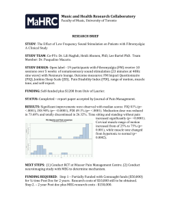


© Copyright 2026