
Endovascular treatment of aortoesophageal and aortobronchial fistulae
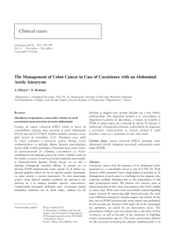
Endovascular treatment of aortoesophageal and aortobronchial fistulae Roberto Chiesa, MD, Germano Melissano, MD, Enrico M. Marone, MD, Andrea Kahlberg, MD, Massimiliano M. Marrocco-Trischitta, MD, PhD, and Yamume Tshomba, MD, Milano, Italy Background: Even when promptly recognized and treated, aortoesophageal (AEF) and aortobronchial (ABF) fistulae are highly lethal conditions. Open surgical repair also carries a high risk of mortality and morbidity. Several alternative strategies have been recently reported in the literature including thoracic endovascular aortic repair (TEVAR). However, relatively little is known about results of TEVAR for AEF and ABF due to their rarity and the lack of large surveys. Methods: A voluntary national survey was conducted among Italian universities and hospital centers with an endovascular program. Questionnaires were distributed by e-mail to participating centers and aimed to evaluate the results of endovascular repair of established AEF or ABF. Results: Seventeen centers agreed to participate and provided data on their patients. Between 1998 and 2008, a total of 1138 patients were treated with TEVAR. In 25 patients (2.2%), the indication to treatment was an AEF and/or an ABF. In 10 of these cases (40%), an associated open surgical procedure was also performed. Thirty-day mortality rate of AEF/ABF endovascular repair was 28% (7 cases). No cases of paraplegia or stroke were observed. Mean follow-up was 22.6 months (range, 1-62). Actuarial survival at 2 years was 55%. Among the 18 initial survivors, five patients (28%) underwent reintervention due to late TEVAR failure. Conclusions: Stent grafting for AEF and ABF represents a viable option in emergent and urgent settings. However, further esophageal or bronchial repair is necessary in most cases. Despite less invasive attempts, mortality associated with these conditions remains very high. ( J Vasc Surg 2010;51:1195-202.) Aortoesophageal (AEF) and aortobronchial (ABF) fistulae are uncommon and, in spite of appropriate and timely treatment, highly lethal. AEF and ABF are most commonly found in association with thoracic trauma, aortic aneurysms, ruptured penetrating aortic ulcers, esophageal or bronchogenic malignancies, and as a complication of thoracic surgery, including aortic surgery in up to 1.7% of cases.1,2 Conventional treatment entails open surgical repair of the thoracic aorta associated with esophageal or tracheobronchial reconstruction. Mortality rates of open surgery are primarily due to hemorrhagic and septic complications, with rates reaching 61% in case of primary etiology and 78% in case of secondary fistulae.3,4 Although several alternative strategies have been reported in the literature, including extra-anatomic bypass5 and in situ repair with cryopreserved homograft,6 there is a lack of consensus concerning the optimal treatment of AEF and ABF. Thoracic endovascular aortic repair (TEVAR) has been proposed as an alternative strategy to surgical management.7-9 Although less invasive, this technique presents From Vascular Surgery, Scientific Institute H. San Raffaele, “Vita-Salute” University School of Medicine. Competition of interest: none. Reprint requests: Andrea Kahlberg, MD, Vascular Surgery, Scientific Institute H. San Raffaele, “Vita-Salute” University School of Medicine, Via Olgettina 60, 20132 - Milan, Italy (e-mail: [email protected]). The editors and reviewers of this article have no relevant financial relationships to disclose per the JVS policy that requires reviewers to decline review of any manuscript for which they may have a competition of interest. 0741-5214/$36.00 Copyright © 2010 by the Society for Vascular Surgery. doi:10.1016/j.jvs.2009.10.130 important limitations in treating AEF and ABF, mainly the high risk of graft contamination. Several authors proposed a variety of combinations of TEVAR with surgical aortic repair, esophageal stent grafting, tracheobronchial or esophageal reconstruction, mediastinal drainage, or even endoscopic use of fibrin glue at the level of the fistula.10-12 However, relatively little is known about results of TEVAR in the treatment of AEF and ABF. This is due to the rarity of these conditions, the relatively recent clinical introduction of endovascular techniques, and a lack of large surveys in the literature. The aim of this study is to report initial and midterm results of TEVAR for AEF/ABF through a national survey conducted in Italy. METHODS We conducted a cross-sectional survey on voluntary basis among Italian universities and hospital centers with a thoracic endovascular program. We developed a text document-based survey in a collaborative effort between several vascular surgeons. The survey was distributed in November 2007 by e-mail to the medical directors of Italian departments of vascular surgery or cardiothoracic surgery with an acknowledged experience in thoracic aorta stent grafting. Centers performing TEVAR on a regular basis, even when lacking direct experience of AEF/ABF treatment, were invited to participate. Accompanying the questionnaire was a letter explaining the aims of the study and the compiling modalities. Each center that failed to respond was contacted by telephone or solicited by e-mail. The survey ended in December 2008. Participating centers were requested to provide data collected between 1998 and 1195 JOURNAL OF VASCULAR SURGERY May 2010 1196 Chiesa et al Table I. TEVAR for AEF/ABF: patient demographics Characteristics Overall Age (mean ⫾ SD) Gender (male) Coronary artery disease Diabetes mellitus Hypertension Cerebrovascular disease Pulmonary disease Renal failure Smoking ASA class 1 2 3 4 5 Etiology of the fistula Primary/unknown Atherosclerotic aneurysm Penetrating ulcer/intramural hematoma Chronic dissection Foreign body/caustic ingestion Esophageal cancer Previous aortic open repair Previous aortic endovascular repair No. patients (%) 25 (100) 66.4 ⫾ 18.8 17 (68) 11 (44) 4 (16) 19 (76) 3 (12) 14 (56) 2 (8) 5 (20) 1 (4) 1 (4) 9 (36) 13 (52) 1 (4) 4 (16) 6 (24) 4 (16) 1 (4) 2 (8) 1 (4) 4 (16) 3 (12) ASA, American Society of Anesthesiologists; TEVAR, thoracic endovascular aortic repair; AEF, aortoesophageal fistulae; ABF, aortobronchial fistulae. 2008. Patient names or identifiers were not requested or provided. The questionnaire had three sections: section 1 consisted of 26 items and inquired about the total number of endovascular procedures performed on the thoracic aorta. Section 2 aimed to determine the results of endovascular repair of an established AEF or ABF. This section consisted of 53 items, inquiring about patient demographics, preoperative risk factors, etiology of fistulae (Table I), clinical features at presentation, preoperative imaging, blood cultures (Table II), modality of treatment, endovascular procedure details, device characteristics (Table III), adjunctive maneuvers, technical success, 30-day mortality and complications, secondary procedures, and mortality at follow-up. Section 3 aimed to determine the rate of AEF/ABF as postoperative complications after TEVAR. Although data from Section 3 were collected within the same investigation, they pertain to different patients with different pathologies and will be reported in a separate publication. Demographics and preoperative risk factors, including coronary artery disease, pulmonary disease, and renal failure were defined as previously reported.13 Anatomic location of the proximal landing zone was defined according to the “aortic arch map” proposed by Ishimaru (0 to 4).14 Results of TEVAR were described according to the reporting standards for endovascular aortic aneurysm repair.15 Renal dysfunction was defined as a rise in serum creatinine exceeding the baseline value by 30% and surpassing an absolute level of 2.0 mg/dL. Myocardial infarction was suggested by electrocardiographic changes and confirmed by elevation of cardiac enzymes, regardless of symptoms. Respiratory failure was defined as ventilator dependence of ⬎72 hours, the need for postoperative reintubation, clinical data or culture confirmation of pneumonia, or the need for tracheostomy. Patients were followed up according to the protocol of each institution. Follow-up always included a thoracic contrast-enhanced computed tomography (CT) or magnetic resonance (MR) imaging and an outpatient clinical evaluation. Preoperative and intraoperative variables of interest were tested for significant association with the principal outcomes using Fisher’s exact test or 2 test for categorical data and unpaired t test or Mann-Whitney test for continuous data, as appropriate. Actuarial survival was computed according to the Kaplan-Meier log-rank method. All analyses were run using SAS 8.02 software (SAS Institute Inc, Cary, NC). RESULTS Among 39 contacted centers, 17 departments of vascular surgery or cardiothoracic surgery, including that of the authors, agreed to participate and provided patient data. Overall, 1138 patients were treated by TEVAR between 1998 and 2008. Ten of 11 centers (91%) with a TEVAR experience exceeding 30 procedures reported at least one case of AEF/ABF treated, whereas only one out of six centers (17%) with a TEVAR experience of less than 30 procedures reported one case of AEF/ABF treated endovascularly. Twenty-five patients underwent TEVAR for an established aortoesophageal or aortobronchial fistula. The fistula involved the esophagus in 14 cases and the left bronchial tree in 12 cases, with one patient presenting concomitant esophageal and bronchial involvement. Patients’ demographics and preoperative risk factors are listed in Table I. Symptoms at presentation and diagnostic studies performed at admission are reported in Table II. The interval between the first reported episode of hematemesis/hemoptysis and the final diagnosis of AEF/ABF ranged from 2 hours to 6 months. CT scan performed at admission showed clear signs of aortic infection or fistulization in all ABF patients and in 13/14 (93%) AEF patients (Fig 1). In two cases of ABF, the diagnosis was confirmed by transesophageal echocardiography, showing a penetrating ulcer of the descending thoracic aorta and the proximity to the left lower segment bronchus.16 In retrospect, several participating surgeons stated that there were suggestive symptoms of the fistula prior to hospitalization, however, imaging studies often failed to show a definitive fistulous tract. Blood cultures were positive in only five cases (20%) at admission. Isolated microorganisms included Escherichia coli, Klebsiella, Salmonella, Streptococcus, and Corynebacterium species. After establishing the diagnosis of AEF or ABF, all procedures were performed as soon as possible, under general anesthesia, in an operating room equipped for prompt open surgical conversion. Stent grafts employed are JOURNAL OF VASCULAR SURGERY Volume 51, Number 5 Chiesa et al 1197 Table II. Clinical features at presentation and preoperative diagnostic studies in patients treated with TEVAR alone compared to patients who received combined (endovascular and surgical) treatment Variable Total patients Symptoms at presentation Hematemesis/hemoptysis Fever Shock Thoracic pain Dyspnea Dysphagia Melena Diagnostic studies showing AEF/ABF Computed tomography Gastrointestinal/bronchial endoscopy Esophagogram Angiography Transesophageal echocardiography Overall No. patients (%) TEVAR alone No. patients (%) Combined No. patients (%) 25 (100) 15 (60) 10 (40) 22 (88) 8 (32) 12 (48) 3 (12) 2 (8) 2 (8) 2 (8) 14 (93) 3 (20) 4 (27) 2 (13) 0 (0) 1 (7) 1 (7) 8 (80) 5 (50) 8 (80) 1 (10) 2 (20) 1 (10) 1 (10) .543 .194 .015 1.0 .150 1.0 1.0 24 (96) 12 (48) 3 (12) 2 (8) 2 (8) 15 (100) 6 (40) 1 (7) 1 (7) 0 (0) 9 (90) 6 (60) 2 (20) 1 (10) 2 (20) .400 .428 .543 1.0 .150 P valuea TEVAR, Thoracic endovascular aortic repair; AEF, aortoesophageal fistulae; ABF, aortobronchial fistulae. a Fisher’s exact test. Table III. Stent grafts implanted Stent graft (manufacturer) Endofit (Endomed Inc, Phoenix, Ariz) Relay (Bolton Medical Inc, Sunrise, Fla) TAG (WL Gore and Assoc, Flagstaff, Ariz) Talent (AVE/Medtronic Inc, Santa Rosa, Calif) Valiant (AVE/Medtronic Inc, Santa Rosa, Calif) Zenith TX1 (William Cook Europe Aps, Bjaeverskov, Denmark) Zenith TX2 (William Cook Europe Aps, Bjaeverskov, Denmark) No. of patients 3 3 4 6 2 2 5 reported in Table III. Mean stent graft proximal diameter was 34.6 ⫾ 6.2 mm. A mean number of 1.1 ⫾ 0.3 devices per patient were implanted. The mean aortic length covered was 140.9 ⫾ 45.4 mm. Proximal landing zone was classified as “zone 1” in 2 patients, “zone 2” in 3 patients, “zone 3” in 10 patients, and “zone 4” in 10 patients. Debranching of supra-aortic vessels was performed for “zone 1” cases by means of right-to-left carotid bypass. Selective revascularization of the left subclavian artery (LSA) was performed in one “zone 1” patient, a 36-year-old left-handed worker, and in one “zone 2” patient, an 89-year-old male with previous coronary artery bypass grafting using the left internal thoracic artery. Cerebrospinal fluid (CSF) drainage was instituted preoperatively in four hemodynamically stable patients (16%) with the aortic lesion to be covered involving critical intercostal arteries (T8 to L2), requiring the coverage of a long descending thoracic aortic segment (ⱖ20 cm) or patients with previous abdominal/thoracoabdominal aortic repair. Mean duration of the operation was 159 ⫾ 185 min, with a mean estimated blood loss of 517 ⫾ 470 mL. Ballooning of the stent graft was performed selectively in seven cases (28%). One adjunctive endovascular procedure was reported in a patient treated for ABF caused by an atherosclerotic aneurysm, who underwent positioning of a proximal cuff for type I endoleak 48 hours after the primary endovascular operation. Concomitant or postponed planned adjunctive surgical procedures, other than supra-aortic vessels debranching, were performed in 10 patients (40%), including 5 esophageal resections and/or reconstructions, 2 cervical esophagostomies, 2 jejunostomies, 1 esophageal stenting, 1 pneumonectomy, and 3 cases of thoracic or mediastinal drainage. Both patients presenting with a fistula due to foreign body or caustic ingestion were treated by means of TEVAR with early simple esophageal repair. Assisted primary technical success was 100%. No intraoperative deaths were recorded. Thirty-day mortality was 28% (7/25) due to multiple organ failure (n ⫽ 4), respiratory failure (n ⫽ 2), and myocardial infarction (n ⫽ 1). Overall 30-day morbidity was 56% (14/25). No cases of paraplegia, paraparesis, or stroke were observed. Postoperative complications included acute renal dysfunction in five patients (20%), respiratory failure in 13 patients (52%), and acute myocardial infarction in two patients (8%). Mean follow-up was 22.6 ⫾ 21.8 months (range, 1-62). Actuarial survival at 2 years was 54.7%. Among the 18 initial survivors, six patients (33%) suffered from recurrent sepsis and/or hemorrhagic complications, resulting in all cases in death or reintervention. Four patients died during observation at follow-up, for an overall mortality at follow-up of 44% (11/25). A 76-year-old female who underwent TEVAR for a primary ABF with an uneventful postoperative course, died 35 months later due to hemorrhagic shock of unknown origin. A 72-year-old male submitted to emergent TEVAR for a ruptured aneurysm caus- 1198 Chiesa et al JOURNAL OF VASCULAR SURGERY May 2010 Fig 1. A, Preoperative computed tomography (CT) scan showing a saccular aneurysm of the descending thoracic aorta with fissurated thrombus. Esophageal compression and wall thickening are evident on axial images (arrows). B, Postoperative CT scan after successful endovascular exclusion of the aortic aneurysm. Esophageal defect was surgically repaired via right thoracotomy 6 days after thoracic endovascular aortic repair (TEVAR). ing an AEF and presented 5 months later with fever and hemorrhagic shock. He underwent extra-anatomic bypass arising from the ascending aorta (“ventral aorta”), thoracic stent graft removal, ligation of the arch and of the diaphragmatic aorta, and esophageal simple repair. He died within a few hours from multiorgan failure. A 16-year-old female submitted to TEVAR for an AEF following previous open surgery for aortic coarctation, underwent multiple reinterventions during the next 15 months, including esophageal reconstruction with colonic interposition and aortic reconstruction with cryopreserved homografts under hypothermic circulatory arrest. She died 8 months after the last operation due to septic complications. Finally, a 71-yearold female submitted to “zone 4” TEVAR for a primary AEF, followed by the repair of the esophagus via right thoracotomy, presented 3 months later with evidence of bleeding at the level of the aortic proximal neck. She underwent positioning of a proximal stent graft placed below the origin of the LSA, but she died perioperatively from multiorgan failure. Overall, five patients (28%) underwent reintervention due to late TEVAR failure, with a mean interval from primary TEVAR of 5.6 ⫾ 5.4 months. Three patients (reported above) died. Of the two survivors, one was a 61-year-old male submitted to TEVAR and esophageal stent grafting for a secondary AEF following previous thoracoabdominal aortic repair.11 He presented 3 months later with septic shock and was submitted to esophageal stent graft removal via right thoracotomy, followed by esophageal reconstruction with gastric interposition. He is alive and well at 6 months of follow-up. The other patient was a 58-year-old female submitted to emergent TEVAR for a posttraumatic concomitant AEF and ABF. She underwent multiple reinterventions for recurrent bleeding and persistent sepsis, including re-TEVAR, esophageal repair with gastric interposition, left inferior pulmonary lobectomy, and in situ reconstruction of the descending thoracic aorta using bovine pericardium graft, under hypothermic circulatory arrest with antegrade cerebral perfusion. The patient is alive and well at 30 months of follow-up. No significant differences in terms of 30-day mortality and overall mortality at follow-up were found between AEF and ABF. However, duration of the procedure and intraoperative blood loss were higher in the ABF group (235 ⫾ 234 min vs 94 ⫾ 66 min, P ⫽ .026; and 754 ⫾ 512 mL vs 392 ⫾ 286 mL, P ⫽ .039, respectively). Patients presenting with shock were significantly younger than stable patients (58.5 ⫾ 21.0 years vs 73.6 ⫾ 13.5 years; P ⫽ .026) and were more frequently affected by AEF (77% vs 31%; P ⫽ .047). Early and late mortality were similar in both groups (25% vs 31%, P ⫽ 1.0, and 33% vs 11%, P ⫽ .576, respectively). Positive micro-organism findings in the blood cultures were not associated with an increased overall mortality (2/5 patients died, one soon after TEVAR, and one after multiple reoperations for recurrent sepsis). There was no statistically significant difference in early and late mortality between patients treated for primary and secondary fistulae (22% vs 43%, P ⫽ .355; and 33% vs 71%, P ⫽ .177, respectively). However, patients with secondary AEF/ABF presented with preoperative fever more often than patients with primary fistulae (71% vs 20%, P ⫽ .017). Patients treated for a fistula due to foreign body or caustic ingestion had both a postoperative uneventful course and are alive and well at 48 and 7 months of follow-up, respectively. No differences in operative variables and outcome were identified concerning type of stent graft used or manufacturer. Patients who underwent TEVAR alone, without any scheduled (concomitant or postponed) procedure were overall comparable in terms of clinical features at presenta- JOURNAL OF VASCULAR SURGERY Volume 51, Number 5 Chiesa et al 1199 Fig 2. Survival by surgical management (Kaplan-Meier log rank test, P ⫽ .373). tion, to patients who received a combined treatment, consisting of TEVAR associated with or followed by a scheduled esophageal or bronchial procedure, except for preoperative shock that was significantly more frequent in the combined group (P ⫽ .015, Table II). Patients treated with TEVAR alone had a 30-day mortality of 40% (6/15) and an overall mortality at follow-up of 53% (8/15). A better outcome was recorded in patients who received combined treatment (Fig 2), with a 30-day mortality of 10% (1/10) and an overall mortality at follow-up of 30% (3/10). These differences, however, did not reach statistical significance (P ⫽ .179 and P ⫽ .414, respectively; Kaplan-Meier logrank test, P ⫽ .373). DISCUSSION Although AEF and ABF are believed to be extremely uncommon, we found that this pathology was the indication in 2.2% of all TEVAR procedures performed from 1998 to 2008. Furthermore, the survey showed that most centers performing more than 30 TEVAR procedures reported at least one case of AEF/ABF endovascular treatment. In the English literature, however, a relatively low number of TEVAR for AEF/ABF is published to date, consisting primarily of single case reports or small case series with less than 12 patients.17 In addition, most large international TEVAR trials do not include or mention this specific pathology18-21 due to the important differences in treatment modalities and results compared with aneurysms. This may account for a considerable underestimation of the problem. Since AEF and ABF were first described by Dubrueil in 1818 and by Girardet in 1914, respectively,22,23 they are usually considered as two separate clinical entities. Nevertheless, they present many similar features in terms of etiology, pathogenesis, presentation, and natural history, and they occasionally occur together.24 In our study, no significant differences in terms of perioperative and late mortality were observed between AEF and ABF. The endovascular procedure in case of ABF presented with increased procedural times and blood loss. A possible explanation could be that ABF are usually located more “proximally” compared with AEF, resulting in more complex procedures. Consistently, both “zone 1” and “zone 2” patients who required LSA revascularization belonged to the ABF group, and this may have contributed to increase the mean procedure duration and blood loss value. If left untreated, both AEF and ABF are uniformly fatal. In a review of AEF, Coselli and Crawford reported that more of 60% of patients presenting with a herald gastrointestinal bleed die within 6 hours.25 Thus, surgical management must be immediate and must include hemostasis and prevention of septic complications. In this survey, nearly 90% of patients presented with hematemesis or hemoptysis, and half of them were already in shock. CT scan is the first imaging study performed in most cases, as it is fast and easy to carry out in emergency conditions. Although a CT scan rarely allows detecting a fistulous tract, in this survey, suggestive signs of AEF/ABF were present in most patients, including air bubbles into the thrombus, periaortic fluid collection, esophageal or bronchial wall thickening, and lung consolidation. Endoscopy is known to be the most sensitive and specific method for the diagnosis of AEF/ABF26 but often requires sedation and entails the risk of dislodging clots during progress 1200 Chiesa et al of the endoscope that may cause fatal bleeding.27 Also, we found that transesophageal echocardiography may help to confirm the presence of an ABF in selected cases. Surgical repair is most commonly performed via a left posterolateral thoracotomy, with simple cross-clamping of the descending thoracic aorta. However, this maneuver entails the risk of spinal cord and visceral ischemia, and cardiopulmonary bypass may be required. Moreover, access to the aorta may be particularly demanding due to dense adhesions in patients with previous thoracic interventions, as is the case of secondary fistulae, with a high risk of adjacent organ lesions and significant blood loss. Direct aortic suture or patch angioplasty are rarely employed and are most often used in the treatment of small lesions associated with foreign body ingestion.28 For larger lesions, aortic replacement is usually required. In situ reconstruction with prosthetic bypass has been successfully performed in the past29 but carries the potential for graft infection and new fistulization. Cryopreserved homografts represent a viable option for in situ aortic reconstruction,6 although they are susceptible to infection as well, and long-term secondary degeneration has been described.30 Extraanatomic bypass arising from the ascending aorta (ventral aorta) reduces the risk of graft infection5 but must be performed as a primary procedure, requires additional sternotomy extending to an upper laparotomy, and is often unfeasible due to fistula hemorrhage. Moreover, it carries the risk of aortic stump infection with potential long-term complications. In addition to repairing the aorta, the esophageal or bronchial defect should be addressed. A small esophageal defect may be treated by direct repair, whereas patients with a larger esophageal defect may undergo esophageal reconstruction with gastric or colonic interposition.31 The repair of the bronchus in cases of ABF may entail primary repair or resection and anastomosis with reinforcement using muscle flaps or pedicled omental flaps.32 Other options include lobectomy or pneumonectomy. Successful use of stent grafts in patients with aortoduodenal and ilioureteral fistulae have been reported in the last decade.33,34 Treatment of a fistula between the thoracic aorta and an adjacent organ by means of TEVAR was first reported in 1996 by Chuter et al35 and in the same year by Campagna et al.36 Subsequently, several cases were published describing the feasibility of endovascular treatment of AEF and ABF.7-12 Since endovascular treatment is simpler, faster, and safer than open surgery in unstable patients, TEVAR is considered by most authors as the first line treatment to obtain immediate control of aortic bleeding. In the overtly moribund patient, TEVAR has been proposed as the most appropriate definitive strategy, as a palliative procedure. Conversely, in good surgical candidates, coverage of the aortic lesion, along with an aggressive antibiotic therapy, may be used to achieve an improvement in the patient’s general conditions, serving as a “bridge” to open surgical treatment of the aortic and/or esophageal/ bronchial defect. Following successful TEVAR, there is no general consensus concerning the need for planned staged JOURNAL OF VASCULAR SURGERY May 2010 surgical intervention, in the absence of clear signs of reinfection or bleeding. In this series, both early and late mortality was increased in cases of TEVAR alone management compared with a combined (endovascular and surgical) approach. Of the 15 patients submitted to TEVAR alone, eight patients died at follow-up (53%). Six patients died within 30 days from TEVAR (40%), one patient died 35 months later due to hemorrhagic shock of unknown origin, and the last patient presented after 5 months with fever and hemorrhagic shock, underwent thoracic stent graft removal and extra-anastomic bypass but died within a few hours from multiorgan failure. Of the 10 patients submitted to combined treatment, overall three patients died at follow-up (30%), and only one patient died within 30 days (10%). The difference in early and late outcome, however, did not reach statistical significance, possibly due to the small sample size. In addition, potential confounders are to be considered when comparing these two groups, mainly the eventuality that the TEVAR alone group may also include patients that would have been candidates to staged surgical repair but who died before surgery could be scheduled. Although clear limitations of the TEVAR alone strategy include the risk of reinfection and septic complication, due to the inability to debride or drain the mediastinum, there has been some success in the literature with this approach.37,38 Some authors suggest considering TEVAR as a potential definitive treatment in patients at high risk for complications with open surgical repair who showed no clinical or imaging evidence of infection (fever, leukocytosis, air or fluid collections on CT).39 However, in addition to the currently reported series, poor late results of TEVAR alone strategies are reported in most cases due to infectious complications or recurrent fatal bleeding.7,30,40,41 The evidence emerging from these results is not strong enough to justify changes in clinical practice. It is clear, however, that TEVAR alone does not provide complete and durable cure for AEF and ABF, and that these patients need to be followed closely to evaluate the opportunity and the appropriate timing for a secondary surgical procedure. Also, treatment strategy and time interval between TEVAR and additional surgical procedures are individualized, according to patient conditions and the grade of sepsis. Patients presenting with fistulae from a foreign body or caustic ingestion are typically young and healthy and may have an uncomplicated course with a low risk of reinfection.42 On the contrary, patients with secondary AEF or ABF are usually elderly and affected by several comorbidities. In this survey, secondary fistulae were associated with a high prevalence of preoperative fever, and this may reflect a severe grade of sepsis due to the presence of an additional infected aortic surgical graft. In these cases, a delayed staged surgical procedure following TEVAR is often preferred to achieve an improved general clinical condition and recovery from sepsis. Another concern regards the fate of the stent graft during long-term follow-up. Although we did not record JOURNAL OF VASCULAR SURGERY Volume 51, Number 5 cases of stent graft migration or collapse, these are wellknown complications of TEVAR and were previously reported in a case of AEF treatment.38 Recurrent sepsis and/or hemorrhagic-related complications were observed in this series in around one-third of cases at a mean follow-up of approximately 2 years. Although reinfection is usually attributed to stent graft contamination, the stent graft itself has the potential to erode into adjacent organs, causing a new fistulization.43 Prolonged antibiotic therapy and life-long surveillance is crucial in these patients, regardless of symptoms or clinical signs of sepsis. In conclusion, the incidence of AEF and ABF is probably underestimated, and centers that regularly perform thoracic aortic surgery will eventually deal with this complex pathology. TEVAR has a predominant role in controlling the massive hemorrhage associated with AEF and ABF in the usual setting of sepsis and medical comorbidities. In cases of minimal local infection, further treatment may be unnecessary. In the other cases, a definitive esophageal or bronchial repair is indicated after stabilization. The authors wish to thank all participating centers who provided patient data: Angelo Argenteri (Presidio Ospedaliero di Lodi, Lodi); Carlo Bertoglio (Ospedale di Imperia, Imperia); Giorgio Maria Biasi, Gaetano Deleo, Cristian Benatti (A.O. San Gerardo, Monza); Stefano Bonardelli (A.O. Spedali Civili, Brescia); Piergiorgio Cao, Fabio Verzini (Ospedale S.M. della Misericordia, Perugia); Gioacchino Coppi, Stefano Gennai (Policlinico di Modena, Modena); Francesco Mascoli (Arcispedale S. Anna, Ferrara); Claudio Novali (A.O. S. Croce e Carle, Cuneo); Attilio Odero, Antonio Bozzani (IRCCS Policlinico San Matteo, Pavia); Domenico Palombo, Giovanni Spinella (A.O. San Martino, Genoa); Vincenzo Rampoldi, Santi Trimarchi (IRCCS Policlinico San Donato, Milan); Mauro Rinaldi, Sergio Trichiolo (A.O. Molinette, Turin); Carlo Setacci, Francesco Setacci (A.O. Policlinico “Le Scotte”, Siena); Francesco Speziale (Policlinico Umberto I, Rome); Francesco Spinelli, Giovanni De Caridi (A.O. Policlinico “G. Martino”, Messina); Domenico Tealdi, Giovanni Nano (IRCCS Policlinico San Donato, Milan). The authors also acknowledge Dr Gliliola Calori, from the Statistical Unit of San Raffaele Hospital, for help with the statistical analysis of the article’s data. AUTHOR CONTRIBUTIONS Conception and design: RC, GM, EM, AK, MM, YT Analysis and interpretation: RC, GM, EM, AK, MM, YT Data collection: AK, MMT Writing the article: RC, GM, AK Critical revision of the article: RC, GM, EM, AK, MM, YT Final approval of the article: RC, GM, EM, AK, MM, YT Statistical analysis: AK Obtained funding: Not applicable Overall responsibility: RC Chiesa et al 1201 REFERENCES 1. Hollander JE, Quick G. Aortoesophageal fistula: a comprehensive review of the literature. Am J Med 1991;91:279-87. 2. Lawrie GM, Earle N, De Bakey ME. Evolution of surgical techniques for aneurysms of the descending thoracic aorta: twenty-nine years experience with 659 patients. J Card Surg 1994;9:648-61. 3. Dossa CD, Pipinos II, Shepard AD, Ernst CB. Primary aortoenteric fistula: part II. Primary aortoesophageal fistula. Ann Vasc Surg 1994;8: 207. 4. Pipinos II, Reddy DJ. Secondary aortoesophageal fistulae. Ann Vasc Surg 1999;13:649-52. 5. Madan AK, Santora TA, Disesa VJ. Extra-anatomic bypass grafting for aortoesophageal fistula: a logical operation. J Vasc Surg 2000;32: 1030-3. 6. Kieffer E, Chiche L, Gomes D. Aortoesophageal fistula: value of in situ aortic allograft replacement. Ann Surg 2003;238:283-90. 7. Léobon B, Roux D, Mugniot A, Rousseau H, Cérene A, Glock Y, et al. Endovascular treatment of thoracic aortic fistulas. Ann Thorac Surg 2002;74:247-9. 8. Taylor BJ, Stewart D, West P, Dunn JT, Cisek P. Endovascular repair of a secondary aortoesophageal fistula: a case report and review of the literature. Ann Vasc Surg 2007;21:167-71. 9. Marone EM, Baccari P, Brioschi C, Tshomba Y, Staudacher C, Chiesa R. Surgical and endovascular treatment of secondary aortoesophageal fistula. J Thorac Cardiovasc Surg 2006;131:1409-10. 10. Van Doorn RC, Reekers J, de Mol BA, Obertop H, Balm R. Aortoesophageal fistula secondary to mycotic thoracic aortic aneurysm: endovascular repair and transhiatal esophagectomy. J Endovasc Ther 2002; 9:212-7. 11. Civilini E, Bertoglio L, Melissano G, Chiesa R. Aortic and esophageal endografting for secondary aortoenteric fistula. Eur J Vasc Endovasc Surg 2008;36:297-9. 12. Mok VW, Ting AC, Law S, Wong KH, Cheng SW, Wong J. Combined endovascular stent grafting and endoscopic injection of fibrin sealant for aortoenteric fistula complicating esophagectomy. J Vasc Surg 2004;40: 1234-7. 13. Chiesa R, Melissano G, Marrocco-Trischitta MM, Civilini E, Setacci F. Spinal cord ischemia after elective stent-graft repair of the thoracic aorta. J Vasc Surg 2005;42:11-7. 14. Melissano G, Civilini E, Bertoglio L, Calliari F, Setacci F, Calori G, et al. Results of endografting of the aortic arch in different landing zones. Eur J Vasc Endovasc Surg 2007;33:561-6. 15. Chaikof EL, Blankensteijn JD, Harris PL, White GH, Zarins CK, Bernhard VM, et al. Reporting standards for endovascular aortic aneurysm repair. J Vasc Surg 2002;35:1048-60. 16. Pirrelli S, Bozzani A, Arici V, Odero A. Endovascular treatment of acute haemoptysis secondary to aortobronchial fistula. Eur J Vasc Endovasc Surg 2006;32:366-8. 17. Jonker FH, Heijmen R, Trimarchi S, Verhagen HJ, Moll FL, Muhs BE. Acute management of aortobronchial and aortoesophageal fistulas using thoracic endovascular aortic repair. J Vasc Surg 2009 (in press). 18. Leurs LJ, Bell R, Degrieck Y, Thomas S, Hobo R, Lundbom J; EUROSTAR; UK Thoracic Endograft Registry collaborators. Endovascular treatment of thoracic aortic diseases: combined experience from the EUROSTAR and United Kingdom Thoracic Endograft registries. J Vasc Surg 2004;40:670-9. 19. Matsumura JS, Cambria RP, Dake MD, Moore RD, Svensson LG, Snyder S; TX2 Clinical Trial Investigators. International controlled clinical trial of thoracic endovascular aneurysm repair with the Zenith TX2 endovascular graft: 1-year results. J Vasc Surg 2008;47:247-57. 20. Makaroun MS, Dillavou ED, Wheatley GH, Cambria RP; Gore TAG Investigators. Five-year results of endovascular treatment with the Gore TAG device compared with open repair of thoracic aortic aneurysms. J Vasc Surg 2008;47:912-8. 21. Fairman RM, Criado F, Farber M, Kwolek C, Mehta M, White R, Lee A, Tuchek JM; VALOR Investigators. Pivotal results of the Medtronic Vascular Talent Thoracic Stent Graft System: the VALOR trial. J Vasc Surg 2008;48:546-54. 1202 Bower 22. Dubrueil O. Observations sur la perforation de l’esophage et de l’aorte thoracique par une portion d’os avale: avec de reflexions. J Univ Sci Med 1818;9:357-63. 23. Girardet A. Doppelte perforation eines tuberkelknotens in die aorta und die bifurcation der trachea. Deutsch Med Wochenschor 1914;40:1425-8. 24. Raghavendran K, Cherr GS, Ford PF, Burkhard PG, Bell-Thomson J. Successful management of concomitant aortoesophageal and aortotracheal fistulae secondary to a thoracic aortic aneurysm: case report and review of literature. J Vasc Surg 2005;42:1218-20. 25. Coselli JS, Crawford ES. Primary aortoesophageal fistula from aortic aneurysm: successful surgical treatment by use of omental pedicle graft. J Vasc Surg 1990;12:269-77. 26. Akaraviputh T, Sriprayoon T, Prachayakul V, Sakiyalak P. Endoscopic diagnosis of secondary aortoesophageal fistula. Endoscopy 2008; 40(Suppl 2):E90. 27. Benson MJ, Rouse D, van Someren N, Wingate DL, Swain CP. Fatal hemorrhage from an aorto-esophageal fistula precipitated by flexible endoscopy. Gastrointest Endosc 1991;37:193-6. 28. Wu MH, Lai WW. Aortoesophageal fistula induced by foreign bodies. Ann Thorac Surg 1992;54:155-6. 29. Bogey WM Jr, Thomas JH, Hermreck AS. Aortoesophageal fistula: report of a successfully managed case and review of the literature. J Vasc Surg 1992;16:90-5. 30. Topel I, Stehr A, Steinbauer MG, Piso P, Schlitt HJ, Kasprzak PM. Surgical strategy in aortoesophageal fistulae: endovascular stentgrafts and in situ repair of the aorta with cryopreserved homografts. Ann Surg 2007;246:853-9. 31. da Silva ES, Tozzi FL, Otochi JP, de Tolosa EM, Neves CR, Fortes F. Aortoesophageal fistula caused by aneurysm of the thoracic aorta (successful surgical treatment, case report, and literature review). J Vasc Surg 1999;30:1150-7. 32. Paull DE, Keagy BA. Management of aortobronchial fistula with graft replacement and omentopexy. Ann Thorac Surg 1990;50:972-4. 33. Deshpande A, Lovelock M, Mossop P, Denton M, Vidovich J, Gurry J. Endovascular repair of an aortoenteric fistula in a high-risk patient. J Endovasc Surg 1999;6:379-84. JOURNAL OF VASCULAR SURGERY May 2010 34. Gibbons M, O’Donnell S, Lukens M, Meglin A, Costabile RA. Treatment of a ureteroiliac artery fistula with an intraluminal endovascular graft. J Urol 1998;159:2083-4. 35. Chuter TA, Ivancev K, Lindblad B, Brunkwall J, Arén C, Risberg B. Endovascular stent-graft exclusion of an aortobronchial fistula. J Vasc Interv Radiol 1996;7:357-9. 36. Campagna AC, Wehner JH, Kirsch CM, Semba CP, Kagawa FT, Jensen WA, et al. Endovascular stenting of an aortopulmonary fistula presenting with hemoptysis. A case report. J Cardiovasc Surg (Torino) 1996; 37:643-6. 37. Bond SE, McGuinness CL, Reidy JF, Taylor PR. Repair of secondary aortoesophageal fistula by endoluminal stent-grafting. J Endovasc Ther 2001;8:597-601. 38. Rodriguez JA, Olsen DM, Shtutman A, Lucas LA, Wheatley G, Alpern J, et al. Application of endograft to treat thoracic aortic pathologies: a single center experience. J Vasc Surg 2007;46:413-20. 39. Flores J, Shiiya N, Kunihara T, Yoshimoto K, Yasuda K. Aortoesophageal fistula: alternatives of treatment case report and literature review. Ann Thorac Cardiovasc Surg 2004;10:241-6. 40. Burks JA Jr, Faries PL, Gravereaux EC, Hollier LH, Marin ML. Endovascular repair of bleeding aortoenteric fistulas: a 5-year experience. J Vasc Surg 2001;34:1055-9. 41. González-Fajardo JA, Gutiérrez V, Martín-Pedrosa M, Del Rio L, Carrera S, Vaquero C. Endovascular repair in the presence of aortic infection. Ann Vasc Surg 2005;19:94-8. 42. Assink J, Vierhout BP, Snellen JP, Benner PM, Paul MA, Cuesta MA, et al. Emergency endovascular repair of an aortoesophageal fistula caused by a foreign body. J Endovasc Ther 2005;12:129-33. 43. Santo KC, Guest P, McCafferty I, Bonser RS. Aortoesophageal fistula secondary to stent-graft repair of the thoracic aorta after previous surgical coarctation repair. J Thorac Cardiovasc Surg 2007;134: 1585-6. Submitted Jul 9, 2009; accepted Oct 17, 2009. INVITED COMMENTARY Thomas C. Bower, MD, Rochester, Minn Primary and secondary aortoesophageal and aortobronchial fistulas are uniformly fatal if untreated, remain a formidable surgical problem, and carry high operative mortality in older, high-risk patients with hemorrhagic shock or sepsis, and in those with secondary communications between the aorta and/or graft and the aerodigestive tract. This article represents one of the larger series, which define the role for thoracic endovascular aortic repair (TEVAR) for these problems. Clearly, TEVAR is becoming the initial primary treatment, but the importance of this article is the analysis of patient outcomes in the group treated with TEVAR alone compared with the group who had TEVAR followed later by aortic reconstruction and treatment of the esophageal and bronchial defects. The authors question the utility of TEVAR as the only mode of therapy because of a higher 30-day and midterm mortality in this group compared with patients treated with a combined approach. Based on their data, TEVAR as sole definitive treatment should be reserved for patients with small aortobronchial communications and no CT evidence of mediastinal infection at initial presentation or during follow up. Since patients in shock with primary or secondary aortoesophageal fistulas had a much higher early and late mortality, the authors appropriately recommend a combined approach to treat them. The choice of conduit and route for aortic reconstruction, and the specific treatment of the fistula are dependent on patient age and comorbid conditions, the severity of infection, the location and type of fistula, and whether a thoracic graft is already in place. Definitive treatment should follow the precepts used to treat aortic graft-enteric erosions or fistulas in the abdomen. Choices for aortic reconstruction include ascending aorta to upper abdominal prosthetic bypass; or in situ replacement with antibiotic-soaked prosthetic or homografts. The authors suggest early resection of the involved segment of esophagus or lung in patients with mediastinal sepsis. Vascularized pericardial or pleural flaps, serratus muscle, or omentum can be used to cover the graft or the aortic stumps. What seems crucial to improve patient outcomes, not addressed in detail in this report, is identification of specific factors that influence timing of the definitive open surgical repair, and the role for suppressive antibiotics. Similar to the treatment of abdominal aortic graft infection, one could argue for a 6 to 8- week course of broad spectrum IV antibiotics in the interval between TEVAR and definitive repair, followed by lifelong oral antibiotic suppression for those with aortoesophageal fistulas, extensive mediastinal contamination, and in situ prosthetic aortic grafts.


© Copyright 2026