
Free PDF
CellR4 2015; 3 (1): e1408
Bone marrow- and cord blood-derived stem cell transplantation
for diabetes therapy
E. Cantarelli, S. Pellegrini, A. Citro, V. Sordi, L. Piemon
Diabetes Research Instute, IRCCS San Raffaele Scienfic Instute, Milan, Italy
Corresponding Author: Lorenzo Piemonti, MD; e-mail: [email protected]
ABBREVIATIONS
T1D: Type 1 Diabetes, T2D: Type 2 Diabetes, BM:
Bone Marrow, HSC: Hematopoietic Stem Cells, EPC:
Endothelial Precursors Cells, MSC: Mesenchymal
Stem Cells, UCB: Umbilical Cord Blood, Tx: Transplantation, HSCT: Hematopoietic Stem Cell Transplantation, GVHD: Graft-Versus-Host Disease, Treg:
regulatory T cells, APC: Antigen Presenting Cells,
NOD: Non-Obese Diabetic, VEGF: Vascular Endothelial Growth Factor, TNC: Total Nucleated Cells.
ABSTRACT
In the last years, the widely consolidated clinical
experience in the field of hematology has encouraged the use of bone marrow (BM)- and
cord blood (CB)-derived stem cells in nonhaematological disease. In the field of diabetes,
a huge amount of clinical trials for the cure of
type 1 and type 2 diabetes, involving BM-derived
HSC and both BM- and CB-derived MSC got
underway, thanks also to the availability of simple protocols for collection, culture and storage
of these stem cells. Many groups have investigated their potential role in tolerance induction
and/or restoration, in pancreatic tissue remodelling as “feeder” cells and in direct differentiation
into insulin-producing cells, with the shared final
goal to preserve β cell function. This review recapitulates the historical use of BM- and CB-derived stem cells in diabetes therapy, alone or in
combination with islet transplantation, and focuses on the most relevant information on preclinical experimental data and provides an
update on the most recent clinical trials.
Keywords: Bone marrow, Mesenchymal stem cell, Umbilical cord blood, Diabetes, Immunomodulation, Tolerance,
Transplantation, Islet.
INTRODUCTION
Diabetes affects 382 million people throughout the
world and this number will rise to 592 millions by
2035 (http://www.idf.org/diabetesatlas/introduction).
Both type 1 (T1D) and type 2 diabetes (T2D) share
a deficit in β cell mass, although due to different
pathogenic events: autoimmunity and insulin resistance, respectively1,2. Exogenous administration of
insulin is routinely used to control both types of diabetes, but it does not sufficiently replace β cells and
the adverse short- and long-term effects of the disease remain. Therefore, the cure for diabetes lies in
the possibility to replace the lost β cell mass with a
new endocrine component capable of assessing
blood sugar levels and secreting appropriate levels of
insulin in the vascular bed. β cell replacement,
through whole pancreas or islet transplantation, is
the only treatment capable of establishing long-term
euglycemia in T1D patients3. Unfortunately these
procedures, despite advances in recent years4, are
hindered by the need of immunosuppression, the use
of many donors for a single recipient and the short
life of the grafts. Accordingly, new approaches
aimed to overcome these limits are strongly required. An attractive possibility to treat diseases like
diabetes could be represented by stem cell therapy.
In the last years, the use of stem cells in clinical protocols is over and over increasing. The remarkable
plasticity of different cell subsets obtained from
human embryonic and adult tissues from different
sources (including bone marrow, adipose tissue, umbilical cord and amniotic fluid) has been the focus
of many efforts in research, also in the field of diabetes5. Among stem cells, those derived from bone
marrow (BM), which mostly comprise hematopoietic stem cells (HSC), endothelial precursors cells
(EPC) and mesenchymal stem cells (MSC), can be
easily recovered and cultured and have been studied
1
2
E. Cantarelli, S. Pellegrini, A. Citro, V. Sordi, L. Piemonti
for a long time (Fig. 1). In this review we will report
the most relevant preclinical and clinical applications of total BM, isolated BM cells subpopulations
and cord blood (CB) cells for the treatment of both
T1D and T2D, alone and in combination with pancreatic islet transplantation (tx) (Table 1).
TOTAL BM TRANSPLANTATION
FOR β CELL REPLACEMENT
At first, the possibility for BM cells to differentiate
into β cells following signals of tissue remodelling,
was reported; in fact, some studies suggested that
undifferentiated BM cells transplanted in vivo could
become glucose-responsive insulin producing cells68
. Using transgenic BM cells that express the green
fluorescent protein (GFP) under the guidance of insulin promoter, Ianus and colleagues were the first to
demonstrate the ability of BM cells to transdifferentiate into insulin producing cells within the pancreatic islets7. These data have resulted in conflicting
reports because several other groups have not been
able to confirm these findings. In fact, although the
infusion of BM cells into diabetic mice lowered the
blood glucose and increased β cell mass, their ability to transdifferentiate into β cells was not supported9-13. In this direction it was reported that the
uptake of exogenous insulin by differentiated cells
could lead to false conclusions about the ability of
BM-derived cells to differentiate in β cells14. There-
fore, the concept of in vivo transdifferentiation of
BM cells into insulin producing cells still remains
elusive. Hess et al gave a new dimension to BM tx
for diabetes therapy demonstrating that the transplanted cells can initiate endogenous pancreatic regeneration by β cell rapid proliferation and
neogenesis15. The experience with unpurified BMderived cells in clinical to treat diabetes is very limited. Few years ago, a Spanish study evaluated the
impact of the infusion into the pancreatic artery of
autologous, unfractionated BM-derived mononuclear cells obtained after mobilization with G-CSF
from the iliac crest of long-standing T1D patients.
The pilot clinical study showed no effects in terms of
C-peptide serum levels, both basal and stimulated,
and no changes in insulin requirement or metabolic
control after tx. Due to the lack of efficacy this study,
initially aimed at enrolling 10 subjects, was stopped
after the third patient by the local research ethic
committee16. Possible criticisms on this trial included
the lack of immune interventions aimed at favouring self tolerance restoration and the selection of a
subpopulation of T1D patients with undetectable Cpeptide levels before tx.
HEMATOPOIETIC STEM CELL TRANSPLANTATION
FOR T1D AND T2D THERAPY:
TOWARD TOLERANCE RESTORATION?
The use of HSC, instead, has aroused much more interest and success. HSC transplantation (HSCT) is
now widely recognized as a curative therapy for
MSC
HSC
EPC
Bone Marrow
Umbilical Cord
ESC
Figure 1. Schematic representation of the different kind of stem cells that can be obtained from bone marrow or umbilical cord.
MSC: Mesenchymal Stem Cells, HSC: Hematopoietic Stem Cells, EPC: Endothelial Precursor Cells, ESC: Embryonic Stem Cells.
Table 1. Clinical trials on BMand UCB-derived HSC and
MSC therapy. References or
ClinicalTrilas.gov Identifier
were reported for completed or
still active clinical studies, respectively.
Bone marrow transplantation in diabetes
T1D
T2D
In association with Islet Tx
BM-HSC
[36-37, 38, 39, 40, 41,
42, 43, 44, 45]
NCT01121029
NCT01285934
[47, 48, 49, 50, 51]
NCT00767260
NCT00465478
-
BM-MSC
[86]
NCT02057211
NCT00690066
NCT01576328
[158]
NCT00646724
UCB
[114-115, 116, 117]
NCT01996228
[118, 121-122]
-
many high-risk hematological diseases. Over the last
two decades, it has also been extensively investigated as a therapeutic opportunity for patients affected by severe autoimmune diseases considered
refractory to conventional therapies17. The idea that
a strong relationship between HSC and the organ target of the autoimmune response exists, was further
supported by the evidence that either susceptibility
or resistance to autoimmunity could be transferred
by HSCT, as widely confirmed in animal models for
many autoimmune diseases18,19 including systemic
lupus erythematosus, experimental autoimmune encephalomyelitis, adjuvant arthritis, antiphospholipid
syndrome and T1D20. In particular, the definitive
proof-of-principle that HSCT may represent a promising therapeutic opportunity for T1D patients was
borne accidentally from finding a patient who, after
undergoing HSCT for hematological indications, developed the autoimmune disease21. The rationale of
HSCT for the cure of autoimmune diseases is the
substitution of the defective immune system by an
healthy one that can start from scratch and regenerate undergoing tolerization to self antigens, hopefully in the absence of the supposed accidental
environmental circumstances that have led to the initial autoimmune response.
HSCT consists in the administration of HSC
which are self-renewing cells identified as CD34+
CD59+ Thy1+ CD38low/- c-Kit-/low and Lin- in humans
(CD34low/- Sca-1+ Thy1+/low CD38+ c-Kit+ and Lin- in
mice) able to give rise to all mature hematopoietic
cells and possibly to some non-hematopoietic cells.
In the clinical routine, recipients undergoing HSCT
are pre-conditioned with a potent immunosuppressive therapy before autologous (auto-HSCT; cells
harvested from the recipient before pre-conditioning) or allogeneic HSCT (allo-HSCT; cells harvested from donor other than the recipient). In both
3
instances, HSC are mobilized from the BM to the
peripheral blood before conditioning by using different protocols, many of which involve granulocyte colony-stimulating factor (G-CSF) and/or
cyclophosphamide, a myelosuppressive drug that
leads to a ‘rebound’ mobilization of these cells. The
first choice between allo-HSCT and auto-HSCT in
the clinical practice is influenced by the balance between the risk to develop graft-versus-host-disease
(GVHD) and the effectiveness to initiate an hopeful
graft-versus-autoimmunity (GVA) response. GVHD
arises from the attack of donor allogeneic T cells on
recipient antigens, while GVA is the result of the
immune-mediated destruction of residual recipient’s
memory T and B cells by host’s T cells, a mechanism applicable to allo-HSCT only.
Despite the well-documented clinical success of
HSCT in correcting autoimmune diseases22, an accurate explanation of the mechanisms of action of
this treatment is still tricky. Clearly, HSCT relies on
an extensive debulking of the recipient’s immune
system by potent immunosuppressive conditioning
protocols such as total body irradiation (TBI), cyclophosphamide, anti-CD2 antibodies, anti-CD52
antibodies, fludarabine and anti-thymocyte globulin (ATG), which leads to profound long-lasting
lymphopenia and persistently reduced levels of
long-living autoantibody-producing plasma cells23.
It was demonstrated both in animal models and clinical trials that the use of these lymphoablative therapies alone (without HSCT) for the conditioning
regimen can halt or slow per se the progression of
autoimmune diseases24. Aside this quite non-specific immunosuppression, there are growing evidences that auto-HSCT not only has a role in
shortening aplasia, but also holds the potential to reestablish the immunological tolerance thanks to: (1)
an increase in the number of CD4+ FoxP3+ regula-
4
E. Cantarelli, S. Pellegrini, A. Citro, V. Sordi, L. Piemonti
tory T cells (Treg) that are crucial for tolerance
preservation25; (2) the reactivation of thymic function re-establishing T cell receptor heterogenicity as
showed by the presence of recent thymic emigrating
cells (TREC) and CD31 expression26,27. However,
autoimmunity relapse, mainly due to the persistence
of autoreactive cells such as surviving memory T
cells, memory B cells and long-lived plasma cells
in genetically predisposed recipients, may occur.
Further studies are strongly required for the evaluation of the optimal condition regimen in relation to
the duration and stability of the induced remission.
Mild conditioning treatments that do not completely
ablate donor’s HSC followed by allo-HSCT leads
to a condition called “mixed hematopoietic
chimerism” in which donor and recipient HSC, and
therefore multi-lineage hematopoietic populations,
co-exist. Thus in the recipients, a life-long source
of donor antigen presenting cell (APC) pool that
most effectively presents antigens to T cells positively selected in the recipient thymus is present. In
this context, T cells with high affinity for self peptide-MHC complexes are deleted, ensuring tolerance towards donor and recipient antigens (Figure
2). Mixed chimerism induction after allo-HSCT
prevents the development of autoimmune diseases
with greater efficacy than auto-HSCT does, as
demonstrated in animal models28. Kaminitz A et al
compared auto- and allo-HSCT using the NOD
mouse model and explored the degree of donor
hematopoietic chimerism required to prevent T1D
development. This study demonstrated that: (1) low
levels of allogeneic hematopoietic chimerism were
sufficient to suppress the autoimmune response and
to lead to the resolution of the inflammatory insulitis and (2) transplantation of syngeneic BM cells
was largely not effective in insulitis prevention. In
order to facilitate allo-HSC engraftment overcoming a potential recipient’s T cells response to
donor’s antigens in the periphery (i.e. GVHD), costimulatory blockade of the CD40-CD154 and
CD28-CD80/CD86 pathways has been recently introduced in pre-conditioning non-myeloablative
protocols29. This strategy, together with allo-HSCT,
β cell production
Defined small molecules
Differentiation
Islet
Genetic modifications
β cell
Tolerance induction
Mixed
Tx
Recipient
Donor
Chimerism
Secretion of
Tx
Infiltrated islet
immunomodulatory
cytokines
β cell function improvement
Tx
Secretion of
Co-culture
growth factors
Co-Tx
Graft support
with islet
Figure 2. Potential therapeutic application of HSC, MSC, EPC and ESC
for the treatment of diabetes. (1) β cell
production: MSC and CB-ESC differentiate in vitro into insulin producing β-like cells; (2) Tolerance
induction: transplantation of donor
HSC induces mixed chimerism in recipient, eliminating autoreactive T
cells. Transplantation of MSC or recipient HSC blocks immune attack
against islets by cytokine secretion;
(3) β cell function improvement:
MSC and EPC, alone or in combination with pancreatic islets, improve
islet survival and function by secreting cytokines and growth factors able
to stimulate vascularization and protect β cells.
is able to lead to tolerance toward existing alloreactive CD4+ T cells in the periphery through anergy
(followed by deletion) of these cells, due to the presentation of donor antigens on APCs in the absence
of an activation signal. Focusing our attention on
T1D among all the autoimmune diseases, the availability of murine models of spontaneous T1D such
as Biobreeding (BB) rat30 and Non-Obese Diabetic
(NOD) mouse31 has allowed to investigate the potential of HSCT in this context. The potential use of
BM to alter the course of T1D pathogenesis was
first proposed in 1985 in NOD mice through allogeneic BM transplantation32. In the last years, alloHSCT and the induction of mixed hematopoietic
chimerism received greatest attention for T1D therapy. Numerous studies have demonstrated that alloHSCT resulted effective for diabetes prevention and
remission in NOD mice33-35. Despite the promising
results obtained in NOD mice by using allo-HSCT,
in the clinical practice the auto-HSCT procedure has
been preferred over allo-HSCT because of the lower
risk of severe toxicity. Firsts clinical trials were designed to demonstrate that auto-HSCT is safe and
feasible for the achievement of a stable normoglycemic state in T1D patients with sufficient residual β cell mass. The first attempt to determine safety
and efficacy of a non-myeloablative immunosuppression regimen followed by auto-HSCT in early
onset T1D patients comes from a Brazilian phase
I/II clinical trial by Voltarelli and colleagues (ClinicalTrials.gov Identifier: NCT00315133). In this
study 23 new onset patients (aged 13-31 years)
within 6 weeks from T1D diagnosis underwent
HSC mobilization with cyclophosphamide and
daily G-CSF administration, followed by collection
and cryopreservation. Before the reinfusion of autologous HSC, patients received an intensive immunosuppressive conditioning therapy with ATG
and cyclophosphamide. During a 7- to 58-month
follow-up (mean 29.8 months), 20 out of the 23 patients became insulin independent. Twelve patients
maintained this status for 31 months (range 14-52
months) and 8 patients relapsed and resumed insulin
use albeit at low dose (0.1-0.3 IU/kg). There was no
treatment-related mortality, although two patients
developed bilateral nosocomial pneumonia, three
late endocrine dysfunction and nine of them
oligospermia36,37. In 2009 Snarsky at al reported the
safety and feasibility of auto-HSCT in a 28-yearsold patient with a 4-week history of T1D. Insulin
independence was achieved 3 weeks after the transplant, thus confirming the results obtained in the
Brazilian group38. Subsequently, a Polish group ap-
Bone marrow transplantation in diabetes
5
plied the same protocol for HSC mobilization, recipient pre-conditioning and auto-HSCT to a larger
number of subjects with T1D diagnosis no longer
than 6 weeks. All eight transplanted patients
reached insulin independence and achieved a good
glycemic control with average HbA1c levels decreasing from 12.3% at T1D diagnosis to 6.2% at 6
months after auto-HSCT. During the follow-up,
only one patient resumed low-dose insulin 7 months
after transplant39. Li et al reported in a cohort of
Chinese T1D patients diagnosed within the previous 12 months, that intravenous administration of
autologous HSC resulted in: (i) a significant reduction in insulin requirement for an adequate glycemic
control in 11 out of 13 patients; (ii) insulin independence in 3 out of 11 patients maintained for 7
months, more than 3 or more 4 years; (iii) normal
HbA1c levels for 2 years in 7 out of 8 patients (ClinicalTrials.gov Identifier: NCT01341899)40. Using a
similar treatment, the same group published a case
report demonstrating that insulin independence can
be achieved after auto-HSCT in a patient with new
onset T1D and concomitant diabetic ktoacidosis
(DKA)41. Although this successful case report, Gu et
al showed in a prospective phase II clinical trial on
28 patients with T1D that auto-HSCT can be an effective long-term treatment to reach insulin independence, but that it’s possible to achieve greater
efficacy in subjects without DKA at diagnosis42. The
same group performed a phase II clinical trial (ClinicalTrials.gov Identifier: NCT00807651) in 9 patients diagnosed with T1D within the previous 6
months trying to specifically evaluate whether autoHSCT was safe when chemotherapy and immunotherapy were combined together. Six of the 9
patients became insulin free, while the remaining
three still required insulin injection, although with
reduced dosage. Immuno-monitoring of these patients during the 6 months follow-up revealed that:
(i) there was no significant differences in immune
cell populations (CD4+ and CD8+ T, B and NK
cells) despite insulin independence achievement;
(ii) T cells differentiated toward Th1 subset after
auto-HSCT; (iii) the pro-inflammatory IFNγ signalling pathway was the most significantly modified pathway in patients that remained
insulin-dependent43. Although the application of
auto-HSCT has shown increasing potential for the
cure of T1D in adult patients, the above mentioned
clinical studies did not contain data from children
with T1D. To address this point, a Chinese group
6
E. Cantarelli, S. Pellegrini, A. Citro, V. Sordi, L. Piemonti
designed a clinical study to determine the safety and
efficacy of immune intervention combined with
auto-HSCT and conventional insulin therapy in the
treatment of 42 children (aged 1.5-12.5 years) with
newly diagnosed T1D. The study included a case
group of 14 patients undergoing auto-HSCT within
the first 3 months after T1D diagnosis and a control
group of 28 patients with newly diagnosed T1D enrolled in the same period. During the 3-5 years follow-up, the auto-HSCT lead to: (i) a stop of the
insulin therapy in 3 out of 14 patients for 2, 3 and 11
months respectively; (ii) no DKA in all the patients
that have received auto-HSCT; (iii) significant
lower HbA1c levels in control in comparison to the
transplanted group and (iv) no significant differences in insulin requirement and serum C-peptide
levels between the two groups44. The results of a
multicenter clinical study involving two Chinese
and one Polish centers in 65 individuals with new
onset T1D was published in the last months, with
the aim to determine the safety and the efficacy of
autologous non-myeloablative HSCT. Insulin independence was achieved in 59% of the patients
within the first 6 months after the pre-conditioning
therapy with ATG and cyclophosphamide and a single infusion of auto-HSCT and maintained in 32%
of individuals at the last time point of their followup. In all treated patients HbA1c levels were decreased and serum C-peptide levels increased.
Despite the encouraging results on the possibility of
T1D remission by combining auto-HSCT and immunosuppression, 52% of treated subjects experienced adverse events including one death,
suggesting that safer HSC-based therapies are still
required and strongly encouraged45. Beyond the
above mentioned studies, other clinical trials are
still active due to ongoing patients recruitment or
waiting for a longer follow-up, or they are completed but results have not yet been published.
Among them, in Mexico a phase I/II clinical trial
tested the efficacy of non-myeloablative auto-HSCT
in 15 T1D patients (aged 2-35 years) (ClinicalTrials.gov Identifier: NCT01121029) in order to determine whether it can induce prolonged and
significant increases in C-peptide levels and/or absence or reduction of daily insulin injections. Patients enrolled in this clinical protocol received a
combination of filgrastim and cyclophosphamide to
mobilize HSC and were then pre-conditioned with
cyclophosphamide and fludarabine before autoHSCT. The study was completed, but results have
not yet been published. Another phase I/II clinical
trial is ongoing in Brazil and will be finished in December 2017 (ClinicalTrials.gov Identifier:
NCT01285934). The protocol design include an experimental group undergoing auto-HSCT and a control group treated with intensive insulin injections.
Despite numerous clinical studies have been performed, the majority of them did not include in their
design a randomized control group that either received no intervention or received only immunosuppression or immunomodulation. Furthermore,
only long-term monitoring of β cell function over
the coming months and years could finally established how long the achieved clinical results could
be maintained and then prove whether the cost/benefit ratio of this approach can support the procedure.
Although these clinical data collectively suggested
that auto-HSCT could be beneficial for pancreatic β
cell function preservation and/or improvement in
T1D patients, the question whether this is due to β
cell regeneration or to the blockade of the autoimmune destruction of the residual β cells, or both, remains open.
As BM tx has been demonstrated to improve β
cell function and/or mass increasing C-peptide levels and potentially leading to insulin independence
achievement, thus opening new perspectives in the
management of T1D, similarly its potential has been
investigated for the treatment of T2D, where β cell
loss is due to metabolic exhaustion. The rationale
for the use of HSC in T2D included: (i) the secretion
of different growth factors such as hepatocyte
growth factor (HGF) and vascular endothelial
growth factors (VEGF) by HSC resulting in angiogenesis and stimulation of growth, differentiation
and survival of the β cells; (ii) trans-differentiation
of HSC into β cell and (iii) islet regeneration due to
pancreatic stem cells around the pancreatic ducts46.
Taking advantage from these potential mechanisms
of action, HSC were directly injected into the pancreas through the dorsal pancreatic artery. Twentyfive patients with T2D enrolled between March
2004 and October 2006 at the Stem Cells Argentina
Medical Center of Buenos Aires, received a combination therapy of intra-pancreatic auto-HSCT along
with peri-infusion hyperbaric oxygen treatment.
The results of this prospective phase I study were
recently published: all metabolic variables tested
(fasting glucose, HbAlc, fasting C-peptide, C-peptide/glucose ratio and insulin requirements) showed
significant improvement over a period of one-year
follow-up when compared to the baseline47. Improvement in glucose control and decrease in in-
sulin requirement and oral hypoglycemic agents
were reported also in 31 patients with T2D enrolled
at the Central Hospital of Wuhan in China48. In a recently published study, Hu et al demonstrated the
long-term (3 years follow-up) efficacy and safety of
autologous BM mononuclear cells infusion in comparison to intensive insulin therapy in 118 patients
with T2D. The transplanted group achieved significantly lower HbA1c levels with reduction in oral
hypoglycemic drugs and insulin requirement in
comparison to the control group. One of the critical
points for this clinical study is that it is not conducted in double-blind but patients were allowed to
choose among the different treatment, thus potentially leading to wrong conclusions49. Intrapancreatic autologous stem cell infusion was also reported
as a safe and effective treatment to improve β cell
function in 10 patients with T2D at Postgraduate Institute of Medical Education and Research in
India50. The results of the phase II clinical trial
(ClinicalTrials.gov Identifier: NCT00644241) performed to test safety and efficacy of auto-HSCT for
the cure of T2D in the same center were recently
published. Patients enrolled in this study received a
super-selective injection of HSC under fluoroscopic
guidance through the superior pancreaticoduodenal
artery which is feeding the head and the part of the
body of the pancreas composed by a relatively
higher density of β cells. Six out of 10 patients
showed a reduction in insulin requirement by 74%
as compared to the baseline and one patients
achieved and maintained insulin independence till
the end of the study (15 months follow-up) without
any adverse events. Responder patients showed a
reduction in HbA1c levels and a significant improvement in glucagon-stimulated C-peptide levels
and Quality Of Life scores. However, non-responder patients did not show any significant changes in
these parameters51. Further randomized controlled
clinical trials will be required to confirm these findings. Phase I/II clinical trials of intra-arterial pancreatic infusion of total autologous BM and/or BM
derived stem cell are currently underway in China
for the treatment of T2D at Fuzhou General Hospital (in combination with hyperbaric oxygen therapy;
ClinicalTrials.gov Identifier: NCT00767260) and at
Shandong University (ClinicalTrials.gov Identifier:
NCT00465478).
Altogether these up-to-date clinical trials involving auto-HSCT and, although to a small extent,
allo-HSCT to cure T1D and T2D supported the increasing evidences on the crosstalk between BMderived cells and pancreatic islets. However, both
Bone marrow transplantation in diabetes
7
higher number of transplanted patients and longer
duration of follow-up are required to substantiate
these observations. Future studies should also evaluate and clarify the effect of HSCT on prevention
and cure of diabetes by unravelling the mechanisms
involved, allowing the identification of new molecular pathways and the development of new pharmacological strategies to improve both safety and
efficacy.
MESENCHYMAL STEM CELL TRANSPLANTATION
FOR T1D AND T2D THERAPY:
DIFFERENTIATION INTO INSULIN PRODUCING
β CELLS AND/OR IMMUNOMODULATION?
MSC constitute another cellular component of the
BM and are an essential HSC niche component. Together with HSC, MSC have been the object of extensive research for decades. More than thirty
thousands papers regarding MSC have been published in indexed journals and their capacity to differentiate into multiple lineages, to support
hemopoiesis, to exert immunoregulation and secrete
growth factors/cytokines have been described. This
field of study has gone widening in the last 20 years
as new features of these cells were discovered52-54.
In fact at the beginning MSC were isolated only
from BM and classified as the postnatal, self-renewing, and multipotent stem cells for the mesenchymal lineage (bone, fat, cartilage) and as a key
player in maintaining HSC in their niche55,56. A
panel of minimal criteria to define an MSC was then
reported and is still greatly in use: ability to adhere
to plastic surfaces when cultured under standard
conditions, expression of a defined panel of phenotypic markers (CD73+ CD90+ CD105+ CD45- CD14CD11b-CD19- HLA-DR- CD34-) and capacity to
differentiate into osteogenic, chondrogenic and adipogenic lineages when cultured in specific inducing media57. Afterwards, in a second period, MSC
have started to be isolated from virtually all post
natal tissues (adipose tissue, Wharton’s jelly, dental
pulp, pancreas, amniotic fluid, liver) and their capacity to differentiate also along ectodermic and endodermic lineages has been reported. As a matter of
fact, some studies suggested that MSC might differentiate into nerve cells, heart muscle cells, liver
cells and endothelial cells58, although controversial59. In the third and most recent phase, the interest for MSC has shifted from their plasticity to their
ability to modulate the function of host tissues, also
thanks to the deeper experience acquired with the
8
E. Cantarelli, S. Pellegrini, A. Citro, V. Sordi, L. Piemonti
in vivo use of these cells. In fact, a large number of
studies reported that MSC hold immunomodulatory
and feeder cell functions which are exerted by direct cell-to-cell contacts, secretion of cytokines
and/or by a combination of both mechanisms60 (Fig.
2). The discovery that MSC contribute to tissue regeneration by modulating inflammation ushered in
a new interest in MSC as a promising therapeutic
tool to suppress inflammation and down-regulate
pathogenic immune responses in GVHD, Chron’s
disease and autoimmune disorders such as diabetes,
multiple sclerosis and rheumatoid arthritis.
Some key points about MSC immunomodulatory potential has been established by now, and excellently reviewed recently by Wang and
colleagues60. Briefly:
Migration. When MSC are exogenously administered by intravenous infusion a large number of
cells remains trapped in the lungs, but some MSC
migrate to damaged tissue sites such as infarcted
myocardium, traumatic brain injury, fibrotic liver
and chemically damaged lungs, where they participate in tissue repair61.
Engraftment. The rate of MSC engraftment in
vivo is poor, and engrafted MSC tend to be shortlived, which suggest a “hit-and-run” effect of MSC
on target tissue54.
Cytokine release. In response to inflammatory
mediators, MSC produce a large number of cytokines, growth factors and cell-mobilization factors able to regulate inflammation and tissue.
Among the factors produced there are TNF-α, IL-1,
IL-6, IFN-γ, transforming growth factor-β (TGF-β),
HGF, epidermal growth factor (EGF), insulin
growth factor (IGF), fibroblast growth factor (FGF),
platelet-derived growth factor (PDGF), keratinocyte
growth factor (KGF), angiopoietin-1 (Ang-1),
prostaglandin E2 (PGE2), VEGF, stromal cell-derived factor-1 (SDF-1), tryptophan-catabolic enzyme IDO, nitric oxide (NO) and inducible nitric
oxide synthase (iNOS)62.
Anti/pro inflammatory action. MSC have the capacity to modulate immune response both as suppressor and as enhancer, depending on the type and
on the intensity of the signals they receive from the
microenvironment.
Effect on immune cells. MSC exert an effect on
cells of the innate and adaptive immune systems and
in particular they are able to suppress the function of
T and B cells, NK cells, dendritic cells,
macrophages and neutrophils63.
Feeder cell action. In the process of tissue repair,
MSC are thought to exert an action also on endogenous cells of damaged tissue, for instance protecting
cells from apoptosis or stimulating cell proliferation64.
This path of knowledge described until here
about MSC and their use, from multipotent cells to
cells that secrete key factors for the immune response and tissue remodelling, was likewise followed in the field of diabetes. In fact the first efforts
have been focused on in vitro transdifferentiation of
MSC into insulin producing cells, with the aim to
provide a tissue source for autologous cell tx.
IN VITRO DIFFERENTIATION INTO β CELLS
Many attempts have been made to differentiate isolated MSC in vitro into insulin producing cells (Fig.
2). Several studies reported the appearance of insulin mRNA in cultures of MSC treated with defined combinations of growth factors65-67. To give
an example, also very recently a study was published about the differentiation of MSC into β cells:
a protocol of 18 days of differentiation with the addition of FGF-β, EGF, activinA and β-cellulin. Differentiated cells formed cell clusters some of which
resembled pancreatic islet, stained positive with
dithizone and were able to produce C-peptide68. The
limits of this and of many studies published before
is that, at a deeper look, none of these differentiated
cells exhibit the necessary conditions to be defined
as β cells: insulin secretion in response to glucose
stimuli and capacity to normalize glycemia in diabetic animal models. Moreover, safety is an issue
when stem cells are forcedly converted in another
cell type. For instance, in a recent study, MSC were
induced to differentiate into islet-like clusters:
newly formed islet-like cells expressed multiple
genes related to islet development and β cell function, produced insulin, demonstrated time-dependent glucose-stimulated insulin release, and the
ability to ameliorate hyperglycemia in chemicallyinduced diabetic mice, but, when transplanted in diabetic immunocompromised mice, differentiated
cells became tumorigenic69. So far, although knowing that the risk of neoplastic transformation may
be even greater, the most convincing data of MSC
reprogramming to functional β cells involve the use
of genetic modifications. To this purpose, pancreatic transcriptional factors are the mostly used candidates70. This approach is mainly based on the
forced expression of pancreatic duodenal homeobox-1 (Pdx1) and/or Ngn3 in MSC, as reported for
MSC derived from BM71-74 and from CB75-78. Pdx1
gene is crucial for the transdifferentiation to pan-
creatic endocrine cells: in fact, it can shuttle to the
nucleoplasm of MSC under high glucose stimulus,
then initiate the expression of Ngn3 and recruit
other proteins, resulting in transactivation of relevant genes (including insulin) and generating β cell
phenotype. For example, MSC transfected with
Pdx1 cDNA were shown to secrete insulin in response to glucose stimulus and the formed islet-like
structure resulted positive to dithizone staining79.
Besides, in a recent paper by Guo and colleagues a
procedure for induction of insulin-producing cells
from murine BM-MSC based on the transfection
and expression in these cells of a combination of the
pancreatic transcription factors Pdx-1, NeuroD1
(neurogenic differentiation-1), and MafA (V-maf
musculoaponeurotic fibrosarcoma oncogene homolog A) genes was reported. With this procedure
insulin biosynthesis and secretion were induced in
MSC, and transplantation of the transfected cells
into mice with streptozotocin-induced diabetes resulted in the reversal of the glucose challenge80.
This strategy of MSC transdifferentiation through
genetic manipulations still needs improvements to
increase the efficacy in order to generate a good
candidate for β cell replacement, although it is obviously anyhow limited by the risk of tumorigenesis. This year a new interesting contribution came
with the use of Laminin 411 for the induction of
MSC differentiation into β cells: in fact Laminin
411 strongly promoted the expression of the genes
Foxa2 and Sox17 which leads to up-regulation of
insulin transcription and translation. Besides, treatment with Laminin 411 was able to induce the expression of Pdx1 and Ngn3 and the insulin
producing cells obtained with this treatment were
able to normalize glycemia and improve the survival of diabetic rats76.
MSC EXERT IMMUNOMODULATORY
AND FEEDER CELL ACTIVITY IN VIVO
The immunomodulatory capacities displayed by
MSC have been tested as beneficial agents for autoimmune diseases and in particular for prevention
and treatment of T1D. Several studies in preclinical
models have shed lights on different aspects of
MSC effect. First, the role of MSC as feeder cells
for endogenous pancreatic cells (Fig. 2). Lee and
colleagues reported that MSC home to and promote
repair of pancreatic islets and renal glomeruli in diabetic mice81. In this paper human MSC were delivered via multiple intracardiac infusions in
hyperglycemic NOD/scid mice. MSC infusion was
able to lower blood glucose levels in diabetic mice
Bone marrow transplantation in diabetes
9
and mouse insulin measurement was higher in the
MSC-treated compared with untreated group, but
the presence of human insulin in the serum was not
detected. Rare islets containing human cells that colabelled for human insulin or Pdx-1 were found into
mouse pancreases, but most of the β cells within the
islets were cells that expressed mouse insulin,
demonstrating that MSC effect was mainly exerted
on recipient pancreatic cells82. A single intravenous
injection of MSC in diabetic mice was subsequently
tested in order to study the recovery of pancreatic
and renal function and structure. One week after tx,
only MSC-treated diabetic mice exhibited significant reduction in their blood glucose levels, reaching nearly euglycemic values a month later.
Reversion of hyperglycemia and glycosuria remained for 2 months at least. An increase in the
number of morphologically normal pancreatic islets
was observed only in MSC-treated diabetic mice.
Thus, MSC administration resulted in pancreatic
islets regeneration and prevented also renal damage
in diabetic animals. In an attempt to enhance the effect of MSC, BM cells were administered together
with syngeneic or allogeneic MSC into sublethally
irradiated diabetic mice83. Blood glucose and serum
insulin concentrations rapidly returned to normal
levels, accompanied by efficient tissue regeneration
after a single injection of a mixture of BM cells and
MSC. Successful treatment of diabetic animals was
not due to the reconstitution of the damaged islet
cells, since no donor-derived β cells were found in
the recovered animals, indicating a graft-initiated
endogenous repair process. Moreover, MSC injection caused the disappearance of β cell-specific T
cells from diabetic pancreas. These evidences suggest that BM cells and MSC were able to induce the
regeneration of recipient-derived pancreatic insulinsecreting cells and that MSC inhibited T-cellmediated immune responses against newly formed
β cells. As reported above, MSC exert an effect on
many types of immune cells and indeed they were
shown to protect NOD mice from diabetes by inducing Treg cells84; in this paper, MSC were able to
suppress in vitro both allogeneic and insulin-specific proliferative responses and this suppressive effect was associated with the induction of
IL10-secreting FoxP3+ T cells. Moreover, MSC infusion reduced the capacity of diabetogenic T cells
to infiltrate pancreatic islets in vivo and to transfer
diabetes. Finally, MSC co-transfer inhibited the decrease in levels of Treg induced by injection of di-
10
E. Cantarelli, S. Pellegrini, A. Citro, V. Sordi, L. Piemonti
abetogenic T cells. The effect of MSC on immune
cells is mainly sustained by cytokine secretion. In
one study diabetic mice transplanted with intravenous injection of syngeneic MSC reverted their
hyperglycemia state even if presented no donor-derived insulin-producing cells. In contrast, 7 and 65
days post-tx, MSC were engrafted into secondary
lymphoid organs. This correlated with a systemic
and local reduction in the abundance of autoreactive T cells together with an increase in Treg cell
number. Additionally, in the pancreas of mice
treated with MSC, a cytokine profile shift from proinflammatory to anti-inflammatory was observed.
Besides, EGF circulating levels were found increased in MSC transplanted mice. This study underlined the capacity of MSC to restore the balance
between Th1 and Th2 immunological responses and
to modify the pancreatic microenvironment12.
The experiences in vitro and in animal models of
diabetes, together with the increasing number of
data regarding clinical applications of MSC in other
diseases85, has led to the development of trials also
in diabetes field. Among these clinical trials, until
today only one has been completed and data have
been published86. This study (ClinicalTrials.gov
Identifier: NCT01068951) was performed at the
University of Uppsala (Sweden) and was aimed to
evaluate the safety and efficacy of BM derived autologous MSC tx in patients with recent onset of
T1D. The starting hypothesis was that an increased
number of circulating MSC would provide immunomodulation, and thereby stop the immune
process causing progressive β-cell death in islets.
Twenty patients were randomized in MSC or control group. Safety of treatment was proved, since
autologous treatment with MSC was well tolerated
and no side effects were observed. Changes during
the first year in C-peptide response to a Mixed Meal
Tolerance Test (MMTT) were evaluated as primary
efficacy end point. In response to MMTT, patients
in the control arm had an expected decrease in both
C-peptide peak values and C-peptide when calculated as Area Under Curve (AUC) during the first
year; in contrast, these responses were preserved in
MSC-treated patients. These encouraging results
opened the way to a larger, randomized, and double-blinded study, with a longer follow-up, to validate the findings obtained. This new study
(ClinicalTrials.gov Identifier: NCT02057211) is
now recruiting participants and the estimated completion date is May 2017. Another important clini-
cal trial was performed by Mesoblast International
Srl in partnership with Juvenile Diabetes Research
Foundation. This study (ClinicalTrials.gov Identifier: NCT00690066) was a phase II, multicenter,
randomized, double-blind, placebo-controlled study
aimed to test safety and efficacy of Prochymal®, a
human BM-derived MSC line, in recently diagnosed T1D patients. The interim assessment at one
year showed that systemic infusions of Prochymal®
were well-tolerated and there were no differences
in adverse event rates between the Prochymal® and
the placebo groups. At that early time point no significant differences in disease progression, as measured by stimulated C-peptide levels, have been
observed; however, there was a trend towards fewer
hypoglycemic events for patients Prochymal-treated
compared to controls. This study is now concluded
and a complete analysis of the data is expected. A
new
study
(ClinicalTrials.gov
Identifier:
NCT01157403) is ongoing at the Third Military
Medical University of Chongqing, in China and it is
at the moment recruiting patients. In this case the
aim of the trial is to test the effect of autologous tx
of BM-MSC administered intravenously in recently
diagnosed T1D patients.
The potential of MSC to ameliorate hyperglycemia in diabetic animals by the release of
trophic factors have pushed the research on the use
of MSC also in T2D. In fact it was recently reported
that multiple intravenous MSC infusions may reverse hyperglycemia in T2D rats87. Briefly, allogeneic MSC were administrated to T2D rats
intravenously once every 2 weeks; hyperglycemia
decreased only transiently after a single infusion in
early-phase (1 week) T2D rats, but normoglycemia
was achieved after at least three infusions and maintained for at least 9 weeks. Serum concentrations of
both insulin and C-peptide were dramatically increased after serial MSC infusions. Oral glucose tolerance tests revealed that glucose metabolism was
significantly improved. In another paper the hypothesis that MSC might also contribute to amelioration of the insulin resistance was tested88. MSC
infusion was performed during an early (7 days) or
a late phase (21 days) after diabetes induction to test
their therapeutic effects. Infusion of MSC during the
early phase not only promoted β cell function, but
also ameliorated insulin resistance, whereas infusion
in the late phase had a mild positive effect. The therapeutic potential of MSC infusion was investigated
also through infusion into the pancreatic artery of diabetic macaques89. Six weeks after BM-MSC tx,
blood glucose and lipid levels were significantly
lower in the treated compared to the control group.
Additionally, the serum C-peptide levels were significantly increased and an intravenous glucose tolerance test and C-peptide release test showed
significant changes to the AUC. Si et al proposed
that MSC can enhance β cell function by elevating
phosphorylation of insulin receptor substrate 1 (IRS1) and Akt (protein kinase B) in insulin target tissues
thereby reducing hyperglycemia88. Collectively,
these reports suggested that the secreted trophic factors or the MSC themselves had a positive effect on
T2D outcome by either protecting the remaining β
cells or stimulating the generation of endogenous β
cells from resident stem cells, or by reducing the peripheral insulin resistance. Also clinical trials of
MSC therapies for the treatment of T2D have been
approved, but the final results have not yet been published. Currently, two clinical trials are ongoing: the
first is a study by Mesoblast (ClinicalTrials.gov
Identifier: NCT01576328), the same company that
was testing Prochymal®, which is conducting a study
of mesenchymal precursor cells transplantation in
T2D. It is a randomized, placebo-controlled, doseescalation study with the aim to assess safety and
tolerability of a single intravenous infusion of allogeneic MSC in patients sub-optimally controlled on
metformin. The other is a Chinese study (ClinicalTrials.gov Identifier: NCT01954147) which is testing a combined therapy of umbilical cord derived
MSC tx and Liraglutide in T2D patients. The investigators hypothesized that this combined treatment
will allow stem cells differentiation into insulin producing cells, improve their survival, protect the
residual β cells and improve insulin secreting function, so as to achieve a favourable glucose homeostasis. Another study (ClinicalTrials.gov Identifier:
NCT01759823) focused on efficacy and safety of
autologous MSC tx is now recruiting patients in
India. The hypothesis was that intra-pancreatic MSC
infusion in T2D patients may lead to increased angiogenesis, secretion of various cytokines and
VEGF, upregulation of pancreatic transcription factors and contribute to create a microenvironment
which supports β cell survival and resident stem cell
activation. Other clinical trials, aimed to establish
safety and efficacy of MSC infusion in T2D patients,
are still recruiting patients: in Florida (ClinicalTrials.gov Identifier: NCT01453751) with autologous
adipose-derived MSC, which will be intravenously
implanted; in China (ClinicalTrials.gov Identifier:
NCT02302599) with allogeneic UCB-MSC; in India
(ClinicalTrials.gov Identifier: NCT01759823) with
autologous BM-derived MSC.
Bone marrow transplantation in diabetes
11
UMBILICAL CORD AS A SOURCE OF STEM CELLS
Another source of stem cells with differentiation potential and immunomodulatory capacities comparable to BM-derived stem cells is umbilical cord
blood (UCB), which consists in the blood left over
in the placenta and in the umbilical cord (UC) after
childbirth. In humans, UC normally contains two
umbilical arteries and one vein, included within the
surrounding connective tissue called Wharton’s
jelly90. Following the first UCB tx in 1988 for the
treatment of Fanconi’s anemia91, the past decades
have led to increased use of UCB as a source of
cells for tx to treat many hematological and nonhematological diseases92. In fact, compared to other
stem cells, UCB-derived cells can be easily collected, cryopreserved and stored for years without
significant loss of viability93,94. In the last decades,
because of the increased demand of UCB storage,
public and private banking of UCB became more
widespread in many parts of the world95. The umbilical cord contains about 60-200 ml CB96 and harvesting UCB can yield an average of 10x106 total
nucleated cells (TNC) per ml of tissue collected97.
UCB is composed of red blood cells, white blood
cells, plasma, platelets and is also rich of cord blood
stem cells (CB-SC) that are self-renewable multipotent/pluripotent progenitor with the potential to
differentiate into various lineages98. In contrast to
adult BM-derived HSC, CB-SC display many advantages, including an eightfold greater proliferative potential, a higher cell-cycle rate and a
relatively longer telomere length99. Moreover, because of the immunological immaturity of this tissue, unrelated UCB tx tolerates greater HLA
disparity between the donor and the recipient and
may result in reduced severe acute GVHD100,101.
UCB has been reported to be a source of many different kinds of stem cells, including embryonic stem
cells, EPC, MSC and HSC98,99 (Fig. 1). CB embryonic stem cells are a recently discovered cell population characterized by cells with very small size
and low density102 that express the embryonic markers Oct4, Nanog and SSEA-4103 and are considered
to be virtually totipotent. CB-derived EPC are
CD133+ CD34+ VEGFR2+ cells and are considered
as the most promising source of stem cells for integration into vascular structures with the goal of regenerating vascularization processes104. MSC are
identified as CD44+ CD73+ CD90+ CD105+ cells
with the potential to differentiate into various line-
12
E. Cantarelli, S. Pellegrini, A. Citro, V. Sordi, L. Piemonti
ages such as chondrogenic, adipogenic and osteogenic. These cells can be easily collected from
UCB and Wharton’s jelly90 and utilized for both differentiation studies or in vivo tx as discussed before.
Related and unrelated CB-derived HSC are now
considered the most appropriate cells for tx procedures for the treatment of hematological and non
hematological diseases105 for the majority of patients
who are unable to identify a fully matched donor92.
During the past years, CB cells tx for the regulation of immune imbalance in various autoimmune
diseases has gained great interest106-108. In particular, the application of UCB-derived cells for the
treatment of diabetes has a high therapeutic potential due to the variety of stem cells available in this
tissue; in fact, all the key issues of this disease can
be addressed such as control of autoimmunity
through induction of hematopoietic chimerism and
immune tolerance restoration or overcoming the
shortage of insulin-producing cells through differentiation processes. Indeed, it has been demonstrated that CB-SC can be driven in vitro to become
insulin secreting cells, as confirmed by the production of insulin and C-peptide, but their engraftment
and survival in vivo has not been tested109,110. The
presence of human insulin+ cells was also reported
within pancreatic tissue after in vivo differentiation
of CB-SC transplanted into immunodeficient normoglycemic mice111 albeit with a very low efficiency (<1%). In another study the efficiency of in
vivo differentiation of UCB-derived cells into insulin producing cells was incremented when mice
underwent pancreatectomy two weeks after tx, but
these cells were not glucose-responsive112.
Despite these promising works, the greatest interest regarding the use of UCB-SC for diabetes
treatment still remains related to their potential role
in restoring immune regulation. The fact that UCB
contains a large population of immature unprimed
highly functional subpopulation of CD4+ T cells, the
CD4+ CD25+ FoxP3+ Treg, has become the base of
the first clinical trial for UCB tx in patients with
T1D113. CB Treg cells in fact have the potential to
decrease the inflammatory cytokine response and
anergize the effector T cells that play a key role in
cellular-mediated autoimmune processes114 restoring immune tolerance. In the first pilot study 15
children (mean age of 5.5 years) with recently diagnosed T1D (mean time of 4.1 months since diagnosis) have been infused with autologous UCB and
monitored for immunologic and metabolic assess-
ment every 3 to 6 months. At 6 months, an increased
Treg population in the peripheral blood, a slowing
of the loss of endogenous insulin production and no
significant adverse events associated with UCB infusion, were observed113. One year post-infusion
however, no changes were observed in insulin requirement, C-peptide measurement, autoantibody
titers or Treg cell numbers, indicating that the procedure is feasible and safe, but has yet to demonstrate efficacy115. The same results were observed at
the end of the study (2 years follow-up), concluding
that a single infusion of minimally manipulated autologous UCB in young children with T1D fails to
preserve C-peptide116, neither when infusion was
followed by 1 year of supplementation with immunomodulatory agents such as vitamin D and docosahexaenoic acid117. One reason for the failure of
these trials could be that an insufficient number of
cells carrying regenerative or immunoregulatory capacity may have been transferred into patients. In
fact, in a recently published work in which seven
children with newly diagnosed T1D underwent a
single autologous UCB infusion, Giannopoulou et
al demonstrated that patients who received more
TNC per kg showed better preservation of residual
β cell function, as assessed by C-peptide measurement after stimulus118. To address this issue, strenuous efforts are ongoing to isolate and expand
specific cell populations within UCB in order to increase their therapeutic potential117. In another study
the efficacy of UCB tx has been tested also in T2D
patients. UCB cells were infused by micro-catheter
into the dorsal pancreatic artery in 3 subjects with
different diabetic histories. The most important observations of the study were that after UCB tx (i) Cpeptide levels increased in all patients by the third
month and (ii) the requirement for insulin and oral
hypoglycemic agents was reduced. The positive
outcome of this study compared to those performed
in T1D patients was possibly due to a less serious
immune injury, a better microenvironment surrounding transplanted cells in T2D patients and a
different method of UCB perfusion119.
A different approach has been designed by Zhao
et al, who discovered that UCB-SC displayed immunomodulatory effects in vitro on human allogeneic
T lymphocytes120. Recently, the same group demonstrated that co-culture of human UCB-SC with purified NOD mouse spleen cells was able to induce a
subpopulation of Treg CD4+ CD62L+ but CD25- that
reversed established diabetes in NOD mice. The treatment with these autologous unconventional subset of
Tregs was able to eliminate hyperglycemia promot-
ing islet β cell regeneration, reducing insulitis and inducing apoptosis of infiltrated leukocytes in pancreatic islets121. The same strategy, called “Stem Cell
Educator therapy”, has been then translated to human:
15 subjects (median age of 29 years old, range 15-41)
with a median diabetic history of 8 years (range 1-21)
were infused with autologous blood-derived T lymphocytes “re-educated” through the exposure to allogeneic CB-SC. Stem Cell Educator therapy markedly
improved C-peptide levels, reduced the median
HbA1C values, and decreased the median daily dose
of insulin in both patients with residual (n = 6) or no
evident (n = 6) β cell function, indicating that this
therapy is able to control the immune response sufficiently to allow regeneration of the native β cell population. Moreover, patients who received educated
cells exhibited an increase in the number of Treg cells
and in the production of the immunoregulatory cytokine TGF-β1 four weeks after infusion. The therapy was well-tolerated and no adverse effects were
reported122. An open-label, phase I/II Stem Cell Educator therapy study has been performed also in 36 patients with long-standing T2D. Clinical findings one
year after infusion of autologous educated cells indicated that treated patients achieved improved metabolic control (significantly reduced median HbA1C
and increased insulin sensitivity) and reversed immune dysfunctions through immune modulation of
monocytes/macrophages and balance of Th1/Th2 cytokine production123. The efficacy and safety of this
innovative approach is currently being tested in a
phase I/II clinical trial in children with T1D (ClinicalTrials.gov Identifier: NCT01996228).
In conclusion, among the broad array of potential
cell-based therapies, the use of autologous UCB as
a source of immunomodulatory cells or exposing a
patient’s lymphocytes to CB-SC represent two
promising strategies for the treatment not only of
diabetes, but also of other autoimmune diseases.
BM-DERIVED STEM CELLS IN COMBINATION
WITH PANCREATIC ISLET TRANSPLANTATION
The physical replacement of the β cell mass constitutes the rationale for islet tx. Allogeneic pancreatic
islet tx is a minimally invasive and safe option for
patients with T1D able to induce restoration of physiological glucose sensing and insulin delivery. Sustained graft survival is achieved in the majority of
islet tx recipients, but the rate of insulin independence may progressively decline after tx reaching
about 10-50% at 5 years124. Several factors contribute
to the progressive islet graft failure observed over
time and limit the widespread application of this pro-
Bone marrow transplantation in diabetes
13
cedure: (i) the generation of nonspecific inflammation early after tx, which leads to loss of a substantial
mass of the implanted islets, (ii) the increase of hypoxia, due to a delayed revascularization, (iii) the activation of allo- and auto-reactive T cells, (iv) the
need for life-long immunosuppressive therapy125. Cotx of islet with stem cells is a promising option to improve their survival and function, overcoming the
current challenges of islet tx. Among BM-derived
stem cells, the best candidate for a protective therapy
in diabetes are EPC, MSC and HSC.
STEM CELLS AND ISLET TX:
RE-VASCULARIZATION OF THE GRAFT
BM-EPC are one of the main experimental tools
aimed at improving revascularization (Fig. 2). A recent report by Quaranta et al. suggested that vascularization is a crucial step to achieve stable
normoglycemia. Syngeneic islets and GFP+ EPC were
co-transplanted in diabetic rats; recipients co-tx with
islets and EPC exhibited a better glycemic control
than the control group transplanted with islets alone,
thus highlighting the importance of a newly formed
viable vascular network to obtain a functional graft126.
Recently also another group emphasized the relevance
of BM-EPC infusion in a preclinical model of islet tx;
indeed, BM-EPC co-transplanted with islets improved
the outcome of marginal mass islet transplantation by
promoting revascularization and preserving islet morphology127.
Also MSC have been investigated for their possible action on islet revascularization after tx. The
capacity of MSC, by secretion of a large number of
cytokines, chemokines and other factors, to produce
repair and functional improvement in injured tissues
is well known and was detailed before in this review.
Park et al reported that MSC secreted numerous
trophic molecules such as IL6, IL8, HGF, insulin like
growth factor binding protein 4 (IGFBP4), VEGFA,
Von Willebrand factor and TGF-β128. Other studies
in animal models described the ability of MSC cotransplanted with islets to enhance graft function and
survival by increasing islet revascularization128-130.
In vitro co-culture of MSC with islets before tx increase their ability to reverse hyperglycemia in vivo,
thus suggesting that pre-conditioning could exert a
positive effect on islet tx outcome131. Figliuzzi et al
demonstrated that MSC co-tx with the islets under
the kidney capsule improved graft function and
revascularization by secreting VEGF132. Accordingly, we also demonstrated that co-localization of
14
E. Cantarelli, S. Pellegrini, A. Citro, V. Sordi, L. Piemonti
MSC and islets in a marginal mass islet tx promoted
graft function and vascularisation133. The same observations were reported also when syngeneic islet
and MSC were co-tx into the liver of diabetic rats.
One week after infusion the histological analysis revealed a well-preserved and vascularized graft only
in the rats co-tx with MSC and islets130. Similarly, in
a preclinical monkey model of allogeneic islet tx,
Berman et al demonstrated that the co-tx with MSC
enhanced the engraftment providing an increase in
the revascularization process132. Other mechanisms
of action behind the ability of MSC to sustain islet
function after tx was recently reported by Remuzzi
et al. The authors described a “double” effect exerted
by MSC: the release of trophic factors increased islet
survival, while the expression of Pdx1, induced by
direct contact of MSC with islets, resulted in their
differentiation into insulin releasing cells134. An innovative strategy adopted in order to overcome the
limitation of current islet tx strategy has been the tx
of co-encapsulated islets and MSC under the kidney
capsule. Results demonstrated the ability of MSC to
improve graft revascularization and insulin content
and secretion both in vitro and in vivo135. Pancreatic
islets were also co-cultured with MSC on a silkbased scaffold incorporating ECM proteins
(Laminin and Collagen IV) able to improve insulin
secretion and gene expression of functional genes
such as insulin I, insulin II, glucagon, somatostatin
and Pdx1136. Further development of this system
may become a suitable platform for in vivo islet delivery. EPC and MSC were also combined together
with human islets on a composite structure to promote islet revascularization before the infusion in
immune-deficient animal model, in order to enhance
neo angiogenesis and islet survival137.
Overall, the local and systemic effects of multiple infusions of stem cells could provide new perspectives in islet tx, whereby these cells support
pancreatic β cell replacement providing them an adequate supply of survival and trophic factors and inducing revascularization.
STEM CELLS AND ISLET TX: IMMUNOMODULATION
OF THE GRAFT SITE
One of the principal goal to be addressed in islet tx
is the optimization of immunosuppressive therapies
thus limiting the undesired side effects. In this way
a relevant aspect of stem cell therapy application in
islet tx is the possibility to modulate the immune response against graft antigens. As previously de-
scribed, BM-derived MSC and HSC could exert immunomodulatory activity. In particular, recent studies revealed that MSC affect several mechanisms of
different cellular components of both innate and
adaptive immunity (Fig. 2). In this context, it has
been demonstrated that MSC strongly act on T cells
by (i) efficiently suppressing the proliferation of
CD4+ and CD8+ T cells138-140; (ii) reducing IFN-γ
production by CD4+ Th1 cells and IL-17 release by
CD4+ Th17 cells, whereas increasing IL-4 secretion
by CD4+ Th2 cells141-143; (iii) impairing the cytolytic
potential of Cytotoxic T lymphocyte (CTL)144; (iv)
markedly promoting the expansion and the inhibitory capacity of regulatory T cells145. Other studies have also shown that MSC have the capacity to
modulate DC by (i) impairing the differentiation of
human blood monocytes into immature DC as well
as DC maturation146-148; (ii) inhibiting endocytosis
and IL-12 production by DC148; (iii) suppressing the
capacity of DC to stimulate T cell proliferation, reducing DC-mediated polarization of naïve CD4+ T
lymphocytes into pro-inflammatory Th1 cells and
promoting the induction of Th2 cell responses. In
few studies was also investigated the impact of
MSC on macrophages149 and NK cells141,144,150. In
2009 a pioneer study demonstrated for the first time
in a preclinical model of marginal mass islet tx, the
efficacy of MSC in prolonging graft function and
survival with a low dose of immunosuppressive
drugs151. Ding et al then dissected the molecular
mechanism at the basis of immunomodulation when
MSC were combined with allogeneic islet tx. They
clearly described that MSC are responsible of the
modulation of T cell response by reducing CD25
expression through the secretion of matrix metalloproteinases (MMP)-2 and 9. In vitro the abrogation
of MMP-2 and 9 completely abolished MSC-induced suppression of T cell proliferation and restored CD25 expression in T cells and their
sensibility to IL-2152.
Yeung et al recently demonstrated that MSC were
able to protect islets from cytokines-induced damage. Human islets co-cultured with BM- and pancreas-derived MSC and exposed to IFN-γ, TNF-α
and IL-1β, were protected from inflammatory-induced damage and apoptosis thanks to the release of
the cytoprotective factors HGF and MMP-2 and 9
by MSC153. In a preclinical mouse model, MSC cotransplanted with islets under the kidney capsule
were able to delay graft rejection by inhibiting the
proliferation and the development of alloreactive effector T cells and potently enhancing the induction
of regulatory T cells154. In order to further improve
the outcome of human islet and MSC co-tx, Mundra
et al genetically modified MSC by inducing the expression of hIL-Ra and VEGF. Islet co-transplanted
with these modified MSC into diabetic immunodeficient mice showed improved glycemic control and
better islet viability after cytokines stimuli155. In
2010 Berman et al published a promising study
about the co-tx of islets, MSC and BM in a cynomolgus monkey model. Allogeneic MSC were intra-portally infused simultaneously with islet and a HSC
suspension was injected intravenously at days 5 and
11. They observed that MSC significantly improved
islet engraftment and function 30 days after tx.
Moreover, the additional infusion of HSC determined a reversion of the rejection episodes and prolonged islet function in a small number of monkeys.
Immunophenotype analysis of T cells in recipients
with stable graft function showed an increase in the
total number of Treg in peripheral blood156.
To test the real immunosuppressive ability of stem
cells in islet tx setting, is crucial to study their effect
in a model of autoimmune diabetes. The fascinating
hypothesis that donor cell chimerism is necessary to
obtain a central tolerant state and prevent autoimmune response, triggered different protocol to combine HSC or MSC prior or with islet infusion. In a
NOD mouse model of islet tx Kang et al. reported
that infusion of unfractionated BM before the onset
of T1D was able to prevent the disease in all treated
mice for one year after tx, while the same treatment
performed at 2 weeks after the onset was unsuccessful. Moreover, in order to test whether tolerance to
islets was achieved, islets from the same allogeneic
donor strain as the BM cells were transplanted two
weeks after BM infusion in four recipients: two of
them showed graft acceptance and reversion of the
disease157. Another similar experience was reported
by Itakura and colleagues: diabetic rats were cotransplanted with islets, allogeneic BM cells and
MSC after a pre-conditioning total body irradiation.
Although all the recipients rejected the islets, half of
them developed a stable mixed chimerism and donorspecific immune tolerance, as shown by the engraftment of a second islet transplant158.
Altogether the evidences obtained studying the
preclinical models promoted the development of
several clinical trials. Many of them showed an increase in allograft islet survival and a reduction of
adverse events by using high doses of donor alloHSC. Diabetes Research Group in Miami was one
of the first to co-tx stem cells and pancreatic islets
into diabetic patients; since 1994 to 2007 they started
to combine islet tx to the use of BM stem cells with
Bone marrow transplantation in diabetes
15
different clinical indications. The unfractionated BM
or HSC used in these studies were obtained from the
vertebral bone of the same allogeneic pancreas
donors. The primary end points in all the trials were
tolerance induction against islet graft and
hematopoietic chimerism. The first trial started in
1994 and enrolled eight patients; seven of them received simultaneously islets and kidney (SIK) and
only one islet after kidney (IAK). In this report,
chimerism was achieved and maintained for 12
months, but the loss of islet function was observed
within in the first 6 months after tx. From 1998 to
the latest trial in 2007 Miami group collected three
different experiences of islet alone tx (IAT) co-infused with HSC. In the latest clinical trial the five
patients enrolled received a single islet infusion on
day 0 with an Edmonton-like immunosuppression
therapy and two intravenous injection of donor HSC
at day 5 and 11. Clinical primary endpoint in this
study was the acceptance of islets after weaning of
immunosuppression thanks to the induction of
hematopoietic chimerism. Unfortunately, the co-tx
did not lead to a solid chimerism and the islet function was prematurely lost during the follow up or
after the suspension of immunosuppressive regimen159. Another clinical trial was started in 2008 in
China, at Fuzhou General hospital. This trial, with
an estimate study completion in 2014, is aimed to
the evaluation of the safety and the efficacy of co-tx
of islets and MSC in T1D patients. The rational of
the trial is based on the hypothesis that MSC infusion could improve engraftment in the transplant site
and protect the graft from inflammatory damage and
allo- and auto-immune reaction (ClinicalTrial.gov
Identifier: NTC00646724). Although the preclinical
experience highlighted the pivotal role of stem cells
in improving islet tx during engraftment of the islets
and immune reaction, clinical experience is limited
to a reduced number of cases. In these reports the
failure of the expected clinical outcomes is probably
related to some limitations in these studies, including the use of conventional immunosuppression and
the lack of myeloablative strategies. Since preclinical experience suggested the use of MSC as
paracrine cells able to modulate the transplant microenvironment, probably the co-localization of
these cells and islets within the tx site could improve
the efficacy also in the clinical practice. More clinical trials are probably needed in order to better display the stem cell potency also from the clinical
point of view.
16
E. Cantarelli, S. Pellegrini, A. Citro, V. Sordi, L. Piemonti
CONCLUSIONS
BM transplantation, either autologous or allogeneic,
is successfully used to treat hematopoietic diseases.
BM transplantation was firstly used to treat leukemia
in 1978 and recent research has suggested its efficacy also in non-hematological diseases such as autoimmune diseases, aging-related disorders and
malignant tumors. In diabetes field, a huge amount
of clinical trials for the cure of T1D and T2D, involving BM-derived HSC and both BM- and CBderived MSC are ongoing, thanks also to the
availability of simple protocols for collection, culture and storage of these stem cells. Despite this,
consistent and reproducible results are still lacking
and only a small subset of patients with early-onset
T1D could benefit from this kind of approach. To
date, the results obtained with MSC in preclinical
and in particular in the first clinical experiences are
only preliminary, and require higher numbers and
longer follow up. Instead, pioneering studies using
HSC have assessed the efficacy of autologous BM
reconstitution following immunosuppressive therapy, especially in new onset T1D patients. Overall,
these studies, based on the harvest by aphaeresis of
mobilized BM progenitors under the coverage of cyclophosphamide to prevent add back of effector lymphocytes, suggest that immunosuppressive therapy
and auto-HSCT decrease exogenous insulin requirement in approximately 60% and 40% of the patients
for one and two years, respectively. Despite this, the
short-term outcome of these clinical transplants is
similar to the predictions drawn from NOD mice:
the debulking of diabetogenic cells by immunosuppressive drugs is ineffective, and resetting of immune homeostasis does not restrain autoimmunity.
Finally, all the described cell therapy retain the concern for potential adverse effects. In the case of autoHSCT for example, even if there are not enough data
from T1D patients, this procedure has been used to
treat other autoimmune diseases in children or adults
for more than 15 years. Instead, over time we learned
that allogeneic stem cells transplantation is associated with significant morbidity and mortality, and is
therefore not yet considered the standard of care for
non hematological diseases. In the early post-HSCT
phase, bacterial or fungal infections occur and therapy-associated lymphopenia sets patients at risk for
reactivation of endogenous viruses and other opportunistic infections. During re-activation of lymphopoiesis after transplant, de novo autoimmunity
may develop through loss of central or peripheral
control mechanisms. Late effects of auto-HSCT, like
a potentially increased frequency of secondary malignancies, are also of concern. In the case of transplantation of other stem cells, such as MSC, the
knowledge of these kind of short- and long-term
complications is even more limited by the reduced
number of clinical studies ongoing.
ACKNOWLEDGMENTS
This study was supported by the Italian Minister of
Health (Ricerca Finalizzata RF-2009-1469691 and
GR-2009-1606803) and EU (HEALTH-F5-2009241883-BetaCellTherapy). EC is Ph.D. Student in
in Molecular Medicine, Program in Basic and Applied Immunology, San Raffaele University, Milano
(Italy). SP is Ph.D. Student in Experimental and
Translational Medicine, University of Insubria,
Varese (Italy).
CONFLICT OF INTEREST
The authors declare no conflict of interest.
REFERENCES
1. Keymeulen B, Vandemeulebroucke E, Ziegler AG, et al.
Insulin needs after CD3-antibody therapy in new-onset
type 1 diabetes. N Engl J Med 2005; 352(25): 2598-2608.
2. Butler PC, Meier JJ, Butler AE, Bhushan A. The replication
of beta cells in normal physiology, in disease and for therapy.
Nat Clin Pract Endocrinol Metab 2007; 3(11): 758-768.
3. Shapiro AM, Ricordi C, Hering BJ, et al. International
trial of the Edmonton protocol for islet transplantation. N
Engl J Med 2006; 355(13): 1318-1330.
4. Barton FB, Rickels MR, Alejandro R, et al. Improvement
in outcomes of clinical islet transplantation: 1999-2010.
Diabetes Care 2012; 35(7): 1436-1445.
5. Liew A, O’Brien T. The potential of cell-based therapy
for diabetes and diabetes-related vascular complications.
Curr Diab Rep 2014; 14(3): 469.
6. Banerjee M, Kumar A, Bhonde RR. Reversal of experimental diabetes by multiple bone marrow transplantation.
Biochem Biophys Res Commun 2005; 328(1): 318-325.
7. Ianus A, Holz GG, Theise ND, Hussain MA. In vivo derivation of glucose-competent pancreatic endocrine cells
from bone marrow without evidence of cell fusion. J Clin
Invest 2003; 111(6): 843-850.
8. Iskovich S, Goldenberg-Cohen N, Stein J, et al. Elutriated stem cells derived from the adult bone marrow differentiate into insulin-producing cells in vivo and reverse
chemical diabetes. Stem Cells Dev 2012; 21(1): 86-96.
9. Taneera J, Rosengren A, Renstrom E, et al. Failure of
transplanted bone marrow cells to adopt a pancreatic
beta-cell fate. Diabetes 2006; 55(2): 290-296.
10. Choi JB, Uchino H, Azuma K, et al. Little evidence of
transdifferentiation of bone marrow-derived cells into
pancreatic beta cells. Diabetologia 2003; 46(10): 13661374.
11. Lechner A, Yang YG, Blacken RA, et al. No evidence for
significant transdifferentiation of bone marrow into pancreatic beta-cells in vivo. Diabetes 2004; 53(3): 616-623.
12. Ezquer F, Ezquer M, Contador D, et al. The antidiabetic
effect of mesenchymal stem cells is unrelated to their
transdifferentiation potential but to their capability to restore Th1/Th2 balance and to modify the pancreatic microenvironment. Stem Cells 2012; 30(8): 1664-1674.
13. Bell GI, Broughton HC, Levac KD, et al. Transplanted
human bone marrow progenitor subtypes stimulate endogenous islet regeneration and revascularization. Stem
Cells Dev 2012; 21(1): 97-109.
14. Hansson M, Tonning A, Frandsen U, et al. Artifactual insulin release from differentiated embryonic stem cells.
Diabetes 2004; 53(10): 2603-2609.
15. Hess D, Li L, Martin M, et al. Bone marrow-derived stem
cells initiate pancreatic regeneration. Nat Biotechnol
2003; 21(7): 763-770.
16. Esmatjes E, Montaña X, Real MI, et al. Regeneration of
insulin production by autologous bone marrow blood autotransplantation in patients with type 1 diabetes. Diabetologia 2010; 53(4): 786-789.
17. Farge D, Labopin M, Tyndall A, et al. Autologous
hematopoietic stem cell transplantation for autoimmune
diseases: an observational study on 12 years’ experience
from the European Group for Blood and Marrow Transplantation Working Party on Autoimmune Diseases.
Haematologica 2010; 95(2): 284-292.
18. Ikehara S, Good RA, Nakamura T, et al. Rationale for bone
marrow transplantation in the treatment of autoimmune diseases. Proc Natl Acad Sci U S A 1985; 82(8): 2483-2487.
19. Ikehara S. Autoimmune diseases as stem cell disorders:
normal stem cell transplant for their treatment (Review).
Int J Mol Med 1998; 1(1): 5-16.
20. Hinterberger W, Hinterberger-Fischer M, Marmont A.
Clinically demonstrable anti-autoimmunity mediated by
allogeneic immune cells favorably affects outcome after
stem cell transplantation in human autoimmune diseases.
Bone Marrow Transplant 2002; 30(11): 753-759.
21. Lampeter EF, Homberg M, Quabeck K, et al. Transfer of
insulin-dependent diabetes between HLA-identical siblings by bone marrow transplantation. Lancet 1993;
341(8855): 1243-1244.
22. Sykes M, Nikolic B. Treatment of severe autoimmune
disease by stem-cell transplantation. Nature 2005;
435(7042): 620-627.
23. Zand MS, Vo T, Pellegrin T, et al. Apoptosis and complement-mediated lysis of myeloma cells by polyclonal rabbit
antithymocyte globulin. Blood 2006; 107(7): 2895-2903.
24. Brodsky RA, Petri M, Smith BD, et al. Immunoablative
high-dose cyclophosphamide without stem-cell rescue for
refractory, severe autoimmune disease. Ann Intern Med
1998; 129(12): 1031-1035.
25. Roord STA, de Jager W, Boon L, et al. Autologous bone
marrow transplantation in autoimmune arthritis restores
immune homeostasis through CD4+CD25+Foxp3+ regulatory T cells. Blood 2008; 111(10): 5233-5241.
26. Alexander T, Thiel A, Rosen O, et al. Depletion of autoreactive immunologic memory followed by autologous
hematopoietic stem cell transplantation in patients with
refractory SLE induces long-term remission through de
novo generation of a juvenile and tolerant immune system. Blood 2009; 113(1): 214-223.
Bone marrow transplantation in diabetes
17
27. Muraro PA, Douek DC, Packer A, et al. Thymic output
generates a new and diverse TCR repertoire after autologous stem cell transplantation in multiple sclerosis patients. J Exp Med 2005; 201(5): 805-816.
28. Kaminitz A, Mizrahi K, Yaniv I, et al. Low levels of allogeneic but not syngeneic hematopoietic chimerism reverse autoimmune insulitis in prediabetic NOD mice. J
Autoimmun 2009; 33(2): 83-91.
29. Davies JK. Costimulatory blockade with monoclonal antibodies to induce alloanergy in donor lymphocytes. Int J
Hematol 2011; 93(5): 594-601.
30. Nakhooda AF, Like AA, Chappel CI, Wei CN, Marliss
EB. The spontaneously diabetic Wistar rat (the “BB” rat).
Studies prior to and during development of the overt syndrome. Diabetologia 1978; 14(3): 199-207.
31. Makino S, Kunimoto K, Muraoka Y, et al. Breeding of a
non-obese, diabetic strain of mice. Jikken dobutsu. Experimental Anim 1980; 29(1): 1-13.
32. Ikehara S, Ohtsuki H, Good RA, et al. Prevention of type
I diabetes in nonobese diabetic mice by allogenic bone
marrow transplantation. Proc Natl Acad Sci U S A 1985;
82(22): 7743-7747.
33. Kang EM, Zickler PP, Burns S, et al. Hematopoietic stem
cell transplantation prevents diabetes in NOD mice but
does not contribute to significant islet cell regeneration
once disease is established. Exp Hematol 2005; 33(6): 69705.
34. Beilhack GF, Landa RR, Masek MA, Shizuru JA. Prevention of type 1 diabetes with major histocompatibility
complex-compatible and nonmarrow ablative hematopoietic stem cell transplants. Diabetes 2005; 54(6): 17701779.
35. Serreze D V, Osborne MA, Chen Y-G, et al. Partial versus full allogeneic hemopoietic chimerization is a preferential means to inhibit type 1 diabetes as the latter induces
generalized immunosuppression. J Immunol 2006;
177(10): 6675-6684.
36. Voltarelli JC, Couri CEB, Stracieri ABPL, et al. Autologous nonmyeloablative hematopoietic stem cell transplantation in newly diagnosed type 1 diabetes mellitus.
JAMA 2007; 297(14): 1568-1576.
37. Couri CE, Oliveira MC, Stracieri AB, et al. C-peptide levels and insulin independence following autologous nonmyeloablative hematopoietic stem cell transplantation in
newly diagnosed type 1 diabetes mellitus. JAMA 2009;
301(15): 1573-1579.
38. Snarski E, Torosian T, Paluszewska M, et al. Alleviation
of exogenous insulin requirement in type 1 diabetes mellitus after immunoablation and transplantation of autologous hematopoietic stem cells. Pol. Arch. Med.
Wewnętrznej 2009; 119(6): 422-426.
39. Snarski E, Milczarczyk A, Torosian T, et al. Independence of exogenous insulin following immunoablation
and stem cell reconstitution in newly diagnosed diabetes type I. Bone Marrow Transplant 2011; 46(4): 562566.
40. Li L, Shen S, Ouyang J, et al. Autologous hematopoietic
stem cell transplantation modulates immunocompetent
cells and improves β-cell function in Chinese patients
with new onset of type 1 diabetes. J Clin Endocrinol
Metab 2012; 97(5): 1729-1736.
18
E. Cantarelli, S. Pellegrini, A. Citro, V. Sordi, L. Piemonti
41. Shen S, Li L, Ouyang J, Xu J, Zhu D. Remission induced
by autologous hematopoietic stem cell transplantation in
one newly diagnosed type 1 diabetes patient with diabetic
ketoacidosis: a case report. J Diabetes 2012; 4(4): 359-361.
42. Gu W, Hu J, Wang W, et al. Diabetic ketoacidosis at diagnosis influences complete remission after treatment
with hematopoietic stem cell transplantation in adolescents with type 1 diabetes. Diabetes Care 2012; 35(7):
1413-1419.
43. Zhang X, Ye L, Hu J, et al. Acute response of peripheral
blood cell to autologous hematopoietic stem cell transplantation in type 1 diabetic patient. PLoS One 2012;
7(2): e31887.
44. Gu Y, Gong C, Peng X, et al. Autologous hematopoietic
stem cell transplantation and conventional insulin therapy in the treatment of children with newly diagnosed
type 1 diabetes: long term follow-up. Chin Med J (Engl)
2014; 127(14): 2618-2622.
45. D’Addio F, Valderrama Vasquez A, Ben Nasr M, et al.
Autologous nonmyeloablative hematopoietic stem cell
transplantation in new-onset type 1 diabetes: a multicenter analysis. Diabetes 2014; 63(9): 3041-3046.
46. Alvarez SS, Jiménez LM, Murillo AZ, et al. A new approach for bone marrow-derived stem cells intrapancreatic autotransplantation in diabetic rats. Microsurgery
2006; 26(7): 539-542.
47. Estrada EJ, Valacchi F, Nicora E, et al. Combined treatment of intrapancreatic autologous bone marrow stem
cells and hyperbaric oxygen in type 2 diabetes mellitus.
Cell Transplant 2008; 17(12): 1295-1304.
48. Wang L, Zhao S, Mao H, et al. Autologous bone marrow
stem cell transplantation for the treatment of type 2 diabetes mellitus. Chin Med J (Engl) 2011; 124(22): 36223628.
49. Hu J, Li C, Wang L, et al. Long term effects of the implantation of autologous bone marrow mononuclear cells
for type 2 diabetes mellitus. Endocr J 2012; 59(11): 10311039.
50. Bhansali A, Upreti V, Khandelwal N, et al. Efficacy of
autologous bone marrow-derived stem cell transplantation in patients with type 2 diabetes mellitus. Stem Cells
Dev 2009; 18(10): 1407-1416.
51. Bhansali A, Upreti V, Walia R, et al. Efficacy and safety
of autologous bone marrow derived hematopoietic stem
cell transplantation in patients with type 2 DM: A 15
months follow-up study. Indian J Endocrinol Metab 2014;
18(6): 838-845.
52. Bianco P. “Mesenchymal” stem cells. Annu Rev Cell Dev
Biol 2014; 30: 677-704.
53. Phinney DG, Prockop DJ. Concise review: mesenchymal
stem/multipotent stromal cells: the state of transdifferentiation and modes of tissue repair--current views. Stem
Cells 2007; 25(11): 2896-2902.
54. Prockop DJ, Kota DJ, Bazhanov N, Reger RL. Evolving
paradigms for repair of tissues by adult stem/progenitor
cells (MSCs). J Cell Mol Med 2010; 14(9): 2190-2199.
55. Phinney DG. Building a consensus regarding the nature
and origin of mesenchymal stem cells. J Cell Biochem
Suppl 2002; 38: 7-12.
56. Heike T, Nakahata T. Stem cell plasticity in the
hematopoietic system. Int J Hematol 2004; 79(1): 7-14.
57. Dominici M, Le Blanc K, Mueller I, et al. Minimal criteria for defining multipotent mesenchymal stromal cells.
The International Society for Cellular Therapy position
statement. Cytotherapy 2006; 8(4): 315-317.
58. Pittenger MF, Mackay AM, Beck SC, Jaiswal RK, Douglas R, Mosca JD, Moorman MA, Simonetti DW, Craig S,
Marshak DR. Multilineage potential of adult human mesenchymal stem cells. Science 1999; 284(5411): 143-147.
59. Phinney DG. Functional heterogeneity of mesenchymal
stem cells: implications for cell therapy. J Cell Biochem
2012; 113(9): 2806-2812.
60. Wang Y, Chen X, Cao W, Shi Y. Plasticity of mesenchymal stem cells in immunomodulation: pathological and
therapeutic implications. Nat Immunol 2014; 15(11):
1009-1016.
61. Auletta JJ, Lazarus HM. Immune restoration following
hematopoietic stem cell transplantation: an evolving target. Bone Marrow Transpl 2005; 35(9): 835-857.
62. Kyurkchiev D, Bochev I, Ivanova-Todorova E, et al. Secretion of immunoregulatory cytokines by mesenchymal
stem cells. World J Stem Cells 2014; 6(5): 552-570.
63. Glenn JD, Whartenby KA. Mesenchymal stem cells:
Emerging mechanisms of immunomodulation and therapy. World J Stem Cells 2014; 6(5): 526-539.
64. Sordi V, Piemonti L. Mesenchymal stem cells as feeder
cells for pancreatic islet transplants. Rev Diabet Stud
2010; 7(2): 132-143.
65. Bhandari DR, Seo K-W, Sun B, et al. The simplest
method for in vitro β-cell production from human adult
stem cells. Differentiation 2011; 82(3): 144-152.
66. Dave SD, Vanikar A V, Trivedi HL. In-vitro generation
of human adipose tissue derived insulin secreting cells:
up-regulation of Pax-6, Ipf-1 and Isl-1. Cytotechnology
2014; 66(2): 299-307.
67. Timper K, Seboek D, Eberhardt M, et al. Human adipose
tissue-derived mesenchymal stem cells differentiate into
insulin, somatostatin, and glucagon expressing cells.
Biochem Biophys Res Commun 2006; 341(4): 11351140.
68. Czubak P, Bojarska-Junak A, Tabarkiewicz J, Putowski
L. A modified method of insulin producing cells’ generation from bone marrow-derived mesenchymal stem
cells. J Diabetes Res 2014; 2014: 628591.
69. Tang D-Q, Wang Q, Burkhardt BR, et al. In vitro generation of functional insulin-producing cells from human
bone marrow-derived stem cells, but long-term culture
running risk of malignant transformation. Am J Stem
Cells 2012; 1(2): 114-127.
70. Bernardo AS, Hay CW, Docherty K. Pancreatic transcription factors and their role in the birth, life and survival of the pancreatic beta cell. Mol Cell Endocrinol
2008; 294(1-2): 1-9.
71. Karnieli O, Izhar-Prato Y, Bulvik S, Efrat S. Generation
of insulin-producing cells from human bone marrow mesenchymal stem cells by genetic manipulation. Stem Cells
2007; 25(11): 2837-2844.
72. Li Y, Zhang R, Qiao H, et al. Generation of insulin-producing cells from PDX-1 gene-modified human mesenchymal stem cells. J Cell Physiol 2007; 211(1): 36-44.
73. Limbert C, Päth G, Ebert R, et al. PDX1- and NGN3-mediated in vitro reprogramming of human bone marrowderived mesenchymal stromal cells into pancreatic
endocrine lineages. Cytotherapy 2011; 13(7): 802-813.
74. Moriscot C, de Fraipont F, Richard MJ, et al. Human
Bone Marrow Mesenchymal Stem Cells Can Express Insulin and Key Transcription Factors of the Endocrine
Pancreas Developmental Pathway upon Genetic and/or
Microenvironmental Manipulation In Vitro. Stem Cells
2005; 23(4): 594-603.
75. Van Pham P, Thi-My Nguyen P, Thai-Quynh Nguyen A,
et al. Improved differentiation of umbilical cord bloodderived mesenchymal stem cells into insulin-producing
cells by PDX-1 mRNA transfection. Differentiation 2014;
87(5): 200-208.
76. Qu H, Liu X, Ni Y, et al. Laminin 411 acts as a potent inducer
of umbilical cord mesenchymal stem cell differentiation into
insulin-producing cells. J Transl Med 2014; 12: 135.
77. Anzalone R, Lo Iacono M, Loria T, et al. Wharton’s jelly
mesenchymal stem cells as candidates for beta cells regeneration: extending the differentiative and immunomodulatory benefits of adult mesenchymal stem
cells for the treatment of type 1 diabetes. Stem Cell Rev
2011; 7(2): 342-363.
78. Wu L-F, Wang N-N, Liu Y-S, Wei X. Differentiation of
Wharton’s jelly primitive stromal cells into insulin-producing cells in comparison with bone marrow mesenchymal
stem cells. Tissue Eng Part A 2009; 15(10): 2865-2873.
79. Yuan H, Li J, Xin N, Zhao Z, Qin G. Expression of Pdx1
mediates differentiation from mesenchymal stem cells
into insulin-producing cells. Mol Biol Rep 2010; 37(8):
4023-4031.
80. Guo Q-S, Zhu M-Y, Wang L, et al. Combined transfection
of the three transcriptional factors, PDX-1, NeuroD1, and
MafA, causes differentiation of bone marrow mesenchymal stem cells into insulin-producing cells. Exp Diabetes
Res 2012; 2012: 672013.
81. Lee RH, Seo MJ, Reger RL, et al. Multipotent stromal cells
from human marrow home to and promote repair of pancreatic islets and renal glomeruli in diabetic NOD/scid mice.
Proc Natl Acad Sci U S A 2006; 103(46): 17438-17443.
82. Ezquer FE, Ezquer ME, Parrau DB, et al. Systemic administration of multipotent mesenchymal stromal cells
reverts hyperglycemia and prevents nephropathy in type
1 diabetic mice. Biol Blood Marrow Transplant 2008;
14(6): 631-640.
83. Urban VS, Kiss J, Kovacs J, et al. Mesenchymal stem
cells cooperate with bone marrow cells in therapy of diabetes. Stem Cells 2008; 26(1): 244-253.
84. Madec AM, Mallone R, Afonso G, et al. Mesenchymal
stem cells protect NOD mice from diabetes by inducing
regulatory T cells. Diabetologia 2009; 52(7): 1391-1399.
85. Sharma RR, Pollock K, Hubel A, McKenna D. Mesenchymal stem or stromal cells: a review of clinical applications and manufacturing practices. Transfusion 2014;
54(5): 1418-1437.
86. Carlsson P-O, Schwarcz E, Korsgren O, Le Blanc K. Preserved Beta-Cell Function in Type 1 Diabetes by Mesenchymal Stromal Cells. Diabetes 2015; 64(2): 587-592.
87. Hao H, Liu J, Shen J, et al. Multiple intravenous infusions
of bone marrow mesenchymal stem cells reverse hyperglycemia in experimental type 2 diabetes rats. Biochem
Biophys Res Commun 2013; 436(3): 418-423.
88. Si Y, Zhao Y, Hao H, et al. Infusion of mesenchymal stem
cells ameliorates hyperglycemia in type 2 diabetic rats:
identification of a novel role in improving insulin sensitivity. Diabetes 2012; 61(6): 1616-1625.
Bone marrow transplantation in diabetes
19
89. Pan X, Song Q, Dai J, et al. Transplantation of bone marrow mesenchymal stem cells for the treatment of type 2
diabetes in a macaque model. Cells Tissues Organs 2013;
198(6): 414-427.
90. Schugar RC, Chirieleison SM, Wescoe KE, et al. High
harvest yield, high expansion, and phenotype stability of
CD146 mesenchymal stromal cells from whole primitive
human umbilical cord tissue. J Biomed Biotechnol 2009;
2009: 789526.
91. Gluckman E, Broxmeyer HA, Auerbach AD, et al.
Hematopoietic reconstitution in a patient with Fanconi’s
anemia by means of umbilical-cord blood from an HLAidentical sibling. N Engl J Med 1989; 321(17): 11741178.
92. Liao Y, Geyer MB, Yang AJ, Cairo MS. Cord blood transplantation and stem cell regenerative potential. Exp
Hematol 2011; 39(4): 393-412.
93. Mugishima H, Harada K, Chin M, et al. Effects of longterm cryopreservation on hematopoietic progenitor cells
in umbilical cord blood. Bone Marrow Transplant 1999;
23(4): 395-396.
94. Laroche V, McKenna DH, Moroff G, et al. Cell loss and
recovery in umbilical cord blood processing: a comparison of postthaw and postwash samples. Transfusion 2005;
45(12): 1909-1916.
95. McCullough J, McKenna D, Kadidlo D, Schierman T,
Wagner J. Issues in the quality of umbilical cord blood
stem cells for transplantation. Transfusion 2005; 45(6):
832-841.
96. Waller-Wise R. Umbilical cord blood: information for
childbirth educators. J Perinat Educ 2011; 20(1): 54-60.
97. M-Reboredo N, Díaz A, Castro A, Villaescusa RG. Collection, processing and cryopreservation of umbilical
cord blood for unrelated transplantation. Bone Marrow
Transplant 2000; 26(12): 1263-1270.
98. Francese R, Fiorina P. Immunological and regenerative
properties of cord blood stem cells. Clin Immunol 2010;
136(3): 309-322.
99. Van de Ven C, Collins D, Bradley MB, Morris E, Cairo
MS. The potential of umbilical cord blood multipotent
stem cells for nonhematopoietic tissue and cell regeneration. Exp Hematol 2007; 35(12): 1753-1765.
100. Cairo MS, Wagner JE. Placental and/or umbilical cord
blood: an alternative source of hematopoietic stem cells
for transplantation. Blood 1997; 90(12): 4665-4678.
101. Bradley MB, Cairo MS. Cord blood immunology and
stem cell transplantation. Hum Immunol 2005; 66(5):
431-446.
102. Zuba-Surma EK, Klich I, Greco N, et al. Optimization of
isolation and further characterization of umbilical-cordblood-derived very small embryonic/ epiblast-like stem
cells (VSELs). Eur J Haematol 2010; 84(1): 34-46.
103. Kucia M, Halasa M, Wysoczynski M, et al. Morphological and molecular characterization of novel population of
CXCR4+ SSEA-4+ Oct-4+ very small embryonic-like
cells purified from human cord blood: preliminary report.
Leukemia 2007; 21(2): 297-303.
104. Nagano M, Yamashita T, Hamada H, et al. Identification
of functional endothelial progenitor cells suitable for the
treatment of ischemic tissue using human umbilical cord
blood. Blood 2007; 110(1): 151-160.
20
E. Cantarelli, S. Pellegrini, A. Citro, V. Sordi, L. Piemonti
105. Ljungman P, Bregni M, Brune M, et al. Allogeneic and autologous transplantation for haematological diseases, solid
tumours and immune disorders: current practice in Europe
2009. Bone Marrow Transplant 2010; 45(2): 19-34.
106. Dejaco C, Duftner C, Grubeck-Loebenstein B, Schirmer
M. Imbalance of regulatory T cells in human autoimmune
diseases. Immunology 2006; 117(3): 289-300.
107. Sun L, Wang D, Liang J, et al. Umbilical cord mesenchymal stem cell transplantation in severe and refractory systemic lupus erythematosus. Arthritis Rheum
2010; 62(8): 2467-2475.
108. Carrion F, Nova E, Ruiz C, et al. Autologous mesenchymal stem cell treatment increased T regulatory cells with
no effect on disease activity in two systemic lupus erythematosus patients. Lupus 2010; 19(3): 317-322.
109. Denner L, Bodenburg Y, Zhao JG, et al. Directed engineering of umbilical cord blood stem cells to produce Cpeptide and insulin. Cell Prolif 2007; 40(3): 367-380.
110. Sun B, Roh K-H, Lee S-R, Lee Y-S, Kang K-S. Induction of human umbilical cord blood-derived stem cells
with embryonic stem cell phenotypes into insulin producing islet-like structure. Biochem Biophys Res Commun 2007; 354(4): 919-923.
111. Yoshida S, Ishikawa F, Kawano N, et al. Human cord
blood--derived cells generate insulin-producing cells in
vivo. Stem Cells 2005; 23(9):1409-1416.
112. Parekh VS, Joglekar M V, Hardikar AA. Differentiation
of human umbilical cord blood-derived mononuclear
cells to endocrine pancreatic lineage. Differentiation
2009; 78(4): 232-240.
113. Haller MJ, Viener H-L, Wasserfall C, et al. Autologous
umbilical cord blood infusion for type 1 diabetes. Exp
Hematol 2008; 36(6): 710-715.
114. Han P, Hodge G, Story C, Xu X. Phenotypic analysis of
functional T-lymphocyte subtypes and natural killer cells
in human cord blood: relevance to umbilical cord blood
transplantation. Br J Haematol 1995; 89(4): 733-740.
115. Haller MJ, Wasserfall CH, McGrail KM, et al. Autologous
umbilical cord blood transfusion in very young children with
type 1 diabetes. Diabetes Care 2009; 32(11): 2041-2046.
116. Haller MJ, Wasserfall CH, Hulme MA, et al. Autologous
umbilical cord blood transfusion in young children with
type 1 diabetes fails to preserve C-peptide. Diabetes Care
2011; 34(12): 2567-2569.
117. Haller MJ, Wasserfall CH, Hulme MA, et al. Autologous
umbilical cord blood infusion followed by oral docosahexaenoic acid and vitamin D supplementation for C-peptide preservation in children with Type 1 diabetes. Biol
Blood Marrow Transplant 2013; 19(7): 1126-1129.
118. Giannopoulou EZ, Puff R, Beyerlein A, et al. Effect of a
single autologous cord blood infusion on beta-cell and
immune function in children with new onset type 1 diabetes: a non-randomized, controlled trial. Pediatr Diabetes 2014; 15(2): 100-109.
119. Tong Q, Duan L, Xu Z, et al. Improved insulin secretion following intrapancreatic UCB transplantation in patients with
T2DM. J Clin Endocrinol Metab 2013; 98(9): E1501-1504.
120. Zhao Y, Huang Z, Qi M, Lazzarini P, Mazzone T. Immune
regulation of T lymphocyte by a newly characterized
human umbilical cord blood stem cell. Immunol Lett
2007; 108(1): 78-87.
121. Zhao Y, Lin B, Darflinger R, et al. Human cord blood
stem cell-modulated regulatory T lymphocytes reverse
the autoimmune-caused type 1 diabetes in nonobese diabetic (NOD) mice. PLoS One 2009; 4(1): e4226.
122. Zhao Y, Jiang Z, Zhao T, et al. Reversal of type 1 diabetes
via islet β cell regeneration following immune modulation by cord blood-derived multipotent stem cells. BMC
Med 2012; 10: 3.
123. Zhao Y, Jiang Z, Zhao T, et al. Targeting insulin resistance in type 2 diabetes via immune modulation of cord
blood-derived multipotent stem cells (CB-SCs) in stem
cell educator therapy: phase I/II clinical trial. BMC Med
2013; 11: 160.
124. 2007 update on allogeneic islet transplantation from the
Collaborative Islet Transplant Registry (CITR). Cell
Transplant 2009; 18(7): 753-767.
125. Mineo D, Pileggi A, Alejandro R, Ricordi C. Point: steady
progress and current challenges in clinical islet transplantation. Diabetes Care 2009; 32(8): 1563-1569.
126. Quaranta P, Antonini S, Spiga S, et al. Co-transplantation
of endothelial progenitor cells and pancreatic islets to induce long-lasting normoglycemia in streptozotocintreated diabetic rats. PLoS One 2014; 9(4): e94783.
127. Oh BJ, Oh SH, Jin SM, et al. Co-transplantation of bone
marrow-derived endothelial progenitor cells improves
revascularization and organization in islet grafts. Am J
Transplant 2013; 13(6): 1429-1440.
128. Park KS, Kim YS, Kim JH, et al. Trophic molecules derived from human mesenchymal stem cells enhance survival, function, and angiogenesis of isolated islets after
transplantation. Transplantation 2010; 89(5): 509-517.
129. Izumida Y, Aoki T, Yasuda D, et al. Hepatocyte growth factor is constitutively produced by donor-derived bone marrow cells and promotes regeneration of pancreatic beta-cells.
Biochem Biophys Res Commun 2005; 333(1): 273-282.
130. Ito T, Itakura S, Todorov I, et al. Mesenchymal stem cell
and islet co-transplantation promotes graft revascularization and function. Transplantation 2010; 89(12): 14381445.
131. Rackham CL, Dhadda PK, Chagastelles PC, et al. Preculturing islets with mesenchymal stromal cells using a
direct contact configuration is beneficial for transplantation outcome in diabetic mice. Cytotherapy 2013; 15(4):
449-459.
132. Figliuzzi M, Cornolti R, Perico N, et al. Bone marrow-derived mesenchymal stem cells improve islet graft function
in diabetic rats. Transpl Proc 2009; 41(5): 1797-1800.
133. Sordi V, Melzi R, Mercalli A, et al. Mesenchymal cells
appearing in pancreatic tissue culture are bone marrowderived stem cells with the capacity to improve transplanted islet function. Stem Cells 2010; 28(1): 140-151.
134. Scuteri A, Donzelli E, Rodriguez-Menendez V, et al. A double mechanism for the mesenchymal stem cells’ positive effect on pancreatic islets. PLoS One 2014; 9(1): e84309.
135. Kerby A, Jones ES, Jones PM, King AJ. Co-transplantation of islets with mesenchymal stem cells in microcapsules demonstrates graft outcome can be improved in an
isolated-graft model of islet transplantation in mice. Cytotherapy 2013; 15(2): 192-200.
136. Davis NE, Beenken-Rothkopf LN, Mirsoian A, et al. Enhanced function of pancreatic islets co-encapsulated with
ECM proteins and mesenchymal stromal cells in a silk
hydrogel. Biomaterials 2012; 33(28): 6691-6697.
137. Johansson U, Rasmusson I, Niclou SP, et al. Formation of
composite endothelial cell-mesenchymal stem cell islets:
a novel approach to promote islet revascularization. Diabetes. 2008;57(9):2393–2401.
138. Krampera M, Cosmi L, Angeli R, et al. Role for interferon-gamma in the immunomodulatory activity of
human bone marrow mesenchymal stem cells. Stem Cells
2006; 24(2): 386-398.
139. Krampera M, Glennie S, Dyson J, et al. Bone marrow
mesenchymal stem cells inhibit the response of naive and
memory antigen-specific T cells to their cognate peptide.
Blood 2003; 101(9): 3722-3729.
140. Di Nicola M, Carlo-Stella C, Magni M, et al. Human
bone marrow stromal cells suppress T-lymphocyte proliferation induced by cellular or nonspecific mitogenic
stimuli. Blood 2002; 99(10): 3838-3843.
141. Aggarwal S, Pittenger MF. Human mesenchymal stem
cells modulate allogeneic immune cell responses. Blood
2005; 105(4): 1815-1822.
142. Bai L, Lennon DP, Eaton V, et al. Human bone marrowderived mesenchymal stem cells induce Th2-polarized
immune response and promote endogenous repair in animal models of multiple sclerosis. Glia 2009; 57(11):
1192-1203.
143. Rafei M, Campeau PM, Aguilar-Mahecha A, et al. Mesenchymal stromal cells ameliorate experimental autoimmune encephalomyelitis by inhibiting CD4 Th17 T cells
in a CC chemokine ligand 2-dependent manner. J Immunol 2009; 182(10): 5994-6002.
144. Maccario R, Podestà M, Moretta A, et al. Interaction of
human mesenchymal stem cells with cells involved in alloantigen-specific immune response favors the differentiation of CD4+ T-cell subsets expressing a
regulatory/suppressive phenotype. Haematologica 2005;
90(4): 516-525.
145. English K, Ryan JM, Tobin L, et al. Cell contact,
prostaglandin E(2) and transforming growth factor beta 1
play non-redundant roles in human mesenchymal stem
cell induction of CD4+CD25(High) forkhead box P3+
regulatory T cells. Clin Exp Immunol 2009; 156(1): 149160.
146. Zhang W, Ge W, Li C, et al. Effects of mesenchymal stem
cells on differentiation, maturation, and function of
human monocyte-derived dendritic cells. Stem Cells Dev
2004; 13(3): 263-271.
147. Nauta AJ, Kruisselbrink AB, Lurvink E, Willemze R,
Fibbe WE. Mesenchymal stem cells inhibit generation
and function of both CD34+-derived and monocyte-derived dendritic cells. J Immunol 2006; 177(4): 20802087.
Bone marrow transplantation in diabetes
21
148. Spaggiari GM, Abdelrazik H, Becchetti F, Moretta L.
MSCs inhibit monocyte-derived DC maturation and function by selectively interfering with the generation of immature DCs: central role of MSC-derived prostaglandin
E2. Blood 2009; 113(26): 6576-6583.
149. Kim J, Hematti P. Mesenchymal stem cell-educated
macrophages: a novel type of alternatively activated
macrophages. Exp Hematol 2009; 37(12): 1445-1453.
150. Sotiropoulou PA, Perez SA, Gritzapis AD, Baxevanis
CN, Papamichail M. Interactions between human mesenchymal stem cells and natural killer cells. Stem Cells
2006; 24(1): 74-85.
151. Solari MG, Srinivasan S, Boumaza I, et al. Marginal mass
islet transplantation with autologous mesenchymal stem
cells promotes long-term islet allograft survival and sustained normoglycemia. J Autoimmun 2009; 32(2): 116124.
152. Ding Y, Xu D, Feng G, et al. Mesenchymal stem cells prevent the rejection of fully allogenic islet grafts by the immunosuppressive activity of matrix metalloproteinase-2
and -9. Diabetes 2009; 58(8): 1797-1806.
153. Yeung TY, Seeberger KL, Kin T, et al. Human mesenchymal stem cells protect human islets from pro-inflammatory cytokines. PLoS One 2012; 7(5): e38189.
154. Xu DM, Yu XF, Zhang D, et al. Mesenchymal stem cells
differentially mediate regulatory T cells and conventional
effector T cells to protect fully allogeneic islet grafts in
mice. Diabetologia 2012; 55(4): 1091-1102.
155. Mundra V, Wu H, Mahato RI. Genetically modified
human bone marrow derived mesenchymal stem cells for
improving the outcome of human islet transplantation.
PLoS One 2013; 8(10): e77591.
156. Berman DM, Willman MA, Han DM, et al. Mesenchymal Stem Cells Enhance Allogeneic Islet Engraftment in
Nonhuman Primates. Diabetes 2010; 59(10): 2558-2568.
157. Kang EM, Zickler PP, Burns S, et al. Hematopoietic stem
cell transplantation prevents diabetes in NOD mice but
does not contribute to significant islet cell regeneration
once disease is established. Exp. Hematol.
2005;33(6):699–705.
158. Itakura S, Asari S, Rawson J, et al. Mesenchymal stem
cells facilitate the induction of mixed hematopoietic
chimerism and islet allograft tolerance without GVHD in
the rat. Am J Transplant 2007; 7(2): 336-346.
159. Fotino C, Ricordi C, Lauriola V, Alejandro R, Pileggi A.
Bone marrow-derived stem cell transplantation for the
treatment of insulin-dependent diabetes. Rev Diabet Stud
2010; 7(2): 144-157.

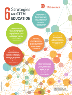


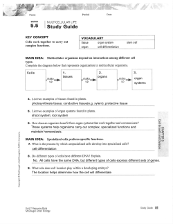

© Copyright 2026