
Document 75199
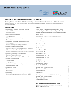
Early diagnosis and treatment
are necessary to ensure the
best possible outcomes in
children with hypopituitarism.
Wassily Kandinsky (1866-1944). Improvisation 26, 1912.
Hypopituitarism in Childhood
Mitchell E. Geffner, MD
Background: Childhood hypopituitarism may be present at birth or may be acquired. Early diagnosis and
treatment promote the best possible outcomes.
Methods: The anatomy, etiologies, clinical presentation, diagnostic testing strategies, and current treatments
relevant to childhood hypopituitarism are reviewed.
Results: Children with congenital hypopituitarism may present with life-threatening hypoglycemia, abnormal
serum sodium concentrations, shock, microphallus in males, and, only later, growth failure. Causes of
congenital hypopituitarism include septo-optic dysplasia, other midline syndromes, and mutations of
transcription factors involved in pituitary gland development. Children with acquired hypopituitarism
typically present with growth failure and may have other complaints depending on the etiology and the extent
of missing pituitary hormones. Acquired hypopituitarism may result from tumors (most commonly
craniopharyngioma), radiation, infection, hydrocephalus, vascular anomalies, and trauma.
Conclusions: An MRI of the head is critical in determining the etiology. Testing for pituitary hormone
deficiencies is undertaken along with appropriate hormonal replacement and, in some cases, direct treatment
of the cause of the hypopituitarism. All children with hypopituitarism require coordination of medical care by
a pediatric endocrinologist and, when older, transition to the care of an internist endocrinologist. Referrals to
a reproductive endocrinologist may be required as fertility issues arise.
Introduction
From the Childrens Hospital Los Angeles, Keck School of Medicine, University of Southern California, Los Angeles, California.
Submitted September 7, 2001; accepted November 8, 2001.
Address reprint requests to Mitchell E. Geffner, MD, Division of
Endocrinology, Childrens Hospital Los Angeles, 4650 Sunset Boulevard, Mailstop #61, Los Angeles, CA 20027. E-mail: mgeffner@chla.
usc.edu
Supported in part by grants from the Carolan Foundation,
Pharmacia, Inc, Genentech, Inc, Eli Lilly and Co, and Novo-Nordisk,
Inc. No significant relationship exists between the author and the
companies/organizations whose products or services may be referenced in this article.
212 Cancer Control
Hypopituitarism refers to the absence or reduction
in function of two or more hormones produced by the
pituitary gland. When all pituitary hormones are affected, the term panhypopituitarism should be used. In
childhood cases, one must consider whether the hypopituitarism has its origin before or at birth or is acquired at
any time after birth. The younger the child is at the time
of presentation, the more likely the etiology is to be conMay/June 2002, Vol. 9, No.3
genital. However, on occasion, congenital forms may
present or be diagnosed well after birth and, conversely,
some children with acquired forms are discovered relatively early in life. The purpose of this review is to discuss the causes of, relevant diagnostic modalities for, and
treatment of hypopituitarism in childhood.
mal hormonal production by the pituitary gland could
be restored. Thus, the term hypopituitarism, while convenient, is technically incorrect. Children with congenital hypopituitarism and adults with hypopituitarism are more likely to have primary diseases of the
pituitary gland compared to children with acquired
hypopituitarism, in whom hypothalamic and stalk disease is much more common.
Anatomy
The adult pituitary gland is a pea-sized midline
structure located below the optic chiasm. It hangs
down on a pedicle (the hypothalamic-pituitary stalk)
from the inferior portion of the hypothalamus. The
pituitary gland contains two anatomical components.
The anterior portion, also known as the adenohypophysis, receives its signals through the portal system of
blood vessels that arise in the hypothalamus and traverse the stalk. The posterior section, also known as
the neurohypophysis, receives its signals through axons
that arise in neural bodies within the hypothalamus and
also traverse down the stalk. Embryologically, the anterior pituitary gland is derived from Rathke’s pouch, an
evagination of the stomodeal ectoderm. In contrast, the
posterior pituitary arises in the infundibulum, a part of
the diencephalon.1
Most cases of childhood hypopituitarism, especially those that develop after birth, result from diseases of
the hypothalamus and the stalk that result in a failure of
the normal hypothalamic signals to reach the pituitary
gland. In other words, the pituitary gland is usually fully
functional but is unable to produce normal levels of its
stimulating hormones because it does not receive the
proper stimulation by the hypothalamic-releasing hormones or factors.2 Therefore, if patients with hypopituitarism could be treated with the appropriate types
and doses of hypothalamic-stimulating hormones, nor-
As can be seen on a normal head magnetic resonance imaging (MRI) scan, the anterior pituitary, on T1weighted imaging, appears dark and equal in intensity
to gray matter, while the posterior pituitary gland
appears white and is referred to, radiologically, as the
posterior “bright spot” (Fig 1A).3 This difference may
reflect a layer of fat within or around the posterior pituitary. In some cases of congenital hypopituitarism, this
“bright spot” is completely absent and correlates well
with the clinical presence of diabetes insipidus (DI). In
other cases of congenital hypopituitarism, the “bright
spot” is found ectopically in an anatomical location in
the normal pathway of posterior pituitary embryologic
development (either at the base of the hypothalamus or
in the stalk), in which case DI is usually absent (Fig 1B).
Such radiological findings are sometimes helpful in
securing the congenital nature of hypopituitarism. In
acquired forms of hypopituitarism in which DI develops either spontaneously or postoperatively, the “bright
spot” is typically absent.
Causes of Congenital Hypopituitarism
Congenital hypopituitarism may occur as a result
of birth trauma and/or asphyxia or as part of one of a
number of midline anatomical defects (Table 1). It may
also occur secondary to a genetic mutation involving
the anatomic development of the pituitary gland. The
Fig 1. — (A) Normal head MRI showing a sagittal view through the pituitary gland. The large arrow indicates the anterior portion of the pituitary gland,
whereas the small arrow indicates the posterior (normally whiter) portion of the pituitary gland. (B) Coronal head MRI with posterior pituitary bright spot
noted ectopically at the base of the hypothalamus (arrow). (C) Sagittal MRI view of an infant pituitary gland showing a transected hypothalamic-pituitary
stalk (arrow).
May/June 2002, Vol. 9, No.3
Cancer Control 213
Table 1. — Causes of Hypopituitarism
Congenital
Acquired
Birth trauma/asphyxia
Tumors in the hypothalamic-pituitary
region (eg, craniopharyngioma)
Midline defect syndromes
(eg, septo-optic dysplasia)
Mutations of genes encoding
pituitary transcription factors
Radiation to the head and
neck region
Hydrocephalus
Vascular abnormalities of
hypothalamic-pituitary region
tion can be successfully performed, it will show one or
both optic nerve heads to have a reduced diameter. If
the involvement is subtle, fundal photography may be
required for direct measurement and comparison to
age-matched normative data. It should be noted that
most children with optic nerve hypoplasia do not actually have associated hypopituitarism; however, for any
child first ascertained to have optic nerve hypoplasia, at
least a one-time referral to a pediatric endocrinologist
is warranted, especially if there is any associated
growth delay.
Major head trauma
association of hypopituitarism with specific prenatal or
birth trauma is sometimes difficult to prove. However,
the pregnancy histories of the mothers of affected children have a higher than expected occurrence of complications late in gestation or around the time of birth,
including placenta previa, abruptio placenta, and difficult deliveries.4 A congenital basis for the hypopituitarism is strongly suggested by the presence of a transected (or interrupted) hypothalamic-pituitary stalk on
MRI scan5 (Fig 1C).
Congenital hypopituitarism may also occur as a
component of one of several “midline defect syndromes.” The most common disorder in this group is
known as septo-optic dysplasia, or de Morsier syndrome. This disorder includes absence of the septum
pellucidum (the function of which is uncertain) in 50%
of cases (hence, the designation,“septo”) and underdevelopment of the optic nerves, associated with variable
degrees of reduced vision ranging from mild loss to
complete blindness.6 Affected children may present
with “wandering” or “searching” nystagmus, an irregular
jerkiness of the eyeballs, representing, in a sense, a
search for vision. An ophthalmologic examination may
be technically difficult in affected children if the degree
of nystagmus is moderate to severe but, if the examina-
Other midline associations with hypopituitarism
include the presence of a fused deciduous upper central maxillary incisor (Fig 2A),7 cleft lip and/or palate,
choanal atresia, anomalous and/or absent vascular supply to the central nervous system, and encephaloceles
(either of the extrusive variety or completely internal
emanating from the sphenoid sinus8 [Fig 2B]). If there
are no associated clinical, biochemical, or radiological
abnormalities, the hypopituitarism is said to have an
idiopathic basis.
On rare occasions, congenital hypopituitarism may
have a genetic basis involving one of a series of relatively recently identified pituitary transcription factors
that regulate the formation of the gland and its cellular
organization.9 One of the earliest known transcription
factor genes involved in embryogenesis of the pituitary
gland is Rpx (Rathke’s pouch homeobox, also called
HESX1), mutations of which have been found in a few
cases of septo-optic dysplasia.10 Other early transcription factor genes important in the formation of various
pituitary cell populations are LHX3, LHX4, and PROP-1
(Prophet of Pit-1). Lastly, PIT-1 (also known as
POU1F1) is a pituitary-specific transcription factor that
is necessary for the development of somatotroph, lactotroph, and thyrotroph lineages. Inherited cases of
hypopituitarism in humans have been described in conjunction with mutations of LHX3 (associated with defi-
Fig 2. — (A) MRI depiction of a fused upper central maxillary incisor (arrow) in a child with hypopituitarism. (B) Sagittal head MRI showing a transsphenoidal encephalocele (arrows) in an adolescent with hypopituitarism. (C) Lateral view cerebral angiogram showing a suprasellar arteriovenous varix at the
apex of the basilar artery (arrow). Fig 2B is reproduced with permission of Springer-Verlag GmbH & Co: Kjellin IB, Kaiserman KB, Curran JG, et al. Pediatr Radiol. Aplasia of right internal carotid artery and congenital hypopituitarism. 1999;29:586-588. Fig 2C is reproduced with permission from Blackwell
Science Ltd: Koral K, Geffner ME, Curran JG. Trans-sphenoidal and sphenoethmoidal encephalocele: report of two cases and review of the literature.
J Australas Radiol. 2000;44:220-224.
214 Cancer Control
May/June 2002, Vol. 9, No.3
ciencies of all anterior pituitary hormones except corticotropin [ACTH], along with a rigid cervical spine
leading to limited head rotation), PROP-1 (associated
with deficiencies of growth hormone [GH], prolactin,
thyroid-stimulating hormone [TSH], and sometimes
gonadotropins and ACTH, along with limited elbow
extensibility, blue sclerae, and, surprisingly, a large sella
turcica in some affected individuals), and PIT-1 (associated with deficiencies of GH, prolactin, and TSH). Due
to mutations of pituitary gene transcription factors,
some cases of hypopituitarism include variability in the
timing of presentation of the different hormone deficiencies. For example, GH deficiency typically manifests early in life, whereas other hormone deficiencies
somewhat inexplicably present later.
Unique Features of
Congenital Hypopituitarism
The most important presenting feature, and perhaps most common feature, of congenital hypopituitarism is hypoglycemia.11 This occurs secondary to
the presence of GH deficiency with or without associated ACTH deficiency (the latter leading to cortisol
deficiency). Both GH and cortisol are counterregulatory (“anti-insulin”) hormones that protect against
hypoglycemia, especially during fasting. Without one
or both, insulin acts in an unopposed fashion and,
because of immaturity of other protective mechanisms against hypoglycemia in infants, this population
is at an undue risk for low blood sugar. Thus, in infants
(and sometimes in toddlers and older children) with
unrecognized and/or untreated or undertreated
hypopituitarism, hypoglycemia may often be recurrent and severe, potentially leading to permanent
brain damage if not managed aggressively. The hypoglycemia usually resolves with replacement of GH
and/or cortisol, although it may occasionally remain
problematic in older children during stressful periods
associated with reduced oral intake. An important
caveat is that, in any term infant who develops hypoglycemia with no underlying risk factor (such as prematurity, intrauterine growth retardation, infant of a
diabetic mother, etc), the diagnosis of hypopituitarism
must be considered. Additionally, severe cortisol deficiency may result in presentation with hyponatremia
(as cortisol is necessary for the action of anti-diuretic
hormone [ADH, also known as vasopressin] to facilitate the excretion of free water by the kidneys) and/or
outright shock due to its effects on blood pressure
maintenance. If DI were present, shock with hypernatremia would be manifest.
Another unique feature of congenital hypopituitarism is the presence at birth of a microphallus
May/June 2002, Vol. 9, No.3
(small penis) in some affected males. This may result
from isolated GH deficiency or from combined GH
and gonadotropin deficiencies (the latter leading to
testosterone deficiency in the second and third
trimesters and postnatally).12 The microphallus may
enlarge solely in response to treatment with GH, suggesting that GH has a critical role in penile growth at
least in fetal and early postnatal life. Thus, some pediatric endocrinologists will initiate a 6-month trial of
GH therapy alone to stimulate penile enlargement. If
the response were inadequate, then a short course of
low-dose testosterone therapy would follow. This can
be in the form of 25 mg of long-acting testosterone
enanthate given as four equally spaced intramuscular
injections over a 12-week period. This approach is
usually successful in equalizing penile size with that
of other male infants and with few or no side effects.
There are, however, other pediatric endocrinologists
who would administer testosterone immediately,
along with GH.
Lastly, some children with congenital hypopituitarism develop a unique, noninfectious form of hepatitis. The condition is suspected if there is liver enlargement and abnormal liver function tests (predominantly
indicating cholestasis) and is confirmed by the presence of characteristic giant-cell transformation of hepatocytes as seen on biopsy.13 The exact cause of this
giant-cell hepatitis is unknown, and the condition usually remits on its own over the first few months of life
without any permanent damage to the liver.
Causes of Acquired Hypopituitarism
The most concerning cause of acquired hypopituitarism is a tumor in the hypothalamic-pituitary region.
The most common tumor that arises in this region in
childhood is a craniopharyngioma. While craniopharyngiomas are derived from epithelial remnants of
Rathke’s pouch, the same tissue that forms the anterior
pituitary gland, they are not usually found exclusively
within the pituitary gland, but more likely they are confined to the suprasellar region or are found in both
suprasellar and intrasellar locations.14 The presenting
symptoms are dependent both on the main location of
the tumor and on the direction(s) that the tumor
grows. For example, if the tumor grows downward, it
may cause alterations in the anterior and posterior clinoid bones or in the floor of the sella turcica and/or
compress the pituitary gland. If the tumor grows
upward, it may compromise vision by effects on the
optic nerves. While medical textbooks typically
describe the most common visual change in patients
with craniopharyngioma as “bitemporal hemianopsia”
(loss of the outer visual fields of both eyes) due to comCancer Control 215
Table 2. — Hierarchy of Hypothalamic-Pituitary-Target Organ Hormones
Hypothalamus
Pituitary
Target Organ
Hormone/Function
Growth hormone-releasing hormone (GHRH)
Anterior: Growth hormone (GH)
Cartilage/liver
Insulin-like growth factor-1
(IGF-1)
Thyrotropin-releasing hormone (TRH)
Anterior: Thyroid-stimulating hormone (TSH)
Thyroid gland
T4/T3
Gonadotropin-releasing hormone (GnRH)
Anterior: Luteinizing hormone (LH)
Follicle-stimulating hormone (FSH)
Ovary
Testicle
Estradiol
Testosterone
Corticotropin-releasing hormone (CRH)
Anterior: Corticotropin (ACTH)
Adrenal glands
Cortisol
Prolactin-inhibitory factor
Anterior: Prolactin
Breast
Milk
Hypothalamic factors
Posterior: Antidiuretic hormone (ADH)
Kidney
Urine concentration
pression of the optic chiasm, this rarely occurs so precisely. More likely is the development of “quadrianopsias,” which are smaller, more irregular field cuts. If the
tumor grows even farther upward beyond the visual
tracts, it may block the third ventricle and cause
obstructive hydrocephalus, leading to headaches, vomiting, and/or blurry vision (in association with
papilledema). Craniopharyngiomas are benign tumors
histologically, but they are often described as “geographically malignant” based on their location and ability to wrap around vital structures (eg, optic nerves)
precluding complete surgical removal. As stated previously, tumors originating in the pituitary gland are
uncommon causes of acquired hypopituitarism in children. However, any surgery to remove a tumor in the
hypothalamic-pituitary region may lead to hypopituitarism if it is not already present.
Another cause of acquired hypopituitarism is radiation treatment of a cancerous tumor in the head or
neck region. More specifically, the radiation that is
required to cure the child’s tumor may, of necessity,
damage normal tissue in its path or beyond. When
hypopituitarism ensues in this situation, it is usually
the result of radiation-induced damage to the hypothalamus, as the pituitary gland is relatively resistant to
radiation. Different hypothalamic-pituitary axes have
different sensitivities to radiation. Doses as low as 18
Gy using conventional fractionation can interfere with
GH dynamics; doses higher than 40 Gy can cause deficiencies of gonadotropins, TSH, and ACTH, while >50
Gy may cause hyperprolactinemia, especially among
young women.
Other causes of acquired hypopituitarism in childhood include previous brain infection (encephalitis
and/or meningitis), hydrocephalus (even without an
underlying tumor), vascular abnormalities (such as a
varix in the hypothalamic-pituitary area)15 (Fig 2C), and
major head trauma usually associated with a significant
loss of consciousness.16
216 Cancer Control
Potential Anterior Pituitary
Hormone Deficiencies
In childhood hypopituitarism, GH is the most commonly underproduced pituitary hormone, often as the
result of loss of hypothalamic GH-releasing hormone
(GHRH) (Table 2). The deficiency of GH primarily leads
to short stature and slow height velocity. Untreated GH
deficiency in children also causes disturbed body composition, with a reduction in lean body mass (ie, muscle)
and an excess of fat, the latter accumulating predominantly in the cheeks of the face and in the abdomen, creating a cherubic or angel-like appearance.17
Deficiency of hypothalamic thyrotropin-releasing
hormone (TRH) or pituitary TSH causes central
hypothyroidism. Unlike children whose hypothyroidism is due to thyroid gland damage, those with
hypopituitarism typically have somewhat higher thyroid hormone levels and thus may have few or no
symptoms. In other cases, as occurs in patients with
primary thyroid disease, short stature and slow height
velocity, relative weight excess, constipation, dry skin,
cold intolerance, and fatigue may be present.
Younger children with deficiencies of either hypothalamic gonadotropin-releasing hormone (GnRH) or
the pituitary gonadotropins typically show no abnormalities since luteinizing hormone (LH) and folliclestimulating hormone (FSH) levels are normally low
prior to puberty. In contrast, adolescent-aged children
with deficiencies of the gonadotropins present with
failure to start or progress through puberty (breast
development and menstrual periods in girls and
enlargement of the testicles and penis in boys). Infrequently and paradoxically, central sexual precocity can
be seen in the setting of hypopituitarism.
Loss of hypothalamic corticotropin-releasing
hormone (CRH) or pituitary ACTH results in an ability
of the adrenal zonas fasciculata and reticularis to
May/June 2002, Vol. 9, No.3
manufacture normal amounts of cortisol (central
adrenal insufficiency). If deficient, this hormone is
most likely to place a child in a life-threatening situation. While there would likely be no symptoms under
normal circumstances, except maybe mild fatigue,
lack of cortisol in the setting of infection, fever,
surgery, trauma, etc, may cause vomiting, dehydration,
shock, and even death. Biochemical correlates of cortisol deficiency include hypoglycemia and hyponatremia (with normokalemia). In this setting, mineralocorticoid function is completely normal, as aldosterone production by the zona glomerulosa of the
adrenal cortex is regulated by the renin-angiotensin
and not the CRH-ACTH system.
Potential Anterior Pituitary
Hormone Excess
Serum levels of prolactin are usually normal or only
mildly increased (if there is disruption of the hypothalamic-pituitary stalk) secondary to the loss of a normally predominant inhibitory signal from the hypothalamus. Although there are typically no symptoms in this
situation, in rare cases a small amount of galactorrhea
might occur.
Potential Posterior Pituitary
Hormone Excess
As stated previously, a deficiency of ADH causes
central or neurogenic DI. Infants and toddlers manifest
DI with excessively wet diapers. If unrecognized and
hence untreated, dehydration with elevated serum sodium concentrations will ensue in this age group as
young children cannot report and easily satisfy heightened thirst. Older children with DI typically present
with excessive day- and night-time urination, new onset
of bed-wetting, and increased thirst. DI most often
occurs unintentionally as a result of surgical treatment
of a hypothalamic-pituitary tumor, such as a craniopharyngioma. In some cases, DI is temporary due to
local postsurgical edema, but it will be permanent if
surgical sacrifice close to the hypothalamus or of the
stalk itself is required for complete cure of a brain
tumor in the suprasellar region.18 DI may also occur in
association with a diencephalic germinoma.
Diagnosis of Hypopituitarism
The final diagnosis of GH deficiency must integrate clinical, auxological (growth), and biochemical
data.19 GH deficiency should be suspected if a child
has reduced height velocity along with a delayed bone
age, and low serum levels of the GH surrogates, insulinMay/June 2002, Vol. 9, No.3
like growth factor-1 (IGF-1), and insulin-like binding
protein-3 (IGFBP-3) not otherwise explained by young
age and/or undernutrition. The next step is then to
measure serum GH levels. Unfortunately, beyond the
first month of life, measurement of GH levels in a random blood sample is without value (to secure a diagnosis of GH deficiency) because most of the body’s GH
is made during sleep, with several significant peaks
linked to deep (stage III-IV EEG) sleep, the first cycle
of which usually occurs at approximately 60 to 90 minutes after falling sleep. Random GH measurements
during the day, even in tall individuals, are frequently
low and thus potentially misleading.
If most or all of these criteria are met, GH testing is
then recommended by any of several physiologic or
pharmacologic methods.
The most commonly
employed physiologic stimulus is exercise, but this is
often difficult to standardize. Overnight serial blood
sampling every 20 to 30 minutes was common in the
early 1990s but lost favor because of inconvenience,
cost, and lack of reproducibility.
A more common approach is stimulation testing
(also referred to as provocative testing) with medications known to activate the physiologic regulation of
GH secretion, notably the α-adrenergic pathway (eg,
clonidine20 at the following dosages according to body
weight: 50 µg for body weight 5 to 15 kg, 75 µg for
15.1 to 20 kg, 100 µg for 20.1 to 25 kg, 150 µg for 25.1
to 35 kg, 200 µg for 35.1 to 50 kg, and 250 µg for more
than 50 kg).
Blood samples are procured 60 and 90 minutes
after the clonidine is taken. Clonidine may cause sleepiness and a mild fall in blood pressure, but these effects
are transient and usually resolve within 90 minutes. To
mimic the other physiologic pathway of GH regulation
involving the dopaminergic system, L-dopa combined
with Inderal (propranolol) has been used, but L-dopa
was recently taken off the market. These and all other
stimulation tests must be performed in the fasting state
(and, hence, early in the morning), as glucose intake suppresses GH secretion. GH provocation tests can also be
performed using glucagon (0.1 mg/kg, maximum dose 1
mg)21 or insulin (0.05 to 0.1 units of regular insulin per
kilogram, depending on the index of suspicion), the latter known as an insulin tolerance test (ITT).22 In both
cases, hypoglycemia is induced. In the case of glucagon,
blood sugar initially rises followed by stimulation of
endogenous insulin leading to late hypoglycemia. For a
hypoglycemic stimulus to be deemed adequate to stimulate GH, the plasma glucose concentration needs to fall
by at least 50% from baseline. In the glucagon test,
blood sampling is performed every 15 minutes for 1
hour and then every 30 minutes for the next 2 hours out
Cancer Control 217
to 180 minutes. For the ITT, blood sampling is performed every 15 minutes for 1 hour and then again 30
minutes later, with bedside and laboratory glucose testing, along with GH measurements at all time points and
cortisol measurements at 0, 60, and 90 minutes. To
obtain these multiple blood samples, a heparin lock
should be placed in a large-bore vein at the start of the
study. Glucagon or hypoglycemia itself may cause nausea and vomiting. Hypoglycemia results in uncomfortable adrenergic manifestations such as sweatiness,
excessive hunger, and shakiness, and may also cause
neuroglycopenic manifestations such as loss of consciousness or seizures. These tests can also be used to
assess cortisol production, thus eliminating the need for
an additional stimulation test either on the same day or
on a different day. As such, these tests are moderately
dangerous and need to be performed by experienced
personnel in a hospital outpatient unit with resuscitative equipment and expertise as well as with a physician
in attendance throughout the test. If dangerous hypoglycemia associated with diminished mental status
occurs, 2 mL/kg of 25% dextrose should be immediately administered intravenously. The test will still be valid
because the hypoglycemic stimulus was delivered so all
blood samples should continue to be collected.
Arginine is another GH secretagogue that requires a
45-minute intravenous infusion (5 mL/kg of 10% arginine
hydrochloride).22 Arginine is an amino acid that is a nonspecific stimulator of multiple hormones including
insulin and glucagon,in addition to GH. There are no side
effects with its use. Lastly, intravenous GHRH (at a dose
of 1 µg/kg body weight) may be employed with sampling
at 0, 5, 30, 45, 60, 90, and 120 minutes.23 Its only frequent
side effects are local injection site reactions.
GH deficiency is traditionally defined by all GH
levels on all tests being less than 10 ng/mL as measured
by traditional double-antibody radioimmunoassay. In
certain clinical situations, such as known surgical
transection of the hypothalamic-pituitary stalk or the
administration of high-dose radiation to the hypothalamic-pituitary region, less rigorous GH testing is required.
Of note, the blood of children with untreated panhypopituitarism is mildly acidotic secondary to the
absence of GH and its ability to regulate acid-base
metabolism by the kidney.24 Its presence is detected by
finding a slightly low serum bicarbonate level that corrects completely with GH replacement.
The diagnosis of central hypothyroidism due to
TSH deficiency requires low levels of free and/or total
thyroxine (T4) in serum. On occasion, the free T4 level
is at the low end of the normal range. A TSH level generally is not helpful. With pituitary failure, the TSH level
218 Cancer Control
would obviously be low or not measurable. Surprisingly, if the problem arises in the hypothalamus, the TSH is
usually in the normal range or occasionally even slightly elevated for uncertain reasons. In some situations,
administration of synthetic TRH, the hypothalamic factor that controls pituitary TSH secretion, may be necessary. This test can help distinguish whether the deficiency lies at the level of the pituitary (in which case
the TSH level is very low and fails to increase) or at the
hypothalamus (in which case the TSH level rises excessively and stays elevated for a longer period of time
than occurs normally).25
The diagnosis of LH and FSH deficiencies in childhood can be the most problematic of all the pituitary
hormones as throughout most of the first 10 years of
life the serum concentrations of children are naturally
low. Because LH and FSH levels are slightly higher in
the first 6 months of life and then increase significantly
after 10 years of age, random testing at these ages may
indicate low levels.26 Such testing is most informative
when, in the teenager, there is also a failure to show
clinical pubertal changes by age 13 years in girls (lack
of breast development, sexual hair, and menses) and by
age 14 years in boys (lack of testicular enlargement,
penile growth, and sexual hair). In females, radiological
confirmation of lack of pubertal initiation by pelvic
ultrasound may be helpful if the ovaries and uterus are
prepubertal in size.
As stated previously, cortisol deficiency is the most
important parameter to consider in view of its role in
regulating vital functions during stressful situations.
Using random blood testing for this diagnosis requires
an understanding of both the age-related changes
(lower levels in younger than in older children) and
“diurnal” changes (by time of day) in cortisol levels.27
More specifically, in older children and adults, the highest cortisol levels are found at 8:00 AM. Thus, a low 8:00
AM serum cortisol level (less than 3 µg/dL in children
between the ages of 1 and 16 years) would suggest deficiency. An indirect clue to the presence of cortisol deficiency is an increased percentage of eosinophils on a
peripheral blood smear. As mentioned previously, more
rigorous testing can be accomplished as part of the
glucagon or insulin hypoglycemia GH tests or after
administration of metyrapone, CRH, or low-dose synthetic ACTH, drugs that stimulate the adrenal glands to
produce more cortisol if they can. The use of ACTH
provocation may occasionally give a misleading picture
of adequate adrenal reserve as sufficient cortisol may
be produced in response to exogenous ACTH stimulation but is not produced under daily living conditions.
Prolactin excess is present if there is an elevated
prolactin concentration in a random blood sample.
May/June 2002, Vol. 9, No.3
ADH deficiency or central DI is suspected if urination and thirst are excessive, and it is strongly suggested
by the presence of increased levels of serum sodium
along with dilute urine. If the diagnosis remains
unclear, a water deprivation test can firmly prove or disprove this diagnosis.28 This test involves fasting by the
child (no food or liquids) for up to 8 hours and observing the child’s urine output. A normal child will reduce
urine output to avoid dehydration since there is no fluid
intake. Under the same circumstances, a child with DI
will continue to urinate and, if not monitored carefully,
could become dehydrated and develop an elevated
serum sodium concentration. Specific mathematical criteria that are derived from blood and urine test results
during the water deprivation test are used to make the
diagnosis of DI. This test should always be done under
close medical supervision in either an experienced outpatient procedure unit or an inpatient setting.
Once the diagnosis of hypopituitarism has been
made by appropriate hormonal testing, a head MRI
scan (with gadolinium contrast) must be performed to
look for a possible organic or structural basis. Nonserious abnormalities that can be associated with hypopituitarism include a small pituitary gland (for age) with
filling of the sella with cerebrospinal fluid (empty sella)
(Fig 3A) and an ectopic posterior pituitary gland.
A midline syndrome is suggested by the absence of the
septum pellucidum and/ or the corpus callosum. The
most critical reason for obtaining the MRI scan is to
detect a brain tumor in the hypothalamic-pituitary area,
the most common of which is a craniopharyngioma.
Radiological evidence of craniopharyngiomas can be
seen on a lateral skull-ray in 89% of cases, which makes
this an important screening test when this diagnosis is
suspected in a child29 (Fig 3B). Findings include erosion of the normal sellar architecture and/or the presence of suprasellar calcification. On MRI scans, the
tumor usually contains a mixture of solid and cystic
components (Fig 3C) and may contain a ring of calcification around a cystic component (the latter best seen
on a computed tomographic scan) (Fig 3D). Other
abnormalities that can be
seen on the MRI scan
include a transected stalk,
small optic nerves, hydrocephalus, and vascular
abnormalities.
Treatment of
Hypopituitarism
Fig 3. — (A) Sagittal head MRI showing compressed pituitary contents (small arrow) with filling of sella turcica
by cerebrospinal fluid (empty sella) (large arrow). (B) Lateral skull radiograph showing erosion of sella turcica
(large arrow) and suprasellar calcification (small arrow) caused by a craniopharyngioma. (C) Coronal head MRI
showing a craniopharyngioma in the suprasellar region containing a large cyst (small arrow) within a contrastenhanced thin shell, along with mild hydrocephalus (large arrow). (D) Head CT scan showing contrast-enhanced
ring of calcification around a craniopharyngioma and internal calcification (arrow).
May/June 2002, Vol. 9, No.3
If an underlying cause
is discovered on the MRI
of the head, such as a
tumor and/or hydrocephalus, appropriate neurosurgical intervention is
required.
For example,
removal or debulking of a
tumor and shunting for
hydrocephalus, respectively,
would be undertaken. Additionally, radiation therapy
may be required to treat certain types of tumors.
Chemotherapy is used less
often to treat brain tumors.
In most cases, these treatments, while potentially lifesaving, do not reverse the
hypopituitarism. The diagnosis of specific hormonal
deficiencies present prior
to any neurosurgical procedure has limited imporCancer Control 219
tance as all deficiencies will persist and all intact axes
will likely be sacrificed during the surgical procedure.
Since these patients uniformly receive high-dose glucocorticoid (dexamethasone) treatment to reduce any
swelling around the tumor before and during the
surgery, this serves to prevent possible adrenal insufficiency that, if present and not managed aggressively,
could lead to great morbidity and even death. If DI is
present preoperatively, it should be readily apparent
on clinical and biochemical grounds and easily managed by a capable anesthesiologist. If GH, thyroid hormone, and/or gonadotropin deficiencies are present
preoperatively and not recognized and treated, there
would be no significant effect on the immediate postoperative recovery.
To treat GH deficiency in a child with remaining
growth potential, replacement with GH is usually
undertaken. GHRH, the hypothalamic stimulator of
GH, is also approved by the US Food and Drug Administration (FDA) for this purpose, but it is effective only
if the pituitary gland is fully functional. Unfortunately,
optimal dosing and frequency of administration of
GHRH remain unknown. Thus, GH itself is preferentially administered. It is typically given subcutaneously
either with a 31-gauge needle and syringe, pen system
with a 31-gauge needle, or “needleless” injector system.
The usual dose range is between 0.18 and 0.3 mg per
kilogram of body weight per week divided into 6 or 7
daily doses administered generally in the evening to
simulate the fact that most of the GH manufactured by
the body is at night during sleep (nyctohemeral
rhythm).30 A long-acting, once- or twice-monthly formulation of GH administered subcutaneously with a
special 22-gauge needle has recently been approved by
the FDA. If the hypopituitarism is caused by a brain
tumor or its treatment, commencement of GH treatment is usually delayed for 1 year until the clinical status of the tumor is clearly defined. This practice is
based on logic because there are no scientific data
showing that GH causes tumor growth or recurrence.
Other GH formulations delivered intranasally or by
inhalation are under development. When the growth
potential of a child with hypopituitarism has been fully
or nearly fully attained, a transition to adult GH dosing
should be considered (approximately one sixth to
one third the pediatric dose). While the need for GH
retesting is recommended for children with isolated
GH deficiency before commencing adult dosing (as
approximately 50% will have normal GH responses on
retesting31), this should not be required when there are
multiple hormone deficiencies.
One unusual circumstance, which usually arises following surgery for a craniopharyngioma, occurs when
the child begins to gain excess weight and grow nor220 Cancer Control
mally to excessively in height with documented GH
deficiency and without institution of GH treatment.
The cause of this paradox, dubbed “the growth without
GH syndrome,”32 is unknown, but the condition can be
debilitating secondary to large weight accumulation.
There is no proven treatment for this situation,
although research trials with appetite depressants, antineurosis/anti-anxiety drugs, and analogs of somatostatin that inhibit GH, insulin, and other hormones are
in progress.33
Oral levothyroxine (brand name or generic) is the
specific treatment for central hypothyroidism, as TRH
and thyrotropin (TSH), while available as drugs for
dynamic testing of thyroid function, are not FDAapproved for treatment purposes. The dose of levothyroxine for treatment of central hypothyroidism in children is approximately 50 µg/m2 per day (or approximately half that used to treat primary hypothyroidism).
The adequacy of the dose can be readily determined by
measuring the serum level of free T4. TSH levels should
not be monitored as the patients are TSH-deficient.
For documented deficiencies of LH and FSH, a
more practical approach than replacing the
gonadotropins is replacing the corresponding sex
steroid(s), ie, estrogen (orally or by patch) and progesterone (orally) in girls and testosterone in boys (by
long-acting monthly injection in the early adolescent
period or by injection, daily skin patch, or daily skin gel
in the late adolescent period). Some pediatric endocrinologists will also prescribe a low dose of testosterone
to adolescent girls without which they may not have
any pubic or axillary hair development, although this is
not currently the general standard of care. These
approaches will provide appropriate adolescent physical development and protect against osteoporosis but,
as such, will not restore fertility. When affected subjects
reach the age when they are imminently ready to have
children, their hormone replacement regimens need to
be altered based on the location of the damage. More
specifically, if the hypothalamus is damaged and the
pituitary gland is proven to be intact, treatment with
the hypothalamic hormone GnRH by pump will stimulate the subject’s pituitary gland to produce LH and FSH
and, thus, induce egg or sperm development. If pituitary function is damaged, coordinated treatment with
synthetic LH (as human chorionogonadotropin [hCG])
and FSH (either as human menopausal gonadotropin
[hMG] or recombinant FSH) needs to be initiated. In
either case, the treatment program is complex and
requires the supervision of an experienced endocrinologist with expertise in reproductive disorders.
The most practical approach to restoration of normal adrenal function is replacement of cortisol (also
May/June 2002, Vol. 9, No.3
known as hydrocortisone), since CRH and ACTH are
approved only for testing of the adrenal axis. In children, oral hydrocortisone is usually given 2 or 3 times
daily at a dose of 10 to 15 mg/m2 per day. This dose is
equal to approximately 1.5 to 2 times the body’s normal
daily production rate of cortisol but provides appropriate replacement as only 50% to 90% of an oral dose is
absorbed. Many of the commonly used replacement regimens use a higher dose in the morning than at other
times of the day in an attempt to simulate the normal
diurnal variation in cortisol production, in which the
adrenal glands make more cortisol earlier than later in
the day. Recently, liquid suspensions of cortisol were
removed from the market so that only tablet formulations remain, with the lowest pill dose containing 5 mg.
To provide a liquid glucocorticoid formulation as
required by infants and toddlers, prednisone suspension
(5 mg/5 mL) can be used, remembering that prednisone
is 4 times the milligram-for-milligram potency of cortisol.
Thus, a 10-mg dose of cortisol is equal to a 2.5-mg dose
of prednisone. Another glucocorticoid formulation that
can be used (and is available in both tablets and liquid)
is dexamethasone that is 25 to 100 times more potent
than cortisol. Because of the wide range of potency
equivalence between dexamethasone and hydrocortisone, use of this formulation is sometimes problematic
as it is easy to unintentionally overdose. As stated previously, there is no need to prescribe replacement of mineralocorticoid as, with central adrenal insufficiency, the
kidney still responds normally in terms of sodium and
potassium balance that is regulated by the reninangiotensin system. With significant illness, stress,
and/or injury, the glucocorticoid dose must be doubled
or tripled for 1 or more days depending on the length
and severity of the illness, stress, and/or injury. If there
is significant vomiting, a child with adrenal insufficiency
should be taken to the physician or to the emergency
room. As a precaution, all children should have
injectable hydrocortisone at home (in the form of 100or 250-mg Solu-Cortef Act-o-Vials). Lastly, older children
(who are not with their parents all the time) and adolescents must wear Medic-Alert identification indicating the
presence of hypopituitarism and adrenal insufficiency.
For the treatment of central DI, replacement of
ADH may be accomplished with a number of formulations. During surgery and other critical situations,
ADH should be administered as a continuous intravenous infusion. In most day-to day situations, desmopressin acetate is the preferred replacement therapy.
This formulation has been structurally modified to prolong its duration of action and to eliminate any tendency of the drug to cause hypertension. Desmopressin acetate was originally developed for intranasal
administration using a “rhinal tube”(allowing provision
of doses as low as 2.5 µg that might be required in a
May/June 2002, Vol. 9, No.3
young child). In infants, desmopressin acetate use
requires even more attention to fluid intake and formulation. Subcutaneous administration may be the
optimal method of delivery in this age group. More
recently, the nasal preparation became available as a
fixed-dose, metered pulse spray (similar to asthma
medication), with each pulse providing a 10-µg dose.
Most recently, an oral formulation of desmopressin
acetate has become available as 0.1- or 0.2-mg tablets.
In prescribing oral desmopressin acetate, it is important to remember that, since most of the drug is inactivated in the stomach, a 20-fold higher oral dose (compared to the nasal dose) must be prescribed.34
Conclusions
Hypopituitarism in childhood may have either a
congenital or an acquired basis. A head MRI scan is critical in attempting to determine the specific cause in
either case. Identified hormone abnormalities require
treatment with appropriate hormonal replacement
therapies, including GH, usually for life. All children
with hypopituitarism require supervision of medical
care by a pediatric endocrinologist and, when older,
appropriate transition to an internist endocrinologist
or to a reproductive endocrinologist.
References
1. Thorner MO,Vance ML, Horvath E, et al. The anterior pituitary.
In: Wilson JD, Foster DW, eds. Williams’ Textbook of Endocrinology.
8th ed. Philadelphia, Pa: WB Saunders Co; 1992:222-224.
2. Rosenfeld RG. Disorders of growth hormone and insulin-like
growth factor secretion and action. In: Sperling MA, ed. Pediatric
Endocrinology. Philadelphia, Pa: WB Saunders Co; 1996:141-143.
3. Root AW, Martinez CR. Magnetic resonance imaging in
patients with hypopituitarism. Trends Endocrinol Metab. 1992;3:
283-287.
4. Craft WH, Underwood LE,Van Wyk JJ. High incidence of perinatal insult in children with idiopathic hypopituitarism. J Pediatr.
1980;96:397-402.
5. Pinto G, Netchine I, Sobrier ML, et al. Pituitary stalk interruption syndrome: a clinical-biological-genetic assessment of its pathogenesis. J Clin Endocrinol Metab. 1997;82:3450-3454.
6. Siatkowski RM, Sanchez JC,Andrade R, et al. The clinical, neuroradiographic, and endocrinologic profile of patients with bilateral
optic nerve hypoplasia. Ophthalmology. 1997;10:493-496.
7. Kjellin IB, Kaiserman KB, Curran JG, et al. Aplasia of right
internal carotid artery and congenital hypopituitarism. Pediatr Radiol. 1999;29:586-588.
8. Koral K, Geffner ME, Curran JG. Trans-sphenoidal and sphenoethmoidal encephalocele: report of two cases and review of the
literature. J Australas Radiol. 2000;44:220-224.
9. DiMeglio LA, Hofman PL, Pescovitz OH. Molecular defects of
the growth hormone axis. In: Smith RG,Thorner MO, eds. Human
Growth Hormone: Basic and Clinical Research. Humana Press, Inc:
Totowa, NJ; 2000:169-189.
10. Lopez-Bermejo A, Buckway CK, Rosenfeld RG. Genetic
defects of the growth hormone-insulin-like growth factor axis.
Trends Endocrinol Metab. 2000;11:39-49.
11. Job JC. Early diagnosis and early treatment of growth hormone deficiency. Horm Res. 1989;31:149-152.
12. Urzola A, Leger J, Czernichow P. Three cases of congenital
growth hormone deficiency with micropenis and hypospadias: what
Cancer Control 221
does growth hormone have to do with it? Horm Res. 1999;51:101104.
13. Spray CH, Mckiernan P, Waldron KE, et al. Investigation and
outcome of neonatal hepatitis in infants with hypopituitarism. Acta
Paediatr. 2000;89:951-954.
14. Lafferty AR, Chrousos GP. Pituitary tumors in children and
adolescents. J Clin Endocrinol Metab. 1999;84:4317-4323.
15. Martin NA, Macagba-Crain CL, Geffner ME, et al. Isolated
growth hormone deficiency associated with a giant arteriovenous
varix. Neurosurgery. 1990;27:295-298.
16. Benvenga S, Campenni A, Ruggeri RM, et al. Clinical review
113: hypopituitarism secondary to head trauma. J Clin Endocrinol
Metab. 2000;85:1353-1361.
17. Carrel AL,Allen DB. Effects of growth hormone on body composition and bone metabolism. Endocrine. 2000;12:163-172.
18. Blevins LS Jr, Wand GS. Diabetes insipidus. Crit Care Med.
1992;20:69-79.
19. Consensus guidelines for the diagnosis and treatment of
growth hormone (GH) deficiency in childhood and adolescence:
summary statement of the GH Research Society. J Clin Endocrinol
Metab. 2000;85:3990-3993.
20. Lanes R, Recker B, Fort P, et al. Low-dose oral clonidine: a simple and reliable growth hormone screening test for children. Am J
Dis Child. 1985;139:87-88.
21. Vanderschueren-Lodeweyckx M, Wolter R, Malvaux P, et al.
The glucagon stimulation test: effect on plasma growth hormone and
of immunoreactive insulin, cortisol, and glucose in children. J Pediatr. 1974;85:182-187.
22. Penny R, Blizzard RM, Davis WT. Sequential arginine and
insulin tolerance tests on the same day. J Clin Endocrinol Metab.
1969;29:1499-1501.
23. Achermann JC, Brook CG, Hindmarsh PC. The GH response
to low-dose bolus growth hormone-releasing hormone (GHRH(129)NH2) is attenuated in patients with longstanding post-irradiation
GH insufficiency. Eur J Endocrinol. 2000;142:359-364.
24. Glaser NS, Shirali AC, Styne DM, et al. Acid-base homeostasis
in children with growth hormone deficiency. Pediatrics. 1998;102:
1407-1414.
25. Kolesnick RN, Gershengorn MC. Thyrotropin-releasing hormone and the pituitary. New insights into the mechanism of stimulated secretion and clinical usage. Am J Med. 1985;79:729-739.
26. Hayes FJ, Crowley WF Jr. Gonadotropin pulsations across
development. Horm Res. 1998;49:163-168.
27. Deshpande S, Platt MP,Aynsley-Green A. Patterns of the metabolic and endocrine stress response to surgery and medical illness in
infancy and childhood. Crit Care Med. 1993;21(9 suppl):S359-361.
28. Adam P. Evaluation and management of diabetes insipidus.
Am Fam Physician. 1997;55:2146-2153.
29. Thomsett MJ, Conte FA, Kalpan SL, et al. Endocrine and neurologic outcome in childhood craniopharyngioma: review of effect
of treatment in 42 patients. J Pediatr. 1980;97:728-735.
30. Van Cauter E, Copinschi G. Interrelationships between
growth hormone and sleep. Growth Horm IGF Res. 2000;10(suppl
B):S57-S62.
31. Consensus guidelines for the diagnosis and treatment of
adults with growth hormone deficiency: summary statement of the
Growth Hormone Research Society workshop on adult growth hormone deficiency. J Clin Endocrinol Metab. 1998;83:379-381.
32. Geffner ME. The growth without growth hormone syndrome. Endocrinol Metab Clin North Am. 1996;25:649-663.
33. Lustig RH, Rose SR, Burghen GA, et al. Hypothalamic obesity
caused by cranial insult in children: altered glucose and insulin
dynamics and reversal by a somatostatin antagonist. J Pediatr.
1999;135:162-168.
34. Fjellestad-Paulsen A, Paulsen O, d’Agay-Abensour L, et al. Central diabetes insipidus: oral treatment with dDAVP. Regul Pept.
1993;45:303-307.
222 Cancer Control
May/June 2002, Vol. 9, No.3


© Copyright 2026