
© Copyright 2000, David J. Leffell. MD. All rights reserved.
COM1\ION SKIN
PR()BtEMS
© Copyright 2000, David J. Leffell. MD. All rights reserved.
© Copyright 2000, David J. Leffell. MD. All rights reserved.
Common Skin
Conditions
Jimmy scratches all day. When he's under stress,
it gets worse and then the scratched skin gets
injected. Ifeel so badfor him. It's hard to see your
child suffer like this.
-Leanne, 38, mother of an 8-year-old
with severe eczema
T
he bad news about common skin conditions is that
they are just that-common. The good news is that
the majority of problems you and your skin may face over
a lifetime are treatable and can be fixed. In some cases,
the skin ailment is chronic, waxing and waning in severity, so knowing how to handle it on an ongoing basis will
make you a comfortable partner with your condition. In
other cases, the problem is acute and once it's over, it's
done with (some of these acute problems are covered in
"Skin Emergencies," Appendix 3).
• XEROSIS
Dry skin, what doctors call xerosis (pronounced zirOR-sis), is both common and annoying. It is caused in
part when the skin cannot retain water. Although young
© Copyright 2000, David J. Leffell. MD. All rights reserved.
272
Common Skin Problems
>
FIXING CRACKED FINGERTIPS: KRAZY WHAT?
Cracked fingertips can be a big problem for; people with dry skin.
When deep, painful fissures occur,try to apply Aqu~phor or some other
thick oiNment. If this fails or objects startto slip from your grasp, I adVise
patients to apply a thin layer of cyanoacrylate (one brand is Krazy Glue).
This forms a water-protective coating that sheds in time when your top
layer of epidermal cells sloughs off naturally. Be very careful when using
this glue, since it is not officially recommended for this use and.can stick
your fingers together. BE CAREFULl
people can develop xerosis, as the skin ages its water-retaining abilities
wane, so dry skin especially becomes a problem of older individuals. Dry
skin is often exacerbated by a cold, dry climate, the use of forced-air
heating, and excessive washing of the skin without appropriate moisturization.
The main complaint of people with dry skin is itching. The skin
appears rough, cracked, and scaly. The natural markings of the skin
become pronounced. Look at the back of your hand under a magnifying
glass and you will see many fine crisscrossing lines surrounding the hair
follicles. These are the so-called natural skin markings. In severe cases of
xerosis, there may be horizontal superficial cracks or fissures, which have
been likened to the appearance of a cracked, dry riverbed.
In all cases of xerosis, prevention is the key. Simple steps such as
decreasing the frequency of washing, using gentle and non-irritating soap,
and frequently applying moisturizers are recommended.
For most mild cases of xerosis treatment with bland emollients such
as Eucerin cream is effective. Thicker creams and ointments are the
best moisturizers to use and these should always be applied after any
type of hand washing or bathing. In more severe cases of xerosis-those
in which fine cracking or superficial fissuring is present-a week of topical corticosteroid may often be necessary to reverse the changes.
Heavy-duty moisturizers such as Lac-Hydrin, which contains lactic acid,
can be helpful-although it stings a bit at first on irritated skin.
© Copyright 2000, David J. Leffell. MD. All rights reserved.
( 0
m m 0 n Ski nCo n d i t ion 5
273
• ITCHING
Itching, also known as pruritus, is perhaps the most common symptom
of all skin diseases. You feel an unpleasant sensation that elicits a compelling desire to scratch. There are many potential causes of itching in the
skin. Just a few of the physical stimulants that can trigger it are vibrations,
chemical irritants, certain drugs, various underlying internal diseases, dry
skin, aging of the skin, and various forms of eczema. Stress and other psychological factors can also playa role in itching.
Because dryness of the skin is one of the most common reasons for a
person to experience itching, the initial treatment of pruritus should
always include rehydration of the skin, with frequent application of an
effective moisturizer. If you have tried this general treatment and the itching persists, or if there are obvious signs on the skin suggesting another
problem, see your dermatologist.
Treatment of pruritus includes identifying the underlying cause and
some general symptomatic relief measures. Avoid extremes in temperature
at home and at work. Also avoid very hot showers and overly warm clothing. Hot environments, hot showers, and the like usually make the itching
worse. Generous application of an effective moisturizer frequently
throughout the day can often help. There are also several over-the-counter
itch preparations that provide excellent relief; one such product is Sarna
lotion, which contains menthol and phenol. In severe cases, a physician
may prescribe oral antihistamines, such as Benadryl or Zyrtec, to provide
additional relief. Avoid topical lotions that contain diphenhydramine, the
active ingredient in Benadryl, because it may worsen the situation by causing an allergic reaction of the skin.
In rare situations, persistent unexplained itching may be a sign of serious internal disease. If you've had unremitting itching, consult your doctor.
• DERMATITIS
A word that simply means inflammation of the skin, dermatitis is often
used synonymously with eczema. Both words are broad general terms that
need to be further qualified by the type of dermatitis and its location.
Regardless of the type three stages are often recognized: acute, subacute,
and chronic. Each of these represents a stage in the evolution of the
inflammatory process that underlies dermatitis.
Acute eczematous inflammation is an intense redness of the skin with
© Copyright 2000, David J. Leffell. MD. All rights reserved.
274
Common Skin Problems
tiny little blisters. Severe itching is often present. In the subacute stage,
there may be redness, scaling, and overlying cracking or fissuring of the
skin. Itching is also a common symptom at this stage, as are pain, stinging,
and burning. In the chronic stage of eczematous inflammation, there is
thickening of the skin with accentuation of the normal skin lines, in addition to cracking, fissuring, and evidence of scratching. Dermatologists can
usually tell you've been scratching by the long scratch lines where your fingernails wandered in search of relief.
Let's take a look at the main types of dermatitis or eczema.
• ASTEATOTIC ECZEMA
Asteatotic eczema, which is also known as eczema craquele or dry
skin eczema, is actually a severe form of xerosis. It arises after excess
drying of the skin and is most common in elderly people and during the
dry winter months. The inflammation process can be seen on almost any
skin surface area, but by far occurs mostly on the lower legs. In addition
to rough and scaly skin, there are often thin, red raised patches with
cracks (now you know where "craquele" comes from), which can bleed.
Pain rather than itching is associated with these patches. Further
scratching of the skin or applying agents that further dry out the skin (for
instance, calamine or alcohol-based lotions) will invariably worsen the
condition.
When asteatotic eczema is mild to moderately severe, it can be treated
simply with bland lubrication such as Vaseline or Aquaphor and a lowpotency topical steroid ointment such as hydrocortisone ointment 1%
twice daily. In its more severe form, you may need to resort to open wet
CHICKEN SOUP FOR YOUR SKIN
An excellent way to soothe and heal dry or otherwise irritated skin is to apply open
wet dressings. Follow these instructions:
1. Soak a cotton pillowcase or handkerchief in tepid tap water.
2. Wring it out so it is still damp.
3. Apply to the affected area and leave in place for 10 to 15 minutes.
4. Apply lubricant after removing.
5. Repeat several times a day as needed.
© Copyright 2000, David J. Leffell. MD. All rights reserved.
Common Skin Conditions
275
dressings (see box, page 274), followed by the application of a moderatestrength topical steroid ointment.
Once the condition subsides, prevention is key. As with all types of
xerosis, you should pay particular attention to avoiding activities and substances that excessively dry out the skin, such as frequent bathing, harsh
soap, and lack of lubrication. Once the problem has been reined in, concentrate on daily lubrication of the skin with an over-the-counter thick
moisturizing cream or ointment.
• ATOPIC DERMATITIS
Atopic dermatitis is a chronic eczematous condition that frequently
flares into an acute stage. It usually begins early in life and waxes and
wanes. At various stages throughout life, the disease may behave differently. Infants and very young children often have outbreaks on the face
and either patchy or generalized eczema of the body. In adolescents and
those adults still affected-the condition often abates in adulthood-the
eczema is generally localized in a symmetric fashion in such areas as
where the arms bend and the back of the knees. The hands may also be
involved.
Several factors are thought to play a role in this disorder: genetic susceptibility (it often runs in families); a personal or family history of atopy,
meaning the presence of hay fever, very dry skin, asthma, or eczema; alterations in the immune system; and, possibly, allergies to such airborne substances as house dust, mites, or mold.
An outbreak of atopic dermatitis usually starts with redness and severe
itching. Scratching leaves the skin dry, scaly, and thickened. This scratching causes more itching, creating more scratching-this is a textbook
example of the "itch-scratch cycle" made famous in television commercials. There can also be a superficial infection that results from breaking
the protective barrier of the skin; this is characterized by a honey-colored
crust overlying the eczematous areas. As the injured area heals, areas of
lightened or darkened skin may linger, although they gradually improve
with time.
Other features associated with atopic dermatitis may include keratosis
pilaris (tiny rough red bumps on the upper arms), darkening of the skin
around the eyes, an increased number of lines on the palms, and marked
sensitivity to irritants such as wool, clothing, fabric softeners, and cold dry
weather.
© Copyright 2000, David J. Leffell. MD. All rights reserved.
276
Common Skin Problems
Several factors are known to worsen atopic dermatitis and trigger acute
exacerbations. If you understand these aggravating stimuli and try to control them, you'll have a better record of keeping this disorder in check.
Anything that increases dryness or aggravates the sensation of itching,
stimulating the desire to scratch, can trigger an outbreak. Avoid extremes
of and changes in temperature, activities that cause profound sweating,
decreased humidity, excessive washing of the skin, and contact with topical irritants such as harsh soap and detergents or irritating chemicals. As
with any chronic condition, stress can be an aggravating stimulus. Avoiding stressful situations is easier said than done, but if you can manage your
life in a way that reduces stress, your skin will love you. Certain foods may
also provoke an acute flare-up of atopic dermatitis.
When topical treatments such as moisturizers, open wet dressings, and
corticosteroid creams and ointments fail, depending on how severe the
problem is, oral prednisone or an injection of corticosteroid may be used
to end the need to scratch and give your skin a chance to recover. Antibiotics may even be used if there is also superficial infection of the skin, such
as impetigo.
Spend time trying to understand which specific factors trigger your
atopic dermatitis, and try to come up with ways to avoid them or minimize
their presence in your life. Adjust your environment to become more
agreeable. This may include maintaining a cool stable temperature in the
home, avoiding"overdressing and situations of excessive sweating, humidifying the house, and minimizing airborne allergens and dust. Relaxation
techniques such as meditation work well for some people.
• NUMMULAR DERMATITIS
Nummular dermatitis is a common and often chronic condition that
usually occurs in middle-aged and older adults. Its coin-shaped red lesions
are often quite itchy, starting as small marks with tiny blisters and expanding and coalescing into larger patches. There is often crusting over the center of these lesions and evidence of superficial infection. The usual locations
are the back of the hand, the forearms and calves, the flanks, and the hips.
It is not clear what causes nummular dermatitis, but in most other
kinds of eczema, these lesions are more common during the winter months.
As with all types of dermatitis, it's a good idea to use a gentle soap,
avoid frequent washing, and keep your skin well lubricated. During an outbreak of nummular dermatitis, the acute, subacute, and chronic stages
© Copyright 2000, David J. Leffell. MD. All rights reserved.
Common Skin Conditions
277
may all be present at once, so a combination of treatments may be used.
Treatment options include a strong topical corticosteroid, oral antibiotics,
open wet dressings, and anti-itch medications such as oral antihistamines.
CONTACT DERMATITIS: WHEN YOUR SKIN TOUCHES
SOMETHING IT SHOULDN'T
Dermatitis that is caused by allergy to certain compounds is one of the most frequent
skin problems. It occurs when cells in your skin react to chemicals or compounds to
which they have become sensitized in the past. Through a very complex mechanism,
your immune system remembers that it does not like a particular "allergen," and, in
response, mounts afull-blown defense against it. Cells march to the area of contact and
pour out chemicals that cause severe itching, blistering, and even breakdown of the skin.
Once the itching and blistering have resolved, hyperpigmentation, or discoloration of the
skin, may last for some time.
Allergic dermatitis due to poison ivy or similar plants is usually obvious. Avoiding the
plant is the best defense. When you have developed a reaction to another compound,
such as nickel, nail polish, perfume, or latex, but it is not clear what you are actually
allergic to, patch testing by your dermatologist will help identify the culprit. In this procedure tiny amounts of dozens of chemicals are placed on your back and, a few days
later, are studied for a reaction.
The best way to deal with an acute allergic contact dermatitis like poison ivy, sumac,
or oak is:
1. Wash the area with soap and water.
2.
3.
4.
5.
6.
7.
Wash your clothes to remove the resin.
Apply a topical corticosteroid cream.
Use a moisturizer.
Take an antihistamine pill for itch.
Do open wet dressings (see page 274).
In severe cases, where swelling is uncomfortable and itching severe, your doctor may
prescribe several days of an oral corticosteroid called prednisone.
• HAND DERMATITIS
An eczematous inflammation of the hands, which may be uncomfortable and can interfere with work, is usually caused by irritant contact dermatitis or allergic contact dermatitis. It is no surprise that people in
certain occupations, such as cleaners, hairdressers, nurses, and others
© Copyright 2000, David J. Leffell. MD. All rights reserved.
278
Com m 0 n Ski n Pro b I ems
who wash their hands frequently or come in contact with chemicals and
other irritants, are more prone to develop hand dermatitis.
The symptoms are similar to those of other dermatitis conditions. A
detailed history by your dermatologist can help sort out exposure to irritants or substances that may cause an allergic contact dermatitis. You may
be asked to keep a diary for a week or two in order to reveal a pattern that
zeroes in on the offending agent or situation.
If an allergic contact dermatitis of the hands is indeed suspected, your
physician will most likely order a series of patch tests to try to identify the
causative agent. Your condition will improve if you can eliminate exposure
to the chemical that is causing the reaction. However, other conditions can
be mistaken for hand dermatitis, so your doctor should also check for fungal infection and psoriasis.
In severe cases of hand dermatitis, which do not respond to topical
treatments such as liberal use of moisturizers and topical corticosteroid
creams, ultraviolet phototherapy can sometimes be helpful. This is a
treatment prescribed and monitored by dermatologists in which a light
booth is used to deliver carefully controlled ultraviolet radiation to skin.
• DYSHIDROTIC ECZEMA
Dyshidrotic eczema is a reaction that develops on the hands and feet.
The exact cause of this condition, which is characterized by tiny itchy blisters, is not known, but stress seems to playa role.
Dyshidrotic eczema seems to go through several stages, beginning first
with moderate to severe itching with subsequent eruption of numerous fine
blisters on the palms, the soles, and the sides of the fingers and toes. The
tiny blisters slowly resolve in several weeks, followed by peeling of the
palms and soles.
Treatment is similar to that for other forms of eczema. Identifying and
eliminating stressful circumstances in your life may also be helpful.
• STASIS DERMATITIS
Stasis dermatitis occurs most often on the lower legs in patients with
bad venous circulation. Venous insufficiency, another term to describe the
circulation problem, simply means that the blood flow from these far
reaches of your body back to the heart is impaired. Signs of venous insufficiency may include swelling of the lower legs and varicose veins. The
© Copyright 2000, David J. Leffell. MD. All rights reserved.
Common Skin Conditions
279
eczematous eruption, however, does not develop in all patients with
venous insufficiency, and the reason for its presence in certain individuals
is not clear.
Acute inflammatory stasis dermatitis shows up as a very itchy isolated
red patch on the lower leg. There often is weeping of fluid, crusting, and at
times tiny blisters. In severe cases, a more generalized itchy eruption can
occur on various other parts of the body. This is called an id reaction.
In the chronic form of stasis dermatitis, a brawny, reddish brown discoloration of the lower calves develops. As the problem gets worse, a reddish brown lesion with some bluish tint is seen on the lower inside calf.
Scarring ensues and this area often becomes firm with overlying skin
thickening. The skin may have a bumpy cobblestone appearance. It is at
this stage that one is at risk for leg ulceration. Because the skin is often
quite tight and scarred, the slightest trauma can break down the skin,
resulting in ulcer formation. Such ulcers are sometimes quite hard to heal,
but the use of new artificial skin is promising. Chronic stasis dermatitis is
best treated with topical corticosteroids and daily compression with prescription compression stockings; the latter is critical for healing and to prevent further acute inflammatory attacks.
• SEBORRHEIC DERMATITIS
Seborrheic dermatitis is a common chronic condition that arises in
oily areas of the head-specifically the scalp, the scalp line, the eyebrows,
around the nostrils and mouth-and on the chest. Less frequently the
armpits, the groin, and the buttocks are involved. The cause is not known,
but a yeast called pityrosporum ovale is probably a player.
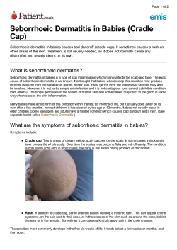
In infants and children, seborrheic dermatitis appears first as cradle
cap and later as dandruff in the scalp. The typical appearance in adults is
that of redness throughout the scalp and scalp line. In addition, scaling
over the eyebrows, nose, beard region, and chest can be seen.
When a moderate to severe amount of fine dry white scaling-commonly known as dandruff-is seen throughout the scalp (or on your navy
blue suit), some people interpret it as dry skin and cut back on washing.
That's not a good idea-by decreasing the frequency of hair washing, more
scale accumulates, which may cause further inflammation throughout the
scalp. Treatment therefore includes more frequent hair washing (daily or
every other day) with an anti-dandruff shampoo that contains selenium
sulfide or zinc. Regardless of which one you choose, the shampoo should
© Copyright 2000, David J. Leffell. MD. All rights reserved.
280
Com m 0 n Ski n Pro b I ems
be lathered up generously throughout the scalp and left on for five minutes before rinsing off. A non-greasy topical corticosteroid solution may
also be prescribed to apply throughout the scalp twice a day to combat the
itching.
For treatment of the red scaly areas on the face or chest, a low-potency
topical corticosteroid cream and an anti-yeast cream (Nizoral) to combat
pityrosporum ovale should help.
Since seborrheic dermatitis tends to be a chronic recurring process,
maintenance therapy with anti-dandruff shampoo and the other treatments mentioned may be necessary.
• PSORIASIS
Psoriasis is a relatively common inherited disease of the skin which is
characterized by overproliferation of the skin layers. While it often bears
the brunt of Madison Avenue glibness ("the heartbreak of psoriasis"), it
can indeed be a difficult problem for those who have it. It affects approximately 1 to 3 percent of the population. The exact cause of psoriasis is not
fully understood, but major advances in the study of this skin condition
have taken place in the last several years and it is becoming clearer that
inherited abnormalities in the immune function of the skin definitely play
a role.
Psoriasis has favorite locations where it likes to set up house, including the scalp, elbows, knees, and buttocks. A typical patch of psoriasis can
be a circle, an oval, or even an irregular shape; it is red (often brick red)
with overlying thick, silvery scales. When the scale is peeled off, one can
usually see tiny areas of bleeding, like pinpoints. On the buttocks, the
armpits, or the groin, psoriasis often appears as a red smooth lesion without much scale. Patches of chronic psoriasis tend to remain fixed in their
one position for months.
Guttate psoriasis is a common form of the condition that often erupts
following a streptococcal sore throat or a viral infection of the upper respiratory tract. It is a generalized eruption of many pinpoint to 1 centimeter pink-red papules with overlying scale.
Certain types of arthritis may coincide with the skin lesions of psoriasis. Factors known to provoke or exacerbate psoriasis are trauma to the
skin, infection such as strep throat, certain medications, low calcium levels,
and stress. Psoriasis may also be more prevalent in the HN-positive population (though of course having psoriasis does not mean you have AIDS).
© Copyright 2000, David J. Leffell. MD. All rights reserved.
Common Skin Conditions
281
Psoriasis is treated with topical creams, oral medication, and ultraviolet light therapy. The exact treatment depends largely on the type of psoriasis and the extent of cutaneous involvement. For limited psoriasis, your
dermatologist will probably prescribe a topical therapy such as corticosteroids, a topical vitamin D compound called calcipotriene (Dovonex), tar
preparations, a topical vitamin A derivative called tazarotene gel, or
anthralin. Should these topical therapies not work or if the extent of the
psoriasis is significant, your dermatologist may recommend an oral medication such as methotrexate, acitretin (a derivative of vitamin A), or
cyclosporine (a medication that affects the immune system). illtraviolet
light therapy may be suggested in tandem with oral psoralen, a compound,
which when taken by mouth and absorbed, interacts with the ultraviolet
light to reduce psoriasis patches.
Various specific treatments are also available when psoriasis has broken out on your scalp, including medicated shampoos containing tar or
salicylic acid, baby oil to put in your hair at night to loosen up the scale,
or topical corticosteroid solutions. Psoriasis tends to be a chronic condition-although it often responds to therapy, it nevertheless frequently
recurs. By rotating several of the treatments that have been outlined,
your dermatologist can help you achieve the best control of this skin
condition.
• PITYRIASIS ROSEA
Pityriasis rosea is a common skin condition that is usually seen in
young adults. The exact cause of this eruption is not known. Typically, its
first symptom is an isolated 1- to 3-inch, round-to-oval pink lesion with a
tiny central collar of scale. This isolated first patch, called the herald
patch, can arise anywhere but is most commonly seen on the chest or
upper arms and legs.
Several days or weeks after the onset of the herald patch, similar but
smaller lesions erupt over the entire trunk, arms, and legs, sometimes in
the pattern of a Christmas tree. (Typically, pityriasis rosea does not
involve the face). Most of these lesions do not cause any discomfort, but
sometimes there is a mild itching sensation.
This skin disease usually runs its course over a period of four to twelve
weeks. No specific treatment is necessary, but in cases with severe itching,
your dermatologist may recommend a topical corticosteroid or ultraviolet
therapy.
© Copyright 2000, David J. Leffell. MD. All rights reserved.
282
(
0
m m 0 n Ski n Pro b I ems
• DERMATOSIS PAPULOSA NIGRA
Dennatosis papulosa nigra is an entirely benign condition that many
black people experience. Multiple brown or black bumps, each no bigger
than a peppercorn or millet seed develop most often on the face and neck.
Under the microscope they look very much like seborrheic keratoses, the
growths I call "barnacles of life." The easiest way to treat this condition is to
gently scrape the papules off. Some physicians like to gently bum or freeze
them, but I prefer a technique that simply scrapes the bumps off at the level
of the epidennis-this minimizes hyperpigmentation, or worse, white spots.
• COMMON PIGMENTATION PROBLEMS
VITILIGO
Vitiligo is a relatively common disorder of pigment loss with great
social impact. People with the condition develop white patches on their
skin where the pigment-producing melanocytes have been destroyed.
Because of the resemblance of vitiligo to some fonns of leprosy, in certain
parts of the world it is confused with the ancient infectious disease. In
those situations, the social stigma historically associated with leprosy is
wrongly attached to patients with vitiligo.
Vitiligo is thought to be an autoimmune disease. Somehow the body
sets up a process whereby the immune system destroys the melanocytes.
In fact, because of the autoimmune nature of the disease, it is sometimes
seen in the skin of people with other diseases in which the immune system
attacks the body's own cells, such as thyroid disease, pernicious anemia,
and collagen-vascular diseases.
Vitiligo, which occurs in all populations but is more noticeable in people of color, usually starts suddenly with white patches on the skin. It
develops most commonly on the hands, feet, genitalia, and face. It also
appears on the cheeks, around the eyes and near the mouth. Vitiligo may
occur in one spot or one segment of the skin, such as on an arm or leg, or
it can be generalized, appearing over the whole body.
Vitiligo usually appears first in childhood. Itching can be an early
symptom-it's probably a sign that the body's immune cells are slugging it
out with the melanocytes. Because of the emotional toll this condition carries, it is especially frustrating to physicians that we cannot easily predict
or control its course.
© Copyright 2000, David J. Leffell. MD. All rights reserved.
Com m 0 n Ski nCo n d it ion s
283
Treatment has generally been unsatisfying and consists of the use of topical corticosteroids. Since tanning in the sun can stimulate pigment production, this is one of the few areas where dermatologists, under carefully
regulated circumstances, make use of ultraviolet radiation combined with an
agent called psoralen. Melanocytes can repopulate the vitiligo patch from pigment cells that survive in the hair follicles and from the adjacent normal skin.
Many people, frustrated by their condition, have tried tattooing, surgical treatments, and cosmetic covers. Because none of these approaches is
predictably successful, some people with vitiligo seek the help that comes
from attending support groups. When there is a 50 percent or greater pigment loss over the whole body, depigmentation may be recommended in
order to make the skin a uniform color. This is an irreversible step: if the
person doesn't like the result, there will be no means of changing back to
the original pigmentation. A topical medicine called monobenzone is used
daily modifying the treatment frequency as the pigment fades.
Surgical solutions to vitiligo have been tried and consist of grafting normally pigmented skin into the depigmented area. Punch grafts of normal
skin have been used but result in a confettilike appearance of pigmentation. Recently, I published a technique that I invented for the treatment of
Vitiligo that has proved simple, quick, easy to perform in the doctor's office,
and does not appear to result in the irregular pigmentation. I call it the
Flip-Top Pigment Procedure and an example of the results are shown in the
"Color Atlas of Your Skin."
HYPERPIGMENTATION AND HYPOPIGMENTATION
In people of color, the effects of trauma to the skin may become more
obvious than in more lightly pigmented individuals. Common causes of
post-inflammatory hyperpigmentation are acne, lacerations, eczema, and
even a special type of reaction to medication called fixed drug eruption. It
can develop in response to ampicillin, tetracycline, sulfa, or other medications. In this case a circular patch of jet black discoloration can develop in
dark-skinned people and persist for months, getting worse with each subsequent exposure to the medication. It is harmless, but until the cause is
known, and then aVOided, it will not resolve.
After skin injury, whether from an abrasion, rash, or other disruption
of the skin surface, post-inflammatory hyperpigmentation can develop.
When the trauma occurs in the epidermis, there is an increase in the transfer of melanin molecules to the surrounding epidermal cells. When the der© Copyright 2000, David J. Leffell. MD. All rights reserved.
284
Com m 0 n Ski n Pro b I ems
mis is also injured, pigment-containing scavenger cells, or melanophages,
set up house in the dermis for a long time, resulting in discoloration of the
skin that can last for years.
There is no perfect treatment for post-inflammatory hyperpigmentation. It will get better with time so patience is essential, but topical corticosteroid cream can help in some cases. Minimizing sun exposure is also
important. In my experience, laser treatment does not work, and may in
fact worsen the situation.
Hypopigmentation can occur for the same reasons as hyperpigmentation and similarly requires patience and time for improvement. Because
hypopigmentation just represents a decrease, rather than complete
absence of pigmentation, recovery can be expected as the pigment cells
from adjacent areas step up to the bat.
In order to determine the nature of your skin pigment problem your
dermatologist will likely examine you under a Wood's light-a black light
that helps determine the extent and depth of pigmentary change.
• DID MEDICINE CAUSE My RASH?
Three out of every thousand prescriptions in this country result in
some sort of allergic reaction. Although all medications come with an
extremely long list of potential side effects, it is important to identify a true
allergic reaction to medication, because taking the same medication again
can result in further problems. Similarly, if you do not actually have an
allergy to a medication but merely couldn't tolerate it in the doses given, you
need to keep that in mind should you need that medication in the future.
It is helpful to distinguish between the latter situation and a true
allergy to a drug. Fewer than 10 percent of adverse drug reactions are due
to a true allergy to the medication. In an allergy the body's immune system
responds to a foreign chemical, typically a protein, and such reaction will
happen every time the person takes the drug.
The most common types of drug reactions, however, are not allergies,
but intolerances. For instance, if erythromycin is taken on an empty stomach, it might cause nausea or vomiting. That is not an allergy, however, it
is an adverse reaction to the drug. Likewise, many women who take antibiotics find that they result in a yeast infection. This is also not an allergy,
but an adverse event due to a change in the bacteria that normally grow in
your gastrointestinal tract.
© Copyright 2000, David J. Leffell. MD. All rights reserved.
Com m 0 n Ski nCo n d it ion s
285
When a drug causes a reaction on the skin, it often will involve wide
areas of the skin. There will also be a correlation between when the drug
was taken and when the rash started.
About half of all drug reactions on the skin are called exanthems. An
exanthem is the splotchy type of flat red rash with clear areas. It may cover
most of the trunk, legs, and even face, but does not usually involve the
palms and soles. This kind of rash will typically begin within ten days of
starting a new medication, and some people develop a fever as well. The
most common drugs causing this type of reaction are antibiotics: ampicillin, amoxicillin, trimethoprimfsulfamethoxazole (Septra, Bactrim, CoTrimoxazole). The rash will typically go away on its own within one to two
weeks of stopping the medication. Scaling and peeling may follow after the
red rash fades.
Hives are also common, constituting about one-fourth of all drug reactions. When you get hives from drugs, it usually happens within thirty-six
hours after starting the medication. An individual spot of hives will last
fewer than twenty-four hours. Again antibiotics are the most common culprits, and 1 in 50 people taking amoxicillin and 1 in 100 people taking
either ampicillin or cefaclor will end up with hives. Upon discontinuing
these medications, the eruptions should cease within one to two weeks, if
not much sooner.
The sun can also cause a number of drug-related reactions. When a
medicine is absorbed by your body, it is distributed throughout the various
tissues, including the skin. When the skin is exposed to ultraviolet light
from the sun or even a tanning booth, certain itchy types of rashes can
occur in uncovered areas. Drugs that commonly cause these type of eruptions include sulfa drugs (including some water pills and diabetes pills),
nonsteroidal anti-inflammatory drugs (such as piroxicam), members of the
tetracycline family, and griseofulvin, a common antifungal medication.
Drug rashes occur in children, and, like those in adults, usually resolve
promptly. Very rarely more serious rashes develop in children and adults
in response to medication and require medical attention.
If you develop a drug rash while taking more than one medication, it
may be necessary to use the process of elimination to determine which
medication is causing the rash. This should be done only in close consultation with the doctor prescribing the medications. The watchword is to be
patient, but your dermatologist, internist, or family doctor will likely be
able to get to the bottom of your drug rash.
© Copyright 2000, David J. Leffell. MD. All rights reserved.
286
Com m 0 n Ski n Pro b I ems
• BIRTHMARKS
Birthmarks, which doctors call hemangiomas, are benign tumors of
blood vessels that appear on the newborn or soon after birth. Some birthmarks disappear on their own during childhood. The term birthmark is
also used for other skin lesions present at birth, but I have found that most
people mean hemangioma when they use the term. Before we take a look
at some of the most common types of birthmarks and treatment options,
let's clear up some myths about their cause.
Birthmarks don't have anything to do with what Mom ate during pregnancy, bad thoughts she might have had, or problems with delivery. These
growths sometimes seem to be stimulated by estrogens, which is why many
resolve over time following birth, as the estrogen levels in the child change
or as those estrogen receptors present on the birthmark itself change.
STRAWBERRY HEMANGIOMAS
A common type of birthmark, the strawberry hemangioma, occurs in
children, developing shortly after birth. A strawberry hemangioma typically starts as a small red bump and grows rapidly over two to three
months. Then its growth stops and a process called involution, when a
hemangioma shrinks in size, begins. In most cases it leaves little evidence
that it was ever there.
Strawberry hemangiomas are red or purple on the surface and are
raised above the surface of the skin. Sometimes the mass or lump under
the skin can be sizable. If it is near the neck or mouth it can interfere with
head movement and eating; near the eye, it can interfere with eyesight and
thus affect the infant's proper development of vision; near the nose, breathing can be affected. Any hemangioma near the mouth, in the mouth, or
near the nose is of special concern. Though it is rare, this can herald the
development of a similar growth in the throat so any child suspected of
having this problem should be evaluated by a pediatric ear, nose, and
throat specialist.
Parents are obviously concerned about the appearance of these lesions
on their children, which can make management of strawberry hemangiomas a bit controversial. On the one hand, a conservative approach is
called for. We know that the majority of hemangiomas of this sort go away
on their own. Ten percent go away by age one and 90 percent will have
vanished by age ten.
© Copyright 2000, David J. Leffell. MD. All rights reserved.
Com m 0 n Ski nCo n d it ion s
287
But what about those that don't go away or resolve too slowly? Most
parents and doctors would like the hemangioma to be gone by age four or
five, the time when the child is about to start school and make new
friends.
Many doctors are resistant to excising, or completely removing, these
growths, whether with traditional or laser surgery, because they can be
large and the resulting permanent scar may cause more cosmetic problem
than the original birthmark. In addition, incomplete excision of the
hemangioma can result in recurrence within scar tissue, which can
become more problematic. Most important, a hemangioma should not be
excised during the rapid growth phase.
If the hemangioma is in a vital location, treatment with corticosteroids-by mouth for a defined period such as a month or two, or even
by direct injection by a skilled physician-can slow or even reverse
growth. In rare cases where life is at risk, interferon, a naturally occurring
chemical that affects the immune system, can be used as well.
Whether to excise or not is a decision that should be made in consultation with experts who treat hemangiomas as a routine part of their
practice. Dermatologists, plastic surgeons, and ear, nose, and throat surgeons may all have expertise in this area and should consult closely with
the child's pediatrician. My advice is to be conservative when feasible;
when function is compromised, as when the birthmark is close to the
eyes, nose, or mouth, of course be aggressive. When the situation falls in
between, consider excision if the plastic surgeon believes the resulting
permanent scar will be superior to that which would result from natural
resolution.
EROSIONS
A common problem that does occur with hemangiomas is that during
the involution phase the surface skin may break down, causing a depressed
area or erosion. This can be painful for the child. Proper wound care can
help speed healing and eliminate the pain. Follow your doctor's instructions carefully; this will probably involve using an antibiotic cream such as
Bactroban and keeping the area covered with a nonstick dressing such as
Telfa. In more advanced cases, a special dressing called Vigilon, which is a
soothing gelatinlike material, can help a great deal (keep it refrigerated
between uses so it will have a cooling effect as well). Never use alcohol or
peroxide, which sting terribly and are not helpful; instead, tap water and
© Copyright 2000, David J. Leffell. MD. All rights reserved.
288
Com m 0 n Ski n Pro b I ems
gentle soap will do the trick. A topical anesthetic cream such as ELA-Max
may help control some of the pain that the child feels.
PORT WINE STAINS
Port wine stains, another common birthmark, are flat red or purple discolorations of the skin that is visible at birth. Some port wine stains can be
associated with a condition called Sturge-Weber syndrome; your pediatrician will know whether this possibility should be further investigated,
depending on the size and location.
When lasers first became available to treat these tumors, there was
much excitement about their potential to remove the entire hemangioma.
We now know that there are birthmarks of this type that can get 50 to 80
percent improvement, but complete eradication with current technology is
not always possible.
Each treatment course must be tailored to the child. For example, with
a very young child, parents must discuss the use of general anesthesia with
the dermatologist and pediatrician. Although this approach does allow the
dermatologist to be more complete in treating the birthmark than office
treatments done with topical anesthetic, there are minimal risks associated with anesthetizing a young child that must be taken into consideration. New lasers that cool the skin make it much easier to treat large areas
on children in the office setting.
By the time a person reaches adulthood, port wine stains have often
evolved from the original pink or red childhood mark to a purplish birthmark.
When lasers were first introduced, we thought that only pale birthmarks
responded to the treatment but, happily, this has not proven to be true. Medical insurance does not normally cover treatment for such birthmarks
because it is considered cosmetic.
Whatever approach you take with laser, remember that it is a gentle,
prolonged approach that slowly eliminates the growth under the surface of
the skin (see chapter 13).
STORK BITES
Stork bites on the back of the neck are a form of port wine stain that
do not resolve on their own but are not an issue for most people. You don't
see them every day and hair covers them. Similar lesions over the eyes, socalled angel's kiss, tend to resolve on their own.
© Copyright 2000, David J. Leffell. MD. All rights reserved.
Com m 0 n Ski nCo n d it ion s
•
289
FUNGUS OR CANCER:
THE STORY OF LYMPHOMA OF THE SKIN
A group of skin rashes that look something like early psoriasis or mild
eczema may actually be precursors to outright lymphoma, a form of cancer of the white blood cells. This serious condition is important to know
about because it may occur in areas that are not sun-exposed, and may be
mistaken for eczema or psoriasis. It can also develop as early as the teen
years. In general if such a rash does not go away with topical corticosteroid
medication, it should be biopsied by your dermatologist.
The disease can pass through several stages, including a flat or patch
stage, a stage with large, raised scaly areas, and a tumor stage in which
nodules are present on the skin. In a small number of people, the rashes
progress to involve the bloodstream and the lymph nodes.
The key player in what is called cutaneous T-cell lymphoma (CTCL)
is the T-cell type of white blood cell (the same type of cell that becomes
infected with HIV). When these special T-cells in the skin start growing out
of control, they can cause several different types of skin lesions.
In its earliest stages, CTCL can be treated with topical therapies
including super-potent steroids or topical nitrogen mustard. Photochemotherapy, or the use of an oral photosensitizing drug along with
ultraviolet A light (PUVA), is also a helpful therapy for such patients.
Dr. Richard Edelson, chairman of dermatology at Yale since 1985, pioneered the use of a clever therapy called photopheresis. In this treatment,
the patients ingest the same photosensitizing drug that would be used in
PUVA. The patients then have their blood filtered, as though on dialysis,
and about 10 percent of their white blood cells are removed. These white
blood cells are then exposed to the same ultraviolet A light that is used in
PUVA therapy for psoriasis. Finally, these cells are then injected back into
the patients. In some patients, this therapy can result in improvement of
the more severe forms of the disease.
An indication of how qUickly science progresses is the fact that even
this procedure, relatively new by conventional standards, is giving way to
more specific ways of manipulating the abnormal T-cells that are at the
root of the condition.
© Copyright 2000, David J. Leffell. MD. All rights reserved.


© Copyright 2026