
Molecular Basis for the Hormonal Regulation of the
Molecular Basis for the Hormonal
Regulation of the Tyrosine
Aminotransferase and Tryptophan
Oxygenase Genes
GÜNTHER SCHÜTZ, WOLFGANG SCHMID,
MICHAEL JANTZEN, ULRICH DANESCH, BERND
GLOSS, UWE STRÄHLE, PETER BECKER, AND
MICHAEL BOSHART
Institute 0/ Cell and Tumor Bi%gy
German Cancer Research Center
D-6900 Heide/berg, Federal Republic 0/ Germany
INTRODUCTION
A classical example for hormonal control of gene expression is the induction of
the enzymes tyrosine aminotransferase (TAT) and tryptophan oxygenase (TO).1-3
These two enzymes are expressed exclusively in the parenchymal cells of the liver.4
The expression ofboth genes is affected by glucocorticoids.s- lO In nuclear run-on assays
it could be shown that hormonal induction involves changes in the relative rates of
transcription of these two genes. Apart from glucocorticoid induction, the activity of
the TAT gene is also regulated by cAMP."- 14 The accumulation ofTAT mRNA after
cAMP stimulation is a consequence of transcriptional activation of the TAT gene. IS
Combined dexamethasone and cAMP treatment leads to higher TAT mRNA concentrations than treatment with either inducer alone, which implies that dexamethasone and cAMP act by distinct mechanisms. IS
In addition to hormonal regulation, both TAT and TO genes are under developmental control. Whereas TAT enzyme activity appears around birth, TO enzyme
activity can be detected only 2 weeks later. 16 The activity of the TAT gene is affected
by two distinct genetic loci, which appear to operate in trans on the expression of the
TAT gene. By genetic and biochemical analysis of several albino lethai mutants of
the mouse, a control region required for expression and inducibility of several liver
enzymes including TAT has been assigned to the region of the albino locus on
chromosome 7 of the mouse. l7 The basal level of TAT mRNA is severely decreased,
and the concentration of the mRNA is no longer inducible by glucocorticoids and
cAMP, aIthough the structural gene for TAT is still present. 18 Because the mouse
TAT structural gene is located on chromosome 8,'9 the effect of this presumptive
regulatory locus must operate in trans.
Another locus, the so-called tissue-specific extinguisher-l (Tse-l) has been mapped
to chromosome 11 of the mouse. 20 When chromosome 11 of a fibroblast cell is
93
ANNALS NEW YORK ACADEMY OF SCIENCES
94
introduced by microcell fusion into a hepatoma cell expressing TAT, TAT gene activity
is selectively extinguished, suggesting that a negatively acting factor encoded in chromosome 11 is responsible for this effect.
To study the complex control mechanism operating on these two liver-specific
the TAT and TO genes from the rat
genes at the molecular level, we have isolated
,
as well as the TAT gene of the mouse. ·•· IO•l • The availability of these DNA clones
have allowed us to determine the level at which hormonal regulation of expression
of these two genes occurs and, furthermore, anowed us to identify those sequences
that are required for hormonal induction in cis. We demonstrate here that these sites
represent binding sites for the glucocorticoid receptor.
2
1
5
4
3
6
8 9 10 11
7
TAT
A
BC
F
4
3
1 2
TO
E
D
G
H
I J
K
C
D
8
6
E
G
Size of
gene
(kb)
(kb)
2.4
11.0
20
19.0
L
10
11
[}-ß
AB
Srze of
mRNA
H
K
>---t
lkb
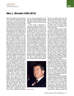
FIGURE 1. Exon-intron structure of the rat TAT and TO genes. Open boxes represent the
exons; connecting lines represent the intervening sequences, and are drawn to scale. The size of
the mRNA and the size of the gene are indicated in kb pairs.
RESULTS
To allow analysis at the molecular level of the sequences important for the hormonal regulation of expression of the TO and TAT genes, we have cloned cDNA
sequences representing these two mRNAs·· 1O as wen as genomic DNA containing
these two genes. " l9 The exon-intron organization and the start site of transcription of
these two genes was determined by electron microscopic analysis of heteroduplexes
,
of mRNA with the cloned genomic DNA, as wen as by SI nuclease mapping. ,10
FIGURE 1 shows the exon-intron organization of these two genes. The coding portion
of these genes is interrupted by multiple intervening sequences; thus the genes are
much longer than the respective mRNAs. In order to allow studies of the molecular
effects of the albino deletion mutations, we have isolated and characterized the mouse
TAT gene and have shown that the rat and mouse TAT genes are very similar in
exon-intron structure. Sequence comparison around the 5' end of the rat and mouse
genes shows extensive homology over the entire sequence (approximate1y 1 kb), indicating the importance of these sequences for the regulation of these two genes. ' •
In order to obtain clues to possible regulatory sequences, we have analyzed the
chromatin structure in the vicinity of the 5' end of the TO and TAT genes with regard
to DNase I hypersensitivity. Hypersensitive sites frequently map near the 5/ end of
actively transcribed genes. Their presence has been correlated in many systems with
the state of expression. We have found DNase I hypersensitive sites at each of the
two promoters in liver cens of the rat'2 and mouse (FIG. 2). DNase I hypersensitive
SCHÜTZ et al.: MOLECULAR BASIS FOR HORMONAL REGULATION
~~
-dex
.2
.4
95
.65
.2
.4
.5 ONase I u/Jl9
Hili
FlGURE 2. A glucocorticoid-inducible DNase
I hypersensitive site rar upstream of the mouse
TAT gene. Nuclei from hormone-induced (+
dex) and uninduced (- dex) mouse livers were
treated with increasing amounts of DNase 1.
After purification, the DNAs were cut with the
restriction enzyme abutting with the probe used,
separated on a 1% agarose gel, and biotted to
nitrocellulose filters. The end-Iabeling technique
was used for visualizing tbe preferential DNase
I c1eavage sites. On the right part of the figure
these hypersensitive sites are aligned with the 5'
ftanking region of the TAT gene.
sites of both genes are absent in kidney nuclei and therefore appear to be specific for
the tissue expressing the genes. 22 Most remarkable is the alteration in chromatin
structure following glucocorticoid induction (FIG. 2). A strong DNase I hypersensitive
site is detected about 2.4 kb upstream of the start site of transcription of the mouse
TAT gene. This dexamethasone-dependent hypersensitive site suggested to us the
possibility that glucocorticoid response elements might be located far upstream of the
start site of transcription.
To identify sequences important for the hormonal control of these two genes by
glucocorticoids, fusion genes were constructed containing the 5' fianking DNA of
these genes upstream of a suitable indicator gene. We used either the bacterial gene
for neomycin resistance (neo) or the gene for the enzyme chloramphenicol transacetylase (CAT). A typical hybrid gene is shown in FlGURE 3. Because chromatin
studies have suggested that the control sequence might be remote from the cap-site22
(FlG. 2), 2.9 kb of 5' fianking DNA was included in the construction of this hybrid
gene. In the case of the TO promoter, a CAT fusion gene containing 1.9 kb of 5'
flan king DNA was constructed. These fusion genes were introduced into hepatoma
ceHs and fibroblasts, and the effect of steroid administration on expression of these
genes was analyzed by CAT enzyme activity measurements or analysis of the RNA
with RN A filter hybridization experiments and/or with SI nuclease analysis. Both
FIGURE 3. Structure of pTATneo, a fusion gene of the
TAT 5' ftanking DNA and the bacterial neomycin resistance
gene.
ANNALS NEW YORK ACADEMY OF SCIENCES
96
TO - ONA
-eoCI
EXTENIlIHG TO
-3M
+
+
+
FIGURE 4. Expression of TO-CAT recombinants in mouse L cells. TO-CAT recombinants
containing approximately 1930 bp, 600 bp, and 340 bp of 5' ftanking sequences were transferred
into mouse L cells. Expression in the presence and absence of dexamethasone was followed by
measuring CAT activity in extracts of the cells 48 hr after transfection.
genes, when introduced into heterologous or homologous cells, are expressed, and the
expression is regulated by glucocorticoids (data not shown). In order to delineate the
important regulatory elements in more detail, deletion mutants of these parental
plasmids were constructed and tested. A typical experiment is shown in FIGURE 4
for the TO-CAT recombinants expressed in mouse L cells. It is seen that as long as
1930 nuc1eotides remain of 5' ftanking sequences of the TO fusion gene, strong inTAT
-------.-
CAT
DEX INDUCTION
OF CAT ACTIVITY
-2561
I
I
I
-2474
I
I
I
RECEPTOR
BINDING
17.9 x
+
24.4 x
+
1.7x
-
1.9x
N.D.
2.0x
-
I
I
I
I
1
I
I
I
-2424
I
FIGURE 5. A remote DNA sequence confers glucocorticoid responsiveness and receptor binding. The upper part of the figure shows the 5' ftanking part of the TAT gene fused onto the
CAT gene. Deletion derivatives as indicated by the deletion end points were tested for dexamethasone inducibility of expression of the CAT recombinants in L cells 12 hr after hormone
treatment as weil as for receptor binding. The approxirnate position of the hexanucJeotide
(TGTTCT) indicative of glucocorticoid receptor binding sites is shown.
SCHÜTZ et al.: MOLECULAR BASIS FOR HORMONAL REGULATION
97
pTAT·CAT
l
Bam HI
l
l
SmaI
LABELLING
?
,/ GRE
FILTER BINDING
BamHI
l
I
PstI<
"
,,~,
PsII
l
7-------~-----------:
GEL ELECTROPHORESIS
roo~rt-~
__----~----------4---
__----------2561
50%
-1890
-1300
;0
250
500
1000
1200
ngCTDNA
FIGURE 6. Binding ofthe glucocorticoid receptor to 5' deletions ofthe TAT-CAT recombinant.
The upper part of the figure shows the method employed to determine binding of the purified
glucocorticoid receptor to 5' deletion derivatives of the TAT-CAT plasmids. The lower part of
the figure shows the results of a filter binding experiment with three plasmids containing 5'
ftanking TAT DNA as indicated in the presence of increasing amounts of competing calf thymus
DNA (CT DNA). The inset shows the autoradiogram of the fragments preferentially retained
on nitrocellulose filters. The top set of fragments represents the deletion ending at - 2561; the
middle set, the deletion ending at -1890; and the lower set, the deletion ending at -1300. For
quantitation the bands were cut out and counted. The data are plotted as percentages of binding
of the deletion ending at -2561.
ANNALS NEW YORK ACADEMY OF SCIENCES
98
duction of expression by glucocorticoids can be achieved. Deletion of 5' flanking
sequences up to about -500 resulted in considerable loss of inducibility, even though
some induction of the TO-CAT recombinant can still be observed. Further deletion
to -314 abolished all inducibility. These findings suggest the presence oftwo hormoneresponsive elements in the 5' flanking region of the TO gene. These results were further
confirmed by more finely spaced deletion mutants.
A similar series of experiments was conducted with the TAT fusion genes, which
were introduced in rat hepatoma cells and mouse fibroblasts. In mouse L cells the
TAT 5' flanking sequence of 2.95 kb conferred strong inducibility of expression.
Expression studies of 5' deletion derivatives of the parental TAT-CAT recombinant
-265u
G G Tel, G ,~ G C C T C T G GAG G Ace C C T G A A G T [ T [ T T [ T C A GfTGTTCil[ TAT [
[ [ A G T [ T [ G GAG A [ [ T [ [ T G G G G A [ T ] [ A G I, G A AGA 5 T [~G A TAG
A [ A G G ~ AGA G [ T G T LAG [ [ C C T G G I, ArG T G G i T [ T I, ] G T [ TAG ,\ A A ~, [ T {"
T G T l [ [ T [ T [ G " C A G T C G G G G " [ [ 1 ; A [ A [ [ I (, G A TAL A G f.. 1 [ 1 1 T 1 G A 1
-25:0
-25CO
.
,
1 C [ [ i, 1 A A ,\ 1 A ,\ [ A G G A A G [ C C ii " S G ; 1 1 I [ [ A A ] [ T [ T G [ 1 G T A C A G G A
A S G ,',lA 1 1 rAT T G T [ [ ] ] [ G G G T T [ C I, A A ] G G T 1 I, G J' G A C G A C A T G Tee T
. . ..
,
,
r.
--..-:;.,..""''''',
'
a::..:~=.o:I'
C G A T G A A A ] A A A C G ] TAT C ] ] T ] AGA C ] ] ] [ A A A G G G G ] A C A G
t
G eTA C T T TA] ] ] G C A I, TAG A ~ A A ] [ T G I, J\ i, G T ] 1 [ C C [ A T G T C
T~
-2450
CA AC AA AC
A C A A : Tee T G C G ] AG] [ G C [ 1 G T C G G ] T T C T G G
G T T G T T C T G AIu..rll.IJr T G T T C A G G A [ G ( A T [ A G C G G A [ A G [ [ A A AGA C [
-2400
G T G T G G ] G G TA] A G [ [ C 1 G ] A A ] C C [ A G [ A ] ] ] G G G A A G [ ] GAG G T G G G A
[ A [ I, [ [ A [ [ A TAl [ G G G A [ A ] ] A G G G 1 [ G ] A A A [ [ [ ] ] [ G AC] C [ A [ [ [ T
-23;0
G G A T [ G G GAG] ] [ A A G G ] [ A G [ ] ] G G G [ T A [ T ] AGA A AGA [ [ 1 ] G r [ ] [ A
[[] AG C [[] [AA G 1 T[[ AG T [G AA[[ [G A] G AAT [ ] ] ] C] GGAA[A G A Gl
FIGURE 7. Sequence ofthe far 5' flanking DNA ofthe TAT gene. The DNA sequence between
- 2650 and - 2200 of the TAT gene is presented. The three hexanuc1eotides TGTTCT characteristic
of receptor binding sites are shown in boxes. The regions protected from DNase I digestion after
binding of the glucocorticoid receptor in vitro are shown in boldface type. DNase I hypersensitive
regions are shown with dots. Protected and enhanced guanine residues as evidenced from dirn ethyl
sulfate proteetion experiments are shown with upward- and downward-pointing arrows, respectively. The DNase land dirnethyl sulfate protection experiments have been perforrned with the
upper strand only.
defined important control elements in the region that showed a local alteration in
chromatin structure in the previous DNase I hypersensitivity studies (FIG. 5). Inspection of the DNA sequence within this region revealed sequences homologous to
the glucocorticoid receptor binding sites of the mouse mammary tumor virus,23.24
methallothionin,2' and lysozyme 26•27 genes.
To determine whether this control element located so far upstream of the initiation
site of transcription functions as a glucocorticoid receptor binding site, filter binding
studies with the purified glucocorticoid receptor and the 5' deletion mutants were
performed. Binding of the glucocorticoid receptor to TAT 5' deletion derivatives was
investigated as shown in the upper panel of FIGURE 6. The lower panel shows that
SCHÜTZ et al.: MOLECULAR BASIS FOR HORMONAL REGULATION
the purified receptor interacts preferentially with the deletion mutant that contains
the control sequence identified in vivo, but not with derivatives ending at -1890 or
-1300. The DNA filter binding studies were complemented by DNase I footprint
analyses as weIl as by dimethyl sulfate protection experiments (data not shown). Two
footprinting regions could be identified in the far upstream sequence of the T AT gene
using a purified glucocorticoid receptor. These protected regions are indicated by the
shaded sequence in FIGURE 7. Dimethyl sulfate protection experiments indicate contact
sites of the receptor. Protections and enhancements indicated by the arrows in FIGURE
7 are seen in the sequences defined by the DNase I footprints. These nuclease protection
experiments identify partially homologous receptor binding sites located in the region
that confer glucocorticoid responsiveness in vivo.
CONCLUSIONS
Induction of enzyme activity for TO and TAT, two gluconeogenic enzymes, results
from increased expression of the respective genes at the transcriptional level. The
effect of the hormone on transcription is rapid and does not require new protein
synthesis, suggesting a direct effect of the hormone receptor complex on the activity
of the gene. In order to identify sequences important for regulation, fusion genes
containing the presumptive control sequences were constructed and their expression
studied under the influence of steroid hormones. This has allowed identification of
two control sequences in the case of the TO gene located around -420 and around
-1190 and control elements located at about -2500 bp upstream of the TAT initiation
site. The responsible sequences show strong sequence homology to each other as well
as to previously characterized glucocorticoid control elements?3-27 What role these
hormonal control elements play in the course of deve10pmental activation of these
genes will now be tested by analysis of expression of these genes in transgenie animals.
REFERENCES
1.
2.
3.
4.
5.
6.
7.
8.
9.
10.
11.
12.
F'EIGELSON, P. & O. GREENGARD. 1962. J. Bio!. ehern. 237: 3714-3717.
SCHIMKE, K. T., E. A. SWEENEY & C. M. BERLIN. 1965. J. Bio!. Chern. 240: 322-331.
KENNEY, F. T. 1962. J. Bio!. Chern. 237: 3495-3498.
HARGROVE, J. L. & R. B. MACKIN. 1984. J. Bio!. Chern. 259: 386-393.
BAXTER, J. D. & G. G. ROUSSEAU, Eds. 1979. G1ueoeortieoid Hormone Action. Springer
Verlag. Berlin.
SCHÜTZ, G., L. KILLEWICH, G. CHEN & P. FEIGELSON. 1975. Proc. Nat!. Aead. Sei. USA
72: 1017-1020.
SHiNOMIYA, T., G. SCHERER, W. SCHMID, H.-W. ZENTGRAf & G. SCHÜTZ. 1984. Proe.
Nat!. Aead. Sei. USA 81: 1346-1350.
DANESCH, U., S. HASHIMOTO, R. RENKAWITZ & G. SCHÜTZ. 1983. J. Bio!. Chern. 258:
4750-4753.
SCHERER, G., W. SCHMID, C. M. STRANGE, W. RÖWEKAMP & G. SCHÜTZ. 1982. Proc.
Natl. Aead. Sei. USA 79: 7205-7208.
SCHMID, W., G. SCHERER, U. DANESCH, H.-W. ZENTGRAF, P. MATTHIAS, C. M. STRANGE,
W. RÖWEKAMP & G. SCHÜTZ. 1982. EMBO J. 1: 1287-1293.
WICKS, W. D. 1974. Adv. Cyclie Nucleotide Res. 4: 335-438.
ERNEST, M. J. & P. FEIGELSON. 1978. J. Bio!. Chern. 253: 319-322.
100
ANNALS NEW YORK ACADEMY OF SCIENCES
NOGUCHI, T., M. DIESTERHAFf & D. GRANNER. 1978. J. Bio!. Chern. 253: 1332-1335.
CULPEPPER, J. A. & A. Y.-c. LJU. 1983. J. Bio!. Chern. 258: 13812-13819.
HASHIMOTO, S., W. SCHMID & G. SCHÜTZ. 1984. Proe. Nat!. Acad. Sei. USA 81:
6637-664l.
16. GREENGARD, O. 1970. In Biochernieal Aetions of Hormones. G. Litwaek, Ed. Vo!. 1:
53-87. Aeadernie Press. New York, NY.
17. GLUECKSOHN-WAELSCH, S. 1979. Ce1l16: 225-237.
18. SCHMID, W., G. MÜLLER, G. SCHÜTZ & S. GLUECKSOHN-WAELSCH. 1985. Proc. Natl.
Aead. Sei. USA 82: 2866-2869.
19. MÜLLER, G., G. SCHERER, H.-W. ZENTGRAF, S. RUPPERT, B. HERRMANN, H. LEHRACH
& G. SCHÜTZ. 1985. J. Mol. Bio!. 184: 367-373.
20. KILLARY, A. M. & R. E. K. FOURNIER. 1984. Cell 38: 523-534.
21. ELGlN, S. C. R. 1981. Cell 27: 413-415.
22. BECKER, P., R. RENKAWITZ & G. SCHÜTZ. 1984. EMBO J. 3: 2015-2020.
23. PAYVAR, F., D. DEFRANCO, G. L. FIRESTONE, B. EDGAR, Ö. WRANGE, S. OKRET,
J.-A. GUSTAFSSON & K. R. YAMAMOTO. 1983. Ce1l35: 381-392.
24. SCHEIDEREIT, E., S. GEISSE, H. M. WESTPHAL & M. BEATO. 1983. Nature 304: 749-752.
25. KARIN, M., A. HASLINGER, H. HOLTGREVE, R. I. R!CHARDS, P. KRAUTER, H. M.
WESTPHAL & M. BEATO. 1984. Nature 308: 513-519.
26. VAN DER AHE, D., S. JANICH, C. SCHEIDEREIT, R. RENKAWITZ, G. SCHÜTZ & M. BEATO.
1985. Nature 313: 706-709.
27. RENKAWITZ, R., G. SCHÜTZ, D. VAN DER AHE & M. BEATO. 1984. Cell 37: 503-520.
13.
14.
15.


© Copyright 2026