
book here
The Cell
Structure and Function
Objectives
By the end of this exercise you should be able to:
1. Understand the differences between prokaryotes
and eukaryotes and identify structures characteris~
tic of each.
2. Prepare a wet mount to view cells with a com~ pound microscope. 3. Understand the function of organelles visible with
a light microscope .
4. Examine a cell's structure and determine whether
it is from a plant, animal, or protist.
ells are considered the basic unit of living organisms be~ cause they perform all of the processes we collectively
call "life." All organisms are made of cells. Although most
individual cells are visible only with the aid of a microscope,
some may be a meter long (e.g., nerve cells) or as large as a
small orange (e .g., the yolk of an ostrich egg). Despite these
differences, all cells are designed similarly and share funda~
mental features.
C
Cytology is the study of cellular structure and func~
tion. The major tools of cytologists are light microscopy,
electron microscopy, and cell chemistry. By studying the
anatomy of a cell, we can find clues to how the cell works.
To understand the life processes of organisms, in to~
day's lab you will study some of the features and variations
among living cells. Prior to this exercise, review in your
textbook the general features of cellular structure and
function.
PROKARYOTIC CELLS
Bacteria and cyanobacteria are prokaryotes (fig. 4.1), and
their diversity is considerable (> 5000 species). Prokaryotes
do not contain a membrane~bound nucleus or any other
membrane~bound organelles. Organelles are organized
structures of macromolecules having a specialized function
and are suspended in the cytoplasm. The cytoplasm of
prokaryotes is enclosed in a plasma membrane (cellular
membrane) and is surrounded by a supporting cell wall
covered by a gelatinous capsule. Flagella and hairlike
Ribosomes
\
Flagellum
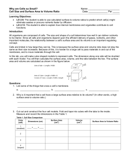
Figure 4.1
The structure of a bacterial ce ll. Bacteria lack a nuclear membrane. All prokaryotic (bacte ri a l) cells have a nucleoid regi on, ribosomes, plasma
membrane, cytoplasm, and cell wall, but not a ll have flagella (lSOO x).
4-1
33
outgrowths called pili are common in prokaryotes; flagella
are used for movement, and pili are used to attach some
types of bacteria to surfaces or to exchange genetic material
with other bacteria. Within the cytoplasm of prokaryotes
are ribosomes (small particles involved in protein synthesis)
and nucleoid regions (concentrations of DN A). Prokary~
otes do not reproduce sexually, but they have mechanisms
for genetic recombination (see Exercise 16).
Procedure 4. 1
Examine cyanobacteria
1. Examine a prepared slide of Oscillatoria, a filament of
cells, and one of Gloeocapsa, a loosely arranged
colony (fig 4.3). Review Exercise 3 for the correct
steps to use the microscope.
2. Focus with the low~power objective.
high~power objective into place to see
filaments and masses of cells.
3. Rotate the
Cyanobacteria
The largest prokaryotes are cyanobacteria, also called blue~
green algae. They contain chlorophyll a and accessory pig~
ments for photosynthesis, but these pigments are not
contained in membrane~bound chloroplasts. Instead, the
pigments are held in photosynthetic membranes called
thylakoids (fig. 4.2). Cyanobacteria are often surrounded by
a mucilaginous sheath. Their ability to photosynthesize
made them the primary contributors to the early oxygena~
tion of the ancient earth's atmosphere.
4. Prepare a wet mount of Oscillatoria and one of
Gloeocapsa. Review procedure 3.5 in Exercise 3 for
preparing a wet mount.
5. Observe the cellular structures and draw the cellular
shapes and relative sizes of Oscillatoria and Gloeocapsa
in the following space. Use an ocular micrometer to
measure their dimensions.
Gloeocapsa
Oscillatoria
SAFETY FIRST Before coming to lab, you were
asked to read this exercise so you would know
what to do and be aware of safety issues. In the
space below, briefly list the safety issues associated
with today's procedures. If you have questions
about these issues, contact your laboratory assis
Filament of cells
tant before starting work.
I.~ .
- - + - - Mucilagenous
sheath
. . ....,.....-ilr--
Vegetative cells
PhotosynthetiC membranes
Figure 4.2
(b)
Electron micrograph of a photosynthetic bacterial cell, Prochloron,
showing extensively folded photosynthetic membranes. The DNA is
in the clear area in the central region of the cell; it is not membrane
bound (5200 X).
Common cyanobacteria. (a) Oscillatoria (40x).
(b) Gloeocapsa (400x).
34
EXERC ISE
4
Figure 4.3
4-2
Question 1
a. Where are the pigments located in these
cyano bac teria?
h. Are nuclei visible in cyanobacterial cells?
c. Which of these two genera has the most prominent
mucilaginous sheath?
d. How many cells are held within one sheath of
Gloeocapsa?
Bacteria
of the cell and is contained by the plasma membrane. Within
the cytoplasm are a variety of organelles. Chloroplasts are el~
liptical green organelles in plant cells. Chloroplasts are the
site of photosynthesis in plant cells and are green because
they contain chlorophyll, a photosynthetic pigment capable
of capturing light energy. Mitochondria are organelles found
in plant and animal cells. These organelles are where aerobic
respiration occurs. When viewed with a conventional light
microscope, mitochondria are small, dark, and often difficult
to see. All of the material and organelles contained by the
plasma membrane are collectively called the protoplast.
Eukaryotic cells are structurally more complex than
prokaryotic cells. Although some features of prokaryotic
cells are in eukaryotic cells (e.g., ribosomes, cell membrane),
eukaryotic cells also contain several organelles not found in
prokaryotic cells (table 4.1). In the following exercise you
will investigate some of these organelles.
Most bacteria are much smaller than cyanobacteria and do
not contain chlorophyll. Yogurt is a nutrient~rich culture of
bacteria. The bacterial cells composing most of the yogurt
are Lactobacillus, a bacterium adapted to live on milk sugar
(lactose). Lactobacillus converts milk to yogurt. Yogurt is
acidic and keeps longer than milk. Historically, Lactobacillus
has been used in many parts of the world by peoples defi~
cient in lactase, an enzyme that breaks down lactose. Many
Middle Eastern and African cultures use the more digestible
yogurt in their diets instead of milk.
Procedure 4.3
Examine living Elodea cells and chloroplasts
1. Remove a young leaf from the tip of a sprig of Elodea.
Elodea is a common pond weed used frequently in
studies of photosynthesis, cellular structure, and
cytoplasmic streaming.
Procedure 4.2
2. Place this leaf, with the top surface facing up, in a
drop of water on a microscope slide. The cells on the
upper surface are larger and more easily examined.
Add a coverslip, but do not let the leaf dry. Add
another drop of water if necessary.
3. Examine the leaf with your microscope. Review
Exercise 3. First use low, then high, magnification to
bring the upper layer of cells into focus (fig. 4.6). Each
of the small, regularly shaped units you see are cells
surrounded by cell walls made primarily of cellulose
(fig. 4.7). Cellulose is a complex carbohydrate made of
glucose molecules attached end~to~end. The plasma
membrane lies just inside the cell wall. Sketch what
you see.
PLANT CELLS
Examine bacteria
1. Place a tiny dab of yogurt on a microscope slide.
2. Mix this small amount of yogurt in a drop of water,
add a coverslip, and examine the yogurt with a
compound microscope. Review Exercise 3.
3. Focus with the low~power objective.
4. Rotate the high~power objective (40X) into place to
see masses of rod~shaped cells.
5. Observe the simple, external structure of the bacteria
and draw their cellular shapes in the following space:
Question 3
a. What three~dimensional shape are Elodea cells?
Question 2
How does the size of Lactobacillus compare with that of
Oscillatoria and Gloeocapsa?
h. Examine various layers of cells by focusing up and
down through the layers. About how many cells
thick is the leaf that you are observing?
c.
What are the functions of the cell wall?
EUKARYOTIC CELLS
Eukaryotic cells contain membrane~bound nuclei and other
organelles (figs. 4.4, 4.5). Nuclei contain genetic material of
a cell and control metabolism. Cytoplasm forms the matrix
4-3
The Cell
35
Plasma membrane o;.:~..::.;.=:....--r=r.o=~'=:.i~------1r-
Nuclear
membrane
~t'ttllki1~ffi~~~~---;- Golgi
complex
Nucleolus
•,.---=-....,.-..:.....~~=.::=~"'"'rri-- Mitochondrion
Golgi complex Plasma
membrane
Cytoskeleton
Lysosome
Nuclear envelope Nucleolus
Mitochondrion
Rough
endoplasmic
reticulum
Cytoplasm
Figure 4.4
Structure of animal cells. Most organelles of animal cells are not visible with common light microscopes- therefore, our understanding of cellular
structures is based mainly on research using electron micrographs, as shown with the upper photo and diagram. Cells are surrounded by a bilayered
plasma membrane containing phospholipids and proteins . The nucleus houses chromosomal DNA and is surrounded by a double-membraned
nuclear envelope. Centrioles organize spindle fibers during cell division. Endoplasmic reticulum (ER) is a system of membranes inside the cell.
Rough ER has many ribosomes, and smooth ER has fewer ribosomes. Mitochrondria are sites of oxidative respiration and ATP synthesis. Microvilli
are cytoplasmic projections that increase the surface area of some specialized animal cells. Golgi complexes are flat sacs and vesicles that collect
and package substances made in the cell. Ribosomes are aggregations of proteins that conduct protein synthesis. Lysosomes contain enzymes
important in recycling cellu lar debris (17,500 x ).
36
EXERCISE
4
4-4
It
e
e
e
e
e
Vacuole
Nuclear envelope
Chloroplasts
Nucleolus
It
Cell wall
e
e
e
e
Nucleus
Ribosomes
Intercellular space
Plasmodesma
I:
Endoplasmic reticulum
Mitochond ri a
Chloroplast
containing starch
Cell wall Plasma membrane Chloroplasts
Central --+-+--=--1---
vacuole
Plasmo
desmata
-j-7'---=-+---+-+-I---
-+-.- Lysosome
Ribosomes
Golgi apparatus Nucleus
Nucleolus
Nuclear
envelope
Rough
endoplasmic
reticulum
Cytoplasm
Figure 4.5
Structure of plant cells. This illustration shows relative proportions of the different parts of a plant cell. Most mature plant cells contain large
central vacuo les, which occupy most of the volume of the cell. Cytoplasm is often a thin layer between the vacuole and the plasma membrane.
Cytoplasm contains the cell's organelles (17,500X).
4-5
The Ce ll
37
TARL E
S
4.1
ME OF THE MAJOR DIFFERENCE S RETWEEN PROKARYOTIC AND EUKARYOTIC CELLS AND BETWE EN
PLANT AND ANIMAL C~ELLS
Eukaryote
Prokaryote
Animal
Plant
Cell wall
Present (protein-polysaccharide)
Absent
Present (cellulose)
Cell membrane
Present
Present
Present
Flagella
May be present (single strand)
May be present
Absent except in sperm of a few species
Absent
Usually present
Usually present Ribosomes
Present
Present
Present Microtubules
Absent
Present
Present Centrioles
Absent
Present
Absent Golgi complex
Absent
Present
Present Nucleus
Absent
Present
Present
Mitochondria
Absent
Present
Present
Present
EXTERIOR STRUCTURES
INTERIOR STRUCTURES
ER
OTHER ORGANELLES
Chlmoplasts
Absent
Absent
Chromosomes
A single circle of naked DNA
Multiple; DNA-protein complex
Multiple; DNA-protein complex
Vacuoles
Absent
Absent or small
Usually a large single vacuole
Figure 4.6
(a) Elodea cells showing abundant
chloroplasts (400X) . (b) The cellular
structure of Elodea (lSO X ).
(a)
Central
Middle
lamella
38
EXERCISE
4
4-6
e
e
e
e
e
e.
It Microfibril-
'.
e
......... e
ttl
e.
e
e
e
tt l
Figure 4.7
Cellulose is the most abundant organic compound o n earth and is a polymer of glucose molecules. Free hydroxyl (OH - ) groups of the glucose
molecules form hydrogen bonds between adjacent cellulose molecules to form cohesive microfibrils. Microfibrils align to form strong cellulose
fibers that resist metabolic breakdown. Because humans cannot hydrolyze the bonds between glucose molecules of cellulose, cellulose is
indigestible and its energy is unavailable. Cellulose passes through the human digestive tract as bulk fiber (20x).
d.
Use an ocular micrometer or refer to the dimensions
of the field of view calculated in Exercise 3 to
measure the dimensions of an Elodea cell. What are
the cell's approximate dimensions?
4.
Chloroplasts appear as moderately sized green
spheres within the cells (figs. 4.6, 4.8). Locate and
sketch cells having many chloroplasts; estimate the
number of chloroplasts in a healthy cell. Remember
that a cell is three-dimensional, and some
chloroplasts may obscure others.
e
e
e
Cellulose chain
. ..... ....... . I
as a faint gray sphere the size of a chloroplast or
larger. Staining the cells with a drop of iodine may
enhance the nucleus. If your preparation is
particularly good, a nucleolus may be visible as a
dense spot in the nucleus.
7. 8. Warm the slide with intense light for about 10 min
and search for movement of the chloroplasts. You
may need to search many cells or make a new
preparation. This movement is called cytoplasmic
streaming, or cyclosis. Chloroplasts are not motile;
instead, they are being moved by the activity of the
cytoplasm. Add water if the cells appear to be
drying out.
Question 4
a. What shape are the chloroplasts? What is their
function?
b. Where are the chloroplasts located within the Elodea
cell-toward the perimeter or centrally located?
5. Determine the spatial distribution of chloroplasts
within a cell. They may be pushed against the
margins of the cell by the large central vacuole
containing mostly water and bounded by a vacuolar
membrane. The vacuole occupies about 90% of the
volume of a mature cell. Its many functions include
storage of organic and inorganic molecules, ions,
water, enzymes, and waste products.
6. Search for a nucleus; it mayor may not be readily
visible. Nuclei usually are appressed to the cell wall
4-7
Search for some cells that may appear pink due to
water-soluble pigments called anthocyanins. These
pigments give many flowers and fruits their bright
reddish color.
9. In the following space sketch a few cells of Elodea;
compare the cells with those shown in figure 4.6.
The Cell
39
Inner membrane
Thylakoid
disk
Granum
Stroma
Thylakoid membrane
Figure 4.8
Chloroplast structure . The inner membrane of a chloroplast is fused to form stacks of closed vesicles called thylakoids. Photosynthesis occurs
within these thylakoids. Thylakoids are typically stacked one on top of the other in columns called grana.
10. When you are finished examining Elodea, dispose of
the Elodea as specified by your instructor.
Question 5
a. Can you see nuclei in Elodea cells?
b. Cell Walls
Cell walls include an outer primary cell wall deposited dur~
ing growth of the cell and a middle lamella, the substance
holding walls of two adjacent cells together. The protoplasm
of adjacent cells is connected by cytoplasmic strands called
plasmodesmata that penetrate the cell walls (fig. 4.9).
What are the functions of nuclei?
Procedure 4.4
c. Which are larger, chloroplasts or nuclei?
Examine cell walls and plasmodesmata
1. d. What is the approximate size of a nucleus?
e. Why is the granular-appearing cytoplasm more
apparent at the sides of a cell rather than in the
middle?
Question 6
a. Are all cellular components moving in the same
direction and rate during cytoplasmic streaming?
b.
40
2. Prepare a wet mount of Elodea and examine the cell
walls. Always begin your examination at the lowest
magnification and cautiously move to higher
magnifications. The middle lamella may be visible as
a faint line between cells.
Obtain a prepared slide of tissue showing
plasmodesmata. This tissue may be persimmon
(Oiospyros) endosperm, which has highly thickened
primary walls. Sketch what you see.
What do you conclude about the uniformity of
cytoplasmic streaming?
EXERCLSE
4
4-8
It
e'
e
2. Snap the leaf backward and remove the thin piece of
the inner epidermis formed at the break point
(fig. 4.10), as demonstrated by your lab instructor.
3. Place this epidermal tissue in a drop of water on a
microscope slide, add a coverslip, and examine the
tissue. This preparation should be one cell thick. Always begin your examination with the lowest
magnification.
Plasmodesmata
e
e
e
4. Stain the onion cells by placing a small drop of 0.1 %
e'
e' e:
neutral red at the edge of the coverslip. Draw the
neutral red across the specimen by wicking. To wick
the solution, hold the edge of a small piece of paper
towel at the opposite edge of the coverslip and it wiu
withdraw some fluid. This will cause the neutral red
to flow over the onion and will not disturb the tissue
under the coverslip.
Middle
lamella
e:
e,
ei
5. Stain the tissue for 5-10 min.
e
e
e;
6. Carefully focus to distinguish the vacuole surrounded
by the stained cytoplasm.
7. Search for the nucleus of a cell (fig. 4.11). The
nucleus may appear circular in the central part of the
cell. In other cells it may appear flattened.
Figure 4.9
The thickened primary cell walls of persimmon endosperm show
plasmodesmata connecting adjacent cells. Middle lamella appear as
faint lines parallel to the cell surface (950 X ).
Question 8
How do you explain the differences in the apparent shapes
and positions of the nuclei in different cells?
3. Locate the middle lamella as a faint line between cell
8. walls.
4. Locate the plasmodesmata appearing as darkened
lines perpendicular to the middle lamella and
connecting the protoplasts of adjacent cells (fig. 4.9).
9.
Repeat steps 1-7 and stain a new preparation of
onion cells with other available stains, such as
methylene blue.
In the following space sketch a few of the stained
onion cells.
Question 7
a. What are the functions of plasmodesmata?
b. Why do you suspect that there are so many
plasmodesmata connecting the cells in this fruit?
Question 9
a. What cellular structures of onion are more easily seen
in stained as compared to unstained preparations?
Onion Cells
Staining often reveals the structure of cells and cell or~
ganelles more clearly. A specimen is stained by adding a dye
that preferentially colors some parts of the specimen but not
others. Neutral red is a common stain that accumulates
in the cytoplasm of the cell, leaving the cell walls clear.
Nuclei appear as dense bodies in the translucent cytoplasm
of the cells.
Procedure 4.5
Examine stained onion cells
1. 4-9 Cut a red onion into eighths and remove a fleshy leaf.
b.
Which of the available stains enhanced your
observations the most?
c. Do onion cells have chloroplasts? Explain.
d.
Use an ocular micrometer or the dimensions of the
field of view (FOV) calculated in Exercise 3 to
measure the dimensions of an onion epidermal cell.
Are these cells larger or smaller than the Elodea cells
you examined in procedure 4.3?
The C ell
41
Figure 4.10
Preparing a wet mount of an onion epidermis.
Inner membrane
,/
,/
Nuclear pores - - Nuclear pore
-
Outer membrane
Figure 4.11
The nucleus. The nucleus consists of a double membrane, called a nuclear envelope, enclosing a fluid -filled interior containing the DNA. In the
cross section, the individual nuclear pores extend through the two membrane layers of the envelope; the material within the pore is protein, which
controls access through the pore (1765 X).
Mitochondria
2. Mitochondria are surrounded by two membranes (fig 4.12).
The inner membrane folds inward to form cristae, which
hold respiratory enzymes and other large respiratory mole
cules in place. Some DNA also occurs in mitochondria.
Chloroplasts also are double-membraned and contain DNA.
3. Procedure 4.6
Examine mitochondria in onion cells
1. 42
On a clean glass slide mix two or three drops of the
stain Janus Green B with one drop of 7% sucrose.
EXERCISE
4
Prepare a thin piece of onion epidermis (as
instructed in procedure 4.5) and mount it in the
staining solution. The preparation should be one cell
thick. For mitochondria to stain well, the onion cells
must be healthy and metabolically active. Add a
coverslip.
Search the periphery of cells to locate stained
mitochondria. They are small blue spheres about
1 J.Lm in diameter. The color will fade in 5-10 min,
so examine your sample quickly and make a new
preparation if needed.
4-10
e
e
e
e
Outer membrane Inner membrane
\
e
e
It
It
e'
e
e
e
e
e
e
e
e
e
e
e
e
e
e
e
e
Figure 4.12
Mitochondrion in cross section, Mitochondria evolved from bacteria that long ago took up residence within the ancestors of present-day
eukaryotes (80,OOOX).
Plastids
b. Plastids are organelles where food is made and stored. You
have already examined chloroplasts, a type of plastid in
which photosynthesis occurs. Other plastids have different
functions. We will examine amyloplasts, plastids that store
starch and therefore will stain darkly with iodine.
What can you conclude about the location of starch
in storage cells of potato?
c. What are the functions of amyloplasts in potatoes?
d. Why are potatoes a good source of carbohydrates?
Procedure 4.7
Examine amyloplasts
1. Use a razor blade to make a thin section of a potato
tuber. Make the section as thin as you can.
2.
Place the section in a drop of water on a microscope
slide and add a coverslip. Add another drop of water
to the edge if needed.
3.
4. Stain the section by adding a drop of iodine to the
edge of the coverslip. Iodine is a stain specific for
starch (see Exercise 6, "Biologically Important
Molecules"). If necessary, pull the stain under the
coverslip by touching a paper towel to the water at
the opposite edge of the coverslip.
It
e
e
e
e
e
Question 10
a.
Are any cellular structures other than amyloplasts
stained intensely by iodine?
e
e
e
Locate the small, clam-shaped amyloplasts within
the cells. High magnification may reveal the
eccentric lines distinguishing layers of deposited
starch on the grains.
4-11
ANIMAL CELLS
Animals, like plants, are eukaryotes. They share many simi
larities, and also have several differences (table 4.1).
Human Epithelial Cells
Human epithelial cells are sloughed from the inner surface
of your mouth. They are flat cells with a readily visible
nucleus.
Procedure 4.8
Examine human epithelial cells
1. Gently scrape the inside of your cheek with the
broad end of a clean toothpick.
2. Stir the scrapings into a drop of water on a
microscope slide, add a coverslip, and examine with
your compound microscope. Dispose of used
toothpicks in a container designated by your
instructor.
The Cell
43
3. 4. Stain the cells by placing a small drop of methylene
blue at one edge of the coverslip and drawing it
under the coverslip with a piece of absorbent paper
towel placed at the opposite side of the coverslip.
Procedure 4.9
Examine Amoeba
1. Use an eyedropper to obtain a few drops from the
bottom of an Amoeba culture. Examining the culture
with a dissecting microscope may help you locate
some organisms.
Prepare another slide and stain the cells with Janus
Green B. Observe the mitochondria.
5. Use an ocular micrometer or the dimensions of the
FOV calculated in Exercise 3 to measure the
dimensions of a human epithelial cell.
2. Place the organisms on a microscope slide.
3. Question 11
a. What structures visible in the stained preparation
were invisible in the unstained preparation?
Add a coverslip and use a compound microscope to
locate a living Amoeba. Your instructor may allow
you to view the Amoeba without using a coverslip,
but view them only on 4x or lOx magnification.
4. Decrease the light intensity and observe an Amoeba
for a few minutes.
5. Locate the structures shown in figure 4.13.
b. Were mitochondria as abundant in human epithelial
cells as in onion epidermal cells (procedure 4.6)?
Explain.
c. What similarities and differences are there between
plant and animal cells?
d. How do the size and shape of a human epithelial cell
differ from those of the Elodea and onion cells that
you examined earlier?
e. 6. Why do Elodea and onion cells have more consistent
shapes than human epithelial cells?
6. Examine a prepared slide of stained Amoeba; then
observe a demonstration of Amoeba on a dark~field
microscope if one is available.
7. Sketch an Amoeba in the following space.
Question 12
a. List the organelles found in plant cells, in Amoeba,
and common to both.
b. Does Amoeba have a cell wall? How can you tell?
c. How do the appearances of Amoeba differ in live cells
and preserved ce lls?
After viewing the preparation, put the slides and
coverslips in a container of 10% bleach.
PROTISTS
Paramecium
Amoeba and Paramecium are unicellular members of a large
Like Amoeba, Paramecium is also a single~celled organism
(fig. 4.14).
group of eukaryotic organisms called protists. You will learn
more about protists in Exercises 24 and 25. In today's exer~
cise, you'll examine Amoeba and Paramecium.
Procedure 4. 1 0
Examine Paramecium
1. Place a small ring of methylcellulose on a microscope
slide to slow the Paramecium.
Amoeba is an irregularly shaped protist with many internal
organelles (fig. 4.13) . Amoeba move via amoeboid move~
2. Place a drop from a culture containing Paramecium
inside the methylcellulose ring.
ment. Amoeboid movement occurs by means of pseudo~
podia, temporary protrusions of the cell. Pseudopodia also
surround food particles and create food vacuoles, where food
is digested. Another important structure in Amoeba is the
contractile vacuole that accumulates and expels water and
waste products.
3. Use a toothpick to mix the methylcellulose with the
Amoeba
44
EXERCISE
4
drop of water from the culture of Paramecium.
4. Add a coverslip and examine Paramecium with your
compound microscope. On the surface of Paramecium
are cilia, short hairlike structures used for
locomotion.
4-12
e
e
e
e
e
e
e
e
e
e
(b)
Figure 4.13
(a ) D iagram of A m oeba. (b) Light micrograph of a living A m oeba (1 60 X ). e
e
e
--
e
e
e
e
e
--
e
e
e
Anterior contractile vacuole --~F---+
Food vacuole Micronucleus
Gullet
Macronucleus
Pellicle
---~~
Cilia
Posterior contractile vacuole --~~---~
"wH---=------
Cytoproct
(a)
(b) Figure 4.14
(a) Diagram of Paramecium (150 x ). (b) Light micrograph of a liv ing Param ecium . Note the abundant cilia (150 X ). e
e
e
e
e
Ie
e
e
e
4 - 13
The Cell
45
5.
6. Examine a prepared slide of stained Paramecium.
In the following space, sketch a Paramecium.
Question 13
a. How does movement of Paramecium compare to that
of Amoeba?
b. How do shape and body consistency differ between
Amoeba and Paramecium?
c. What structures in Amoeba and Paramecium also
occur in plant cells? What structures in Amoeba and
Paramecium do not occur in plant cells?
Procedure 4. 1 1
You will be given a slide of an unknown organism. Use what
you've learned in today's lab to identify the cells as prokary~
otic or eukaryotic; if eukaryotic, identify the cells as plant,
animal, or protist. Complete table 4.2 before leaving the
lab. If instructed to do so, turn in table 4.2 before leaving
the lab.
INVESTIGATION
The Responses of Single ..Celled Organisms to Environmental Stimuli
Observation: Single-celled protists such as Paramecium and
Amoeba live in water and are sensitive to environmental
stimuli.
Question: How are the movements of single~celled protists af~
fected by temperature?
a. Establish a working lab group and obtain Investigation
Worksheet 4 from your instructor.
b. Discuss with your group well~defined questions relevant to
the preceding observation and question. Choose and
record your group's best question for investigation.
46
EXERCISE
4
c. Translate your question into a testable hypothesis and
record it.
d. Outline on Worksheet 4 your experimental design and sup~
plies needed to test your hypothesis. Ask your instructor to
review your proposed investigation.
e. Conduct your procedures, record your data, answer your
question, and make relevant comments.
f. Discuss with your instructor any revisions to your questions,
hypothesis, or procedures. Repeat your work as needed.
4-14
e
e
e
e
TARLE
4.2
USING DISTINGUISHING FEATURES TO IDENTIFY AN UNKNOWN ORGANISM
NAME _ _ _ _ _ _ _ _ _ __ _
OVERAll DESCRIPTION OF SPECIMEN:
UNKNOWN No: _______
e
e
LAB SECTK"")N: _ _ _ _ _ _ __
e
e
e
e
e
e
e
-
e
e
e
e
e
Ie e
e
-
e
e
BASED ON THE ABOVE, MY UNKNOWN ORGANISM IS A:
(Circle One)
IF THE SPECIMEN IS A EUKARYOTE, IT IS A(N):
(Circle One)
Prokaryote
Eukaryote
Plant
Animal
Protist
e
e
e
e
e
e
e
e
e
4-15
The Cell
47
Questions for Further Thought and Study
1. What is a cell?
2.
Describe the structure and function of each cellular part that you observed with a light microscope.
3. Would you expect a cell of a multicellular organism to be more complex than the cell of a unicellular organism? Less
complex? Why?
4. What is the purpose of using a biological stain when microscopically examining cellular components?
5. How are eukaryotic cells different from prokaryotic cells? How are they similar?
Determine the total surface areas and volumes of
the chloroplasts in a typical Elodea cell. Assume
that each chloroplast is a sphere of 5 j.l..m diameter. {The
What criteria might you use to distinguish
colonial organisms, such as many cyanobacteria,
from truly multicellular organisms?
surface area of a sphere = 1Td 2; the volume of a sphere
= (}) 1Tr3.) What is the significance of these surface areas and
volumes? Would it be advantageous for a cell to be
filled with chloroplasts? Why or why not?
48
EXERCISE
4
4-16


© Copyright 2026