
WavSTAT4 Optical Biopsy System for Colorectal Cancer Diagnosis
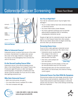
Technology
ALERT
Horizon Scanning Centre
February 2015
WavSTAT4 Optical Biopsy System® for
colorectal cancer diagnosis
TECHNOLOGY
WavSTAT4 Optical Biopsy
System® has been developed
by SpectraScience for the real
time
diagnosis
of
precancerous
colon
polyps
(≤5mm). WavSTAT4® uses
laser-induced autofluroescence
to obtain an objective, in vivo
analysis of polyps during
colonoscopy.
©SpectraScience
A low-intensity and non-damaging laser light is directed from the WavSTAT4® mobile
console through an optical fibre mounted in the single use WavSTAT4® optical biopsy
forceps. These are directed through the working channel of the colonoscope (a standard
endoscope) and brought into direct contact with the polyp. The polyp tissue absorbs the
laser light, resulting in the excitation of the tissue being examined. The resulting
autofluorescent signal is sent back and analysed by a waveform analysis algorithm in the
console, to diagnose precancerous colon polyps. A green ‘non-suspect’ or a red ‘suspect’
result is displayed on the console's screen within seconds. A biopsy sample can be taken
using the forceps.
WavSTAT4® is CE marked and is currently in use at St James University Hospital in Leeds,
as part of a European trial. A full commercial launch in the UK is expected in Q2 2015.
WavSTAT® has been developed through the National Institute for Health Research (NIHR)
Colorectal Therapies Healthcare Technology Co-operative (HTC). The HTC engages
industry, NHS, scientists, charities, engineers, patients and public in identifying areas within
colorectal therapies where development of new technology would be of benefit to patients.
POTENTIAL FOR IMPACT
Colorectal cancer (CRC) is the third most common cancer in the UK. The majority of CRCs
This alert presents independent research funded by the National Institute for
Health Research (NIHR). The views expressed are those of the author and
not necessarily those of the NHS, the NIHR or the Department of Health.
NIHR Horizon Scanning Centre, University of Birmingham
Email: [email protected]
Web: www.hsc.nihr.ac.uk
NIHR Horizon Scanning Centre
develop from benign adenomatous polyps in the bowel wall. A series of genetic alterations
usually results in the transformation of normal epithelium to an adenoma, which in some
instances mutates into a carcinoma. The disease progresses slowly and takes approximately
10 years for an adenoma to develop into cancer. This long latency period provides an
opportunity for early detection of the disease.
Colonoscopy is the endoscopic examination of the large bowel and the distal part of the
small bowel with a fibre optic camera on a flexible tube passed through the anus. It can
provide a visual diagnosis and provides the opportunity for removal of suspected polyps.
Once polyps are removed, they can be studied with the aid of a microscope to determine if
they are precancerous or not. Removal of these adenomatous polyps reduces the risk of
future CRC. Macroscopic differentiation between a hyperplastic (has virtually no chance of
becoming cancerous) and an adenomatous polyp during colonoscopy is difficult.
According to the company, WavSTAT4® has the potential to distinguish between hyperplastic
and adenomatous polyps during colonoscopy. The company claim that WavSTAT4® may
reduce the risks associated with polyp removal during the colonoscopy (e.g. bleeding and
perforation). If WavSTAT4® proves to be effective, it has the potential to reduce the number
of histological biopsies and costs involved in processing current biopsies.
This technology is predicted to have an impact on the following domain of the NHS
Outcomes Framework (see: www.england.nhs.uk/resources/resources-for-ccgs/out-frwrk):
Domain 1 Preventing people from dying prematurely.
EVIDENCE
PUBLISHED PAPERS AND ABSTRACTS
Kuiper T, Alderlieste YA, Tytgat KM et al. Automatic optical diagnosis of small colorectal
lesions by laser-induced autofluorescence. Endoscopy 2015; 47 (1): 56-62.
http://www.ncbi.nlm.nih.gov/pubmed/25264763
Neumann H, Vieth M, Günther C et al. Rapid computerized prediction of histology in
colorectal lesions - A pilot study. Gastrointestinal Endoscopy 2013; 77 (5).
http://www.giejournal.org/article/S0016-5107(13)00626-3/abstract
Rex DK, Kahi C, O'Brien M et al. The American Society for Gastrointestinal Endoscopy PIVI
(Preservation and Incorporation of Valuable Endoscopic Innovations) on real-time
endoscopic assessment of the histology of diminutive colorectal polyps. Gastrointestinal
Endoscopy 2011; 73 (3): 419-22.
http://www.ncbi.nlm.nih.gov/pubmed/21353837
Benes Z and Antos Z. Optical biopsy system distinguishing between hyperplastic and
adenomatous polyps in the colon during colonoscopy. Anticancer Research 2009; 29 (11):
4737-9.
http://www.ncbi.nlm.nih.gov/pubmed/?term=wavstat+optical+biopsy
ONGOING STUDIES
ClinicalTrials.gov. The MORDIS Study Clinical Investigational Plan.
http://clinicaltrials.gov/ct2/show/NCT01980134 Accessed 21 January 2015.
2
NIHR Horizon Scanning Centre
COMPANY INFORMATION
According to the company, future generations of WavSTAT4® will incorporate scattering
spectroscopy diagnostic technology, together with laser-induced fluorescence, to address
the diagnosis of precancerous lesions or other cancers in the colon and to improve the
overall diagnostic capability.
INFORMATION FROM
This Alert is based on information from the company and a time-limited internet search.
3


© Copyright 2026