
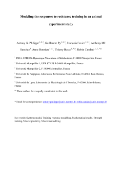
Corticotropin-Releasing Factor Mediates Pain
International Journal of Neuropsychopharmacology Advance Access published February 25, 2015
Corticotropin-Releasing Factor Mediates Pain-Induced Anxiety through the
ERK1/2 Signaling Cascade in Locus Coeruleus Neurons
Gisela Patrícia da Silva Borges, Msc1,2,3; Juan Antonio Micó Segura, PhD1,4, Fani
Lourença Moreira Neto, PhD2,3, Esther Berrocoso, PhD4,5*
Neuropsychopharmacology and Psychobiology Research Group, Department of
Neuroscience, University of Cádiz, 11003 Cádiz, Spain;
ip
t
1
2
Departamento de Biologia
us
cr
Experimental, Centro de Investigação Médica da Faculdade de Medicina da
Universidade do Porto (CIM-FMUP), 4200-319 Porto, Portugal;
3
Grupo de
Morfofisiologia do Sistema Somatossensitivo, Instituto de Biologia Molecular e Celular
(IBMC), Porto, Portugal; 4 Centro de Investigación Biomédica en Red de Salud Mental
(CIBERSAM),
Instituto
de
Salud
Carlos
III,
Madrid,
Spain;
5
an
Neuropsychopharmacology and Psychobiology Research Group, Department of
Psychology, University of Cádiz, 11510 Cádiz, Spain.
author:
Esther
Berrocoso
M
*Corresponding
PhD,
Neuropsychopharmacology
Psychobiology Research Group, Psychobiology Area, Department of Psychology,
pt
ed
University of Cádiz, 11510 Cádiz, Spain. Tel.:+34956015224 Fax: +34956015225.
Email: [email protected].
Category: Brief report - Psychiatry
Ac
ce
Word count:
Abstract: 150
Body of the manuscript: 2502
Number of references: 25
Number of figures: 2
Supplemental material: 1 file and 1 figure
© The Author 2014. Published by Oxford University Press on behalf of CINP.
This is an Open Access article distributed under the terms of the Creative Commons Attribution Non-Commercial License (http://creativecommons.org/licenses/
by-nc/4.0/), which permits non-commercial re-use, distribution, and reproduction in any medium, provided
the original work is properly cited. For commercial re-use, please contact [email protected]
1
ABSTRACT
Background: The corticotropin-releasing factor (CRF) is a stress-related neuropeptide
that modulates Locus Coeruleus (LC) activity. As LC has been involved in pain and
stress-related patologies, we tested whether the pain-induced anxiety is a result of the
ip
t
CRF released in the LC.
Methods: Complete Freund's adjuvant (CFA)-induced monoarthritis (MA) was used as
us
cr
inflammatory chronic pain model. α-helical CRF receptor antagonist was microinjected
into the contralateral LC of four weeks MA animals. The nociceptive and anxiety-like
behaviors, as well as phosphorylated extracellular signal-regulated kinases 1/2
an
(pERK1/2) and CRF receptors expression were quantified in the Paraventricular
Nucleus (PVN) and LC.
M
Results: MA rats manifested anxiety and increased pERK1/2 levels in the LC and PVN,
although the expression of CRF receptors was unaltered. α-helical CRF antagonist
LC.
pt
ed
administration reversed both the anxiogenic-like behavior and the pERK1/2 levels in the
Conclusions: Pain-induced anxiety is mediated by CRF neurotransmission in the LC
Ac
ce
through ERK1/2 signaling cascade.
Keywords: Anxiety; Corticotropin-releasing factor; Locus Coeruleus; Pain; pERK1/2.
2
INTRODUCTION
Corticotropin-releasing factor (CRF) is a neuropeptide released from neurons in the
paraventricular nucleus (PVN) of the hypothalamus that activates stress-related
hypophysial structures (Bale and Vale, 2004). Extrahypophysial CRF operates as a
ip
t
neurotransmitter in several brain areas, influencing different actions related to the stress
response (Valentino and Wehby, 1988). Also, it may exacerbate many chronic diseases,
us
cr
in particular, those involving severe pain like arthritis (Zautra et al., 2007), a prevalent
inflammatory condition. Equally, the emergence of anxiety due to persistent pain is a
negative factor commonly reported by arthritic patients (Gyurcsik et al., 2014),
an
representing in itself a stressful situation (Hummel et al., 2010) and inducing similar
effects to other stressors (Vierck et al., 2010). Indeed, it is estimated that up to 20% of
M
patients with arthritis will develop depression and/or anxiety (Covic et al., 2012).
Although inflammatory pain is a stressor that may modulate the hypothalamic-pituitary-
pt
ed
adrenal (HPA) axis (Bomholt et al., 2004), the neurobiological features and behavioral
repercussion of such association remain poorly understood.
CRF acts at different sites in important regulatory pain structures, directly implicating
Ac
ce
this molecule in pain modulation (Lariviere and Melzack, 2000). In particular, the
Locus Coeruleus (LC), the major noradrenaline source in the brain, is one important
target for CRF neurotransmission (Valentino and Van Bockstaele, 2008). Besides its
important role in modulating ascending and descending pain pathways, the LC also
represents a convergent nucleus that is correlated with adaptive responses to stress
(Valentino and Van Bockstaele, 2008). Thus, the activity of CRF in the LC could
influence chronic inflammatory pain and it would not be unreasonable to hypothesize
3
that the onset of emotional changes produced by pain might be the result of stressinduced CRF release.
The extracellular signal-regulated kinases 1/2 (ERK1/2) cascade is a strong candidate to
ip
t
mediate the effects of CRF in pain and it is known to participate in CRF receptor
signaling in neuronal cells (Hauger et al., 2006). In addition, CRF administration into
us
cr
the LC region promotes c-Fos and ERK1/2 activation in the prefrontal cortex (Snyder et
al., 2012). Accordingly, we demonstrated that animals suffering chronic inflammation
also display an anxio-depressive phenotype, with an enhancement of ERK1/2 activation
an
in the prefrontal cortex (Borges et al., 2014).
M
To evaluate whether CRF neurotransmission in the LC triggers the development of
anxiety in chronic inflammation (e.g., a model of rheumatoid arthritis), an antagonist of
pt
ed
the CRF receptors was microinjected into the LC and the nociceptive and anxiety
Ac
ce
behavior, as well as the activation of ERK1/2 in the LC, were evaluated.
4
METHODS
Animals
Harlan Sprague-Dawley male rats (250-300g) were provided by the Experimental Unit
ip
t
of the University of Cádiz (ES110120000210). Animals were housed 2-4 per cage with
ad libitum access to food and water, and kept under controlled conditions of lighting
us
cr
(12h light/dark cycle), temperature (22 ºC) and humidity (45-60%). The protocols
followed the European Communities Council Directive of 22 September 2010
(2010/63/EC), Spanish Law (RD 1201/2005) and the ethical guidelines for investigation
of experimental pain in animals (Zimmermann, 1983), and they were reviewed and
M
an
approved by the Institutional Ethical Committee for animal care and use.
Monoarthritis model of inflammatory pain
pt
ed
Monoarthritis (MA) was induced as described previously (Butler et al., 1992), injecting
the left tibiotarsal joint of rats anaesthetized with isoflurane (4% to induce and 2% to
maintain: Abbott, Spain) with 50 µL of complete Freund's adjuvant (CFA) solution,
containing 30 mg of desiccated Mycobacterium butyricum (Difco Laboratories, USA),
Ac
ce
paraffin oil (3 mL), saline (2 mL) and Tween®80 (500 µL). Animals that developed
polyarthritis were excluded. The control rats were injected with the vehicle solution
(paraffin oil, saline and Tween®80). The experimental design is represented in
Supplementary Fig. S1a.
5
Surgery and intra-LC drug administration
Animals were anesthetized with an intraperitoneal injection of ketamine (100 mg/kg)
and xylazine (20 mg/kg), and placed in a stereotaxic apparatus with the head tilted at an
angle of 15ᵒ to the horizontal plane. A guide cannula (22 gauge, 15 mm length) was
ip
t
implanted into the contralateral LC (lambda: AP=-3.2 mm, ML -1.1 mm, and DV -6.2
mm; Fig. 1a and Supplementary Fig. S1b) and was fixed with skull screws and dental
us
cr
cement. A stainless steel wire was inserted into the guide cannula to prevent occlusion.
Five days after recovery, animals were immobilized and the steel wire was cut.
Microinjection was performed by inserting an injector cannula (30 gauge) that was 1
an
mm longer than the guide cannula (i.e.: 16 mm). Animals received 28 ng or 34 ng of αhelical CRF(9-41) (αCRF) dissolved in sterile water (0.5 µL; Sigma Aldrich, Ref. C246),
which blocks the CRF I and II receptors, inhibiting the endogenous CRF activity. Sterile
M
water was used as a vehicle. Behavioral tests or sacrifice for western blot procedures
were performed 10-25 minutes after drug/vehicle administration (see Supplementary
pt
ed
Material and Fig. S1a). As no differences between doses were observed in terms of
pERK1/2 expression, the behavioral effects were analyzed in animals receiving the 28
ng αCRF. Behavior was assessed in groups of 6 animals and random animals were
Ac
ce
selected for histological verification of the cannula implantation.
Health parameters, nociceptive and anxiety-like behavior
The body weight (g) and rectal temperature (°C) were recorded weekly and nociceptive
mechanical allodynia (automated von Frey test) and hyperalgesia ( paw-pinch test) were
assessed as described in the Supplementary Material. Anxiety-like behavior was
evaluated in the elevated zero maze (EZM) test, which consisted in a black circular
platform divided into four quadrants , with two opposing open quadrants with 1 cm high
6
clear curbs to prevent falls and two opposing closed quadrants with 27 cm high black
walls. A 5 min trial under the same lighting conditions began with the animal placed in
the centre of a closed quadrant. The SMART software was used to analyze the time
spent in the open arms and the total distance travelled by each rat. Increases in the time
ip
t
spent in the closed areas were correlated with anxiety-like behavior (Borges et al.,
us
cr
2014).
Immunohistochemistry
Another set of control and MA rats (4 weeks) was used for quantification of the
an
expression of pERK1/2 in the PVN and CRFI/II receptors in the LC (Suplementary
Statistical analysis
M
Material).
All data is represented as mean ± SEM and were analyzed using STATISTICA 10.0 or
pt
ed
GraphPad Prism 5 software, using either an unpaired Student t test (two-tailed) or oneway, two-way or repeated measures analysis of variance (ANOVA), followed by the
Ac
ce
appropriate post-hoc tests. The level of significance was considered as p<0.05.
7
RESULTS
Effect of MA on health parameters and nociceptive responses
The behavior of control animals was normal, with no signs of an inflammatory reaction.
CFA injection produced a stable MA, with the signs of inflammation restricted to the
ip
t
injected joint and evident a few hours after induction, persisting into the fourth week.
The weight gain of MA rats was significantly lower than that of the control animals 1
us
cr
and 3 weeks after CFA injection (repeated measures, Bonferroni test; p<0.05: Fig. 1b),
while their body temperature remained normal during the experiment (Fig. 1c).
Regarding the pain threshold, a significant decrease in the withdrawal threshold of the
an
ipsilateral paw was evident when compared with the contralateral paw, indicative of
mechanical hyperalgesia (repeated measures, Bonferroni test; p<0.05 for week 1, 2 and
M
4, p<0.01 for week 3: Fig. 1d). Additionally, there was a significant decrease in the paw
withdrawal threshold to von Frey stimulation by the ipsilateral-inflamed paw of MA
pt
ed
rats when compared with the contralateral paw, indicative of mechanical allodynia
(repeated measures, Bonferroni test; p<0.001: Fig. 1e).
Ac
ce
Effect of intra-LC microinjection of an α-helical CRF receptor antagonist on the
pERK1/2 levels in the LC
The administration of the α-helical CRF receptor antagonist in the LC normalized the
pERK1/2 values in MA4W rats at both doses of the compound used (one-way ANOVA,
Dunnett’s test;; p>0.05 MA4W 28ng and MA4W 34ng vs control; Fig. 1f), suggesting
that CRF acts through the ERK1/2 signaling cascade in LC neurons.
8
Effect of intra-LC microinjection of an α-helical receptor antagonist on MA-induced
pain
In the paw withdrawal threshold, a significant increase in pain sensitivity was observed
when the ipsilateral paw was compared with the contralateral paw in the MA4W rats
ip
t
before (pre-drug, two-way ANOVA, Bonferroni test; p<0.001) and after microinjection
of the αCRF into the LC (two-way ANOVA, Bonferroni test; p<0.01: Fig. 1g). Thus,
us
cr
the intra-LC administration of the α-helical CRF receptor antagonist (28 ng) had no
significant effect on the paw withdrawal threshold of MA4W rats (p>0.05; Fig. 1g). In
the force threshold, microinjection of the αCRF receptor antagonist into the LC had no
an
effect on the ipsilateral sensitivity to innocuous stimulation. Indeed, the significant
decrease in the force supported by the ipsilateral paw of MA4W rats when compared to
the contralateral paw was present before and after administration of the drug (two-way
pt
ed
M
ANOVA, Bonferroni test; p<0.001: Fig. 1h).
Effect of intra-LC microinjection of an α-helical CRF receptor antagonist on MAinduced anxiety.
In the EZM, MA4W rats that received the vehicle alone (MA4W-vehicle) spent
Ac
ce
significantly less time in the open arms than control animals, indicative of anxiety-like
behavior (two-way ANOVA, Bonferroni test; p<0.05: Fig. 1i). By contrast, those
animals that received an intra-LC microinjection of the αCRF receptor antagonist
(MA4W-αCRF) spent significantly longer time in the open arms when compared to the
MA4W-vehicle animals (two-way ANOVA, Bonferroni test; p<0.05: Fig. 1i). No effect
of microinjecting the αCRF receptor antagonist into the LC was observed in the control
rats. Moreover, no differences were observed in the total distance travelled in the EZM
(Fig. 1j), ruling out any influence of locomotor impairment on the experimental results.
9
Regarding the number of entries into the open arms, the MA4W-vehicle rats appeared to
enter these arms less frequently than the control animals that received the vehicle alone,
although this difference was not significant. No such difference was observed in the
Expression of CRF receptors in the LC and pERK1/2 in the PVN
ip
t
MA4W-αCRF rats (Fig. 1k).
us
cr
When the expression of the CRF I and II receptors was studied in the LC, no differences
were observed between the control and MA4W rats (Fig. 2a, b and c). Most of the
neurons expressing CRFI/II receptors also expressed TH, indicating their noradrenergic
an
nature and demonstrating that the CRFI/II receptors were expressed by neurons in the
LC area (Fig. 2d).
M
The expression of pERK1/2 was also studied in the PVN of the hypothalamus in order
to determine the activity of this structure as a readout of CRF stimulation in the central
pt
ed
nervous system. Interestingly, an increase in pERK1/2 was observed in MA4W rats
Ac
ce
compared with control rats (unpaired Student’s t-test; p<0.001: Fig. 2e-g).
10
DISCUSSION
This study shows that the action of CRF on LC neurons is involved in the development
of anxiety-like symptoms associated with prolonged inflammatory pain. As expected,
four weeks after CFA injection, rats displayed signs of pain and anxiety, consistent with
ip
t
previous reports (Borges et al., 2014). We also observed a significant increase in
ERK1/2 phosphorylation in the LC, in accordance with previous data (Borges et al.,
us
cr
2014), and this increased ERK1/2 activation in the LC seems to be related with the
development of anxiety-like behaviors in chronic inflammatory conditions. This raises
the question as to what produces this increase in ERK1/2 activation in the LC when
an
painful conditions develop. CRF is a molecule linked with the endocrine and behavioral
response to stress (Bale and Vale, 2004), and the role of CRF in different pain
conditions has been studied (Lariviere and Melzack, 2000), although not its effects after
M
prolonged times of inflammation (e.g., 4 weeks). Here, we studied the PVN nucleus, a
CRF-producing structure, and we found that pERK1/2 levels increase in MA4W rats
pt
ed
when compared with control rats, suggesting that PVN hyperactivation occurs in
association with chronic inflammatory pain. As the PVN and LC have reciprocal
excitatory connections (Perez et al., 2006), we hypothesized that this would underpin
Ac
ce
the ERK1/2 activation in the LC of MA4W rats. Indeed, the LC is rich in CRF receptors
(Reyes et al., 2006; Mousa et al., 2007) and it has already been shown that CRF
activates LC neurons (Valentino and Foote, 1988). Here, we found no significant
differences in CRFI/II receptor expression in the LC of control and MA4W rats, which
indicates that while enhanced neurotransmission might originate in the PVN when
chronic inflammatory pain is established, it is not accompanied by changes in the
expression of the CRFI/II receptors in the LC. The co-localization of CRFI/II receptors
11
with the TH protein, as described previously (Reyes et al., 2006), confirmed the
specificity of this labeling.
To better understand how CRF neurotransmission influences the role of the LC in
nociception and anxiety behavior, an antagonist blocking the CRF receptors was
ip
t
microinjected into the contralateral LC. This strategy was adopted in order to study the
ascending pain pathway passing through the LC given its important projections to
us
cr
corticolimbic areas (Fig. 1a). The dose of the α-helical CRF antagonist used was based
on previous studies (Mousa et al., 2007), and at both 28ng and 34ng this antagonist
successfully dampened pERK1/2 expression in the LC of MA4W rats. Thus, the effects
an
of the lower dose alone (28 ng) were evaluated on behavior. This procedure had no
effect on pain sensitivity in the ipsilateral/inflamed or contralateral paws of MA4W rats.
M
In contrast, microinjection of the α-helical CRF receptor antagonist reverses the
anxiety-like behavior observed in MA4W rats without interfering with locomotor
pt
ed
activity. Indeed, the decrease in the time MA4W rats spent in the open arms was no
longer observed when they received this antagonist. These results suggest that the
increased CRF neurotransmission in chronic inflammatory conditions enhances the LC
Ac
ce
driven activation of corticolimbic areas, which may be responsible for the development
of anxiety. Indeed, it has already been shown that CRF infused into the LC increases
anxiety, a behavioral effect of CRF associated with increased noradrenergic
neurotransmission in LC terminal areas like the amygdala and hypothalamus (Butler et
al., 1990; Weiss et al., 1994). Moreover, the α-helical CRF receptor antagonist prevents
the development of anxiety induced by a neuropeptide Y receptor antagonist, while not
producing any significant change when administered in a non-anxious state (Kask et al.,
1997; Donatti and Leite-Panissi, 2011). Similarly, we did not find a significant effect of
the α-CRF antagonist in control non-inflamed animals. Overall, the effects in the
12
ipsilateral paw and on anxiety-like behavior in MA rats are consistent with studies
showing that administration of the α-helical CRF receptor antagonist to the basolateral
or central nuclei of the amygdala has no effect on the nociceptive threshold but that it
reduced innate fear behavior (Donatti and Leite-Panissi, 2013). Nevertheless, the lack of
ip
t
changes in nociception might be related to the use of a broad-spectrum CRF antagonist
that blocks, nonspecifically the signaling of CRF1 and CRF2 receptors. Indeed, when
us
cr
NBI27914, a specific CRF1 receptor antagonist, was microinjected into the amygdala,
the withdrawal thresholds of the arthritic rats, as well and the anxiety-like behavior,
were reversed (Ji et al., 2007).
an
Concluding, CRF signaling through the ERK1/2 cascade in the LC appears to be an
important mechanism related with anxious behavior associated with chronic
Ac
ce
pt
ed
M
inflammatory conditions.
13
ACKNOWLEDGMENTS
We would like to acknowledge the help provided by Ms Raquel Rey-Brea, Mr José
Antonio García Partida, Mr Jesus Gallego-Gamo, Ms Paula Reyes Perez, Mr Santiago
Muñoz and Ms Elisa Galvão.
work
was
supported
by
“Cátedra
Externa
del
Dolor
Fundación
ip
t
This
Grünenthal/Universidad de Cádiz" which paid a grant to the first author; "Cátedra em
us
cr
Medicina da Dor from Fundação Grünenthal-Portugal and Faculdade de Medicina da
Universidade do Porto"; Fondo de Investigación Sanitaria (PI12/00915, PI13/02659);
CIBERSAM G18; Junta de Andalucía (CTS-510, CTS-7748); Fundación Española de
Ac
ce
pt
ed
None to declare
M
Statement of Interest
an
Dolor (travel fellowship granted to Gisela Borges – 1536).
14
REFERENCES
Ac
ce
pt
ed
M
an
us
cr
ip
t
Bale TL, Vale WW (2004) CRF and CRF receptors: role in stress responsivity and other
behaviors. Annu Rev Pharmacol Toxicol 44:525-557.
Bomholt SF, Harbuz MS, Blackburn-Munro G, Blackburn-Munro RE (2004)
Involvement and role of the hypothalamo-pituitary-adrenal (HPA) stress axis in
animal models of chronic pain and inflammation. Stress 7:1-14.
Borges G, Neto F, Mico JA, Berrocoso E (2014) Reversal of monoarthritis-induced
affective disorders by diclofenac in rats. Anesthesiology 120:1476-1490.
Butler PD, Weiss JM, Stout JC, Nemeroff CB (1990) Corticotropin-releasing factor
produces fear-enhancing and behavioral activating effects following infusion
into the locus coeruleus. J Neurosci 10:176-183.
Butler SH, Godefroy F, Besson JM, Weil-Fugazza J (1992) A limited arthritic model for
chronic pain studies in the rat. Pain 48:73-81.
Covic T, Cumming SR, Pallant JF, Manolios N, Emery P, Conaghan PG, Tennant A
(2012) Depression and anxiety in patients with rheumatoid arthritis: prevalence
rates based on a comparison of the Depression, Anxiety and Stress Scale
(DASS) and the hospital, Anxiety and Depression Scale (HADS). BMC
Psychiatry 12:6.
Donatti AF, Leite-Panissi CRA (2011) Activation of corticotropin-releasing factor
receptors from the basolateral or central amygdala increases the tonic immobility
response in guinea pigs: An innate fear behavior. In: Behav Brain Res, pp 23-30.
Donatti AF, Leite-Panissi CRA (2013) Activation of the corticotropin-releasing factor
receptor from the basolateral or central amygdala modulates nociception in
guinea pigs. Advances in Bioscience and Biotechnology 4:7.
Gyurcsik NC, Cary MA, Sessford JD, Flora PK, Brawley LR (2014) Pain anxiety and
negative outcome expectations for activity: Negative psychological profiles
differ between the inactive and active. Arthritis Care Res (Hoboken).
Hauger RL, Risbrough V, Brauns O, Dautzenberg FM (2006) Corticotropin releasing
factor (CRF) receptor signaling in the central nervous system: new molecular
targets. CNS Neurol Disord Drug Targets 5:453-479.
Hummel M, Cummons T, Lu P, Mark L, Harrison JE, Kennedy JD, Whiteside GT
(2010) Pain is a salient "stressor" that is mediated by corticotropin-releasing
factor-1 receptors. Neuropharmacology 59:160-166.
Ji G, Fu Y, Ruppert KA, Neugebauer V (2007) Pain-related anxiety-like behavior
requires CRF1 receptors in the amygdala. Mol Pain 3:13.
Kask A, Rägo L, Harro J (1997) α‐Helical CRF9‐41 prevents anxiogenic‐like effect of
NPY Y1 receptor antagonist BIBP3226 in rats. NeuroReport 8:3645-3647.
Lariviere WR, Melzack R (2000) The role of corticotropin-releasing factor in pain and
analgesia. Pain 84:1-12.
Mousa SA, Bopaiah CP, Richter JF, Yamdeu RS, Schafer M (2007) Inhibition of
inflammatory pain by CRF at peripheral, spinal and supraspinal sites:
involvement of areas coexpressing CRF receptors and opioid peptides.
Neuropsychopharmacology 32:2530-2542.
Perez H, Ruiz S, Nunez H, White A, Gotteland M, Hernandez A (2006) Paraventricularcoerulear interactions: role in hypertension induced by prenatal undernutrition in
the rat. Eur J Neurosci 24:1209-1219.
Reyes BA, Fox K, Valentino RJ, Van Bockstaele EJ (2006) Agonist-induced
internalization of corticotropin-releasing factor receptors in noradrenergic
neurons of the rat locus coeruleus. Eur J Neurosci 23:2991-2998.
15
Ac
ce
pt
ed
M
an
us
cr
ip
t
Snyder K, Wang WW, Han R, McFadden K, Valentino RJ (2012) Corticotropinreleasing factor in the norepinephrine nucleus, locus coeruleus, facilitates
behavioral flexibility. Neuropsychopharmacology 37:520-530.
Valentino RJ, Wehby RG (1988) Corticotropin-releasing factor: evidence for a
neurotransmitter role in the locus ceruleus during hemodynamic stress.
Neuroendocrinology 48:674-677.
Valentino RJ, Foote SL (1988) Corticotropin-releasing hormone increases tonic but not
sensory-evoked activity of noradrenergic locus coeruleus neurons in
unanesthetized rats. J Neurosci 8:1016-1025.
Valentino RJ, Van Bockstaele E (2008) Convergent regulation of locus coeruleus
activity as an adaptive response to stress. Eur J Pharmacol 583:194-203.
Vierck CJ, Green M, Yezierski RP (2010) Pain as a stressor: effects of prior nociceptive
stimulation on escape responding of rats to thermal stimulation. Eur J Pain
14:11-16.
Weiss JM, Stout JC, Aaron MF, Quan N, Owens MJ, Butler PD, Nemeroff CB (1994)
Depression and anxiety: Role of the locus coeruleus and corticotropin-releasing
factor. Brain Res Bull 35:561-572.
Zautra AJ, Parrish BP, Van Puymbroeck CM, Tennen H, Davis MC, Reich JW, Irwin M
(2007) Depression history, stress, and pain in rheumatoid arthritis patients. J
Behav Med 30:187-197.
Zimmermann M (1983) Ethical guidelines for investigations of experimental pain in
conscious animals. Pain 16:109-110.
16
LEGENDS
Fig. 1: a) Schematic representation of the anatomical pathways implicated. Briefly, the
contralateral LC indirectly receives inputs from the inflamed paw (red dashed line;
ascending pathways) and, subsequently, the information is sent to corticolimbic areas.
ip
t
Additionally, the LC sends direct projections to the spinal cord (blue straight line;
descending pathways). b) Body weight of the control and MA rats. c) Body rectal
us
cr
temperature of control and MA rats. d) Mechanical hyperalgesia represented by a
significant decrease in the paw withdrawal threshold of the ipsilateral paw of MA rats.
e) Mechanical allodynia represented by a significant decrease in the force threshold of
an
the ipsilateral paw of MA rats. *p<0.05, **p<0.01 and ***p<0.001; repeated measures
followed by a Bonferroni post-hoc test comparing control vs MA for the same week (b
and c) or comparing the ipsilateral vs contralateral paw for the same week (d and e). f)
M
Graph depicting the expression of pERK1/2 in the LC after intra-LC administration of
the αCRF receptor antagonist, showing that the significant increase of pERK1/2 in
pt
ed
MA4W animals was no longer observed when this antagonist was administered:
*p<0.05 (one-way ANOVA followed by Dunnett’s post-hoc test). g) Graph showing
that the local administration of the αCRF antagonist had no significant effect on
Ac
ce
mechanical hyperalgesia in MA4W rats. h) Graph showing that local administration of
the α-helical CRF antagonist had no significant effect on mechanical allodynia in the
ipsilateral paw of MA rats. **p<0.01 and ***p<0.001 (two-way ANOVA followed by
Bonferroni post-hoc test). i) Graph showing that the time spent in the open arms
decreased in MA4W rats receiving the vehicle alone but this effect was successfully
reversed by administration of the αCRF antagonist. *p<0.05 (two-way ANOVA
followed by Bonferroni post-hoc test). j) Graph showing that local administration of the
α-helical CRF antagonist had no significant effect on the total distance travelled in the
17
elevated zero maze. k) Graph showing that local administration of the α-helical CRF
antagonist reversed the decrease in the number of entries into the open arms observed in
MA4W rats receiving the vehicle alone. B=Baseline; LC=Locus Coeruleus;
αCRF=antagonist of the corticotropin-releasing factor receptor I and II; W=Week;
ip
t
MA=Monoarthritis.
us
cr
Fig. 2: Expression of CRFI/II receptors in the LC of control and MA4W rats. a) and b)
Photomicrographs showing the expression of CRFI/II receptors in control and MA4W
rats, respectively. c) Graph showing that there were no significant changes between
an
control and MA4W rats in terms of CRFI/II receptor expression in the LC. d)
Immunofluorescence photomicrographs showing that almost all the neurons expressing
M
CRFI/II receptors (green) are noradrenergic neurons, since they co-localize with TH
immunolabeling. e) Graph showing the increase in pERK1/2 expression in the PVN
pt
ed
nucleus of MA4W rats: ***p<0.001 (unpaired two-tailed Student’s t-test). f) and g)
Photomicrographs of pERK1/2 expression in control and MA4W rats, respectively.
Scale bar=100µm. AU=Arbitrary units; W=Week; MA=Monoarthritis. W=Week;
Ac
ce
MA=Monoarthritis.
Fig. S1: a) Schematic representation of the experimental protocol followed in this study
and b) Schematic representation of the local cannula implantation.
18
ce
Ac
d
pt
e
cr
ip
t
us
an
M
Figure 1
ce
Ac
d
pt
e
cr
ip
t
us
an
M
Figure 2
Supplementary material
Nociception
Mechanical allodynia was evaluated using an electronic version of the von Frey test
(Dynamic Plantar Aesthesiometer, Ugo Basile, Italy), applying an increasing vertical
ip
t
force (from 0 to 50 g) to both the ipsi- and contralateral paws over a period of 20 s. In
addition, the presence of mechanical hyperalgesia was tested using the paw-pinch test
us
cr
(Randall and Selitto, 1957). Increasing pressure (from 30g of pressure) was gradually
applied to the dorsal side of the paw using a graded motor-driven device (Ugo Basile,
Italy). A 250g cut-off was used to prevent damage to the paw. In both tests, three
an
measures were taken on each paw at 5 min intervals and the average value was used.
Western blotting
M
Fresh tissue samples from the contralateral LC of a preliminary group including control,
MA4W-vehicle and MA4W-α-CRF rats (28 ng or 36 ng) were obtained after sacrificing
pt
ed
them by decapitation, 10-25 minutes after administration. After the tissue was lysed, an
aliquot of the sample (50 µg) was separated in a 10% polyacrylamide gel and then
transferred to a polyvinylidene difluoride membrane (PVDF; BioRad, Spain). After
Ac
ce
washing in TBST (Tris-buffered saline containing 0.1% Tween-20), the membranes
were blocked with 5% Bovine Serum Albumin (BSA; Sigma, Spain) in TBST and
probed overnight at 4 ºC with a rabbit anti-phospho-ERK1/2 (1:5,000; Neuromics,
USA) and a mouse-anti ERK1/2 (1:2,000; Cell Signaling; Izasa, Spain) antibody diluted
in 5% BSA-TBST. After thorough washing, these primary antibodies were detected by
incubating for 1 hour at room temperature with IRDye 800CW goat anti-rabbit (green)
or IRDye 680LT goat anti-mouse (red) secondary antibodies (1:10,000; Bonsai
Advanced Technologies, Spain). After 3 final washes with TBST, antibody binding was
detected using a LI-COR Odyssey® two-channel quantitative fluorescence imaging
1
system (Bonsai Advanced Technologies, Spain). Digital images of the Western blots
were analyzed by densitometry using the Image J free access software and the data were
expressed as the pERK1/2 expression relative to the total ERK1/2. Three assays were
ip
t
performed on LC samples from 3 rats per group.
us
cr
Immunohistochemistry (IHC)
Control and MA rats 4 weeks after CFA injection (MA4W; N=5 per group) were
anesthetized with 8% chloral hydrate (400mg/kg) and they were transcardially perfused
through the ascending aorta with 250 mL of oxygenated Tyrode’s solution followed by
an
750 mL of paraformaldehyde 4% in phosphate buffer (PB, 0.1M [pH 7.2]). Brains were
removed and processed for free-floating immunohistochemistry. One in five sequential
M
transverse brain sections (30 µm) containing the PVN from each rat were washed,
blocked and incubated with a rabbit antiserum against the phosphorylated ERK1 and
pt
ed
ERK2 isoforms (pERK1/2; 1:1000; 48 hours at 4-8ºC: Neuromics, USA).
Immunodetection was achieved with a biotinylated donkey anti-rabbit antiserum (1:500;
1 hour; Jackson ImmunoResearch, USA), followed by an ABC solution (1:200, 1 hour;
Ac
ce
ABC Elite kit, Vector Laboratories, UK) and a colorimetric reaction with 3,3diaminobenzidine tetrahydrochloride (DAB; 10 min) in 0.05M Tris-HCl buffer
containing 0.003% hydrogen peroxide (Cruz et al., 2005). Sections were then washed in
PBS, mounted on gelatin-coated glass slides, cleared in xylene, cover-slipped with DPX
and analyzed by light microscopy. Furthermore, immunohistochemistry to detect CRF
receptors I/II in LC sections was performed following a similar protocol with an antiCRFI/II rabbit antiserum (1:50; Santa Cruz), a biotinylated donkey anti-rabbit antibody
and streptavidin 488 (1:1000, Invitrogen). Additionally, to co-localize the CRF I/II
receptors with tyrosine hydroxylase (TH) positive neurons, a sheep anti-TH antibody
2
was used (1:4000; abcam, UK), which was detected with an Alexa 568 conjugated
donkey anti-sheep antibody (1:1000, Invitrogen). The specificity of the antibodies was
controlled by omitting the primary antibodies in various sets of immunoreactions.
Quantification of the expression of pERK1/2 in the PVN and CRF I/II receptors in the
ip
t
LC was performed by densitometry as described elsewhere (Borges et al., 2013). As no
differences were observed in ipsilateral side when comparing with the contralateral side,
an
us
cr
the values obtained in each area were averaged and used for statistical purposes.
References
Ac
ce
pt
ed
M
Borges GS, Berrocoso E, Ortega-Alvaro A, Mico JA, Neto FL (2013) Extracellular signal-regulated
kinase activation in the chronic constriction injury model of neuropathic pain in
anaesthetized rats. Eur J Pain 17:35-45.
Cruz CD, Neto FL, Castro-Lopes J, McMahon SB, Cruz F (2005) Inhibition of ERK phosphorylation
decreases nociceptive behaviour in monoarthritic rats. Pain 116:411-419.
Randall LO, Selitto JJ (1957) A method for measurement of analgesic activity on inflamed
tissue. Arch Int Pharmacodyn Ther 111:409-419.
3
Ac
ce
pt
e
d
M
an
us
cr
ip
t
Supplementary figure 1


© Copyright 2026