
Document 8094
EFFECT OF RENIN.AI\GIOTEI\SIN SYSTEM BLOCKADE
ON CARDIAC REMODELIIIG IN HEART FAILURE
AFTER MYOCARDIAL INFARCTION
BY
XIAOBING GUO
A Dissertation Submitted to
The Faculty of Graduate Studies
In Partial Fulfillment of Requirements for the Degree of
DOCTOR OF PHILOSOPHY
nstitute of Cardiovascular Sciences
Department of Physiology, Faculty of Medicine
University of Manitoba
Winnipeg, Manitoba
I
@
August 2004
THE UNIVERSITY OF MANITOBA
FACULTY OF GRADUATE STUDIES
*****
COPYRIGHT PERMISSION
EFFECT OF RENIN.ANGIOTENSIN SYSTEM BLOCKADE
ON CARDIAC REMODELING IN HEART FAILURE AFTER
MYOCARDIAL INFARGTION
BY
XIAOBING GUO
A Thesis/Practicum submitted to the Faculty of Graduate Studies of
The University of Manitoba
in partial fulfillment of the requirement of the degree
of
Doctor of Philosophy
XIAOBING GUO @2004
Permission has been granted to the Library of the University of Manitoba to lend
or sell copies of this thesis/practicum, to the National Library of Canada to
microfilm this thesis and to lend or sell copies of the film, and to University
Microfilms lnc. to publish an abstract of this thesis/practicum.
This reproduction or copy of this thesis has been made available by authority of
the copyright owner solely for the purpose of private study and research, and
may only be reproduced and copied as permitted by copyright laws or with
express written authorization from the copyright owner.
ACKNOWLEDGEMENTS
I would like to
express my appreciation to all the people who have assisted me in
accomplishing my Ph.D. program. First,
it is my great pleasure to extend my deepest
gratitude to my advisor Dr. Naranjan S. Dhalla, for his warrn heart, fabulous perception,
countless care and incredible patience. Without his thoughtful guidance,
been impossible for me
to face all
it would
have
these challenges during my training. His sharp
knowledge and help have kept me on the right track towards my goal.
I
would also like to acknowledge my advisory committee, whose critical
comments as well as encouragement have greatly helped me to
fulfill my task. Dr. Pawan
K. Singal has continuously supported me through the past seven years of my
study. Dr. Ian
graduate
M. C. Dixon always "fixes the problem" for me immediately. Special
thanks are extended to Dr. Paramjit S. Tappia for his help with my project and the editing
of my thesis. In addition, it is he, who opened the window of advanced education at the
University for me, and has showed me the amazing nature of nutrition science.
I would also like to express my gratitude towards all members of the Experimental
Cardiology Laboratory. Every achievement
in my work
could not have
been
accomplished without their efforts. Their everlasting solid support and sincere friendship
have kept me enjoying science and life.
I would
Institute, who never let me lose hope.
also wish to thank all the members in our
It is also time to acknowledge
and thank the
I
appreciate the
marvellous academic training given in the Department of Physiology.
sustained help of the University of Manitoba and friends in V/innipeg.
I
want to thank Manitoba Health Research Council and the University of
Manitoba, for the financial assistance. Also
I want to thank all the persons who offered
Ii
me the academic assistance from as far away as USA and Europe. Also,
I would like to
thank all the friends and families in China, who have encouraged me to achieve my goal.
I
dedicate my thesis to my parents and parents-in-law for their unlimited love.
I
am highly indebted to them, to my sister, to my brother-in-law and sister-in-law, whose
enorïnous supports and sacrifices are always my sources of strength and power. Finally, I
am thankful to my wife,
Dr. Jingwei Wang, who deserves the best words in the world.
She always brings me the love, wisdom, encouragement and energy, and always brings
the honour, pleasure, harmony and comfort to the family.
All of the best wishes
are given to my lovely son, Jiabo, and my adorable daughter,
and love
Amy. Their understanding
and patience are crucial for my research work. During the most challenging period of my
life,
I have been granted with confidence, happiness
endurance, talent and collaboration.
and pride, from their astonishing
III
ABSTRACT
Congestive heart failure (CHF) is one of the major challenges affecting society.
Specifically, myocardial infarction (MI) is the major cause of CHF in the Western world.
Cardiac remodeling occurs post
modification
of cardiac
MI
and contributes to the establishment
remodeling is important
of CHF,
and
in both prevention and treatment of
cardiac dysfunction post ML Activation of the renin-angiotensin system (RAS) is critical
in the cardiac remodeling process following ML Also, the blockade of RAS has shown
significant therapeutic effects in research and clinical practice. By employing a rat model
of coronary artery ligation, the current project focuses on the mechanism of the beneficial
effects of RAS blockade in the failing heart post MI. The angiotensin converting enzyme
(ACE) inhibitor (enalapril) and angiotensin II type
1 receptor
(ATrR) antagonist (losartan)
were utilized to explore the therapeutic potential of RAS blockade on the heart at the
subcellular and molecular levels.
To investigate the mechanisms underlying the depressed sarcolemmal (SL) Na*K*-ATPase activity in the failing heart, different isoforms of Na*-K*-ATPase protein
contents and gene expression were examined
in the left ventricle (LV) at 8
weeks
following MI. In addition, these parameters were also studied after 5 weeks of treatment
with RAS blockade by enalapril (10 mglkglday) and/or losartan (20 mglkglday) starting
at 3 weeks after the coronary ligation. An infarcted heart features the formation of a scar
and significant cardiac hypertrophy as is evident from the increased heart weight and
heart weight/body weight ratio. CHF was also evident from increased lung wet
weighVdry weight ratio (index for pulmonary edema) and the impaired heart function
associated with depressed
LV pressure development (+dPldt) and pressure decay (-dPldt)
IV
as \Ã/ell as elevation
in LV
end-diastolic pressure (LVEDP).
In the isolated SL
preparations, we found that the protein contents for Na*-K*-ATPase isoforms underwent
significant changes corresponding to similar alterations in gene expression in CHF. These
changes were associated
with the depressed Na*-K*-ATPase activity in the failing heart.
The protein contents and the mRNA expression for Na*-K*-ATPase øz isoform were
decreased, whereas the protein content and
mRNA level for Na*-K*-ATPase atisoform
were greatly increased. On the other hand, no changes were observed in the protein
contents or
6RNA levels for Na*-K*-ATPase at
activity of Na*-K*-ATPase
øz
and p7 isoforms. The
higher enzyme
isoform in comparison to the Na*-K*-ATPase ø¡ isoform
may explain the reason for the depressed Na*-K*-ATPase activity in the failing heart post
ML The
changes
in the corresponding mRNA of Na*-K*-ATPase
isoforms may
contribute to the remodeling of different Na*-K*-ATPase isoform proteins. The treatment
of infarcted animals with enalapril, losartan and combination of enalapril and losartan
significantly attenuated lung congestion, cardiac hypertrophy and improved the heart
function. The blockade of RAS also partially prevented the remodeling of Na*-K*ATPase isoforms, attenuating the depression in øz isoform and the increase in ø3 isoform,
at both the protein and mRNA levels. However, the combination therapy with enalapril
and losartan did not produce the expected additive effects.
Impaired Ca2*-transport
is
another important factor that contributes
to
the
pathogenesis of heart failure. To examine the remodeling of SL Caz*-regulatory proteins
in the failing heart post MI, a second series of
experiments were conducted
in
the
hemodynamically assessed infarcted animals with or without drug treatment. Three
proteins including SL Na*-Ca2*-exchanger Q.{CX), Ca2*-pump and Ca2*-channel were
V
examined in the isolated SL preparations.
At 8 weeks post MI, the protein content for
NCX was increased in the viable LV (P < 0.05). Similar increase in NCX mRNA was
identified by Northern blot analysis. These changes were associated with significant
cardiac hypertrophy and heart dysfunction. The protein content
for SL
Ca2*-pump
(PMCAi) was markedly increased in the failing heart; this was associated with
a
significant elevation in the corresponding mRNA expression as determined by RT-PCR.
A
decrease
in the SL Ca2*-charnel alsubunit and an increase in the SL Ca2*-channel p
subunit without any change
in the 612 subunit were
compared to sham control. The remodeling
in the failing
heart
as
of SL NCX, PMCA1 and Ca2*-channel
as
evident
well as heart dysfunction were partially prevented by blockade of RAS with enalapril and
losartan. These results suggest that the beneficial effects of RAS blockade in CHF may be
due to partial prevention of SL remodeling with respect to Ca2* transport.
Although, the right ventricle (RV) is as important as the LV in pumping the blood,
less information is available regarding gene expression in the RV, especially in the failing
heart post
MI. In these experiments, both LV and RV were utilized for the isolation of
total RNA, in order to investigate
if
similar remodeling occurs in cardiac gene expression
in the LV and RV of the same failing heart. Most of the SL, sarcoplasmic reticular (SR)
and myofibrillar proteins were equally expressed in both ventricles of the heart. However,
higher expression of PMCA1 and the lower expression of pmyosin heavy chain (ÊlVffIC)
as
well as ftactin were observed in the RV in comparison to the LV. Following MI,
a
reduction in PLB and ø-myosin heavy chain (ø-MHC) gene expression, and an increase
in NCX and þ}i1rHC occurred in both the RV and the LV. On the other hand, the
depressions
in Na*-K*-ATPase ø2 subunit, RYR, and SERCA2, were only significant in
VI
the LV. The difference in the remodeling process between the LV (showing hypertrophy
and heart failure) and
RV (showing hypertrophy) may partially be due to the differential
in
changes in cardiac gene expression between the two ventricles. However, alterations
gene expression in both the
RV and the LV
were partially prevented by treatment
of
as
well
as hypertrophy and heart dysfunction
animals showing CHF with enalapril and
losartan. These results further support the concept that improvement
in
cardiac
performance in CHF by the blockade of RAS may be associated with partial prevention
SL, SR and myofibril remodeling in the heart.
of
VII
TABLE OF CONTENTS
........
I.
LITERATURE REVIE\ry
1.
Introduction...............
2.
Cardiac Remodeling after MI
3.
Ca2*Homeostasis in Heart
4.
SL Na*-K*-ATPase in Heart
5.
SL Ca2*-Channel in Heart
6.
SL NCX and Caz*-pump in Heart
7.
SR Ca2*-Channel in Heart
8.
SERCA in Heart
9.
PLB and CQS in Heart
10.
MHC and MLC in Heart
11.
RAS in Cardiovascular
12.
Activation of RAS in
13.
Blockade of RAS in
14.
RAS Blockade and Subcelluar Remodeling
............
.....................
1
......................
1
...............2
Failure......
.............. 6
Failure......
Failure
......... 10
..................12
Failure
Failure
...... 1'4
................. 16
Failure
..............l7
Failure
....20
Failure
....................21
System
......................23
MI
..............28
MI
............... 31
..............
......47
VIII
15.
Summary
il.
STATEMENT OF THE PROBLEM AND HYPOTHESES TO
BE TESTED
....................47
.............
.................. 50
ilI.
MATERTALS AND METHODS
..............
1.
Experimental
2.
Treatment with Enalapril and
3.
EKG Measurement and Analysis
4.
Hemodynamic
5.
General Assessment and Tissue Preparation
6.
Isolation of Cardiac SL
7.
Measurement of Na*-K*-ATPase
8.
Analysis of SL Protein
9.
RNA Isolation
10.
Northem Blot Analysis
............
11.
Semi-quantitative PCR
........
12.
Data Analysis
....................54
Model..
....................54
Losartan....
....... 55
..............
..... 55
Studies
.................56
............
Membrane............
.......51
..... 58
Activity
.....59
Content
..................... 59
..........
....................62
...................... 63
........ 66
............
...................67
IV. RESULTS
................. 68
1.
SL Na*-K*-ATPase Remodeling in Failing Rat
Heart
2.
SL Ca2*-regulatory Proteins Expression in Failing Rat
3.
Changes in Left and Right Ventricular Gene Expression in Heart Failure...... .....87
V.
DISCUSSION
1.
SL Na*-K*-ATPase Remodeling in Failing Rat
Heart
..........
..... 68
........... 78
................. r02
Heart
...102
IX
Heart
2.
SL Ca2*-regulatory Proteins Expression in Failing Rat
3.
Changes in Left and Right Ventricular Gene Expression in Heart Failure......... 108
VII.
SUMMARY AND
VIIII.
REFERENCES
CONCLUSIONS
.........
.......... 105
.,......IT2
................ r14
X
LIST OF FIGURES
release.......
Figure
1.
Calcium induced calcium
Figure
2.
Renin-angiotensin system in cardiovascular
Figure
3.
Blockade of renin-angiotensin
Figure
4.
Histological alterations at 8 weeks after coronary artery ligation in rats..... 69
Figure
5.
Changes in cardiac sarcolemmal Na*-K*-ATPase and Mg2*-ATPase
activities of the left ventricles
Figure
6.
7.
8.
9.
aisoforms
pisoforms
.......
10.
.....................72
.................14
.................75
.............76
mRNA abundance for cardiac sarcolemmal Na*-K*-ATPase different
isoforms
Figure
.....................32
Typical Northern blots of cardiac sarcolemmal Na*-K*-ATPase different
isoforms ..........
Figure
...........25
Typical immunoblots and protein contents of cardiac sarcolemmal Na*-K*ATPase different
Figure
............
system.......
Typical immunoblots and protein contents of cardiac sarcolemmal Na*-K*ATPase different
Figure
system
......7
.............77
Typical immunoblots and protein contents of cardiac sarcolemmal Na*-Ca2*exchanger in the left ventricles
............
................... 81
Figure I 1. Typical Northem blots and mRNA abundance of cardiac sarcolemmal Na*Ca2*-exchanger in the left
Figure
12.
ventricles
.....82
Typical immunoblots and protein contents of cardiac sarcolemmal Caz*pump in the left ventricles
............
......... 83
XI
Figure
13.
Typical RT-PCR and mRNA abundance of cardiac sarcolemmal Ca2*-pump
in the left ventricles
Figure
............
................... 85
14. Typical immunoblots and protein contents of cardiac sarcolemm al C** channel different subunits in the left ventricles
Figure
1
5.
............
............ 86
Typical Northem blots of cardiac sarcolemmal Na*-Ca2*-exchanger, Na*K*-ATPase different isoforms in the viable tissue of both left and right
ventricles
Figure
16.
........... 90
mRNA abundance for cardiac sarcolemmal Na*-K*-ATPase different
subunits in the viable tissue of both left and right ventricles ...................... 91
Figure
17.
Typical RT-PCR and mRNA abundance of cardiac sarcolemmal Ca2*-pump
in the viable tissue of both left and right
Figure
ventricles...........
18. Typical Northem blots of cardiac myofibrillar
........92
proteins and sarcoplasmic
reticular proteins in the viable tissue of both left and right ventricles
Figure
19.
... .. ...
. 94
mRNA abundance for cardiac ø-myosin heavy chain, fmyosin heavy chain,
myosin light chain, and þactin in the viable tissue of both left and right
ventricles
Figure
20.
........... 95
mRNA abundance for cardiac ryanodine receptor, sarco-endoplasmic
reticulum Ca2*-ATPase, phospholamban and calsequestrin in the viable
tissue of both left and right
Figure
21.
ventricles...........
...........96
Comparison of mRNA abundance for cardiac sarcolemmal Na+-Caz+exchanger, Na*-K*-ATPase different isoforms, and Ca2*-pump in the viable
tissue of both left and right
ventricles...........
........... 98
XII
Figure22.
Comparison of mRNA abundance for cardiac ø-myosin heavy chain,
þ
myosin heavy chain, myosin light chain, and ftactin in the viable tissue of
both left and right
Figure
23.
ventricles
.................99
Comparison of mRNA abundance for cardiac ryanodine receptor, sarcoendoplasmic reticulum Ca2*-ATPase, phospholamban and calsequestrin in
the viable tissue of both left and right ventricles
..........
.......... 100
XIII
LIST OF TABLES
1.
Alterations in Cardiac Gene Expression Following MI.............................'...
5
Table 2.
Alterations in SL and SR Proteins Gene Expression Following MI..............
8
Table 3.
Alterations of Myofibrillar Proteins Gene Expression Following MI........... 9
Table 4.
Possible beneficial effects of RAS blockade after
Table 5.
Clinical trials for ACE inhibitors in heart failure
Table 6.
ACE inhibitors in MI
Table
Clinical trials for AT1R antagonists in
Table
7
.
model
model
MI
.............. 33
...............
....... 36
................ 38
MI
.............43
............44
Table 8.
ATrR antagonists in MI
Table 9.
General characteristics and hemodynamic parameters in myocardial
infarcted rats
..........
.............70
Table 10.
Modification of Na*-K*-ATPase isoforms in myocardial infarcted rats ...73
Table
General characteristics and hemodynamic parameters of myocardial
1
1.
infarcted rats
Table 12.
..........
.............79
General characteristics and hemodynamic parameters of myocardial
infarcted rats
..........
............. 88
XIV
LIST OF ABBREVIATIONS
*dPldl(max)
(the maximum) rate of pressure development
-dPld/in'o*¡
(the maximum) rate of pressure decay
ø-MHC
cx,-myosin heavy chain
Ê}|4}lC
pmyosin heavy chain
ACE
angiotensin converting enzyme
ACE2
ACE-related c arboxyp eptidase
ADEPT
Addition of the AT1 receptor antagonist eprosartan to ACE
inhibitor therapy in chronic heart failure trial
AIRE:
Acute Infarction Ramipril Efficacy Study
Ang I
angiotensin
I
angiotensin
II
Ang
II
ANP
atrial natriuretic peptid
AR
angiotensin II receptor
ATrR
angiotensin II type 1 receptor
ATIuR
angiotensin II type 1 a subtype receptor
ATrrrR
angiotensin II type 1 b subtype receptor
ATzR
angiotensin II type 2 receptor
BP
blood pressure
Icu'*],
the intracellular concentration of free Ca2*
CATS
Captopril and Thrombolysis Study
CHF
congestive heart failure
CICR
calcium induced calcium release
XV
CO
cardiac output
COM
combined enalapril and losartan treated infarcted rats
CONSENSUS
Cooperative New Scandinavian Enalapril Survival Study
CONSENSUS
Cooperative New Scandinavian Enalapril Survival Study
CQS
calsequestrin
CVP
central venous pressures
DAG
diacylglycerol
DHPS
dihydropyridiens
DS
Dahl salt-senstive (rat)
ECC
excitation-contraction coupling
EDTA
ethylenedi aminetetraac etate
EGTA
ethylene glycol-bis(paminothyl ether)-N,N,N' -Tetra-acetic acid
EKG
electrocardiography
ELITE
Evaluation of Losartan in the Elderly Study
ENP
enalapril treated infarcted rat
FAMIS
Fosinopril in Acute Myocardial Infarction Study
G-proteins
guanine nucleotide-binding proteins
GAPDH
glyc eraldehydes-3 -phosphate dehydro genase
GISSI-3
the Third Gruppo Italiano per lo Studio della Soprav vivenza nel
Infarto Miocardico
H+E
hematoxylin and eosin stain
HOCI
hypochlorous acid
HOPE
Heart Outcomes Prevention Evaluation
KVI
HRP
horseradish peroxidase
lNvcu
Na*-C a2*-exchanger current
IP:
phosphatidylinositol 1,4,5-triphosphate
ISIS-4
Fourth International Study of Infarct Survival
kDa
kilo-dalton
LOS
losartan treated infarcted rat
LV
left ventricle
LVEDP
left ventricle end-diastolic pressure
LVSP
left ventricular systolic pressure
MAP
mean arterial blood pressure
MDA
malondialdehyde
MF
myofibril
MHC
myosin heavy chain
MI
myocardial infarction
MLC
myosin light chain
MLCK
myosin light chain kinase
MOPS
3-
NCX
Na*-Ca2*-exchanger
ND
not detected
OPTIMAAL
Optimal Therapy in Myocardial Infarction with the Angiotensin
fN-morpholino] -propanesulfonic acid
Antagonist Losartan study
PiP2
phosphatidylinosital 4,5-bisphosphate
PKA
protein kinase A
PKC
protein kinase C
II
XVII
PKG
cGMP-dependent protein kinase
PLB
phospholamban
PLC
phospholipase C
PMCA
Ca2*-pump of the plasma membrane
PVDF
polyvinylidene difluoride
RAS
renin-angiotensin system
RT
reverse transcription
RV
right ventricle
RYR
ryanodine receptor
SAVE
Survival and Ventricular Enlargement (Trial)
SERCA
sarco-endoplasmic reticulum Ca2*-ATPase
SDS
sodium dodecyl sulfate
SDS-PAGE
sodium dodecyl sulfate polyacrylamide gel electrophoresis
SHR
spontaneously hypertensive rats
SMILE
Survival of Myocardial Infarction Long-Term Evaluation
SOLVD
Studies of Left Ventricular Dysfunction
SL
sarcolemma
SNS
sympathetic nervous system
SR
sarcoplasmic reticulum
SVR
systemic vascular resistance
TBS
Tris-buffered saline
TBS-T
Tris-buffered saline with
TCA
trichloroacetic acid
TGF-p
transforming growth factor - p
0. lo/o Tw een-20
XVIII
TRACE
Trandolapril Cardiac Evaluation
TzuCH
Masson's trichrome stain
VEGF
vascular endothelial growth factor
wt
weight
I.
LITERATURE REVIEW
1.
Introduction
Congestive heart failure (CHF) has high mortality and morbidity rates
l1l
and is
regarded as a major challenge affecting both the health care system and the society. In the
United States alone, more than 550,000 new cases are identified annually, while over
5
million patients are suffering from CHF and 250,000 deaths occur each year [2]. On the
other hand, current health care system and emphasis on quality life style have greatly
improved and elongated human life. For example, more patients are surviving from acute
myocardial infarction (MI) [3,4]. Nonetheless, prevalence of CHF has also increased,
especially in the patients who have survived an acute
MI [5]. In patients with MI,
successful reperfusion therapy within 2 hours is associated with the greatest degree
of
myocardial salvage [6,7]. The majority of patients, however, miss this opportunity and
this results in the loss of myocardium in the infarcted area. To compensate for the damage
inflicted upon the heart, the sympathetic nervous system (SNS), renin-angiotensin system
(RAS) and various other neurohumoral mechanisms are activated [8]. Under the influence
of these neurohormones thus released, the infarcted heart undergoes cardiac remodeling,
resulting in CHF. In addition to the initial insult during acute MI, the cardiac remodeling
has an important impact on the ultimate outcome of the patient and thus its therapy is
important [9,10].
It is well
known that the remodeling process following acute MI is
considerably more complex than that
in any other pathophysiological condition of
hemodynamic overload, however, the way it affects the function of the ventricle and the
prognosis ofsurvival are poorly understood [1 1].
2.
Cardiac Remodeling after MI
One important prognostic predictor for
dysfunction
MI patients is the degree of ventricular
ll2).Left ventricular (LV) remodeling after MI
refers to the alterations in the
topography of both the infarcted and non-infarcted regions of the ventricle and
immediately after
MI
and progresses
to the chronic
stage
it
starts
of heart failure [9]. The
important factors in the genesis of CHF post-Ml include reduced myocyte mass (replaced
by scar tissue) and nonmyocyte factors, such as increased wall
stress, altered LV
geometry, and changes in the myocardial interstitium [13].
Within hours of acute MI, necrosis, edema and inflammation are localized to the
infarcted area, which are followed by a long-term period of fibroblast proliferation and
collagen deposition. This is referred to as scar formation, which is completed within
weeks to months depending on the species, and for rats, the period of scar healing is about
3 weeks 19,T4,I51. Usually, vessel growth, predominantly in the area adjacent to the scar,
results
in basal normalization within 7
days, and complete normalization
vasodilatory capacity occurs within 35 days post
MI [16]. A discrete thin
within 7 to 19 days after MI, and further LV enlargement occurs
of
coronary
scar is formed
as a result
of continuous
alterations in the residual myocardium [17]. At the end-stage of heart failure in human,
remodeling of the
LV following MI is commonly
associated with interstitial fibrosis in
the noninfarcted hypertrophic myocardium, which is remote from the scar area U8-231.
Of course, there are different opinions regarding whether remodeling post MI in humans
is associated with interstitial fibrosis of non-infarcted myocardiuml24l.
Cardiac growth is another important issue following
combination
MI
of both concentric and eccentric types of
and, in fact,
it occurs
cardiac hypertrophy
as a
1251.
J
Hypertrophy
in individual cardiomyocyte is significantl25,26l;
l8o/o at" 1 week 1271, I0% at
these become longer by
3 weeks [28], and l3lo/o at 6 weeks post-Ml l29l in
comparison to sham control. Left ventricular weight (wt)/body wt ratio increases 45o/o at 6
weeks post
Mi
129).
It is interesting to note that the cardiomyocytes isolated from right
ventricle (RV) following MI, expanded longitudinally by 23o/o and transversely by 24%
1271.
However, when the changes in cardiac dilatation and geometry reach a criticai point
[30], the remodeling of heart itself may contribute to progression of CHF [31]. In both
LV
and RV, the elevations in
filling
pressures are associated
with the hypertrophy of that
ventricle 1321. This suggests that the cardiac hypertrophy process occurs
in
both
ventricles of the heart. Progressively developing heart failure is associated with cardiac
remodeling following MI. Time course of the hemodynamic changes in rats with healed
severe
MI shows that at 4 weeks after MI, peak LV blood pressure (BP), LV maximum
pressure development (+dPldrmo*), mean arterial blood pressure (MAP), and heart rate are
significantly reduced and maintained for a long time, in addition to the increase in the LV
end-diastolic pressure (LVEDP) t33]. Cardiac output (CO) and systemic vascular
resistance (SVR) are progressively reduced, as
well as lung and heart weights
are
significantly increased; these changes are associated with the reduction in lung dry wt/wet
wt ratio, and the reduction of blood flow to stomach, small intestine, and kidney 133].
Generally, the increased lung wet and dry
wt l29l or the increase of lung wet
wtldry wt ratio [34] as well as increased LVEDP, depression in both +dPldt and pressure
decay (-dPldt) in infarcted rats are regarded as evidence for the presence of CHF 129,34371.
It is not common for fluid
retention such as pleural effusion and ascites at early
weeks to be seen in this model [38], while there is presence of significant ascites at 16
weeks [3a]. On the other hand, the subsequent changes
in the intracellular
signal
4
transduction pathway initiate alterations of cardiac gene expression which are phenotypic
changes [8] and are a part of the cardiac remodeling process. Following MI, immediate
early genes 1291, contractile proteins 1391, Caz*-regulating proteins [40], and signal
transduction pathway proteins 141,42), as
significantly altered (Table
well as the component of RAS l43l
are
1).
In view of the importance of Caz* in cardiac contractile function and intracellular
signal transduction, the cardiac remodeling in Ca2*-regulatory proteins has attracted
a
great deal of interest. In addition, the activation of the SNS and RAS is known to produce
an enhancement in loading conditions in the failing ventricle and may accelerate the
progression of cardiac remodeling. RAS affects not only the structural remodeling of the
LV, but also the myocyte function in MI. The release of neurohorrnones especially due to
the activation of RAS in CHF not only exacerbates the hemodynamic abnormalities but
their continuous release also initiates a series of self-reinforcing events, which lead to LV
dysfunction and CHF [44] including hypertrophy. Therefore, cardiac remodeling is
characterized by expansion
of the infarcted myocardium, compensatory hypertrophy of
the viable myocardium, progressive dilation of the
LV chamber, neurohormonal
changes
and heart failure [11]. CHF is regarded as a complicated syndrome, initiates with the
myocardial damage with subsequent neurohumoral and cytokine activation. Nonetheless,
a complex series of changes in both myocyte and non-myocyte elements following
referred
to
MI, is
as the ventricular remodeling process and/or phenotype alteration, associated
with the presence of disturbed ventricular function and progressive development to CHF
[8,45].
Table 1. Alterations in cardiac Gene Expression Following MI
Gene
c-myc
c-fos
ANP
LV
RV
1
ND
ND
ND
ND
1
ND
ND
ND
Notes
rat cardiomyocytesl to 3 weeks after MI [29]
rat cardiomyocytesf29]
Isolated rat LV cardiomyocytes 1-6 weeks after MI [29]
1
LV viable tissue t2 weeks afrer MI [46]
1
1
ANP mRNA increase d, at 4 and, 12 weeks after MI in rats l47l
VEGF
I
rat cardiomyocytes 1 to 3 weeks after MI [29]
TGF-/r
rat cardiomyocytes 1 to 3 weeks after MI [29]
TGF-fi
rat cardiomyocytes i to 3 weeks after MI [29]
Angiotensinogen
1
5 and 25 weeks infarcted rat 142)
ACE
ND
I
LV viable tissue 12 weeks after MI [46]
ATIR
ND
LV viable tissue 12 weeks after MI [46]
I
Prepro-ET-1
ND
LV viable tissue 12 weeks after MI [46]
1
AT¡'R
ND
4 weeks infarcted ratl43l
I
ATrbR
ND
4 weeks infarcted ratl43)
Collagen I
1
Collagen I mRNA increased at 4 and.12 weeks after MI in rats [47]
1
Colla n III
1
Collagen III mBNA increased at 4 and,12 weeks after MI in rats [47
1
ACE: angiotensin converting eîzyme;ANP: atrial natriuretic peptid; ATiuR: angiotensin II type t a s"Utyp" teceptot; et,oRI
angiotensin II type 1 b subtype receptor; ATrR: angiotensin II type 1 receptor LV: left ventricle; ND: not detecteà; RV: right ventricle;
TGF-P: transforming growth factor-p; VEGF: vascular endothelial growth factor; 1: increase; J: decrease;
-: no change.
3.
Caz* Homeostasis
Ca2* homeostasis
in Heart Failure
is a part of
excitation-conhaction coupling (ECC) where
cardiomyocyte contraction and relaxation are mainly regulated
by
changes
in
the
intracellular concentration of free Ca2* ([Cu'*]') [48-50]. Different studies with the failing
heart have shown that the magnitude of contraction is directly dependent upon the ¡Ca2*]i
121,51,52). At 3 weeks post MI, the maximal systolic pressure in the isolated heart, and
the maximal extent of cell shortening in the isolated cardiomyocyte are significantly
reduced. These depression are associated with the reduction
decreased systolic [Cu'*],
in peak [Ca2*]¡ [28]. This
in the LV is associated with depressed LV function, including
the elevation of LVEDP and depression of +dP/dt 1271. It has been shown that this
reduction
in
[Ca2*]¡
is not normalized by isoproterenol treatment [53]. In
diastolic lCut*l,in infarcted heart
is
contrast,
higher than [53] or equal to l54l that in the sham
operated animals. In the failing human heart, the capacity to restore a low resting Ca2*
level during diastole is diminished [55-57]. This increased diastolic [Cu'*], in LV is
associated with depressed
LV relaxation. However, the RV function was preserved but
the RV diastolic lCu'*),is elevated [27]. Nonetheless, it is clear that impaired cardiac
homeostasis and altered ECC
Ct*
is of significant relevance for the pathophysiology of
myocardial dysfunction and consequent CHF, but the identification of the cellular and
subcellular mechanisms
in failing heart is
complicated, including differences in
experimental animal models and disease progression 145,561. Cardiac remodeling occurs
at different steps of the ECC, and multiple sites and genes are involved, including
sarcolemma (SL) and sarcoplasmic reticulum (SR) and myofibrillar components (Fig.1
and Table
2 and3).
s
$n
Y
$l
s
üA
T
s
o
T
L
o
E
L
E
'ffi
'"'!
ca2*pump i
Galcium lnduced Galcium Release (CICR)
Figure
L.
Calcium induced calcium release. CQS: calsequestrin; MF: myofibril; NCX: Na*-Ca2*-exchanger; PLB: phospholamban;
RYR: ryanodine receptor; SERCA: sarco-endoplasmic reticulum Ca2*-ATPase; SL: sarcolemma; SR: sarcoplasmic
reticulum.
Tzble
2. Alterations in SL and SR Proteins
Gene
NCX
SL Ca2*-pump
Ca2*-channel
RYR
Gene Expression Following
LV
RV
1
1
ND
1t-
1t-
ND
ND
ND
ND
ND
ND
ND
ND
ND
ND
J
J
SERCA2
.J,
J,
J
I
J
J
J
J
-
ND
ND
ND
PLB
CQS
MI
Notes
Human heart failure sample [58,59]
Protein contents of NCX in failing human myocytes from dilated
cardiomyopathic and ischemic cardiomyopathic heart [60].
mRNA level of NCX in failing rat heart was decreased at 4 weeks
after MI, but not at 12 weeksþ7}
8 weeks infarcted rat [40]
12 weeks after
LV viable tissue
MI [46]
rat cardiomyocytes 6 weeks after MI [29]
8 weeks infarcted rat [40,61]
LV viable tissue 12 weeks after MI [46]
4 weeks infarcted ratl43,62)
mRNA level of SERCA2 in failing rat heart RV was decreased at 12
weeks after MI, but not at 4 weeks; no changes of SERCA2 in LV
1471.
Protein contents of SERCA2a in failing human myocytes from
dilated cardiomyopathic and ischemic cardiomyopathic heart [60].
rat cardiomyocytes 6 weeks after MI [29]
8 weeks infarcted rat 140,6ll
8 weeks infarcted,rat [40]
CQS:calsequestrin;LV:leftventricle;MI:myocardialinfarction;NCX:Na*-Ca2*
receptor; PLB: phospholamban; SERCA: sarco-endoplasmic reticulum Ca2*-ATPase; 1: increase;
available.
J:
decrease;
-:
no change; ND: not
Table
3. Alterations in Myofïbrillar
Gene
LV
e"-MHC
Proteins Gene Expression Following
RV
ND
JJ
It-
MI
Notes
rat cardiomyocytes 6 weeks after MI [29]
rat viable tissue at 8 weeks post
It-
MI
139]
ø-MHC mRNA decreased at 4 weeks after MI in rats, but not at
12
weeks [47]
ÊMHC
1
ît-
ND
rat cardiomyocytes 1 to 6 weeks after MI[29]
It-
ÊMHC mRNA increased at 4 weeks after MI in rats, but not at 12
weeks 1471.
I
1
MLC
'
e'-actin (cardiac)
rat viable tissue at 8 weeks post
MI [39]
rat viable tissue at 8 weeks after
MI [39].
d-actin (cardiac) did not change at 4 and 12 weeks after MI in rat
l47l
ø"-actin (skeleton)
1
ND
I
rat cardiomyocytes 6 weeks after
a-actin(skeleton) increased at
LV:leftventricle;MHC:myosinheavychain;MI:myocardialinfarctio'';iurC
MI [29]
4 and,12 weeks after MI inratl4Tl
10
4.
SL Na*-K*-ATPase in Heart Failure
Na*-K*-ATPase is ubiquitously expressed
in all animal cells and
serves as the
principal active regulator of intracellular ion homeostasis, especially it is responsible for
generating and maintaining transmembrane ionic gradients 163,641. Na*-K*-ATPase
maintains the cross membrane Na* gradient, which is the trigger and power for Na*-Ca2+exchanger CNCX).
It
belongs to a large family of P-type þhosphor-intermediate type)
ATPase; generally, P-type ATPases are inhibited
by vanadate. Although the precise
molecular structure and 3 dimensional organization of Na*-K*-ATPase in the plasma
membrane is not elucidated, the complete genome of NalKn-ATPase is clear
in
some
species (not human and rat) [64].
It is well known that Na+-K*-ATPase is composed of
three polypeptide subunits (a,
and y) embedded in the plasma membrane, forming a
þ,
heterodimeric protomer (a-B)2 [65] and undergoing conformational changes as part of
their reaction cycle [63]. Na*-K*-ATPase ø subunit is regarded as a catalytic subunit with
a molecular wt
of
100-112 kilo-dalton (kDa), containing the binding sites for Na*, K*,
ATP and cardiac glycosides. Multiple genes encode the Na*-K*-ATPase ø subunit and
four different ø subunits (ø1, &2, d3, and aa) have been identified at both the genetic and
protein levels [64]. Among them, øl subunit is regarded as the "housekeeping" isoform,
due to its abundance and ubiquitous cellular distribution in majority of the tissues 163],
and
it
(especially
arþt) is essential for life of all mammalian cells [64], except
for
reticulocytes. Immunohistochemistry and confocal microscopy studies have identified
that Na+-K*-ATPase øl subunit was readily labelled along the entire ventricular SL and
intercalated disks and, to a lesser extent, in the transverse tubules [66]. The øz isoform is
expressed most abundantly
in cardiac muscle, skeletal muscle, adipose tissue and glial
11
cells in the brain [63], and
it is identified
as a regulator
of
Caz*
in the mouse heart;
heterozygous a2hearts are hypercontractile as a result of increased Ca2* transients during
the contractile cycle 1671. It should be pointed out that the expression
lower in the human heart compared to
e
in
øz isoform is
and ø¡ isoforms [63]. Jewell and
Lingrel [68]
have compared the substrate dependence properties of the rat Na*-K*-ATPase and have
identified that the affinity of the ø3 subunit is two to three times lower than the dl and
ø2
isoforms. The a3 isoform is detected in high concentration in neurons of the central
nervous system and cardiac muscle 163]. The øq has also been identified
at the
transcriptional level in mammalian testis [69], it has been shown to be important for the
flagellar motor and the mobility of spermatozoa164l.
The Na*-K*-ATPase p subunit is referred to as the "regulatory" subunit and is
required for the biogenesis and activity of the Na*-K*-ATPase complex. The molecular
wt of the p subunit ranges from 35 to 55 kDa, depending on the attached .¡/-linked
[63]. There are three Na*-K*-ATPase pisoforms (þt,
is ubiquitously expressed in all tissues [63] and
h,
and
ft);
sugars
however, the pl isoform
it is the predominate one in the human
heart [63]. Some investigators have reported that the p3 isoform was not detected in rat
heart by Northem blot [63], but it is detected by RT-PCR in human's heart 1701. It should
also be mentioned that Na+-K*-ATPase
¡
subunit is a transmembrane protein, with tr¡¡o
isoforms [64] and molecular wt ranging 6.5 -10 kDa [63] but its function is not clear.
In view of the essential role of Na*-K*-ATPase in the different aspect of cell life,
the alteration of cell specific Na*-K*-ATPase isoform as well as their function, are much
more important 164l
in both
In
the
hypothyroid hearts, cardiac Na*-K*-ATPase activities are red.uced, while they
are
pathophysiological and pharmacological studies.
12
increased
in the hyperthyroid heart [71]. In the kidney, Na*-K*-ATPase
been identified and
is
associated
regulation, and with angtiotensin
cx
subunit has
with steroid and thyroid hormones for long term
II
(Ang
II),
endothelin, parathyroid hormone and
dopamine for the short term regulation [6a]. In addition, Na*-K*-ATPase ø¡ subunit can
be phosphorylated by protein kinase
A (PKA)
and protein kinase C (PKC) in both in
vitro
and in intact cells, both inactivation and activation of Na*-K*-ATPase have been reported
[64]. Previous experiments have shown that incubation of SL vesicles from normal
porcine hearts with hypochlorous acid (HOCi) reduced the Na*-K*-ATPase activity in a
concentration- and time- dependent manner; this change is associated with the reduction
of Na*-K*-ATPase p1-subunit [72]. Similarly, xanthine plus xanthine oxidase increase the
malondialdehyde (MDA) content and reduced the Na*-Kn-ATPase activity in isolated SL
vesicles [73]. In the MI induced failing heart, our laboratory has reported a reduction in
the Na*-K*-ATPase activity; this was associated with the depression in LV contractile
function 174,751. Treatment with L-carnitine [75] is found to partially attenuate the
reduction of Na*-K*-ATPase activity in this model.
It is interesting to know
that when
Na*-K*-ATPase is inhibited by strophanthidin, the [Na+]¡ is increased and associated with
the increased lCa2*]¡
5.
and. Ca2*
transient, and results in arrthymias [76].
SL Ca2*-Channel in Heart Failure
The L{ype and T-type SL Ca2*-channel are known to be present in the heart but
the L-type Ca2*-channel is predominant.
Ca2*
It is via the SL
Ca2*-channel that extracellular
influx occurs and triggers the Ca2* release from the SR. There are several factors
that influence the function of Ca2*-channel (L-type current), as it is increased by activated
13
þadrenergic receptors and PKA and
is decreased by dihydropyridiens
(DHPS),
phenylalkylamines and bezothiazipines 1771. The molecular structure-function relation of
the cardiac Caz*-channel has attracted some attention in the heart. The L-type
Ca2*-
channel is believed to be a multimeric protein complexes with ø1, dz, þ, y and 6 subunits,
which are encoded by separate genes
1771.
The ør subunit contains the ion-conducting
pore and the binding sites for channel blockers 178], and overexpression of dr subunit can
significantly increases [Cu'*], 1791, wheraàs üz subunit is thought to be located on the
extracellular surface
of the SL, and co-operates with ä
subunit as dz6 subunit to
accelerate channel opening and closing t78]. The psubunit
of the
Ca2*-channel is
important in the assembly of the channel complex, and also in the regulation of channel
activity by protein kinases. The ¡subunit gene expression has been shown to be restricted
to skeletal muscle 180,81]. SL Ca2*-channel is also enhanced when stimulating cGMP, via
the cGMP-dependent protein kinase (PKG) dependent phophorylation [82]. The
contribution of altered L-type Ca2*-channel function to heart failure is controversial [45].
While some researchers have reported that no change of peak L-type currents in failing
rat heart following MI
136,83-85] and hypertension
[86], others have
reported
significantly reduced L{ype current [38], after the ascending aortic banding of the guinea
pig. Nitrendipine binding is reduced in heart failure following
MI [87]. The L-type
current density was decreased in association with increasing myocyte membrane atea,
irrespective of any specif,rc effects, hypertrophy and heart failure [88]. The DHP binding
decreased in both SL vesicles and intact myocytes from
1çu;
MI rat but there was no change of
forskolin and dibutyryl adenosine 3',5'-cyclic monophosphate, unlike isoproterenol,
significantly increased
16n
in MI myocytes [84]. On the other hand, pre-treatment with
l4
Ca2*-channel antagonist reduced infarct size and increased septum thickness,
mibefradil and amlodipine also prevented LV dilatation and cardiac fibrosis [89].
6.
SL NCX and Ca2+-pump in Heart Failure
The cloning of canine cardiac NCX is accomplished in the Philipson's lab [90],
confirming a mature protein of 938 amino acids [91] and the nonglycosylated
Mr I08
kDa [90]. Using monoclonal antibody R3F1 [90-92), there are three bands available
at
160 kDa (at non-reducing gel condition), 120 kDa and 70 kDa (at SDS-PAGE); the 70
kDa band appears to be derived from the 120 kDa band, especially after chymotrypsin
treatment of the sample. There are atleast2 NCX inhibitors. SEA0400 is a better cardiac
protector than KB-R7943, which has side effects at high dose (> 1 pM). The side effects
include depressions of heart rate and left ventricular systolic pressure (LVSP) as well
as
+dPldt [93]. In the failing human heart due to coronary artery disease, NCX mRNA level
and protein content are significantly increased [58]. Also
it is noted that sympathetic
activation may enhance the expresson of NCX in end-stage heart failure patients 159]. In
addition, isolated LV myocytes from 8 weeks infarcted rabbit heart exhibit an increase in
the maximal NCX current (1NycJ density [38]. Opposite results are obtained in
cardiomyocytes from 3 week post MI rats, reverse 1¡lyco (3 Na* out : 1 Ca2* in¡ is greatly
reduced in addition to the caffeine induced SR Ca2*-release [85]. Neither hypothyroidism
nor hyperthyroidism affects the cardiac NCX activities [71]. It is reported that, the protein
level of NCX is not changed in hypertophied human heart 1941, as well as in the failing
human heart [95]. In our laboratory previous work has shown a reduction in the NCX
activity 175,961in MI induced failing heart, as well as depressed gene expression and
15
protein content (unpublished data). Nonetheless, treatments with L-carnitine attenuate the
reduction of NCX function [75]. Although there are different observations regarding the
SL NCX density and corresponding Ca2*-current, Hasenfuss et al [45] have concluded
that NCX is increased in failing hearts of both human patients and experimental models.
In
explanted hearts
of patients with
severe heart failure, thapsigargin
(a
SERCA2a
inhibitor) abolishes the phasic component of action potential Ca2* transient, as well
as
caffeine induced Ca2* transient (from SR); whereas NCX inhibitor No7943 eliminates the
tonic component of action potential Ca2* transient and reduces the magnitude of the
phasic component [97].
The Ca2*-pump of the plasma membrane (PMCA) is widely expressed in human
and rat tissues [98], and encoded by four different genes
(PMCAI, PMCA2, PMCA3 and
PMCA4). But in the heart tissue, only PMCA4 was found in humans and PMCA1 in rats
[98]. Due to the very small amount of PMCA in most plasma membranes,
the
biochemical studies of PMCA have been very difficult; however, in the overexpression of
PMCA4 rats heart,
it is found that SL Ca2*-pump
has little relevance for beat-to-beat
regulation of ECC, but likely to play a role in regulating myocardial growth, possibly
through modulation of caveolar signal transduction [99]. Our laboratory has shown that
there is no change of Ca2*-pump activity at any time point after
MI (4-,8-, and 16-week)
[96]. By contrast, there are reports of depressed Ca2*-pump activity in different models
including diabetic cardiomyopathy [100], perfused heart with oxygen free radicals
11011.
In the hypothyroid heart, cardiac Ca2*-pump activities are reduced, but no great changes
in the hyperthyroid heart are observed [71].
t6
7.
SR Ca2*-Channel in Heart Failure
SR Ca2*-channel is also known as the ryanodine receptor (RYR), named due to
the fact that a plant alkaloid, ryanodine, is found to both stimulate (low concentration
<0.01
¡t}y'r) and
inhibit (high concentration > 10 pM) the Ca2*-efflux from the SR to the
cytosol [102]. Such inhibition is long lasting becasue removal of ryanodine from the
perfusion medium does not recover the channel condition to a fully conducting state
Generally, RYR can be activated by micromolar amounts of
Ct*
[03].
or millimolar amounts
of ATP. The RYR is inhibited by millimolar amount of Mgz* or micromolar amount of
ruthenium red [104]. The cDNA of cardiac RYR is cloned in Maclennan's laboratory
16,532 base pairs, which encodes a protein of 4,969 amnio acids with a M,
as
of 564,7II
[105]. The electron microscopy revealed a four-leaf clover structure (feet) that spans the
transverse tubule
- SR junction [104,106]. This meets the requirement
for its involvement
in calcium induced calcium release (CICR) [107]. The developed force transients of
skinned fiber of
MI rat is significantly
f3H]-ryanodine binding activities
the
reduced; this is associated with a depression in
in homogenates and SR-enriched fractions,
while
receptor affinity (IÇ) is not changed 135,371. Treatment with trandolapril attenuates the
reduction of ryanodine binding activities 137). Periasamy's laboratory has reported that
the mRNA level of RYR was significantly reduced in both
LV
and RV from human
failing heart [108]. By contrast, Hasenfuss's laboratory has reported that there is no
change of RYR
reduction
in failing human dilated cardiomyopathy by Western blot [109]. The
of SR function is
significant for both Ca2*-uptake by sarco-endoplasmic
reticulum Ca2*-ATPase (SERCA) and Ca2*-release by RYR [37], because less Ca2* is
available
in the SR lumen for release. A similar reduction of [3H]-ryanodine binding
t7
activities has been observed in volume overloaded cardiac hypertrophy model induced by
aortocaval shunt [110], but it has been noted that the RYR density in cardiac hypertrophy
model induced by pressure-overload is increased [111], but does not change in the
prehyp ertrophi c c ardiomyop athi c hamster heart
8.
ll
I 21.
SERCA in Heart Failure
The SERCA protein is uniformly distributed in the free SR membrane [113], and
the SR Ca2*-ATPase isoform 2 (SERCA2) gene encodes both SERC A2a (cardiac/slowtwitch muscle) [114-116] and SERCA2b (smooth/non-muscle and ubiquitously) proteins
[114,115]. Two functional copies of the SERCA2 gene are necessary to maintain normal
levels of SERCA2 mRNA, protein, and Ca2n-sequestering activity, otherwise reported to
result in impaired cardiac contractility and relaxation
lIITl.In
addition to SERCA2,there
are also SERCAl (restricted to fast-switch striated muscle) and SERCA3 [118] (intestine,
thymus and cerebellum) [115]. The decreased level
of
SERCA2a is regarded as the
marker of ventricular dysfunction, which has been shown to occur in different models
of
CHF, i.e. marker of the transition from compensatory hypertrophy to heart failure in
ascending aortic banding induced heart hypertrophy and heart failure model
streptozotocin-induced diabetic
ll1g,I20l,
rat, as well as in hypothyroid mice U2Il. In
the
overexpressed SERCA2a mouse, transgenic hearts show significantly higher contractile
function and relax function. This is associated with a doubling in Ca2* transient amplitude
lI22l, even in the hypothyroid condition [121]. Furthermore, adenovirus
gene transfer
of
SERCA2a improves LV systolic function of the failing heart in aortic-banded rats U231.
It is interesting to note that skeletal muscle SERCA1 overexpression in the heart has
also
18
shown similar results 1124,1251.
Periasamy s laboratory has reported the depression of SERCA2a mRNA in both
LV
and RV from the human failing heart 11081. Hasenfu.rs's laboratory has found a
significant depression (4I%)
in
SERCA2a
in failing human heart of
dilated
cardiomyopathy by Western blot, even relative to RYR (37%) and phospholamban (PLB)
(28%) protein contents [109]. Also the protein levels of SERCA2a are closely related to
the force-frequency behaviour of the human myocardium, suggesting that SERCA
determines the systolic contractile reserve 1126l.It has also been reported that SERCA is
reduced and inversely related to the degree of the cardiac hypertrophy in both primary
and secondary hypertrophied human myocardium [94].
protein and activity are significantly decreased
decrease
in -dPldt
In addition, cardiac
in senescent hearts,
SERCA2a
associated with
U271. By contrast, others have reported that the heart of human
idiopathic dilated cardiomyopathy exhibits the same protein level of SERCA2a as normal
heart 1128-130], however, the depressed SR Ca2*-uptake function is identified in the endstage heart failure patient. This includes longer
initial beat duration, depressed contraction
amplitude following a rest interval, abolishment of positive staircase by treatment of
thapsigargin (a SERCA2a inhibitor) [131]. Here, one point should be made that the
mRNA level of SERCA2 is depressed in human heart failure lI32l, even in Linck's study
in which the protein level of SERCA2a does not change U291. Likewise, Schwinger's
[130] has reported that Ca2*-uptake, Caz*-ATPase activity and mRNA of SERCA2 are
reduced
in human dilated
cardiomyopathy. Furtherïnore, both
LV
and RV SERCA2
mRNA to 18S RNA ratios show significant correlations with several indices of heart
function ll32l. Similar depression has been identified in animal models at 16 weeks after
MI
L34,40), SR Ca2*-pump activity is depressed
in LV, without changes in the affinities
19
of the enzqe for Ca2* and ATP; SR Ca2*-pump activity in the presence of both PKA and
Ca2*-calmodulin stimulation are significantly reduced and the 32P incorporation in the
presence
of PKA or Ca2*-calmodulin is
depressed 134]. The reduction
of Ca2*-pump
activity is confirmed at 4 weeks in infarcted rats, and associated with the depression of
SERCA2 gene expression at the mRNA level [43,62f, and such reductions become worse
at 8 and 16 weeks post
MI
1621.
At
8 weeks after
MI, rat heart shows
decreased SERCA
activity, protein contents and gene expression [61]. In failing rat heart following MI,
thapsigargin induced relaxation time is significantly shortened compared to sham groups
[36]. Similarly, in explanted hearts of patients with severe heart failure, thapsigargin
abolishes the phasic component
of action potential
Ca2* transient, as
well as caffeine
induced Ca2* transient (from SR). On the other hand, NCX inhibitor No7943 eliminates
the tonic component of action potential Ca2* transient and reduces the magnitude of the
phasic component as well [97]. There are also controversial opinions about SERCA2a
undergoing a depression [a0] or no change 126,291in
MI model. It is interesting
that
Tajima, et al 126l have reported that SERCA is not depressed in failing heart following
MI but growth
hormone improves these failing hearts function
by increasing gene
of SERCA. On the other hand, SR exhibits
increased
Ca2*-uptake at 8 weeks, but not 15 weeks in RV of infarcted rabbit hearts after
MI [133].
expression and protein contents
Furthermore, overexpressed SERCA2a in human ventricular myocytes from failing heart
has produced an increase in both protein expression and pump activity
of
SERCA,
associated with a faster contraction velocity, enhanced relaxation velocity, and reduced
diastolic
Ct.
¡tZ+1. Overexpression
of
SERCA2a significantly improves the heart
function, otherwise depressed in senescent rat
ll27l.In
the human RV, no statistically
different changes in SERCA2a are found in end-stage heart failure lI32l. Post-translation
20
regulation of SERCA2a function involves [K*]i and ATP, which stimulates the enzyme
activity [135]
9.
as
well as the inhibition effect of PLB [136].
PLB and CQS in Heart Failure
PLB is a small homopentameric SR membrane protein with 52 amino acids (M,
6,000) f 1361. Actually, PLB localized in SR has a direct interaction with SERCA2a and
inhibits it in the non-phosphorylated state whereas the phosphorylation of PLB reverses
this inhibition [136]. The phosphorylation of PLB can be initiated by PKA 1137,1381 as
well as calmodulin [138], another Ca2*-binding protein. For example, in transgenic hearts
overexpressing
a
non-phosphorylatable form
of PLB (5164, T17A), the maximal
inhibition of SERCA is achieved 11391. Similarly, negative mutant of PLB in mouse
enhances SERCA2 activity and myocyte contractility as
well
as heart
contractility U40].
In patients with idiopathic dilated cardiomyopathy, the protein level of PLB is same as in
normal heart [128-130], and there is no change
of PLB
between hypertrophied and
normal human heart [94]. Laboratories of both Linck and Schwinger have reported
depression
of PLB mRNA in human failing heart [108,129,130,141] and Hasenfuss's
laboratory has reported that PLB protein level (pentameric form) is decreased by 18% (P
< 0.05) in failing
depression
human dilated cardiomyopathy 11091. Periasamy has reported the
of PLB in RV of the failing human heart [108]. In additron,
Gwathmey's
laboratory has reported that SERCA and PLB are not changed in failing human heart, but
cAMP-dependent phosphorylation level of PLB is significantly reduced
ll42l.
In the failing rat heart following MI, PLB protein contents and gene
expression
are markedly reduced in LV; these changes are associated with the reduction of heart
2l
function and SERCA activity 161]. Our previous work has shown depressed PLB in
failing heart associated with reduced SERCA2a gene and protein expression [40]. It is
interesting to note that at 3 weeks after MI, PLB protein and mRNA level are sustained
but not basal levels of p16-PLB and p17-PLB, which are significantly reduced, associated
with increased protein phosphatase 1 activity. Stimulation of B-adrenergic receptor or
adenyly cyclase attenuates these changes [143]. One more important finding is that
infection of cultured rat neonatal cardiomyocytes with antisense expression of PLB
significantly reduces the PLB mRNA level and protein contents in 48 and 72 hours by
30%o and 24Yo respectively and these reductions are associated
function lI44). On the other hand, catheter-based
with improvement of heart
in vivo gene
transfer
of
PLB
significantly increases the PLB expression at protein level and also depresses LV
contractility [1a5].
It
should be mentioned that
in
patients with idiopathic dilated
cardiomyopathy, the protein level of calsequestrin (CQS) is identical with normal human
heart [109,128]. Moreover, in hypertrophied human heartl94), failing rat heart [40] and
failing human heart [95], there is no change in CQS but Periasamy repoúed the reduction
of CQS in LV of failing human heart 1108]. Our previous works have not found change of
CQS in failing heart following
10.
MI [40].
MHC and MLC in Heart Failure
Cardiac myosin heavy chain (MHC) isoforms play a key role
dynamic contractile behaviour
in defining
of the heart during development. In
the
addition,
propylthiouracil (0.6Yo) induced hypothyroid rats exhibits a transition in the ventricular
cardiac MHC isoforms expression f'rom a-myosin heavy chain (ø-MHC) to pmyosin
22
heavy chain (Êly''Hc), while there is a significant desensitization in the Ca2*-sensitivity
of
tension development in pMHC-expressing ventricular myocytes, indicating the
activation properties of the thin filament are in part MHC isoform dependent [146]. Also,
hearts from transgenic mice expressing removal of the myosin light chain (MLC) binding
domain in the lever arm of MHC are asymmetrically hypertrophied with increases in
mass primarily located
to the cardiac anterior wall while there is marked
cellular
hypertrophy, myocyte disorganization, small vessel coronary disease, and severe valvular
pathology, decreased Ca2* sensitivity
of tension and decreased relaxation rate ll47l.
However, Holt's study has not revealed the changes
in the calcium-myof,rlament
sensitivity in the failing heart following MI [36]. The switching of MHC isforms from a-
MHC to pMHC has been regarded as the marker of ventricular dysfunction, which
occurs in different models of heart diseases, such as streptozotocin-induced diabetic rat
f
1481 and
MI induced CHF in rals
129,39]. From the study of transgenic mice,
it is found
that even small shifts in the myosin isoform compostion of the myocardium can result in
physiologically significant changes in cardiac contractility U491. However,
it
is
interesting to note that the relative increase of pMHC expression is significant at 1 day
compared
to 1 week and 6 weeks after MI
129). On the other hand, dexamethasone
induced cardiac hypertrophy in newborn rats is accompanied by increased expression ø-
MHC by 83% at one day and I48% at 7 days after injection of dexamethasone
(1
mg/kg/day), while the steady state level of pMHC mRNA declines by 25% at day one
and shows a maximum decrease of 54o/o at day seven [150]. Although, some reports
indicate no change [141] or reduction [108]
heart,
of
P-}I4HC gene expression
in failing human
it is now accepted that increase of þM}JC is a marker of heart failure. In our
23
previous studies, the shift between ø-MHC and pMHC in both LV and RV is significant
post MI; this is associated with the depressed myofibrillar Ca2*-ATPase activity and heart
function
1391.
This remodeling of MHC can be attenuated by the treatment of imidapril,
in addition to the improvement of both myofibrillar Caz*-ATPase activity and heart
function.
The protein and gene expression of MLC remains at the normal level in the failing
heart following
MI [39], which is in agreement with the observation in human idiopathic
dilated cardiomyopathic hearts [151]. The regulatory mechanism
of MLC is via the
phosphorylation by Caz*lcalmodulin-dependent MLC kinase (MLCK) and is considered
to play a modulatory role in the activation of myofibrillar Ca2*-stimulated ATPase. At
8
weeks following MI, the MLC phosphorylation (measured as MLC kinase and ratio of
phosphorylated MLC) has increased significantly in the RV and septum but decreased
markedly in the viable
decline
LV [152]. Similarly, Bing's
in phosphorylation of
portions of the
regulatory MLC
LV U53]. On the other hand,
phosphorylation
of
regulatory MLC [153].
laboratory has reported a striking
in both infarcted and non-infarcted
isoproternol significantly increases the
In the streptozotocin induced diabetic
cardiomyopathy in rats, MLC, MLCK, MLC phosphorylation are significantly reduced,
which is associated with the reduction of myofibrillar Ca2*-stimulated ATPase as well
as
the depression of heart function [154], which can be partially prevented by the insulin
treatment.
11.
RAS in Cardiovascular System
RAS is one of the major mechanisms for the regulation of cardiovascular function
24
and includes several components (see Fig. 2). Angiotensinogen,
globular glycoprotein of 55-56 kD, is the precursor of Ang
II
a 452 amino acid
and is mainly localized in
the pericentral zone of the liver lobules. The plasma is the major reservoir of
angiotensinogen and thus
is the major determinant of RAS activity [155]. Renin,
glycoproteolytic single-chain aspartyl protease
of
a
37-40 kD, is highly specific for its
substrate, angiotensinogen, and is generated mainly
in the juxtaglomerular cells of
the
afferent arterioles of kidney. In fact, the renin mRNA is found in almost all organs of the
body but its level is rather low in the heart. Renin cleaves a leucine-valine bond in the Nterminal region of angiotensiongen for the generation of angiotensin I (Ang
I)
[155,156].
Ang I is composed of i0 amino acids and does not have any significant effect of its own;
however,
it is converted by angiotensin-converting enzpe (ACE) to Ang II. ACE,
a
dipeptidyl carboxypeptidase, is a member of the family of zinc metallopeptidases, derived
from the lung and other organs in the body. ACE is predominately attached to
the
endothelial cells [15], and in addition to converting Ang I to Ang II, it inactivates a wellknown vasodilator, bradykinin. Recently, a novel ACE-related carboxypeptidase (ACE2)
has been identified
in failing heart [157]. Ang II shows almost all the effects of RAS
activation when it combines with its receptor. Ang II receptor (AR) heterogenity has been
defined primarily by the use of non-peptides to show that there are two distinct types
of
AR (see Fig. 2). one of these receptors is Ang II type 1 receptor (ATrR), which is
composed of 359 amino acids and divided into ATru (ATr"R) and AT16 (ATlbR) subtypes;
bovine and rat have a high degree of sequence identity of ATIR with human [158]. It
mediates most of physiological and pathophysiological effects
of Ang II, involves the
translocation of PKC from the cytosol to the membranes and is blocked by the specific
çl'e,,
w
d,J
&td
Blood Vessel
\
1 tt+ AcrH
-ffi
\
"rþ
Ww..
.......-oW eM
Local Tissues
ffi..,.
Lung
ffi
{eiÉ.
"ffi ffi
W
ÂngI
Blood
vessel
d
-ffi ^ry-
"/Weninç
ffi
Angiotmsinor* N
- /
\
Kidney&
Adrenal
gland
2.
hypertrophy
contnctility
yelæd/afteñoad
hypertrcphy of Sltæ
hyperplasia of ìntima
1 vasocpns'trÌction
l
l
catecholamine
aldosterone
J reninrcIease
Ren i n-a ngiotensi n System I n Cardiovasc u lar
Figure
I
I
t
I
ffi
W
tn¡rc1vasoprcssrn
1
Heart
ffi
l
System
Renin-angiotensin system in cardiovascular system. ACE: angiotensin converting enzyme; Ang I: angiotensin I; Ang II:
angiotensin II; R: angiotensin II receptor.
NJ
26
ATrR antagonist, losartan 115,159]. The other receptor is the Ang
(AT2R), which is present
II
type 2 receptor
in the heart during fetal development and is blocked
PDl23l77 U601. Activation of ATzR is fully capable of activating Gi [161]
by
and inducing
cardiac hypertrophy. Furthermote, enhancement of padrenergic receptor upregulation by
ACE inhibitor (captropril) treatment is thought to be mediated by activation of bradykinin
B2 receptors and PKC, while
antagonist
it is abolished by B2 receptor
antagonist, but not ATrR
U62l.In addition, ATzR is not found in skeletal muscle in human with CHF
U63]. ATIR subtype is considered to play
a major
role in adult ventricular myocytes [15].
There is now evidence available for the existence of a local RAS in the heart
the tissue RAS
in the heart is
indicated
angiotensinogen, ACE and pro-renin
in
by
showing the presence
|6al;
of mRNA for
cardiomyocytes [15]. Cardiac ACE has been
observed mainly around the cardiac valves, the adventitial and endothelial layers of major
arteries, and very minutely in the endocardium [165]. An alternative pathway for cardiac
Ang
II generation is through a chymotrypsin-like porteinase (ch¡imase),
which is not
regulated by ACE inhibition U661. There is no difference in chymase activity or mRNA
levels between normal and failing hearts, while chymse levels are highest in both LV and
RV [i67]. AR is widely distributed throughout the heart and each receptor
subtype
accounts for about 50% of the specific binding by losartan or PD 1 23 177 . The density
of
AR is high in the atrioventricular node [160]; the ratio of ATzR:ATrR in the human atria
is about
2:I
and the ratio
in rats is about 7:3. Most of the Ang II effects are mediated by
the ATrR only 11591. But in rats, AT¡R and AT2R are equally distributed over the
myocardium, and a twofold increase in the density after birth reaching the maximum on
day
2
and decreasing towards prenatal values thereafter, suggest that these aÍe
27
developmentally regulated [160]. Myocardial hypertrophy induced
by
oxirane
carboxylates is belived to occur via the activation of ATrR, independent of changes in
systemic hemodlmamics [168]. The stimulation of AT¡R activates phospholipase C (PLC)
to produce phosphatidylinosital 4,5-bisphosphate
(PIP2), which forms diacylglycerol
(DAG) and phosphatidylinositol 1,4,5-triphosphate (IP¡). IP¡ has been shown to induce
Ca2*-release from SR and increase cardiac contractile force development [169,170]. DAG
activates PKC and thus stimulates cardiac growth [170]. Ang
II
is observed to increase
the [Ca2*]¡ in rat cardiomyocytes as measured by Fura-2/AM fluorescence spectroscopy;
this increase is dependent on the concentration of Ang
II (0.01 to 10 ¡zM),
as
well
as
both
losartan and PD 123319 attenuated this increase of [Ca2*]i [171]. In cardiac hyperhophy,
ATzR is upregulated and associated with antiproliferative effects counteracting
growth-promoting effects
of the AT¡R,
whereas inhibition
amplies the immediate LV growth response to Ang
It should be mentioned that Ang II
vasculature, acting directly
of ATzR by PD
the
I233T9
IIlI72l.
has multiple effects on the heart and peripheral
or indirectly on myocytes for the regulation of growth,
vascular resistance and contractile force development. The effects of Ang
II are mediated
via its receptors, which are located on ventricular and smooth muscle cells and are linked
to guanine nucleotide-binding proteins (G-proteins) that control the generation of various
downstream second messenger pathways [15]. Mechanical stretch has been shown to
initiate the release of Ang II from cardiomyocyte which acts as an initial mediator of the
hypertrophic response in cardiac myocytes U731. Cardiac local RAS interacts with the
SNS; Ang
II modifies
the release of norepinephrine from sympathetic nerve endings and
promotes presynaptic norepinephrine release.
In
addition, Ang
iI
blocks presynaptic
28
norepinephrine re-uptake, incteases catecholamine synthesis and potentiates postsynaptic
actions of norepinephrine U64).In cardiac hbroblasts, Ang
the activity of metalloproteinase
II
has been shown to decrease
I, which is the key enzyme for interstitial collagen
degradation [30].
12.
Activation of RAS in MI
Neurohormonal activation occurs almost immediately after the patient experiences
acute
MI
as well as there is a positive relationship between these elevation of plasma
neurohormones, including atrial natriuretic peptid (AltIP),
catecholamines and size
of infarction U74). The
degree
of
Ang II,
aldosterone,
neurohumoral activation
during the first period after MI may be crucial to the resulting degree of LV dilatation
[164]. It has been suggested that the RAS and its primary effecter peptide, Ang II, play an
important role in the pathophysiology of MI, cardiac hypertrophy and heart failure
Decreased cardiac output following
MI
leads to a reduction
lI75l.
in renal blood flow
and
therefore promotes the release of renin from the kidney. Renin acts on angiotensinogen to
form Ang I, which reacts with the ACE from the lungs and produces Ang II. The RAS
has been traditionally thought
of a circulating system which is involved in the regulation
of blood pressure and fluid homeostasis [176]. However, accumulating evidence indicates
that locally synthesized Ang
II within
the cardiac tissue may be an autocrin elparacnne
modulator of cardiac function and structure
lI77l.It
has been indicated that most Ang
within the heart is produced in situ, and most of cardiac Ang
II is synthesized by the
conversion of local Ang I, instead of the systemic one [178]. In addition, both Ang
Ang
II
I
I
and
levels are much higher in the interstitial fluid and intravascular space of dog hearl.,
29
which were over 100 folds of that in blood plasma. As well as the cardiac Ang I and Ang
II
levels were not affected by circularing Ang
I
concentration [179]. This suggested that
Ang II production and/or degradation in the heart is compartmenlalized and mediated by
different enzymatic mechanism in the interstitial and intravascular spaces. In the first six
hours after MI, plasma Ang I and Ang
is observed. In LV, however, Ang
Ang
II
increases
I
II
are increased but thereafter only minor increase
is gradually increased in the first 8-9 weeks, while
in the first two weeks only. This phenomenon explained as local
production, plays the major role in the increases in cardiac Ang
II post-Ml [180]. ACE
activity was found higher in atria than ventricles, and higher in RV than in LV [181].
Furthermore, the cardiac RAS is thought to be capable of regulating Ang
II levels locally
within the heart 1I79,I82]. Ang II increases aldosterone release from the adrenal cortex
and causes peripheral vasoconstriction. The vasoconstriction as well as sodium and water
retention enhance both preload and afterload, which may further increase the remodeling
process
in compromised myocardium [183]. Ang II also has characteristics as a local
growth factor and may contribute to the compensatory LV hypertrophy following MI
[183]. Once formed, Ang II may potentiate the effects of other vasoconstrictor hormones
in heart failure, specifically by facilitating the central and peripheral effects of the SNS
and by enhancing the release of vasopressin from the pituitary; all these three systems
may then act in concert to greatly exacerbate loading conditions in the failing heartl44l.
In the rat model of acute MI, the angiotensinogen mRNA levels are significantly
elevated
in the non-infarcted portion of the LV within 5 days of MI, and shows
a
significant correlation with infarct size and increased LVEDP. However, such alterations
in LV disappear al25 days after acute MI, without significant changes in RV and atria
þ21. ln this experimental model of acute MI, an increased expression of c-myc and c-jun
30
is demonstrated in the viable myocytes of RV and LV at 2 to 3 days after MI while the
myocyte volume increases [15]. ACE is increased in the infarct area and in fibrous tissue
from the viable areas in the infarcted rat heart [18]. The cardiac ACE activity is increased
markedly in the infarcted area and moderately in the viable hypertrophied myocardium
without any alteralion of the affinity to the inhibitors [184]. The up-regulation of ACE
mRNA has been shown in the Ml-induced heart failure [15]. In addition,
reported that ACE activity is upregulated in
LV
it
aneurysms in patients with
has been
MI [18i].
This upregulation of ACE in the hypertrophied heart induced by chronic aortic stenosis
has been documented to be associated
with an increased intracardiac conversion of Ang I
to Ang II, and such an increased conversion can be blockade over
70o/o
by enalapril [185].
As well as, cardiac bradykinin-(1-9) is increased and braykinin-(1-7) is reduced after MI,
due to the activation
of cardiac ACE [186]. On the other hand, there is evidence to
indicate that AR expression in the heart is regulated in
MI and heart failure. The AR,
mainly located on the non-myocytes and fibroblasts [159], is found to be elevated in rats
after MI; the ATI'R mRNA level is increased while the ATruR mRNA did not change
[43]. Increased mRNA expression in ATI^R and ATzR has been demonstrated in the
infarcted and non-infarcted areas of
LV
subsequent
to coronary ligation in rats, but the
increase of ATrR (3.2 fold) and ATzR (3.5 fold) receptors density is not associatedwith
any change of receptor affinities [187], One week after MI, the ventricular myocytes are
found to possess the ATrR subtype exclusively. On the other hand, the Ang Il-stimulated
phosphoinositol turnover is enhanced 3.7-fold and 2.5-fold in the LV and RV myocytes,
respectively [188].
In addition, cardiomyocytes isolated from post-infarcted failing
heart show increased ATrR density in both
rat
LV and RV by 1.84- and 1.85- fold without
the changes of affinity of AR, which may explain the enhanced susceptibility to Ang
II in
3t
both LV and RV [188]. It is pointed out that RAS in involved in pathologic myocardial
fibrosis because Ang
collagenase activity
II
was reported to increase the collagen synthesis and inhibit the
in cultured adult cardiac fibroblasts [15]. The
suggests that the RAS may be
evidence at hand
of potential physiological and clinical importance for
remodeling of the infarcted LV. In fact, inhibition of the RAS has become the therapeutic
strategy used in clinical practice (see Fig. 3). The inhibition of the RAS can be achieved
either by reduction of Ang II by using ACE inhibitors or by blockade of AR t1891.
13.
Blockade of RAS in MI
The most important development in the treatment of
emergence
of
agents which are used
MI and heart failure is the
to limit cardiac remodeling and dysfunction via
neurohumoral blockade (see Fig. 3). In particular, there has been considerable interest in
the role of ACE inhibition on LV remodeling. ACE inhibitors are introduced in 1980 for
the treatment of hypertension [190]. Twenty years later, ACE inhibitors have become the
main agents for treatment of MI and CHF. These have been found to improve ventricular
function and long-term survival after MI [191-194] (see Table 4).
Although it is understood that ACE inhibitors decrease both plasma and LV Ang
II
content following
MI
effects and duration
1180], the mechanisms of action, time frame for the beneficial
of therapy with ACE inhibitors are still unknown [190]. ACE
inhibitors are considered to affect heart failure following
combination
of
neurohormoanl, hemodynamic,
LV
MI
as a consequence
of a
structural remodeling and other
effects 177,37,195-1991(see Table 4). First, ACE inhibitors directly decrease the
circulating and tissue effects of Ang II by reducing its production and may suppress the
frìÍh
ffid
Blood Vessel
\t
I
--b
*t.
{
@@._.a
loänrrì"r---5-ttgi
ffi
.a
t 2
W
N
Lung
tu
AngI
w
Ltver
%,
w*@
Angiotensinogcn
Blockade of Renin-ang iotensin System
FÍgure
3.
Blockade of rening-angiotensin system. ACE: angiotensin converting enzyme; Ang I: angiotensin I; Ang II: angiotensin II;
ATrR: angiotensin II type
1
receptor; ATzR: angiotensin II type 2 receptor.
(,J
N)
Table
4. Possible beneficial effects of RAS blockade after MI
A. Effects on left ventricle only
¡
.
o
.
Decreased left ventricular wall stress
Decreased intracavity pressures
Decreased infarct expansion
Improved left ventricular geometric shape
B. Hemodynamic effects
.
.
.
Reduced afterload
Reduced preload
Absence of chronotropic stimulation
C. Neurohormonal effects
o
o
.
¡
o
Decreased circulating angiotensin
II level
Decreasedtissue angiotensin-converting enzymeactivity
Decreased catecholamine level
Decreased aldosterone and secondary fluid retention
Decreasedbradykinin degradation
D. Anti-ischemic effects
.
o
Improved coronary flow distribution
Potential plaque stablilization and vascular protection
E. Other effects
o
.
Inhibition of cell growth due to angiotensin II
Possible direct cellular effects of angiotensin-converting enzyme (a carboxypeptidase inhibitor)
(/J
t¿J
34
sympathetic-mediated ventricular dilation due to increased cardiac workload or direct
inotropic effects on the myocardium [15,190,195]. ACE inhibitors are tested to be safe in
treatment
of MI patients and suitable to be combined with other drugs (recombinant
tissue-type plasminogen activator) 1200]. For example,
measurements
of MI
in serial echocardiographic
patients, early treatment with captopril (first generation ACE
inhibitor) is found to attenuate infarct expansion and favourably influence early LV
remodeling
l20ll. Long-term
treatment with zofenopril markedly reduces the
LV cavity
volume in rats with moderate-to-large infarctions which is accompanied by a significant
reduction in the total heart wt to body wt ratio 12021. Short-term treatment with zofenopril,
however, neither prevents cardiac hypertrophy nor improves heart function 1202].
It
also reported that there is no positive effect of acute administration of enalapril after
is
MI
12031,
As measured in the perindopril treated aorta-constricted rabbit, plasma ACE
activity is inhibited by 93 to
12041. On the other hand,
95o/o, compared
to placebo-treated animals or sham controls
ramipril significantly reduces the uptake of 3H-noradrenalin into
the varicosities of the heart by l0o/o, associated with the decrease in neuronal
extraneuronal uptake
of
catecholamines 12051.
and
In the pacing-induced heart failure pig
model, benazeprilat alone or combined with valsartan significantly reduces plasma
norepinephrine, which is not affected by valsartain sole treatment 1206]. These drugs may
also diminish the level of plasma aldosterone and prevent secondary sodium retention and
fluid accumulation [15,190,195]. The circulatory catecholamines are reduced, while the
degradation
of bradykinin is diminished by ACE inhibitors [15,195]. However, ACE
inhibitors do not completely inhibit the production of Ang II because it is also produced
through non-ACE pathway 1170]. Secondly, these drugs can induce veno- and arteriolar
35
dilation resulting in improved stroke volume and reduce atrial and ventricular diastolic
volume without chronotropic stimulation [15,190,195]. In patients treated with captopril,
ischemia-related events are reduced during the first 3 to 12 months after MI but there is a
rebound phenomenon after the withdrawal
of the treatment
12071.
ACE inhibitors
containing sulfhydryl groups may have additional beneficial effects in scavenging free
radicals in reducing reperfusion damage 1208]. Accumulating evidence supports the idea
that chronic ACE inhibition reduces cardiac hypertrophy and chamber dilatation while
improving long-term survival
in MI
inhibitors not only reduces the
LV filling
reduces the dilatation of
of early
animal models (Table 5) [195,19],2091. ACE
LV (Table 6) without reducing the infarct size l3Tl.Irrespective
versus delayed ACE inhibition
venous pressure (CVP) are reduced;
decreased
ll97l. In
pressure and ventricular distension but also
in rats after MI, LVSP, LVEDP and
LV wt, cavity
central
size and collagen density
are
addition, ACE inhibitors are found to decrease the transmural wall
stress, compensatory dilatation and compensatory increase
of end-diastolic and
end-
systolic volume. ACE inhibitors directly or indirectly alter remodeling process of the LV
and improve the coronary flow distribution
in both surface and transmural areas
[15,190,195]. On a long-term basis (6 to 8 weeks), treatment with ACE inhibitors also
reduces the abnormal accumulation of myocardial collagen in
MI rats 115]. Blockade of
RAS by trandolapril attenuates the reduction of SR function and heart fuction in MI
induced heart failure 1371.
In addition to the cardiovascular
improves the respiratory and skeletal muscle performanc e
benehts, ACE inhibition
p}al.
Clinical studies have been carried out to evaluate the effects of ACE inhibition
following MI (see Table 5). It has been shown that ACE inhibitors attenuated ventricular
remodeling, minimized the subsequent development
of severe CHF and ultimately
Table
5. clinical trials for ACE inhibitors in heart failure
Drug
Captopirl
Dose in Human
12.5-2.5 (up to 50)
Major Clinical
Trial
SAVE [210]
mg t.i.d.
Therapeutic Effects
Improvement in survival and reduction in morbidity and mortarity,
as
well as reduction of both fatal and nonfatal major cardiovascular events.
rsrs-4 [211]
Significant (7o/o) reduction in 5-week mortality, especially in high-risk
patients.
25 mg t.i.d.
cArs [2i2]
very early treatment with captopril after MI significantly
reduces the
occurrence of early dilation and the progession to heart failure.
Enalapril
Fosinopril
2.5-40 mg/day
soLVD Ugt,t94l
CONSENSUS r [1e2]
Reduces mortality and improves symptoms.
2.5-20 mg/day
coNSENSUS-rr [203]
No improvement of survival in fîrst 180 days after MI
5-20 mglday
FAMrS [213]
Fosinopirl shows a30o/o reduction in the 2-year incidence of death or
2.5-20 mglday
Improves clinical symptoms and reduces mortality and recurrence of MI
moderate-to-severe CHF in MI patients.
Lisionpril
20 mglday divided
Grssr-3 [214]
Improves prognosis in terms of survival rate and ventricular dysfunction
ArRE [21s]
Ramipril reduces premature death from all causes of MI patients MI
does
Ramipril
5 mg b.i.d.
(27%o reduction
of observed risk)
(,)
Table 5
Drug
Ramipril
(Cont'd)
Dose in Human
10 mg/day
Major Clinical
Trial
HOPE [216]
Therapeutic Effects
Ramipril lowers the risk of MI by
22o/o, cardiovascular death
by 3?"/"
high risk patients
Trandolapril
2-4 mg/day
rRACE [217]
Reduces the risk of overall mortality, mortality from cardiovascular
causes, sudden death, and the development
Zofenopril
7.5-30 mg/day
sMrLE [218]
^
of severe heart failure
Zofenopril improves both short-term and long-term outcome,
significantly reduced death or severe CHF
AIRE:AcuteInfarctionRamiprilEfficacyStudy;CAT
CONSENSUS: Cooperative New Scandinavian Enalapril Survival Study; FAMIS: Fosinopril in Acute Myocardial
Infarction Study;
GISSI-3: the Third Gruppo Italiano per lo Studio della Sopravivenza nel Infarto Miocaidico; HopE: Heart
Outcomes prevention
Evaluation; ISIS-4: the Fourth Intemational Study of Infarct Survival; MI: myocardial infarction; SAVE:
Survival and Ventricular
Enlargement; SMILE: Survival of Myocardial Infarction Long-Term Evaluation; SOLVD: Studies of
Left Ventricular Dysfunction;
TRACE: Trandolapril Cardiac Evaluation.
UJ
\ì
Table
6. ACE inhibitors in MI model
Dru
Captopril
Model
Dose
Female Wistar
rats
Female Wistar
rats
Beneficial Effects
2 glL drinking
water
2 g/L drinking
water
The captopirl treatment starting at 14 days after MI, significantly improves LV
function and lessened dilatation, as well as improved the one yeart surviv al
l}09l.
Attenuates LV dilation, rightward shift of the pressure-volume relation; and
lowers LV filling pressures and LV dilatation, as well as prolongs survival rate
Male Wistar rats
500 pLglkg/hr
Male Wistar rats
500 pglkg/hr
Male SD rats
2 g/L drinking
water
Early captopril treatment (one day to 21 days after MI) had tachycardia together
with a decreased stroke volume lZIg).
Later captopril treatment (starting at2I days after MI) improves cardiac function,
without effects on MAP, central venous pressure and heart rate l2l9l.
captopril has similar effect with losartan in survival rate for MI lzz0l.
Male'Wistar rats
I0 mglkglday
Male SD rats
2 g/L drinking
Female SD rats
Male SD rats
u7l.
water
2 g/L drinking
water
2 g/L drinking
water
g/L dnnking
SD rats
2
Wistar rats
water
2 g/L drinking
water
Decreases MAP and LVEDP, increases cardiac output and stoke volume; reverses
the reduction in ATP, creatine phosphate, creatine and the mitochondrial oxygen
consumption rate [ 1 96].
Captopril reduces LVEDP and increases venous compliance U99].
Captopril improves heart function, reduces LV volume and lower left and right
ventricular weights, without effects on scar size l22l).
Captopril improves heart function, SR Ca2*-uptake and SERCA activities; as well
as attenuates the depression of SERCA and PLB at both protein and gene
expression level [61].
Captopirl prevents the hypertrophy in both LV and RV, by reducing the increased
cell length atLY free wall, septum and RV l2ZZl.
Captopril prevents the reduction of toall, MM-, and mitochondrial creatine kinase
activity, as well as the depression of total cratine, phosphocratine and CK velocity
Table 6
Dru
(Cont'd)
Model
Female SD rats
SD rats
Male Wistar rats
Male SD rats
10 mg/kg/day
Male SD rats
0.5 mg/kg/day
Female SD rats
Male Wistar rats
2 mgkglday
10 mglkg/day
Male SD rats
0.5 mg/kg/day
Male Vy'istar rats
Lisinopril
drinking water
25 mglL drinking
water
2 glL drinking
water
30 mg/kg, daily,
intraperitoneally
10 mglkg/day
Female SD rats
Enalapril
Dose
10 pglml
Beneficial Effects
Enalapril produces thicker and less expanded scar in MI rat, which may be due to
the hypertrophy of viable myocytes.fzzal.
Enalapril prevents LV and RV hypertrophy, as well as the increase in white blood
count, neutrophils and lymphocyte count. There is significant increase in the T
helper:suppressor ratio, and attenuates T cells and immunoglobulin G antibody
l22sl.
Captopril attenuates LV remdoeling, decreases interstitial fibrosis and prevents the
increase of TGF-BI mRNA expression 1226).
Captopril reduces similary postinfarction hypertrophy and collagen depostition in
viable myocardiu m 1227 l.
Decreases MAP and LVEDP, increases cardiac output and stoke volume; reverses
the reduction in ATP, creatine phosphate, creatine and the mitochondrial oxygen
consumption rate [196].
Enalapril inhibits nonmyocyte proliferation and limits myocardial hypertrophy, as
well as diminished collagen content [198].
Enalapril reduces cardiac hypertrophy, restores minimal coronary vascular
resistance, attenuates the development of myocardial interstitial fibrosis in the
noninfarcted LV [19]
Enalapril prevents LV dilation by limiting infarct expansion lZ2Sl.
Both early versus delayed treatment increase survival rate, lower LVEDP, cntral
venous pressures; meanwhile accompanying with reduction of systolic pressure
and LV wt, LV cavity circumference, and LV collagen density U9il.
Low dose of lisinopril attenuates the desease in skeletal muscle capillary density
and capillary-to-muscle fiber ratio l-20'ì.
(}J
\c)
Table 6
(Cont'd)
Model
Dose
5 mg/kg/day
Ramipril
Male albino Wistar
I mg/kg/day
rats
I mglkglday
Imidapril
Trandolapril
Male SD rats
Male Wistar rats
3 mglkglday
Quinapril
Male Wistar rats
Male Wistar rats
6 mg/kg/day
3 mgkglday
Zofenopril
Male Wistar rats
12-t5
mglkglday
BenefÏcial Effects
High dose of lisionpril reduces the increased media thinckness of intramuscular
resistance vessels, in addition to attenuation of the skeletal muscle capillary density
and capillary-to-muscle fiber ratio [20].
Ramipril improves contraction rate, and suppresses the development of hypertrophy
l22e).
Imidapril attenuates the changes of SL PLC isoenzymes [230].
Decreases MAP and LVEDP, increases cardiac output and stroke volume; reverses
the reduction in ATP, creatine phosphate, creatine and the mitochondrial oxygen
consumption rate [196].
Quinapril improves cardiac performance and reduces LV wt [231].
Decreases LVEDP and improves the developed force transients of the skinned fibre
and attenuates the reduction of RYR 137].
Long-term (42 days) treatment with zofenopril reduces the LV cavity volume in rats
with moderate-to-large infarctions, and also reduces the septal wall thickness in
both sham and MI rats12021.
è
O
4l
improved survival after
Survival Study
I
MI
12321.
The Cooperative North Scandinavian Enalapril
(CONSENSUS I) is the first clinical trial to demonstrate the beneficial
effects of the ACE inhibitor on survival
ll92l.In
the Fourth International Study of Infarct
Survival (ISIS-4), captopril is reported to be associated with significant improvement in
the overall survival rate
in
acute
MI
patients 12331. The Survival and Ventricular
Enlargement (SAVE) Trial demonstrated that use
of captopril in the post-infarction
period is associated with a decrease in sudden death, recurrent MI, fewer ventricular
arrhythmias and a reduction of infarct expansion
l2l0l. The results in the Acute Infarction
Ramipril Efficacy (AIRE) Study shows that ramipril substantially reduces premature
death, severe heart failure and recurrent
MI after it
has been administered to the patient
with heart failure following MI [215]. AIRE Study also demonstrates that ramipril
significantly decreases SVR, lowers LV filling pressure and improves LV function. Based
on these clinical trials, ACE inhibitors have been utilized in hemodynamically stable
patients within the first 24 hours after acute MI, asymptomatic LV dysfunction after MI,
and CHF 1215,2181. However, an experimental study in rats with
MI differs from
these
clinical results l2I9l. There is no significant benefit when the ACE inhibitor is given
within the first 3 weeks after MI; captopril therapy in chronically infarcted rats improves
cardiac function when therapy
is
started after completion
of the healing process.
Nonetheless, recent studies from our laboratory have revealed the beneficial effects
captopril and imidapril
administered within
on cardiac
of
dysfunction when these ACE inhibitors are
I to 3 hours after the induction
of MI in rats 161.,234); imidapril is
also observed to attenuate Ml-induced anhythmias [235]. It is pointed out that the major
side effects of ACE are cough, renal dysfunction and first dose hypotension, which are
due to ACE inhibitor-induce bradykinin formation but there are
still some patients having
42
poor
LV function
despite ACE inhibitor treatment 12361. Some of the ACE inhibitors
which have been evaluated clinically for their benef,rcial effects in CHF are shown in
Table 6.
An important discovery occurred in 1990 in terms of the biological activity of the
first orally active antagonist (losartan potassium, old name was DuP 753) of AR [237],
which has effects on Ca2*-transport, as blockade of Ang Il-induced Ca2* efflux in rat
aortic smooth muscle cell, without partial agonistic effect 12371. Several AR antagonists,
including valsartan, candesartan cilexetil, irbesartan, telmisartan and losartan,
are
currently used for the treatment of patients with hypertension [238]. The ATrR antagonist
are more specific than ACE inhibitor in that these block the Ang
level and could inhibit the effects of Ang
pathways as well
as do the
II
II actions at the receptor
formed through both the RAS and non-RAS
l239l.In addition, ATrR blockers do not affect bradykinin metabolism
ACE inhibitors, therefore, they do not cause cough. It has been reported that AR
blockers are not as effective as ACE inhibitors in preventing cardiac remodeling, but
ATIR antagonists do have the same effect in attenuating cardiac hypertrophy and longterm mofiality as compared to ACE inhibitors in ligated rats 12201and other beneficial
effects 1198,199,240] (see Table 7 and 8). However, the therapeutic effect of losartan on
nonmyocyte proliferation and collagen deposition are limited 1198]. Losartan improves
the symptoms in CHF after MI or other diseases [241]. Furthermore, losartan is reported
to enhance the peak exercise capacity and alleviate the clinical symptomslza\ in CHF
patients who are severely symptomatic even during treatment
with optimal
doses
(maximally recommend or tolerated) of ACE inhibitors. In normal rat cardiomyocytes,
losartan is shown to block the increase of [Ca2*]; and cell beating due to Ang
II [171]. It is
demonstrated that either losartan alone or combined with enalapril could
limit cardiac
Table
7. Clinical trials for ATIR antagonists in Ml
Dru
Losartan
Candesartan
Dose in Human
Ma or Clinical Trial
50 mglday
OPTTMAAL 1243)
50 mg/day
ELrrEl244l
25 or 50 mglday
166 CHF patients [245]
50 mg/day
20 CHF patientl246l
8 mg/day
23 CHF patients 12471
218 CHF patients [248]
Eprosartan
400-800 mg/day
ADEPT [249]
Beneficial Effects
Losartan has non-signif,rcant difference in total mortality in favour
of captopril, but losartan has better tolerance than captopril.
In elderly heart-failure patients (65 years old or more), losartan
treatment has a lower mortality and better tolerated, compared with
captopril.
Losartan and enalapril are of comparable efficacy and tolerability
in short-term treatment of moderate or severe CHF.
Losartan blunts the hypertensive response to exercise, increasing
exercise tolerance and improves quality of life.
Candesartan decreases plasma levels of TNFø IL-6, sICAM-1 and
sVCAM-1, without significant alteration of hemodynamic
parameters.
Candesarlan demonstrates significant short-term and long-term
improvements in hemodynamic, neurohormonal, and
symptomatic status and was well tolerated in patients with CHF.
There is no change in left ventricle ejection fraction after 2 months
of therapv with eprosartan.
CHF: congestive heart failure; OPTIMAAL: The Optimal Therapy in Myocardial Inarction with the Angiot¿t*tr 11 A"t"g*trt
Losartan; ELITE: Evaluation of Losartan in the Elderly Study; ADEPT: Addition of the AT¡ receptor antagonist eprosartan to ACE
inhibitor therapy in chronic heart failure trial
UJ
Table
8.
AT1R antagonists in MI model
Drug
Losartan
Model
Beneficial Effects
Dose
Male SD rat
40 mg/kg/day
Male SD rats
Male Wistar rats
40 mglkg day
15 mglkglday
Male SD rats
2 g/L drinking
Male SD rats
water
3 mg/kglday
Male Wistar rats
I0 mglkg/day
Female SD rats
2 g/L drinking
water
15 mglkglday
Losartan limits the collagen deposition, and limits the increase of heart wt/body wt
ratio, without therapeutic effecs on nonmyocyte and collagen deposition [198].
Dup 753 reduces LVEDP and increases venous compliance [199].
Losartan reduces cardiac hypertrophy, and completely inhibits collagen deposition
12401.
Male SD rats
Male SD rats
Irbesartan
Male SD rats
Candesartan Male Wistar rats
2 g/L dnnking
water
40 mg/kg/day
I
mg/kg/day
Losartan has the same effect as captopril in surviv al rate for MI, but associated with
increase in heart rate and decreae in peak developed pressure 12201.
Losartan reduces cardiac hypertrophy, restores minimal coronary vascular
resistance, attenuates the development of myocardial interstitial fibrosis in the
noninfarcted LV [19].
Losartan initiates 1 week after MI attenuates LV dilation and limited the thinning of
the scar 12501.
Losartan attenuates LV remodeling, decreases interstitial fibrosis and prevents the
increase of TGF-p1 mRNA expression 12261.
Losartan attenuates the activation of TGF- 81. intarget tissue, normalize total Smad2
overexpression in scar and viable tissue, as well as Smad4 in scar l25ll.
Losartan decreases LV and RV weights as well as MAP and LVSP, and associated
with the reduction of interstitial hbrosis [21].
Irbesartan attenuates the cardiac hypertrophy, prolonged time constant of
isovolumic relaxation (tau), and increases ANP expression 12521.
Attenuates the mRNA changes in both LV and RV, including B-MHC, a-skeletal
actin, ANP, collagens I and III, NCX, and SERCA 1471.
À
Þ
45
hypertrophy in acute
MI rats; however, losartan does not have marked effects on non-
myocyte cellular proliferation [198]. The SL L-type C]*-chamel and SERCA densities
are reduced
attenuated
in the failing pig heart induced by rapid pacing, and these changes
by a combination
are
therapy with benazepnlat and valsartan; neither ACE
inhibitor nor ATrR blocker alone has such effects 1253].
Although only a small improvement in cardiac ouþut in the rat MI model is
observed in losartan treated group, losartan does reduce
LV hypertrophy and completely
prevents cardiac interstitial collagen accumulation 12401. When losartan is administered
locally in the coronary circulation of dogs, there is a greater increase in coronary corsssection area and coronary blood flow than the results observed in the ACE inhibitor
(enalapril) frial 12541. It has been suggested that nitric oxide released from endothelial
cells of epicardial vessels may be involved in this vasodilatory effect of losartan because
the response is blocked by L-NAME
treatment
12391.
Although Ang
II
may rise during the
with losartan, the deleterious effects of the increased Ang II still
blocked by losartan. Additionally, the available circulating Ang
II
remain
may stimulate the
unblocked AT2R, which is thought to regulate several beneficial cardiovascular processes,
including vasodilation and antiproliferation through the production of cGMP and protein
dephosphorylation 1255-2571, as well as inhibition of growth factor-induced proliferation
1255), and offset the growth promoting effects mediated
by the ATrR [256]. The
postulated benefits of AR blocker over ACE inhibitors have not been clearly proven, but
preliminary data indicates that AR blocker may be at least as effective as ACE inhibitors
in terms of their therapeutic effect on patients with heart failure 12441.
it
has already been shown that ACE inhibitors and AR antagonist
have
comparable efficacy and tolerability in patients with moderate and severe CIJF 12451,
46
reducing cardiac hypertrophy in acute MI rats, and restoring minimal coronary vascular
resistances, as well as attenuating the development of myocardial interstitial fibrosis in
viable LV tissue 1191. In the Optimal Therapy in Myocardial Infarction with the Ang
II
Antagonist Losartan (OPTIMAAL) study [258], losartan has been compared with
captopril in high risk patients with acute MI. Losartan is found to be significantly better
tolerated than captopril, associated with significantly fewer discontinuations, but a nonsignificant difference in total mortality is in favour of captopril l243l.In order to answer
the question whether the combination of ACE inhibitors and AR blockade
will provide
benefits over using ACE inhibitor or AR antagonist alone, both experimental animal
models
of CHF and clinical
studies have been designed
with combination of ACE
inhibitor/AR blocker therapy and the preliminary results indicate that the agents may be
additive with respect to reduction
of circulating endothelin level [206,2531. A
large
clinical trial, Valsartan in Heart Failure, is being carried out to investigate the benefits of
combining treatment of patient of heart failure.
It has been suggested that by adding an
ATrR antagonist to the ACE inhibitor therapy, more benefits will be provided to
patients both from the bradykinin actions and a complete blockade of Ang
the
IIl259l.
Some researchers have found that the combination of ACE inhibitor and ATrR
antagonist is more beneficial than monotherapy for treatment of heart failure. In Dahl
salt-senstive (DS) rats, the combination of ACE inhibitor benazepnl (5 mg/kg) and ATrR
antagonist valsartan (15 mg/kg), independent
of the hypotensive effect, improves LV
phenotypic change and increased LV endothelin-1 production and collagen accumulation,
diastolic dysfunction, and survival in a rat heart failure model more effectively than either
agent (benazepril 10 mg/kg or valsartan 30 mg/kg) alone [260].
47
14.
RAS Blockade and Subcelluar Remodeling
Although pharmacological therapy with ACE inhibitors and ATrR has proven to
be effective in humans with CHF, the experimental basis of this effect has not yet been
addressed clearly. Treatment
of CHF patients with ACE inhibitors and AR antagonists is
commonly considered to produce beneficial effects by attenuating cardiac remodeling in
terms
of changing the
shape and size
of cardiomyocytes. These actions are primarily
attributed to alteration in the extracellular matrix. Extensive studies in rats have revealed
marked changes in SL Na*-K* ATPase, Ca2*-channels, NCX and G-protein activities, SR
Ca2*-uptake and -release activities as
hearts due
to MI
remodeling
of cardiac
well as myofibrillar ATPase activities in failing
L74,81,96,261,262]. Accordingly,
it is proposed that there occurs
membranes and contractile proteins during the development
of
heart failure 12631and it is likely that this remodeling of subcellular organelles is affected
by RAS blockade. This view is supported by the fact that different ACE inhibitors have
been observed to prevent changes
in SR, extracellular matrix and myocardial metabolism
in the failing rat hearts due to MI [61,196,2641.It should be mentioned that remodeling of
subcellular organelles should be viewed in terms of changes in their molecular structure.
It
appears that remodeling
of subcellular organelles during the development of CHF
is
occurring at the level of gene expression and both ACE inhibitiors and AR antagonists
may be acting to prevent these molecular alterations in the failing heart. Such an effect
of
RAS blockade may not be limited to cardiomyocytes but vascular myocytes have also
been shown to be affected by treatment of
15. Summary
MI
rats with losartan 12651.
48
From the foregoing discussion, it is evident that MI results in cardiac hypertrophy
and subsequent CHF, but the exact mechanisms of these pathophysiological alterations
are still not fully understood. For the phenotypic alterations following
MI,
immediate
early genes 129), contractile proteins 1391, Caz*-regulating proteins [40], and signal
transduction pathway proteins 141] are significantly changed. These alterations are
associated
with the ventricular remodeling and heart function, while
pharmacological intervention are considered
to modify them and improve the heart
it is impossible
and not
it is possible and necessary to investigate
the key
function 139-411. Although not all the proteins are studied (as
practicable at current condition),
different
proteins, which are involved in the ECC in the heart following MI, the major cause of
CHF.
An immediate induction of the fetal/embryonic transcriptional gene program
1291,
is followed by myocyte hypertrophy and CHF in rats with post-Ml. In failing hearts due
to MI in rats, the diastolic [Ca2*]¡ is increased and the systolic peak [Ca2*]¡ is decreased.
However, alterations of SL Ca2*-chanrtel, SL Na*-K*-ATPase, SL NCX, and SL Ca2*pump are not well defined following
gene expression
in RV in
MI.
comparison
Since RV is part of the whole heart, a study
to that in LV can be seem to yield
of
valuable
information. Factors such as neurohumoral activation, including RAS, are crucial in terms
of ventricular topographic changes during the development of cardiac hypertrophy
and
heart failure. RAS affects not only structural remodeling of the LV but also the myocyte
function following MI. In clinical studies and animal experiments, blockade of RAS with
different ACE inhibitors or AR antagonists has been shown to produce beneficial effects
in Ml-induced CHF. The blockade of RAS following MI is still of critical importance
even
if
new therapeutic methods including cell transplantation 1266-268] have
been
49
emerged for research and clinical trial, The beneficial actions of RAS blockade on cardiac
function in CHF are considered to be due to its ability to limit infarct size, enhance scar
formation and reduce infarct expansion to decrease ventricular wall stress as well
prevent the ventricular remodeling after
MI. It has been shown that various
as
ACE
inhibitors and AR antagonists have comparable efficacy and tolerability in patients with
moderate and severe CHF. Since alterations in subcellular organelles such as SL, SR and
myofibrils in CHF are attenuated by ACE inhibitors and AR antagonists, it is suggested
that the beneficial effects of RAS blockade in heart failure may be due to the actions
these agents on subcellular remodeling.
of
50
II.
STATEMENT OF THE PROBLEM AND HYPOTHESES
TO BE TESTED
MI is known to
in cardiomyocyte size
and
shape) and leads to cardiac hypertrophy and subsequent CHF [191]. Following MI,
it is
cause cardiac remodeling (changes
clearly evident that there is activation of the RAS
in the failing
heart, which may
contribute to the subcellular and molecular changes that occur in cardiomyocytes during
the cardiac remodeling process. Thus,
it is hypothesized that the blockade of the RAS
may prevent the cardiac remodeling at gene expression and protein level, associated with
improvement of heart function.
Cardiac remodeling has been shown to be associated with alterations in SL Na*-
K*-ATPase 12691. For example, in the failing ralheart post MI, the activity of Na*-K*ATPase is reduced and associated with the depressed LV contractility 174l.However, the
mechanism of the reduced Na*-K*-ATPase activity has not been fully elucidated. SL Na*-
K*-ATPase manipulates intracellular homeostasis
of
[Na*]¡ and [K*]' as well
myocardial function regulating movements of the Na* and
149,270). Generally, Na*-K*-ATPase is composed
K*
as
across cell membrane
of one ø isoform and one p isoform,
while there are 3 a and 3 pisoforms of Na*-K*-ATPase in rats l27Il. The increase in the
Na*-K*-ATPase a3 isoform (low affinity for Na*) is involved in cardiac remodeling in the
aortic constriction induced hypertrophied heart 1272), the ischemic-reperfusedhearll2T3l
as well as in the failing human heart 1274). However, there is not enough information
regarding the status
of the SL Na*-K*-ATPase subunits in CHF post MI. Thus, it
is
proposed that examination of alterations in the different subunits of Na*-K*-ATPase in
51
heart failure
will yield the required information about the cardiac remodeling that occurs
following MI. In addition, the gene expression of different Na*-K*-ATPase subunits will
also be tested.
The RAS is vital to cardiac remodeling in clinical studies as well as in animal
experiments and blockade of the RAS has become the major therapeutic method in CHF
[2]. However, the mechanism of the effects of RAS on SL Na*-K*-ATPase during CHF
has not been fully investigated. In order to study the therapeutic mechanisms of RAS
blockade, changes in enzyme activities, protein content and steady state mRNA levels of
SL Na*-K*-ATPase need to be evaluated upon treatment with enalapril, an ACE inhibitor,
and/or losartan, an ATlR antagonist following ML
Since the RAS is activated in CHF and its blockade has been shown to prevent
cardiac remodeling and improve cardiac function in CHF due to
and animal models 139,40,6I,196,2751,
MI in humans U91,2151
it is likely that SL Na*-K*-ATPase remodeling in
the failing heart is prevented by the blockade of RAS. Although enalapril and losartan
have been reported to produce beneficial actions on cardiac remodeling and heart failure
140,260,276], the effects
of enalapril and losartan on changes in SL Na*-K*-ATPase
activity and different isoforms in CHF remain to be determined. Thus, the present study
was undertaken to test the hypothesis that improvement
of LV
cardiac function is
associated with prevention of changes in gene and protein expression as well as activities
of Na*-K*-ATPase isoforms in the failing heart upon treatment with enalapril or losartan.
Combination therapy with enalapril and losartan was used to treat infarcted animals in
order to test the hypothesis that the effects of enalapril and losartan aÍe additive.
Alterations
changes
in SL
Ca2*-transport
in different types of failing heart are due to
in the expression of genes specific to SL Ca2*-regulatory proteins; however, the
52
understanding of the SL NCX changes following
MI is controversial. Furthermore, SL
Ca2*-pump and Ca2*-channel have not been examined previously. Due to the increased
diastolic [Cu'*], and reduced activity of SERCA2a following MI, it is hypothesized that
the SL Ca2*-pump may be upregulated to compensate for the reduction in SR function.
is therefore proposed that the protein level of cardiac PMCA1 in the failing heart post
be examined. The gene expression of PMCA1
will
It
MI
also be measured at the same time. On
the other hand, the decrease in systolic [Ca2*]¡ and reduced amount of SL Caz*-current
following MI gives rise to the hypothesis that reduced L-type Ca2* channel current is due
to the remodeling of the SL Ca2*-channel. Therefore, different subunits of cardiac SL
Ca2*-channel
will be assessed at the protein level. Furtherïnore, the gene expression
and
protein level of NCX will also be tested in this study. Interestingly, due to the significant
therapeutic effects of RAS blockade in SR remodeling in our previous work [40],
logically assumed that activation of the RAS also involves the remodeling of SL
regulatory proteins. Nonetheless, the effects
of
it
is
Ca2*-
enalapril and losartan, alone and in
combination, on gene expression and protein contents of SL Ca2*-regulatory proteins will
be examined in failing heart due to ML
Although the heart is an integrated organ composed of
LV
and RV, the inter-
relationship and interaction of these two ventricles is critical for cardiac function. The RV
is significantly different from the LV in both anatomical and physiological parameters, as
well as its alterations in the failing heart following ML There is some data related to
changes
in RV
Moreover,
cardiac gene expression after
all the
studies examined
MI but such
information
is
limited.
LV or RV independently without showing
the
relationship between them. It is therefore logical to hypothesize that there is a difference
between
LV and RV
gene expression. The screening
of available ECC related cardiac
53
gene mRNA level
and
will give general information about cardiac
RV from the normal heart
and RV following
as
well
gene expression in both
as the differential response
LV
of cardiac genes in LV
MI. The blockade of RAS by enalapril and/or losartan in the failing
heart post
MI
expression
in different studies, however, no link between the differential
has shown benehcial effects with regarding to different cardiac gene
been established. Therefore, our study
changes has
will provide an integrated picture of the RAS
blockade during cardiac remodeling at the gene expression level.
54
III.
1.
MATERIALS AND METHODS
Experimental Model
All
animal experiments were conducted
Research Centre,
in St. Boniface
General Hospital
in accordance with the "Guide to the Care and Use of Experimental
Animals" issued by the Canadian Council of Animal Care. All experimental protocols
were approved by the Animal Care Committee of the University of Manitoba.
induced
in male Sprague-Dawley
rafs (I75-200g)
MI
was
by surgical occlusion of the left
coronary artery as described earlier in our laboratory 140,61,87,26I,2771.In brief, rats
were anesthetized with isofluorane mixed with 95Yo oxygen and
intermittent positive pressure ventilation.
carbon dioxide under
5o/o
A left thoracotomy
was performed,
and
following the incision of skin and muscle, a purse string suture was prepared without
closure. The fourth and fifth ribs were incised left of and adjacent to the sternum; the
pericardial sac was then removed and the heart was exteriorized from the chest cavity
gently. The left coronary artery was then quickly ligated at about 2mmfrom origin of the
aorta with a 6-0 silk suture. The heart was repositioned back in the chest and the incision
was closed with a purse string suture as described previously. Sufficient suction was
accomplished with
a 100 mL syringe to
restore negative pressure
in the chest cavity.
Additional suture closure was made for the chest skin.
Mortality
of
experimental rats was 30-35% within the first 48
hr and rats
surviving the first week following operation were expected to live until the end of the
experiments. Sham operated rats were treated in the same way however, that the coronary
artery was not ligated and the sutures were removed immediately. Electrocardiography
(EKG) was performed before and after the procedure to test the success of the ligation,
as
55
well
as at 3 weeks and 8 weeks as required
standard care, kept at
by the CHF protocol. All animals were given
12lv daylnight cycle, fed regular rat chow and provided with water
ad libitum. The assessment of cardiac function and measurement of the histological and
biochemical changes was carried out 8 weeks after the coronary artery ligation.
2.
Treatment with Bnalapril and Losartan
All
rats were randomly divided into 5 groups: sham control (sham), infarcted
(MI),
enalapril treated infarcted (ENP), losartan treated infarcted (LOS), and combined
enalapril and losartan treated infarcted (COM). Three weeks after the operation, enalapril
(10 mg/kg/day) and/or losartan (20 mg/kglday), or tap water were given orally via an oral
gavage
to sham and infarcted groups for 5 weeks. Enalapril and losartanare both water
soluble and were administered once a day in tap water. These two drugs, however, cannot
be mixed together before administration since a white cotton-like precipitation develops.
This precipitation can be dissolved by heating or vigorous stining. Usually, there was 20
to 30 min interval between the deliveries of enalapril and losartan in the
combined
therapy group. The selection of the doses for enalapril and losartan was based on our
previous study [40] in which enalapril and losartan exhibited therapeutic effects on the
SR protein and gene expression in
MI induced CHF model without significant alteration
in vascular function, such as systolic pressure and diastolic pressure in the aorta. Both
enalapril and losartan were supplied by Merck Research Laboratories (Rahway, NJ, USA).
3.
EKG Measurement and Analysis
The EKG recordings of the operated rats were performed in stabilized animals
56
under isoflurane anesthesia with computer software (AcqKnowledge for Windows 3.0.,
Harvard Apparatus, Montreal, Canada). Six lead electrocardiograms (limb leads I, II, III,
aVR, aVL and aVF) were recorded simultaneously at four time points: before opening the
chest, after closure of chest, just before starting the therapy (3 weeks after operation) and
8 weeks after the surgery. According to the modified method of QRS scoring system in
humans 1278-2821, only the amplitude of Q wave in lead I, -aVR and aVL were measured
in mV to estimate the gross size of MI in the rats. The presence of Q (> 1 mV) waves in
the limb (I, -aVR and aVL) was used as the criteria for a large MI, this method is over
95Yo conect and
4.
in accordance with previous studies [199].
Hemodynamic Studies
All
animals were assessed hemodynamically before sacrificing. The animals were
anesthetized with an intraperitoneal injection of a mixture of ketamine (60 mg/kg) and
xylazine (10 mg/kg). The hemodynamic condition of rats became stable at 5 to 10 min
after the injection and usually the analysis was performed 10 min later. The right carotid
artery was exposed and a carurula with a microtip pressure transducer (model SPR-249,
Millar Instruments, Houston, TX) was introduced through proximal arteriotomy [394I,87,261,2771. The catheter was advanced carefully through the lumen of the carotid
artery, until
it entered the LV
(confirmed by the characteristic changes of pressure and
dPldt curves), and was then secured with a silk ligature around the artery. The readings
were recorded with a computer software (AcqKnowledge for Windows 3.0., Harvard
Apparatus, Montreal, Canada). The LVSP, LVEDP, heart rate, +dPldt, -dPldt, aorta
systolic and diastolic pressure were measured and MAP was calculated
139-
51
4r,6r,87 ,261,2171.
5.
General Assessment and Tissue Preparation
After finishing of the hemodynamic measurements, the abdominal cavity
was
opened and blood was taken from the descending aorta and hearts were quickly removed.
The LV (including septum) and RV as well as the scar tissue were quickly dissected out,
washed twice with a solution containing 10 mM 3-fN-morpholino]-propanesulfonic acid
(MOPS) and 10 mM sodium ethylenediaminetetraacetate (EDTA), weighed, frozen in
liquid nitrogen and stored at-70'C. All procedures were carried out on top of an ice-pad
within 3 min to protect the tissue during handling. The lung wetldry wt ratio, an index of
pulmonary congestion, as well as the heart wt/body wt ratio (including both ventricles and
infarct scar), an index of cardiac hypertrophy, were measured in these animals. The scar
of the LV was removed and kept separately from viable LV tissue. The infarct size was
determined while the viable myocardium (including septum) was used for a biochemical
study. Scar wt/total LV wt (including septum and infarcted tissue) ratio was found to
exhibit a linear relationship with infarct size (as measured morphometrically) 140,2751.
Therefore, the scar
wt was used as a marker to
determine the extent
of
scar size
140,61,87,261,277]. Generally,l0%o of the total infarcted rats showed a small infarct (scar
wt/total LV ratio
<
I5o/o corresponding
to scar size <30o/o of the free LV wall). The
hemodynamic data from these animals showing small infarct were not included and the
cardiac tissue from these animals was discarded. The noninfarcted LV (including septum)
and RV tissues from the infarcted animals were employed in this study. From previous
studies [39], the average scar
wtllV wt ratio in the untreated and imidapril
treated
58
animals were24.4 + 0.8 and 23.6+ 0.6, respectively; these values were not statistically
different (P > 0.05) from each other and corresponded to infarct size of about 40Yo of the
free LV wall area.
6.
Isolation of Cardiac SL Membrane
Isolation of cardiac SL membrane was performed according to a modihed method
of Pitts [283]. Generally, all the procedures were performed in cold room (4"C), and the
frozen viable LV tissue without scar was ground by crushing with mortar and pestle in
liquid nitrogen. The powdered samples were suspended
containing 0.6
M
in
15 mL ice cold buffer
sucrose and 10 mM imidazole-HCl (pH 7.0) and homogenized with a
Pol¡ron (Polytron PT-MR3000, Brinkmann Instruments; Mississauga, Ontario,
Canada)
at 12,000 rpm,20 sec x 5 with 1 min intervals in between. The resulting homogenate was
centrifuged at 12,000 x g, compensation -1 (model J2-HS, Beckman Instruments Canada
Inc; Mississauga, Ontario, Canada), for 30 min at 4'C. The supernatant was collected and
diluted with 3 volume of 20 mM MOPS and 160 mM KCI (pH 7.4), centrifuged at
100,000 x g (model L70 ultracentrifuge, Beckman Instruments Canada Inc; Mississauga,
Ontario, Canada) for 60 min. The supernatant was discarded, and the pellet was resuspended
in 5 mL 20 mM MOPS and
160
mM KCI (pH 7.4), and layered over 20 mL
0.88 M sucrose buffer with 100 mM TrisÆICl, 50 mM Na2pyrophosphate and 300 mM
KCI (pH 8.3), and centrifuged at 100,000 x g for 90 min. The white layer of SL at the
interface between upper and lower layers was carefully collected with a disposable glass
pipette, and diluted with 3 times 20 mM MOPS and 160 mM KCI (çH 7.4). The
homogenate was centrifuged at 100,000
x g for 30 min,
and pellet was collected and
59
suspended
in 0.5 mL 250 mM sucrose with 10 mM L-histidine (pH 7.0). Isolated sample
were divided into 50 ¡rL each tube and stored at -70"C.
7.
Measurement of Na+-K*-ATPase Activity
Cardiac SL Na*-K*-ATPase activity was measured by the method described
before L74,2731with some modification. After the protein assay with Lowry
t
method, 10
pg of isolated SL membrane was incubated at 37 oC for 5 min with 61 mM L-histidine
(pH 7.4), I.2 mM ethylene glycol-bis(þaminothyl ether)-N,N,N'-Tetra-acetic
acid
(EGTA)-Tris (pH 7.4),6.1 rnM NaN3, 6.7 mM MgCl2, 111.1 mM NaCl, and 11.1 mM
KCl. The reaction was initiated by adding 25 ¡tL 80 mM NazATP (pH 7.4),
and
terminated 10 minutes later with 0.5 mL ice-cold I2o/o trichloroacetic acid (TCA). After
centrifuging at 1,159 x g (model GS-l5R, Beckman Instruments Canada Inc; Mississauga,
Ontario, Canada) at 4 "C for 10 min, the liberated phosphate was measured by the method
of Taussþ
and
Shorr
pïal;
reading absorbance at 69I nm by SPECTRAma*@ PLUS"4
(Molecular Devices, Sunnyvale, CA, USA). Na*-K*-ATPase activity was calculated
as
the difference between activities with and without Na* plus K* and Mg2*-ATPase activity
was estimated as the difference between the activities with and without Mg2* in the
absence of Na* and
K* in the reaction medium. All reaction
samples were carried out
in
duplicate.
8.
Analysis of SL Protein Content
The relative protein contents of SL Na*-K*-ATPase subunits, NCX, Ca2*-channel
isoforms, and PCMAI was obtained by running I0o/o or I2%o mini sodium dodecyl sulfate
60
polyacrylamide gel electrophoresis (SDS-PAGE), with a 4o/o stacking gel followed by
Western blot analysis with isolated cardiac SL membrane from viable LV tissue 140,2731.
The SL membranes (1 mg/ml) were added to the SDS-PAGE loading buffer containing
0.25 M Tris-HCl (pH 6.8), 8% (wlv) sodium dodecyl sulfate (SDS), 45o/o glycerol,
20o/o
þmercaptoethnol, and 0.006% bromophenol blue in a ratio of 3:1. The sample loads for
each group were of the same volume as 15 ¡rg total SL protein in each lane. The SDS-
PAGE was carried out at 200 voltage for 45 to 60 minutes. The separated proteins were
then electroblotted to polyvinylidene difluoride (PVDF) Western blotting membrane
(Roche Diagnostics GmbH; Inaianapolic, IN, USA) in a transfer buffer containing 25 mM
Tris-HCl, 192 mM glycine and 4Yo methanol (vlv) at 0.5 mA. The transferred membranes
were shaken for 2 hr in blocking buffer containing Tris-buffered saline (TBS, 10 mM
Tris-HCl, 150 mM NaCl) and 5o/o fat-free powdered milk.
The membranes were incubated for
t
hr at room temperature with a) protein G-
purihed mouse monoclonal IgG anti-rabbit Na+-K*-ATPase ø¡ antibody (1:5,000, Upstate
Biotechnology; Lake Placid, NY, USA), b) rabbit polyclonal antiserum anti-rat Na*-K*ATPase a2 antibody (1 :1,000, Upstate Biotechnology; Lake Placid, NY, USA), c) protein
A-purified rabbit polyclonal IgG anti-rat Na*-K*-ATPase a3 antibody (1:1,000, Upstate
Biotechnology; Lake Placid, NY, USA), d) protein G-purified mouse monoclonallgG2¡¡
anti-rabbit Na*-K*-ATPase p1 antibody (1:5,000, Upstate Biotechnology; Lake Placid,
NY, USA), e) recombinant rabbit polyclonal antiserum anti-rat Na*-K*-ATPase þ
antibody (i:1,000, Upstate Biotechnology; Lake Placid, NY, USA), f) protein A-purified
rabbit polyclonal IgG anti-rat Na*-K*-ATPase h antibody (1:1,000,
Biotechnology; Lake Placid,
NY, USA), g) lyophilized, affinity-purified
Upstate
mouse
6t
monoclonal IgG anti-canine NCX antibody (1:5,000, Swant; Bellinzona, Switzerland), h)
anti-NCX R3F1 antibody (1:5,000, courtesy of Dr. K. D. Philipson, UCLA, Los Angles,
CA, USA),
j) mouse monoclonal
Swant; Bellinzona, Switzerland),
anti-rabbit Ca2*-channel ø¡-subunitantibody (1:3,000,
k)
mouse monoclonal anti-rabbit Ca2*-chann
el øz-
subunit antibody (1:3,000, Swant; Bellinzona, Switzerland), 1) mouse monoclonal anti-
rabbit Ca2*-channel fsubunit antibody (1:3,000, Swant; Bellinzona, Switzerland), and m)
lyophilized whole serum rabbit monoclonal anti-rabbit Ca2*-ATPase (PMCAI) antibody
(i:3,000, Swant; Bellinzona, Switzerland). Then the membranes were subsequently
incubated for
t
hr with second goat anti-mouse IgG (H+L)-horseradish peroxidase (HRP)
conjugate antibody (1:3,000, Bio-Rad Laboratories Canada Ltd; Mississauga, Ontario,
Canada)
or goat anti-rabbit IgG (H+L)-Hnp conjugate antibody (1:3,000,
Bio-Rad
Laboratories Canada Ltd; Mississauga, Ontario, Canada) respectively. The blots were
rinsed in the TBS-T (TBS and 0.2Yo Tween 20) buffer 4 times (10 min each time)
between each
of the preceding
steps. For chemiluminescent detection, the membrane
sheets were dipped into the luminal substrate solution (ECL
kit, Amersham Biosciences
Inc; Baie d'Urfe, Quebec, Canada) and the chemilumigrams were developed on ECLhyperfilm (Amersham Biosciences Inc; Baie d'Urfe, Quebec, Canada) to visualize each
protein. The bands were analyzed by the model GS-800 Calibrated Densitometer (Bio-
Rad Laboratories Canada Ltd; Mississauga, Ontario, Canada) with the Quantity One
software (version 4.4.0, Bio-Rad Laboratories Canada Ltd; Mississauga, Ontario, Canada)
and were expressed
in relation to
sham control values as a percentage.
A
purified
microsomal preparation from a rat brain (Upstate Biotechnology; Lake Placid, NY, USA)
was used a positive control to identify different Na*-K*-ATPase subunits.
62
9.
RI\A lsolation
Total myocardial RNA was extracted from the viable LV and RV of sham, MI,
ENP, LOS, COM rats by the acid guanidinium thiocyanate-phenol-chloroform method
(TRIzol reagent, GIBCO-BRL Life Technologies; Burlington, Ontario, Canada) with
modification to the manufacturer's instructions, which is compatible with the original
Chomczynsfri s method 1285].
each heart ground
Briefly, frozen samples were chopped around 100 mg from
with mortar and pestle while immersed in liquid nitrogen. Powdered
samples were suspended
in
1.5 mL TRIzol Reagent and subjected to pol¡ron
homogenization (Polytron PT3000, Brinkmann Instruments; Mississauga, Ontario,
Canada) at 12,000 rpm, 15 sec
x 2 with 20 sec intervals in between. The mixture
was
cooled on ice for an additional 15 min and was centrifuged aÍ.12,000 x g (model J2-HS,
Beckman Instruments Canada Inc; Mississauga, Ontario, Canada) for 10 min at 4oC. The
clear supernatant homogenate solution was transferred to a fresh capped tube and kept for
5 minutes at room temperature (22.5"C), and 0.3 mllsample chloroform was added
followed by vigorously hand shaking for 15 sec and incubation at room temperature for
another 5 minutes. Mixture was centrifuged at <12,000 x g for 15 min at 4"C. The RNA-
containing colorless upper aqueous phase was transferred to a fresh 1.5 mL capped-tube,
mixed with 0.75 mL of isopropyl alcohol and kept at -20" C for at least 4 hrs. Following
centrifuge at <12,000
xg
(Eppendorf Centrifuge 5415C, Brinkmann Instruments;
Mississauga, Ontario, Canada) for 10 min at 4"C, gel-like RNA pellets were suspended in
75% ethanol (molecular biology grade diluted with DEPC-treated water). After
sedimentation at <12,000 x g for 10 min at 4"C, RNA pellets were washed for a second
time in
7
5o/o ethanol,
centrifuged at <12,000 x g for 10 min at 4"C and vacuum dried by
63
Speed Vac
(SCl10, Savant Instruments Inc. Farmingdale, NY). Samples were dissolved
in DEPC-treated water and the RNA concentration was calculated from the absorbance at
260 and280 nm with SPECTRA,'*@ PLUS384 (Molecular Devices, Sunnyvale, CA, USA).
10.
Northern Blot Analysis
Steady-state levels
of different
cardiac proteins mRNA were determined by
Northern hybridization analysis;20 pg of total RNA was denatured in 50o/o formamide,
7%o formaldehyde,
20 mM MOPS (pH 7 .4), 2 mM EDTA (pH 8.0), 0.1% SDS and was
subjected to electrophoresis
in a 1 .2Yo agarose/formaldehyde gel to size fractionate
the
mRNA transcripts. The fractionated RNA was transferred (capillary) to nylon transfer
membranes CNTYTRAN@ supercharge, Schleicher
& Schuell, Keene, NH, USA) . After 24
to 48 hr, the filter was removed and nucleic acids were covalently crosslinked to
the
matrix using IJV radiation (LIV Stratalinker 2400, Stratagene, Cedar Creek, TX, USA).
After 5 to 12hr prehybridization at 42"C in an Innova 4080 incubator Q.{ew Brunswick
Scientific; Edison, NJ, USA) with continuous oscillation at 65 rpm the 3zP-labeled cDNA
probes were added to the prehybridization solution, and each membrane was hybridized
at 42C for 24 hr. Following cardiac protein cDNA probes and glyceraldehydes-3phosphate dehydrogenase (GAPDH) oDNA probes were radio-labeled with a-¡32PldCTP
(DuPont NENrM Life Sciences Products; Boston, MA, usA) by a Random primer DNA
Labeling System using Klenow fragment (GIBCO-BRL Life Technologies; Burlington,
Ontario, Canada) 12861. Specific isoform cDNA probes derived from rat brain sequences
were used to identify specif,rc isoforms of Na*-K*-ATPase as follows 1273): a) 0.332-kb
CDNA fragment (nucleotides, abbreviated as
nt
89-421) from rat brain for ar-isoform
64
(American Type Culture Collection, Rockville, MD, USA); b) 0.3S1-kb cDNA fragment
(nT.l2l-502) from rat brain for az-isoform (American Type Culture Collection, Rockville,
MD, USA); c) 0.278-kb cDNA fragment (nt 53-331) from rat brain for a3-isoform
(American Type Culture Collection, Rockville, MD, USA); and d) 0.271-kb cDNA
fragment (nt 913-1,184) from rat brain for p1-isoform (American Type Culture Collection,
Rockville, MD, USA). In addition, e) 1.O-kb cDNA fragment from the dog heart for the
NCX (courtesy of Dr. K. D. Philipson, UCLA, Los Angeles, CA, USA). Specific isoform
oDNA probes were employed for SR Ca2*-regulation proteins as follows: Ð 2.2-kb cDNA
fragment
(nt 7,900-10,090) from rabbit origin for RYR2 (courtesy of Dr. D.
Maclennan, University
of
Toronto, Toronto, Ontario, Canada);
fragment from rabbit origin for SERCA2a (courtesy
g)
H.
0.762-kb oDNA
of Dr. A. K. Grover, McMaster
University, Hamilton, Ontario, Canada); h) 0.507-kb cDNA fragment (-I77 to +330 nt)
from rabbit origin for PLB (courtesy of Dr. D. H. Maclennan, University of Toronto,
Toronto, Ontario, Canada);
(courtesy
i)
2.5-kb cDNA fragment from rabbit origin for CQS
of Dr. Ã. Zilberman, University of Cincinnati, Cincinnati, OH, USA). For
myofibrillar proteins,
j)
0.607-kb cDNA fragment from human fetal muscle for MLC
(American Type Culture Collection, Rockville, MD, USA); and
fragment from Homo sapiens hippocampus
for þactin
k)
1.1-kb cDNA
(American Type Culture
Collection, Rockville, MD, USA). For internal standard, s) 1.2-kb fragment from human
for GAPDH (American Type Culture Collection, Rockville, MD, USA) was utilized.
For cardiac myofibrillar ø-MHC , a 39-meroligonucleotide derived from the 3'untranslated region of the rat a-MHC gene is as follows: 5'-GGG ATA GCA ACA GCG
AGG CTC TTT CTG CTG GAC AGG TTA-3' (American Type Culture Collection,
65
Rockville, MD, USA), and for the cardiac d-}l4HC probe, a 3O-meroligonucleotide was
derived from the 3'-untranslated region of the gene (rat genome) and was 5'-CAG GCA
TCC TTA GGG TTG GGT AGC ACA AGA-3' (American Type Culture Collection,
Rockville, MD, USA). For 18S ribosomal RNA, a 25-mer synthetic oligonucleotide
complementary to the rat 18S rRNA sequence (1,046-I,010 nt) (American Type Culture
Collection, Rockville, MD, USA) was utilized. Synthetic oligonucleotide were 5'-end
radio-labled with T4 Polynucleotide Kinase (GIBCO-BRL Life Technologies; Burlington,
Ontario, Canada) and y-32P¡ATPI (DuPont NENrM Life Sciences Products; Boston, MA,
usA).
Following the washing in 1 x SSC/I% SDS at oscillation at 62 rpm for 20-40 min,
filters were exposed to x-ray film (Biomax MS film, Eastman Kodak Company,
Rochester, NY, USA) at -80"C with intensifying screens. Results of the autoradiographs
from Northern blot analysis were quantified by densitometry (model GS-800 Calibrated
Densitometer, Bio-Rad Laboratories CanadaLtd; Mississauga, Ontario, Canada) and the
Quantity One software (version 4.4.0, Bio-Rad Laboratories Canada Ltd; Mississauga,
Ontario, Canada). The densitometric values for all the bands were divided by the
respective GAPDH value. Therefore, signals of Na*-K*-ATPase subunits
(at, az, e and
pr), NCX, RYR, SERCA2a, PLB, CQS, a-MHC, Ê}l4HC, MLC, and þactin mRNA
were norrnalized to that of corresponding GAPDH mRNA to account for differences in
loading and/ortransfer.
All of these signalswere also testedwith
18S signal. The signal
for GAPDH mRNA in infarcted hearts did not change when normalized with respect to
that for the 18S or 28S. The mRNA signal for each band was expressed as a percentage
the mean value observed for the sham control group.
of
66
11.
Semi-quantitatÍvePCR
Reverse transcription (RT) was conducted
for 45 min at 48'C using the
Superscript Preamplification System for First Strand cDNA Synthesis (GIBCO-BRL Life
Technologies; Burlington, Ontario, Canada) as described elsewhere 12871. One primer for
rat PMCAl RT-PCR was from Mamic s paper in 2000 12881, which amplified a 120-bp
amplicon and was as follows: forward primer 5'-GGC GAC TTT GGC ATC ACA CT-3'
and reverse primer 5'TTT CAA CTT GGT GCA AAT TCC A-3'. Another PMCA1
primer, which amplifing a 177-bp (nt 642-818) was designed from rat PMCA1 genome
(gene bank) with Primer3 online software
(http://frodo.wi.mit.edulcgi-
bin/primer3/primer3_www.cgi) as forward primer 5'-ATT GCC CAA GTG AAG TAC
GG-3' and reverse primer 5'CAT CCT TCC AGA ACC TTC CA-3'. Amplification of
the oDNA of PMCA1 gene was performed using specifîc primers and the Superscript
Preamplification System (GIBCO-BRL Life Technologies; Burlington, Ontario, Canada).
Temperatures used for PCR were as follows: denaturation at 94"C for 30 sec, annealing at
60'C for 60
sec, and extension at 68"C
for 120
sec
with a final extension for 7 min.30
amplification cycles for each individual primer sets were carried out by thermal cycler
(Techne Genius@, Fisher Scientific, Nepean,
oN, canada). GApDH primers, 5'-TGA
AGG TCG GTG TCA ACG GAT TTG GC-3' (forward) and 5'-GCA TGT cAG ATC
CAC AAC GGA TAC-3' (reverse) were utilized as internal standards to normalize of the
data and to amplify GAPDH gene as a multiplex with the PCMA1 gene. The PCR
products were analysed by 2o/o agarose gel electrophoresis for 35 min at 50 mA. The
intensity
of the bands was photographed and quantified with a Molecular
Dynamics
STORM860 scanning system (Amersham Biosciences Inc; Baie d'Urfe, Quebec, Canada)
6l
as a ratio of
12.
PMCA1 gene over GAPDH.
Data Analysis
Data are expressed as mean + SE. The differences among various groups were
evaluated statistically by one-way ANOVA followed by the Newman-Keuls test.
value < 0.05 was taken to represent a significant difference.
A
P
68
IV.
RESULTS
1.
SL I\a*-K*-ATPase Remodeling in Failing Rat Heart
Occlusion of the left coronary artery resulted in scar formation in the LV as shown
in the histological study (Fig. 4), while the remaining cardiac muscle in the 8 weeks
infarcted animals underwent hypertrophy as indicated
compared to sham control values (Table 9).
by
increased ventricular wt
A significant increase in wet wt/dry wt ratio
of the lungs indicated the presence of pulmonary congestion in MI rats. The liver wet
wtldry wt ratio was not altered. The depression in contractile function in the 8 weeks
untreated MI group was evident from a significant increase in LVEDP and a depression in
both +dPldt and -dPldt (Table 9). Changes in the general characteristics
hemodynamics were partially prevented
combination
by
treatment
and
with enalapril, losartan or
of enalapril and losartan. In addition, MAP in the untreated and treated
groups was not different from the sham control rats (Table 9). The alterations in treated
and untreated
MI
groups observed in this study were similar to those reported earlier
139,40,611.It was interesting that the combination therapy did not produce any additive
effects as these values were not different from those obtained by treatments with either
enalapril or losartan alone (Table 9). The scar wt in the untreated MI group was not
different from the treated groups.
SL preparation isolated from the viable LV of infarcted hearts exhibited a lower
(14.4
t
(24.3
!2.0 ø:;rol Pi/mg/hr). Blockade of RAS with enalapril and/or losartan partially (P <
1.5 l¡rtol Pi/mglhr) Na*-K*-ATPase activity compared to sham-operated
0.05) normalizedthe Na*-K*-ATPase activity (19.5
t
1.1, 19.8 + 1.4, and 18.7 + 1.7
rats
lmol
Sham
MI
TRICH
H+E
Figure
4.
Histological alterations at 8 weeks after coronary artery ligation in rats. Sham: sham control rats;
infarcted rats; TRICH: Masson's trichrome stain; H
*
MI:
myocardial
E: hematoxylin and eosin stain.
\o
Table
9. General
characteristics and hemodynamic parameters in myocardial infarcted rats with or without
enalapril, and/or losartan treatment for 5 weeks starting at 3 weeks after coronary occlusion.
MI
Sham
Body wt (BW; g)
Heart wt (mg)
Scar wt (mg)
r 8.1
514
+
1101
18.8
ND
512
t s.6
1262 + 23.7 *
233
t
ENP
LOS
COM
507 + 7.3
509 + 7.7
504 + 8.5
tt42 + 223 #
2lI
tI.9
+ t2.7
#
1160 + 16.0
224 + 9.9
tl26 + 27.0#
210
r 10.5
Heart wt/BW (mg/g)
2.15 + 0.03
2.47 + 0.04 *
2.26 x 0.04
#
2.29 + 0.04#
2.24 + 0.05
#
Lung wet/dry wt
4.50 + 0.09
4.88 + 0.06 *
4.69 + 0.06
#
4.65 + 0.07
#
4.72 + 0.05
#
Liver wet/dry wt
2.89
r 0.06
3.00
r
0.04
2.97 + 0.05
2.91!0.06
2.95 t0.04
Heart rate (beat/min)
25T + 3.8
250 + 6.3
250
x
5.2
25t + 5.9
251+ 5.3
LVSP (mm Hg)
128
r 3.8
5.4 r 0.8
125 + 3.5
II9 !
4.5
r23 + 3.2
119+5.1
I4.9 + 1.2 *
t0.2 + 0.6#
10.g + 0.9
+dP/dl (mm Hg/sec)
tT9t7 + 328
9185 + 296 *
10356
r
t0r57 +274#
t0255 + 360
-dPldt (mm Hg/sec)
12604
r
107g5
+260#
LVEDP (mm Hg)
MAP (mm Hg)
t
449
103 + 2.4
9641X 316
t00
!2.9
g.l + 0.9 #
288
97 x2.4
#
10831 + 353
99
!2.8
#
10882
96
#
#
+ 375#
!3.3
Values are mean + SE of 2l to 30 (n) animals in each group. MI: myocardial infarcted; ENP: myocardial infarcted treated with
enalapril; LOS: myocardial infarcted treated with losartan; COM: myocardial infarcted treated with both enalapril and losartan; ND:
not detected; LVEDP: left ventricular end diastolic pressure; LVSP: left ventricular systolic pressure; MAP: mean arterial pressure; *
P < 0.05 compared with sham group; # P < 0.05 compared with MI group.
{
7l
Pilmgltr) in ENP, LoS and CoM groups, respectively (Fig. 5). Furthermore,
significant alterations of SL Mg2*-ATPase activity (34.5
40.3
t
4.1 and 36.4
!
no
13.8,39.1+ 2.4,39.5 + 3.3,
4.2 pmol Pi/mg/hr) were found among different groups (P > 0.05)
(Fig. s).
The relative protein content
of SL Na*-K*-ATPase
was determined from
immunoblots using anti-Na*-K*-ATPase antibodies (Table 10).
protein content for Na*-K*-ATPase
cr2
isoform was reduced significantly while that for a3
isoform was increased markedly (Fig. 6); Na*-K*-ATPase
reduced by
In the MI group, the
c¿z
isoform content was
2l% compared to sham-control rats (P < 0.05) while that
of
cr¡ isoform
increased by 34% (P < 0.05) as shown in Fig. 6 and Table 10. On the other hand, protein
content
for
Na*-K*-ATPase
þ
and,
pj
decreased signihcantly
by 26% and 33%
respectively in MI groups (Fig. 7). No changes in the protein contents for Na*-K*-ATPase
Q
p¡ isoforms were evident in the infarcted rats. Blockade of RAS with ENP, and/or
and
LOS partially prevented the decrease in Na*-K*-ATPase az,
the increase in Na*-K*-ATPase
cx2
h
and,
ftisoforms
as
well as
isoform due to MI. However, the combination therapy
did not show any additive effects.
The steady-state mRNA levels for Na*-K*-ATPase subunits were determined by
Northem blot analysis (Fig. 8 and 9). Northern blot analysis showed that in the failing
heart following MI, the level of Na*-K*-ATPase ø2-subunit was reduced by 3I%o while
that
of
d3-subunit was increased by 660/o wilhout significant changes
in at-
and
þr
subunits compared to sham control groups. These changes were partially reversed by
treatment with ENP, LOS and COM.
ft
Na*-K*-ATPase
I
Mg2*-ATPase
50
L
tr
'õ
o
L
CL
40
30
U)
E
f=
L
õ
20
E
10
0
Sham
Figure 5.
Changes in cardiac sarcolemmal Na*-K*-ATPase and Mg2*-ATPase activities of the left ventricles from sham,
infarcted (MI), enalapril (10 mglkglday) treated infarcted (EI.IP), losartan (20mglkglday) treated infarcted (LOS), and
combined enalapril and losartan treated infarcted (COM) rats. ENP and/or LOS were given orally for 5 weeks starting at 3
weeksafterinducingMl.Valuesaremeans+SEof5experimentalsamplesforeachgroup.*P<0.05vsSham;uP.
0.05 vs MI.
-ì
N.)
Table l0.Modification of Na*-K*-ATPase Isoforms in myocardial infarcted rats with or without enalapril,
and/or losartan treatment for 5 weeks starting at 3 weeks after coronary occlusion.
MI
Sham
Na*-K*-ATPase ø1 isoform
r00%
-4.6
x4.I%
ENP
-t0.1 t2.2%
LOS
COM
t
3.1%
-12.4 + 4.7yo
-10.8
Na*-K*-ATPase
ø2
isoform
100%
-21.4 + 3.4o/o *
-10.1 + I.6o/o#
-7.I + 2.5o/o#
-12.9 + 2.Io/o#
Na*-K*-ATPase
ø3
isoform
100%
+34.0 + 5.zyo *
+I4.7 + 4.2yo#
+13.6 + 5.5o/o#
+6.6 + 83yo#
Na*-K*-ATP ase plisoform
T00%
-16.4 + 14.5%
-t4.r x94%
+I.9 + 9.\yo
-r9.0 t6.0%
Na*-K*-ATP ase þisoform
t00%
-26.4 + 3.|yo *
-I.4 + 8.5o/o#
-0.8 + 8.5yo#
-13.3 + 8.6o/o#
Na*-K*-ATPase
r00%
-33.2 + l.60/0 *
-18.9 + 4.5o/o#
-9.7 + 6.7o/o#
-15.g
p3
isoform
t
3.6yo#
Values are mean + SE of 5 experimental samples in each group as shown in Fig. 2 and,3. MI: myocardial infarcted; ENP: myocardial
infarcted treated with enalapril; LOS: myocardial infarcted treated with losartan; COM: myocardial infarcted treated with both
enalapril and losartan; *: increase; -: decrease. * P < 0.05 compared with sham group; u P. 0.05 compared with MI group.
{
(!
112k,affi*
11*'affi"
11*DaW"
Ë
o
cr.'
100
Eb
oo
Èb
ñ
õ
t
C: cq Næ-K*-ATPase
co
150
o^
oõ
cÈ
100
o:
a)o
o-o
O\o
.>Ð
g
Ë3so
fLo
Oro
.zÐ
õ
Figure 6.
cO^
OE
Es
Næ-K--ATPase
c
o^
Oõ
eo
c
o
o\o
.>Ð
Sham MI ENP LOS COM
A:
B: ca Næ-K--ATPase
to
SHAM MI
ENP
ENP LOS
COM
Typical immunoblots and protein contents of cardiac sarcolemmal Na*-K*-ATPase different ø isoforms at
and at (C), in the left ventricles from sham, infarcted (MI), enalapril (10 mg/kglday) treated infarcted (El'trP),
mglkg/day) treated infarcted (LOS), and combined enalapril and losartan treated infarcted (COM) rats. ENP
were given orally for 5 weeks starting at 3 weeks after inducing MI. Values are means + SE of 5 experimental
each group. * P < 0.05 vs Sham; n P. 0.05 vs MI.
(A),
az (B),
losartan (20
and/or LOS
samples for
{À
B: Æ Na--K--ATPase
c
5o/6okDaW"
Ë
o^
(Jõ
4s/sskDaWu
ËË
oo
so
>Ð
40
tb
O\O
**o"MA
Sham Ml
A:
c
9
120
$f,
roo
¡s
el
lLo
.!E
ENP LOS
Á Næ-K--ATPase
COM
60
õ20
É.
0
C: pr Næ-K'-ATPase
Ë
eno
tr
3ãroo
'õF 80
80
bo
60
öb
.ËE
*20
É.
40
É.0
SHAM
Figure 7.
Ë
12o
100
60
¿o
0
MI
ENP LOS
COM
Typical immunoblots and protein contents of cardiac sarcolemmal Na*-K*-ATPase different pisoforms
þt(A), p2(B),
and B3(C), in the left ventricles from sham, infarcted (MI), enalapril (10 mglkglday) treated infarcted (EIIP), losartan (20
mglkg/day) treated infarcted (LOS), and combined enalapril and losartan treated infarcted (COM) rats. ENP and/or LOS
were given orally for 5 weeks starting at 3 weeks after inducing MI. Values are means + SE of 5 experimental samples for
each group. x P < 0.05 vs Sham; u P < 0.05 vs MI.
\¡
76
q
3.7 kb
5.3
tft
q
2.7 Rb
l
Ha.-K*-ATPase
3.7 kb
2.7
%
kb___r
Ft )
2,4 Rb--r
1.4
tö
GAPDH
28S
185
SHAM MI ENP LOSCOM
Figure
8.
Typical Northern blots of cardiac sarcolemmal Na*-K*-ATPase different isoforms
d¡, d2, d3, ãnd þt, in the left ventricles from sham, infarcted (MI), enalapril (10
mglkglday) treated infarcted (ENP), losartan (20 mglkglday) treated infarcted
(LOS), and combined enalapril and losartan treated infarcted (COM) rats. ENP
and/or LOS were given orally for 5 weeks starting at 3 weeks after inducing MI.
Blots for dt, d2, d3, àr1dl1 Na*-K*-ATPase mRNA were obtained by using specific
molecular probes. GAPDH: mRNA blot for glyceraldehydes-3-phosphate
dehydrogenase; 285/18S: the quality of mRNA preparation is evident from the
ethidium bromide staining of the 28S and 18S ribosomal RNA.
A:
q
Na'-K.ATPase
-r:o
c: % Na--K*.ATPase
c
200
b
r50
o
o
ô\
I
o
100
À
Ióuo
ã.0
E
B:
or2
N*-K*-ATPase
:o
èc
sl-tAM
Mt
D: Ár Na'-K-.ATPase
150
o
o
9 roo
ö
I
o
&uo
Io
É.0
E
Figure 9.
ST{AM
MI
mRNA abundance for cardiac sarcolemmal Na*-K*-ATPase different isforms ør (A), az(B), at(C), and (D), in the left
þt
ventricle from sham, infarcted (MI), enalapril (10 mglkglday) treated infarcted (EÌ.IP), losartan (20 mg/kgláay) treated
infarcted (LOS), and combined enalapril and losartan treated infarcted (COM) rats. ENP and/or LOS werð giván orally
for 5 weeks starting at 3 weeks after inducing MI. The values were noÍnalized with respect to glyceraldehydes-3phosphate dehydrogenase (GAPDH) mRNA levels and were expressed as a percentage of ,rãIn.. for sham. Values are
means + SE of 8 animals for each group. * p < 0.05 vs Sham; n p.0.05 vs MI.
\ì
\¡
78
2.
SL Ca2*-regulatory Proteins Expression in Failing Rat Heart
In
another set
of
experiments, both treated and untreated animals were also
assessed hemodynamically before monitoring
the Ca2*-regulatory protein
expression.
Occlusion of the left coronary artery resulted in extensive LV infarction and the viable
tissue of both LV and RV underwent marked hypertrophy at 8 weeks post-ligation. These
changes are associated
scar wt-to-total
with the presence of a large scar, as indicated by the 23%
- 24 %
LV wt ratio in different infarcted groups, corresponding to the average
scar size value in the range of 45o/o
-
47% of the LV wall [a0]. The heart wt (including
LV, RV and scar) and heart wt/body wt ratio were markedly increased, which were due to
the cardiac hypertrophy post MI, but there was no significant difference in the body wt of
different groups as shown
in Table 1 1. In
addition, the average scar
wt
(and
corresponding scar size) ranged from 209 g to 233 g without a significant difference in
the different groups of infarcted animals. Therefore, the differential alterations of heart wt
in
infarcted hearts with
or without treatment were due to the alteration of
viable
myocardium. In contrast, there were significant increases in lung wet/dry wt ratio in the
infarcted group (MI) compared to the sham group, indicating the presence of pulmonary
edema (Table 11). This increase in the lung wet/dry wt ratio was attenuated by ENP, LOS
and COM groups. There was no great difference in the liver wet/dry wt ratio in this study.
An increase in LVEDP
as
well
as a decrease
in both +dP/dt and-dP/dt was observed in
infarcted animals at 8 weeks post-ligation; however, no difference in heart rate, LVSP or
MAP between the infarcted and sham groups was observed (Table 11). Treatment with
enalapril and/or losartan (EI.IP, LOS and COM) showed beneficial effects for attenuating
the depression in contractile function of LV, including changes in +dPldt, -iPld,t and
Table 11. General characteristics and hemodynamic parameters of myocardial infarcted rats with or without
enalapril, and/or losartan treatment for 5 weeks starting at 3 weeks after coronary occlusion.
MI
Sham
Body wt (BW; g)
Heart wt (mg)
Scar wt (mg)
501 + 8.2
1099 + 22.7
ND
513
r
6.3
1269 + 27.9
233
+
+ t5.I
ENP
LOS
COM
504 + 7.2
506 + 8.2
5t2 + 9.3
tr64 + t63#
1148 + 28.2#
1160
r
24.2#
2r0 + t2.9
223
!
10.4
209
!9.6
Heart wt/BW (mg/g)
2.17 + 0.04
2.49 + 0.06 *
2.30 + 0.04#
2.3t + 0.05 #
2.25 + 0.05
#
Lung wet/dry wt
4.61r0.04
4.90 + 0.09 *
4.70 + 0.06 #
4.68 1 0.07
#
4.7r + 0.06
#
Liver wet/dry wt
2.85 + 0.05
2.97 + 0.04
2.97 + 0.04
Heart rate (beat/min)
252 + 5.3
259 + 5.9
251
4.8
252 + 6.2
249 + 5.0
LVSP (mm Hg)
t29 X 4.2
127 + 5.0
120 + 4.8
123 + 3.5
LVEDP (mm Hg)
6.2 X t.0
r 5.6
10.5 r 0.8 #
+dP/dt (mm Hg/sec)
1t952
r
-dP/dt (mm Hg/sec)
12607
+ 461
MAP (mm Hg)
386
102 + 3.2
t
2.88
r
0.0s
15.3
+ 1.4 *
9.5 + i.0 #
t0.2 t.0.7 #
9019
I 348 x
i0119 !335 #
t0042 + 289 #
936t x322 *
98
!
4.2
t0495
!328
97 + 2.4
#
10705
+376#
98 + 3.1
2.94 + 0.03
118
10107
!377
#
10654!399 #
94
t3.7
Values are mean + SE of 17 Io 23 (n) animals in each group. MI: myocardial infarcted; ENP: myocardial infarcted treated with
enalapril; LOS: myocardial infarcted treated with losartan; COM: myocardial infarcted treated with both enalapril and losartan; ND:
not detected; LVEDP: left ventricular end diastolic pressure; LVSP: left ventricular systolic pressure; MAP: *"un arterial pressure; *
P < 0.05 vs Sham; #P < 0.05 vs MI.
{\o
80
LVEDP, without afffecting the LVSP and MAP. These results are similar to those
described earlier and are consistent with previous observations in this experimental model
139-411. The combination
of enalapril and losartan did not result in an additive effect
as
no significant change was evident among ENP, LOS and COM groups of experimental
animals.
In this study, 'Western blots of SL
Caz*-regulatory proteins were obtained by
employing specific antibodies for NCX, PMCA1 and Caz*-channel subunits in isolated
SL samples from sham and infarcted animals with enalapril and/or losartan treatment.
Densitometric analysis
of the immunoblots
significant increase (34.1
t
(anti-NCX-antibody
R3Fl) revealed
a
I .0%) in the relative SL NCX protein content in the infarcted
hearts (Fig. 10). Both bands at 116/112
lÐa were regarded
as
NCX as explained by the
developer of this antibody 192) in ECL exposure film. On the other hand, the l0 kDa band
was not included in the calculation. Treatment with enalapril, with or without losartan
significantly attenuated the increase of NCX; the values were 108.4 + 4.2o/o, 107.4 X7
and 112.1 +
3.3o/o
.4o/o,
of that in non-infarcted animals in ENP, LOS and COM groups,
respectively (Fig. 10). The mRNA level for NCX determined by Northem blot analysis
(Fig. 11) showed a significant increase (136 +
2.8o/o
in MI group). Such
changes were
partially prevented in ENP, LOS and COM groups (118.5 + 6.50/0, 111.1 + 9.3o/o and
105.6
+ lI.3% respectively).
In the isolated SL preparations, the relative protein level of PMCA1 was also
tested (Fig. 12). Signals at the molecular mass range of the PMCA1 (130-135 kDa) were
detected, similar results have been described in rat brain 1289).
as 264.9
t
34.0% (P
A marked increase
< 0.01), was identified in infarcted animal, This
as
high
increase was
116 kDa
1 12 kDa
Sham
ENP
LOS
COM
Þ(
(J
zta-
150
Ëe
OE
{¡, I58
oç
100
O5.
¡-
(õ
et
9,r
tro
50
rO
Oo\
.=*
t,
-(õ
o
É.
Figure
10.
0
Sham Ml
ENP
LOS
coM
Typical immunoblots and protein contents of cardiac sarcolemmal Na*-Ca2*-exchanger (NCX) in the left ventricles from
sham, infarcted (MI), enalapril (10 mglkglday) treated infarcted (EI.IP), losartan (20 mglkglday) treated infarcted (LOS),
and combined enalapril and losartan treated infarcted (COM) rats. ENP and/or LOS were given orally for 5 weeks starting
at 3 weeks after inducing MI. Values are means + SE of 5 experimental samples for each group. * P < 0.05 vs Sham; # p
< 0.05 vs MI.
oo
82
NCX
GAPDH
28S
18S
Sham
MI
EN
P LOS
COM
E
-LC
oo
tLo
Eõ
(r)
ô
Éo
qGl
o\
Sham
Figure
11.
ENP
LOS
coM
Typical Northern blots and mRNA abundance of cardiac sarcolemmal Na*Ct*-exchanger Q.{CX) in the left ventricles from sham, infarcted (MI),
enalapril (10 mg/kglday) treated infarcted (ENP), losartan (20 mglkglday)
treated infarcted (LOS), and combined enalapril and losartan treated
infarcted (COM) rats. ENP and/or LOS were given orally for 5 weeks
starting at 3 weeks after inducing MI. The values were norrnalized with
respect to glyceraldehydes-3-phosphate dehydrogenase (GAPDH) mRNA
levels and were expressed as a percentage of values for sham. 28Sll8S: the
quality of mRNA preparation is evident from the ethidium bromide staining
of the 28S and 18S ribosomal RNA. Values are means + SE of 8 animals for
n
each group. x P < 0.05 vs Sham; P < 0.05 vs MI.
135 kDa .+
Sham
E-^
oEL
¡¡ {-,
o=
t, tJ
trtJ
OE
(Jh
MI
ENP
LOS
coM
*
300
ìL,
¡-F.
F
I-
.õ
200
tÍt
gö
¡hf la-
o-s
oÞri
100
Eil
0
F{
-9(J
o5
Sham
Figure
12.
MI
ENP
LOS
coM
Typical immunoblots and protein contents of cardiac sarcolemmal Ca2*-pump (PMCAI) in the left ventricles from sham,
infarcted (MI), enalapril (10 mgk<glday) treated infarcted (EI.IP), losartan (20 mg/kgday) treated infarcted (LOS), and
combined enalapril and losartan treated infarcted (COM) rats. ENP and/or LOS were given orally for 5 weeks starting at 3
weeks after inducing MI. Values are means + SE of 5 experimental samples for each group. * P < 0.05 vs Sham; n p <
0.05 vs MI.
oo
(¡.)
84
significantly attenuated (P < 0.05) in each of the treatment groups (182.3 + 17.6o/0,209.1
+ 21.7, and 183.2 t I8.5% for ENP, LOS and COM respectively). The mRNA level for
PMCA1 was also determined using semi-quantitative PCR by isolated total RNA from
viable tissues of animals from each group. As shown in Fig.
t
ligated rats was significantly higher (162
i3, PMCAI mRNA in
24.2%) than in sham animals (P < 0.05).
Treatments with enalapril and/or losartan attenuated this increase (112.6 + 2.60/0,116.3
I2.0% and 115.8
t
t
20.5% in ENP, LOS and COM groups respectively). There was no
difference in GAPDH mRNA in each experimental group.
The relative protein levels of SL Ca2*-channel subunits were tested by 'Westem
blot (Fig. 14). It was observed that there is a significant decrease in the ø1 subunit (P <
0.05) and a marked decrease in the
/
subunit (P < 0.05) in the failing heart, without a
significant change in the ø2 subunit. The results showed that the protein levels
subunit protein were 67.1
o/o
+ 6.4o/o,9116
t
4.8o/o,96.5 + 4.8yo, and 95.1
t
of at
10/% of that
in sham animals in MI, ENP, LOS and COM groups respectively. Such changes were
partially attenuated by enalapril and/or losartan treatments (P < 0.05). On the other hand,
there was a significant increase in the SL Ca2*-channel p subunit (P < 0.05) in the failing
heart, which was attenuated by the treatment with enalapril and/or losartan. Compared to
relative protein levels in the sham group, SL Ca2*-channel
Ill.9
+
4.7o/o, 126.8
+
9.8%o and 115.5
t
/
subunit was 148.6
+
I0.lo/o,
5.5%; the values for treatment groups (EÌ.IP,
LOS and COM) and MI were significantly different (P < 0.05). There was no change in
the SL Ca2*-channel ø2 subunit (P > 0.05) in this model as the relative protein contents in
MI, ENP, LoS and CoM were93.9 + 8.3yo,86.0 t
12.5% of that in sham animals.
5.2o/o, 101.6
t
5.9o/o, and
1r0.4+
85
GAPDH'+
.F
PMCA1
--. s'{r$*#ü+Ps $WS.S
<-
GAPDH
-)
{-
PfUlCAl r+
200
T¡- E 150
EË
o{ë
(}tr
F(E
d-c
¡¡ r/t
EË
fLv
È)
100
50
qGl
¡¡\
Sham
Figure
13.
Ml
ENP
LOS
COM
Typical RT-PCR and mRNA abundance of cardiac sarcolemmal Ca2*-pump
(PMCAI) in the left ventricles from sham, infarcted (MI), enalapril (10
mglkglday) treated infarcted (EI.IP), losartan (20 mg/kg/day) treated
infarcted (LOS), and combined enalapril and losartan treated infarcted
(COM) rats. ENP and/or LOS were given orally for 5 weeks starting at 3
weeks after inducing MI. The values were normalized with respect to
glyceraldehydes-3-phosphate dehydrogenase (GAPDH) mRNA levels and
were expressed as a percentage of values for sham. Values are means * SE
of4 animals for each group. * P < 0.05 vs Sham; u P < 0.05 vs MI.
B: cr, Caz*-channel
c(¡)
coÃ
150
165 kDa
-+
oõ 1æ
135 kDa
-Þ
¡Þo
(Lb
*
ËE
g¿õ
55 kDa
(Í
Sham Ml ENP LOS
A:
E
(l)
õ
COM
0
Sham MI
COM
2æ
O^
oõ
uEtæ
ENP LOS
C: pCd*-channel
c(l)
c
150
c
150
Lg
-L
oX
Ou
LL
-!
þb
o(J
Lr
o-o
ej,
.>
I
>
E
G'
õ
É.
t-? so
tD
1æ
ro
e_)
50
õ
fif
É.0
0
Sham Mt ENp LOS COM
14.
È.
q Cd*-channel
Oa
Figure
50
Sham Ml ENp LOS
COM
Typical immunoblots and protein contents of cardiac sarcolemm al Ct+-channel different subunits ø1 (A), az(B), and, p2
(C), in the left ventricles from sham, infarcted (MI), enalapril (10 mg/kglday) treated infarcted (El.trp), losartan (10
mglkglday) treated infarcted (LOS), and combined enalapril and losartan treated infarcted (COM) raìs. ENp and/or LOS
were given orally for 5 weeks starting at 3 weeks after inducing MI. Values are means + SE of 5 experimental samples for
each group. * P < 0.05 vs Sham; o p < 0.05 vs MI.
oa
87
3.
Changes in Left and Right Ventricular Gene Expression in Heart
Failure
In order to examine if changes in LV gene expression occur differently from that
in the RV in heart failure due to MI, animals with and without drug treatments were
assessed hemodynamically.
As previously described, rats that underwent coronary artery
ligation were treated with enalapril (10 mg/kglday) and/or losartan (20 mglkg/day)
starting at 3 weeks after surgery for 5 weeks. The infarcted rats without treatment showed
significant cardiac hypertrophy which was evident from the increased heart
wt
and
increased heart wt/body wt ratio (Table 12). This kind of cardiac hyperhophy occurred
in
both LV and RV; RV wt increased 3I% (272 + 1 i.4 mg in MI group vs 208 + 7 .21mg in
Sham group, P < 0.05). The LV viable tissue also underwent significant hypertrophy but
it
was difficult to calculate the increase due to the loss of necrotic myocardium and
replacement
of scar tissue in the ligated animal. As shown in Table 12, the average
amount of total heart wt (including
LV viable tissue, scar and RV) in MI group (1314 +
29.8 mg) was increased compared to the Sham group (1120
t
I7.4 mgXP <
0.05).
However, there was no significant difference in scar wt among each group. Enalapril
and/or losartan treatment significantly prevented the cardiac hypertrophy in both
RV. Total heart wt was reduced to 1199
whereas RV wt was reduced to 233
t
t
40.9,
9.77,240
t
r20l +
35.0 and 1208
LV
and
+ 34.4 mg,
7.6 and 233 + 10.9 mg, in ENP, LoS
and COM groups respectively.
LV systolic
and diastolic functions were significantly impaired in the
MI group,
including reduced +dPldt and -dP/dt as well as increased LVEDP (Table 12).In addition,
the lung wet wt/dry wt ratio was markedly increased in infarcted rats, 5.04 + 0.14 vs 4.19
Table 12. General characteristics and hemodynamic parameters of myocardial infarcted rats with or without
enalapril, and/or losartan treatment for 5 weeks starting at 3 weeks after coronary occlusion.
MI
Sham
Body wt (BW; g)
Heart wt (mg)
Scar wt (mg)
RV wt (mg)
Heart wt/BW (mg/e)
Lung wet/dry wt
Liver wet/dry wt
Heart rate (bpm)
LVSP (mm Hg)
LVEDP (mm Hg)
+dPldt (mm Hg/sec)
-dPldt (mm Hg/sec)
MAP (mm Hg)
+ 12.5
1120 + t7.4
514
ND
208 + 7.21
510
r
223 +
LOS
r
520 + 10.3
510
10.1
1314 + 29.9
ENP
+
It.8
1
r
222 !
199
10.6
#
40.9
tt.7
r20r + 35.0
273
!
COM
515 + 19.9
#
II.3
+ 34.4#
1208
221t15.0
272 + i1.4 *
233 + 9.77
0.05
2.59 + 0.09 *
2.35 + 0.04#
2.32 + 0.0g
#
2.35 + 0.07
#
0.13
5.04 + 0.14 *
4.57 + 0.0g
#
4.55 + 0.0g
#
4.56 + 0.05
#
2.87 + 0.07
2.9T + 0.06
2.94 + 0.08
2.88 + 0.07
2.9r !0.04
249 + 3.5
246 + 7.2
!
25t + 7.5
247 + 6.4
I2l
120
119 + 8.6
g.6 + 0.9 #
r
4.19 r
2.19
4.8
+ 4.4
r
0.s
116t7 + 458
12093 + 528
104 + 3.5
! 6.3
248
#
240 + 7.6#
t0.3
i18r6.2
IZI + 3.9
14.1 + 1.6 *
8970 + 289 *
7.3+0.9#
10200 +334#
9.7 + 0.6#
9254 + 225 *
10551 + 358
98 + 4.6
97
!3.0
#
233
+215#
t0773 + 266#
10008
106
r 3.9
+ 10.9 #
10186 + 477
10568
97
#
+ 436#
!6.9
Values are mean t SE of 9 to 12 animals (n) in each group. MI: myocardial infarcted; ENP: myocardial infarcted treated with
enalapril; LOS: myocardial infarcted treated with losartan; COM: myocardial infarcted treated with both enalapril and losartan; ND:
not detected; LVEDP: left ventricular end diastolic pressure; LVSP: left ventricular systolic pressure; MAP: mean arterial pressure; t
#
P < 0.05 vs Sham; P < 0.05 vs MI.
oo
oo
89
+ 0.13 as compared to Sham group (P < 0.05); this indicates the presence of pulmonary
edema, which is the standard index
of LV heart failure. In contrast, the RV
cardiac
function was preserved because there was no significant difference in liver wet wldry wt
ratio among all the experimental groups (P > 0.05). Thus, it appeared that the LV of the
infarcted heart was at the moderate stage of heart failure whereas the RV was in the
hypertrophic stage without clinical signs of liver congestion. Enalapril and/or losartan
treatment partially prevented the cardiac hypertrophy
attenuated the
in both LV and RV as well as
LV dysfunction (Table I2). There was no change of MAP among each
group after treatment with enalapril and/or losartan in failing heart (P > 0.05).
In this hemodynamically
assessed
MI model, a series of Northern blots for
SL
cation transporting proteins were performed for both LV and RV from the same heart (Fig.
15). These values were norrnalized with respect to GAPDH mRNA levels, expressed as a
percentage of the value of the Sham group. The SL NCX mRNA level was significantly
increased in both
LV
and
RV (Fig. 16) by 32.1
t
6.0 and 25.6 + 5.4o/orespectively. The
increase of NCX mRNA was partially attenuated with the treatment of ENP, LOS and
COM (P < 0.05). For SL Na*-K*-ATPase isoforïns, a significant increase in the
isoform (37.0
t
a3
6.4%) and a marked decrease in the øz isoform (37.3 X 4.7%) in the LV
of MI rats (P < 0.05) were observed without significant changes in the Ø
andfi
isoforms.
On the other hand, there were no significant changes in the SL Na*-K*-ATPase isoforms
(dt,
ø2, d3,
àt7d p1
isoforms) in the RV (P > 0.05).
The mRNA level for PMCA1 was determined using semi-quantitative PCR in
both the LV and the RV. As shown in Fig. 17, PMCA1 expression in ligated rats was
significantly increased to 762
t
24.2% as compared to sham animals (P < 0.05), and
90
2.7
kb'fWNa..caz,-exchanger
;l
Næ.K*.ATPase
qr
Pr)
GAPDH
28S RNA
18S RNA
duu-.Èo-,ep""-.u-*$.f g*
<_
LV-____+
<_
RV_______+
Næ.Ca2*.exchanger
E
(l
150
.c
o
$
roo
I
o
ù
o
Iz
Éo
È
Figure
15.
LV
Typical Northern blots of cardiac sarcolemmal Na*-Ca2*-exchanger (NCX),
Na*-K*-ATPase different d¡, dz, d3, and pl isoforms in the viable tissue of
both left and right ventricles from the same heart in sham, infarcted (MI),
enalapril (10 mglkglday) treated infarcted (ENP), losartan (20 mglkglday)
treated infarcted (LOS), and combined enalapril and losartan treated
infarcted (COM) rats. ENP and/or LOS were given orally for 5 weeks
starting at 3 weeks after inducing MI. mRNA abundance for cardiac
sarcolemmal Na*-Ca2*-excahnger in the viable tissue of both left and right
ventricles from sham, infarcted (MI), enalapril (10 mglkglday) treated
infarcted (El.trP), losartan (20 mglkglday) treated infarcted (LOS), and
combined enalapril and losartan treated infarcted (COM) rats. The values
were normalized with respect to glyceraldehydes-3-phosphate
dehydrogenase (GAPDH) mRNA levels and were expressed as a percentage
of values for sham. 285/18S: the quality of mRNA preparation is evident
from the ethidium bromide staining of the 28S and 18S ribosomal RNA.
Values are means + SE of 6 animals for each group. * P < 0.05 vs Sham; # P
< 0.05 vs MI.
A:
crr
Na--K*-ATPase
C: % Na--K.-ATPase
E 150
!
E
(ú
ñ
U'
o
h
Þ
o
o
^\ 100
T
o
oo
E
s
1æ
fL
50
-o
50
(9
Iz
t
0
LV
zt
RV
E
0
B: cr, Na*-K--ATPase
*o
o
,o 100
o\
100
T
I
Ê
û-
50
o
a
z
É.
E
Figure
16.
pi Næ-K*-ATPase
-c
v,
an
L
(J
RV
E 150
aú
-c
o
fL
LV
D:
E 150
aú
.o
6\
?k
150
-É.
0
LV
RV
E
50
0
LV
RV
mRNA abundance for cardiac sarcolemmal Na*-K*-ATPase different isoforms at (A), az(B), at(C), and p1 (D), in the
viable tissue of both left and right ventricles in sham, infarcted (MI), enalapril (10 mglkglday) treated infarcted (El.trP),
losartan (20 mgkglday) treated infarcted (LOS), and combined enalapril and losartan treated infarcted (COM) rats. ENP
and/or LOS were given orally for 5 weeks starting at 3 weeks after inducing MI. The values were norïnalizedwith respect
to glyceraldehydes-3-phosphate dehydrogenase (GAPDH) mRNA levels and were expressed as a percentage of values for
sham.Valuesaremeans+SEof6animalsforeachgroup.xP<0.05vsSham;#P<0.05vsMI.
\o
92
A: LV
GAPDH
GAPDH
->
"$
^no
è*
a+q
GAPDH
è
oØ
""-
_>
F
<_
s
e,
tus
&
PMCAI
c
PCMAl
n
F
150
smN
INt
-o
o\
E
;'o
(L
õso
1oo
Vo
erup
N los
@
z
17.
{-
PMCAl
ct
Figure
PMCA1
->
è
fo
{-
LV
coM
RV
Typical RT-PCR and mRNA abundance of cardiac sarcolemmal Ct*-pump
(PMCAI) tested in the viable tissue of both left (A) and right (B) ventricles
from the same heart in sham, infarcted (MI), enalapril (10 mglkglday)
treated infarcted (EI.IP), losartan (20 mg/kglday) freated infarcted (LOS),
and combined enalapril and losartan treated infarcted (COM) rats. The
values were normalized with respect to glyceraldehydes-3-phosphate
dehydrogenase (GAPDH) mRNA levels and were expressed as a percentage
of values for left ventricle in sham. Values are means + SE of 4 animals for
each group. * P <0.05 vs Sham; #P < 0.05 vs MI.
93
treatments with enalapril and/or losartan attenuated this increase (113 + 2.6,116 + 12.0%
and 116
t
20.5% in ENP, LOS and COM groups, respectively). On the other hand, there
was a small reduction of PMCA1 expression in RV after MI but it was not significant (P
> 0.05).
A
second series of Northern blots was performed using the same
used in the first series
RNA samples
as
of studies. Gene expression from four myofibrillar genes a-MHC,
&lr4HC, MLC and þactin were tested (Fig. 18). In the infarcted heart, a myosin "shift"
was evident from a significant decrease in ø-MHC and a marked increase in pMHC in
both LV and RV (Fig. 19). In
MHC increased to 374.0
t
LV, a-MHC in MI
group reduced to 54 + 5.8% whereas
ft
44.2% of that in the Sham group; similar but larger changes
occurred in RV in ø-MHC (81.1
t
2.9o/o,
P < 0.05) and pMHC (575
t
74.5yo, P < 0.01).
Treatment with enalapril and/or losartan significantly but not completely prevented the
myosin
"shiff in both LV
and RV (the decrease
in ø-MHC and increase in fMHC).
There was no change in MLC or ftactin mRNA levels in both LV and RV. Although, the
mRNA level of þactin in LOS group was increased in LV samples, it did not reach the
significant range (P > 0.05).
The gene expression for SR Caz*-regulating proteins (including RYR, SERCA2,
PLB and CQS) was also examined in the same RNA sample as used earlier (Fig.
Significant reductions in RYR (77
t
7.Iyo), SERCA2 (70
t
5.4%) and PLB (79
t
18).
3.9%)
were identified in the LVof post MI rats, compared to those in the Sham group (Fig. 20).
PLB was also reduced in the RV to 70 +
2.5o/o;
however, RYR, SERCA2 and CQS were
not changed in RV. The changes of RYR and SERCA2 tnLY as well as PLB in both LV
and RV were partially prevented by treatment
with enalapril and/or losartan (P <0.05).
94
æMHC
ÉMHC
MLC
þaclin
RYR
*Y.ri*
gryw&eS
SERCA2a
PLB
CQS
GAPDH
28S RNA
18S RNA
Figure
18.
Typical Northem blots of cardiac myofibrillar protein ø-myosin heavy chain
(ø-MHC), fmyosin heavy chain (ÉMHC), myosin light chain (MLC), ft
actin, and sarcoplasmic reticular proteins ryanodine receptor (RYR), sarcoendoplasmic reticulum Ca2*-ATPase (SERCAZa), phospholamban (PLB)
and calsequestrin (CQS) in the viable tissue of both left and right ventricles
from the same heart in sham, infarcted (MI), enalapril (10 mglkg/day)
treated infarcted (EI'IP), losartan (20 mg/kg/day) treated infarcted (LOS),
and combined enalapril and losartan treated infarcted (COM) rats. ENP
and./or LOS were given orally for 5 weeks starting at 3 weeks after inducing
ML GAPDH: mRNA blot for glyceraldehydes-3-phosphate dehydrogenase;
28S/18S: the quality of mRNA preparation is evident from the ethidium
bromide staining of the 28S and 18S ribosomal RNA.
E
Ê
al
roo
a\t
-c
-c
u,
U'
o
o
s
ro
Ë
ã50
û-
100
o-
o
P
t
Ë0
zt
E0
Iz
E
(,
zso
150
E
(ú
-c
at
la
o
E
+
o
500
-o
6\
T
o
È
1æ
-o
È
250
õ
zt
EO
o
50
Iz
É.
LV
Figure
C: MLC
rso
19. mRNA abundance for cardiac
E
0
LV
RV
ø-myosin heavy chain (ø-MHC)(A), pmyosin heavy chain (ÉMHCXB), myosin light
chain (MLCXC), and ftactin (D), in the viable tissue of both left and right ventricles in sham, infarcted (MI), enalapril
(10 mglkg/day) treated infarcted (EI.IP), losartan (20 mgkglday) treated infarcted (LOS), and combined enalapril ànd
losartan treated infarcted (COM) rats. ENP and./or LOS were given orally for 5 weeks starting at 3 weeks after inducing
MI. The values were norrnalized with respect to glyceraldehydes-3-phosphate dehydrogenase (GAPDH) 6RNA levels
and were_expressed as a percentage of values for sham. Values are means t SE of 6 animals for each group. * p <0.05 vs
Sham;*P<0.05vsMI.
A: RYR
E'
E1æ
(f
150
rÌt
Ø
-c
tJt
g1æ
s
o
o
IE^
ow
o
a
z
o
a
E0
EO
:E
o-
fL
zú_
ú.
I
F
rso
It
o
u,
o
too
Etæ
õso
ìz
Ë0
õso
Iz
S
T
o
t-
I
o
fL
tc
É_
FÍgure 20.
rso
-c
-c
LV
RV
-0
LV
RV
mRNA abundance for cardiac ryanodine receptor (RYRXA), sarco-endoplasmic reticulum Ca2*-ATPase (SERCA2a)(B),
phospholamban (PLB)(C) and calsequestrin (CQS)(D), in the viable tissue of both left and right ventricles in sham,
infarcted (MI), enalapril (10 mglkgday) treated infarcted (El.trP), losartan (20 mglkg/day) treated infarcted (LOS), and
combined enalapril and losartan treated infarcted (COM) rats. ENP and/or LOS were given orally for 5 weeks starting at 3
weeks after inducing MI. The values were nonnalized with respect to glyceraldehydes-3-phosphate dehydrogenase
(GAPDH) mRNA levels and were expressed as a percentage of values for sham. Values are means + SE of 6 animals for
n
each group. * P < 0.05 vs Sham; P < 0.05 vs MI.
\o
o\
91
The loading of both LV and RV samples into one gel provided an opportunity to
directly compare the
LV
and RV cardiac gene expression. Analysis
of the data of
Northem blots from both LV and RV was carried in comparison to the LV and expressed
as a percentage
of the value of Sham LV. Results of the Sham and MI groups are shown
in Fig. 2I,22, and 23; NCX was expressed at almost the same level in both LV and RV
(102
!
4.2o/o,
P > 0.05), and as reported previously, the NCX mRNA levels
were
significantly increased in both LV and RV post MI (Fig. 21). There were no differences
in mRNA levels for Na*-K*-ATPase dt, d2, and p¡ isoforms in the LV and RV (Fig. 21);
however, the gene expression of Na*-K*-ATPase a3 isoform was higher in RV (126
!
I0.5%) but not significant (P > 0.05). By contrast, the gene expression in PMCA1 (Fig.
2l) of Sham
animals was much higher in the RV (187 + 24.60/0, P < 0.05). As stated
previously, there was a small reduction in PMCA1 in the RV following MI, but the MI
value in the LV was higher than that of Sham group (P < 0.05).
In the sham group, there was no difference in ø-MHC between LV and RV (i01
t
8.2%); however, Êli/'Ijrc was about one third in RV (27.9 + 4.8o/o, P < 0.05) of that in LV
(Fig.22). Even as shown in Fig. 19, Ê}l4.}JC increased almost six fold in RV post MI (P <
0.001), but this value in the
LV group was lower. No
signif,rcant difference in mRNA
level for MLC from Sham or infarcted groups was observed between the LV and RV;
however, RV post MI had signficantly lower content of MLC (P < 0.05) as compared to
Sham LY
. þactin
gene expression was significant lower in
RV
(81 .9
t
3.8%) than in LV
(P < 0.0s).
Although there was lower expression of PLB and higher expression of RYR and
CQS
in the RV, the differences were not significant (P > 0.05), The RV had similar
98
!
sHAM
r
rill
A: Na*-Ca2+-exchanger
t50
150
+È
.9oo
Ê
1{U'
Ê .9oo
q¡lf)
9>
;J
p>
ätsso
E-
äEuo
'È+
0
0
B: cz' Na*-K*-ATPæe
E:
f.'
Ha*-K*-ATPase
150
¿t
Q sr00
u!
(/,
Ê 9oo
4ul
€
!J>
9>
Ë:so
iEro
0
0
=J
=J
C:
c" Na'-l{*-ATPæe
t50
250
¿È
-O€dof¡
Ê (/¡
-Eroo
* #so
9>
à {00
<
9>
>J
ii
so
+ô
Ë*ro
0
0
LV
Figure
21.
LV
Comparison of mRNA abundance for cardiac sarcolemmal Na+-Ca2+exchanger (NCXXA), Na*-K*-ATPase different isoforms ø1 (B), az(C), az
(D), and þt(E), and Ca2*-pump (PMCAIXF) in the viable tissue of both left
and right ventricles from sham and infarcted (MI) rats. The values were
normalized with respect to glyceraldehydes-3-phosphate dehydrogenase
(GAPDH) mRNA levels and were expressed as a percentage of values for
left ventricle in sham. Values are means + SE of 6 animals for each group
(except 4 for PMCAI). * P < 0.05 vs Sham LV.
A: c¿-MHC
1æ
150
ã
Ert
fL -c
<U'
IF
IJÑ
ô- -c
g>
õE
3i
É:
di *
Fo\
50
EÐ
0
0
LV
5æ
rÊ
LV
B: ÉMHC
1s0
4m
fL -c
4Ø
Eì
ãl .o
{J
ft2
EÐ1æ
Fo\
û
0
LV
22.
Éact¡n
:rtr
Ê õ100
R-E sm
<U'
o>
ãJ 2æ
Figure
D:
RV
LV
RV
Comparison of mRNA abundance for cardiac ø-myosin heavy chain (ø-MHC)(A), pmyosin heavy chain (ÊMHCXB),
myosin light chain (MLCXC), and ftactin (D), in the viable tissue of both left and right ventricles from sham and
infarcted (MI) rats. The values were norrnalized with respect to glyceraldehydes-3-phosphate dehydrogenase (GAPDH)
mRNA levels and were expressed as a percentage of values for left ventricle in sham. Values are means + SE of 6 animals
for each group. * P < 0.05 vs Sham LV.
\o
\o
1m
TF
oõ
lL -c
õE
'150
o=
o-
Si
ú.:
tsJ
ft.2
Fo\
dJ
0
0
150
150
-Ê
a õlm
o- -c
<s)
g>
<J
ã150
50
Eù,
0
0
LV
Figure
23.
so
EÙ,
ræ
o'
ù-c-L
<U'
g>
eE
Fo\
€r*
-c
4u,
p>
50
RV
LV
RV
Comparison of mRNA abundance for cardiac ryanodine receptor (RYRXA), sarco-endoplasmic reticulum Ca2*-ATpase
(SERCA2a)(B), phospholamban (PLBXC) and calsequestrin (CQSXD), in the viable tissue of both left and right
ventricles from sham and infarcted (MI) rats. The values were norTnalized with respect to glyceraldehydes-3-phosphate
dehydrogenase (GAPDH) mRNA levels and were expressed as a percentage of values for left ventricle in sham. Values
are means + SE of 6 animals for each group. * P < 0.05 vs Sham LV.
O
101
changes post
MI
as seen in the
LV, including a decrease of gene expression in RYR,
SERCA2a and PLB. The reduction of SERCA2a in RV post MI was not significant (P >
0.05) compared to RV Sham, but
it was significant (P < 0.05) compared to LV
gene
expression in the sham group. Also a depression in mRNA level for CQS was observed in
RV following MI, when compared to LV Sham (P < 0.05).
In summary, the RV had higher mRNA expression of PMCA1
expression
of ft}i4$C
as
well as lower
and þactin in the sham group, and the difference was significant
(P < 0.05). The rest of the changes in cardiac gene expression in the LV and RV were
similar. Following MI, the reduction in PLB and a-MHC gene expression as well as the
increase in NCX and
øz isoform,
pMHC
occurred bi-ventricular. The decreases in Na*-K*-ATPase
RYR, and SERCAZa,however, were only significant in the LV, but not in the
RV. Also, the increases in the LV in PMCA1 and Na*-Kn-ATPase ø3 isoform were
replaced by decreases in RV, although not significant.
102
V.
DISCUSSION
1.
SL Na*-K*-ATPase Remodeling in Faiting Rat Heart
In this
study, both +dPldr and -dPldt were decreased whereas LVEDP was
markedly increased indicating cardiac dysfunction in the infarcted animals. These hearts
were hypertrophied as the heart wt/body wt ratio was increased and the animals showed
signs of lung congestion as the lung wet wt/dry wt ratio was also increased. In addition,
heart dysfunction due to
MI was found to be associated with depressed SL Na*-K*-
ATPase activity and changes of Na*-K*-ATPase isoforms. An increase
ATPase
cx3
in SL Na*-K*-
isoform protein and gene expression as well as a decrease in Na*-K*-ATPase
dz,
h,
MI.
These results, showing depressed cardiac function and SL Na*-K*-ATPase activity as
and
fi
isoform protein and gene expression were also evident in hearts following
well as changes in SL Na*-K*-ATPase isoforms protein and gene expression, are in
agreement with our results reported previously
in this experimental model
139,40,741.
With respect to changes in Na*-K*-ATPase ø3 isoform, it is pointed out that there are
different studies available for the distribution and activation, including retinoic acid
induced increased Na*-K*-ATPase ø3 isoform and associated cardiac hypertrophy 12901
and ouabain-induced depression in the gene expression of Na*-K*-ATPase ø3 isoform
129I,292]. However, sufficient information about Na*-K*-ATPase
a3 isoform in the
failing heart following MI is not available. The observed alterations in the relative values
for protein contents of Na*-K*-ATPase isoforms are seen to result in changes in the
composition as well as molecular structure of Na*-K*-ATPase and reduced SL Na*-K*-
ATPase activity, thereby representing the process
of SL Na*-K*-ATPase
remodeling
103
during the development of heart failure. On the other hand, the function of Na*-K*ATPase
þ
and
þ
pj
and
/3 isoform
as
well
as the mechanism
isoforms in the failing heart following
measure changes in any components of RAS,
of the depression in Na*-K*-ATPase
MI
are not clear. Although we did not
it can assumed that the observed changes in
Na*-K*-ATPase isoforïns are related to the activation of RAS because enalapril and
losartan therapy modulated their reduction at protein level in failing heart. Since increase
in ø: isoform
12691,
has been regarded as re-induction of a fetal program in heart failure models
it is reasonable to conclude that this process of SL remodeling in the failing heart
may occur at the level of gene expression. Our data showed that mRNA levels for Na*-
K*-ATPase dz,
h,
and
þ
isoform were decreased and those for Na*-K*-ATPase c¡
isoform were increased. Since such a remodeling has also been shown to occur in the SR
135,40), myofibrils [39], and PKC isozymes [41]
in CHF due to MI, it is likely
that
remodeling of these subcellular components may play a role in determining the strength
of the contraction and relaxation of cardiomyocytes as well as in the adaption of the heart
to the stress due to CHF. In view of the critical role of SL Na*-K*-ATPase isoforms in
determining the action potential and, Caz* transportation across the cardiomyocyte
membrane that is associated with the depressed the cardiac contractility,
it is likely that
the depressed SL Na*-K*-ATPase activity in the failing heart may be due to the observed
alterations of SL Na*-K*-ATPase isoforms. Due to the development of isoform-specif,rc
monoclonal antibodies to Na+-K*-ATPase isoforms 12931, it is possible for us to examine
the individual isoforms in the failing heart. Although the total protein density of SL Na*-
K*-ATPase was not tested, it appears that there occurs a "shift" in SL Na*-K*-ATPase a
isoforms from u2 (high afünity to Na*) to
ø3
(low affinity to Na*) in this model, and this
104
may explain the reduced SL Na*-K*-ATPase activity. This shift is exactly the reversal of
postnatal changes of Na*-K*-ATPase isoforms from as to ø2 observed in rats [294]. Such
a change in the protein contents of NalKn-ATPase seems specihc because the protein
contents of both Na*-K*-ATPase
a¡
and
þt isoforms in the failing hearts were unchanged.
There are reports showing no changes in Na*-K*-ATPase
e
and p7 isoforms
in different
experimental models of heart failure 1272,273,290,292). Since we did not determine the
absolute values of the Na*-K*-ATPase isoforms in the failing heart by employing ELISA,
some caution should be exercised while interpreting the observed changes in Na*-K*ATPase isoforms in terms of quantitative alterations in Na*-K*-ATPase isoforms protein
contents. In addition, although there is evidence that all human Nan-K*-ATPase a subunit
isoforms have a similar affinity for cardiac glycosides 12951, the activity of different
isoforms in rat is different [63]. The development of cardiac hypertrophy as well as the
observed alterations
of
contractile dysfunction may not be
fully explained by
the
impairment of Na*-K*-ATPase. Nevertheless, due to the importance of Na*-K*-ATPase in
heart function,
it
may at least contribute to the remodeling of heart following ML
Treatment of infarcted animals with enalapril was observed to partially prevent alterations
in cardiac hypertrophy, lung congestion, heart function, SL Na*-K*-ATPase activity and
alteration in Na*-K*-ATPase d2, d3, p2, andp3 isoform protein as well as gene expression.
Since neither enalapril nor losartan affected the protein contents and mRNA levels for
Na*-K*-ATPase ø¡ and þt isoforms, it is evident that the observed effect of these drugs in
Na*-K*-ATPase protein and gene expression is specific in nature. In view of the fact that
the effects of both enalapril and losartan were simulated by combined therapy of enalapril
and losartan,
it appears that the benehcial actions of both enalapril and losartan on cardiac
105
function and SL Na*-K*-ATPase remodeling are due to the blockade of RAS in animals
with heart failure. Both enalapril and losartan as well as other ACE inhibitors such as
captopril, imidapril and trandolapril have also been shown to partially prevent cardiac
remodeling in the failing hearts
137
,39-4I,61,2961. Thus, it is evident that the blockade
of
RAS may play a critical role in preventing cardiac and subcellular remodeling in infarcted
animals. Such effects of the RAS blockade may not be due to reduction in the afterload
because we did not observe any change in the MAP upon treatments with ACE inhibitors
or losartan. Since ACE inhibitors are also known to prevent the breakdown of bradykinin
1297,2981,
it
can be argued that the beneficial effects of enalapril are mediated through
the actions of bradykinin. Although we have not carried out any experiments to rule out
this possibility, this mechanism in the experimental model used may not be of any major
significance. This view is based on our observation that the combination therapy with
both enalapril and losartan did not produce an additive effect on heart dysfunction,
myo
2.
fibrillar remodelin g 12991 or SL Na*-K*-ATPase alterations.
SL Caz*-regulatory Proteins Expression in Failing Rat Heart
In this study, we have observed that cardiac dysfunction in the infarcted animals
was associated with an increase in protein content for SL NCX.
It should be mentioned
that the changes in NCX protein content are based on the densitometer analysis of the
1161112 kDa bands in'Westem blots whereas a 70-kDa protein band, which is
likely to be
proteolyic fragment [90], was not included in our measurement. Furthermore, it should be
noted that mRNA levels for SL NCX were also significantly increased in the viable LV at
8 weeks post
MI.
These results are in agreement with the report of end-stage human heart
106
failure due to coronary artery disease in which NCX has been reported to be increased
significanly with respect to both mRNA level and protein contents [58]. However, there
are some controversial reports about the changes of NCX following MI.
in view of the
depressed SR function, as evident from the decreased RYR and SERCA2a
in the infarcted
rats [40], it is reasonable to assume that the elevated levels of NCX in SL may serve as a
compensatory mechanism
to
accommodate the increased diastolic
[Cu'*], due to
the
reduced Ca2*-uptake in the SR. It has been suggested earlier that the changes of NCX and
SERCA2a in the failing heart may compensate for each other [45,300]. However, on the
basis
of information in this study, it is difficult to conclude
depression
in
SERCA2a
induces the elevation of NCX. Although anhythmias have been reported to be associated
with the elevated NCX [45,301], it was not observed in our study.
It
was interesting to observe the increased SL Ca2*-pump protein content and
corresponding mRNA level in the failing heart following MI. In view of the small amount
of PMCA1 in sham control heart and less than
physiological significance
of
600/o increase
increased PMCA1
in proteins contents,
in failing heart is not of
the
any major
consequence 199]. Increased PMCA1 may compensate the reduced SERCA2a function to
attenuate the increased diastolic lCut*], in the infarcted heart. However, our previous data
in the infarcted rat showed that SL Ca2*-pump activity was not reduced [96]. Therefore,
the
post-translation regulation
of
Ca2*-pump
by
calmodulin 13021 and acidic
phospholipids [303] may play an important role in determining the SL Ca2*-pump activity
in the failing heart. On the other hand, the depressed SL Ca2*-channel activity in CHF due
to MI may be a consequence of alterations in SL Caz*-channel subunits. In this regard, it
was observed that protein content for ø1-subunit was decreased whereas that for
psubunit
101
was increased without any change in cr2-subunit in the infarcted heart. However, due to
of
insufficient knowledge about the structure and function
Ca2*-channel, the exact
interpretation of the results in terms of SL Ca2*-channel activity is difficult. Nonetheless,
it
appears that the reduced L-type current during heart failure may be due
to the
remodeling of the Ca2*-channel components. Particularly, there was a reduction
in
ø1
subunit, which is considered to contain the cation-conducting pore and the binding sites
for channel blockers. These results are consistent with the observation in human failing
heart showing reduced Ca2*-channel mRNA expression t304].
The increased protein content for NCX and PMCA1 and a decrease in o1-subunit
for SL Ca2*-channel in the failing heart may be associated with depressd LV function.
The changes
in SL were attenuated after treatment with
enalapril and losartan. The
benefical effects of blockade of RAS were observed at both protein contents and the
corresponding mRNA levels for NCX and PMCA1. The increases in NCX and PMCA1
in failing heart are explained on the basis of elevated gene expression of
these two
proteins. There was no difference between enalapril and losartan treatment on these two
proteins, indicating that the SL Ca2*-regulatory protein gene may predominately be under
ATrR influence apart from AT2R. Moreover, the Ca2*-channel subunits are altered when
ATrR is activated. In accordance with our previous work, blockade of RAS has shown
benehcial effects on both SL and SR Ca2*-regulatory proteins. Although it is difficult to
conclude which component of the SL membrane is more important, it is clear that cardiac
remodeling of the SL is a result of RAS activation in failing heart post ML
108
3.
Changes in Left and Right Ventricular Gene Expression in Heart
Failure
Although MI induced rat model of CHF [305] is hampered by high mortality and
marked variability in infarct size and cardiac dysfunction, it is regarded as a useful model
for studying the progression of cardiac dysfunction as well as for evaluating different
therapeutic approaches. During the past decades, several modifications have been implied
for improvements in this model; these include different species of animals such as Lewis
inbred rat [306], mice l307land dogs [308-313] as well as improvements of surgical
methods 1226,3141. Although both RV and LV are important for the proper functioning
of
the heart, in view of relatively less information available for the RV in comparison to the
LV especially post MI, it should
both
LV
and RV
in
be noted that the development of cardiac hypertrophy
infarcted rats was confirmed
increased heart wt (including
by different
of
parameters such as
LV viable tissue, scar and RV) and increased RV wt. In vivo
hemodynamic data provided the evidence of impaired LV systolic and dystolic function.
The increased lung wet wt/dry wt ratio was the proof for the presence of LV heart failure.
In this model, LV was at a stage of moderate to severe heart failure while RV was under
hypertrophy without signs of heart failure 126I,277,315]. Since no liver congestion was
evident,
it is confirmed that the RV was not in the failing stage. It
should, however, be
mentioned that some investigators have reported that both left and right ventricular end-
diastolic pressures were significantly increased and the depression
pressure that was observed from 1 day
of RV
systolic
to 6 weeks after coronary artery ligation was
explained as acute and chronic biventricular failure [29].
This study has revealed that the observed differential changes in gene expression
109
in RV and LV are consistent with earlier reports showing
differences
in SR, Ca2*-
transport, ftadrenoreceptor linked signal transduction and contractile proteins
126Í,277,315,3161. The results reported
depressed significantly
in this study indicate that
SERCA2 was
in LV while there was no significant change in RV. SERCA2a
plays a major role in the ECC and is responsible for transporting Ca2* into the lumen of
SR. The post-transcriptional regulation of SERCA2 is important since elevated mRNA
level can lead to the increase of SERCA2aprotein contents as well as its activity [317].
In LV from the failing human heart, the protein level of SERCA2a protein and mRNA
levels were significantly reduced but there was a difference between the endo- and epi-
myocardium [95]. PLB, the post-translation regulator
of SERCA2a, was signif,rcantly
reduced in both the LV and the RV in this project, while it is reported to be reduced in the
LV in human heart failure [95]. This is consistent with the view that reduction in PLB
may indicate the cardiac remodeling of the failing heart with respect to the posttranslational modification
of SERCA2a. This will partially release the inhibition upon
SERCA2a activity under conditions
of
increased diastolic [Cu'*],
in the failing
heart.
However, with the development of heart failure and long term increased expression
of
NCX, the SERCA2a undergoes the depression and results in the development of cardiac
remodeling. When this compensatory mechanism reaches its limits,
it results in the
reduction of SERCA2a function.
The NCX gene expression in heart failure was significantly elevated in LV, which
shows depression in SERCA2a. However, such a relationship was not observed for the
RV
because the elevated
SERCA2a in RV.
NCX gene expression in RV occurred without any change in
It is possible that cardiac remodeling in NCX occurs before that in
110
SERCA2a in RV. This contradicts the view that the increased NCX compensates for the
reduction
in
SERCA2a during heart failure,
or that the reduced SERCA2a is
compensatory response to increased NCX. Further experiments regarding time-course
changes in both RV and
LV during the development of CHF
conclusions in this regard. However,
are needed to make any
it should be noted that mRNA levels for PMCA1 in
the RV are higher than those in the
LV
and these are slightly reduced rather than
markedly increased in the infarcted animals. Such changes in PMCA1 mRNA level may
be due to greater stress in the
LV in comparison to the RV.
Nonetheless, these results
provide additional evidence regarding differential changes in gene expression in RV and
LV.
It
can be argued that attenuation of depression in
LV
and RV gene expressions for
ø-MHC and PLB proteins in the infarcted heart are the consequence of improvement in
heart function upon treatments with enalapril and losartan. However, this may not be the
case because attenuation
of depressions in LV gene expression for Na*-K*-ATPase
a2
isoform, RYR and SERCA2a were not associated with any effect on the RV. FurtheÍnore,
the increased mRNA levels for NCX and pMHC proteins in both RV and LV in the
infarcted animals were decreased upon treatment with enalapril and losartan. Although
the increased mRNA levels for Na*-K*-ATP ø3 isoform and PMCA1 in the LV were
normalized upon treatment with these agents, no changes in the RV were seen with or
without drug treatment. Such differential effects of both enalapril and losartan in LV and
RV may, therefore, be neither due to the direct effect of these agents on the genetic
machinery nor are these the results of improved cardiac performance. Since enalapril has
been seen to reduce the formation of Ang
II where losartan would anfagonize the effect of
111
Ang II, the observed differential effects of these agents are most probably due to the
blockade
of RAS. Such differential changes in LV and RV gene expression in
the
infarcted hearts with or without drug treatment may be due to the differential changes in
cardiac RAS in both ventricles rather than changes in the circulating RAS. This view is
supported by the fact that differential changes in RV and
shown to occur
LV SNS activations have been
in the infarcted myocardium [318]. Although compartmentalization of
Ang II formation in the heart has been observed [179], the exact status of RAS in both LV
and RV remains to be demonstrated. Nonetheless, the effects of both enalapril and
losartan on changes in cardiac performance and gene expression in the infarcted heart
appears to be due to the blockade of RAS because combination therapy
did not show any additive effects in the infarcted animals.
with these
agents
t12
VI.
SUMMARYAND COI\CLUSIONS
This study was undertaken to test the hypothesis if the beneficial effects of RAS
blockade
by enalapril and/or losartan on
cardiac performance are associated with
attenuation of subcellular remodeling in protein and gene expression levels for SL Na*-
K*-ATPase, NCX, Ca2*-pump, and Ca2*-channel. It was also the objective of this study to
investigate if there occurs a differential remodeling for SL and SR Ca2*-transport proteins
as
well as contractile components at mRNA level in both LV and RV due to CHF. On the
basis of the results obtained in this study, the following conclusions can be made:
1.
Cardiac hypertrophy and heart failure
in the LV are accompanied by RV
hypertrophy at 8 weeks post MI in rats.
2.
Decreased SL Na*-K*-ATPase
is due to changes in Na*-K*-ATPase isoforms,
which are associated with alterations in corresponding gene expression following
MI.
3. Remodeling in SL Ca2*-regulatory
proteins is associated with elevations in NCX
and PMCA1 at both protein and mRNA levels as well as the alterations of
Ct*-
channel subunits.
4.
In control animals, RV has a similar pattern of gene expression for cardiac SL and
SR Ca2*-regulatory proteins and contractile components as that in the LV, except
for the higher level of PMCA1 and lower level for the
5. The differential
gene expression in the
LV
fMHC
and
þactin.
and RV is evident for SL, SR and
myof,rbrillar components following MI.
6. Activation of RAS in infarcted animals causes cardiac
hypertrophy and heart
failure as these changes are attenuated by treatment with enalapril, an ACE
113
inhibitor, or losartan, an ATIR antagonist.
7.
Treatment of infarcted animals with enalapril or losartan significantly modifies the
cardiac remodeling with respect
to
gene expression
for the SL, SR and
myofibrillar components as this is associated with attenuation of corresponding
protein contents.
8. The beneficial effect of
enalapril and losartan on cardiac and subcellular
remodeling is not additive indicating that the actions of these agents are mediated
by the blockade of RAS.
Thus, the RAS activation following
MI contributes to the cardiac remodeling in
failing heart, particularly the alterations of cardiac gene expression and corresponding
protein contents. The blockade of the RAS prevents cardiac remodeling at the
gene
expression and protein level. This is associated with an improvement in heart function
post MI.
114
REFERENCES
11] McMurray
JJ, Stewart S. Epidemiology, aetiology, and prognosis of heart failure,
Heart 2000; 83: 596-602.
t2l Goldstein S. Heart failure therapy at the turn of the century. Heart Fail Rev 2001;
6:7-14.
t3l Maclntyre K, Capewell S, Stewart S, Chalmers JW, Boyd J, Finlayson A, Redpath
A, Pell JP, McMurray JJ. Evidence of improving prognosis in heart failure: trends
in case fatality in 66 547 patients hospitalized between 1986 and 1995.
Circulation 2000; 102: I126-I13L.
t4l Siniorakis E, Arvanitakis S, Voyatzopoulos G, Hatziandreou P, Plataris G,
Alexandris A, Bonoris P. Hemodynamic classification in acute myocardial
infarction. Chest 2000 ll7:. 1286-1290.
lsl Davis RC, Hobbs FD, Lip GY. ABC of heart failure. History and epidemiology.
BMJ 2000;320: 39-42.
t6l Milavetz JJ, Giebel DV/, Christian TF, Schwartz RS, Holmes DR, Jr., Gibbons RJ.
Time to therapy and salvage in myocardial infarction. J Am Coll Cardiol 1998;31:,
1246-t25t.
17l Brodie BR, Stuckey TD, Wall TC, Kissling G, Hansen CJ, Muncy DB, Weintraub
RA, Kelly TA. Importance of time to reperfusion for 30-day and late survival and
recovery of left ventricular function after primary angioplasty for acute
myocardial infarction. J Am Coll Cardiol 1998;32: L3l2-13I9.
t8l Tomaselli GF, Marban E. Electrophysiological remodeling in hyperhophy and
heart failur e. C ar diov as c Re s 1999 ; 42: 27 0-283.
tel Pfeffer MA, Braunwald E. Ventricular remodeling after myocardial infarction.
Experimental observations and clinical implications. Circulatíon 1990;81: 1161rt72.
[10]
Rumberger JA. Ventricular dilatation and remodeling after myocardial infarction.
Mayo Clin Proc 1994;69:664-674.
[11]
Pfeffer JM, Pfeffer MA, Fletcher PJ, Braunwald E. Progressive ventricular
remodeling in rat with myocardial infarction. Am J Physiol Heart Circ Physiol
I99l ; 2602 H\ 406-Hl4l4.
lr2l
Deedwania PC. Early intervention in patients with heart failure and left ventricular
dysfunction. Med Clin North Am 1995;79: II9I-1203.
115
[13] Anand IS, Liu D, Chugh SS, Prahash AJ, Gupta S, John R,
Popescu F,
Chandrashekhar Y. Isolated myocyte contractile function is normal in postinfarct
remodeled rat heart with systolic dysfunction. Circulation 1997;962 3974-3984.
[14]
Roberts CS, Maclean D, Maroko P, Kloner RA. Early and late remodeling of the
left ventricle after acute myocardial infarction. Am J Cardiol1984;54: 407-4I0.
Fischer TA, Pfeffer MA. Angiotensin-converting enzvrrre inhibition
and ventricular remodeling after myocardial infarction. Annu Rev Physiol 1995;
57: 805-826.
[15] Pfeffer JM,
[16]
Nelissen-Vrancken HJ, Debets JJ, Snoeckx LH, Daemen MJ, Smits JF. Timerelated normalization of maximal coronary flow in isolated perfused hearts of rats
with myoc ardi al infarct ion. C ir c ul ati on 7 99 6 ; 93 : 3 49 -3 5 5 .
U7l
Pfeffer JM. Progressive ventricular dilation in experimental myocardial infarction
and its attenuation by angiotensin-converting enzyme inhibition. Am J Cardiol
1991;68: 17D-25D.
[18] Sun Y, V/eber KT. Angiotensin II
receptor binding following myocardial
infarction in the rat. Cardiovasc Res 1994;28:1623-1628.
119] Schieffer B, Wirger A, Meybrunn M, Seitz S, Holtz J, Riede lIN, Drexler H.
Comparative effects of chronic angiotensin-converting enzpe inhibition and
angiotensin II type I receptor blockade on cardiac remodeling after myocardial
infarction in the rat. Circulation 1994;89:2273-2282.
l20l
Schieffer B, v/ollert KC, Berchtold M, Saal K, Schieffer E, Homig B, Riede [JN,
Drexler H. Development and prevention of skeletal muscle structural alterations
after experimental myocardial infarction. Am J Physiol Heart CÌrc Physiol 1995;
2692 H1507-H1513.
l2Il
Thai HM, Van HT, Gaballa MA, Goldman S, Raya TE. Effects of AT1 receptor
blockade after myocardial infarct on myocardial f,rbrosis, stiffness, and
contractility.Am J Physiol Heart Circ Physiol 1999;276:}J873-H880.
l22l
Van Kerckhoven R, Kalkman EA, Saxena PR, Schoemaker RG. Altered cardiac
collagen and associated changes in diastolic function of infarcted rat hearts.
Cardiovasc Re s 2000; 46: 316-323.
l23l
Pelouch V, Dixon IM, Sethi R, Dhalla NS. Alteration of collagenous protein
profile in congestive heart failure secondary to myocardial infarcti on. Mol Cell
Biochem 1993 ; 129 : I2I -131.
124) Marijianowski MM, Teeling P, Becker AE. Remodeling after myocardial
infarction in humans is not associated with interstitial fibrosis of noninfarcted
myocardium. J Am Coll Cardiol 1997;30: 76-82.
tt6
l25l
Anversa P, Beghi C, Kikkawa Y, Olivetti G. Myocardial infarction in rats. Infarct
size, myocyte hypertrophy, and capillary growth. Circ Res 1986; 58: 26-37.
126) Tajima M, Weinberg EO, Bartunek J, Jin H, Yang R, Paoni NF, Lorell BH.
Treatment with growth hormone enhances contractile reserve and intracellular
calcium transients in myocytes from rats with postinfarction heart failure.
Circulation 1999; 99 z 127 -134.
l27l
Capasso JM, Li P, Anversa P. Cytosolic calcium transients in myocytes isolated
from rats with ischemic heart failure. Am J Physiol Heart Circ Physiol 1993;265:
Hr953-Hrg64.
l28l
Cheung JY, Musch TI, Misawa H, Semanchick A, Elensky M, Yelamarty RV,
Moore RL. Impaired cardiac function in rats with healed myocardial infarction:
cellular vs. myocardial mechanisms. Am J Physiol Cell Physiol 1994;266: C29c36.
l29l
Yue P, Long CS, Austin R, Chang KC, Simpson PC, Massie BM. Post-infarction
heart failure in the rat is associated with distinct alterations in cardiac myocyte
molecular phenotype. J Mol Cell Cardiol 1998;30: 1615-1630.
[30]
Maisch B. Ventricular remodeling. Cardiology 1996;87 Suppl lz 2-1,0.
[31]
Kurrelmeyer K, Kalra D, Bozkurt B, Wang F, Dibbs Z, Seta Y, Baumgarten G,
Engle D, Sivasubramanian N, Mann DL. Cardiac remodeling as a consequence
and cause of progressive heart failure. Clin Cardiol 1998;2lzII4-I19.
132) Pfeffer JM, Pfeffer MA, Fletcher PJ, Braunwald E. Ventricular performance in
rats with myocardial infarction and failure . Am J Med 1984;76: 99-103.
133] DeFelice A, Frering R, Horan P. Time course of hemodynamic changes in rats
with healed severe myocardial infarction. Am J Physiol Heart Circ Physiol 1989;
257:H289-H296.
l34l
Afzal N, Dhalla NS. Sarcoplasmic reticular Ca2* pump ATPase activity in
congestive heart failure due to myocardial infarction. Can J Cardiol 1996; 12:
t065-r073.
135] Yamaguchi F, Sanbe A, Takeo S. Cardiac sarcoplasmic reticular function in rats
with chronic heart failure following myocardial infarction. J Mol Cell Cardiol
1997;29:753-763.
Tonnessen T, Lunde PK, Semb SO, Wasserstrom JA, Sejersted OM,
Christensen G. Mechanisms of cardiomyocyte dysfunction in heart failure
following myocardial infarction in rats. J Mol Cell Cardiol 1998;30: 1581-1593.
[36] Holt E,
117
l37l
Yamaguchi F, Sanbe A, Takeo S. Effects of long-term treatment with trandolapril
on sarcoplasmic reticulum function of cardiac muscle in rats with chronic heart
failure following myocardial infarction. Br J Pharmacol 1998:123:326-334.
Enhanced Na*-Ca2* exchange in the infarcted heart.
Implications for excitation for excitation-contraction coupling. Circ Res 1997;81:
I 083-1093.
t38] Litwin SE, Bridge JH.
[39]
Wang J, Liu X, Ren B, Rupp H, Takeda N, Dhalla NS. Modification of myosin
gene expression by imidapril in failing heart due to myocardial infarction. J Mol
Cell Cardiol 2002; 34: 841-857.
Chapman D, Dhalla NS. Partial prevention of changes in SR gene
expression in congestive heart failure due to myocardial infarction by enalapril or
losartan. Mol Cell Biochem 2003; 254: 163-172.
[40] Guo X,
'Wang
[41]
J, Liu X, Sentex E, Takeda N, Dhalla NS. Increased expression of protein
kinase C isoforms in heart failure due to myocardial infarction. Am J Physiol
He art Circ P hys iol 2003 ; 2842 H227 7 -H2287 .
l42l
Lindpaintner K, Lu W, Neidermajer N, Schieffer B, Just H, Ganten D, Drexler H.
Selective activation of cardiac angiotensinogen gene expression in post-infarction
ventricular remodeling in the rat. J Mol Cell Cardiol 1993;25: I33-I43.
143) hjima K, Geshi E, Nomizo A, Arata Y, Katagiri T. Alterations in sarcoplasmic
reticulum and angiotensin II type 1 receptor gene expression after myocardial
infarction in rats. Jpn Circ J 1998;62: 449-454.
l44l
Packer M. Neurohormonal interactions and adaptations in congestive heart failure.
Circulation 1988; 77 : I 2I-l 30.
[45]
Hasenfuss G, Pieske B. Calcium cycling in congestive heart failure. J Mol Cell
Cardiol 2002; 34: 951-969.
146l Sakai S, Miyauchi T, Yamaguchi L Long-term endothelin receptor antagonist
administration improves alterations in expression of various cardiac genes in
failing myocardium of rats with heart failure. Circulation 2000; l0lz 2849-2853.
147) Hanatani A, Yoshiyama M, Takeuchi K, Kim S, Nakayama K, Omura T, Iwao H,
Yoshikawa J. Angiotensin II type l-receptor antagonist candesartan cilexitil
prevents left ventricular dysfunction in myocardial infarcted rats. Jpn J
Pharmacol 1998; 78: 45-54.
[48]
Dhalla NS, Pierce GN, Panagia V, Singal PK, Beamish RE. Calcium movements
in relation to heart function. Basic Res Cardiol 1982;'77: 1I7-I39.
118
Beamish RE, Naimark B, Takeda N, Nagano M.
Pathophysiology of cardiac dysfunction in congestive heart failure. Can J Cardiol
1993;9: 873-887 .
l49l Dhalla NS, Afzal N,
[50]
Dhalla NS, Wang X, Beamish RE. Intracellular calcium handling in normal and
failing hearts. Exp Clin Cardiol 1996; l: 7-20.
[51]
Morgan HE, Baker KM. Cardiac hypertrophy. Mechanical, neural, and endocrine
dependence. Circulation l99l 83: 13-25.
152) Barry WH, Bridge JH. Intracellular calcium homeostasis in cardiac myocytes.
Circulation 1993; 87 : 1 806-1 8 i 5.
t53]
Zhang XQ, Moore RL, Tenhave T, Cheung fY. ¡Ca2*1i transients in hypertensive
and postinfarction myocytes. Am J Physiol Cell Physiol 1995;269:. C632-C640.
[54] Li P, Park C, Micheletti
R, Li B, Cheng W, Sonnenblick EH, Anversa P, Bianchi
G. Myocyte performance during evolution of myocardial infarction in rats: effects
of propionyl-L-carnitine. Am J Physiol Heart Circ Physiol 1995; 268: HI702-
Ht7t3.
[55] Limas CJ, Olivari MT,
Calcium uptake
by
Goldenberg
cardiac sarcoplasmic reticulum
R s 19 87 ; 21 : 601 - 60 5
cardiomyop athy . C ar di ov as c
[56]
IF, Levine TB, Benditt DG, Simon A.
e
in
human dilated
.
Gwathmey JK, Copelas L, MacKinnon R, Schoen FJ, Feldman MD, Grossman W,
Morgan JP. Abnormal intracellular calcium handling in myocardium from patients
with end-stage heart failure. Circ Res 1987;61:70-76.
151) Morgan JP, Erny RE, Allen PD, Grossman W, Gwathmey JK. Abnormal
intracellular calcium handling, a major cause of systolic and diastolic dysfunction
in ventricular myocardium from patients with heart failure. Circulation 1990; 81:
III2I-III32.
[58]
Studer R, Reinecke H, Bilger J, Eschenhagen T, Bohm M, Hasenfuss G, Just H,
Holtz J, Drexler H. Gene expression of the cardiac Na*-Ca2* exchanger in endstage human heart failure. Circ Res 1994;75:443-453.
[59]
Schillinger W, Schneider H, Minami K, Ferrari R, Hasenfuss G. Importance of
sympathetic activation for the expression of Na*-Ca2* exchanger in end-stage
failing human myocardium. Eur Heart J 2002;23: llIS-1,I24.
[60]
Hasenfuss G, Schillinger'W, Lehnart SE, Preuss M, Pieske B, Maier LS, Prestle J,
Minami K, Just H. Relationship between Na*-Ca2*-exchanger protein levels and
diastolic function of failing human myocardium. Circulation 1999;99: 641-648.
119
[61]
Shao Q, Ren B, Zarain-Herzberg A, Ganguly PK, Dhalla NS. Captopril treatment
improves the sarcoplasmic reticular Ca2* transport in heart failure due to
myocardial infarction. J Mol Cell Cardiol 1999;31: 1663-1672.
L62] Zarain-Herzberg A, Afzal N, Elimban V, Dhalla NS. Decreased expression of
cardiac sarcoplasmic reticulum Ca(2+)-pump ATPase in congestive heart failure
due to myocardial infarction. Mol Cell Biochem 19961'163-164:. 285-290.
[63]
Mobasheri A, Avila I, CozarCastellano I, Brownleader MD, Trevan M, Francis
MJ, Lamb JF, Martin-Vasallo P. Na*, K*-ATPase isozyme diversity; comparative
biochemistry and physiological implications of novel functional interactions.
Biosci Rep 2000;' 20: 5 1-91.
164l
Jorgensen PL. Aspects of gene structure and functional regulation of the isozymes
of Na,K-ATPase. Cell Mol Biol (Noisy Je-grand) 2001;47:231-238.
[65]
Brotherus JR, Jacobsen L, Jorgensen PL. Soluble and enzymatically stable (Na+ +
K+)-ATPase from mammalian kidney consisting predominantly of protomer alpha
beta-units. Preparation, assay and reconstitution of active Nat, K* transport.
Biochim Biophys Acta 1983; 731: 290-303.
[66]
A. Subcellular localization
myocardium.
Am
of the Na*/H* exchanger NHE1 in rat
J Physiol Heart Circ
Physiol 1999;276: H709-H717 .
167l
James PF, Grupp
RA,
Lingrel JB. Identification of a specific role for the Na,K-ATPase alpha 2 isoform
as a regulator of calcium in the heart. Mol Cell 1999;3: 555-563.
[68]
Jewell EA, Lingrel JB. Comparison of the substrate dependence properties of the
rat Na,K-ATPase alpha 1, alpha 2, and alpha 3 isoforms expressed in HeLa cells. ./
Biol Chem l99I;266: 16925-16930.
t69]
Shamraj OI, Lingrel JB. A putative fourth Na*,K*-ATPase alpha-subunit gene is
expressed in testis. Proc Natl Acad Sci U S A 1994;9lz L2952-12956.
Petrecca K, Atanasiu R, Grinstein S, Orlowski J, Shrier
[70] Pierre
IL, Grupp G, Woo AL, Askew GR, Croyle ML,
'Walsh
S, Paganelli F, Sennoune S, Roche R, Schwinger RH, Mesana T, Maixe JM.
RT-PCR detection of the Na,K-ATPase beta3-isoform in human heart. Cell Mol
Biol (Noisy -le-grand) 2001; 47 : 261-264.
lll)
Seppet EK, Kolar F, Dixon IM, Hata T, Dhalla NS. Regulation of cardiac
sarcolemm al Ct* channels and, Caz* transporters by thyroid hormone . Mol Cell
Biochem 1993; l29z 145-159.
l72l
Kato K, Shao Q, Elimban V, Lukas A, Dhalla NS. Mechanism of depression in
cardiac sarcolemmal Na*-K*-ATPase by hypochlorous acid. Am J Physiol Cell
Physiol 1998; 2752 C826-C83 L
120
U3l Shao Q, Matsubara T, Bhatt SK, Dhalla NS. Inhibition of cardiac sarcolemma
Na*-K* ATPase by oxyradical generating systems. Mol Cell Biochem 1995;147:
t39-744.
174) Dixon
IM, Hata T, Dhalla NS. Sarcolemmal
Na*-K*-ATPase activity in
congestive heart failure due to myocardial infarction. Am J Physiol Cell Physiol
1992;262: C664-C671.
l75l
Sethi R, Dhalla KS, Ganguly PK, Ferrari R, Dhalla NS. Beneficial effects of
propionyl L-carnitine on sarcolemmal changes in congestive heart failure due to
myocardial infarction . Cardiovasc Res 1999; 42: 607 -615.
176l
Satoh H, Ginsburg KS, Qing K, Terada H, Hayashi H, Bers DM. KB-R7943 block
of Ca2* influx via Na*/Ca2* exchange does not alter twitches or glycoside inotropy
but prevents Ca2* overload in rat ventricular myocytes. Circui:lation 2000; 101:
r44r-t446.
l77l
Bers DM, Perez-Reyes E. Ca channels in cardiac myocytes: structure and function
in Ca influx and intracellular Ca release. Cardiovasc Res 1999;42 339-360.
[78]
Bangalore R, Mehrke G, Gingrich K, Hofmann F, Kass RS. Influence of L-type
Ca channel alpha 2/delta-subunit on ionic and gating current in transiently
transfected HEK 293 cells. Am J Physiol Heart Circ Physiol 1996;2702 Hl52lH1528.
179) Serikov VB, Petrashevskaya NN, Caruring AM, Schwartz A. Reduction of ¡Ca2*1i
restores uncoupled beta-adrenergic signaling in isolated perfused transgenic
mouse hearts. Circ Res 2001;88: 9-1 1.
[80] Ruth P, Rohrkasten A, Biel M,
Bosse E, Regulla S, Meyer HE, FlockerziY,
Hofmann F. Primary structure of the beta subunit of the DHP-sensitive calcium
channel from skeletal muscle. Science 1989 2452 1 1 15- I 1 18.
181]
[82]
[83]
Jay SD, Ellis SB, McCue AF, Williams ME, Vedvick TS, Harpold MM, Campbell
KP. Primary structure of the gamma subunit of the DHP-sensitive calcium channel
from skeletal muscle. Science 1990;248:. 490-492.
'Wagner
Wang Y,
MB, Joyner RW, Kumar R. cGMP-dependent protein kinase
mediates stimulation of L-type calcium current by cGMP in rabbit atrial cells.
Cardiovasc Res 2000;482 310-322.
Qin D, Zhang ZH, Caref EB, Boutjdir M, Jain P, el Sherif N. Cellular and ionic
basis of anh¡hmias in postinfarction remodeled ventricular myocardium. Circ
Res 1996;'79: 461-473.
[84] Zhang XQ, Moore RL, Tillotson DL, Cheung fY. Calcium currents in
postinfarction rat cardiac myocytes. Am J Physiol Cell Physiol 1995;2692 C1464c1473.
t2t
185] Zhang XQ, Tiliotson DL, Moore RL, Zelis R, Cheung JY. NalC a2* exchange
currents and SR Ca2* contents in postinfarction myocytes. Am J Physiol Celt
Physiol 1996; 271:
[86]
C1 800-C1 807.
Gomez AM, Valdivia HH, Cheng H, Lederer MR, Santana LF, Cannell MB,
McCune SA, Altschuld RA, Lederer V/J. Defective excitation-contraction
coupling in experimental cardiac hypertrophy and heart failure. Science 1997 276:
800-806.
[87] Dixon IM, Lee SL, Dhalla NS. Nitrendipine binding in congestive
due to myocardial infarction.
Circ
heart failure
Res 1990;662 782-788.
current density in single
guinea pig hearts. J Mol Cell
[88] Ming Z, Nordin C, Siri F, Aronson RS. Reduced calcium
myocytes isolated from hypertrophied failing
Cardiol 1994; 26: 1133-1143.
[89]
Sandmann S, Claas R, Cleutjens JP, Daemen MJ, Unger T. Calcium channel
blockade limits cardiac remodeling and improves cardiac function in myocardial
infarction-induced heart failure in rats. J Cardiovasc Pharmacol 2001;37: 64-77.
[90] Nicoll DA, Longoni S, Philipson KD. Molecular cloning and
functional
expression of the cardiac sarcolemmal Na*-Ca2* exchanger. Science 1990; 250:
562-565.
191l Porzig H,Li Z, Nicoll DA, Philipson KD. Mapping of the cardiac sodium-calcium
exchanger with monoclonal antibodies. Am J Physiol Cell Physiol L993; 2652
c748-C156.
l92l
Philipson KD, Longoni S, Ward R. Purification of the cardiac Na*-Ca2* exchange
protein. Biochim Biophys Acta 1988; 945:298-306.
[93] Magee WP, Deshmukh G, DeNinno MP, Sutt JC, Chapman JG, Tracey WR.
Differing cardioprotective eff,rcacy of the Na*-Ca2* exchanger inhibitors,
SEA0400 and KB-R7943. Am J Physiol Heart Circ Physiol2002;
194) Schotten U, Koenigs B, Rueppel M, Schoendube F, Boknik P, Schmitz W,
Hanrath P. Reduced myocardial sarcoplasmic reticulum Ct*-ATPase protein
expression in compensated primary and secondary human cardiac hypertrophy. -r
Mol Cell Cardiol 1999; 31: 1483-1494.
[95] Prestle J, Dieterich S, Preuss M, Bieligk U,
Hasenfuss G. Heterogeneous
gene
transmural
expression of calcium-handling proteins and natriuretic peptides
in the failing human heart. Cardiovasc Res 1999;432 323-331.
196] Dixon IM, Hata T, Dhalla NS. Sarcolemmal calcium transport in congestive heart
failure due to myocardial infarction in rats. Am J Physiol Heart Circ Physiol1992;
262: H1387-H1394.
122
Jeevanandam V, Houser SR' The
sarcoplasmic reticulum and the Na*/Ca2* exchanger both contribute to the Ca2*
transient of failing human ventricular myocytes. Circ Res 1999;84: 435-444.
l97l Dipla K, Mattiello JA, Margulies KB,
t98l
Stauffer TP, Guerini D, Carafoli E, Tissue distribution of the four gene products
of the plasma membrane Ca2* pump. A study using specific antibodies. J Biol
Chem 1995; 27 0: 12184-12190.
[99]
Hammes A, Oberdorf-Maass S, Rother T, Nething K, Gollnick F, Linz KW,
Meyer R, Hu K, Han H, Gaudron P, Ertl G, Hoffmann S, Ganten U, Vetter R,
Schuh K, Benkwitz C, Zimmer HG, Neyses L. Overexpression of the sarcolemmal
calcium pump in the myocardium of transgenic rats. Circ Res 1998; 83: 877-888.
Takeda N, Dhalla NS. Cardiac membrane Ca2*-transport in alloxaninduced diabetes in rats. Diabetes Res Clin Pract 1996'31Suppl: 513-571 .
[100] Golfman LS,
[101] Matsubara T, Dhalla NS. Relationship between mechanical dysfunction
and
depression of sarcolemmal Ca2*-pump activity in hearts perfused with oxygen free
radicals. Mol Cell Biochem 1996;160-161: 179-185.
t1021 Meissner G. Ryanodine activation and inhibition of the Ca2* release charurel of
sarcoplasmic reticulum . J Biol Chem 1986;261: 6300-6306.
[103]
Rousseau E, Smith JS, Meissner G. Ryanodine modifies conductance and gating
behavior of single Ca2* release channel. Am J Physiol Cett Physiol 1981; 2532
c364-C368.
[104] Anderson K, Lai FA, Liu QY, Rousseau E, Erickson HP, Meissner G. Structural
and functional charactenzation of the purified cardiac ryanodine receptor-Ca2*
release channel complex. J
Biol Chem 1989;264: 1329-1335.
[105] Otsu K, Willard HF, Khanna VK, Zorzato F, Green NM, Maclennan DH.
Molecular cloning of cDNA encoding the Ct* release channel (ryanodine
receptor) of rabbit cardiac muscle sarcoplasmic reticulum. J Biol Chem 1990;265:.
13472-13483.
1106] Lai FA, Erickson HP, Rousseau E, Liu QY, Meissner G. Purification and
reconstitution of the calcium release channel from skeletal muscle. Nature 1988;
331:315-319.
1107] Fabiato A. Simulated calcium current can both cause calcium loading in and
trigger calcium release from the sarcoplasmic reticulum of a skinned canine
cardiac Purkinje cell. J Gen Physiol 1985;852 291-320.
1108] Arai M, Alpert NR, Macleman DH, Barton P, Periasamy M. Alterations in
sarcoplasmic reticulum gene expression in human heart failure. A possible
mechanism for alterations in systolic and diastolic properties of the failing
myocardium. Circ Res 1993;722 463-469.
123
[109] Meyer M, Schillinger W, Pieske B, Holubarsch C, Heilmann C, Posival H,
Kuwajima G, Mikoshiba K, Just H, Hasenfuss G, . Alterations of sarcoplasmic
reticulum proteins in failing human dilated cardiomyop athy. Circulation 1995; 922
118-784.
tl10]
Hisamatsu Y, Ohkusa T, Kihara Y, Inoko M, Ueyama T, Yano M, Sasayama S,
Matsuzaki M. Early changes in the functions of cardiac sarcoplasmic reticulum in
volume-overloaded cardiac hypertrophy in rats. J Mol Cell Cardiol 1997;292
1097-1709.
[111]
Ohkusa T, Hisamatsu Y, Yano M, Kobayashi S, Tatsuno H, Saiki Y, Kohno M,
Matsuzaki M. Altered cardiac mechanism and sarcoplasmic reticulum function in
pressure overload-induced cardiac hypertrophy in rats. J Mol Cell Cardiol 1997;
292 45-54.
lll2)
Sapp JL, Howlett SE. Density of ryanodine receptors is increased in sarcoplasmic
reticulum from prehypertrophic cardiomyopathic hamster heart. J Mol Cell
Cardiol 1994; 26: 325-334.
[113]
AO, Shen AC, Daly P, Maclennan DH. Localization of Ca2* + Mg'*ATPase of the sarcoplasmic reticulum in adult rat papillary muscle. J Cell Biol
1982;93: 883-892.
14]
Zarain-Herzberg A, Maclennan DH, Periasamy M. Characterization of rabbit
cardiac sarco(endo)plasmic reticulum Ca2*-ATPase gene. J Biot Chem 1990;2652
4670-4677.
U
Jorgensen
and quantification of
endoplasmic reticulum Ct* -ATPase isoform transcripts. Am J Physiol Cetl
P hys iol 199 5 ; 269 z C7 7 5 -C7 84.
[115] Wu KD, Lee WS,'Wey J, Bungard D, Lytton J.Localization
Sagara Y, Inesi G, Jones LR. Comparative studies of cardiac and
skeletal sarcoplasmic reticulum ATPases. Effect of a phospholamban antibody on
enzyme activation by Caz*. J Biot Chem 1993;268: 17018 -17025.
[116] Cantilina T,
[117]
Periasamy M, Reed TD, Liu LH, Ji Y, Loukianov E, Paul RJ, Nieman ML, Riddle
T, Duffy JJ, Doetschman T, Lorenz IN, Shull GE. Impaired cardiac performance
in heterozygous mice with a null mutation in the sarco(endo)plasmic reticulum
Ca2*-ATPase isoform 2 (SERCA2) gene. J Biol Chem 1999;2742 2556-2562.
[118] Burk SE, Lytton J, Maclennan DH, Shull GE. cDNA cloning, functional
expression, and mRNA tissue distribution of a third organellar Ca2* pump . J Biot
Chem 1989; 2642 18561-18568.
[119] Kiss E, Ball NA, Kranias EG, Walsh RA. Differential changes in cardiac
phospholamban and sarcoplasmic reticular Caz*-ATPase protein levels. Effects on
Ca2* transport and mechanics in compensated pressure-overload hypertrophy and
congestive heart failure. Circ Res 1995; 77:759-764.
t24
U20l
Feldman AM, Weinberg EO, Ray PE, Lorell BH. Selective changes in cardiac
gene expression during compensated hypertrophy and the transition to cardiac
decompensation in rats with chronic aortic banding. Circ Res 1993;73: 184-192.
U2ll
Bluhm WF, Meyer M, Sayen MR, Swanson EA, Dillmann'WH. Overexpression
of sarcoplasmic reticulum Ca2*-ATPase improves cardiac contractile function in
hypothyroid mice. Cardiovasc Res 1999;43: 382-388.
U22l Baker DL, Hashimoto K,
Grupp IL, Ji Y, Reed T, Loukianov E, Grupp G,
Bhagwhat A, Hoit B, Walsh R, Marban E, Periasamy M. Targeted overexpression
of the sarcoplasmic reticulum Ca2*-ATPase increases cardiac contractility in
transgenic mouse hearts. Circ Res 1998; 83: 1205-1214.
ll23l
Miyamoto MI, del Monte F, Schmidt U, DiSalvo TS, Kang ZB, Matsui T,
Guerrero JL, Gwathmey JK, Rosenzweig A, Hajjar RJ. Adenoviral gene transfer
of SERCA2a improves left-ventricular function in aortic-banded rats in transition
to heart failure. Proc Natl Acad Sci U S A 2000;97: 793-798.
Ji Y, Grupp IL, Kirkpatrick DL, Baker DL, Loukianova T, Grupp G,
[124] Loukianov E,
'Walsh
Lytton J,
RA, Periasamy M. Enhanced myocardial contractiiity and
increased Ca2* transport fucntion in transgenic hearts expressing the fast-twitch
skeletal msucle sarcoplasmic reticulum Ca2*-ATPase. Circ Res 1998;83: 889-897.
ll25l
Lalli MJ, Yong J, Prasad V, Hashimoto K, Plank D, Babu GJ, Kirþatrick D,
Walsh RA, Sussman M, Yatani A, Marban E, Periasamy M. Sarcoplasmic
reticulum Ca2* atpase (SERCA) la structurally substitutes for SERCA2a in the
cardiac sarcoplasmic reticulum and increases cardiac Ca2* handling capacity. Circ
Res 20011,892 160-167
U26)
.
Hasenfuss G, Reinecke H, Studer R, Meyer M, Pieske B,Holtz J, Holubarsch C,
H, Just H, Drexler H. Relation between myocardial function and
expression of sarcoplasmic reticulum C**-ATPase in failing and nonfailing
human myocardium. Circ Res 1994;75.. 434-442.
Posival
ll27)
Schmidt U, del Monte F, Miyamoto MI, Matsui T, Gwathmey JK, Rosenzweig A,
Hajjar RJ. Restoration of diastolic function in senescent rat hearts through
adenoviral gene transfer of sarcoplasmic reticulum Ca2*-ATPase. Circulation
2000; l0l:.790-196.
t128] Movsesian MA, Karimi M, Green K, Jones LR. Ca2*-transporting ATPase,
phospholamban, and calsequestrin levels in nonfailing and failing human
myocardium. Circulation 1994;90: 653-657
ll29l
.
Linck B, Boknik P, Eschenhagen T, Muller FU, Neumann J, Nose M, Jones LR,
Schmitz W, Scholz H. Messenger RNA expression and immunological
quantification of phospholamban and SR-Ca2*-ATPase in failing and nonfailing
human hearts. Cardiovasc Res 1996;31: 625-632.
125
f
130] Schwinger RH, Bohm M, Schmidt U, Karczewski P, Bavendiek U, Flesch M,
Krause EG, Erdmann E. Unchanged protein levels of SERCA II and
phospholamban but reduced Ca2* uptake and Ca2*-ATPase activity of cardiac
sarcoplasmic reticulum from dilated cardiomyopathy patients compared with
patients with nonfailing hearts . Circulation 1995 92: 3220-3228.
[131] Davia K, Davies CH, Harding
SE. Effects of inhibition of sarcoplasmic reticulum
calcium uptake on contraction in myocytes isolated from failing human ventricle.
Cardiovasc Res 1997;33: 88-97.
lI32)
Mercadier JJ, Lompre AM, Duc P, Boheler KR, Fraysse JB,'Wisnewsky C, Allen
PD, Komajda M, Schwartz K. Altered sarcoplasmic reticulum Ca2*-ATPase gene
expression in the human ventricle during end-stage heart failure. J Clin Invest
1990; 85: 305-309.
[133] Denvir MA,
MacFarlane NG, Cobbe SM, Miller DJ. Sarcoplasmic reticulum Ca2*
loading in rabbits 8 and 15 weeks after coronary artery ligation. Pflugers Arch
1998;436: 436-442.
lI34l
del M, Harding SE, Schmidt U, Matsui T, Kang ZB, Dec GW, Gwathmey JK,
Rosenzweig A, Hajjar RJ. Restoration of contractile function in isolated
cardiomyocytes from failing human hearts by gene transfer of SERCA2a.
Circulation 1 999; 100: 2308-2311.
U35]
Jones LR, Besch HR, Jr., Watanabe AM. Regulation of the calcium pump of
cardiac sarcoplasmic reticulum. Interactive roles of potassium and ATP on the
phosphoprotein intermediate of the (K*,Ca2)-ATPase. J Biot Chem 1978; 253:
1643-t653.
'Wuytack
F, De Smedt H, Himpens B,
[136] Verboomen H,
Casteels R. Functional
difference between SERCA2a and SERCA2b Caz* pumps and their modulation by
phospholamban. Biochem J 1992;286 ( Pt 2):591-595.
U37l Tada M, Kirchberger MA, Katz AM.
Phosphorylation
of a
22,000-dalton
adenosine 3':5'monophosphate-dependent protein kinase. J Biol Chem 1975;250: 2640-2647 .
component
of the cardiac sarcoplasmic reticulum by
[138] Kirchberger MA, -Antonetz T. Calmodulin-mediated regulation of calcium
transport and (Ca2* + Mg2*)-activated ATPase activity in isolated cardiac
sarcoplasmic reticulum . J Biol Chem 1982;2572 5685-5691.
Carr AN, Schmidt AG, K¡anias EG. Maximal inhibition of SERCA2
Caz* affintty by phospholamban in transgenic hearts overexpressing a nonphosphorylatable form of phospholamban. J Biol Chem 2000;275: 12129-12135.
[139] Brittsan AG,
1140] He H, Meyer M, Martin JL, McDonough PM, Ho P, Lou X, Lew WY, HilalDandan R, Dillmann V/H. Effects of mutant and antisense RNA of
126
phospholamban on SR Ca2*-ATPase activity and cardiac myocyte contractility.
Circulation 1999; 100: 97 4-980.
[141]
Feldman AM, Ray PE, Silan CM, Mercer JA, Minobe W, Bristow MR. Selective
gene expression in failing human heart. Quantification of steady-state levels of
messenger RNA in endomyocardial biopsies using the polymerase chain reaction.
Circulation 1991 ; 83: 1866-1872.
ll42l
Schmidt U, Hajjar RJ, Kim CS, Lebeche D, Doye AA, Gwathmey IK. Human
heart failure: oAMP stimulation of SR Ca2*-ATPase activity and phosphorylation
level of phospholamban. Am J Physiol Heart Circ Physiol 1999;277: H474-H480.
U43) Huang B, Wang S, Qin D, Boutjdir M, el Sherif N. Diminished
basal
phosphorylation level of phospholamban in the postinfarction remodeled rat
ventricle: role of beta-adrenergic pathway, G(i) protein, phosphodiesterase, and
phosphatases. Circ Res 1999;85: 848-855.
ll44l
Eizema K, Fechner H, Bezstarosti K, Schneider-Rasp S, van der LA, Wang H,
Schultheiss HP, Poller WC, Lamers JM. Adenovirus-based phospholamban
antisense expression as a novel approach to improve cardiac contractile
dysfunction: comparison of a constitutive viral versus an endothelin-1-responsive
cardiac promoter. C ircul ation 2000 ; 101 : 2193 -2199 .
[145] Hajjar RJ, Schmidt U, Matsui T, Guerrero JL, Lee KH, Gwathmey JK, Dec GW,
Semigran MJ, Rosenzweig A. Modulation of ventricular function through gene
transfer in vivo. Proc Natl Acad Sci U S A 1998; 95: 5251-5256.
'Wahr
U46l
Metzger JM,
PA, Michele DE, Albayya F, Westfall MV. Effects of myosin
heavy chain isoform switching on Caz*-activated tension development in single
adult cardiac myocytes . Circ Res 1999;842 1310-1317 .
ll47l
welikson RE, Buck sH, Patel JR, Moss RL, vikstrom KL, Factor sM, Miyata S,
Weinberger HD, Leinwand LA. Cardiac myosin heavy chains lacking the light
chain binding domain cause hypertrophic cardiomyopathy in mice. Am J Physiol
Heart Circ P hysiol 1999; 27 6: H2148-H2 1 58.
[148] Depre C, Young ME, Ying
J, Ahuja HS, Han Q, Garza N, Davies PJ, Taegtmeyer
H. Streptozotocin-induced changes in cardiac gene expression in the absence of
severe contractile dysfunction. J Mol Cell Cardiol2000;322 985-996.
ll49l rardiff JC, Hewett TE, Factor SM, vikstrom KL, Robbins J, Leinwand LA.
Expression of the beta (slow)-isoform of MHC in the adult mouse heart causes
dominant-negative functional effects. Am J Physiol Heart Circ Physiol 2000;278:
H4I2-H4I9.
[150] Muangmingsuk S, Ingram P, Gupta MP, Arcilla RA, Gupta M.
induced cardiac hypertrophy
in newborn rats is
accompanied
Dexamethasone
by
changes in
127
myosin heavy chain phenotype and gene transcription. Mol Cell Biochem 2000;
209:165-173.
[151] Holt JC, Caulfield
JB, Norton P, Chantler PD, Slayter HS, Margossian SS. Human
cardiac myosin light chains: sequence comparisons between myosin LCI andLC2
from normal and idiopathic dilated cardiomyopathic hearts. Mol Cell Biochem
1995;145:89-96.
ll52l Liu X, Shao Q, Dhalla NS. Myosin light chain phosphorylation in cardiac
hypertrophy and failure due to myocardial infarction. J Mol Cell Cqrdiol 1995;272
2613-2621.
[153] Akiyama K, Akopian G, Jinadasa P, Gluckman TL, Terhakopian A, Massey B,
Bing RJ. Myocardial infarction and regulatory myosin light chain. J Mol Cell
Cardiol
1997 ; 29
: 2641 -2652.
[154] Liu X, Takeda N, Dhalla NS. Myosin light-chain phosphorylation in
diabetic
cardiomyopathy in rats. Metabolism 1997 ; 46: 7 l-7 5.
[155] Griendling KK, Murphy TJ, Alexander RW. Molecular biology of the reninangiotensin system. Circulation 1993; 87 : 1816-1 828.
[156] DzauYJ, Burt DW,
Pratt RE. Molecular biology of the renin-angiotensin system.
Am J Physiol Renal Physiol1988; 255: F563-F573.
ll57l
Donoghue M, Hsieh F, Baronas E, Godbout K, Gosselin M, Stagliano N, Donovan
M, Woolf B, Robison K, Jeyaseelan R, Breitbart RE, Acton S. A novel
angiotensin-converting enzyme-related carboxypeptidase (ACE2) converts
angiotensin I to angiotensin 1-9. Circ Res 2000;87: E1-E9.
U58]
Furuta H, Guo DF, Inagami T. Molecular cloning and sequencing of the gene
encoding human angiotensin II type 1 receptor. Biochem Biophys Res Commun
1992;183: 8-13.
[159] Timmermans PB, Smith RD. Angiotensin
antagonists and functional correlales. Eur Heart
[160]
II
receptor subtypes: selective
J L994;15 Suppl D: 79-87
.
Sechi LA, Griffin CA, Grady EF, Kalinyak JE, Schambelan M. Charactenzation
of angiotensin II receptor subtypes in rat heart. Circ Res 1992;71 1482-L489.
[161] Hansen JL, Servant G, Baranski TJ, Fujita T, Iiri T, Sheikh SP. Functional
reconstitution of the angiotensin II type 2 receptor and G(i) activation. Circ Res
2000;87: 753-759.
1162l Yonemochi H, Yasunaga S, Teshima Y, Iwao T, Akiyoshi K, Nakagawa M,
Saikawa T, Ito M. Mechanism of beta-adrenergic receptor upregulation induced
by ACE inhibition in cultured neonatal rat cardiac myocytes: roles of bradykinin
and protein kinase C.
Circulation L998;97:2268-2273.
128
U63] Malendowicz SL, Emezat PV, Testa M, Murray L, Sonnenblick EH, Evans T,
LeJemtel TH. Angiotensin II receptor subtypes in the skeletal muscle vasculature
of patients with severe congestive heart failure. Circulation 2000; 102: 2210-2213.
1164) Anthonio RL, van Veldhuisen DJ, van Gilst WH. Left ventricular dilatation after
myocardial infarction: ACE inhibitors, beta-blockers, or both? J Cardiovasc
Pharmacol 1998;32 Suppl 1: S1-S8.
[165] Yamada H, Fabris B, Allen AM, Jackson B, Johnston CI, Mendelsohn AO.
Localization of angiotensin converting enzyme in rat heart. Circ Res 1991; 68:
r4r-149.
1166] Urata H, Kinoshita A, Misono KS, Bumpus FM, Husain A. Identification of a
highly specific chymase as the major angiotensin Il-forming eîzyme in the human
heart. J Biol Chem 1990;265:22348-22357.
1167l Urata H, Boehm KD, Philip A, Kinoshita A, Gabrovsek J, Bumpus FM, Husain A.
Cellular localization and regional distribution of an angiotensin Il-forming
chymase in the heart. J Clin Invest 1.993;91: 1269-1281.
1168] Wolkowicz PE, Urthaler F, Forrest C, Shen H, Durand J, Wei CC, Oparil S,
Dell'Italia LJ. 2-Tetradecylglycidic acid, an inhibitor of camitine
palmitoyltransferase-l, induces myocardial hypertrophy via the ATl receptor. J
Mol Cell Cardiol 1999;31: 1405-7412.
[169] Morgan JP, Perreault CL, Morgan KG. The cellular basis of contraction and
relaxation in cardiac and vascular smooth muscle. Am Heart J 1991; l2l: 96I-968.
[170]
Shao, Q, Panagia, V., Beamish, R. E., and Dhalla, N. S. Role of renin-angiotensin
system in cardiac hypertrophy and failure. In: Angiotensin II Receptor blockade;
Physiological and clinical Implicationr, l't ed. Dhalla, N. S., Zahuadka, P, Dixon,
L M., and Beamish, R. E. (ed). Boston: Kluwer Academic Publishers, 1998 pp.
283-3r0.
Jl\
Shao Q, Saward L, Zahradka P, Dhalla NS. Ca2+ mobilization in adult rat
cardiomyocytes by angiotensin type I and 2 receptors. Biochem Pharmacol 1998;
55: 1413-1418.
'Weinberg
EO, Tajima M, Rohrbach S, Lorell BH. Angiotensin II type
L1721 Bartunek J,
2 receplor blockade amplifies the early signals of cardiac growth response to
angiotensin II in hypertrophied hearts. CirculatÌon 1999;99:22-25.
lI73l
Sadoshima J, Xu Y, Slayter HS, Izumo S. Autocrine release of angiotensin II
mediates stretch-induced hypertrophy of cardiac myocytes in vitro. Cell 1993; 75:
977-984.
ll74)
Sigurdsson A, Held P, Swedberg K. S. Am Heart J 1993;126: T068-1076.
t29
t175] Wollert KC, Drexler H. The renin-angiotensin system and experimental
heart
failure. Cardiovasc Res 1999;43: 838-849.
U76l
Reid IA, Morris BJ, Ganong WF. The renin-angiotensin system. Annu Rev Physiol
1978;402 311-410.
lllll
von Lutterotti N, Catanzaro DF, Sealey JE, Laragh JH. Renin is not synthesized
by cardiac and extrarenal vascular tissues. A review of experimental evidence.
Circulation 1994; 89: 45 8-470.
t178]
van Kats JP, Danser AH, van Meegen JR, Sassen LM, Verdouw PD, Schalekamp
MA. Angiotensin production by the heart: a quantitative study in pigs with the use
of radiolabeled angiotensin infusions. Circulation 1998; 98: 73-81.
LJ, Meng QC, Balcells E, Wei CC, Palmer R, Hageman GR, Durand J,
Hankes GH, Oparil S. Compartmentalization of angiotensin II generation in the
dog heart. Evidence for independent mechanisms in intravascular and interstitial
spaces. J Clin Invest 1997; 100: 253-258 '
llTg) Dell'Italia
t180] Leenen FH, Skarda V, Yuan B, White R. Changes in cardiac ANG II
postmyocardial infarction in rats: effects of nephrectomy and ACE inhibitors. Am
J P hys iol Heart Circ P hysiol 1999 ; 27 62 H317 -H325.
tl81]
Hokimoto S, Yasue H, Fujimoto K, Sakata R, Miyamoto E. Increased angiotensin
converting enzqe activity in left ventricular aneurysm of patients after
myocardial infarction . Cardiovasc Res 1995;29: 664-669.
1182] van Kats IP, Duncker DJ, Haitsma DB, Schuijt MP, Niebuur R, Stubenitsky R,
Boomsma F, Schalekamp MA, Verdouw PD, Danser AH. Angiotensin-converting
eîzyme inhibition and angiotensin II type 1 receptor blockade prevent cardiac
remodeling in pigs after myocardial infarction: role of tissue angiotensin II.
Circulation 2000; l02z 1556-1563.
tl83l
Francis GS, McDonald KM, Cohn JN. Neurohumoral activation in preclinical
heart failure. Remodeling and the potential for intervention. Circulation 1993;872
IV9O-IV96.
t184] Kohzuki M, Kanazawa M, Yoshida K, Kamimoto M, wu XM, Jiang ZL,
Yasujima M, Abe K, Johnston CI, Sato T. Cardiac angiotensin converting enzyme
and endothelin receptor in rats with chronic myocardial infarction. Jpn Circ J
1996; 60: 972-980.
[185]
Schunkert H, Jackson B, Tang SS, Schoen FJ, Smits JF, Apstein CS, Lorell BH.
Distribution and functional significance of cardiac angiotensin converting enzyme
in hypertrophied rat hearts. Circulation 1993;87: 1328-1339.
t1861 Duncan AM, Burrell LM, Kladis A, Campbell DJ. Angiotensin and bradykinin
peptides in rats with myocardial infarction. J Card Fail 1997;32 4l-52.
130
[187] Nio Y, Matsubara H, Murasawa S, Kanasaki M, Inada M. Regulation of gene
transcription of angiotensin II receptor subtypes in myocardial infarction. J Clin
Invest 1995;95: 46-54.
[188]
Meggs LG, Coupet J, Huang H, Cheng W, Li P, Capasso JM, Homcy CJ, Anversa
P. Regulation of angiotensin II receptors on ventricular myocytes after myocardial
infarction in rats. Circ Res 1993;72: 1149-1162.
Zht¿, YZ, Gohlke P, Stauss HM, Unger T. Effects of angiotensinconverting ervyme inhibition and angiotensin II AT1 receptor antagonism on
cardiac parameters in left ventricular hypertrophy. Am J Cardiol 1997; 80: 1104IT7A.
[189] Zhu YC,
f190] Nelson KM, Yeager BF. What is the role of angiotensin-converting enzpe
in congestive heart failure and after myocardial infarction? Ann
harmacother 1996; 30: 986-993.
inhibitors
P
[191] The SOLVD Investigattors, Effect of enalapril on mortality and the development
of heart failure in asymptomatic patients with reduced left ventricular ejection
fractions. N Engl J Med 1992;327: 685-691.
ll92l
The CONSENSUS Trial Study Group. Effects of enalapril on mortality in severe
congestive heart failure. Results of the Cooperative North Scandinavian Enalapril
Survival Study (CONSENSUS). N Engl J Med 1987; 316: 1429-1435.
Ziesche S, Cobb F, Francis G, Tristani F, Smith R,
Dunkman
Loeb H, Wong M, . A comparison of enalapril with hydralazineisosorbide dinitrate in the treatment of chronic congestive heart failure. N Engl J
Med I99l;325:303-310.
Johnson G,
U93] Cohn JN,'WB,
1194] The SOLVD Investigators. Effect of enalapril on survival in patients with reduced
left ventricular ejection fractions and congestive heart failure. N Engl J Med l99I;
325:293-302.
RJ. ACE inhibitors: myocardial infarction and congestive heart failwe. Am
Fam Physician 1995;52: 1801-1806.
[195] Cody
[196]
Sanbe
A, Tanonaka K, Kobayasi R, Takeo S. Effects of long-term therapy with
ACE inhibitors, captopril, enalapril and trandolapril, on myocardial energy
metabolism in rats with heart failure following myocardial infarction. J Mol Cell
C ardiol 199 5 ; 27 : 2209 -2222.
11971 Mulder P, Devaux B, Richard V, Henry JP, Wimart MC, Thibout E, Mace B,
Thuillez C. Early versus delayed angiotensin-converting enzyme inhibition in
experimental chronic heart failure. Effects on survival, hemodynamics, and
cardiovascular remodeling. Circulation 1997 ; 95: 13 I4-1319.
131
[198] Taylor K, Patten RD, Smith JJ, AronovitzMJ, Wight J, Salomon RN, Konstam
MA. Divergent effects of angiotensin-converting enzpe inhibition and
angiotensin ll-receptor antagonism on myocardial cellular proliferation and
collagen deposition after myocardial infarction in rats. J Cardiovasc Pharmacol
i998; 31: 654-660.
1199] Raya TE, Fonken SJ, Lee R'W, Daugherty S, Goldman S, Wong PC, Timmerrnans
PB, Morkin E. Hemodynamic effects of direct angiotensin II blockade compared
to converting enzyme inhibition in rat model of heart failure. Am J Hypertens
I99l; 4:3345-340S.
[200] Nabel EG, Topol EJ, Galeana A, Ellis SG, Bàtes ER, Werns SW, Walton JA,
Muller DW, Schwaiger M, Pitt B. A randomized placebo-controlled trial of
combined early intravenous captopril and recombinant tissue-type plasminogen
activator therapy in acute myocardial infarction. J Am Coll Cardiol l99I; 17: 467473.
l20ll
Oldroyd KG, Pye MP, Ray SG, Christie J, Ford I, Cobbe SM, Dargie HJ. Effects
of early captopril administration on infarct expansion, left ventricular remodeling
and exercise capacity after acute myocardial infarction. Am J Cardiol 1991; 68:
7t3-718.
'wesseling
H. The
1202] Pinto YM, van wijngaarden J, van Gilst wH, de Graeff PA,
effects of short- and long- term treatment with an ACE-inhibitor in rats with
myocardial infarction. Basic Res Cardiol 1991; 86 Suppl l: 165-172.
12031 Swedberg K, Held P, Kjekshus J, Rasmussen K, Ryden L, wedel H. Effects of the
early administration of enalapril on mortality in patients with acute myocardial
infarction. Results of the Cooperative New Scandinavian Enalapril Survival Study
II (CONSENSUS lI). N Engl J Med 1992;327: 678-684.
12041 coirault c, Langeron o, Lambert F, Blanc FX, Lerebours G, claude N, Riou B,
Chemla D, Lecarpentier Y. Impaired skeletal muscle performance in the early
stage of cardiac pressure overload in rabbits: beneficial effects of angiotensinconverting enzyme inhibition. J Pharmacol Exp Ther 1999;Z9lz 70-75.
12051 Dominiak P, Blochl A. Does converting enzqe inhibition change the neuronal
and extraneuronal uptake of catecholamines? Basic Res Cardiol I99I; 86 Suppl3:
149-t56.
12061 Spinale FG, de Gasparo
M, Whitebread S, Hebbar L, Clair MJ, Melton DM,
R, Iannini JP, SJ O. Modulation of the renin-
Krombach RS, Mukherjee
angiotensin pathway through enzyme inhibition and specific receptor blockade in
pacing-induced heart failure: I. Effects on left ventricular performance and
neurohormonal system s. C ir c ul a[ i o n 1997 ; 9 6 z 23 8 5 -23 9 6.
12071 van den Heuvel AF, van Gilst WH, van Veldhuisen DJ, de Vries RJ, Dunselman
PH, Kingma JH. Long-term anti-ischemic effects of angiotensin-converting
132
enzyme inhibition in patients after myocardial infarction. The Captopril and
Thrombolysis Study (CATS) Investigators. J Am Coll Cardiol1997;30: 400-405.
[208] Westlin W, Mullane K. Does captopril
attenuate reperfusion-induced myocardial
dysfunction by scavenging free radicals? Circulation 1988;77: 130-I39,
12091 Pfeffer MA, Pfeffer JM, Steinberg C, Finn P. Survival after an experimental
myocardial infarction: beneficial effects of long-term therapy with captopril.
Circulation 1985 ; 72: 406-412.
l2I0l
Pfeffer MA, Braunwald E, Moye LA, Basta L, Brown EJ, Jr., Cuddy TE, Davis
BR, Geltman EM, Goldman S, Flaker GC, . Effect of captopril on mortality and
morbidity in patients with left ventricular dysfunction after myocardial infarction.
Results of the survival and ventricular enlargement trial. The SAVE Investigators.
N Engl J Med 1992;327: 669-677.
l2lll
ISIS-4 (Fourth International Study of Infarct Survival) Collaborative Group. ISIS4: a randomised factorial trial assessing early oral captopril, oral mononitrate, and
intravenous magnesium sulphate in 58,050 patients with suspected acute
myocardial infarction. Lancet 1995; 345: 669-685.
1212) van Gilst WH, Kingma JH, Peels KH, Dambrink JH, St John SM. 'which patient
benefits from early angiotensin-converting enzpe inhibition after myocardial
infarction? Results of one-year serial echocardiographic follow-up from the
Captopril and Thrombolysis Study (CATS). J Am Coll Cardiol 1996;28: 114-121.
12131 Borghi C, Marino P,Zardini P, Magnani B, Collatina S, Ambrosioni E. Post acute
myocardial infarction: the Fosinopril in Acute Myocardial Infarction Study
(FAMIS). Am J Hypertens 1997; l0:2475-2545.
l2I4l
Gruppo Italiano per lo Studio della Sopravvivenza nell'Infarto Miocardico. Sixmonth effects of early treatment with lisinopril and transdermal glyceryl trinitrate
singly and together withdrawn six weeks after acute myocardial infarction: the
GISSI-3 tiral. J Am Coll Cardiol t996;27: 337-344.
l2l5l
The Acute Infarction Ramipril Efficacy (AIRE) Study Investigators. Effect of
ramipril on mortality and morbidity of survivors of acute myocardial infarction
with clinical evidence of heart failure. Lancet 1993;342: 82I-828.
1216) Yusuf
s, Sleight P, Pogue J, Bosch J, Davies R, Dagenais G. Effects of
an
angiotensin-converting-enzyme inhibitor, ramipril, on cardiovascular events in
high-risk patients. The Heart Outcomes Prevention Evaluation Study Investigators.
N Engl J Med2000; 342: 145-153.
12171 Kober L, Torp-Pedersen C, Carlsen JE, Bagger H, Eliasen p, Llmgborg K,
Videbaek J, Cole DS, Auclert L, Pauly NC. A clinical trial of the angiotensinconverting-enzyme inhibitor trandolapril in patients with left ventricular
133
dysfunction after myocardial infarction. Trandolapril Cardiac Evaluation (TRACE)
Study Group. N Engl J Med 1995; 333: 1670-1676.
[218] Ambrosioni E, Borghi C, Magnani B. The effect of the angiotensin-convertingenzyme inhibitor zofenopril on mortality and morbidity after anterior myocardial
infarction. The Survival of Myocardial Infarction Long-Term Evaluation (SMILE)
Study Investigators. N Engl J Med 1995;332:80-85.
l2l9l
HA, Smits JF. Delayed but not
immediate captopril therapy improves cardiac function in conscious rats,
following myocardial infarction . J Mol Cell Cardiol I99I;23: I87 -197
Schoemaker RG, Debets JJ, Struyker-Boudier
.
12201 Milavetz JJ, Raya TE, Johnson CS, Morkin E, Goldman S. Survival after
myocardial infarction in rats: captopril versus losartan. J Am Coll Cardiol 1996;
27:714-719.
L22I) Jain P, Korlipara G, Mallavarapu C, Sikand V, Lillis O, Cohn PF. Effects of
captopril therapy after late reperfusion on left ventricular remodeling after
experimental myocardial infarction. Am Heart J 1994;127:756-763.
12221 McDonald KM, Chu C, Francis GS, Carlyle W, Judd DL, Hauer K, Hartman M,
Cohn JN. Effect of delayed intervention with AcE-inhibitor therapy on myocyte
hypertrophy and growth of the cardiac interstitium in the rat model of myocardial
infarction. J Mol Cell Cardiol1991;292 3203-3210.
12231 Hugel S, Hom M, de Groot M, Remkes H, Dienesch C, Hu K, Ertl G, Neubauer S.
Effects of ACE inhibition and beta-receptor blockade on energy metabolism in
rats postmyocardial infarction. Am J Physiol Heart Circ Physiol 1999; 277:
H2167-H2175.
12241 Alhaddad IA, Sahgal P, Mir R, Brown EJ, Jr. Early angiotensin converting
enzyme inhibitor therapy enhances the benefits of late coronary artery reperfusion
on infarct expansion. Coron Artery Dis 2000;11: 183-189.
12251 Waltman TJ, Harris TJ, Cesario D, Ziegler M, Maisel AS. Effects of enalapril on
T and B cell function in rats after myocardial infarction. J Card Fail 1995; l:293302.
12261 Youn TJ, Kim HS, Oh BH. Ventricular remodeling and transforming growth
factor-beta 1 mRNA expression after nontransmural myocardial infarction in rats:
effects of angiotensin converting enzyme inhibition and angiotensin II type I
receptor blockade. Basic Res Cardiol 1999;94: 246-253.
12271 Milanez MC, Gomes MG, Vassallo DV, Mill JG. Effects of captopril on
interstitial collagen in the myocardium after infarction in rats. J Card Fail 1997;32
189-197.
134
12281
Ali SM, Brown
EJ, Jr., Nallapati SR, Alhaddad IA. Early angiotensin converting
enzyme inhibitor therapy after experimental myocardial infarction prevents left
ventricular dilation by reducing infarct expansion: a possible mechanism of
clinical benefits. Coron Artery Dis 1998;9: 815-821.
12291 Wagner KD, Theres H, Born A, Strube S, Wunderlich N, Pfitzer G, Baumann G,
Gunther J. Contractile function of papillary muscle from rats \¡/ith different infarct
size after beta-adrenergic blockade and AcE-inhibition. J Mol Cell Cardiol 1997;
29:294T-2951.
12301 Tappia PS, Liu SY, Shatadal S, Takeda N, Dhalla NS, Panagia V. Changes in
sarcolemmal PLC isoenzymes in postinfarct congestive heart failure: partial
correction by imidapril. Am J Physiol Heart Circ Physiol 1999;277: H40-H49.
l23ll
Hu K, Gaudron P, Arders HJ, Weidemann F, Turschner O, Nahrendorf M, Ertl G.
Chronic effects of early started angiotensin converting enzyme inhibition and
angiotensin ATl-receptor subtype blockade in rats with myocardial infarction:
role of bradykinin. Cardiovasc ftes 1998; 39:401-412.
RI. Comparing angiotensin-converting enzpe inhibitor trial results in
patients with acute myocardial infarction. Arch Intern Med 1994;154: 2029-2036.
1232) Cody
12331 ISIS-4 collaborative group. Fourth International Study of Infarct Survival:
protocol for a large simple study of the effects of oral mononitrate, of oral
captopril, and of intravenous magnesium . Am J Cardiol 1991; 68: 87D-100D.
12341 Shao Q, Takeda N, Temsah N, Dhalla KS, Dhalla NS. Prevention of
hemodynamic changes due to myocardial infarction by early treatment with
imidapril. Cardiovasc Pathobiol 1996; l: 1 80-186.
12351 Ren B, Lukas A, Shao Q, Guo M, Takeda N, Aitken RM, Dhalla NS.
Electrocardiographic Changes and Mortality Due to Myocardial Infarction in Rats
With or Without Imidapril Treatment. J Cardiovasc Pharmqcol Ther i998; 3: 1122.
B. Importance of angiotensin-converting enzyme inhibitors in myocardial
infarction and congestive heart failure: implications for clinical practice.
Cardiology 1995;86 Suppl lz 41-45.
12361 Pitt
12371 Chiu AT, McCall DE, Price WA, Wong PC, Carini DJ, Duncia JV, Wexler RR,
Yoo SE, Johnson AL, Timmernans PB. Nonpeptide angiotensin II receptor
antagonists. VII. Cellular and biochemical pharmacology of DuP 753, an orally
active antihypertensive agent. J Pharmacol Exp Ther 7990;252: 711-718.
12381 Siragy H. Angiotensin II receptor blockers: review of the binding characteristics.
Am J Cardiol 1999;84:3S-8S.
135
12391 Sweet CS, Rucinska EJ. Losartan in heart failure: preclinical experiences and
initial clinical outcomes. Eur Heart J 1994;15 Suppl D: 139-144.
12401 Smits IF, van Krimpen C, Schoemaker RG, Cleutjens JP, Daemen MJ.
Angiotensin II receptor blockade after myocardial infarction in rats: effects on
hemodynamics, myocardial DNA synthesis, and interstitial collagen content. -/
Cardiovasc P harmacol 1992; 20: 172-77 8.
l24l)
Regitz-Zagrosek V, Neuss M, Fleck E. Effects of angiotensin receptor antagonists
in heart failure: clinical and experimental aspects. Eur Heart J 1995;16 Suppl N:
86-91.
12421 Hamroff G,Katz SD, Mancini D, Blaufarb I, Bijou R, Patel R, Jondeau G, Olivari
MT, Thomas S, Le Jemtel TH. Addition of angiotensin II receptor blockade to
maximal angiotensin-converting enzyme inhibition improves exercise capacity in
patients with severe congestive heart failure. Circulation 1999;99: 990-992.
12431 Dickstein K, Kjekshus J. Effects of losartan and captopril on mortality and
morbidity in high-risk patients after acute myocardial infarction: the OPTIMAAL
randomised trial. Optimal Trial in Myocardial Infarction with Angiotensin II
Antagonist Losartan. Lanc e t 2002; 3602 7 52-7 60.
12441 Pitt B, Segal R, MartinezFA, Meurers G, Cowley AJ, Thomas I, Deedwania PC,
Ney DE, Snavely DB, Chang PI. Randomised trial of losartan versus captopril in
patients over 65 with heart failure (Evaluation of Losartan in the Elderly Study,
ELITE). Lancet 1997 ; 349: 7 47 -7 52.
12451 Dickstein K, Chang P, Willenheimer R, Haunso S, Remes J, Hall C, Kjekshus J.
Comparison of the effects of losartan and enalapril on clinical status and exercise
performance in patients with moderate or severe chronic heart failure. J Am Coll
Cardiol 1995; 26: 438-445.
12461 Warner JG, Jr., Metzger DC, Kitzman DW, Wesley DJ, Little WC. Losartan
improves exercise tolerance in patients with diastolic dysfunction and a
hypertensive response to exercise. J Am Coll Cardiol 1999;33: 1567-I572.
12471 Tsutamoto T, Wada A, Maeda K, Mabuchi N, Hayashi M, Tsutsui T, Ohnishi M,
Sawaki M, Fujii M, Matsumoto T, Kinoshita M. Angiotensin II type 1 receptor
antagonist decreases plasma levels of tumor necrosis factor alpha, interleukin-6
and soluble adhesion molecules in patients with chronic heart failure. J Am Coll
Cardiol 2000; 35: 714-721.
1248) Mitrovic V, Willenbrock R, Miric M, Seferovic P, Spinar J, Dabrowski M,
Kiowski W, Marks DS, Alegria E, Dukat A, Lenz K, Arens HA. Acute and 3month treatment effects of candesartan cilexetil on hemodynamics,
neurohormones, and clinical symptoms in patients with congestive heart failure.
Am Heart J 2003;145: EI4.
t36
1249) Murdoch DR, McDonagh TA, Farmer R, Morton JJ, McMurray JI, Dargie HJ.
ADEPT: Addition of the AT1 receptor antagonist eprosartan to ACE inhibitor
therapy in chronic heart failure trial: hemodynamic and neurohormonal effects.
Am Heart J2001;141: 800-807.
[250] Jain M, Liao R, Ngoy S, Whittaker P, Apstein CS, Eberli FR. Angiotensin II
receptor blockade attenuates the deleterious effects of exercise training on post-Ml
ventricular remodelling in rats. Cardiovasc Res 2000;462 66-12.
l25ll
Hao J, Wang B, Jones SC, Jassal DS, Dixon IM. Interaction between angiotensin
II and Smad proteins in fibroblasts in failing heart and in vitro. Am J Physiol
Heart Circ Physiol 2000; 279: H3020-H3030.
12521 Ambrose J, Pribnow DG, Giraud GD, Perkins KD, Muldoon L, Greenberg BH.
Angiotensin type 1 receptor antagonism with irbesartan inhibits ventricular
hypertrophy and improves diastolic function in the remodeling post-myocardial
infarction ventricle. J Cardiovasc Pharmacol 1999;33:. 433-439.
12531 Spinale FG, Mukherjee R, Iannini JP, Whitebread S, Hebbar L, Clair MJ, Melton
DM, Cox MH, Thomas PB, de Gasparo M. Modulation of the renin-angiotensin
pathway through ervyme inhibition and specific receptor blockade in pacinginduced heart failure: II. Effects on myocyte contractile processes. Circulation
1997;96:2397-2406.
12541 Sudhir K, MacGregor JS, Gupta M, Barbant SD, Redberg R, Yock PG, Chatterjee
K. Effect of selective angiotensin II receptor antagonism and angiotensin
converting enzyme inhibition on the coronary vasculature in vivo. Intravascular
two-dimensional and Doppler ultrasound studies. Circulation 1993;87: 931-938.
12551 Meffert S, Stoll M, Steckelings IIM, Bottari SP, Unger T. The angiotensin II AT2
receptor inhibits proliferation and promotes differentiation in PCl2W cells. Mol
Cell Endouinol 7996;122: 59-67 .
12561 Stoll M, Steckelings lIM, Paul M, Bottari SP, Metzger R, Unger T. The
angiotensin AT2-receptor mediates inhibitìon of cell proliferation in coronary
endothelial cells, -/ Clin Invest 1995; 95: 65I-657.
12571 Siragy HM, Carey RM. The subtype-2 (AT2) angiotensin receptor regulates renal
cyclic guanosine 3', 5'-monophosphate and AT1 receptor-mediated prostaglandin
E2 production in conscious rats. -/ Clin Invest 1996;97: 1978-1982.
[258] Dickstein K, Kjekshus J. Comparison of the effects of losartan and captopril
on
mortality in patients after acute myocardial infarction: the OPTIMAAL trial
design. Optimal Therapy in Myocardial Infarction with the Angiotensin II
Antagonist Losartan. Am J Cardiol 1999; 83: 477-48L
12591 Unger T. Neurohormonal modulation in cardiovascular disease. Am Heart J 2000;
139: S2-S8.
137
12601 Kim S, Yoshiyama M, Izumi Y, Kawano H, Kimoto M, Zhan Y, Iwao H. Effects
of combination of ACE inhibitor and angiotensin receptor blocker on cardiac
remodeling, cardiac function, and survival in rat heart failure. Circulation 2001;
103:148-154.
126Il
Afzal N, Dhalla NS. Differential changes in left and right ventricular SR calcium
transport in congestive heart failure. Am J Physiol Heart Circ Physiol 1992;262:
H868-H874.
1262] Geenen DL, Malhotra A, Scheuer J. Regional variation in rat cardiac myosin
isoenzymes and ATPase activity after infarction. Am J Physiol Heart Circ Physiol
1989;256: H745-H750.
12631 Dhalla, N. S., Shao, Q, and Panagia, V. Remodeling of cardiac membranes during
the development of congetive heart failure. In: Heart Failure Reviews, 2 ed.
Boston: Kluwer Academic Publishers, 1998 pp. 26T-272.
12641 Dixon IM, Ju H, Jassal DS, Peterson DJ. Effect of ramipril and losartan on
collagen expression in right and left heart after myocardial infarction. Mol Cell
Biochem 1996; 165: 31 -45.
1265) Sharma SK, Chapman D, Temsah R, Netticadan T, Brasil DP, Dhalla NS.
Prevention of Vascular Apoptosis in Myocardial Infarction by Losartan. -/
Cardiovasc Pharmacol Ther 1999; 4: 77 -84.
12661 Sakai T, Li RK, Weisel RD, Mickle DA, Kim EJ, Tomita S, Jia ZQ, yau TM.
Autologous heart cell transplantation improves cardiac function after myocardial
itUury. AnnThorac Surg 1999; 68: 2074-2080.
12671
Li RK,
Jia ZQ, Weisel RD, Merante F, Mickle DA. Smooth muscle cell
transplantation into myocardial scar tissue improves heart function. J Mol Cell
Cardiol 1999; 3l : 513-522.
12681 Li RK, Mickle DA,'Weisel RD, Mohabeer MK,Zhang J, Rao V, Li G, Merante F,
Jia ZQ. Natural history of fetal rat cardiomyocytes transplanted into adult rat
myocardial scar tissue . Circulation 1997;96: II-179-II-187.
12691 Swynghedauw B. Molecular mechanisms of myocardial remodeling. Physiol Rev
1999;792 215-262.
1210) Dhalla NS, Ziegelhoffer A, Harrow JA. Regulatory role of membrane systems in
heart function. Can J Physiol Pharmacol 1977;55: l21l-I234.
l27Ll
Urayama O, Shutt H, Sweadner KJ. Identification of three isozyme proteins of the
catalytic subunit of the Na,K-ATPase in rat brain. J Biol Chem 1989;264: 827I8280.
138
12721 Charlemagne D, Orlowski J, Oliviero P, Rannou F, Sainte BC, Swynghedauw B,
Lane LK. Alteration of Na,K-ATPase subunit mRNA and protein levels in
hypertrophied rat heart. J Biol Chem 1994;269: I54l-1541 .
12731 Elmoselhi AB, Lukas A, Ostadal P, Dhalla NS. Preconditioning attenuates
ischemia-reperfusion-induced remodeling of Na*-K*-ATPase in hearts. Am J
Physiol Heart Circ Physiol 2003; 285: H1055-H1063.
12741 Shamraj OI, Grupp IL, Grupp G, Melvin D, Gradoux N, Kremers'W, Lingrel JB,
De Pover A. Characterisation of Na/K-ATPase, its isoforms, and the inotropic
response to ouabain in isolated failing human hearts. Cardiovasc Res 1993' 27:
2229-2237.
1215) Pfeffer JM, Pfeffer MA, Braunwald E. Hemodlmamic benefits and prolonged
survival with long-term captopril therapy in rats with myocardial infarction and
heart failur e. C ir cul a t i o n 1987 ; 7 5 : Il 49 -Il 5 5 .
12761 KhaperN, Singal PK. Modulation of oxidative stress by a selective inhibition of
angiotensin II type 1 receptors in MI rats. J Am Coll Cardiol200I;37: 146I-1466.
12771 Sethi R, Dhalla KS, Beamish RE, Dhalla NS. Differential changes in left and right
ventricular adenylyl cyclase activities in congestive heart failure. Am J Physiol
Heart Circ Physiol 1997 ; 272: H884-H893.
12781 Sharpe N. Ventricular remodeling following myocardial infarction. Am J Cardiol
1992;'70: 20C-26C.
1279) Wagner, G. Myocardial infarction. In: Marriott's practical electrocardiography,
9th ed. Pine, J. (ed). Baltimore: Williams &'Wilkins, 1994pp.154-171r
[280] Wagner GS, Freye CJ, Palmeri ST, Roark
SF, Stack NC, Ideker RE, Harrell FE,
Jr., Selvester RH. Evaluation of a QRS scoring system for estimating myocardial
infarct size. I. Specificity and observer agreement. Circulation 1982;65:342-347.
[281] Hindman NB, Schocken DD, Widmann M, Anderson WD, White RD, Leggett S,
Ideker RE, HinoharaT, Selvester RH, Wagner GS. Evaluation of a QRS scoring
system for estimating myocardial infarct size. V. Specificity and method of
application of the complete system. Am J Cardiol1985; 55: 1485-1490.
12821 Dellborg M, Herlitz I, Risenfors M, Swedberg K. Electrocardiographic
assessment of infarct size: comparison between QRS scoring of l2-lead
electrocardiography and d¡mamic vectorcardiography. Int J Cardiol L993; 402
167-172.
12831 Pitts BJ, Stoichiometry of sodium-calcium exchange in cardiac sarcolemmal
vesicles. Coupling to the sodium pump. J Biol Chem 1979;2542 6232-6235.
139
12841 TAUSSKY HH, SHORR E. A microcolorimetric method for the determination of
inorganic phosphorus . J Biol Chem 1953;202: 675-685.
[285] Chomczynski P, Sacchi N. Single-step method of RNA isolation by
acid
guanidinium thiocyanate-phenol-chloroform extraction. Anal Biochem 1987;162:
1s6-ts9.
12861 Tso JY, Sun XH, Kao TH, Reece KS, Wu R. Isolation and characterization of rat
and human glyceraldehyde-3-phosphate dehydrogenase cDNAs: genomic
complexity and molecular evolution of the gene. Nucleic Acids Res 1985; 13:
2485-2502.
12871 Asemu G, Tappia PS, Dhalla NS. Identification of the changes in phospholipase C
isozymes in ischemic-reperfused rat heart. Arch Biochem Biophys 2003;4lll- 174-
r82.
12881 Mamic TM, Holman NA, Roberts-Thomson SJ, Monteith GR. PMCA1 mRNA
expression in rat aortic myocytes: a real-time RT-PCR study. Biochem Biophys
Res Commun 2000;276: 1024-1027 .
12891 Stauffer TP, Guerini D, Celio MR, Carafoli E. Immunolocalization of the plasma
membrane CaZ+ pump isoforms in the rat brain. Brain Res 1997;748:21-29.
12901 Chin S, He H, Gick G. Selective induction of Na,K-ATPase alpha3 subunit
mRNA abundance in cardiac myocytes by retinoic acid. J Mol Cell Cardiol 1998;
30:2403-2410.
l29ll
Xie Z, Kometiani P, Liu J, Li J, Shapiro JI, Askari A. Intracellular reactive oxygen
species mediate the linkage of Na*/I(*-ATPase to hypertrophy and its marker
genes in cardiac myocytes. J Biol Chem 1999;274: 19323-19328.
12921 Huang L, Kometiani P, Xie Z. Differential regulation of Na/K-ATPase alphasubunit isoform gene expressions in cardiac myocytes by ouabain and other
hypertrophic stimuli. J Mol Cell Cardiol 1997 ; 29: 3157 -3167 .
12931 Arystarkhova E, Sweadner KJ. Isoform-specific monoclonal antibodies to Na,KATPase alpha subunits. Evidence for a tissue-specific post-translational
modilrcation of the alpha subunit. J Biol Chem 1996; 271: 23407 -23417 .
12941 Lucchesi PA, Sweadner KJ. Postnatal changes in Na,K-ATPase isoform
expression in rat cardiac ventricle. Conservation of biphasic ouabain affinity. J
Biol Chem l99I;266: 9327-9331.
12951 Wang J, Velotta JB, McDonough AA, Farley RA. All human Na*-K*-ATPase
alpha-subunit isoforms have a similar affinity for cardiac glycosides. Am J
Physiol Cell Physiol 200T; 281: C1336-C1343.
140
1296) Watanabe M, Kawaguchi H, Onozuka H, Mikami T, Urasawa K, Okamoto H,
Watanabe S, Abe K, Kitabatake A. Chronic effects of enalapril and amlodipine on
cardiac remodeling in cardiomyopathic hamster hearts. J Cardiovasc Pharmscol
1998;322 248-259.
1297) Linz W, Scholkens BA. A specific B2-bradykinin receptor antagonist HOE 140
abolishes the antihypertrophic effect of ramipril. Br J Pharmacol 1992;105: 77I772.
[298] Gohlke P, Linz W,
Scholkens BA, Kuwer I, Bartenbach S, Schnell A, Unger T.
Angiotensin-converting enzyme inhibition improves cardiac function. Role of
bradykinin. Hypertension T994; 232 4ll-418.
12991 Wang J, Guo X, Dhalla NS. Modification of myosin protein and gene expression
in failing hearts due to myocardial infarction by enalapril or losartan. Biochim
Biophys Acta 2004 (in press);
t300] Pieske B, Maier LS, Bers DM,
Hasenfuss G. Ca2* handling and sarcoplasmic
reticulum Ca2* content in isolated failing and nonfailing human myocardium. Circ
Res 1999;85: 38-46.
[301] Wu Y, Roden DM, Anderson ME. Calmodulin kinase inhibition
prevents
development of the anhythmogenic transient inward current. Circ Res 1999;842
906-9t2.
1302) Carafoli E. The plasma membrane calcium pump. Structure, function, regulation.
Biochim Biophys Acta 1992;1101: 266-267 .
[303] Suju M, Davila M, Poleo G, Docampo R, Benaim G. Phosphatidylethanol
stimulates the plasma-membrane calcium pump from human erythrocytes.
Biochem J 1996;317 ( Pt 3): 933-938.
Takahashi T, Allen PD, Lacro RV, Marks AR, Dennis AR, Schoen FJ, Grossman
[304] 'W,
Marsh JD, Izumo S. Expression of dihydropyridine receptor (Ca2* channel)
and calsequestrin genes in the myocardium of patients with end-stage heart failure.
J Clin Invest 1992;90:927-935.
'WB,
Jr., Abelmann WH. Hemodynamic spectrum of left
[305] Kumar R, Hood
ventricular failure in experimental myocardial infarction. Am Heart
713-7t4.
J
T97l;
822
[306] Liu YH, Yang XP, Nass O, Sabbah IIN, Peterson E, Carretero OA. Chronic heart
failure induced by coronary artery ligation in Lewis inbred rats. Am J Physiol
Heart Circ Physiol 1997; 272: H722-H727
.
13071 Lutgens E, Daemen MJ, de Muinck ED, Debets J, Leenders P, Smits JF. Chronic
myocardial infarction in the mouse: cardiac structural and functional changes.
Cardiovasc Res 1999;41: 586-593.
141
[308] Jugdutt BI, Schwarz-Michorowski BL, Khan MI. Effect of long-term captopril
therapy on left ventricular remodeling and function during healing of canine
myocardial infarction. J Am Coll Cardiol 1992;19:713-72L
[309]
Jugdutt BI. Effects of amlodipine versus enalapril on left ventricular remodelling
after reperfused anterior myocardial canine infarction. Can J Cardiol 1,997;13:
94s-954.
[310] Kambayashi M, Miura T, Oh BH, Murata K, Rockman HA,
Parra G, Ross J, Jr.
Myocardial cell hypertrophy after myocardial infarction with reperfusion in dogs.
Circularion 1992; 86: 1935-1944.
[311] Horrigan MC, Maclsaac AI, Nicolini FA, Vince DG, Lee P, Ellis SG, Topol EJ.
Reduction in myocardial infarct size by basic fibroblast growth factor after
temporary coronary occlusion in a canine model. Circulation 1996; 94; 1927t933.
l3I2l
de Lorgeril M, Ovize M, Delaye J, Renaud S. Importance of the flow perfusion
deficit in the response to captopril in experimental myocardial infarction. -/
Cardiovasc Phqrmacol 1992; l9z 324-329.
[313]
Richard V, Murry CE, Reimer KA. Healing of myocardial infarcts in dogs. Effects
of late reperfusion. Circularion 1995;92: 1891-1901.
[314] Ye J, Yang L, Sethi R, Copps
J, Ramjiawan B, Summers R, Deslauriers R.
A new
technique of coronary artery ligation: experimental myocardial infarction in rats in
vivo with reduced mortality. Mol Cell Biochem 1997;176l. 227-233.
[315]
Sethi R, Elimban V, Chapman D, Dixon IM, Dhalla NS. Differential alterations in
left and right ventricular G-proteins in congestive heart failure due to myocardial
infarction. J Mol Cell Cardiol 1998;30: 2153-2163.
[316]
Green HJ, Duscha BD, Sullivan MJ, Keteyian SJ, Kraus WE. Normal skeletal
muscle Na*-K* pump concentration in patients with chronic heart failur e. Muscle
Nerve 2001;242 69-76.
l3l7l
Ribadeau-Dumas A, Brady M, Boateng sY, schwartz K, Boheler KR.
Sarco(endo)plasmic reticulum Ca2*-ATPase (SERCA2) gene products are
regulated post-transcriptionally during rat cardiac development. Cardiovqsc Res
1999;43: 426-436.
[318]
Ganguly PK, Dhalla KS, Shao Q, Beamish RE, Dhalla NS. Differential changes in
sympathetic activity in left and right ventricles in congestive heart failure after
myocardial infarction. Am Heart J 1997;133:340-345.


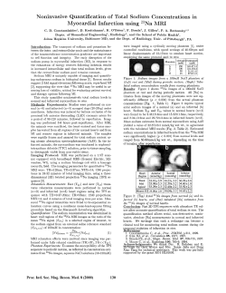


© Copyright 2026