
Printable version
MB, ChB Phase II – Theory Modules Musculoskeletal System 52302 371 2015 MB, ChB Phase II – Theory Modules 2015 MUSCULOSKELETAL SYSTEM 52302 371 MODULE SUPERVISOR: Dr ICM Robertson MUSCULOSKELETAL SYSTEM DESCRIPTION OF MODULE THEME 1: TERMINOLOGY .................................................................................................................. 17 Session 1: Terminology...................................................................................................................... 17 THEME 2: OSTEOLOGY ...................................................................................................................... 20 Session 1: Osteology ......................................................................................................................... 20 THEME 3: JOINTS ................................................................................................................................ 27 Session 1: Joints ................................................................................................................................ 27 THEME 4: THE VERTEBRAL COLUMN ............................................................................................. 39 Session 1: The Vertebral Column ...................................................................................................... 39 THEME 5: SKELETAL MUSCLES ....................................................................................................... 42 Session 1 – 13: Skeletal Muscles ...................................................................................................... 43 Practical Programme.......................................................................................................................... 49 THEME 6: BONE AND CARTILAGE ................................................................................................... 59 Session 1-4: Lectures ........................................................................................................................ 59 Session 5: Practical ........................................................................................................................... 60 Session 6 en 7: Tutorials On The Computer...................................................................................... 61 THEME 7: MUSCULAR ACTIONS ....................................................................................................... 62 Session 1-4: Muscular Action ............................................................................................................ 62 NEURO SCIENCES SESSIONS........................................................................................................... 67 Session 1: Anatomy Of The Spinal Cord ........................................................................................... 67 Session 2: Spinal Cord....................................................................................................................... 68 Session 3: Trauma Of The Spinal Cord ............................................................................................. 68 Pathology Of Trauma Of The Spinal Cord ......................................................................................... 69 Session 4: The Pathology Of Diseases Of The Spinal Cord And Lmn, Including Anterior Horn Cell, Neuromuscular Nerve Junction, Muscular And Peripheral Nerves ........................... 69 Session 5: Muscle Disease/Proximal Weakness ............................................................................... 70 Session 6: Peripheral Neuropathy ..................................................................................................... 70 Session 7:Anatomy, Physiology & Clinical Features Of The Autonomic Nervous System ............... 71 Session 8: Pharmacology Of Neuropathic Pain ................................................................................. 71 Session 9: Localisation Of A Lower Motor Neuron (LMN) Lesion ..................................................... 72 Session 10: The Autonomic Nervous System.................................................................................... 72 THEME 8: INTRODUCTION TO THE CLINICAL MUSCULOSKELETAL BLOCK - 371 ................... 73 Session 1: Introduction Lecture ......................................................................................................... 74 Session 2: Terminology...................................................................................................................... 74 Session 3: Biomechanics and Physics .............................................................................................. 77 Session 4: Principles of Applies Anatomy.......................................................................................... 78 Session 5: Examination of the Orthopaedic Patient .......................................................................... 78 Session 6: Orthopaedic Special Investigations - Radiology .............................................................. 79 Session 7: Principles of Nuclear Medicine ......................................................................................... 80 THEME 9: ORTHOPAEDIC TRAUMA - GENERAL PRINCIPLES...................................................... 81 Session 1: Approach to the Orthopaedic Trauma Patient - Resuscitation ........................................ 81 Session 2: Approach to the Orthopaedic Trauma Patient - General Principles of Fracture Management ...................................................................................................................... 82 Session 3: Approach to the Orthopaedic Trauma Patient - Complications ....................................... 83 Session 4: Approach to the Orthopaedic Trauma Patient - Paediatric .............................................. 84 Session 5: Approach to the Orthopaedic Trauma Patient - Spinal Cord Injury ................................. 85 THEME 10: ORTHOPAEDIC TRAUMA - UPPER LIMB ...................................................................... 89 Session 1: Approach to Shoulder and Arm Trauma .......................................................................... 87 Session 2: Approach to Elbow, Fore-arm Trauma ............................................................................. 88 Session 3: Approach to Wrist, Hand and Tendon Trauma ................................................................ 91 THEME 11: ORTHOPAEDIC TRAUMA - AXIAL, SKELETAL AND LOWER LIMB ........................... 93 Session 1: Vertebral Column Fractures ............................................................................................. 93 Session 2: Approach to Pelvic Girdle Trauma ................................................................................... 95 Session 3: Approach to Hip and Femur Shaft Trauma ...................................................................... 96 Session 4: Approach to Knee Trauma ............................................................................................... 97 Session 5: Approach to Tibial Trauma ............................................................................................... 98 Session 6: Approach to Foot and Ankle Trauma ............................................................................. 100 THEME 12: ORTHOPAEDIC TRAUMA - SOFT TISSUE TRAUMA.................................................. 101 Session 1: Mamangement of Wounds and Lacerations .................................................................. 101 Session 2: Principles fo Wound Healing .......................................................................................... 102 THEME 13: MUSCULOSKELETAL INFECTION ........................................................................... 104 Session 1: General Pathological Principles of Infection .................................................................. 104 Session 2: General Microbiology Principles of Infection .................................................................. 106 Session 3: Antibiotics in Musculoskeletal Infection .......................................................................... 107 Session 4: Approach to Acute Musculoskeletal Infections .............................................................. 108 Session 5: Approach to Chronic Musculoskeletal Infections ........................................................... 109 Session 6: Approach to Musculoskeletal Infective Conditions of the Spine .................................... 111 Session 7: Approach to Hand Infections .......................................................................................... 112 THEME 14: ORTHOPAEDIC ONCOLOGY ........................................................................................ 114 Session 1: Oncology Pathological - Principles - Part 1 .................................................................... 114 Session 2: Oncology Pathological - Principles - Part 2 .................................................................... 114 Session 3: Approach to Benign Bone Tumours ............................................................................... 116 Session 4: Approach to Malignant and Metastatic Bone Tumours .................................................. 117 Session 5: Approach to Soft Tissue Tumours ................................................................................. 119 THEME 15: PAEDIATRIC ORTHOPAEDICS CONGENITAL AND DEVELOPMENT ...................... 120 Session 1: Approach to the Limping Child ....................................................................................... 121 Session 2: Development Dysplasia of the Hip ................................................................................. 122 Session 3: Perthe's Disease ............................................................................................................ 123 Session 4: Slipped Capital Femoral Epiphysis ................................................................................ 124 Session 5: Approach to Mal-Alignment of the Lower Limb .............................................................. 125 Session 6: Approach to Gait Analysis .............................................................................................. 126 Session 7: Paediatric Foot Deformities ............................................................................................ 127 Session 8: Approach to Neuro Muscular Conditions ....................................................................... 128 Session 9: Approach to Paediatric Spinal Deformities .................................................................... 129 THEME 16: ORTHOPAEDIC CONDITIONS OF THE SPINE ............................................................ 131 Session 1: Approach to Back Ache .................................................................................................. 131 Session 2: Approach to Inflammatory Conditions of the Spine........................................................ 132 Session 3: Approach to Adult Spinal Deformities and Degeneration .............................................. 133 THEME 17: MUSCULOSKELETAL CONDITIONS OF THE UPPER LIMB ...................................... 135 Session 1: Approach to Shoulder Pain ............................................................................................ 135 Session 2: Approach to Shoulder Instability .................................................................................... 136 Session 3: Appraoch to Degenerative Shoulder Conditions ............................................................ 137 Session 4: Approach to Elbow Pain ................................................................................................. 139 Session 5: Approach to Wrist and Hand Pain - Part 1 ..................................................................... 140 Session 6: Approach to Wrist and Hand Pain - Part 2 ..................................................................... 141 Session 7: Approach to Peripheral Nerve Conditions of the Upper Limb ........................................ 143 THEME 18: MUSCULOSKELETAL CONDITIONS OF THE LOWER LIMB ..................................... 145 Session 1: Approach to Adult Hip Pain ............................................................................................ 145 Session 2: Approach to Adult Knee Pain ......................................................................................... 146 Session 3: Sports Injuries ................................................................................................................ 147 Session 4: Approach to Heel and Ankle Pain .................................................................................. 147 Session 5: Approach to Medial Arch Pain of the Foot ..................................................................... 148 Session 6: Approach to Metatarsalgia ............................................................................................. 149 THEME 19: APPROACH TO ARTHRITIS .......................................................................................... 151 Session 1: Approach to a Patient presenting with Musculoskeletal Complaints ............................. 152 Session 2: Rheumatoid Arthritis (RA) .............................................................................................. 152 Session 3: Approach to Mono-Arthritis (MO) and Osteo-Arthritis (OA) ........................................... 153 Session 4: Crystalarthropathies ....................................................................................................... 153 Session 5: Interactive Session ......................................................................................................... 154 Session 6: Spondylo-Arthropathies .................................................................................................. 154 Session 7: Infections and Rheumatology......................................................................................... 154 Session 8: Soft Tissue Rheumatism and Generalised Pain ............................................................ 155 Session 9: Paediatric Rheumatism - Part 1 ..................................................................................... 155 Session 10: Paediatric Rheumatism - Part 2 ................................................................................... 155 Session 11: Interactive Session ....................................................................................................... 156 THEME 20:APPROACH TO CONNECTIVE TISSUE DISEASES .................................................... 157 Session 1: Recognising a Patient with a Connective Tissue Disease ............................................. 157 Session 2: Systemic Lupus Erythematosus (SLE) .......................................................................... 158 Session 3: Systemic Sclerosis and Inflammatory Myopathies......................................................... 158 Session 4: Sjogrens Syndrome and Vasculopathies ....................................................................... 159 Session 5: Interactive Session ......................................................................................................... 159 THEME 21: TREATMENT OF RHEUMATOLOGICAL CONDITIONS .............................................. 160 Session 1: Pharmacology ................................................................................................................ 160 Session 2: Paramedical (OT, Physio, SW) ...................................................................................... 161 THEME 22: THE ROLE OF OCCUPATIONAL THERAPY AND PHYSIOTHERAPY IN REHABILITATION ......................................................................................................................... 162 Session 1: OT And PT In Rehabilitation Of Orthopaedic Conditions ............................................... 162 Session 2: Guidelines For Referral .................................................................................................. 163 TIME TABLE ....................................................................................................................................... 164 MUSCULOSKELETAL SYSTEM INTRODUCTION This module comprises the study of the musculoskeletal system with regards to the different components, microscopic and macroscopic structures, functional characteristics, physiological aspects and biochemical composition, so that you can understand the relevant disease processes, clinical pictures and management of the conditions. OVERVIEW MUSCULOSKELETAL BLOCK This musculoskeletal course lasts approximately eight weeks. The initial section consists of four weeks of basic science lectures including Anatomy and Physiology, the neurology of the peripheral nerves, as well as applied Anatomy and Physics. The last four weeks of this course is clinically orientated and consists of musculoskeletal conditions such as Trauma, Paediatric Orthopaedics and Elective Orthopaedics at the level of what a competent General Practitioner would be expected to know. Seventeen lectures are devoted to Rheumatology and there are also lectures on subjects such as Physiotherapy, Occupational Therapy and Nuclear Medicine. There will be many evaluation opportunities. In the basic science section (first four weeks), a practical assessment, as well as a theoretical test will be held. At the end of the clinical section (second four week block) a written test covering this section will be completed. The final examination covering all material studied in this course will be held in November. START OF MODULE This module starts at 08:00 on Tuesday 28 April 2015 (with lectures in Lecture Hall 7, Teaching Block). LOCATIONS All lectures are presented in Lecture Hall 7, Teaching Block. All Anatomy practicals are presented in practical locations F123/124, F237/238 and F225, Fisan Building. All Histology practicals are presented in the Histology Laboratory (Room 5035, Fifth Floor, Education Block). STARTING POINT The starting point is that you are primarily responsible for your own learning process. This starting point is maintained throughout this module. In this module, you will be required to work independently and continuously so that you can complete the outcomes of each theme by the end of the module. A number of activities are presented to make the learning process easier and to complete the outcomes. These activities include formal lectures, structured unaccompanied group work (practicals), and directed self-study. 7 Phase II (Theory): MUSCULOSKELETAL SYSTEM – 52302 371 © Faculty of Medicine and Health Sciences, Stellenbosch University. 2015 All rights reserved. SELF-STUDY ACTIVITIES As already mentioned, you are responsible for your own learning process. Directed self-study activities must therefore be given the necessary attention since they often serve as preparation for practicals and also contribute to making the outcomes more easily attainable. In this module, you must work independently and continuously so that the outcomes of each theme can be achieved at the end of the module (for example, the theme dealing with Osteology and Joints follows closely on the theme covering Skeletal muscles: you must consequently cover the theme dealing with Skeletal muscles while independently acquiring knowledge on aspects that have a relationship to the themes that have been covered in Osteology and Joints). You will consequently only be able to complete the outcomes at the end of the module. Consult the necessary resources in good time so that the specific aspects (as described in this study guide in the Self-study programme) can be studied. OUTCOMES The outcomes for each theme are described to give you an indication of what must eventually be completed by the end of the module. The aim is not to answer the outcomes in writing. You should know all the information after completing the module. The themes serve as a control for the contents that have been covered, as well as the required depth. It will be a waste of time to answer all the outcomes in writing and, if you try, you will waste time that should be used for other activities. PRESCRIBED RESOURCES: (for pre-clinical module of musculoskeletal course) 1. th Moore and Dalley. 2011/2013. Clinically Oriented Anatomy. 6 or 7 th Edition, Lippincott, William and Wilkens. (References are indicated as M6/M7 in the study guide). (NB! All page references in this study guide refer to Moore!) th 2. Tank, PW, (Sauerland, EK). Grant’s Dissector, William and Wilkens. (14th or 15 Edition). 3. Netter, F. Atlas or Human Anatomy. Novartis. 4. Young, Lowe, Stevens and Heath. 2006. Wheater’s Functional Histology. 5 th Churchill Livingstone, or 4 5. th Edition, Edition, 2000 Silverthorn. 2000. Human Physiology: An Integrated Approach. th 5 Edition, Pearson Benjamin Cummings. 6. Meisenberg & Simmons. Medical Biochemistry. St. Louis, Mosby. 7. John T. Hansen. Netter’s Clinical Anatomy, 2 nd rd or 3 edition, (Saunders, Elsevier). RECOMMENDED RESOURCES 1. Ross and Lamperti. 2006. Thieme Atlas of Anatomy. Neck and internal organs. Thieme, New York. 2. Sadler TW. Langman’s Medical Embryology. 3. Anne M Gilroy. Atlas of Anatomy. Thieme, Stuttgart, New York. 4. http://www.harford.edu/faculty/EDuncan/links.htm 5. http://www.health24.com/tools/gallery/anatomy/start.asp 8 Phase II (Theory): MUSCULOSKELETAL SYSTEM – 52302 371 © Faculty of Medicine and Health Sciences, Stellenbosch University. 2015 All rights reserved. TAKE NOTE This theme (which deals with skeletal muscles) is spread over a period of 3½ weeks. The major component of this theme is a practical program (during which a structured dissection program is followed), coupled to a self-study program (as preparation for the practicals) a pre-assessment (forms 10% of class mark) and a lecture- program (supplemented where necessary). Theme 1 starts on TUESDAY 28 April 2015 at 08:00. All students must meet on TUESDAY 28 April 2015 at 08:00 in Venue 7, Teaching Block for a lecture. The practical sessions start WEDNESDAY 29 April 2015 in locations F123/124, F237/238, F225 and Histology lab. Attendance of these practical programmes is compulsory. You are working with human tissue (bones and wet specimens). You must make sure of the “Notice in compliance with Article 65 of the Anatomy Act, 1983” which will appear on the notice board. It is your responsibility to complete and sign the registration form, which will be handed out to you. This registration form with your signature is compulsory in accordance with the Anatomy Act (1983), which grants you permission to handle human tissue. The Inspector of Anatomy checks this registration form during his annual visit to the Department. There are specific conditions for the loan of skeletal bones (also see “notice” on the notice board in connection with the conditions for lending of skeletal bones. Two students share an unarticulated skeleton for use during the first part of this module. If any student suspends their course, they must inform the Department of Anatomy and Histology immediately and hand the bones in. You are not allowed to make any marks on the bones. You will be informed of dates to hand bones in. Each student must pay an amount of R500.00 for leasing of the skeletons. The fee will be deducted from your student’s account NB! An amount of R300 per student is payable for the loan of the skeletons during the year. This amount will not be refunded. NB! An amount of R200 will be paid back only if all material handed back is complete and in a satisfactory condition. You are not allowed to make any marks on the bones. Handing in late: handing in late will only be accepted if you hand in a written request with valid reasons to the Division Anatomy and Histology 2 weeks prior to the handing in date. Also arrange a time with her when the bones will be handed in. NB! A fine of between R50 to R250 is payable for late handing in of bones. Skeletal bones must be handed back in person at the Division. Arrangements will be confirmed at the end of the module. Strict rules apply according to Article 65 of the Anatomy Act, 1983 when students are in possession of human tissue (i.e. skeletal bones). If this ordinance is transgressed, the Inspector of Anatomy may instigate further investigations in accordance with Article 65 of the Anatomy Act, 1983. Each skeletal bone supplied to you, is on record. Each bone MUST later be handed back to the Division of Anatomy and Histology in a neat and acceptable condition. Handing in of bones will start on the day of writing the Musculoskeletal module examination and the next successive two days. Bones of different skeleton sets may not be exchanged or mixed. Handing back of bones takes place in the ossuary, Fisan Building. 9 Phase II (Theory): MUSCULOSKELETAL SYSTEM – 52302 371 © Faculty of Medicine and Health Sciences, Stellenbosch University. 2015 All rights reserved. CODE OF CONDUCT FOR THE DIVISION ANATOMY AND HISTOLOGY All lectures and practicals are compulsory. The class will be divided into the same groups as for Cardiovascular and Respiratory Modules (2014). Ensure that you know in which group and dissection hall you are working. You are not allowed to change groups. No visitors are allowed for students in the museum and dissection halls. In exceptional circumstances you can organize a visit with the head of the department or for the museum, with the curator. It is strictly forbidden to take photographs of any human tissue in the lecture or dissection halls or the museum. Guilty students run the risk of being expelled. Your group is responsible for the care of the cadaver. Exposure to air is the main cause of damage to the tissue. Therefore, expose only the area where you work and keep the rest of the cadaver covered with the wet flannel cloths and plastic. Keep the skin of the cadaver and replace it, together with the cloth and plastic after the practical. The flannel cloths should be wet when you cover the cadaver. Use the fluid provided at your table. All the dissected tissue should be placed into the specific container for your cadaver. Clean the work area and leave it in a tidy condition. The use of latex gloves is optional. Place all used sharp instruments (blades) into the special sharps containers. Be careful not to injure yourself or your fellow students. Students are allowed into the dissection halls from Monday to Friday from 08:00 – 16:00. It is compulsory to wear a clean white coat, closed shoes and long trousers (for men) to the dissection practical. You will not be allowed into the practical without the correct clothing. The same rules apply to the practical assessing. Your general appearance should be neat and respectable. It is compulsory to wear shoes in the Fisan building. Smoking is prohibited in the buildings of the Faculty of Health Sciences. All human tissue in the department is under the control of the Human Tissue Act (1983) and the Inspector of Anatomy. The skeletal bones used by students must be handed back to the Department from the the day of writing the Musculoskeletal module examination in November and the next successive two days, or earlier should you end your course. Late handing in of skeletal bones will result in fines and loss of deposits. Failure to give the bones back will lead to prosecution by the police. Eating, drinking and the use of radios are prohibited in the dissection halls. Despite the condition (death) of the cadaver, it is still human. That human had thoughts, he/she saw the beauty of nature, he/she held a baby, and his /her feet carried him/her on their path of life. Treat the remains of that person with the necessary respect and compassion. Times for the museum: Monday to Friday 08:00 – 16:00. The museum is closed on weekends and public holidays. Only students and personnel of the faculty are allowed into the museum, unless a specific visit is organized. Silence and irreproachable behaviour is expected in the museum. Do not sit on the tables, use the chairs provided. Any problems in the museum should be reported to Prof. B.J. Page, Room F162 on the First floor, Fisan Building. 10 Phase II (Theory): MUSCULOSKELETAL SYSTEM – 52302 371 © Faculty of Medicine and Health Sciences, Stellenbosch University. 2015 All rights reserved. ICONS The following are used so that you can easily find your way around this study guide: … your destination for the theme, i.e. the outcomes … contact session with a lecturer … an assignment that you must complete, i.e. self-study … group work with your fellow students … a practical … a case study … the use of prescribed reading material … the use of computers … the use of video material … the use of internet material … the use of slides … take note! … a brain “workout”: something extra for those looking for a challenge … self-assessment, an opportunity to ascertain whether you are on the right path … assessment during the module, that contributes to the class mark 11 Phase II (Theory): MUSCULOSKELETAL SYSTEM – 52302 371 © Faculty of Medicine and Health Sciences, Stellenbosch University. 2015 All rights reserved. ASSESSMENT Section 1 – Anatomy (A), Histology (B), Physiology (C) and Neurosciences (D) General At the end of this section, there will be a theoretical (written) and a practical (identification) assessment. Detail on the nature of these assessments will be supplied to you at a later date. Section 1 test mark The test mark will be calculated as follows: Practical assessment (Spot test): Written (theoretical) assessment: GERGA (theoretical) assessment: 35% Section A, B and C 35% Section D 20% Pre-practical Sunlearn test: 10% Practical Assessment st Date: Thursday 21 May 2015 Time: The group-division, venue and times for meeting will be made known at a later stage on the notice board, Fisan Building. 11:30 – 14:30 Location: GERGA and Examination location, 5th Floor, Teaching Block Type: Identification (identification of anatomical and histological structures). Percentage contribution: 50% Theoretical Assessment Date: nd Friday 22 May 2015 Time: 10:00 – 12:30 The group-division (GERGA, BOYD) and times for meeting will be made known at a later stage on the notice board, Fisan Building. Location: GERGA and Examination location, 5th Floor, Teaching Block Type: True -and-false questions; short questions; paragraph questions; long questions; Multiple choice questions on WebCT Percentage contribution: 50% Because the practical assessment contributes 50% to the marks of the preclinical part of this module, group activities during the practicals are of great importance. If you do not use your time optimally during the practical sessions (directed self-study and preparation is thus a prerequisite), you cannot expect to do well in the practical evaluation. You should thus work independently and continuously and use your practical sessions optimally, if you wish to complete this module successfully. 12 Phase II (Theory): MUSCULOSKELETAL SYSTEM – 52302 371 © Faculty of Medicine and Health Sciences, Stellenbosch University. 2015 All rights reserved. Clinical Assessment Date: Friday 31st July 2015 Time: 09:00 – 12:00 Location: GERGA Type: Multiple Choise Questions Examination (Written & Computer MCQ) The examination for MB,ChB-students takes place on Wednesday 4th November 2015(at 09:00) in GERGA and the Examination Location, Fifth Floor, Teaching Block (details to be confired at a later stage). Global Mark Allocation 1) Test a. Practical anatomy test (Thursday 21 May 2015) b. First theory test [Will include Anatomy, Histology, Physiology and Neurology and will cover all subjects studied up to the date of the test (Friday 22 May 2015)] c. Second theory test will include Clinical Orthopaedics, Rheumatology and other subjects st studied since the first test. (Friday 31 d. 2) July 2015) Test mark allocation = (Preclinical tests [a +b] and portofolio + Clinical test [c])/2 th Exam (Wednesday 4 November 2015) a. Anatomy and Neurology (90 marks) b. Orthopaedics and Rheumatology (90 marks) Final mark will be calculated as follows (Test mark [1.d] + Exam mark [2])/2 13 Phase II (Theory): MUSCULOSKELETAL SYSTEM – 52302 371 © Faculty of Medicine and Health Sciences, Stellenbosch University. 2015 All rights reserved. INTRODUCTION CLINICAL SECTION Welcome to the clinical section of the musculoskeletal system module. Conditions of the musculoskeletal system are particularly common, and will occupy much of your time in general practice. In the USA, it is one of the most common reasons for patients consulting their general practitioner. Approximately 75% of all people will, for example, at some time present with complaints of back pain. Trauma and violence are presently epidemic in South Africa, and with the increase in high velocity injuries and gunshot wounds, your practice with continually be overflowing with musculoskeletal injuries. Other common conditions include joint diseases, bone diseases and deformities. To better understand these, as well as the neoplastic and infective conditions of the musculoskeletal system, appropriate pathological as well as microbiological aspects will be covered. The relevance of the various imaging modalities will also be put into prospective throughout. During this block, the presentations will, as far as possible, be multidisciplinary since treatment of these problems usually requires a multidisciplinary approach. There will be a continual emphasis on a practical approach with regards to the most common conditions affecting the musculoskeletal system. The most important aspects of the clinical diagnosis, radiological interpretation and treatment will be emphasized. A number of practical self-study assignments will be expected from you; you are encouraged to complete these assignments as quick as possible and to liase with your class mates for future reference. Review Sessions: At the end of most themes there will be a review session. All lecturers who were involved in the session will participate in this. Prior to this session students are encouraged read over the study material relevant to the theme, and to prepare questions to ask this panel. The lecturers will answer questions and discuss and clarify topics covered in the recently completed theme. Members of the panel will also present case studies of relevant clinical conditions. REFERENCES AND TEXTBOOKS FOR THE CLINICAL SECTION OF THE MODULE Please make use of the references that appear in your study guides. All references can be found in the library of the Faculty of Health sciences. You also have access to numerous videos, which are worth using to establish practical guidelines. You are recommended to buy the following textbooks. The majority of information that you require appears within. 1. Concise System Of Orthopaedics And Fractures – AG Apley/L Solomons 2. Apleys System Of Orhtopaedics And Fractures – AG Apley/L Solomons 3. Clinical Orthopaedic Examination - Ronald Mcrae 4. Practical Fracture Treatment - Ronald Mcrae 5. Dorlands Medical Dictionary – Saunders 14 Phase II (Theory): MUSCULOSKELETAL SYSTEM – 52302 371 © Faculty of Medicine and Health Sciences, Stellenbosch University. 2015 All rights reserved. Websites 1. www.sun.ac.za/ortho 2. webstudies.sun.ac.za (module 371) LOCATIONS The locations referred to in the timetables for the activities of the themes are as follows: Lectures HL Lecture Hall 7, Ground Floor, Teaching Block PL Practical Locations Histology Laboratory, Room 5035, 5th Floor, Education Block 15 Phase II (Theory): MUSCULOSKELETAL SYSTEM – 52302 371 © Faculty of Medicine and Health Sciences, Stellenbosch University. 2015 All rights reserved. LECTURERS These are the names and contact details of the lecturers that are involved with the module: NAME Prof B Page Dr V Tchokonte-Nana Ms M Alblas Dr SH Kotze Dr D van Vuuren Prof J Carr Dr AJ Vlok Dr F Henning Dr D Zaharie Dr R Gounden Dr J du Toit Dr I Robertson Dr H de Jongh Dr A Ikram Dr S Pretorius Dr N Terblanche Dr G du Preez Dr J Davis Dr D Hugo Prof J Schneider Prof J Apffelstaedt Dr F Graewe Dr R Hoffmann Dr J Warwick Dr D Whitelaw Dr M Manie Dr R du Toit Dr B Viljoen Dr L du Plessis Dr M Esser Dr F Bouwer Ms S Schmutz Ms S de Klerk DEPT/DIV Div of Anatomy & Histology Div of Anatomy & Histology Div of Anatomy & Histology Div of Anatomy & Histology Div of Physiology Div of Neurology Div of Neuro Surgery Div of Neurology Div of Neuro Pathology Div of Pharmacology Div of Orthopaedic Surgery Div of Orthopaedic Surgery Div of Orthopaedic Surgery Div of Orthopaedic Surgery Div of Orthopaedic Surgery Div of Orthopaedic Surgery Div of Orthopaedic Surgery Div of Orthopaedic Surgery Div of Orthopaedic Surgery Div of Anat Pathology Div of Surgery Div of Plastic Surgery Div of Microbiology Div of Nuclear Medicine Div of Rheumatology Div of Rheumatology Div of Rheumatology Div of Rheumatology Div of Rheumatology Pediatric Rheumatology Div of Reumatology Div of Physiotherapy Div of Occupational Therapy E-MAIL [email protected] [email protected] [email protected] [email protected] [email protected] [email protected] [email protected] [email protected] [email protected] [email protected] [email protected] [email protected] [email protected] [email protected] [email protected] [email protected] [email protected] [email protected] [email protected] [email protected] [email protected] [email protected] [email protected] [email protected] [email protected] [email protected] [email protected] [email protected] [email protected] [email protected] [email protected] [email protected] [email protected] TEL 021-938 9430 021-938 9413 021-938 9394 021-938 9428 021-938 9390 021-938 9627 021-938 9265 021-938 9512 021-938 9535 021-938 9334 021-938 9266 021-938 9266 021-938 9266 021-938 9266 021-938 9266 021-938 9266 021-938 9266 021-938 9266 021-938 9266 021-938 4041 021-938 9273 021-938 9432 021-938 5193 021-938 4352 021 938 5527 021 938 5527 021 938 5527 021 938 5527 021 938 5527 021-938 4032 021 938 5527 021-938 9300 021-938 9038 Contact People Helga Oldewage: Suzette Olivier: Marie de Beer: Lyndal Swanepoel: Division of Orthopaedics, Tel: 021- 938 9266, E-mail: [email protected] Department of Internal Medicine, Tel: 021-938 9376, E-mail: [email protected] Division of Anatomy, Tel: 021-938 9397, E-mail: [email protected] Department of Neuro Sciences, Tel: 021-938 9478 E-mail: [email protected] 16 Phase II (Theory): MUSCULOSKELETAL SYSTEM – 52302 371 © Faculty of Medicine and Health Sciences, Stellenbosch University. 2015 All rights reserved. THEME 1: TERMINOLOGY AIM OF THE THEME At the end of the theme, you should realise the importance of internationally acceptable anatomical terms and also be able to apply anatomical terms correctly since anatomical terminology forms a large portion of general medical terminology. OVERVIEW OF THE THEME Session Date Venue Activity Lecturer 1 28/04/2015 09:00 Venue 7 Lecture Dr V Tchokonte-Nana THEME 1: TERMINOLOGY SESSION 1: TERMINOLOGY OUTCOMES 1. Describe and explain the anatomical positions. 2. Describe and explain the different anatomical levels. 3. Describe and explain terms that demonstrate the relative position of structures to each other (terminology with regards to relationships). 4. Describe and explain terms that describe movements of limbs and other parts of the body (terminology with regards to movement). RESOURCES 1. Moore 6, Moore 7 ANYTHING SPECIFIC YOU SHOULD DO? Lecture and Directed Self-study During the lecture, selected aspects of terminology will be explained and demonstrated to you. During the directed self-study, you should study the sections on terminology in preparation for the application thereof in this section. Self-study The following aspects should be dealt with during the self-study so that the anatomical position, anatomical planes and the specific meaning of certain terms can be defined and explained: the anatomical position (M6: 5; M7: 5) 17 Phase II (Theory): MUSCULOSKELETAL SYSTEM – 52302 371 © Faculty of Medicine and Health Sciences, Stellenbosch University. 2015 All rights reserved. anatomical planes (M6: 5; M7: 5) terminology with regards to relationships (M6: 6; M7: 6) terminology with regards to movements (M6: 7; M7: 7) Self-assessment After completion of the self-study, see if you can answer the following questions: 1. 2. 3. 4. 5. The correct definition for abduction is: (a) Moving away from the median plane (b) Moving away from the median plane in a coronal plane (c) Moving away from the median plane in a sagittal plane (d) Moving the upper or lower limb away from the body (e) Moving away from the axial line of the body Lateral means: (a) Further away from the coronal plane (b) Further away from the median plane (c) Further away from the sagittal plane (d) Further away from the body (e) On the opposite side of the body Inferior means: (a) Away from the head (b) Distally (c) Downwards (d) Nearer to feet (e) Nearer to the floor Superior means: (a) Upwards (b) Proximally (c) Nearer to the roof (d) Nearer to the head (e) Away from the feet What is the anatomical definition of the following terms: (a) Median plane (b) Sagittal plane (c) Flexion (d) Extension (e) Rotation (f) Medial rotation (g) Lateral rotation (h) Abduction (i) Adduction (j) Circumduction (k) Proximal 18 Phase II (Theory): MUSCULOSKELETAL SYSTEM – 52302 371 © Faculty of Medicine and Health Sciences, Stellenbosch University. 2015 All rights reserved. (l) Distal (m) Pronation (n) Supination (o) Eversion (p) Inversion (q) Dorsiflexion (r) Plantar flexion (s) Medial (t) Lateral 19 Phase II (Theory): MUSCULOSKELETAL SYSTEM – 52302 371 © Faculty of Medicine and Health Sciences, Stellenbosch University. 2015 All rights reserved. THEME 2: OSTEOLOGY AIM OF THE THEME At the end of the theme, you should be able to describe, explain, identify and demonstrate the organisation and composition of the skeleton. OVERVIEW OF THE THEME Session Date Venue Activity Lecturer 1 28/04/2015 10:00 Venue 7 Lecture Dr V Tchokonte-Nana THEME 2: OSTEOLOGY SESSION 1: OSTEOLOGY OUTCOMES 1. Explain the organisation and composition of the human skeleton. 2. Name the functions of bones. 3. Identify and name all the bones of an articulated human skeleton. 4. Describe and explain the principles of the blood supply and nerve supply of bone (selfstudy). 5. Name the different characteristics of bones and identify each on a bone. RESOURCES 1. Moore 6/7 2. The unarticulated skeleton supplied to you on loan. 3. Articulated skeletons in practical locations. ANYTHING SPECIFIC YOU SHOULD DO? Lecture and Self-study During the lecture, selected aspects of osteology will be explained and demonstrated to you, with the help of certain selected bones. During the directed self-study, you should consult your resources to study the bones and their specific parts. During the practicals (on muscle groups), you will also have the opportunity to name and identify the relevant osteological structures that have a reference to muscle groups or specific muscles (for example attachments of muscles). 20 Phase II (Theory): MUSCULOSKELETAL SYSTEM – 52302 371 © Faculty of Medicine and Health Sciences, Stellenbosch University. 2015 All rights reserved. Self-study Consult your resources to study the following aspects in preparation for practicals (that follow later) so that the relevant osteological aspects are already known to you. This results in the required outcomes being attained more easily: Name the functions of bones (M6; 19; M7: 19). Explain and demonstrate the concepts axial and appendicular skeleton (M6; 19; M7:19). Explain the meaning of the following terms (M6; 20;M7: 20): condyles fossa epicondyle notch supracondylar ridge head tubercle neck tuberosity shaft trochanter linea aspera ridge interosseous border capitulum foramen trochlea groove spina ramus malleolus articular processes/surfaces line (“linea”) projection Name and identify the following bones with their specific parts: 1. Occipital bone (M6; 822;M7: 822) occipital condyles 2. Vertebrae (M6: 76;440-453;982-984;M7; 76;440-453; 982-984) 3. body vertebral foramen pedicle lamina transverse process spinous process articular surfaces foramen transversarium (C1-C7) dens (on C2) sacrum (5 fused sacral vertebrae) Sacrum (M6: 451-453; M7: 451-453) sacral canal sacral foramina (intervertebral foramina) anterior surface (pelvic surface) posterior surface sacral promontorium (anterior margin of body of S1) base (superior surface of S1) 21 Phase II (Theory): MUSCULOSKELETAL SYSTEM – 52302 371 © Faculty of Medicine and Health Sciences, Stellenbosch University. 2015 All rights reserved. 4. 5. 6. 7. apex (attached to Co1) median crest (median crest) lateral crest sacral hiatus intermediary crest auricular surface (articulatory surface with ilium) sacral cornu Sternum (M6: 76-78; M7: 76-78) manubrium body xiphoid-process jugular notch (suprasternal notch) clavicular notch attachment-area for cartilage of rib 1 costal notches 2-7 (for articulation with costal cartilages 2-7) manubrio-sternal joint (sternal angle; angle of Louis) xiphi-sternal joint transverse ridges Rib (M6: 74; M7: 74) head neck shaft tubercle costal angle costal groove articular surfaces external surface internal surface Clavicle (M6: 673-675; M7: 673-675) medial extremity (sternal extremity) lateral extremity (acromial extremity) shaft superior surface inferior surface anterior margin posterior margin Scapula (M6: 675-676 ; M7 : 675-676) spina supraspinous fossa infraspinous fossa subscapular fossa acromion coracoid process 22 Phase II (Theory): MUSCULOSKELETAL SYSTEM – 52302 371 © Faculty of Medicine and Health Sciences, Stellenbosch University. 2015 All rights reserved. 8. glenoid cavity superior angle inferior angle medial border lateral border Humerus (M6: 676-677 ; M7 : 676-677) proximal portion head neck (anatomy neck) surgical neck greater tubercle lesser tubercle intertubercular groove (bicipital groove) body (body) deltoid tuberosity distal portion lateral supracondylar ridge medial supracondylar ridge lateral epicondyle medial epicondyle capitulum (M6 : 677 ; M7 : 677) trochlea (M6 : 677 ; M7 : 677) coronoid fossa (M6 : 677 ; M5 :677) olecranon fossa (M6 : 677 ; M7 : 677) 9. Ulna (M6: 677-678 ; M7 : 677-678) 10. Proximal portion olecranon trochlear notch coronoid process radial notch (for articulation with head of radius) tuberosity of ulna body distal portion head styloid process (of ulna) Radius (M6: 678-679 ; M7 : 678-679) proximal portion head neck tuberosity of radius body distal portion ulnar notch (for articulation with head of ulna) styloid process (of radius) 23 Phase II (Theory): MUSCULOSKELETAL SYSTEM – 52302 371 © Faculty of Medicine and Health Sciences, Stellenbosch University. 2015 All rights reserved. 11. Carpal bones (M6: 680; M7:680) scaphoid trapezium lunate trapezoid triquetrum capitate pisiform hamate 12. Metacarpal bones (M6: 680; M7: 680) 13. Phalanges (of hand) (M6: 680; M7: 680) 14. proximal middle distal Hip bone (os coxae) (M6: 514-516; M7: 514-516) ilium iliac ridge iliac fossa iliac tuberosity articular surface (for articulation with sacrum) anterior superior iliac spina anterior inferior iliac spina posterior superior iliac spina posterior inferior iliac spina greater sciatic notch ala (wing) of ischium (postero-lateral surface) ischium body of ischium ischial spina ischial tuberosity ramus of ischium lesser sciatic notch pubis body of pubis superior ramus of pubis inferior ramus of pubis pubic crest pubic tubercle pecten pubis (pectineal line) obturator foramen acetabulum (ilium, ischium and pubis contribute to acetabulum) acetabular fossa acetabular notch articular surface (for articulation with head of femur) 24 Phase II (Theory): MUSCULOSKELETAL SYSTEM – 52302 371 © Faculty of Medicine and Health Sciences, Stellenbosch University. 2015 All rights reserved. 15. Femur (M6: 516-520 ; M7 : 516-520) 16. 17. proximal portion head (with fovea capitis to which ligamentum capitis femoris is attached) neck greater trochanter lesser trochanter intertrochanteric crest (ridge) intertrochanteric line trochanteric fossa quadrate tubercle pectineal line (continuous with base of lesser trochanter) body anterior surface lateral surface medial surface linea aspera lateral lip of linea aspera medial lip of linea aspera gluteal tuberosity distal portion lateral supracondylar line medial supracondylar line popliteal surface adductor tubercle lateral epicondyle medial epicondyle lateral condyle medial condyle intercondylar fossa patellar surface (for articulation with patella) Patella base apex anterior surface posterior surface Tibia (M6: 520-521; M7: 520-521) proximal portion medial condyle lateral condyle medial tibial plateau tibial tuberosity body borders (anterior; interosseous; medial) surfaces (medial; lateral; posterior) 25 Phase II (Theory): MUSCULOSKELETAL SYSTEM – 52302 371 © Faculty of Medicine and Health Sciences, Stellenbosch University. 2015 All rights reserved. 18. distal portion medial malleolus articular surface Fibula (M6: 521-522; M7: 521-522) proximal portion head (with apex) body borders (anterior; interosseous; posterior) surfaces (medial; lateral, posterior) distal portion lateral malleolus articular surfaces 19. Tarsal bones (M6: 522-524; M7: 522-524) calcaneous talus navicular bone cuboid bone cuneiform bones (3): medial; intermediary; lateral 20. Metatarsal bones (M6: 524; M7:524) 21. Phalanges of foot (M6: 524; M7:524) proximal middle distal 26 Phase II (Theory): MUSCULOSKELETAL SYSTEM – 52302 371 © Faculty of Medicine and Health Sciences, Stellenbosch University. 2015 All rights reserved. THEME 3: JOINTS AIM OF THE THEME At the end of the theme, you should be able to describe and explain the classification and characteristics of the different joints. OVERVIEW OF THE THEME Session Date Venue Activity Lecturer 1 28/04/2015 14:00 Venue 7 Lecture Dr V TchokonteNana THEME 3: JOINTS SESSION 1: JOINTS OUTCOMES 1. Explain the structural classification of joints (fibrous; cartilagenous; synovial) and name the most important characteristics of each type of joint. 2. Name, identify and classify (according to above-mentioned classification system) all the joints that appear in the human skeleton. 3. Name all the movements that are present at each specific synovial joint. 4. Discuss the anatomy of the following synovial joints: atlanto-occipital joint wrist joint atlanto-axial joint hip joint shoulder joint knee joint elbow joint ankle joint RESOURCES 1. Moore 6/7 27 Phase II (Theory): MUSCULOSKELETAL SYSTEM – 52302 371 © Faculty of Medicine and Health Sciences, Stellenbosch University. 2015 All rights reserved. ANYTHING SPECIFIC YOU SHOULD DO? Lectures, Directed Self-study and Practicals During the lectures, selected aspects of the relevant joints, as well as an approach to any synovial joint described, will be explained and demonstrated to you. During the directed self-study, you should consult your resources to study specific aspects of joints (see self-study). During the practicals (on muscle groups), you will have the opportunity to name, identify and classify the relevant joints, as well as an opportunity to name and identify the important aspects of specific joints (see self-study). Self-study 1. Study the anatomy of the following synovial joints: atlanto-occipital joint (M6: 466-467; M7: 466-467) atlanto-axial joint (M6: 467-468; M7: 467-468) shoulder joint (M6: 796; M7: 796) elbow joint (M6: 800; M7: 800) wrist joint (M6: 809; M7: 809) hip joint (M6: 626; M7: 626) knee joint (M6: 634; M7: 634) ankle joint (M6: 647; M7:647) NB: Use the following framework (recipe) to describe each of above-named synovial joints: typical characteristics of synovial joints articulating surfaces of the relevant bones (for example the head of humerus articulates with the glenoid fossa of the scapula) capsule and bursa ligaments (that stabilise joints) intracapsular structures movements by joint important muscles for each of the specific movements blood supply and nerve supply of the joint important clinical aspects 2. The following joints should be named, identified and classified according to the classification-system: fibrous primarily cartilagenous (synchondroses) secondary cartilagenous (symphysis) synovial skull sutures(M6: 822-836; M7: 822-836) tempero-mandibular joint (M6: 916; M7: 916) atlanto-occipital joint (M6: 467; M7: 467) atlanto- axial joint (M6: 467; M7: 467) joints of the vertebral bodies (intervertebral joints) (M6: 464; 28 Phase II (Theory): MUSCULOSKELETAL SYSTEM – 52302 371 © Faculty of Medicine and Health Sciences, Stellenbosch University. 2015 All rights reserved. M7: 464) joints of the vertebral arches (“zygopophyseal joints”) (M6: 466; M7:466) costo-vertebral joints (M6: 79-80; M7:79) between head of rib and vertebral body between tubercle of rib and transverse process of vertebra costo-chondral joints (M6: 80; M7:80) interchondral joints (M6: 81 ; M7 : 80;) sternocostal joints (M6: 81; M7: 81;) manubrio-sternal joint (; M6: 78; M7: 78) xiphi-sternal joint (M6: 78; M7:78) lumbo-sacral joint (M6: 332; M7:332) sacro-coccygeal joint (M6: 332; M7:332) sacro-iliac joint (M6 330; M5; 330) pubic symphysis (M6: 332; M5; 332) sterno-clavicular joint (M6: 794; M7:794) acromio-clavicular joint (M6: 796; M7:796) shoulder joint (gleno-humeral joint) (; M6: 796; M7:796) elbow joint (M6: 800; M7: 800) proximal radio-ulnar joint (M6: 804 ;M7:804) distal radio-ulnar joint (M6: 806 ; M7 : 806) wrist joint (; M6: 809 ; M7 : 809) hand joints: (M6: 809-813 ; M7; 809-813) (intercarpal joints; carpo-metacarpal joints; intermetacarpal joints; metacarpo-phalangeal joints; interphalangeal joints) hip joint (M6: 626; M7: 626) knee joint (M6: 634; M7: 634) proximal tibio-fibular joint (M6: 645; M7:645) distal tibio-fibular joint (M6: 647; M7:647) ankle joint (M6: 647; M7:647) foot joints (M6: 650; 652 table 5.18; M7:650-652 table 5.18;) (subtalar joint; talo-calcaneo-navicular joint; calcaneo-cuboid joint; tarso-metatarsal joints; intermetatarsal joints; metatarso-phalangeal joints; interphalangeal joints) Information Examples of how bony aspects, ligaments, muscles and other structures contribute to the stabilisation or mobilisation of the joint: the large head of the humerus relative to the small glenoid-fossa of the scapula contributes to increasing mobilisation as a result of few limiting bony aspects that can restrict movement; the level glenoid-fossa of the scapula contributes to decreased stabilisation (or increased mobilisation) because the head of the humerus does not fit firmly in the level glenoidfossa; the communal function of the rotator-cuff muscles (supraspinatous, infraspinatous, teres minor and subscapularis) is stabilisation of the head of the humerus against the glenoidfossa of the scapula; 29 Phase II (Theory): MUSCULOSKELETAL SYSTEM – 52302 371 © Faculty of Medicine and Health Sciences, Stellenbosch University. 2015 All rights reserved. the origin of the long head of biceps brachii (which is intracapsular), contributes to stabilise the head of the humerus against the glenoid-fossa of the scapula; the “lax” inferior portion of the capsule of the shoulder joint does not restrict abduction of the humerus and thus contributes to increased mobility of the shoulder joint (M6: 799 Fig 6.95 A; M7: 799 Fig 6.95A) the glenoid-labrum (a fibro-cartilagenous structure) is deep in the glenoid-fossa and thus contributes to the stability of the shoulder joint; extra-capsular ligaments present in the shoulder joint exert minimal limiting effects with regards to the area of movement possible by the shoulder joint (M6: 799 Fig 6.95 B; M7: 799 Fig 6.61 B); the ball-and-socket architecture of the shoulder joint and hip joint contribute to a wide range of movements at this joints; the head of the femur is inserted deep in the acetabulum of the hipbone: this contributes to increased stability of the hip joint; the ligament of the head of the femur (ligamentum capitis femoris) that is situated intracapsularly, as well as other extracapusular ligaments of this joint, contribute to the stability of the hip joint; the numerous ligaments of the knee joint (patellar ligament; medial collateral ligament; lateral collateral ligament; popliteal ligaments; cross ligaments; etc) contribute to the stability of the knee joint; the menisci (C-shaped fibro-cartilage plates) deep within the articulating surface of the tibia, contribute to the stabilisation of the knee joint. Classification of joints (with examples) FIBROUS JOINTS Sutural joints Syndesmosis between a tooth and the bone wherein it is implanted CARTILAGENOUS JOINTS distal tibio-fibular joint Gomphosis skull sutures Primary cartilagenous joints (synchondrosis) first sternocostal joint costo-chondral joint xiphi-sternal joint Secondary cartilagenous joints (symphysis) manubrio-sternal joint intervertebral joints between vertebral bodies sacro-coccygeal joint pubic symphysis SYNOVIAL JOINTS atlanto-occipital joint 30 Phase II (Theory): MUSCULOSKELETAL SYSTEM – 52302 371 © Faculty of Medicine and Health Sciences, Stellenbosch University. 2015 All rights reserved. atlanto-axial joint sterno-clavicular joint acromio-clavicular joint 2nd – 7th sterno-costal joints interchondral joints costo-vertebral joints sacro-iliac joint facet-joints (“zygopophyseal joints”) tempero-mandibular joint shoulder joint; elbow joint; wrist joint proximal and distal radio-ulnar joints hip joint; knee joint; ankle joint proximal tibio-fibular joint hand joints and foot joints Important characteristics of each joint type fibrous joint bones are attached by fibrous tissue no joint cavities movement that is possible, is a function of the length of the membrane/ligament holding the bones together skull sutures: no movement in adults interosseous membrane: good movement (e.g. between ulna and radius) tooth in alveolar process: very limited movement cartilagenous joint bones attached by hyaline cartilage or fibrocartilage no joint cavities primary cartilagenous joint (synchondroses): bones attached by hyaline cartilage; limited movement in early life; joint ossifies by adulthood secondary cartilagenous joint (symphysis): bones attached by fibrocartilage (e.g. the fibrocartilagenous intervertebral discs between the bodies of vertebrae); strong joints that allow limited movement; bones attached together by fibrocartilage-disc synovial joint capsule (exist with synovial membrane on inside) joint cavities hyaline cartilage (on articulating surfaces of bones) 31 Phase II (Theory): MUSCULOSKELETAL SYSTEM – 52302 371 © Faculty of Medicine and Health Sciences, Stellenbosch University. 2015 All rights reserved. Self-assessment Answer the following questions and review the answers to check whether you have answered the questions correctly. It is a waste of time to guess the answers or to copy from someone else since the self-assessment is there to give you an indication of the extent to which you understand the structure and classification of joints. MARK THE CORRECT ANSWER (Each question has only one correct answer) 1. 2. 3. 4. 5. Which one of the following joints is a synovial joint? (a) Interchondral joints (b) Costochondral joints (c) Distal tibio-fibular joint (d) Manubrio-sternal joint (e) Sacro-coccygial joint Which one of the following joints is a primary cartilaginous joint (Synchondroses)? (a) First sterno-costal joint (b) Second sterno-costal joint (c) Manubrio-sternal joint (d) Joints between vertebral bodies (e) Sacro-coccygial joint Which one of the following joints is a secondary cartilaginous joint (Symphysis)? (a) Sutures of the skull (b) Costo chondral joints (c) Sacro coccygial joint (d) Sterno-clavicular joint (e) Acromio-clavicular joint Which one of the following joints is a fibrous joint? (a) (b) Sutures of the skull Costo chondral joints (c) Manubrio-sternal joints (d) Sacro-coccygial joint (e) Distal radio-ulnar joint The main movement(s) at the atlanto axial joint: (a) (b) (c) (d) (e) 6. Gliding movement Rotation Flexion and extension Flexion, lateral flexion, extension and rotation Flexion, extension, abduction, adduction and circumduction The main movement(s) at the atlanto occipital joint: (a) (b) (c) (d) (e) Gliding movement Rotation Flexion and extension Flexion, lateral flexion, extension and rotation Flexion, extension, abduction, adduction and circumduction. 32 Phase II (Theory): MUSCULOSKELETAL SYSTEM – 52302 371 © Faculty of Medicine and Health Sciences, Stellenbosch University. 2015 All rights reserved. 7. The main movement(s) at facet joints: (a) (b) (c) (d) (e) 8. The following are all the movements at the elbow joint: (a) (b) (c) (d) (e) 9. (e) Rotation Flexion and extension Flexion, extension and rotation Flexion, extension, abduction, adduction and circumduction. Flexion, extension, abduction, adduction, circumduction and rotation The following are all the movements at the interphalangeal joints: (a) (b) (c) (d) (e) 13. Rotasie/Rotation Fleksie en ekstensie/Flexion and extension Fleksie, ekstensie en rotasie/Flexion, extension and rotation Fleksie, ekstensie, abduksie, adduksie en sirkumduksie/Flexion, extension, abduction, adduction and circumduction. Fleksie, ekstensie, abduksie, adduksie, sirkumduksie en rotasie/Flexion, extension, abduction, adduction, circumduction and rotation The following are all the movements at the metacarpo-phalangeal joints: (a) (b) (c) (d) (e) 12. Rotation Flexion and extension Flexion, extension and rotation Flexion, extension, abduction, adduction and circumduction. Flexion, extension, abduction, adduction, circumduction and rotation The following are all the movements at the wrist joint: (a) (b) (c) (d) 11. Rotation Flexion and extension Flexion, extension and rotation Flexion, extension, abduction, adduction and circumduction. Flexion, extension, abduction, adduction, circumduction and rotation The following are all the movements at the proximal radio-ulnar joint: (a) (b) (c) (d) (e) 10. Gliding movement Rotation Flexion and extension Flexion, lateral flexion, extension and rotation Flexion, extension, abduction, adduction and circumduction. Rotation Flexion and extension Flexion, extension and rotation Flexion, extension, abduction, adduction and circumduction. Flexion, extension, abduction, adduction, circumduction and rotation At the wrist joint the distal part of the radius articulates with the: (a) (b) (c) (d) (e) Only the scaphoid Scaphoid and trapezoid Scaphoid, trapezoid and trapezium Scaphoid, lunate and triquetrum Scaphoid, pisiform, lunate and triquetrum 33 Phase II (Theory): MUSCULOSKELETAL SYSTEM – 52302 371 © Faculty of Medicine and Health Sciences, Stellenbosch University. 2015 All rights reserved. 14. 15. 16. The head of the first rib articulates with: (a) Costal facet on the body of T1 (b) Costal facet on the pedicle of T1 (c) Costal facet on the transverse process of T1 (d) Demi- (half) facets on the bodies of C7 and T1 (e) Demi- (half) facets on the bodies of T1 and T2 The proximal radio-ulnar joint is located between: (a) Head of radius and capitulum of ulna (b) Head of radius and radial notch of ulna (c) Head of radius and olecranon of ulna (d) Head of radius and trochlear notch and ulna (e) Head of ulna and ulnar notch of radius Articular surfaces at the elbow joint: (a) Trochlea of humerus with head of radius; and trochlear notch of humerus with capitulum of ulna (b) Trochlear notch of humerus with trochlea of radius; and capitulum of humerus with head of ulna (c) Trochlear notch of humerus with trochlea of ulna; and capitulum of humerus with head of radius (d) Trochlea of humerus with trochlear notch of radius; and capitulum of humerus with head of ulna (e) Trochlea of humerus with trochlear notch of ulna; and capitulum of humerus with head of radius 17. 18. 19. Which one of the following joints is NOT a synovial joint? (a) Tempero mandibular joint (b) Atlanto-occipital joint (c) Sacro-iliac joint (d) Sacro coccygial joint (e) Facet joints of vertebrae Which one of the following joints is NOT a synovial joint? (a) Atlanto-aksiale gewrig/Atlanto-axial joint (b) Kosto-vertebrale gewrigte/Costo-vertebral joints (c) Sterno-klavikulêre gewrig/Sterno-clavicular joint (d) Kosto-kondrale gewrigte/Costo-chondral joints (e) Inter-falangeale gewrigte/Interphalangeal joints Which one of the following joints is NOT a cartilaginous joint? (a) Costo vertebral joints (b) Costo chondral joints (c) Pubic symphysis (d) Xiphi-sternal joint (e) Joints between vertebral bodies 34 Phase II (Theory): MUSCULOSKELETAL SYSTEM – 52302 371 © Faculty of Medicine and Health Sciences, Stellenbosch University. 2015 All rights reserved. 20. 21. 22. 23. Which one of the following joints is an example of a primary cartilaginous joint (Synchondroses)? (a) Atlanto-occipital joint (b) Acromio clavicular joint (c) Sutures of the skull (d) Costo chondral joints (e) Sacro coccygial joint Which one of the following joints is an example of a secondary cartilaginous joint (Symphysis)? (a) Atlanto-occipital joint (b) Acromio clavicular joint (c) Sutures of the skull (d) Costo chondral joints (e) Sacro coccygial joint Which one of the following is the most important criterium of a synovial joint? (a) Ball and socket joint (b) Always a wide range of movement (c) A capsule is present (d) Presence of cartilage (e) Presence of ligaments Which bones are all involved at the knee joint? (a) (b) (c) (d) (e) 24. Which bones are all involved at the hip joint? (a) (b) (c) (d) (e) 25. Femur and tibia Femur, tibia and fibula Femur, tibia and patella Femur, fibula and patella Femur, tibia, fibula and patella Ilium and femur Ischium and femur Pubis and femur Ischium, pubis and femur Ilium, ischium, pubis and femur Which bones are all involved at the ankle joint? (a) (b) (c) (d) (e) Tibia and talus Tibia and calcaneus Tibia, fibula and talus Tibia, fibula and calcaneus Tibia, fibula, talus and calcaneus 35 Phase II (Theory): MUSCULOSKELETAL SYSTEM – 52302 371 © Faculty of Medicine and Health Sciences, Stellenbosch University. 2015 All rights reserved. 26. Which one of the following joints is a fibrous joint? (a) (b) (c) (d) (e) 27. The following are all the movements at the hip joint: (a) (b) (c) (d) (e) 28. 30. 31. 32. Flexion and extension Flexion, extension and rotation Flexion, extension and circumduction Flexion, extension, abduction and adduction Flexion, extension, abduction, adduction, rotation and circumduction. The following are all the movements at the knee joint: (a) (b) (c) (d) (e) 29. Proximal radio-ulnar joint Distal radio-ulnar joint Proximal tibio-fibular joint Distal tibio-fibular joint Manubrio-sternal joint Flexion and extension Flexion, extension and rotation Flexion, extension and circumduction Flexion, extension, abduction and adduction Flexion, extension, abduction, adduction, rotation and circumduction. The following are all the movements at the ankle joint: (a) (b) Flexion and extension Flexion, extension and rotation (c) Flexion, extension and circumduction (d) Flexion, extension, abduction and adduction (e) Flexion, extension, abduction, adduction, rotation and circumduction. The most superior intervertebral disk is located between: (a) (b) Occipital bone and axis Occipital bone and atlas (c) Axis and atlas (d) Axis and C3 (e) Atlas and C3 The head of the seventh rib articulates with: (a) Costal facet on the body of T7 (b) Costal facet on the pedicle of T7 (c) Costal facet on the transvers process of T7 (d) Demi- (half) facets on the bodies of T6 and T7 (e) Demi- (half) facets on the bodies of T7 and T8 Which bone or cartilage also articulates at the manubrio-sternal junction? (a) (b) None Clavicle (c) Costal cartilage of rib 1 (d) Costal cartilage of rib 2 (e) Costal cartilage of rib 3 36 Phase II (Theory): MUSCULOSKELETAL SYSTEM – 52302 371 © Faculty of Medicine and Health Sciences, Stellenbosch University. 2015 All rights reserved. 33. 34. 35. The distal radio-ulnar joint is located between: (a) (b) Head of radius and capitulum of ulna Head of radius and radial notch of ulna (c) Head of radius and olecranon of ulna (d) Head of radius and trochlear notch and ulna (e) Head of ulna and ulnar notch of radius Mark the CORRECT statement: (a) (b) Fibrous joints are united only by fibrocartilage Fibrous joints are united only by fibrous tissue (c) Fibrous joints are united by fibrocartilage or fibrous tissue (d) Cartilagenous joints are united only by fibrocartilage (e) Cartilagenous joints are united by fibrocartilage or fibrous tissue Mark the CORRECT statement: (a) (b) Fibrous joints are united only by fibrocartilage Fibrous joints are united by either fibrocartilage or fibrous tissue (c) Cartilagenous joints are united only by fibrocartilage (d) Cartilagenous joints are united by either fibrocartilage or hyaline cartilage which ossifies at a later stage (e) 36. Synoival joints always provide free movement Mark the INCORRECT statement: (a) (b) The periosteum of bone is rich in sensory fibers Synovial joints have a rich nerve supply (c) The articular cartilage of synovial joints is usually of the hyaline type. (d) The elbow joint is a uni (1)-axial joint. (e) The ankle joint is a bi (2)-axial joint. Are the following statements CORRECT or INCORRECT? 37. The tibial collateral ligament (medial ligament) of the knee is attached to the medial meniscus. 38. The fibular collateral ligament (lateral ligament) of the knee is attached to the lateral meniscus. 39. The anterior and posterior cruciate ligaments are called cruciate because they cross each other. 40. The posterior cruciate ligament prevents hyper-extension of the knee joint. 41. The names anterior and posterior (of the anterior and posterior cruciate ligaments) refer to the positions of their attachments to the femur. 42. The names anterior and posterior (of the anterior and posterior cruciate ligaments) refer to the positions of their attachments to the tibia. 43. The names anterior and posterior (of the anterior and posterior cruciate ligaments) do not refer to positions of attachments, but to their locations (i.e. the anterior cruciate ligament is situated anteriorly, and the posterior cruciate ligament is situated posteriorly). 44. The cruciate ligaments of the knee are intracapsular (within the articular capsule of the joint). 37 Phase II (Theory): MUSCULOSKELETAL SYSTEM – 52302 371 © Faculty of Medicine and Health Sciences, Stellenbosch University. 2015 All rights reserved. 45. The cruciate ligaments of the knee are located extra-synovial (outside the synovial joint cavity: M6/7: 637 Fig 5.87. 46. The atlanto-axial joint is a synovial joint. 47. The manubio-sternal joint is a secondary cartilaginous joint. 48. The ilio-sacral joint is a synovial joint. 49. Joints between vertebral bodies are secondary cartilaginous joints. 50. Facet joints of vertebrae are synovial joints. 51. The patellar ligament has an attachment to the tibia. 52. Rotation is one of the movements at the knee joint. 53. The fibula is one of the bones involved in articulation at the knee joint. 54. The fibula is one of the bones involved in articulation at the ankle joint. 55. Eversion is an important movement at the ankle joint. 56. The patella articulates with the tibia in the fully extended knee. 57. The calcaneus is one of the bones involved in articulation at the ankle joint. 38 Phase II (Theory): MUSCULOSKELETAL SYSTEM – 52302 371 © Faculty of Medicine and Health Sciences, Stellenbosch University. 2015 All rights reserved. THEME 4: THE VERTEBRAL COLUMN AIM OF THE THEME At the end of the theme, you should be able to describe and explain the characteristics, bones, joints and movements of the vertebral column, as well as the important muscles involved in vertebral column movements and posture (namely the intrinsic back muscles). OVERVIEW OF THE THEME Session Date Venue Activity Lecturer 1 28/04/2015 15:00 Venue 7 Lecture Dr V TchokonteNana THEME 4: THE VERTEBRAL COLUMN SESSION 1: THE VERTEBRAL COLUMN OUTCOMES 1. Name the functions of the vertebral column. 2. Describe the typical characteristics of the vertebral column. 3. Describe the characteristics of a typical vertebra as well as the regional (cervical; thoracic; lumbar; sacral; coccygeal) characteristics of vertebrae. 5. Name, identify and classify all the joints of the vertebral column. 6. Discuss the anatomy of the atlanto-occipital and atlanto-axial joints. 7. Name and identify all the ligaments involved in the vertebral column. 8. Name and identify the intrinsic back muscles and indicate in which movements of the vertebral column these muscles are involved in. RESOURCES 1. Moore 6/7 2. Netter 39 Phase II (Theory): MUSCULOSKELETAL SYSTEM – 52302 371 © Faculty of Medicine and Health Sciences, Stellenbosch University. 2015 All rights reserved. ANYTHING SPECIFIC YOU SHOULD DO? Lectures, Directed Self-study and Practicals During the lectures, selected aspects involving the vertebral column will be explained and demonstrated to you. During the directed self-study, you should consult your resources in order to study specific aspects of the vertebral column (see self-study). During the practicals (on muscle groups), you will also have the opportunity to study the relevant aspects of the vertebral column. Self-study 1. Revise the osteology of the vertebrae in general as well as the regional characteristics (M6: 76; 443-451; M7: 76; 443-451). 2. Name the two parts of the intervertebral disk 3. List all the ligaments playing a role in stabilising the vertebral column. 4. List, classify and identify all the joints of the vertebral column. 5. List all the movements of the vertebral column. 6. Name and identify the following ligaments (indicate the position of the following ligaments on an articulated skeleton, on isolated vertebrae, on pictures or drawings): anterior atlanto-occipital membrane (ligament) (;M6: 466-469 (M6: 467-469 Fig 4.21 A; M7: 466-469 Fig 4.21 A) posterior atlanto-occipital membrane (ligament) Fig 4.21 B; M7: 467-469 Fig 4.15 B) anterior atlanto-axial membrane (ligament) (M6: 466-469 Fig 4.21 A; M7:466-469 Fig 4.21A) posterior atlanto-axial membrane (ligament) (M6: 467-469 Fig 4.21 B; M7: 467-469 Fig 4.21 B) tectorial membrane alar ligaments (“check ligaments”) (M6: 468 Fig 4.20 A; M7: 468 Fig 4.20A) ligamentum nuchae (M6: 468 Fig 4.20 A M7:468 Fig 4.20 A) transverse ligament of atlas (M6:468-469 Fig 4.20 A,B,C ; M7: 468-469 Fig 4.20 A,B,C) anterior longitudinal ligament (M6: 466 Fig 4.17; M7: 466 Fig 4.17 posterior longitudinal ligament (M6: 467 Fig 4.18 B; M7:467 Fig 4.18 B ligamentum flavum (M6: 466 Fig 4.17; M7: 466 Fig 4.17 supraspinous ligaments (M6: 467 Fig 4.18 B; M7: 467 Fig 4.18 B) interspinous ligaments (M6: 467 Fig 4,18 B; M7: 467 Fig 4,18 B intertransverse ligaments (M6: 467 Fig 4.18 B; M7: 467 Ffig 4.18 B NB: anterior atlanto-occipital membrane: runs from the anterior border of foramen magnum to the anterior arch of C1 (M6: 469 Fig 4.21 A; M7: 469 Fig 4.21 A) posterior atlanto-occipital membrane: runs from the posterior border of foramen magnum to the posterior arch of C1 (M6: 469 Fig 4.21 B; M7: 469 Ffig 4.21 B anterior longitudinal ligament: runs on the anterior surface of the body of the vertebrae (from the occipital bone to the sacrum) 40 Phase II (Theory): MUSCULOSKELETAL SYSTEM – 52302 371 © Faculty of Medicine and Health Sciences, Stellenbosch University. 2015 All rights reserved. posterior longitudinal ligament: runs on the posterior surface of the body of the vertebrae from C2 to the sacrum (thus this ligament is situated inside the vertebral canal) tectorial membrane: reaches from the occipital bone to C2 (continues further as the posterior longitudinal ligament) ligamentum nuchae: runs from the occipital bone on the spinous processes to C7 (continues under C7 further as the supraspinous ligaments) supraspinous ligaments: from spinous process of C7 to sacrum ligamentum flavum: runs vertical from the lamina of one vertebra to the lamina of the following vertebra (and thus forms part of the posterior wall of the vertebral canal) (M6: 466 Fig 4.17; M7: 466 Fig 4.17) cruciate ligament: this cruciate ligament is formed by the transverse ligament of the atlas and two longitudinal bands (superior longitudinal band and inferior longitudinal band): the superior longitudinal band runs upwards from the transverse ligament and attaches to the occipital bone; the inferior longitudinal band runs downwards from the transverse ligament and attached to the body of C2 (M6: 468 Fig 4.20 A; M7: 468 Fig 4.20 A). transverse ligament of the atlas: strong band which runs between the tubercles of the lateral masses of C1 (contains the dens of C2 against the anterior arch of C1) (M6: 468 Fig 420 A; M7: 468 Fig 420 A 41 Phase II (Theory): MUSCULOSKELETAL SYSTEM – 52302 371 © Faculty of Medicine and Health Sciences, Stellenbosch University. 2015 All rights reserved. THEME 5: SKELETAL MUSCLES AIM OF THE THEME At the end of the theme, you should be able to describe and explain the different skeletal muscle groups, as well as the muscles in each group, together with their main functions and nerve supply. OVERVIEW OF THE THEME Session Date Venue Activity: Lecture Lecturer * According To Dissection Programme Practical Locations 1a and 2a Practicals: Unaccompanied Structured Groupwork Session Personnel 1 30/04/2015 08:00 Venue 7 Back Muscles Shoulder Girdle Shoulder Joint Dr V Tchokonte-Nana 2 30/04/2015 09:00 Venue 7 Brachial Plexus Axilla Dr V Tchokonte-Nana 3 04/05/2015 09:00 Venue 7 Upper Arm Dr V Tchokonte-Nana 4 07/05/2015 08:00 Venue 7 Forearm Dr V Tchokonte-Nana 5 07/05/2015 09:00 Venue 7 Forearm (Continued) Dr V Tchokonte-Nana 6 11/05/2015 09:00 Venue 7 Hand Dr V Tchokonte-Nana 7 11/05/2015 10:00 Venue 7 Lumbar Plexus Sacral Plexus Dr V Tchokonte-Nana 8 11/05/2015 14:00 Venue 7 Gluteal Area Dr V Tchokonte-Nana 9 12/05/2015 09:00 Venue 7 Upper Leg (Thigh) Femoral Triangle Dr V Tchokonte-Nana 10 13/05/2015 09:00 Venue 7 Lower Leg Dr V Tchokonte-Nana 11 19/05/2015 09:00 Venue 7 Foot Dr V Tchokonte-Nana 12 19/05/2015 14:00 Venue 7 Hip Joint Knee Joint Dr V Tchokonte-Nana 13 19/05/2015 15:00 Venue 7 Ankle Joint Dr V Tchokonte-Nana 42 Phase II (Theory): MUSCULOSKELETAL SYSTEM – 52302 371 © Faculty of Medicine and Health Sciences, Stellenbosch University. 2015 All rights reserved. THEME 5: SKELETAL MUSCLES SESSION 1 – 13: SKELETAL MUSCLES OUTCOMES 1. Name and identify the different muscle groups. 2. Name and identify the different bones involved in the muscle groups. 3. Name and identify the joints involved with the muscle groups. 4. Name and identify the origin and insertion of muscles. 5. Name and identify the main function of the muscle groups and muscles. 6. Name and identify the nerve supply of muscles. 7. Name and identify the structures (muscles, ligaments, nerves, blood vessels, etc) as described in the practical outcomes. 8. 9. Describe the anatomy of the following clinically important areas: axilla cubital fossa carpal tunnel (syndrome) femoral triangle adductor canal popliteal fossa Write complete notes on the following nerve plexuses: brachial plexus lumbar plexus sacral plexus RESOURCES 1. Moore 6/7 2. Netter 3. Dissector 4. Practicals ANYTHING SPECIFIC YOU SHOULD DO? Self-study, Practicals and Lectures During the self-study, you should study the individual muscles with regards to their origin (proximal attachment), insertion (distal attachment), nerve supply and main action. During the practicals, you will have the opportunity to name and identify the relevant structures involved with the skeletal muscles and the joints (see Practical programme). During the lectures, selected aspects pertaining to skeletal muscles (and aspects related to skeletal muscles), will be explained and demonstrated to you. 43 Phase II (Theory): MUSCULOSKELETAL SYSTEM – 52302 371 © Faculty of Medicine and Health Sciences, Stellenbosch University. 2015 All rights reserved. LOCATIONS AND GROUPS The whole class (MB,ChB) is divided into the same 2 groups as for Respiratory and Cardiovascular Modules (2013) for practical sessions in Anatomy. Consult the time table for the allocation of practicals (mostly groups A and B will be dissecting simultaneously). Group A – mainly Afrikaans speaking group. Group B – mainly English speaking group. Allocation based on Respiratory and Cardiovascular modules (2013). GENERAL INFORMATION You will not, due to practical and logistical reasons, necessarily study the muscles dealt with in the lectures in the exact same sequence as in the practical session. You must study topics independently as preparation for practical sessions (these are often only dealt with later in a formal lecture). The lectures on skeletal muscles are only there to: establish a framework for the information that you have already acquired answer questions give you an indication of to what depth each subject must be studied explain important/problematic aspects The lectures are not there to repeat information that has already been dealt with independently in practicals and in self-study You should attach meaning to the names of muscles that you study in the practical sessions, for example: the name of the deltoid muscle refers to the shape of the muscle (triangular); gluteus maximus refers to the position and size of the muscle (a large muscle in the gluteal area); the muscle biceps femoris refers to the number of heads (involving the origin) and the position of the muscle (a muscle with two heads and which is situated in the femur); the muscle triceps brachii refers to the number of heads (involved in the origin) and the position of the muscle (a muscle with three heads and situated in the upper arm or brachium); adductor brevis refers to the function and size of the muscle (a small / short muscle causing adduction of the upper leg). You should be able to explain the main action of the muscles belonging to that muscle group (i.e. you should practice the action of muscle or muscle groups, be able to relate this to the origin and insertion of the muscle, as well as be able to name the joint at which the movement occurs); Remember: If a muscle or muscle group stretches over more than one joint, it can also carry out an action at more than joint, for example M. rectus femoris can result in flexion of the hip joint, as well as extension at the knee joint. M. extensor digitorum can contribute to extension of the medial 4 fingers at the metacarpophalangeal joints, as well as extend the hand at the wrist joint. 44 Phase II (Theory): MUSCULOSKELETAL SYSTEM – 52302 371 © Faculty of Medicine and Health Sciences, Stellenbosch University. 2015 All rights reserved. Remember: Muscles can cause more than one action at a specific joint, for example: Deltoid can cause flexion of the humerus at the shoulder joint (as a result of contraction of its anterior fibres); abduction of the humerus at the shoulder joint (as a result of contraction of its middle fibres); and extension of the humerus at the shoulder joint (as a result of contraction of its posterior fibres). Flexor carpi ulnaris can cause flexion as well as adduction of the hand at the wrist joint (adduction in collaboration with extensor carpi ulnaris). Extensor carpi ulnaris can result in extension as well as adduction of the hand at the wrist joint (adduction in collaboration with flexor carpi ulnaris). Summary The practical sessions serve as an establisment of knowledge. Each practical session should be optimally used for knowledge establishment. Self-Study 1. The brachial nerve plexus should be studied with regards to (M6/7: 721-725): position spinal cord segment origin nerves (branches and terminal branches) muscle groups or muscles supplied by specific nerves sensory innervation (in upper limb) 2. The lumbar nerve plexus should be studied with regards to (M6/7: 312 Fig 2.97; 312-313): position spinal cord segment origin nerves (branches and terminal branches) muscle branches (to muscle groups or muscles) cutaneous branches 3. The sacral nerve plexus should be studied with regards to (M6/7: 357 Fig 3.21; 358;359): position spinal cord segment origin nerves (branches and terminal branches) muscle branches (to muscle groups or muscles) cutaneous branches 4. The axilla should be studied with regards to (M6/7: 713): definition position apex base walls (anterior; posterior; medial; lateral) contents 45 Phase II (Theory): MUSCULOSKELETAL SYSTEM – 52302 371 © Faculty of Medicine and Health Sciences, Stellenbosch University. 2015 All rights reserved. 5. The cubital fossa should be studied with regards to (M6/7: 739): definition position borders roof floor contents 6. The carpal tunnel should be studied with regards to (M6/7: 790): position structures borders roof floor contents carpal tunnel syndrome 7. The femoral triangle should be studied with regards to (M6/7: 551-556;553 Fig 5.27 A&B) position borders base apex roof floor contents 8. The adductor-canal (subsartorial canal; Hunter’s canal) should be studied with regards to (M6/7: 556): definition position beginning of canal end of canal borders of canal contents of canal 9. The popliteal fossa should be studied with regards to (M6/7: 584): position borders roof (posterior aspects) floor (anterior aspects) content 10. Explain the following terms (what do you understand by the following terms?) (M6/7: 552-553): femoral sheaths compartments of femoral sheaths 46 Phase II (Theory): MUSCULOSKELETAL SYSTEM – 52302 371 © Faculty of Medicine and Health Sciences, Stellenbosch University. 2015 All rights reserved. femoral canal femoral ring femoral hernia: (M6/7: 556) 11. Explain the following terms (what do you understand by the following terms?): palmar aponeurosis bicipital aponeurosis plantar aponeurosis bursa fascia lata dermatome ilio-tibial tract myotome flexor retinaculum adductor hiatus extensor retinaculum varicose veins menisci intracapsular/extracapsular ligaments 12. Revise the following synovial joints: shoulder joint (M6/7: 796) hip joint (M6/7: 626) elbow joint (M6/7: 800) knee joint (M6/7: 634) wrist joint (M6/7: 809) ankle joint (M6/7: 647) 13. The precise position of muscle attachments (origin and insertion/”proximal and distal attachments”) of all muscles to the following bones should be indicated and identified: clavicle hipbone (ilium; ischium; pubis) scapula femur humerus tibia sacrum fibula Self-assessment Are the following statements CORRECT or INCORRECT? 1. Tibialis anterior is supplied by the tibial nerve. 2. Tibialis posterior is supplied by the tibial nerve. 3. Flexor digitorum longus is supplied by the deep peroneal nerve. 4. Extensor digitorum longus is supplied by the deep peroneal nerve. 5. Soleus is supplied by the tibial nerve. 6. Biceps femoris is supplied by the femoral nerve. 7. Triceps brachii is supplied by the musculo-cutaneous nerve. 8. Biceps brachii is supplied by the musculo-cutaneous nerve. 9. Flexor digitorum superficialis has an attachment to the humerus. 10. One of the functions of coraco-brachialis is flexion at the elbow joint. 11. One of the functions of brachialis is flexion at the shoulder joint. 12. One of the functions of extensior digitorum is extension of the wrist joint. 13. One of the functions of tibialis anterior is eversion of the foot. 14. One of the functions of tibialis posterior is inversion of the foot. 15. One of the functions of extensor digitorum longus is dorsiflexion at the ankle joint. 16. Pectoralis major has an attachment to the clavicle. 17. Pectoralis major has an attachment to the humerus. 18. Biceps brachii has an attachment to the humerus. 19. Triceps brachii has an attachment to the scapula. 47 Phase II (Theory): MUSCULOSKELETAL SYSTEM – 52302 371 © Faculty of Medicine and Health Sciences, Stellenbosch University. 2015 All rights reserved. 20. Triceps brachii has an attachment to the humerus. 21. Triceps brachii has an attachment to the radius. 22. Triceps brachii has an attachment to the ulna. 23. One of the functions of gluteus maximus is flexion at the hip joint. 24. One of the functions of the quadriceps femoris muscle group is flexion at the knee joint. 25. One of the functions of biceps femoris is extension at the knee joint. 26. One of the functions of semitendinosus is flexion at the knee joint. 27. Trapezius is an intrinsic muscle of the back. 28. Trapezius has an attachment to the clavicle. 29. Latissimus dorsi has an attachment to the humerus. 30. The fascia lata is the deep fascia of the thigh. 31. The ilio-tibial tract is thickened deep fascia. 32. The extensor retinaculum is thickened deep fascia. 33. Popliteus is involved in “locking” the knee joint. 34. The patella articulates with the tibia in the fully extended knee. 35. The calcaneus is one of the bones involved in articulation at the ankle joint. 36. Longissimus forms the lateral part of the erector spinae muscle group. 37. The erector spinae muscle group stretches from the sacrum to the skull. 38. The piriformis muscle runs through the greater sciatic foramen. 39. The sciatic nerve runs through the greater sciatic foramen. 40. Most gluteal muscles are supplied by branches of the lumbar plexus. 41. The sciatic nerve has its origin from the lumbar plexus. 42. Biceps brachii is the main flexor of the fore-arm. 43. Coracobrachialis is the main flexor of the fore-arm. 44. Brachialis is the main flexor of the fore-arm. 45. The saphenous nerve, a branch of the femoral nerve, supplies part of the skin of the foot. 46. The superficial peroneal nerve (superficial fibular nerve) supplies part of the skin of the foot. 47. The deep peroneal nerve (deep fibular nerve) supplies part of the skin of the foot. 48. The sural nerve (which usually originates from both the tibial nerve and common peroneal nerve) supplies part of the skin of the foot. 49. The lateral plantar nerve (a terminal branch of the tibial nerve) supplies part of the skin of the foot. 50. The medial plantar nerve (a terminal branch of the tibial nerve) supplies part of the skin of the foot. 48 Phase II (Theory): MUSCULOSKELETAL SYSTEM – 52302 371 © Faculty of Medicine and Health Sciences, Stellenbosch University. 2015 All rights reserved. PRACTICAL PROGRAMME CONTENTS general information locations and groups care of cadaver dissection programme GENERAL INFORMATION Practicals are a learning opportunity: the aim of the practicals is to serve as a learning opportunity (and not as a dissection opportunity); you should use this learning opportunity optimally; never again in your medical training will you have the privilege of studying Anatomy in this manner. Practicals are structured unaccompanied groupwork: you, together with other members of your practical group, are responsible for the interactive groupwork during the sessions: share your knowledge and also your questions and problems with other group members; ask questions; consult and acquire answers independently; and enlighten your group members with information that you have acquired. Use the time optimally during practicals: practicals are unique learning opportunities aimed a giving you the chance to study anatomical structures independently; the purpose of practicals is not just to complete a set of dissection instructions; dissection instructions are there to assist you in achieving the required outcomes. Directed self-study and pre-practical test (forms 10% of test mark) serve as preparation for practicals. Follow the self-study programme so that you can use and experience the practicals optimally as learning opportunities. The aims of dissection are to demonstrate: a 3-D exposure of structures in the human body an exposure to the texture of different structures the relationship of structures to each other Aspects contributing to optimal use of time during practicals include: preparation (get a theoretical understanding/framework of the area before starting on the dissection) use Moore 6/7 regularly; also understand what the terms mean bony landmarks are the key in the search of soft tissue structures (identify the bony landmarks) do not waste time unnecessarily (e.g. do not waste unnecessary time looking for cutaneous nerves, etc.): maintain a balance between time that is best spent exploring structures and time spent learning anatomical facts. Use anatomical terminology and apply it correctly. Groupwork (interaction between yourself and fellow students) is essential: share your knowledge with fellow students. You and the other group members are responsible for the care of the cadaver: after each practical session, you should wet the cadaver and cover it properly with wet flannel cloths and 49 Phase II (Theory): MUSCULOSKELETAL SYSTEM – 52302 371 © Faculty of Medicine and Health Sciences, Stellenbosch University. 2015 All rights reserved. the plastic cover; clean and tidy up the area where you have been working. You are allocated to a specific table: your input and presence are required at that table. Practicals are compulsory: if you do not attend practicals, you may be refused admission to the examination. It is compulsory to wear a white jacket and shoes during practicals: you may not enter a practical location without a white jacket and shoes. It is compulsory to wear a name tag. You must be present for the full duration of the practicals and punctuality is required. You must look up the necessary references yourself in Moore 6/7 or other textbooks. No other people or students apart from MBChB-students may have access to the practical locations. LOCATIONS AND GROUPS Consult the timetable in this study guide for your practical programme and practical locations. The whole class (MB,ChB) will be divided into 2 groups for practical sessions in Anatomy. The allocation of groups is based on grouping in Respiratory and Cardiovascular modules. CARE OF THE CADAVER You and the other group members are responsible for the care of the cadaver: after each practical session, you should wet the cadaver and cover it properly with wet flannel cloths and the plastic cover; clean and tidy up the area where you have been working. DISSECTION PROGRAMME Dissection of the body is carried out regionally and as far as possible in correlation with the lecture programme. This is not always practically possible. Guidance will be given to you as far as possible in the lectures and during practicals with regards expected dissection goals. If the work of a specific dissection instruction has not yet been discussed in the lectures, you must look up the relevant information independently in the meantime. When it is then dealt with later in the lectures, you have the advantage of already being familiar with this work. Dissection instructions are continuous and you should continue with your dissection programme to the end of the module. Remember to take your dissection handbook as well as Moore 6/7 (OR other textbook) and Atlas to the dissection practicals. It is indispensable that you have these information sources close at hand for reference. During the dissection programme, you will have the opportunity to name and identify the relevant structures (as listed under the Practical outcomes) on a cadaver or on an articulated skeleton (or on isolated bones). Make use of the tables in Moore 6/7 (or other sources). 50 Phase II (Theory): MUSCULOSKELETAL SYSTEM – 52302 371 © Faculty of Medicine and Health Sciences, Stellenbosch University. 2015 All rights reserved. Dissect both limbs. The whole table will be penalized if any limb, or part of the limb, is not fully dissected. Dissection instructions: Week 17 Grant's Dissector: 14th Edition (p. 5-13); 15th Edition (pages 5-14). Study and identify the bony landmarks and surface anatomy of the vertebral column (as well as the characteristics of individual vertebrae in each area of the vertebral column). Remove the skin together with the superficial fascia. Dissect and study the extrinsic (superficial) muscles of the back: trapezius; latissimus dorsi; rhomboid major; rhomboid minor; levator scapulae; serratus posterior (superior; inferior). Study and dissect the intrinsic (deep) and intermediate muscles of the back: first layer: mm. splenius capitis; splenius cervicis second layer: erector spinae group (mm. iliocostalis; longissimus; spinalis) third layer: transversospinalis group (mm. semispinalis; multifidus; rotatores). Study (no dissection) the latter two muscles only. NB: you do not need to dissect the sub-occipital area. Dissection instructions: Week 18 Grant's Dissector: 14th Edition (p. 19-21;22-24, 28-40; 47-49); 15th Edition (pages 21-23, 2426, 30-46, 53-57). Pre-practical assessment (Sunlearn Test 1): Back, Upper extremity and Axilla. Study and dissect the shoulder girdle area. NB: do not dissect the pectoral area, only reflect the muscles. Study and identify the surface anatomy of the upper limb (axilla; anterior axillary fold; posterior axillary fold; rounded shoulder area with deltoid muscle; grooves both sides of biceps brachii; cubital fossa with superficial veins; tendon of palmaris longus; styloid process of radius; styloid process of ulna; thenar eminence; hypothenar eminence; palmar folds). Study and identify the superficial veins of the arm as a whole (before dissection takes place). Study and identify the cutaneous nerves of the arm as a whole (before dissection takes place). Dissect and identify the superficial veins and cutaneous nerves. NB: do not waste unnecessary time looking for the cutaneous nerves. Remove the superficial fascia of the upper limb. Identify the bony landmarks on the scapula. Identify the bony landmarks on the proximal portion of the humerus. Study and dissect the walls of the axilla. Study and dissect the contents of the axilla. Study and dissect the upper arm and cubital fossa. Study and dissect the forearm. 51 Phase II (Theory): MUSCULOSKELETAL SYSTEM – 52302 371 © Faculty of Medicine and Health Sciences, Stellenbosch University. 2015 All rights reserved. Dissection instructions: Week 19 Grant's Dissector: 14th Edition (p. 40-53, 148-167); 15th Edition (pages 46-62, 165-184). Pre-practical assessment (Sunlearn Test 2): Hand, Joints of Upper extremity and Thigh. Study and dissect the hand. Study the following joints: (in the museum) shoulder joint elbow joint wrist joint Study the superficial veins of the lower limb (before starting the dissection). Study the cutaneous nerves of the lower limb (before starting the dissection). Dissect and identify the superficial veins and cutaneous nerves. NB: do not waste unnecessary time looking for the cutaneous nerves. Study and dissect the saphenous opening. Study and dissect the femoral sheath and the compartments thereof (and the contents of each compartment). Study and dissect the anterior compartment of the thigh with specific attention to the femoral triangle, adductor canal and muscles in the anterior compartment. Study and dissect the medial compartment of the thigh. Study and dissect the gluteal area (give particular attention to important landmarks). Study and dissect the posterior compartment of the thigh. Study and dissect the popliteal fossa. Dissection instructions: Week 20 Grant's Dissector: 14th Edition (p. 167-185); 15th Edition (pages 184-203). Pre-practical assessment (Sunlearn Test 3): Gluteal region, Leg, Foot and Joints of Lower extremity. Study and dissect the posterior compartment of the (lower) leg. Study and dissect the lateral compartment of the (lower) leg. Study and dissect the anterior compartment of the (lower) leg. Study and dissect the foot. Study (in the museum) the following joints: hip joint knee joint ankle joint 52 Phase II (Theory): MUSCULOSKELETAL SYSTEM – 52302 371 © Faculty of Medicine and Health Sciences, Stellenbosch University. 2015 All rights reserved. Complete dissection program. PRACTICAL ASSESSMENT The practical assessment (identification), which contributes 50% to your class mark, takes place on Friday 22 May 2015. Students must meet at the correct times (see notice board, ground floor, Fisan Building) for the venue, group division of students and times). PRACTICAL OUTCOMES The following muscles should be studied with regards to the following aspects: areas where the muscles are situated name and identification main function(s) nerve supply A. EXTRINSIC BACK MUSCLES (M6/7: 700-701) 1. 2. 3. 4. 5. B. INTRINSIC BACK MUSCLES (M6/7: 484-489) 1. 2. 3. 4. C. Trapezius. Latissimus dorsi. Levator scapulae. Rhomboid major. Rhomboid minor. Splenius (capitis; cervicis) Erector spinae (iliocostalis; longissimus; spinalis) Transversospinalis (semispinalis; multifidus; rotatores) Interspinalis; intertransversarii; levator costarum PECTORAL MUSCLES (M6/7: 698) 1. Pectoralis major 2. Pectoralis minor 3. Subclavius 4. Serratus anterior D. SCAPULAR MUSCLES (M6/7: 704-705) 1. Deltoid. 2. Supraspinatous. 3. Infraspinatous. 4. Teres minor. 5. Teres major. 6. Subscapularis. 53 Phase II (Theory): MUSCULOSKELETAL SYSTEM – 52302 371 © Faculty of Medicine and Health Sciences, Stellenbosch University. 2015 All rights reserved. E. MUSCLES OF THE UPPER ARM (M6/7: 734-735) 1. 2. F. Anterior compartment. i) Biceps brachii (long-head; short head) ii) Brachialis. iii) Coracobrachialis. Posterior compartment. i) Triceps brachii (long head; medial head; lateral head) ii) Anconeus. MUSCLES OF THE FOREARM 1. Anterior compartment (/7M6: 748-749). i) ii) iii) iv) v) vi) vii) viii) 2. Posterior compartment (M6/7: 751-752). i) ii) iii) iv) v) vi) vii) viii) ix) x) xi) xii) G. Pronator teres. Flexor carpi radialis. Palmaris longus. Flexor carpi ulnaris. Flexor digitorum superficialis. Flexor digitorum profundus. Flexor pollicis longus. Pronator quadratus. Brachioradialis. Extensor carpi radialis longus. Extensor carpi radialis brevis. Extensor digitorum. Extensor digiti minimi. Extensor carpi ulnaris. Anconeus. Supinator. Abductor pollicis longus ] borders of Extensor pollicis brevis ] "anatomical Extensor pollicis longus ] snuff-box" Extensor indicis. INTRINSIC HAND MUSCLES 1. Thenar muscles (M6/7: 776) i) Abductor pollicis brevis. ii) Flexor pollicis brevis. iii) Opponents pollicis. 2. Adductor pollicis (M6/7: 776). 3. Hypothenar muscles (M6/7: 777) i) Abductor digiti minimi. ii) Flexor digiti minimi (brevis). iii) Opponens digiti minimi. 54 Phase II (Theory): MUSCULOSKELETAL SYSTEM – 52302 371 © Faculty of Medicine and Health Sciences, Stellenbosch University. 2015 All rights reserved. 4. H. Lumbricales (4). ii) Interossei: a) dorsal (4): DAB b) palmar (3): PAD luteus maximus Gluteus medius Gluteus minimus Piriformis Obturator internus Gemelli (superior and inferior) Quadratus femoris THIGH 1. 2. 3. J. i) GLUTEAL AREA (M6/7: 564) i) ii) iii) iv) v) vi) vii) I. "Short muscles of the hand". (M6/7: 777). Anterior compartment (M6/7: 546-547) i) Iliopsoas (Psoas major; Iliacus) ii) Tensor fasciae latae (tensor or the fascia lata) iii) Sartorius iv) Quadriceps femoris: (a) Rectus femoris (b) Vastus lateralis (c) Vastus medialis (d) Vastus intermedius Medial compartment (;M6/7: 549) i) Pectineus ii) Adductor longus iii) Adductor brevis iv) Adductor magnus v) Gracilis vi) Obturator externus Posterior compartment (M6/7: 570) i) Semi-tendinosus ii) Semi-membranosus iii) Biceps femoris LOWER LEG ('LEG') 1. Anterior compartment (M6/7: 591) i) Tibialis anterior ii) Extensor hallucis longus iii) Extensor digitorum longus iv) Peroneus tertius (Fibularis tertius) 55 Phase II (Theory): MUSCULOSKELETAL SYSTEM – 52302 371 © Faculty of Medicine and Health Sciences, Stellenbosch University. 2015 All rights reserved. 2. 3. Lateral compartment (M6/7: 591) i) Peroneus longus (Fibularis longus) ii) Peroneus brevis (Fibularis brevis) Posterior compartment 3.1 3.2 K. Superficial group (M6/7: 597) i) Gastrocnemius ii) Soleus iii) Plantaris Deep group (M6/7: 598) i) Popliteus ii) Flexor hallucis longus iii) Flexor digitorum longus iv) Tibialis posterior MUSCLES OF THE FOOT 1. 2. Dorsum of foot (M6/7: 614) i) Extensor digitorum brevis ii) Extensor hallucis brevis Sole of foot (M6/7: 612-613) 2.1 2.2 2.3 2.4 First layer: i) Abductor hallucis ii) Abductor digiti minimi iii) Flexor digitorum brevis Second layer: i) Quadratus plantae (Flexor accessorius) ii) Lumbricales (4) Third layer: i) Adductor hallucis ii) Flexor hallucis brevis iii) Flexor digiti minimi brevis Fourth layer: i) Interossei: (a) plantar (3) : PAD (b) dorsal (4) DAB : The following nerves should be named and identified: 1. cords (lateral; posterior; medial) of brachial plexus 2. terminal branches of brachial plexus (and the further course): axillary nerve 56 Phase II (Theory): MUSCULOSKELETAL SYSTEM – 52302 371 © Faculty of Medicine and Health Sciences, Stellenbosch University. 2015 All rights reserved. 3. 4. 5. 6. 7. 8. 9. 10. 11. radial nerve musculo-cutaneous nerve median nerve ulnar nerve femoral nerve obturator nerve sciatic nerve common peroneal nerve (common fibular nerve) superficial peroneal nerve (superficial fibular nerve) deep peroneal nerve (deep fibular nerve) tibial nerve lateral plantar nerve medial plantar nerve The following areas should be studied with regards to borders/walls; apex; base; roof; floor (true or relative) contents 1. 2. 3. 4. 5. axilla (M6/7: 713) cubital fossa (M6/7: 739) femoral triangle (M6/7: 551) adductor canal (M6/7: 556) popliteal fossa (M6/7: 584) The following arteries should be named and identified: 1. 2. 3. 4. 5. 6. 7. 8. 9. 10. 11. 12. 13. 14. 15. axillary artery brachial artery radial artery ulnar artery palmar arterial arches (superficial; deep) superior gluteal artery inferior gluteal artery external iliac artery femoral artery popliteal artery anterior tibial artery posterior tibial artery dorsalis pedis artery (dorsalis pedis artery; dorsal artery of foot) lateral plantar artery medial plantar artery The following veins should be named and identified: 1. 2. 3. 4. 5. 6. 7. 8. axillary vein cephalic vein basilic vein median cubital vein dorsal venous network (on dorsum of hand) external iliac vein femoral vein popliteal vein 57 Phase II (Theory): MUSCULOSKELETAL SYSTEM – 52302 371 © Faculty of Medicine and Health Sciences, Stellenbosch University. 2015 All rights reserved. 9. venae comitantes (in lower leg; runs with anterior and posterior tibial arteries) 10. vena saphena magna (great saphenous vein) 11. vena saphena parva (small saphenous vein) 12. dorsal venous arches (on dorsum of foot) The following structures should be named and identified (the meaning of the terms should be explained): 1. palmar aponeurosis (thickened central portion of palmar fascia) 2. flexor retinaculum 3. extensor retinaculum 4. fascia lata (deep fascia of the upper leg) 5. ilio-tibial tract (thickened deep fascia on the lateral side of the upper leg) 6. menisci (lateral; medial): fibro-cartilagenous flat discs at the knee joint 7. tibial collateral ligament (medial collateral ligament) 8. fibular collateral ligament (lateral collateral ligament) 9. patellar ligament 10. anterior cruciate ligament 11. posterior cruciate ligament 12. deltoid-ligament (medial ligament of ankle joint) 13. lateral ligament (of the ankle joint) 14. plantar aponeurosis (thickened central portion of plantar fascia) NB: the cruciate ligaments of the knee are situated intracapsular but extra-synovially (M6: 636; M7: 636: “the cruciate ligaments join the femur and tibia, criscrossing within the articular capsule of the joint but outside the synovial cavity”) the deep fibres of the tibial collateral ligament is firmly attached to the medial meniscus while the fibular collateral ligament is not attached to the lateral meniscus (M6: 637 Fig 5.87 B; M7: M6: 637 Fig 5.87 B) 58 Phase II (Theory): MUSCULOSKELETAL SYSTEM – 52302 371 © Faculty of Medicine and Health Sciences, Stellenbosch University. 2015 All rights reserved. THEME 6: BONE AND CARTILAGE AIM OF THE THEME At the end of this theme, you should be able to identify, describe and draw the different types of bone and cartilage, as well as being able to explain the development and growth of bone and cartilage. OVERVIEW OF THE THEME Session Date Venue Activity Lecturer 1 04/05/2015 10:00 Venue 7 Lecture Dr SH Kotze 2 04/05/2015 11:00 Venue 7 Lecture Dr SH Kotze Venue 7 Lecture Dr SH Kotze Venue 7 Lecture Dr SH Kotze HL PRACTICAL Ms A Alblas GERGA Histology tutorials Revision muscle tissue Selfstudy 3 4 5 050520155 08:00 05/05/20142015 09:00 07/05/2015 Group A: 10:00 Group B: 14:00 6&7 12/05/2015 Group A: 14:00 13/05/2015 Group B: 08:00 THEME 6: BONE AND CARTILAGE SESSION 1-4: LECTURES OUTCOMES At the end of this session, you should be able to do the following: 1. Make annotated line drawings of a cross-section and a longitudinal section through a long bone in order to show the histology. 2. Briefly name the components of bone matrix, and explain how the collagen fibres are arranged. 3. Name three types of cartilage, and describe their histology and the composition of the matrix of each type of cartilage. 4. Describe the growth and nutrition of cartilage. 5. Describe membranous bone formation. 6. Annotate a diagram of endochondral bone formation. 7. Explain how and why (with examples) the remodelling of bone occurs. 59 Phase II (Theory): MUSCULOSKELETAL SYSTEM – 52302 371 © Faculty of Medicine and Health Sciences, Stellenbosch University. 2015 All rights reserved. 8. Use Gerga tutorial to revise muscle tissue. RESOURCES 1. 2. Class notes Functional Histology, 4th or 5th edition ANYTHING SPECIFIC YOU SHOULD DO? Session 1-4: Lectures You will receive class notes: “The histology of bone and cartilage” which will be available on Webstudies at the end of the day. THEME 6: BONE AND CARTILAGE SESSION 5: PRACTICAL OUTCOMES At the end of this session, you should be able to do the following: 1. 2. 3. Identify and draw hyaline cartilage and elastic cartilage under the microscope. Identify and draw bone tissue under the microscope. Differentiate between bone tissue and cartilage under the microscope. RESOURCES 1. 2. 3. Your theoretical notes Functional Histology, 2006 OR 2000 Practical notes ANYTHING SPECIFIC YOU SHOULD DO? Session 5 (2 periods): Practical 1. 2. You will see a demonstration at the practical that will assist you in identifying and drawing the tissues. Bring the following items with you: a) b) 3. 4. Your theoretical notes Functional Histology The practical notes will be available in the Histology laboratory. In the practical test at the end of the module, you will be required to identify tissues/structures/cells from this practical work under the microscope. 60 Phase II (Theory): MUSCULOSKELETAL SYSTEM – 52302 371 © Faculty of Medicine and Health Sciences, Stellenbosch University. 2015 All rights reserved. Half of the MB,ChB-class will be busy in the histology laboratory while the other half will be busy with Anatomy practical. Thereafter the two groups will swop. Please see the timetable details. You will get the most from this activity if you have already mastered the information on bone and cartilage from the previous sessions. THEME 6: BONE AND CARTILAGE SESSION 6 EN 7: TUTORIALS ON THE COMPUTER OUTCOMES At the end of this session, you should be able to do the following: 1. Work through the tutorial “Bone and cartilage” and give all the labels correctly. 2. Identify the different tissues/structures/cells in the tutorial. 3. Revise muscle tissue. Use the “Muscle tissue” tutorial on Toolbook under the menu “FOR Foundation Phase (Life forms...)” 4. Use the tutorials for revision before the practical test. RESOURCES 1. 2. Notes (practical and theory) Study guide 3. Tutorials in Gerga 4. Functional Histology, 2006 OR 2000. ANYTHING SPECIFIC YOU SHOULD DO? Session 6 & 7 (1 period each): self-study (own time) TUTORIALS IN GERGA Instructions Use the tutorials for revision of the histology practical before the practical test. Instruction to access the tutorials will be available on Webstudies under the S-drive on the desktop. Use it in conjunction with your lecture and practical notes. You will also be expected to complete a quiz on Webstudies after you have completed the tutorials. Details will be given in the practical class. 61 Phase II (Theory): MUSCULOSKELETAL SYSTEM – 52302 371 © Faculty of Medicine and Health Sciences, Stellenbosch University. 2015 All rights reserved. THEME 7: MUSCULAR ACTIONS AIM OF THE THEME At the end of the theme, you should be able to understand how muscles work with the aim of bringing about controlled movements. OVERVIEW OF THE THEME Session Date Venue Activity Lecturer 1 28/04/2015 11:00 Venue 7 Lecture Dr D van Vuuren 2 28/04/2015 12:00 Venue 7 Lecture Dr D van Vuuren 3 29/04/2015 08:00 Venue 7 Lecture Dr D van Vuuren 4 29/04/2015 09:00 Venue 7 Lecture Dr D van Vuuren Venue 7 Tutorial Dr D van Vuuren 5 05/05/2015 12:00 THEME 7: MUSCULAR ACTIONS SESSION 1-4: MUSCULAR ACTION OUTCOMES At the end of these sessions, you should be able to do the following: 1. Explain what is meant by concentric, isometric and eccentric contraction of skeletal muscle. 2. Distinguish between the factors that determine whole muscle work; including the importance of active and passive elements, muscle fiber type and recruitement. 3. Have an understanding of the relationships between the tension generated by a muscle, the load on that muscle and the work eventually done by that muscle. 4. U nderstand and analyze the musculoskeletal system in terms of a lever and fulcrum system. 5. Discuss factors involved in the regulation of movement; including the importance of agonist and antagonist muscle pairs, as well as mono- and bi-articular muscles. RESOURCES Class notes and your own annotations are the most important sources of knowledge for this theme. It is thus advisable to attend these sessions! Silverthorn (5th ed.), Chapter 12: 406-419, 422-430. 62 Phase II (Theory): MUSCULOSKELETAL SYSTEM – 52302 371 © Faculty of Medicine and Health Sciences, Stellenbosch University. 2015 All rights reserved. Bruce Brownstein & Shaw Bronner. Functional movement in orthopaedic and sports physical therapy: Evaluation, treatment and outcomes. Churchill Livingstone; 1997. Chapter 1. ANYTHING SPECIFIC YOU SHOULD DO? Sessions 1-4: Lectures To get the most out of the lectures it would be to your benefit if you revise the basic principles of skeletal muscle contraction. There are two sources you can use for this: Previous work: Cell and Tissue, Theme 5 (The cellular basis of muscle function): the crossbridge cycle in skeletal muscle, and the meaning of “motor units”. This information is also available on CD-Rom in GERGA. The program’s name is “Essentials of Human Physiology”. Watch Portion II (Muscle Physiology), Chapter 4, 2nd part ("The crossbridge cycle"). Watch especially the animation of the crossbridge cycle (Click on the last icon on the page).Silverthorn (5th ed.): p. 408-420. After the lecture a. Supplement you class notes from the available resources. b. Try and complete all the outcomes. Very important! In preparation for the test and exam, try and complete the following questions: Questions 1 - 29 are “right or wrong” questions. Check the answers with your class notes and class mates. T 1. The mechanical tension in the activated muscle fibres of the arm used to hold postcard (1 g) in front of the eyes (in order to read it), is the same as the tension in the activated muscle fibres used to hold a heavy physiology textbook (1,5 kg) in front of the eyes. Assume that the arm in both cases is in exactly the same pose. 2. To lift up a postcard, more muscle fibres in the musculus biceps (of the upper arm) are activated, than when the postcard is held still in front of the eyes (in order to read it). 3. To place a postcard down on a table (after you have read it), activate skeletal muscle fibres in the musculus triceps (of the upper arm). 4. When a person places a heavy, iron hammer very carefully down on a glass table, the musculus biceps (of the upper arm) contracts eccentrically. Assume that the action is brought about by extension of the elbow. No other movement of the arm occurs. The hammer is placed down on the table in such a way that the glass top is not damaged. 63 Phase II (Theory): MUSCULOSKELETAL SYSTEM – 52302 371 © Faculty of Medicine and Health Sciences, Stellenbosch University. 2015 All rights reserved. F 5. When a person uses a hammer to hit a nail in a horizontal block of wood, then the musculus triceps (of the upper arm) contract concentrically. Assume that the hitting movement (i.e. the power shot) is carried out by extension of the elbow alone. Ignore the action whereby the hammer is lifted up, away from the nail. The nail sits vertically (upwards) in the horizontal block of wood. 6. More active muscle fibres in the quadriceps femoris muscle are used when a person quickly climbs off a ladder than when the person climbs off slowly. The musculus quadriceps femoris is the muscle that provides the most power when climbing a ladder. This question refers to the activity of the quadriceps, only while the foot is on the crossbar of the ladder – not while the legs move from one crossbar to the other. 7. The tension/load per activated skeletal fibre used to push an Olympic weight (7,26 kg iron ball), is greater than the tension/load per activated skeletal fibre used to hold a postcard (without letting it move). Assume that the muscle fibres used for the two activities have exactly the same characteristics. Assume that the person attempts to push the 7,26 kg weight as far as possible. Take note: this deals with the load PER ACTIVATED MUSCLE FIBRE (not the load on the whole muscle). 8. More muscle fibres are activated in the gluteus maximus muscle for sprinting than for jogging. This involves the times when the gluteus maximus is shortened during extension of the hip. Assume that both occur on an even road in the absence of wind. 9. More muscle fibres are activated in the musculus biceps when picking up a heavy case (10 kg), than picking up a ping-pong ball at the same tempo. Assume that the m. biceps is responsible for both these movements. 10. The load per activated skeletal fibre is the same when the musculus biceps is activated to pick up a heavy case (10 kg) than picking up a ping-pong ball at the same tempo. Assume that the m. biceps is responsible for both these movements. R The following questions (11-20) refer to the following situation: 64 Phase II (Theory): MUSCULOSKELETAL SYSTEM – 52302 371 © Faculty of Medicine and Health Sciences, Stellenbosch University. 2015 All rights reserved. W A beaker is lifted to the mouth with the right arm, and then placed down again carefully on the table. The lifting and placing down of the beaker consists of exactly the same movements of the arm, but in opposite directions. The movement of the arm consists mainly of flexion and extension of the elbow. 11. When a beaker is lifted up, the right musculus biceps contracts isometrically. 12. When a beaker is lifted up, the right musculus triceps contracts concentrically. 13. When a beaker is lifted up, the load per activated fibre in the right musculus biceps is less than when the beaker is placed down. 14. When a beaker is lifted up slowly, the load per activated fibres in the right musculus biceps is less than when the beaker is lifter up rapidly. 15. When a beaker is lifted up slowly, the number of activated fibres in the right musculus biceps is less than when the beaker is lifter up rapidly. 16. When the beaker is put down slowly, the right musculus triceps contracts concentrically. 17. When the beaker is put down slowly, the right musculus biceps contracts eccentrically 18. More muscle fibres are activated in the biceps when the beaker is lifter up than 19. More muscle fibres are activated in the biceps when the beaker is put down when it is placed down. quickly than when it is placed down slowly. 20. When the beaker is put down slowly, fewer muscle fibres are activated in the right musculus triceps than in the right musculus biceps. Assume that the muscle fibres in both muscles are the same length and thickness, and contain the same number of myofibrils per cell. 65 Phase II (Theory): MUSCULOSKELETAL SYSTEM – 52302 371 © Faculty of Medicine and Health Sciences, Stellenbosch University. 2015 All rights reserved. The questions 21-29 refer to the following situation: A person holds a pencil (10 g) in the right hand, and a 10 kg iron ball in the left hand. The objects are moved up and down by flexion and extension of ONLY the elbow. Both arms are moved at exactly the same way. Assume that the skeletal muscles in the two arms have the same composition and characteristics. Ignore the influence of friction and the mass of the arms themselves. R 21. If both objects are held still (no movement), with the elbow in 90º flexion, then the load per activated fibre is exactly the same in the musculus biceps of both arms. 22. If both objects are lifted up slowly (both at 0.10 meters per second), then the number of fibres activated in the left m. biceps is exactly 1000 times higher than in the right arm. 23. If both objects are lifted up quickly (both at 1.0 meters per second), then the number of fibres activated in the left m. biceps is exactly 1000 times higher than in the right arm 24. If both objects are placed down slowly (both at 0.10 meters per second), then the m. triceps in the right arm contracts concentrically. 25. If both objects are placed down slowly (both at 0.10 meters per second), then the m. triceps in the left arm contracts eccentrically. 26. If both objects are placed down slowly (both at 0.10 meters per second), then the m. biceps in both arms relax completely (i.e. no active muscle fibres). Ignore muscle “tone” (the background activity that occurs in normal healthy skeletal muscles of awake people). 27. If all the skeletal muscle fibres in the m. biceps of both arms are activated, then the right hand will ascend faster than the left-hand. 28. Isometric contraction of the m. biceps of both arms will enable the right hand to rise, while the left hand drops. 29. If the load per activated muscle fibre in the m. biceps of both arms is exactly Po/3 (one-third of isometric tension), then the right hand will rise slower than the left hand. 66 Phase II (Theory): MUSCULOSKELETAL SYSTEM – 52302 371 © Faculty of Medicine and Health Sciences, Stellenbosch University. 2015 All rights reserved. W NEURO SCIENCES SESSIONS Session Date Venue Activity Lecturer 1 06/05/2015 09:00 – 09:45 Venue 7 Lecture Prof J Carr 2 08/05/2015 12:00 – 12:45 Venue 7 Lecture Prof J Carr 3 11/05/2015 11:00 – 11:45 Venue 7 Lecture Dr Ian Vlok 4 11/05/2015 12:00 – 12:45 Venue 7 Lecture Dr D Zaharie 5 13/05/2015 12:00 –12:45 Venue 7 Lecture Dr F Henning 6 14/05/2015 09:00 – 09:45 Venue 7 Lecture Dr F Henning 7 14/05/2015 12:00 –12:45 Venue 7 Lecture Dr T Kredo 8 18/05/2015 12:00 – 12:45 Venue 7 Lecture Dr F Henning 9 19/05/2015 08:00 – 08:45 Venue 7 Lecture Dr F Henning Venue 7 Lecture Prof J Carr 10 19/05/2015 12:00 – 12:45 NEURO SCIENCES SESSION 1: ANATOMY OF THE SPINAL CORD OUTCOMES After completion of this session, you should be able to do the following: 1. Name the regions of the spinal cord. 2. Describe the position, relations and coverings of the spinal cord. 3. Name and demonstrate the position of the afferent and efferent nerve pathways in the spinal cord. 4. Describe the elements and course of the somatosensory pathways. 5. Describe the elements and course of the descending nerve pathways. 6. Describe a dermatome and the dermatomal pattern of the body. 7. Discuss a few spinal cord lesions. 67 Phase II (Theory): MUSCULOSKELETAL SYSTEM – 52302 371 © Faculty of Medicine and Health Sciences, Stellenbosch University. 2015 All rights reserved. NEURO SCIENCES SESSION 2: SPINAL CORD OUTCOMES At the end of this session, you should be able to do the following: Give a definition/short description of the following: 1. Spinal shock 2. Chronic spinal cord compression 3. Cauda equina syndrome 4. Conus medullaris syndrome 5. Brown-Sequard syndrome 6. Anterior spinal arterial syndrome 7. Syringomyelia 8. Myeloradiculopathy: cervical spondylosis NEURO SCIENCES SESSION 3: TRAUMA OF THE SPINAL CORD OUTCOMES Spinal Injuries At the end of this session, you should be able to do the following: 1. Describe the clinical picture of: 1. Hemisection of the spinal cord. 2. Total damage of the spinal cord. 3. Partial damage of the spinal cord. 4. Compression of the spinal cord. 2. Describe the management of spinal cord injury. 3. Describe the management of spinal cord compression. 68 Phase II (Theory): MUSCULOSKELETAL SYSTEM – 52302 371 © Faculty of Medicine and Health Sciences, Stellenbosch University. 2015 All rights reserved. Neuro sciences SESSION 4: THE PATHOLOGY OF DISEASES OF THE SPINAL CORD AND LMN, INCLUDING ANTERIOR HORN CELL, NEUROMUSCULAR NERVE JUNCTION, MUSCULAR AND PERIPHERAL NERVES OUTCOMES At the end of this session, you should be able to do the following: 1. Define the terms axonal (Wallerian) degeneration, distal axional degeneration, and segmental demyelination. 2. Define the terms mononeuropathy, mononeuritis multiplex and polyneuropathy. 3. Draw a diagram to illustrate the different levels of involvement in myopathic and neurogenic muscle weakness. 4. Give a basic classification of the most important primary conditions that can result in muscle weakness. 5. Make a list of the most important causes of compression of the spinal cord and the nerve roots. RESOURCES 1. Class notes 2. Underwood JCE, General and systemic pathology; 1996, 2nd Ed; pages 813, 838-839 & 872-880. BACKGROUND KNOWLEDGE Basis Of Disease Process Module- Themes 2, 16, 20 and 21. Lecture with opportunity for discussion PowerPoint illustrated lecture with an opportunity for questions – attend the lecture and make supplementary notes to your textbook. The lecture will deal with basic concepts and illustrate the importance of the role of muscle biopsies in the diagnosis of neuromuscular disease. 69 Phase II (Theory): MUSCULOSKELETAL SYSTEM – 52302 371 © Faculty of Medicine and Health Sciences, Stellenbosch University. 2015 All rights reserved. NEURO SCIENCES SESSION 5: LOCALISATION OF A LOWER MOTOR NEURON (LMN) LESION OUTCOMES At the end of this session, you should be able to do the following: 1. 2. 3. Define the motor unit. List the clinical characteristics of a lesion of the anterior horn cell, motor nerve, neuromuscular junction and muscle. Make a sketch of the dermatomes of: a. b. c. 4. Describe the root innervation of the following muscles: a. b. c. d. e. f. 5. 6. The arm The torso The leg Deltoid Biceps Brachioradialis Triceps Quadriceps Tibialis anterior. Describe the physiology of nerve repair, and a classification of the types of injury. Describe the characteristics of the following: a. b. c. d. Carpal tunnel syndrome. Ulnar neuropathy Radial nerve paralysis Peroneal neuropathy RESOURCES 1. Web CT: HTML tutorial: “Lower Motor Neuron” 2. Class notes At the end of this session, you should be able to do the following: NEURO SCIENCES SESSION 6: PERIPHERAL NEUROPATHY OUTCOMES At the end of this session, you should be able to do the following: 1. Describe the differential diagnosis of global distal weakness. 2. Indicate how the disease conditions that involve the anterior horn cell, nerves, neuromuscular junctions and muscle can be distinguished from one another. 3. Describe the pathophysiology of distal length-dependent polyneuropathy. 70 Phase II (Theory): MUSCULOSKELETAL SYSTEM – 52302 371 © Faculty of Medicine and Health Sciences, Stellenbosch University. 2015 All rights reserved. 4. Describe the pathophysiology of the random demyelinisation of the peripheral nerves. 5. Describe the pathophysiology of random axonal damage to the peripheral nerves (mononeuritis multiplex). 6. Describe further special investigations relevant to diseases affecting the peripheral nerves. 7. Make a list of the different acute processes that can involve peripheral nerves. 8. Make a list of the different chronic processes that can involve peripheral nerves. NEURO SCIENCES SESSION 7: PHARMACOLOGY OF NEUROPATHIC PAIN OUTCOMES At the end of this session, you should be able to do the following: 1. 2. 3. 4. Define neuropathic pain. Describe the symptoms of neuropathic pain. Name the drugs used in the management of neuropathic pain. Describe the major adverse effects of and safety monitoring required for each of the above drugs. NEURO SCIENCES SESSION 8: MUSCLE DISEASE/PROXIMAL WEAKNESS OUTCOMES At the end of this session you should be able to do the following: 1. Describe the differential diagnosis of global weakness. 2. Indicate how the disease conditions that involve the anterior horn cell, nerves, neuromuscular junctions and muscle can be distinguished from one another. 3. Describe the pathophysiology of inflammatory muscle disease. 4. Describe the pathophysiology of the muscular dystrophies. 5. Describe the pathophysiology metabolic disorders affecting muscle 6. Describe further special investigations relevant to diseases affecting muscle. 7. Make a list of the different acute processes that can involve muscle. 8. Make a list of the different chronic processes that can involve muscle. 71 Phase II (Theory): MUSCULOSKELETAL SYSTEM – 52302 371 © Faculty of Medicine and Health Sciences, Stellenbosch University. 2015 All rights reserved. NEURO SCIENCES SESSION 9: MOTOR NEURON DISEASE & MYASTHENIA GRAVIS NEURO SCIENCES SESSION 10: THE AUTONOMIC NERVOUS SYSTEM OUTCOMES At the end of this session you should be able to do the following: Discuss 1. Understand the anatomical structures that make up the sympathetic and parasympathetic systems 2. Understand the various neurotransmitters involved in the autonomic nervous system 72 Phase II (Theory): MUSCULOSKELETAL SYSTEM – 52302 371 © Faculty of Medicine and Health Sciences, Stellenbosch University. 2015 All rights reserved. THEME 8: INTRODUCTION TO THE CLINICAL MUSCULOSKELETAL BLOCK - 371 AIM: The aim of this theme is to supply you with essential background information, understanding and knowledge of the basic sciences. This will greatly enhance your ability to understand the tools we use to make sense out of the Musculoskeletal Module. Biomechanics, Applied Anatomy, Structured Examination of the Musculoskeletal Patient and the ability to use Special Investigations, for the correct indication and at appropriate times, will empower you to understand and appreciate the art of Orthopaedics. All the relevant information is provided in your notes under standardized headings. References have been provided for further reading. OVERVIEW OF THE THEME Session Date Activity Topic 1 6/7/2015 8:00 – 8:45 Lecture Welcoming & Introduction Lecture 2 6/7/2015 9:00 – 9:45 Lecture Terminology Dr J du Toit 3 6/7/2015 10:00 – 10:45 Lecture Biomechanics and Physics Dr ICM Robertson 4 6/7/2015 14:00 – 14:45 Lecture Principles of Applied Anatomy Dr ICM Robertson 5 6/7/2015 15:00 – 15:45 Lecture Examination of the Orthopaedic Patient Dr J du Toit 6 7/7/2015 08:00 – 08:45 Lecture Orthopaedic Special Investigations - Radiology Dr J du Toit 7 7/7/2015 09:00 – 09:15 Lecture Principles of Nuclear Medicine Prof J Warwick 8/7/2015 14:00 – 14:45 Lecturer Dr J du Toit Dr ICM Robertson Dr J du Toit Revision Theme 8 Dr ICM Robertson Prof J Warwick 73 Phase II (Theory): MUSCULOSKELETAL SYSTEM – 52302 371 © Faculty of Medicine and Health Sciences, Stellenbosch University. 2015 All rights reserved. THEME 8: INTRODUCTION TO THE CLINICAL MUSCULOSKELETAL BLOCK - 371 SESSION 1: INTRODUCTION THEME 8: INTRODUCTION TO THE CLINICAL MUSCULOSKELETAL BLOCK - 371 SESSION 2: TERMINOLOGY OUTCOMES: At the end of this session, you should be able to do the following: 1. Utilize your ability to describe normal posture, appearance, movements and anatomy of the musculoskeletal system (Theme 1-7) so that you can better understand the musculoskeletal anomalies 2. Recognize and describe the common musculoskeletal anomalies. ANYTHING SPECIFIC YOU SHOULD DO? 1. Revise the Module: The Musculoskeletal System of Phase II 2. Utilize the prescribed resources to study each of the following definitions before attending Session 1: Normal Osteology of a Long Bone: Epiphysis Physis Metaphysis Diaphysis -Algia: A Combining form denoting “Pain in a specified part of the body” Antalgia Arthralgia Causalgia Metatarsalgia Myalgia Neuralgia 74 Phase II (Theory): MUSCULOSKELETAL SYSTEM – 52302 371 © Faculty of Medicine and Health Sciences, Stellenbosch University. 2015 All rights reserved. -Arthro: A Combining form meaning “Joint” OR “Jointed” Arthritis Arthrosis Gon-Arthrosis Arthrodesis Arthroplasty Arthrotomy Subluxation Dislocation -Osteo: A Combining form meaning “Bone” Osteitis Osteomyelitis Osteochondritis Osteochondrosis Osteolysis Osteosclerosis Osteophyte Osteomalacia Osteoclasts Osteotomy Osteoporosis Osteopenia -Osteitis: Inflammation of bone or bony tissue and Related Terminology Osteitis Osteomyelitis Involucrum Sequestrum -Spondylo: A prefix meaning the vertebrae or the spinal column Spondylolysis Spondylolisthesis Spondylosis 75 Phase II (Theory): MUSCULOSKELETAL SYSTEM – 52302 371 © Faculty of Medicine and Health Sciences, Stellenbosch University. 2015 All rights reserved. -Terminology related to the Spine Lordosis Kyphosis Scoliosis Torticollis -Terminology related to normal or pathological processes in bone Dysplasia Dystrophy Exocytosis Lysis Sclerosis Osteoporosis Osteopenia Osteomalacia - Osteoarthritis and Related Terminology Osteoarthritis Osteophyte Osteosclerosis Cysts Subluxation Ankylosis -Musculoskeletal Terminology for Anomalies around the Foot Bunion or Hallux Valgus Pes Cavus Pes Planus Equines Curly Toe Hammer Toe - Musculoskeletal Terminology for Anomalies around the Hip Joint Coxa: The hip bone or hip joint Coxa Breva Coxa Magna Coxa Vara 76 Phase II (Theory): MUSCULOSKELETAL SYSTEM – 52302 371 © Faculty of Medicine and Health Sciences, Stellenbosch University. 2015 All rights reserved. RESOURCES 1. Dorland's Medical Dictionary Publisher: Saunders; 29 edition (June 15, 2012) Language: English ISBN-10: 1455708437 ISBN-13: 978-1455708437 2. Moore’s Clinically Oriented Anatomy Publisher: Lippincott Williams & Wilkins; Seventh, North American Edition (February 13, 2013) ISBN-10: 1451119453 ISBN-13: 978-1451119459 3. Oxford Dictionaries http://www.oxforddictionaries.com CASE STUDIES At the end of this session a cross word puzzle assessing your understanding and knowledge of terminology will be presented, thus enabling us to emphasize the key concepts THEME 8:INTRODUCTION TO THE CLINICAL MUSCULOSKELETAL BLOCK - 371 SESSION 3: BIOMECANICS AND PHYSICS OUTCOMES At the end of this session, you should understand: 1. Lever systems and how the skeletal system uses them. 2. Strength of materials. The relationship between stress and strain. 3. Biomechanics of Musculoskeletal structures. Characteristics of structures under load. Loading of bone and soft tissue and how these may fail fractures, ligament rupture etc. 4. Rigidity – bending of beams – area moment of inertia and modulus of elasticity. 5. Implant materials and how they may fail in the body. 6. Nuclear physics. The periodic table and atomic numbers. Isotopes and how they are used in medicine. The dangers of irradiation to the patient and doctor. 7. How bone growth responds to mechanical factors: The Heulter Volkman law. 8. Failure of bone and other materials by fracture. Why stress fractures progress. 9. Failure of bone in compression. Torus 'fractures'. 77 Phase II (Theory): MUSCULOSKELETAL SYSTEM – 52302 371 © Faculty of Medicine and Health Sciences, Stellenbosch University. 2015 All rights reserved. RESOURCES 1. Basic Orthopaedic Biomechanics, Van C. Mow, Lillincott, ISBN 0-397-54684-3. 2. A primer of biomechanics, George B Cochran, Churchill Livingstone, ISBN 0-443-08027-5. THEME 8: INTRODUCTION TO THE CLINICAL MUSCULOSKELETAL BLOCK - 371 SESSION 4: PRINCIPLES OF APPLIED ANATOMY OUTCOMES After this session you should: 1. Have a better understanding of the clinical importance of anatomy in orthopaedics 2. Understand how the neuro-musculo-skeletal systems integrate to enable controlled motion 3. Understand how to apply anatomy to the examination of the musculo-skeletal system. This lecture provides an overview of the applied anatomy of the musculo-skeletal system. It is the application of the knowledge on anatomy that you have previously acquired. THEME 8: INTRODUCTION TO THE CLINICAL MUSCULOSKELETAL BLOCK - 371 SESSION 5: EXAMINATION OF THE ORTHOPAEDIC PATIENT OUTCOMES After this session you should be able to: 1. Structure an orthopaedic-directed interview and examination a. Be able to obtain a thorough history b. Do a focussed general examination c. Do a systemic musculo-skeletal examination 2. Be able to formulate a differential diagnosis 3. Make a decision on the need for further investigations ANYTHING SPECIFIC YOU SHOULD DO? SELF STUDY: This lecture will provide an overview of the examination of an Orthopaedic patient. Detailed examinations of specific regions will be taught during your clinical rotation. You should perform self-study of the regional examinations to familiarise yourself with these regional examinations. 78 Phase II (Theory): MUSCULOSKELETAL SYSTEM – 52302 371 © Faculty of Medicine and Health Sciences, Stellenbosch University. 2015 All rights reserved. RESOURCES 1. Apleys system of orthopaedics and fractures pg 3-13 2. Clinical Orthopaedic Examination. McCrae 3. Orthopaedic Physical Assessment. D Magee THEME 8: INTRODUCTION TO CLINICAL MUSCULOSKELETAL BLOCK - 371 SESSION 6: ORTHOPAEDIC SPECIAL INVESTIGATIONSRADIOLOGY Outcomes: 1. Indications for requesting a X-ray to diagnose musculoskeletal conditions 2. The actual methodology of requesting a X-ray 3. Be able to evaluate and interpret the X-ray with regards to: General Principles Discussion of the radiological characteristics of fractures Discussion of the radiological characteristics of dislocations 4. Be aware of musculoskeletal conditions which do not justify unnecessary radiological investigations. 5. Indications for requesting additional imaging investigations (e.g. Ultrasound, Nuclear Medicine, Computerized Tomography and Magnetic Resonance) 6. Possible side-effects, risk and costs of these imaging investigations RESOURCES 1. Orthopedic Imaging: A Practical Approach Author: Adam Greenspan Publisher: Lippincott Williams & Wilkins; Fifth, North American Edition (October 4, 2010) ISBN-10: 1608312879 ISBN-13: 978-1608312870 2. Radiation Risk for Medical Imaging- E Lin ,Mayo Clin Proc 2010:85(12):1142-1146 3. Radiation Risk for Computer Tomography ,N Engl J Med 2007:357;2277-84 4. Recommendations for Imaging in Back Pain- Goodyear –Smith, NZFP 2002:29:97-101 5. Accuracy of Ottawa ankle rules to exclude fractures of the ankle and mid-foot: Systematic review. Bachmann LM, Kolb E, Koller MT, Steurer J, ter Riet G ; BMJ. 2003; 326(7386):417 79 Phase II (Theory): MUSCULOSKELETAL SYSTEM – 52302 371 © Faculty of Medicine and Health Sciences, Stellenbosch University. 2015 All rights reserved. ANYTHING SPECIFIC YOU SHOULD DO? Please prepare yourself for this session by doing some background reading on the topic of radiology and the risks thereof: These articles are freely available on the Internet: 1. Radiation Risk for Medical Imaging- E Lin ,Mayo Clin Proc. 2010:85(12):1142-1146 2. Radiation Risk of Computer Tomography ,N Engl J Med 2007;357;2277-84 3. Recommendations for Imaging in Back Pain- Goodyear –Smith, NZFP 2002:29:97-101 4. Accuracy of Ottawa ankle rules to exclude fractures of the ankle and mid-foot: Systematic review. Bachmann LM, Kolb E, Koller MT, Steurer J, ter Riet G;BMJ. 2003; 326(7386):417. CASE PRESENTATIONS: At the end of this session a clinical case scenario will be presented. A 10 minute small group discussion will be devoted to the appropriate imaging modalities and the actual request thereof. THEME 8: INTRODUCTION TO THE CLINICAL MUSCULOSKELETAL BLOCK - 371 SESSION 7: PRINCIPLES OF NUCLEAR MEDICINE OUTCOMES At the end of this session, you should be able to do the following: 1. Describe the pathophysiological basis of skeletal scintigraphy. 2. List the most common indications for skeletal scintigraphy. 3. Explain how the skeletal scintigram will be conducted to a patient referred therefor. 4. List the advantages of skeletal scintigraphy. 5. Explain the role of skeletal scintigraphy in sports medicines to your patient. RESOURCES 1. Phase 2 notes on the physiology of the skeleton 2. Class notes 3. Textbook of Nuclear Medicine. Wilson MA, Lippincott-Raven Publishers, Philadelphia, 1998 ANYTHING SPECIFIC YOU SHOULD DO? Lecture The aim of this session is to enable you to implement Nuclear Medicine selectively and purposefully in order to diagnose or confirm musculoskeletal conditions. 80 Phase II (Theory): MUSCULOSKELETAL SYSTEM – 52302 371 © Faculty of Medicine and Health Sciences, Stellenbosch University. 2015 All rights reserved. Before attending the lecture, please review your Phase 2 notes on the physiology of the skeleton. The subject will be covered during an interactive lecture. 81 Phase II (Theory): MUSCULOSKELETAL SYSTEM – 52302 371 © Faculty of Medicine and Health Sciences, Stellenbosch University. 2015 All rights reserved. THEME 9: ORTHOPAEDIC TRAUMA – GENERAL PRINCIPLES AIM: This session deals with general trauma principles of the Musculoskeletal system. It will equip you to deal with Musculoskeletal Trauma encountered in an emergency setting. The diagnosis and emergency treatment of the multiple injured patient will be discussed as well as the general treatment principles and complications of Orthopaedic injuries. Special emphasis will be placed on Paediatric Trauma and Spinal cord injuries. All the relevant information is provided in your notes under standardized headings. References have been provided for further reading. OVERVIEW OF THE THEME Session Date Activity Topic Lecturer 1 7/7/2015 11:00 – 1145 Lecture Approach to the Orthopaedic Trauma Patient – Resuscitation – Part 1 Dr ICM Robertson 1 7/7/2015 12:00 – 12:45 Lecture Approach to the Orthopaedic Trauma Patient – Resuscitation – Part 1 Dr ICM Robertson 2 7/7/2015 14:00 – 14:45 Lecture Approach to the Orthopaedic Trauma Patient – General Principles of Fracture Management - 1 Dr J du Toit 2 7/7/2015 15:00 – 15:45 Lecture Approach to the Orthopaedic Trauma Patient – General Principles of Fracture Management - 2 Dr J du Toit 3 8/7/2015 8:00 – 8:45 Lecture Approach to the Orthopaedic Trauma Patient – Complications – Part 1 Dr D Hugo 3 8/7/2015 9:00 – 9:45 Lecture Approach to the Orthopaedic Trauma Patient – Complications – Part 2 Dr D Hugo 4 8/7/2015 11:00 – 11:45 Lecture Approach to the Orthopaedic Trauma Patient Paediatric Dr D Hugo 5 8/7/2015 12:00 – 12:45 Lecture Approach to the Orthopaedic Trauma Patient – Spinal Cord Injury Dr J Davis Dr J du Toit 10/7/2015 12:00 – 12:45 Dr ICM Robertson Revision Ledctures 9 & 10 Dr A Ikram Dr D Hugo Dr J Davis 82 Phase II (Theory): MUSCULOSKELETAL SYSTEM – 52302 371 © Faculty of Medicine and Health Sciences, Stellenbosch University. 2015 All rights reserved. THEME 9:ORTHOPAEDIC TRAUMA – GENERAL PRINCIPLES SESSION 1: APPROACH TO THE ORTHOPAEDIC TRAUMA PATIENT - RESUSCITATION OUTCOMES: At the end of this session, you should understand and be able to do the following: 1. Pre-Hospital care and the Golden hour. 2. Hospital workup and resuscitation. 3. Surgical timing and Damage control Orthopaedics. 4. Explain the emergency treatment of an injured limb. 5. Explain emergency immobilisation techniques of the neck, spinal column and limbs. RESOURCES 1. http://www.orthobullets.com/trauma/1005/evaluation-resuscitation-and-dco 2. Apley's System of Orthopaedics and Fractures, Ninth edition 3. ATLS Advanced Trauma Life Support for Doctors - Student Course Manual: Ninth Edition ANYTHING SPECIFIC YOU SHOULD DO? Trauma make a large part of the burden of disease we have to deal with in South Africa and a clear under standing thereof is extremely important to be able to practice medicine effectively.This is a wonderful opportunity to quickly revise and consolidate your understanding of the principles of Resuscitation ,with specific reference to Orthopaedic injuries (presentation, basic treatment and complications). Case presentations: Examples of trauma cases will be presented and discussed . 83 Phase II (Theory): MUSCULOSKELETAL SYSTEM – 52302 371 © Faculty of Medicine and Health Sciences, Stellenbosch University. 2015 All rights reserved. THEME 9:ORTHOPAEDIC TRAUMA – GENERAL PRINCIPLES SESSION 2: APPROACH TO THE ORTHOPAEDIC TRAUMA PATIENT-GENERAL PRINCIPLES OF FRACTURE MANAGEMENT OUTCOMES: At the end of this session, you should be able to do the following: 1. Describe and illustrate fracture patterns in children and adults. 2. Discuss the biological and structural implications of fracture patterns. 3. Discuss displacement and angulation of fractures. 4. Discuss fracture healing according to Perkins‟ rule. 5. Classify open fractures and discuss the basic principles of wound debridement. 6. Briefly discuss the implications of gunshot wounds. 7. Describe and discuss the basic principles pertaining to application of a cast. 8. Discuss the principles of a three point pressure system in a cast. 9. Tabulate and discuss the complications of cast application and the prevention thereof. 10. Describe how a fracture and/or a dislocation is reduced. RESOURCES 1. http://www.orthobullets.com/trauma/1005/evaluation-resuscitation-and-dco 2. Apley's System of Orthopaedics and Fractures, Ninth edition 3. ATLS Advanced Trauma Life Support for Doctors - Student Course Manual: Ninth Edition ANYTHING SPECIFIC YOU SHOULD DO? Trauma make a large part of the burden of disease we have to deal with in South Africa and a clear under standing thereof is extremely important to be able to practice medicine effectively.This is a wonderful opportunity to quickly revise and consolidate your understanding of the principles of Resuscitation, with specific reference to Orthopaedic injuries (presentation, basic treatment and complications). Case presentations: Examples of trauma cases will be presented and discussed. 84 Phase II (Theory): MUSCULOSKELETAL SYSTEM – 52302 371 © Faculty of Medicine and Health Sciences, Stellenbosch University. 2015 All rights reserved. THEME 9:ORTHOPAEDIC TRAUMA – GENERAL PRINCIPLES SESSION 3: APPROACH TO THE ORTHOPAEDIC TRAUMA PATIENT-COMPLICATIONS OUTCOMES At the end of this session, you should be able to do the following: Discuss and understand the complications associated with Trauma under the following headings: Early onset Intermediate onset – Hypovolaemic Shock – SIRS/MOD/MOF - Fat Embolism Syndrome - Complications of prolonged Bed rest Discuss and understand the complications associated with Fractures and Dislocations under the following headings Early onset Late onset – Vascular injuries - Nerve injuries - Compartment syndrome - Avascular necrosis - Post traumatic Osteoarthritis/Joint stiffness - Complex Regional Pain Syndrome - Delayed and Non-unions RESOURCES 4. http://www.orthobullets.com/user/dashboard?id=all&specialty=1&menu=topic 5. Apley's System of Orthopaedics and Fractures, Ninth edition 6. ATLS Advanced Trauma Life Support for Doctors - Student Course Manual: Ninth Edition ANYTHING SPECIFIC YOU SHOULD DO? Trauma make a large part of the burden of disease we have to deal with in South Africa and a clear under standing thereof is extremely important to be able to practice medicine effectively.This is a wonderful opportunity to quickly revise and consolidate your understanding of the principles of Resuscitation, with specific reference to Orthopaedic injuries (presentation, basic treatment and complications). 85 Phase II (Theory): MUSCULOSKELETAL SYSTEM – 52302 371 © Faculty of Medicine and Health Sciences, Stellenbosch University. 2015 All rights reserved. Case presentations: Examples of trauma cases will be presented and discussed. THEME 9: ORTHOPAEDIC TRAUMA – GENERAL PRINCIPLES SESSION 4: APPROACH TO THE ORTHOPAEDIC TRAUMA PATIENT – THE PAEDIATRIC PATIENT OUTCOMES At the end of this session, you should be able to do the following: 1. Explain the skeletal differences between children and adults. 2. Name and describe the fracture types unique to children. 3. Describe the Salter-Harris classification and explain its clinical significance. 4. Describe the steps of initial evaluation and resuscitation of a paediatric patient with polytrauma. 5. Name the important associated non-orthopaedic injuries. 6. Discuss the basic principles of initial management of orthopaedic injuries in the child. Resources - WebCT link to lecture - Paediatric Polytrauma Management. J Pediatr Orthop 2006;26:268 - 277 ANYTHING SPECIFIC YOU SHOULD DO? Preparation Ensure that you have a good working knowledge of the content of this lecture by - reviewing the notes beforehand. Refer to the following article: Paediatric Polytrauma Management. J Pediatr Orthop - 2006;26:268 - 277) Lecture The objective of this session is to provide you with an approach to the child with a fracture as well as the paediatric polytrauma patient. Key topics are: 1. The immature skeleton: where, how and why does it fracture 2. General approach to the polytrauma patient: evaluation, treatment and transport 3. Advanced life support principles 4. Principles of fracture management appropriate to children 86 Phase II (Theory): MUSCULOSKELETAL SYSTEM – 52302 371 © Faculty of Medicine and Health Sciences, Stellenbosch University. 2015 All rights reserved. There will be a 10 mark quiz at the end consisting of fracture recognition and questions relating to the child as a polytrauma patient. THEME 9: GENERAL PRINCIPLES OF ORTHOPAEDIC TRAUMA SESSION 5: APPROACH TO THE ORTHOPAEDIC TRAUMA PATIENT – SPINAL CORD INJURIES Outcomes: After study of the supplied teaching material and attendance of the lecture session – candidates should be able to: 1. Discuss incidence, etiology and mechanism of cord injuries 2. Effectively use classification and descriptive terminology around cord injuries 3. Describe pathophysiology of cord injuries and distinguish between spinal shock and neurogenic shock 4. Prognosticate cord injuries and provide opinion on life expectancy 5. Discuss pre-hospital care of the cord injured patient 6. Discuss emergency treatment of the cord injured patient 7. Provide a plan for detailed in-hospital work-up and relevant imaging 8. Discuss the controversy around steroid therapy 9. Identify the patient requiring surgical treatment 10. Discuss the complications associated with cord injured patients and appropriate avoidance strategies RESOURCES Handbook of Fractures, 3 rd edition, Kenneth J. Koval, Joseph D. Zuckerman – Lippincott Williams & Wilkins, Section II, Chapter 8 Page 65-80 2. Orthobullets (http://www.orthobullets.com) 3. http://www.asia-spinalinjury.org 1. ANYTHING SPECIFIC YOU SHOULD DO? Emphasis is placed on recognizing and appropriately treating life-threatening situations and opportunities to spare or improve functionality and eventual outcomes in patients with cord injuries. It is considered core-knowledge to safely and effectively manage these patients in the emergency environment. 87 Phase II (Theory): MUSCULOSKELETAL SYSTEM – 52302 371 © Faculty of Medicine and Health Sciences, Stellenbosch University. 2015 All rights reserved. Lecture The lecture will emphasise the following: 1. Specific occurrences encountered in dealing with a cord injured patient that if managed correctly – dramatically improves outcomes. 2. Recognizing and dealing with complications. 3. Emergency management of the cord injured patient, as part of a multiply injured patient. Case presentations: Clinical scenarios will be discussed. 88 Phase II (Theory): MUSCULOSKELETAL SYSTEM – 52302 371 © Faculty of Medicine and Health Sciences, Stellenbosch University. 2015 All rights reserved. THEME 10: ORTHOPAEDIC TRAUMA – UPPER LIMB AIM: This theme will ensure that you are able to apply the basic orthopaedic knowledge and understand the basic principles in the fracture management and dislocations of the upper limb. You should be able to describe a fracture/dislocation under the heading of definition, mechanism of injury, clinical examination, classification, appropriate clinical investigations, radiological interpretation, management non-surgical & surgical and relevant complications. All the relevant information is provided in your notes under standardized headings. References have been provided for further reading. OVERVIEW OF THE THEME Session Date Activity Topic Lecturer 1 9/7/2015 8:00 – 8:45 Lecture Approach to Shoulder and Arm Trauma Dr A Ikram 2 9/7/2015 9:00 – 9:45 Lecture Approach to Elbow, Fore-arm Trauma Dr A Ikram 3 9/7/2015 11:00 – 11:45 Approach to Wrist, Hand and Tendon Trauma Dr A Ikram Lecture Dr J du Toit 10/7/2015 12:00 – 12:45 Dr ICM Robertson Revision Ledctures 9 & 10 Dr A Ikram Dr D Hugo Dr J Davis THEME 10: ORTHOPAEDIC TRAUMA – UPPER LIMB SESSION 1: APPROACH TO SHOULDER AND ARM TRAUMA OUTCOMES At the end of this session, you should be able to do the following: 1. Classify clavicle fractures and discuss the treatment in children and adults. 2. Classify acromioclavicular and sternoclavicular dislocations and discuss the treatment thereof. 3. Classify glenohumeral dislocations, describe the clinical presentation, tabulate the radiological characteristics and possible complications thereof, and discuss the different treatment strategies. 4. Classify fractures of the proximal 1/3 of the humerus and briefly discuss the treatment of each. 5. Discuss fractures of the humerus shaft with reference to possible complications and the treatment methods. 89 Phase II (Theory): MUSCULOSKELETAL SYSTEM – 52302 371 © Faculty of Medicine and Health Sciences, Stellenbosch University. 2015 All rights reserved. RESOURCES - Apleys System Of Orthopaedics And Fractures Chapter 24, Page 566 - Practical Fracture Treatment – McRae Chapter 6, Page 99 - Orthopaedic Trauma IS de Wet Pages 268-290 - Journal Of American Family Physicians, Management Of Clavicle Fracture, Jan 1997 - Journal Of American Family Physicians, Shoulder Instability In Young Athletes. 15 May 1999 WWW.aafp.org/afp - How I Examine The Shoulder, Vol 14, No 6, Nov 2000 Pages 435 - www.Orthobullets.com ANYTHING SPECIFIC YOU SHOULD DO? Lecture demonstration This lecture deals with fractures and dislocations of the clavicle and the treatment thereof on a primary health level. Dislocations of the shoulder will be discussed in detail. Guidelines will be given or the conservative treatment of humerus soft fractures and the indications for surgery. Case Studies Case studies illustrating the conditions discussed in this lecture will be shown and discussed. THEME 10: ORTHOPAEDIC TRAUMA – UPPER LIMB SESSION 2: APPROACH TO ELBOW AND FOREARM TRAUMA OUTCOMES At the end of this session, you should be able to do the following: 1. Tabulate the appearance of the ossification centre in the elbow. 2. Describe and interpret the radiological soft tissue signs of the elbow, as well as the necessary lines seen on xray. 3. Classify supracondylar fractures in children, tabulate the possible complications and discuss the various treatment options. 4. Describe the radiological presentation as well as the treatment options for epicondylar fractures. 5. Classify elbow dislocations and discuss the treatment thereof. 6. Classify radial head fractures and discuss the treatment thereof. 7. Classify olecranon fractures and discuss the treatment thereof. 8. Tabulate the possible complications of elbow injuries. 90 Phase II (Theory): MUSCULOSKELETAL SYSTEM – 52302 371 © Faculty of Medicine and Health Sciences, Stellenbosch University. 2015 All rights reserved. 9. Describe a Monteggia fracture dislocation and discuss the treatment principles in children and adults. 10. Describe a Galliazi fracture dislocation and discuss the principles of treatment. 11. Discuss the principles of treatment of mid-shaft forearm fractures. 12. Discuss the principles of treatment of isolated radius and ulna fractures. 13. Describe a Colles fracture clinically and radiologically, and discuss the treatment and natural course thereof. 14. Describe the radiological presentation of a displaced distal radius fracture and discuss the treatment modalities. 15. Classify growth plate injuries of the distal radius and ulna and discuss the treatment. 16. Describe a radial styloid fracture and discuss its treatment. 17. Give a radiological classification of scaphoid fractures, and discuss the treatment and complications thereof. 18. Classify wrist and carpal dislocations, and discuss the emergency treatment thereof. 19. Briefly discuss carpal instability. RESOURCES: - Ortopediese Trauma Izak de Wet, H. 12 Pages 231-258 - Fractures In Children, Rockwood & Green, Ch. 10, Page 653 - Practical Fracture Treatment McRae, Injuries about the elbow, H. 7 Page 129 - Apleys System Of Orthopaedics And Fractures Chapter 24, The elbow Page 577 - Apleys System of Orthopaedics and Fractures Chapter 24, The Forearm Page 592. - Practical Fracture Treatment McRae. Chapter 7, Page 129 - IS de Wet, Ortopediese Trauma 1984, Pages 219-227, 225, 213, 186-190 - F Freuler, U Wiedmer, Cast Manual for Adults & Children: Springer-Verlap, 1979, Pages 28-31 - P Saffar. Scaphoid Fractures. Current Orthopaedics (1999) 13, Pages 275-281 - IS de Wet. Ortopediese trauma HAUM, 1984, Pages 296-205 - F Freuler, U Wiedmer, Cast Manual for Adults and Children Springer-Verlap, 1979, Pages 38-39, 44-45 - CA Rockwood, DP Green, Rockwood & Green’s Fractures in Adults: Lippincott-Raven, 1996, Pages 791-801. - Apleys System Of Orthopaedics And Fractures, Chapter 24, Pages 595 - Practical Fracture Treatment McRae, Chapter 8, P. 155 Chapter 9 Page 7 169 - Journal Of American Family Physicians, Common Wrist Injuries, Feb 1997, Page 857 - www.aafp.org/afp - How I Examine The Wrist, Vol 14, Nol 5 Sept 2000 - www.orthobullets.com ANYTHING SPECIFIC YOU SHOULD DO? The aim of this session is to ensure that you are able to classify common fractures and dislocations of the elbow (presenting in both children and adults) according to radiological presentation and able to discuss treatment options. 91 Phase II (Theory): MUSCULOSKELETAL SYSTEM – 52302 371 © Faculty of Medicine and Health Sciences, Stellenbosch University. 2015 All rights reserved. The objective of this session is to enable you to classify common fracture patterns in the forearms of both children and adults with reference to the radiological presentation and discuss the treatment guidelines for each fracture or fracture/dislocation. The aim of this session is to ensure that you can classify fractures of the distal radius and ulna according to radiological presentation and accordingly formulate treatment guidelines. You will also learn how to identify fractures and dislocations of the wrist joint radiologically and apply emergency treatment at a primary health level. Lecture This lecture discusses the common fractures and dislocations of the elbow joint. You will learn how to interpret the X-ray. The treatment and methods of reduction of fractures and dislocations will be discussed. The guidelines for surgery will be indicated and the possible complications discussed. This lecture also deals with common fractures of the forearm. The fractures and fracture-dislocation patterns of the forearm in adults and children will be discussed. The aim of this session is to ensure that you can classify the fractures of the distal radius and ulna according to radiological presentation and accordingly formulate a treatment strategy. You will also learn how to identify fractures and dislocations of the wrist joint radiologically and apply emergency treatment on primary health level. Lecture This lecture covers the clinical diagnosis of common fractures of the distal radius and wrist joint, namely the Colles’ fracture, disrupted distal radius fracture, scaphoid fractures and acute wrist dislocations and instabilities. The radiological manifestations, conservative treatment and surgical indications will be discussed. Please complete the assignment given to you during this session. Case Studies Case studies illustrating the conditions discussed in this lecture will be shown and discussed. 92 Phase II (Theory): MUSCULOSKELETAL SYSTEM – 52302 371 © Faculty of Medicine and Health Sciences, Stellenbosch University. 2015 All rights reserved. THEME 10: ORTHOPAEDIC TRAUMA – UPPER LIMB SESSION 3: APPROACH TO WRIST, HAND AND TENDON TRAUMA OUTCOMES At the end of this session, you should be able to do the following: 1. Discuss the emergency treatment of an acutely swollen hand after an injury. 2. Classify metacarpal fractures, describe the methods of immobilisation after a displaced fracture, and discuss the clinical and radiological indications for surgery. 3. Define a Bennett’s fracture, describe the radiological presentation thereof and discuss the treatment thereof. 4. Define a «boxer» fracture, describe the radiological presentation thereof and discuss the treatment modality. 5. Give a radiological classification of a phalanx fracture, describe the method of immobilisation and discuss the clinical and radiological indications for surgery. 6. Classify MP and IP dislocations, describe the methods of reduction and immobilisationand tabulate the indications for surgery. 7. Discuss finger tip injuries in children in detail. 8. Discuss the examination of flexor tendons injury and treatment there of. 9. Discuss the extensor tendon injuries levels and treatment thereof. RESOURCES - Practical Fracture Treatment McRae, Chapter 9, Page 191 - Apleys System Of Orthopaedic And Trauma. Page 608 - Management Of Hand And Finger Injuries Update July 1999, Page 23 - Journal Of American Family Physicians, Fingertip Injuries, May 1, 2001, Page 1961 - Journal Of American Family Physicians, Fingertip Amputations, Aug 1 2001, Page 455 - www.orthobullets.com ANYTHING SPECIFIC YOU SHOULD DO? The aim of this session is to ensure that you can classify common fractures and dislocations that occur in the hand and fingers radiologically and formulate appropriate treatment plans. This session also discusses the types of tendon injury and management principals. Lecture During this lecture, you will be shown how to make a diagnosis of fractures in the hand and fingers. The treatment of the common fractures will be discussed. You will be shown how to reduce dislocations. The application of a Z-splint will be demonstrated. You will be also shown the some techniques of tendon repair. 93 Phase II (Theory): MUSCULOSKELETAL SYSTEM – 52302 371 © Faculty of Medicine and Health Sciences, Stellenbosch University. 2015 All rights reserved. Case Studies Case studies illustrating the conditions discussed in this lecture will be shown and discussed. 94 Phase II (Theory): MUSCULOSKELETAL SYSTEM – 52302 371 © Faculty of Medicine and Health Sciences, Stellenbosch University. 2015 All rights reserved. THEME 11: ORTHOPAEDIC TRAUMA – AXIAL, SKELETAL AND LOWER LIMB AIM: The aim of this theme is to educate the student as to common fractures and soft tissue injuries seen due to trauma to the spine, pelvis, lower limb and foot. An approach to clinical and radiological diagnosis will be given. In each region common pitfalls will be pointed out. An awareness of associated injuries will be created; fracture classifications will be explained where appropriate. Management of these injuries will be detailed if they need immediate care. Recognition and stabilization methods will be recommended. All the relevant information is provided in your notes under standardized headings. References have been provided for further reading. OVERVIEW OF THE THEME Session Date Activity Topic Lecturer 1 9/7/2015 12:00 – 12:45 Lecture Vertebral Column Fractures Dr J Davis 2 9/7/2015 14:00 – 14:45 Lecture Approach to Pelvic Girdle Trauma Dr A Ikram 3 9/7/2015 15:00 – 15:45 Lecture Approach to Hip and Femur Shaft Trauma Dr A Ikram 4 10/7/2015 8:00 – 8:45 Lecture Approach to Knee Trauma Dr A Ikram 5 102/7/2015 9:00 – 9:45 Lecture Approach to Tibial Trauma Dr A Ikram 6 11/7/2015 11:00 – 11:45 Lecture Approach to Foot and Ankle Trauma Dr D Hugo 13/7/2015 8:00 – 8:45 Review Theme 11 Dr A Ikram Dr J Davis Dr D Hugo THEME 11: ORTHOPAEDIC TRAUMA – AXIAL, SKELETAL AND LOWER LIMB SESSION 11.1: VERTEBRAL COLUMN FRACTURES Outcomes: After studying the supplied teaching material, as well as attending the lecture session, a candidate should be able to: 1. 2. 3. Define a stable- vs. an unstable fracture or injury pattern of the spinal column Consider the various regions of the spinal column – anatomical peculiarities that causes vulnerability, the various injury patterns encountered and the associated complications Discuss Pathological fractures: Work-up and the Decision making process. 95 Phase II (Theory): MUSCULOSKELETAL SYSTEM – 52302 371 © Faculty of Medicine and Health Sciences, Stellenbosch University. 2015 All rights reserved. 4. 5. Interpret radiological investigations of vertebral column injuries. Understand the strategies in treatment for vertebral column fractures – conservative treatment vs surgical treatment. RESOURCES Handbook of Fractures, 3 rd edition, Kenneth J. Koval, Joseph D. Zuckerman – Lippincott Williams & Wilkins, Section II, Chapter 8 Page 65-80 2. Orthobullets (http://www.orthobullets.com) 3. http://www.asia-spinalinjury.org 1. ANYTHING SPECIFIC YOU SHOULD DO? In this session you will be equipped to understand the mechanism behind failure of the spinal column, thus it is important that you: Recognize the differences between the adult and pediatric spine. Familiarize yourself with the commonly encountered injury patterns, how to identify them and decide on definitive form of treatment. Are able to thoroughly evaluate the various imaging modalities that will provide you with important knowledge and information to enable you to function as a physician dealing with acute trauma on an efficient and responsible manner Lecture The lecture will emphasise the following: 1. Recognizing spinal-column injury patterns. 2. Interpreting special investigations. 3. Deciding on an appropriate course of action to definitively treat these patients. Case presentations: Specific imaging and interpretation thereof will be presented. 96 Phase II (Theory): MUSCULOSKELETAL SYSTEM – 52302 371 © Faculty of Medicine and Health Sciences, Stellenbosch University. 2015 All rights reserved. THEME 11: ORTHOPAEDIC TRAUMA – AXIAL, SKELETAL AND LOWER LIMB SESSION 2: APPROACH TO PELVIC GIRDLE TRAUMA Outcomes: At the end of this session, you should be able to do the following: 1. Discuss the clinical presentation of anterior and posterior hip dislocations, describe the methods of reduction and tabulate the complications thereof. 2. Understand the basic classification of pelvic fractures 3. Initiate emergency treatment for patients with pelvic fractures and prepare patients for referral to appropriate centers 4. Know what other organ systems are commonly injured in conjunction with a pelvic injury 5. Know the basic principles of treatment of pelvic fractures RESOURCES: 1. Apley's System Of Orthopaedics And Fractures. L Solomon;D Warwick; S Nayagam; A Apley; 9th Ed.London : Hodder Arnold 2010 2. Practical Fracture Treatment. Ronald McRae;Max Esser FRCS Ed ORTH FRACS; ebrary, Inc.; 5th ed.Edinburgh : Churchill Livingstone 2008 http://www.orthobullets.com/trauma/1030/pelvic-ring-fractures http://www.orthobullets.com/trauma/1034/acetabular-fractures http://www.orthobullets.com/trauma/1035/hip-dislocation CASE PRESENTATIONS OR REINFORCEMENT: X-rays of pelvic fractures will be presented and thereafter a 10 minute small group discussion will be conducted to discuss the appropriate diagnosis and initial management for the patients. 97 Phase II (Theory): MUSCULOSKELETAL SYSTEM – 52302 371 © Faculty of Medicine and Health Sciences, Stellenbosch University. 2015 All rights reserved. THEME 11: ORTHOPAEDIC TRAUMA – AXIAL, SKELETAL AND LOWER LIMB SESSION 3 APPROACH TO HIP AND FEMUR SHAFT TRAUMA Outcomes: At the end of this session, you should be able to do the following: 1. Give a radiological classification of proximal femur fractures, and describe the guidelines for treatment of both intra-capsular and extra-capsular femur fractures. 2. Give a radiological classification for femur shaft fractures, discuss treatment options on both children and adults and tabulate the complications. 3. Give a radiological classification of distal femur fractures and discuss treatment options 4. Recognize the common injuries associated with femur fractures. 5. Treat a paediatric patient with a femur fracture in Gallows traction 6. Place a Thomas splint unto a patient correctly 7. Be able to administer emergency treatment and prepare a patient for transport to a centre which can provide specialist treatment. 8. Recognise the various surgical options for treatment of a femur fracture. 9. Give a classification of patella fractures and similar injuries to the extensor mechanism of the knee 10. Be able to treat appropriate injuries conservatively or refer patients for specialised care when necessary RESOURCES - Apley's System Of Orthopaedics And Fractures. L Solomon;D Warwick; S Nayagam; A Apley ;9th Ed.London : Hodder Arnold 2010 - Practical Fracture Treatment. Ronald McRae;Max Esser FRCS Ed ORTH FRACS; ebrary, Inc.;5th ed.Edinburgh : Churchill Livingstone 2008 - http://www.orthobullets.com/trauma/1040/femoral-shaft-fractures - http://www.orthobullets.com/trauma/1041/distal-femur-fractures - http://www.orthobullets.com/pediatrics/4018/proximal-femur-fractures--pediatric - http://www.orthobullets.com/trauma/1037/femoral-neck-fractures - http://www0.sun.ac.za/ortho/webct-ortho/general/trac/trac-2.html ANYTHING SPECIFIC YOU SHOULD DO? Please review the notes on the internet on Femoral traction before the lecture. The lecture will only add to the information included on the website. CASE PRESENTATIONS OR REINFORCEMENT: 98 Phase II (Theory): MUSCULOSKELETAL SYSTEM – 52302 371 © Faculty of Medicine and Health Sciences, Stellenbosch University. 2015 All rights reserved. A 10 minute quiz will be held at the end of the lecture on the different types of femur fractures and their appropriate treatment THEME 11: ORTHOPAEDIC TRAUMA – AXIAL, SKELETAL AND LOWER LIMB SESSION 4: APPROACH TO KNEE TRAUMA OUTCOMES: At the end of this session, you should be able to discuss the following aspects of knee injuries: 1. Clinical approach to the injured knee 2. Radiological signs of knee injury. 3. Pitfalls and commonly overlooked injuries. 4. How to recognise and manage an acute knee dislocation. 5. Acute dislocation of the patella. 6. Signs of acute quadriceps and patella tendon ruptures. 7. “Knee Injuries” in the growing child. a. Why these are seldom ligamentous injuries b. Management of tibial spine avulsions in the child. 8. Conservative management of the less severe knee injury. 9. Acute meniscal injury a. Clinical and special investigations for diagnosis of meniscal injury. b. Patterns of meniscal tears. c. Arthroscopic repair and the place of arthroscopy in knee injuries. 10. “Isolated” Anterior Cruciate ligament injuries. a. Diagnosis of AND b. Indications for surgical repair. RESOURCES: Textbooks: Apleys System of Orthopaedics and Fractures, Page 680 Practical Fracture Treatment McRae, Page 295 Web reference: Knee ligament injuries and knee dislocations: http://www0.sun.ac.za/ortho/webct-ortho/plateau/knee.html 99 Phase II (Theory): MUSCULOSKELETAL SYSTEM – 52302 371 © Faculty of Medicine and Health Sciences, Stellenbosch University. 2015 All rights reserved. ANYTHING SPECIFIC YOU SHOULD DO? Revise your anatomy of the knee before attending this session. Pay particular attention to the knee ligaments, the position of the popliteal artery and the peroneal nerve. CASE PRESENTATIONS OR REINFORCEMENT: At the end of this session a series of cases illustrating various knee injuries will be discussed. A “Clicker” session (online quiz) will be given to obtain feedback as to your understanding of the concepts discussed. If possible, bring your internet enabled mobile device to this session, as it will be needed for the “Clicker” session. THEME 11: ORTHOPAEDIC TRAUMA – AXIAL, SKELETAL AND LOWER LIMB SESSION 5: APPROACH TO TIBIAL TRAUMA OUTCOMES: At the end of this session, you should be able to do the following: 1. Give the radiological classification of tibial shaft and plateau fractures. 2. Tabulate the indications for conservative treatment and discuss the methods of conservative treatment. 3. Discuss the treatment of an open tibia fracture. 4. Discuss the implications of an open tibia fracture, as well as the natural course. 5. Understand the principles and options for surgical treatment of tibial shaft fractures RESOURCES: Textbooks Apleys Systems Of Orthopaedics And Fractures, Page 689 Practical Fracture Treatment McRae, Chapter 13, Page 305 Web References Plateau Fractures: http://www0.sun.ac.za/ortho/webct-ortho/plateau/plateau1.html Tibial Shaft: http://www0.sun.ac.za/ortho/webct-ortho/tibia/tibia.html ANYTHING SPECIFIC YOU SHOULD DO? The aim of this session is to ensure that you can recognise classify tibial plateau fractures, shaft fractures and pilon fractures both clinically and radiologically. You will learn the indications for internal fixation. The principles of conservative management will be explained. 100 Phase II (Theory): MUSCULOSKELETAL SYSTEM – 52302 371 © Faculty of Medicine and Health Sciences, Stellenbosch University. 2015 All rights reserved. Lecture Plateau fractures will be discussed: How to diagnose these injuries, and be aware of associated soft tissue and vascular complications A classification method. Indications for conservative management. How to temporarily stabilise these fractures before referral for internal fixation. Prognosis. Tibial shaft fractures: This lecture deals with the fracture patterns of the tibia and fibula. Pitfalls and commonly overlooked injuries. You will be shown how to reduce uncomplicated fractures, if necessary, and to apply an appropriate plaster. How to adequately splint and immobilise the tibial fracture that needs to be referred. A practical guide as to how to follow up a tibial fracture treated in a plaster cast, wedge a plaster and recognise delayed union and other complications. Principle of cast braces and when a patellar bearing cast is appropriate. The treatment of an open fracture – a basic debridement – will be reviewed briefly and you will be shown how to apply an external fixator. What the general practitioner should know about the follow up of a patient with a tibial fracture treated with an external fixator. The morbidity of open fractures of the tibia will be discussed briefly, as well as the long-term implications. Traumatic amputations of the tibia with guidelines as to when an open tibia may have to be amputated. Pilon Fractures A simple classification system will be given. How to prevent skin breakdown and blister formation by immoblisation and elevation. The emphasis of this talk will be about how to manage and appropriately splint these fractures before referral. Self-study Assignment You must please read up on the levels of amputations in the lower limb, the advantages and disadvantages of these levels, as well as the functional abilities of a patient after an amputation. 101 Phase II (Theory): MUSCULOSKELETAL SYSTEM – 52302 371 © Faculty of Medicine and Health Sciences, Stellenbosch University. 2015 All rights reserved. Case Presentation or Reinforcement At the end of this session a series of cases illustrating tibial injuries will be discussed. A “Clicker” session (online quiz) will be given to obtain feedback as to your understanding of the concepts discussed. If possible, bring your internet enabled mobile device to this session, as it will be needed for the “Clicker” session. THEME 11: ORTHOPAEDIC TRAUMA – AXIAL, SKELETAL AND LOWER LIMB SESSION 6: APPROACH TO FOOT AND ANKLE TRAUMA: OUTCOMES: At the end of this session, you should be able to do the following: 1. Apply the Ottawa Rules 2. DEVELOP A BASIC UNDERSTANDING OF THE FOLLOWING TOPICS Ankle sprains Ankle Fractures Calcaneus fractures Talar fractures LisFranc joint injuries 5th MT base fractures Metatarsal foot fractures RESOURCES: 7. http://www.orthobullets.com/trauma/1047/ankle-fractures 8. Apley's System of Orthopaedics and Fractures, Ninth edition 9. http://www.foothyperbook.com/trauma/pilonFx/PilonFracturesIntro.html ANYTHING SPECIFIC YOU SHOULD DO? These are injuries that are frequently seen in casualty and are commonly missed with disastrous results. Revise the notes on this topic prior to the lecture.The didactic lecture will only illustrate the pertinent aspects of the topic. Case presentations: A series of case studies involving patients with foot fractures will be presented and discussed 102 Phase II (Theory): MUSCULOSKELETAL SYSTEM – 52302 371 © Faculty of Medicine and Health Sciences, Stellenbosch University. 2015 All rights reserved. THEME 12: ORTHOPAEDIC TRAUMA – SOFT TISSUE TRAUMA AIM: The aim of this theme is to deal with common traumatic soft tissue wounds and lacerations as well as wound healing. We will teach you the clinical signs and which investigations are pertinent. The appropriate management of these conditions will be emphasized and the relevant complications and pitfalls will be highlighted. All the relevant information is provided in your notes under standardized headings. References have been provided for further reading. OVERVIEW OF THE THEME Session Date Activity Topic Lecturer 1 13/7/2015 9:00 – 9:45 Lecture Management of Wounds and Lacerations Prof F Graewe 2 13/7/2015 10:00 – 10:45 Lecture Principles of Wound Healing Dr A Zulke Prof J Schneider Prof F Graewe 15/7/2015 09:00 – 09: Review Dr R Hoffmann Theme 12 & 13 Dr D Hugo Dr J Davis Dr S Pretorius THEME 12: ORTHOPAEDIC TRAUMA – SOFT TISSUE TRAUMA SESSION 1: MANAGEMENT OF WOUNDS AND LACERATIONS OUTCOMES: 1. After this session, you should be able to identify simple skin lacerations, know the methods of suturing under local anaesthetic, and also know about the after care and bandage techniques of such lacerations. 2. You should know the basic principles of wound debridement. You should also know about the different suture materials and their properties. You should be able to identify wounds requiring secondary cover. 3. You should know the implications of penetrating wounds in different anatomical areas. 4. You should be able to distinguish between rupture of a nerve or tendon. 5. You should know the prognosis of nerve and tendon injuries. 6. You should be able to identify a "degloving" injury and realise the implications thereof. 7. You should know how to treat a penetrating wound in a joint. 8. You should know how to suture muscle, skin, sinews and nerves. 103 Phase II (Theory): MUSCULOSKELETAL SYSTEM – 52302 371 © Faculty of Medicine and Health Sciences, Stellenbosch University. 2015 All rights reserved. 9. You should be able to realise the severity of blunt trauma to the musculoskeletal system and identify the systemic complications of "crush" injuries. RESOURCES: 1. Peripheral Nerve Injuries. SA Bone & Joint Surgery, Vol 7, No 3, Aug 1997, Page 18. 2. Tendon Injuries. CME, July 1996, Vol 14, No 7, Page 963. 3. Nerve Compression Syndromes. CME, July 1996, Vol 14, No 7, Page 995. 4. Wondhegting (Video cassette) WO166 WOM U.S. Library ANYTHING SPECIFIC YOU SHOULD DO? Lecture This session takes the form of a lecture and deals with the identification of lacerations and penetrating wounds of the musculoskeletal system, and the realisation of the importance thereof. The suturing of skin and muscle lacerations will also be discussed. The diagnosis of arterial nerves and tendon lacerations will be discussed and guidelines will be preented for the treatment of these injuries. "Crush" syndrome and "Degloving" injuries will be briefly rementioned for identification. THEME 12: ORTHOPAEDIC TRAUMA – SOFT TISSUE TRAUMA SESSION 2: APPROPRIATE WOUND HEALING AND WOUND COVER OUTCOMES After completion of these sessions, you should: 1. Understand wound healing and know the factors influencing this. 2. Distingush between a hypertrophic wound and a keloid. 3. Understand the indications and technique of skin transplant. 4. Know the reconstructive ladder in terms of wound cover 5. Understand the concept and types of flaps 6. Get a brief introduction into the speciality of plastic surgery 104 Phase II (Theory): MUSCULOSKELETAL SYSTEM – 52302 371 © Faculty of Medicine and Health Sciences, Stellenbosch University. 2015 All rights reserved. RESOURCES 1. Mystery of Plastic and Reconstructive Surgery Editor: Mimis Cohen, First Edition Volume 1: Volume 3: 2. Chapter 1 Page 3 Woundhealth Chapter 4 Page 45 Skin Chapter 132 Page 1828 Leg ulcers Textbook of Plastic, Maximofacial and Reconstructive Surgery Editor: Georgindes, First Edition Volume 1: Chapter 5 Page 29 Skinwounds 3. Basic Wound Care Video Cassette WO185 BAS. US Library 4. Wound Care Video Cassette WO185 WU US Library 5. Wondhegting video cassette WO166 WOM US Library ANYTHING SPECIFIC YOU SHOULD DO? At the end of this theme, you should understand the anatomy of the skin.. You should have an approach to the treatment of these wounds, as well as disturbed wound healing. Pre-study During this session, you should: Revise the anatomy of the skin. Understand the blood supply of the skin. Understand wound healing and the factors influencing this. Understand skin grafts Understand flaps Lecture with slides During this session, wound healing and wound cover will be described by means of slides and clinical cases. 105 Phase II (Theory): MUSCULOSKELETAL SYSTEM – 52302 371 © Faculty of Medicine and Health Sciences, Stellenbosch University. 2015 All rights reserved. THEME 13: MUSCULOSKELETAL INFECTION AIM: This theme is aimed at providing insight into the aetiology, pathogenesis, presentation and sequelae of infections of the musculoskeletal system. You will be equipped to recognise, appropriately evaluate and treat these prevalent conditions. All the relevant information is provided in your notes under standardized. OVERVIEW OF THE THEME Session Date Activity Topic Lecturer 1 13/7/2015 11:00 – 11:45 Lecture General Pathological Principles of Infection Prof JW Schneider 2 13/7/2015 14:00 – 14:45 Lecture General Microbiology Principles of Infection Dr R Hoffmann 3 13/7/2015 15:00 – 15:45 Lecture Antibiotics in Musculoskeletal Infection Dr G du Preez 4 14/7/2015 8:00 – 8:45 Lecture Approach to Acute Musculoskeletal Infections and Septic Arthritis Dr G du Preez 5 14/7/2015 9:00 – 9:45 Lecture Approach to Chronic Musculoskeletal Infections Dr G du Preez 6 14/7/2015 11:00 – 11:45 Lecture Approach to Musculoskeletal Infective Conditions of the Spine Dr J Davis 7 14/7/2015 12:00 – 12:45 Lecture Approach to Hand Infections Dr K Jordaan Prof J Schneider Prof F Graewe 15/7/2015 8:00 – 8:45 Dr R Hoffmann Review Theme 12 & 13 Dr D Hugo Dr J Davis Dr S Pretorius Dr K Jordaan THEME 13: MUSCULOSKELETAL INFECTION SESSION 1: GENERAL PATHOLOGICAL PRINCIPLES OF INFECTIONS OUTCOMES At the end of these sessions, you should be able to do the following: 1. 2. Define acute and chronic osteomyelitis, tuberculous osteomyelitis and infective arthritis. Name the most important causes of osteomyelitis. 106 Phase II (Theory): MUSCULOSKELETAL SYSTEM – 52302 371 © Faculty of Medicine and Health Sciences, Stellenbosch University. 2015 All rights reserved. 3. Discuss the sequential changes in the pathogenesis of osteomyelitis. 4. Discuss and identify the morphological changes of osteomyelitis. 5. Explain the clinico-pathological correlations of osteomyelitis. 6. Name the most important causes of infective arthritis. 7. Discuss the pathogenesis and morphological changes of septic and tuberculous arthritis. 8. Explain the distinguishing clinico-pathological correlations of septic and tuberculous arthritis. BACKGROUND KNOWLEDGE It is important that you review the principles and outcomes of acute and chronic infections, as well as granulomatous infections (refer to the Phase 2 module: Essentials of Disease Processes and Infections). Ensure that you are familiar with the morphological manifestations of infection, including suppuration, abscess formation, spreading infections such as cellulitis and erysipelas, and necrotizing infection. You must also review tissue repair and the local and systemic factors influencing this (Refer again to the Phase 2 module: Basis of Disease Processes; especially Themes 16 and 21). RESOURCES - Underwood, 2009 Pages 720 to 721 and 735 to 736 (Note that the textbook contains limited information especially on tuberculous involvement of the osteoarticular system) - Make supplementary notes during contact sessions ANYTHING SPECIFIC YOU SHOULD DO? Read pages 720-721 and 735-736 in Underwood before attending the lecture. You will also benefit by glancing at pages 200-218, especially the illustrations and tables. During the lecture, the lecturer will explain and illustrate the theory underlying the outcomes and provide relevant guidelines for further study. Lecture with appropriate images and illustrations 1. Make supplementary notes to your textbook and class notes. 2. The lecture is available at the following webaddress: http://www.sun.ac.za/healthsciences/schools/basic_appl_health/anat_path/ppt/ppt.html Assignments for further directed self-study Answer the following questions during your self-study: 1. Define acute and chronic pyogenic osteomyelitis 2. Name the most important causes of osteomyelitis and of infective arthritis. 3. Discuss the sequential changes in the pathogenesis of pyogenic osteomyelitis. 4. Discuss the morphological changes of pyogenic osteomyelitis and explain their clinicopathological correlations (Study figure 25.8 on page 721 in Underwood) 107 Phase II (Theory): MUSCULOSKELETAL SYSTEM – 52302 371 © Faculty of Medicine and Health Sciences, Stellenbosch University. 2015 All rights reserved. 5. Discuss the clinico-pathological characteristics of tuberculous osteomyelitis and arthritis. Refer specifically to the most important differences between this and pyogenic osteomyelitis and arthritis. Make a sketch illustrating the differences. 6. Name other important non-infective causes of arthritis. 7. Discuss the pathogenic and morphological changes of septic arthritis and tuberculous arthritis. 8. Explain the distinctive clinico-pathological correlations of septic and tuberculous arthritis. 9. Briefly discuss the clinico-pathological characteristics of gonococcal arthritis, spirochaetal arthritis and viral arthritis. THEME 13: MUSCULOSKELETAL INFECTION SESSION 2: GENERAL MICROBIOLOGY PRINCIPLES OF INFECTION OUTCOMES At the end of this session, you should know the following: 1. Tabulate the common organisms responsible for acute bone and joint infections. 2. Tabulate the organisms responsible for chronic bone and joint infections. 3. Tabulate the organisms responsible for soft tissue infections. 4. Discuss the pathogenesis of bone and joint infections. 5. Discuss the role of the laboratory in the diagnosis of bacterial infections. 6. Name the antimicrobial drugs and doses used in the treatment of common musculoskeletal infections. 7. Discuss the most important characteristics and diagnosis of musculoskeletal tuberculosis. 8. Tabulate the drugs used in the treatment of musculoskeletal tuberculosis, as well as the dose, duration and side-effects thereof. BACKGROUND KNOWLEDGE Before starting the session, the student should have the following background knowledge: 1. The classification of bacteria 2. Basis of pathogenesis and bacterial virulence 3. Basis of antimicrobial action At the end of the lecture, an assignment will be handed out for completion. RESOURCES - Myint: Medical Microbiology Made Memorable, 1999. Chapter 27, Page 64 - Inglis: Microbiology and Infection, 1998. Chapter 13, Page 131, Chapter 6, Pages 57-58 - Class notes 108 Phase II (Theory): MUSCULOSKELETAL SYSTEM – 52302 371 © Faculty of Medicine and Health Sciences, Stellenbosch University. 2015 All rights reserved. ANYTHING SPECIFIC YOU SHOULD DO? Lecture Objectives: This session deals with the most important characteristics of the general organisms that give rise to acute and chronic infections of soft tissue, bones and joints. You must also know about the less common organisms that give rise to musculoskeletal infection. THEME 13: MUSCKULOSKELETAL INFECTION SESSION 3: ANTIBIOTICS IN MUSCULOSKELETAL INFECTION AIM OF THE THEME This session focuses on the different pharmacological aspects of antibiotics that are commonly used to treat musculoskeletal infections OUTCOMES At the end of the session you should be able to do the following: 1. 2. 3. 4. 5. 6. 7. Classify the antibiotics according to their mode of action. Name and understand the principles of its use. Be able to apply a systematic approach to emperical antibiotics. Pharmacodynamic and pharmacokinetic factors of therapy. Antimicrobial drug toxicity and the management thereof. The importance and place of combination therapy. Pre-operative antimicrobial prophylaxis. RESOURCES - Rang, Dale & Ritter, Pharmacology, 4th Ed., (1999) Article “Antibiotic therapy for musculoskeletal infection, John T. Mader The journal of bone and joint surgery. Vol83-A. Number 12 Dec 2001 ANYTHING SPECIFIC YOU SHOULD DO? Preparation Study and know the various classes of antibiotics and the ways in which they differ Understand the concepts of “empirical” and “prophylactic” antibiotic therapy. 109 Phase II (Theory): MUSCULOSKELETAL SYSTEM – 52302 371 © Faculty of Medicine and Health Sciences, Stellenbosch University. 2015 All rights reserved. Lecture The lecture will discuss: - the antibiotics most frequently used in orthopaedic surgery. - the concepts of treatment including “empirical” and “prophylactic” antibiotic treatment - factors unique to antibiotic use in orthopaedic surgery as seen with infections of bone and metallic implants - complications of treatment including toxicity and resistance CASE PRESENTATION OR REINFORCEMENT: Three clinical scenarios will be discussed. 1. The choice of prophylactic antibiotics before a total hip replacement. 2. The choice of empiric antibiotics for treatment of a contaminated open fracture with large wounds and exposed bone. 3. The antibiotic treatment regimen for a septic arthritis of the hip in a 3 year old child. THEME 13: MUSCULOSKELETAL INFECTION SESSION 4: APPROACH TO ACUTE MUSCULOSKELETAL INFECTIONS AND SEPTIC ARTHRITIS OUTCOMES At the end of this session, you should be able to do the following: 1. 2. 3. 4. 5. 6. 7. 8. 9. 10. 11. Explain how to make the clinical diagnosis of an acute septic arthritis and an acute osteomyelitis, as well as how to distinguish between the two and superficial soft tissue infections. Explain how to diagnose and treat bone and joint infections. Name the necessary special investigations, as well as the interpretation thereof in order to diagnose bone and joint infections. Describe when a bone and joint infection requires surgical treatment. Describe the particular clinical presentation of musculoskeletal infections in the neonate. Tabulate the complications of septic arthritis of the hip. Tabulate the complications of acute infections of bone and joints. Tabulate the appropriate antibiotic doses, as well as the duration of treatment for acute septic arthritis and osteomyelitis. Tabulate the ways in which soft tissue infections may present. Describe the clinical presentation of an abscess and the drainage technique. Describe cellulitis and the treatment thereof. 110 Phase II (Theory): MUSCULOSKELETAL SYSTEM – 52302 371 © Faculty of Medicine and Health Sciences, Stellenbosch University. 2015 All rights reserved. RESOURCES - Apleys System Of Orthopaedics And Fractures – Chapter 2 ( p. 30 – 58 ) Acute Pyogenic Bone And Joint Infection In Children. S A Bone and Joint Surgery Nov 1997 Vol VII No 4 Page 24 ANYTHING SPECIFIC YOU SHOULD DO? Self-study In preparation, review the above resources and make sure you understand the basic concepts, definitions and terms. Please complete the assignment during this session. Lecture During this session you will learn how musculoskeletal infections may present systemically and locally. You will also be told how to differentiate between soft tissue and bony infections as well as how to apply the principles in the treatment thereof. This demonstration will cover clinical presentation, radiological evaluation, appropriate side-room investigations and the treatment of acute septic arthritis and osteomyelitis. There will be a case presentation at the end and an assignment to complete. THEME 13: INFECTIVE CONDITIONS OF THE MUSCULOSKELETAL SYSTEM SESSION 5: APPROACH TO CHRONIC MUSCULOSKELETAL INFECTIONS OUTCOMES At the end of this session, you should be able to do the following: 1. 2. 3. 4. 5. 6. 7. 8. 9. 10. 11. Describe the incidence and clinical presentation of musculoskeletal Tuberculosis. Explain how to distinguish between an acute septic arthritis and tuberculous arthritis clinically, with side-room investigations and radiological. Tabulate the choices of medications for the treatment of skeletal tuberculosis, as well as the dosages and period of treatment. Explain the guidelines used to evaluate the patient’s environment, as well as to ensure continued medication. Describe the signs of drug-resistance. Name the bursas that can be affected by tuberculosis. Describe the presentation and treatment of cold abscesses. Tabulate the factors resulting in chronic osteomyelitis Tabulate and recognise the radiological characteristics of a chronic osteomyelitis. Discuss the role of antibiotics in the treatment of chronic osteomyelitis. Tabulate the complications of chronic osteomyelitis. Describe a Brodies abscess. 111 Phase II (Theory): MUSCULOSKELETAL SYSTEM – 52302 371 © Faculty of Medicine and Health Sciences, Stellenbosch University. 2015 All rights reserved. RESOURCES - Tuberculosis – Apleys System of Orthopaedics And Fractures – Page 47. Chronic Osteomyelitis: Apleys System of Orthopaedics and Fractures - Page 40. The Challenge of Tuberculosis: Current Orthopaedics 2000, 14. Page 18 TB Arthritis – Current Orthopaedics 2000. 14. Page 197. ANYTHING SPECIFIC YOU SHOULD DO? Lecture At the end of this session, you should understand the most important aspects with regards to the presentation, incidence and spread of musculoskeletal tuberculosis. It is important that you know about the clinical presentation, radiological interpretation and the useful special investigations, as well as the recommended treatment modalities. This session will be used to discuss the presentation and anatomical spread of musculoskeletal tuberculosis. ANYTHING SPECIFIC YOU SHOULD DO? Self-study and self-assessment The aim of this self-study session is to meaningfully analyse the clinical problem of chronic osteomyelitis. You are expected to be able to make the diagnosis, interpret the X-rays meaningfully and know how to treat chronic osteomyelitis. Using the recommended references, you must solve the following problem and complete the assignment: A 40-year old man presents with a draining sinus of the lower leg. 1. 2. 3. 4. Tabulate the possible causes Describe the probable radiological appearances. Briefly discuss the organisms responsible therefor. Tabulate the possible complications Check your answers during a group discussion. 112 Phase II (Theory): MUSCULOSKELETAL SYSTEM – 52302 371 © Faculty of Medicine and Health Sciences, Stellenbosch University. 2015 All rights reserved. THEME 13: MUSCULOSKELETAL INFECTION SESSION 6: APPROACH TO MUSCULOSKELETAL INFECTIVE CONDITIONS OF THE SPINE Outcomes: After study of the supplied teaching material and attendance of the lecture session – candidates should be able to: 1. 2. 3. 4. 5. Define spinal infections: discitis / vertebral osteomyelitis Discuss the different pathogens causing spinal infections Describe the pathophysiology of spinal infections – why do certain infections have predilection for certain regions Navigate through a sensible work-up for spinal infections (history, clinical examination, special investigations) Describe treatment for spinal infections 11. Define spinal infections: discitis / vertebral osteomyelitis 12. Discuss the different pathogens causing spinal infections 13. Describe the pathophysiology of spinal infections – why do certain infections have predilection for certain regions 14. Navigate through a sensible work-up for spinal infections (history, clinical examination, special investigations) 15. Describe treatment for spinal infections RESOURCES 1. Orthopaedic Knowledge Update – Spine 3, Jeffrey M. Spivak, Patrick J. Connolly, AAOS, Chapter 38 Page 367-376 2. Orthobullets (http://www.orthobullets.com) 3. Articles: a. (Rasouli, Mirkoohi, Vaccaro, Yarandi, & Rahimi-Movaghar, 2012) Rasouli, M. R., Mirkoohi, M., Vaccaro, A. R., Yarandi, K. K., & Rahimi-Movaghar, V. (2012). Spinal tuberculosis: diagnosis and management. Asian Spine Journal, 6(4), 294–308. doi:10.4184/asj.2012.6.4.294 ANYTHING SPECIFIC YOU SHOULD DO? Familiarize yourself with the local treatment regimes of spinal Tuberculosis, is this is variable according to region and endemic status. This very prevalent disease is commonplace in South Africa and the Western Cape and requires in-depth knowledge 113 Phase II (Theory): MUSCULOSKELETAL SYSTEM – 52302 371 © Faculty of Medicine and Health Sciences, Stellenbosch University. 2015 All rights reserved. Lecture This lecture will: Identify probable causes for spinal infections, and help you discern between the possibilities. The pillars of treatment for spinal infections will be elucidated Case presentations: Examples of common spinal infections will be presented. THEME 13: MUSCULOSKELETAL INFECTION SESSION 7: APPROACH TO HAND INFECTIONS OUTCOMES At the end of this session, you should be able to do the following: 1. 2. Describe the clinical signs of a Paronechia and explain how to drain this. Describe the clinical signs of a web space infection and describe how this should be drained. Describe the clinical signs of a septic arthritis of the hand and describe how this should be drained. Describe the clinical signs of a Tenovaginitis. Tabulate common organisms causing hand infections, as well as the appropriate 3. 4. 5. antibiotics and dosages. 6. Describe the local anaesthetic methods for drainage of hand infections. 7. Discuss the most important aspects in the rehabilitation of the hand after an infection. RESOURCES: 1. The Journal Of Hand Surgery Volume 13, Issue 5, September 1988, Pages 770–775 2. ORTHOP REV (PAVIA). 2012 MAY 9; 4(2): E19 3. Treatment Of Hand Infections CME Aug 1991 Vol 9 No 8 Page 978 4. Hand Infections CME July 1996 Vol 14 No 7 Page 943 5. How To Drain An Absess Modern Medicine Vol 22 No 8 Aug 97 Page 66 ANYTHING SPECIFIC YOU SHOULD DO? Pre-study Please review the anatomy of the hand with specific reference to the spaces and synovial sheath extensions into the forearm. 114 Phase II (Theory): MUSCULOSKELETAL SYSTEM – 52302 371 © Faculty of Medicine and Health Sciences, Stellenbosch University. 2015 All rights reserved. Lecture After completing this session, you should be able to identify the presentation and localisation of the most common hand infections, and distinguish these from each other. You should know the basic principles of treatment, including the performance of simple surgical procedures on a primary health level. Self-study After completing this session, the student should read the references regarding the basic principles of rehabilitation of a hand after drainage of an infection, and then tabulate this. Case Discussions: Specific case studies will be done for a Fight Bite and an Infected Tenosynovitis of the Thumb or 5th finger. 115 Phase II (Theory): MUSCULOSKELETAL SYSTEM – 52302 371 © Faculty of Medicine and Health Sciences, Stellenbosch University. 2015 All rights reserved. THEME 14: ORTHOPAEDIC ONCOLOGY AIM: In this theme musculoskeletal tumours will be discussed. The pathology of bone tumours will be detailed, including the histology and radiology of these conditions. Diagnosis and Management of benign, malignant and soft tissue tumours will be discussed in separate lectures. Case studies illustrating these tumours will be discussed. All the relevant information is provided in your notes under standardized headings. References have been provided for further reading. OVERVIEW OF THE THEME Session Date Activity Topic Lecturer 1 14/7/2015 14:00 – 14:45 Lecture General Pathological Principles – Part 1 Prof J Schneider Lecture General Pathological Principles – Part 2 Prof J Schneider 2 14//7/2015 15:00 – 15:45 3 15/7/2015 9:00 – 9:45 Lecture Approach to Benign Bone Tumours Dr ICM Robertson 4 15/7/2015 11:00 – 11:45 Lecture Approach to Malignant and Metastatic Bone Tumours Dr ICM Robertson 5 15/7/2015 12:00 – 12:45 Lecture Approach to Soft Tissue Tumous Prof J Apffelsteadt Revision Theme 14 Dr ICM Robertson 16/7/2015 09:00 – 09:45 Prof J Schneider Prof J Appfelstead THEME 14: ORTHOPAEDIC ONCOLOGY SESSIONS 1 AND 2: ONCOLOGY PATHOLOGICAL –PRINCIPLES – PART 1 & 2 OUTCOMES At the end of these sessions, you should be able to: 1. Give a basic classification of skeletal and soft tissue tumours. 2. Discuss the most important morphological characteristics and clinicopathological importance of the common benign and malignant bone tumours with reference to the age and sexual distribution thereof, preferential anatomical positions of different tumours, and their biological behaviour and prognosis. 3. Discuss the pathology of metastatic tumours to bone. 4. Explain the concept of clinical-pathological-radiological correlation in the diagnosis of bone tumours. 5. Briefly discuss the more common tumour-like conditions of bone and soft tissue, including fibrous dysplasia, fibromatosis, nodular fasciitis and myositic ossificans. 116 Phase II (Theory): MUSCULOSKELETAL SYSTEM – 52302 371 © Faculty of Medicine and Health Sciences, Stellenbosch University. 2015 All rights reserved. RESOURCES - Underwood, 2009 Pages 723-726 and 745-746 - Make supplementary notes during contact sessions - Class notes (PowerPoint) - Computer-assisted guidance: This website offers numerous examples, illustrations and tutorials which you will be able to access elsewhere. http://www.bonetumor.org ANYTHING SPECIFIC YOU SHOULD DO? Revision It is important that you review the principles of carcinogenesis and neoplasia in order to have a good understanding of tumour nomenclature, the differences between benign and malignant neoplasia, tumour differentiation, and grading and staging. Review carcinogenesis and refer especially to the principles of carcinogensis, neplastic transformation of cells, and host factors that play a role in neoplasia. Take note of the specific aspects that are important with regards to tumours of bone and soft tissue. Ensure that you review the concept of paraneoplastic syndrome and the clinical importance of local and systemic consequences of tumours (refer again to Phase 2 module: Essentials of Disease Processes and Infections; Theme 22). Lecture 1. Read pages 723 to 726, and 745-746 in Underwood before attending the lecture. You will benefit greatly by reviewing pages 222 to 233, and 252 to 258; pay attention to the illustrations and tables. During the lecture, the lecturer will explain the theory underlying the outcomes, and provide illustrations and appropriate guidelines for further study. 2. Make supplementary notes to your textbook and class notes. 3. The lecture is available at the following web address: http://www.sun.ac.za/healthsciences/schools/basic_appl_health/anat_path/ppt/ppt.html Assignments for further directed self-study Answer the following questions during your self-study: 1. 2. 3. Classify tumours of the skeleton and soft tissue. Ensure that you have a logical approach with respect to the principles of tumour nomenclature. Page 724 in Underwood. Discuss the most important morphological characteristics and clnicopathological importance of the more common benign and malignant bone tumours, and refer to the importance of age, sex, and anatomical position in the differentiation of the various tumours. Discuss the prognostic importance of the most important skeletal tumours. 117 Phase II (Theory): MUSCULOSKELETAL SYSTEM – 52302 371 © Faculty of Medicine and Health Sciences, Stellenbosch University. 2015 All rights reserved. 4. Discuss metastatic tumours to bone and highlight the common tumours which are prone to develop skeletal metastases. 5. Explain the importance of clinical-pathological-radiological correlation in the diagnosis of bone tumours. Refer to osteosarcoma and giant cell tumour of bone to illustrate your answer. 6. Explain the diagnostic value and dangers of biopsies of bone tumours. Refer to the role of a general medical practitioner in the diagnosis of bone tumours. THEME 14: ORTHOPAEDIC ONCOLOGY SESSION 3: PRIMARY AND SECONDARY BONE TUMOURS OUTCOMES At the end of this session, you should be able to do the following: 1. 2. Recognize and tabulate the radiological characteristics of benign and malignant bone lesions. Tabulate the spread of common benign bone lesions. 3. Tabulate the age of presentation and localisaton of primary bone tumours. 4. Name the most common benign bone tumours, as well as their localisation and natural course. 5. Make a radiological diagnosis of metastatic bone tumours and tabulate the tumours which commonly metastasize to bone. 6. Define pathological fractures and describe the treatment thereof. 7. Know the supplementary special investigations used to stage tumours. 8. Describe the most important aspects pertaining to biopsies of bone lesions. 9. Describe the applicaton of the various treatment of primary bone tumours. 10. Know the prognosis of different tumours and be able to inform the patient thereof. RESOURCES System of Orthopaedics and Fractures A. Graham Appley – Butterworths Chapter 9 Page 162 Journal of American family Physicians, Pigmental villonodular synovitis, Aug 1, 1999, Page 1909 ANYTHING SPECIFIC YOU SHOULD DO? During this session, the clinical and radiological aspects of the common primary and secondary tumours of bone and joints will be covered. You should be aware if the incidence, presentation and distribution of these tumours, as well as realize the importance of age and localisation in the differentiation. You should be able to differentiate between primary and secondary bone tumours, as well as between benign and malignant bone tumours. 118 Phase II (Theory): MUSCULOSKELETAL SYSTEM – 52302 371 © Faculty of Medicine and Health Sciences, Stellenbosch University. 2015 All rights reserved. The acitivities of Session 3 include 2 clinical problems to solve. 45 minute lecture demonstration 45 minute self-study Self-study Four clinical problems will be presented for discussion. You will be expected to discuss and solve each problem together during the session with the assistanc of your lecturer and tabulate the approach with reference to the relevant clinical history, appropriate xrays and special investigations. A differential diagnosis and therapeutic guidelines must be reached. Lecture The lecture will include the following important clinical problems: 4. Common benign bone tumours 5. Common malignant bone and joint tumours. Emphasis should be placed on the age of presentation. 6. Recommendations should be made to stage a tumour with the assistance of special investigations. 7. Discuss the treatment of benign tumours. 8. Emphasis will be placed on metastatic diseases of the bony skeleton and metastatic tumours. Occult metastases will be discussed as well as the diagnosis thereof. The role of prophylactic internal fixation will be discussed. THEME 14: ORTHOPAEDIC ONCOLOGY SESSION 4: APPROACH TO MALIGNANT AND METASTATIC BONE TUMOURS OUTCOMES 1. At the end of this session, you should be able to do the following: 2. Make a radiological diagnosis of metastatic bone tumours and tabulate the tumours which commonly metastasize to bone. 3. Define pathological fractures and describe the treatment thereof. 4. Know the supplementary special investigations used to stage tumours. 5. Describe the most important aspects pertaining to biopsies of bone lesions. 6. Describe the application of the various treatment modalities of primary bone tumours. 7. Know the prognosis of different tumours and be able to inform the patient thereof. RESOURCES Text book: 119 Phase II (Theory): MUSCULOSKELETAL SYSTEM – 52302 371 © Faculty of Medicine and Health Sciences, Stellenbosch University. 2015 All rights reserved. System of Orthopaedics and Fractures A. Graham Apley – Butterworths Chapter 9 Page 162 Web Links: Primary Malignant Tumours: http://www0.sun.ac.za/ortho/webct-ortho/tumors/malignant.html Metastatic Bone Tumours: http://www0.sun.ac.za/ortho/webct-ortho/tumors/mets.html ANYTHING SPECIFIC YOU SHOULD DO? During this session, the clinical and radiological aspects of the common malignant primary and secondary tumours of bone will be covered. You should be aware if the incidence, presentation and distribution of these tumours, as well as realize the importance of age and localisation in the differentiation. Lecture The lecture will include the following important clinical problems: 9. Common malignant bone tumours. Emphasis will be placed on the age of presentation and the part of the bone involved. 10. How to recognise a bone tumour that is secondary to another condition. 11. Recommendations about how to stage a bone tumour with the assistance of special investigations. 12. Discuss the management of malignant tumours. The place and timing of non-surgical management by other disciplines such as chemotherapy and radiotherapy. 13. The place of limb sparing surgery in the management of malignant bone tumours. Emphasis will be placed on metastatic diseases of the bony skeleton... Occult metastases will be discussed as well as how to diagnose these... A short list will be given of some tumours that commonly metastasize to bone. The role of prophylactic internal fixation, and when it is needed, will be discussed. Case Presentations or Reinforcement A few clinical problems will be presented for discussion by means of X-rays. You will be expected to discuss and solve each problem together during the session with the assistance of your lecturer and tabulate the approach with reference to the relevant clinical history, appropriate x-rays and special investigations. A differential diagnosis and therapeutic guidelines must be reached. 120 Phase II (Theory): MUSCULOSKELETAL SYSTEM – 52302 371 © Faculty of Medicine and Health Sciences, Stellenbosch University. 2015 All rights reserved. THEME 14: ONCOLOGICAL CONDITIONS OF THE MUSCULOSKELETAL SYSTEM SESSIONS 5: APPROACH TO SOFT TISSUE TUMOURS OUTCOMES At the end of these sessions, you should be able to: 1. Describe the pathological characteristics of the common soft tissue tumours. 2. Describe the clinical presentation. 3. Tabulate the most important dianostic investigations. 4. Discuss the basic guidelines for the most important treatment modalities. 5. Describe the prognosis and course of the most important tumours. RESOURCES 1. Preparatory reading – Hohn DC: Soft Tissue Sarcomas, in Way LW (Ed): Current Surgical Diagnosis and treatment; 10th ed., Pages 1253 – 1256 2. Pathology – MacSween, Whaley K (Eds): soft Tissue Tumours and Tumour-like Lesions in Mui’r Textbook of Pathology, 13th edition, Pages 1001 – 1009 3. Further Reading – Eberlein TJ: Soft Tissue Sarcomas. Surg Clin North Am. 2000 April; 80(2): 687 - 708 ANYTHING SPECIFIC YOU SHOULD DO? Lecture This session will be useful in learning to suspect soft tissue tumours of the musculoskeletal system as well as formulating a reference framework. This session will discuss the approach to a patient that presents with a soft tissue mass of the musculoskeletal system. The appropriate special investigations will be discussed, as well as the relevant pathology. You will also be informed of the various therapeutic modalities. The specific tumours that will be discussed are aggressive fibromatosis, lipoma and liposarcoma, fibrosarcoma and variants, as well as synovial sarcoma. 121 Phase II (Theory): MUSCULOSKELETAL SYSTEM – 52302 371 © Faculty of Medicine and Health Sciences, Stellenbosch University. 2015 All rights reserved. THEME 15: PAEDIATRIC ORTHOPAEDICS CONGENITAL AND DEVELOPMENT AIM: The aim of this theme is to empower the student to understand how to approach a paediatric patient that presents with a generic complaint. After understanding the methodology on how to obtain a differential diagnosis and eventually come to the correct diagnoses, important topics in Paediatric Orthopaedics will be discussed. All the relevant information is provided in your notes under standardized. OVERVIEW OF THE THEME Session Date Activity Topic Lecturer 1 15/7/2015 14:00 – 14:45 Lecture Approach to the Limping Child Dr J du Toit Lecture Development Dysplasia of the Hips Dr J du Toit 2 15//7/2015 15:00 – 15:45 3 16/7/2015 8:00 – 8:45 Lecture Perthes’ Disease Dr D Hugo 4 16/7/2015 11:00 – 11:45 Lecture Slipped Capital Femoral Epiphysis Dr J du Toit Lecture Approach to Mal-alignment of the Lower Limb Dr J du Tiot Lecture Approach to Gait Analysis Dr D Hugo Lecture Paediatric Foot Deformities Dr D Hugo Lecture Approach to Neuro Muscular Conditions Dr D Hugo Lecture Approach to Peadiatric Spinal Deformities Dr J Davis 5 6 7 8 9 16/7/2015 12:00 – 12:45 16/7/2015 14:00 – 14:45 16/7/2015 15:00 – 15:45 17/7/2015 8:00 – 8:45 17/7/2015 9:00 – 9:45 Dr J Davis 20/7/20154 8:00 – 8:45 Revision Theme 15 & 16 Dr J du Toit Dr D Hugo 122 Phase II (Theory): MUSCULOSKELETAL SYSTEM – 52302 371 © Faculty of Medicine and Health Sciences, Stellenbosch University. 2015 All rights reserved. THEME 15: PAEDIATRIC ORTHOPAEDICS: CONGENITAL AND DEVELOPMENT SESSION 1: APPROACH TO THE LIMPING CHILD Outcomes: At the end of this session, you should be able to do the following: 1. 2. Have a sound understanding of how to take a problem orientated history and clinical examination to enable you to make a diagnosis or derive a patient specific differential diagnosis Tabulate a differential diagnosis of a limping child with age or the disease process as common denominator 3. Request the most appropriate confirmatory imaging modalities for common limping conditions 4. Judicious use of the appropriate special investigations 5. Utilizing all of the above (Point 1-4) to make an appropriate diagnosis 6. Be aware of the so called “Red Flags” when evaluating a limping child 7. Be aware of and be able to circumvent the “Pitfalls” in the evaluation of a limping child RESOURCES 1. M.W.Beresford, A.G. Clearly: Evaluation of a limping child. Current Paediatrics 2005;15,15-22 ( Essential Knowledge) 2. John M Flynn, R Widmann: The Limping Child: Evaluation and Diagnosis. J Am Acad 3. Orthop Surg 2001;9:89-98 Apley’s System of Orthopaedics and Fractures, Louis Solomon ,9 th edition, Oct 2010, ISBN-13:978-0340942055 ANYTHING SPECIFIC YOU SHOULD DO? Before this session you have to revise the sessions of Theme 15. (Especially the session on Gait).This will enable you to understand the pathologic processes that we encounter when assessing a limping child. Study the article “M.W.Beresford, A.G. Clearly: Evaluation of a limping child. Current Paediatrics 2005; 15, 15-22”, before attending this session. This is a wonderful opportunity to quickly revise and consolidate your understanding of Paediatric Orthopaedics. Case presentations: A series of case studies involving patients with a limp will be presented and discussed 123 Phase II (Theory): MUSCULOSKELETAL SYSTEM – 52302 371 © Faculty of Medicine and Health Sciences, Stellenbosch University. 2015 All rights reserved. THEME 15: PAEDIATRIC ORTHOPAEDICS: CONGENITAL AND DEVELOPMENT SESSION 2: DEVELOPMENTAL DYSPLASIA OF THE HIP (DDH) Outcomes: At the end of this session, you should be able to do the following: 1. Have a very clear understanding of what exactly the spectrum of disease referred to as “Developmental Dysplasia of the Hip” entails Have the ability to identify the “baby at risk” due to a sound knowledge of the etiology of 2. DDH 3. Methods of “Prevention” of DDH 4. Understand the principles and concepts of screening for early detection of DDH 5. Perform an appropriate clinical examination of an infant or toddler to diagnose DDH 6. Have a clear knowledge of the appropriate special investigations to enable us to diagnose DDH 7. Treatment: Birth to 6 Months 8. Pavlik Harness:Tips,Pitfalls,Success,Failure 9. Rx options when Pavlik Harness Fail RESOURCES 1. http://www.hipdysplasia.org/wp-content/uploads/2010/01/IHDI-Ortho-PresentationWeb.pdf 2. http://hipdysplasia.org/developmental-dysplasia-of-the-hip/ 3. http://www.medbullets.com/step2-3-orthopedics/20517/congenital-hip-dislocation 4. Apley’s System of Orthopaedics and Fractures, Louis Solomon ,9 th edition, Oct 2010, ISBN13:978-0340942055 ANYTHING SPECIFIC YOU SHOULD DO? In preparation for Session 15.2 please listen to the following Lecture and Video: http://hipdysplasia.org/for-physicians/pediatricians-and-primary-careproviders/lectures-and-videos/ CASE STUDIES A series of multiple choice questions will emphasize the pertinent and important points to consolidate core knowledge in Session 15.2 124 Phase II (Theory): MUSCULOSKELETAL SYSTEM – 52302 371 © Faculty of Medicine and Health Sciences, Stellenbosch University. 2015 All rights reserved. THEME 15: CONGENITAL AND DEVELOPMENTAL CONDITIONS OF THE MUSCULOSKELETAL SYSTEM SESSION 3:PERTHES’ DISEASE OF THE HIP OUTCOMES At the end of this session, you should be able to do the following with regards to Perthes disease: 1. Discuss the epidemiology and pathogenesis 2. Tabulate its pathological stages (Waldenström) 3. Name the typical clinical features 4. Tabulate the classic radiological features 5. Make a list of the differential diagnosis 6. Name the treatment options available RESOURCES - International Perthes Study Group (http://community.tsrhc.org/Perthes-disease-aboutperthes-disease ) Ortho Bullets - Legg-Calve-Perthes Disease (Coxa plana) Apley's System of Orthopaedics and Fractures, Ninth edition. ( p. 511 - 515 ) Lecture notes available on Web CT ANYTHING SPECIFIC YOU SHOULD DO? Preparation: - Essential to understanding Perthes disease is a good understanding of the tenuous blood supply to the femur head - Refer to the International Perthes Study Group (see link) for excellent X-ray pictures of the typical radiological features as well as the different stages. - Make sure you know the typical clinical picture as this is essential to be able to discern it from the other hip conditions Lecture The lecture will discuss the basic pathology of Perthes disease including the clinical and radiological features. The diagnostic work-up will be discussed and the principles of treatment will be explained. The lecture will conclude with several case presentations and a cross-word puzzle. 125 Phase II (Theory): MUSCULOSKELETAL SYSTEM – 52302 371 © Faculty of Medicine and Health Sciences, Stellenbosch University. 2015 All rights reserved. THEME 15: PAEDIATRIC ORTHOPAEDICS: CONGENITAL AND DEVELOPMENT SESSION 4: SLIPPED UPPER FEMORAL EPIPHYSIS (SUFE) Outcomes: At the end of this session, you should be able to do the following: 1. Understand the applied anatomy that is relevant for SUFE 2. 3. Clearly understand the concept of the disease entity of “Slipped Upper Femoral Epiphysis” Identify the “At Risk” patient 4. Have a clear understanding of the presenting signs and symptoms of a child with SUFE 5. Classify SUFE and understand why we need to classify a child presenting with SUFE 6. Have a comprehensive and in depth knowledge and understanding of the diagnosis of SUFE by means of X-rays 7. Understand the general concepts of treatment of SUFE 8. Have an in-depth knowledge of the complications of SUFE and reflect on the correct manner to discuss this with the parents and why it is extremely important to properly inform the parents before initiating treatment RESOURCES 1. Medbullets (“Essential” Knowledge) 2. Orthobullets (“Essential” and “Good to Know” Knowledge) 3. Slipped Capital Femoral Epiphysis: Current Concepts, David D. Aronsson, Randall T. Loder, Gert J. Breur, and Stuart L. Weinstein, J Am Acad Orthop Surg November 2006; 14:666679. (“Nice to Know” Knowledge) 4. Apley’s System of Orthopaedics and Fractures, Louis Solomon ,9 th edition, Oct 2010, ISBN13:978-0340942055 ANYTHING SPECIFIC YOU SHOULD DO? 1. Revise the Anatomy of the Hip joint and the Range of Movement of the hip 2. Study the Medbullets review of SUFE before Session 15.4 and make sure you have an basic understanding of the topic before attending the session CASE PRESENTATIONS A Clinical scenario will be presented and thereafter a 10 minute small group discussion will be conducted to discuss appropriate management of this child 126 Phase II (Theory): MUSCULOSKELETAL SYSTEM – 52302 371 © Faculty of Medicine and Health Sciences, Stellenbosch University. 2015 All rights reserved. THEME 15: PAEDIATRIC ORTHOPAEDICS: CONGENITAL AND DEVELOPMENT SESSION 5: LOWER LIMB ALIGNMENT Outcomes: At the end of this session, you should be able to do the following: 1. 2. Have a clear understanding of what normal lower limb alignment entail and how alignment change as the child mature into adulthood Tabulate the terminology we use to describe rotational and angular variations and deformities 3. History and Clinical Examination that is problem specific 4. Judicious use of the appropriate special investigations 5. Tabulate and discuss the most common rotational abnormalities 6. Tabulate and discuss the most common angular deformities 7. Understand the principles of treatment for these conditions RESOURCES 1. Pamela Sass, G Hassan: Lower Extremity Abnormalities in Children. Am Fam Physician 2003;68:461-8 2. Todd Lincoln,P Suen :Common Rotational Variations in Children, J Am Acad Orthop Surg 2003;11:312-320 3. Apley’s System of Orthopaedics and Fractures, Louis Solomon ,9 th edition, Oct 2010, ISBN13:978-0340942055 ANYTHING SPECIFIC YOU SHOULD DO? Read both the resources articles before attending this session. Rotational and Angular Deformities in children is an seemingly difficult diagnostic challenge that is in fact relatively easy to understand if utilizing a standardized manner to approach this interesting problem Case presentations: A series of case studies involving patients with lower limb alignment abnormalities will be presented and discussed 127 Phase II (Theory): MUSCULOSKELETAL SYSTEM – 52302 371 © Faculty of Medicine and Health Sciences, Stellenbosch University. 2015 All rights reserved. THEME 15: PAEDIATRIC ORTHOPAEDICS CONGENITAL AND DEVELOPMENT SESSION 6:APPROACH TO NORMAL AND ABNORMAL GAIT OUTCOMES At the end of this session, you should be able to do the following: 1. 2. 3. Define normal gait and describe the phases of normal gait. List the requirements for normal gait Describe the abnormal gait patterns that is commonly seen in orthopaedics, which includes: a. Antalgic gait b. Trendellenberg gait c. Short leg gait d. Drop foot gait e. Equines gait f. Gait patterns seen in Cerebral Palsy Explain the basic pathology of these abnormal gait patterns Pair the abnormal patterns with the typical conditions in which they present. 4. 5. RESOURCES - Apley's System of Orthopaedics and Fractures, Ninth edition. ( p. 229 ) Lecture notes available on Web CT ANYTHING SPECIFIC YOU SHOULD DO? Preparation - Essential to understanding gait is a good understanding of lower limb anatomy - Review the anatomy of the lower limb, with special attention to the neurological supply to the leg. This includes the L1 to S2 myotomes as well as the peripheral nerves. - Review the muscle groups involved in flexion, extension, adduction and abduction of the hip, knee flexion and extension and ankle flexion, extension, inversion and eversion. Lecture The lecture will discuss the normal gait cycle and its phases. The different abnormal gait patterns that are commonly seen in orthopaedics will then be discussed. There will be several case presentations to illustrate the specific gait pattern. The lecture will conclude with a split-class abnormal gait “walk-off” similar to that seen in Zoolander. Points will be awarded to the best abnormal gait interpretation and the class section with the most points will receive the floating trophy – the judge’s decision is final. 128 Phase II (Theory): MUSCULOSKELETAL SYSTEM – 52302 371 © Faculty of Medicine and Health Sciences, Stellenbosch University. 2015 All rights reserved. THEME 15: PAEDIATRIC ORTHOPAEDICS: CONGENITAL AND DEVELOPMENT SESSION 7: PAEDIATRIC FOOT DEFORMITIES OUTCOMES At the end of this session, you should be able to do the following: 1. Discuss the basic anatomical concepts relevant to foot deformities. 2. Describe the deformities found in congenital clubfeet, and be able to discuss the priniciples of conservative treatment according to Ponseti. 3. Be able to discuss the functional prognosis of clubfeet with the parents. 4. Describe the clinical presentation of a metatarsus adductus and the be able to name the differentiating features from a club foot. 5. Describe the clinical elements of a flat foot, tabulate the most important causes and describe how to clinically distinguish between a rigid and a mobile flat foot. 6. Name aids that can be utilised for the treatment of flat feet. 7. Describe the clinical elements of a cavus foot and tabulate the other conditions with which cavus feet can be associated. RESOURCES - Apley's System of Orthopaedics and Fractures, Ninth edition. ( p. 591-598 ) Excellent guide to clubfoot management according to the Ponseti method available at: http://www.global-help.org/publications/books/help_ponsetiuganda.pdf Lecture available on WebCT ANYTHING SPECIFIC YOU SHOULD DO? Preparation 1. Review the anatomy of the foot, hindfoot, midfoot and forefoot as well as the muscles contributing to a balanced foot 2. Refer to the resources and ensure that: o You can name the basic characteristics of each of the deformities eg. The foot deformities in Clubfoot consist of CAVE: Cavus, Adductus, Varus and Equines of the foot. Do the same for metatarsus adductus, flatfeet, ( mobile and rigid ) cavus feet 3. Understand the basic steps in clubfoot correction by the Ponseti method 4. Know the conditions associated with each foot deformity 129 Phase II (Theory): MUSCULOSKELETAL SYSTEM – 52302 371 © Faculty of Medicine and Health Sciences, Stellenbosch University. 2015 All rights reserved. Lecture The objective of this session is to enable you to recognize, identify and distinguish between the general foot anomalies occurring in children. The distinction between clubfeet and metatarsus adductus will be discussed. Flat feet in the child will be discussed with reference to the causes at different ages and how to distinguish between physiological and pathological flat feet. Cavus feet will also be discussed. Several clinical cases will be presented to illustrate the deformities found in each THEME 15: PAEDIATRIC ORTHOPAEDICS CONGENITAL AND DEVELOPMENT SESSION 8: APPROACH TO NEUROMUSCULAR CONDITIONS OUTCOMES At the end of this session, you should be able to do the following: 1. Define cerebral palsy and describe the ways to classification it. 2. Name the causes of contractures and deformities and briefly describe the functional disabilities thereof. 3. Tabulate the possible therapeutic modalities for cerebral palsy and briefly describe the applications of each. 4. Describe the basic pathology of spina bifida and myelomeningocoele. 5. Name the sensory implications of spina bifida and myelomeningocoele and explain the motor implications for the lower limbs. 6. Tabulate the musculoskeletal implications of poliomyelitis, congential multiplex arthrogryposis and Duchenne‟s muscular dystrophy. RESOURCES - 1. Apleys System Of Orthopaedics And Fractures. 9th Edition 2010. Chapter 10 (p. 225 – 266 ) 2. The Physically Disabled Child. CME June 1994, Vol 12, No 6, Page 721 3. Orthopaedic Surgery In Cerebral Palsy: Assessment Treatment And Management, WS342ORT, US Library 130 Phase II (Theory): MUSCULOSKELETAL SYSTEM – 52302 371 © Faculty of Medicine and Health Sciences, Stellenbosch University. 2015 All rights reserved. ANYTHING SPECIFIC YOU SHOULD DO? - Using the resources above, ensure that you understand the definitions of the different neuromuscular conditions that will be discussed in the lecture. - Review the concepts upper and lower motor neuron palsy and the difference between them. Lecture The lecture will briefly review the basic patho-anatomy and concepts underlying the various conditions. The lecture will mainly focus on cerebral palsy and myelomeningocele as prototype conditions and discuss the following: 1. Functional properties, disabilities and deformities 2. Treatment modalities 3. Complications There will be a video clip and case presentation at the end of the lecture for small group discussion. THEME 15: PAEDIATRIC ORTHOPAEDICS CONGENITAL AND DEVELOPMENT SESSION 9: APPROACH TO PAEDIATRIC SPINAL DEFORMITIES Outcomes: After study of the supplied teaching material and attendance of the lecture session – candidates should be able to: 1. Definitions and natural history of various deformities 2. Comprehension of complications resulting from deformity in the growing spine 3. Develop a differential diagnosis based on patient demographic parameters and symptoms and signs. 4. Discuss an effective work-up of a child with spinal deformity (reference to history, clinical examination and special investigations) 5. Describe associated conditions 6. Provide specifics about treatment RESOURCES 1. 2. 3. Orthopaedic Knowledge Update – Spine 3, Jeffrey M. Spivak, Patrick J. Connolly, AAOS, Chapters 45/46/47/48 Pages 433-465 Orthobullets (http://www.orthobullets.com) Articles: 131 Phase II (Theory): MUSCULOSKELETAL SYSTEM – 52302 371 © Faculty of Medicine and Health Sciences, Stellenbosch University. 2015 All rights reserved. (Trobisch, Ducoffe, Lonner, & Errico, 2013) Trobisch, P. D., Ducoffe, A. R., Lonner, B. S., & Errico, T. J. (2013). Choosing fusion levels in adolescent idiopathic scoliosis. The Journal of the American Academy of Orthopaedic Surgeons, 21(9), 519–28. doi:10.5435/JAAOS-21-09-519 ANYTHING SPECIFIC YOU SHOULD DO? Recognize conditions associated with paediatric spinal deformity Recognize as a “Red Flag” and consider potential for progression and complications. Lecture This lecture will: Provide an approach to spinal deformity in children. Guidelines will be provided to aid in diagnosis and predict natural history Case presentations: Various examples of paediatric spinal deformity will be presented. 132 Phase II (Theory): MUSCULOSKELETAL SYSTEM – 52302 371 © Faculty of Medicine and Health Sciences, Stellenbosch University. 2015 All rights reserved. THEME 16: ORTHOPAEDIC CONDITIONS OF THE SPINE AIM: This theme will provide you with insight into the recognition and appropriate management of back ache and the associated pathological conditions of the spinal column. All the relevant information is provided in your notes under standardized headings reference have been provided for further reading. OVERVIEW OF THE THEME Session Date Activity Topic Lecturer 1 17/7/2015 10:00 – 10:45 Lecture Approach to Back Ache Dr J Davis Lecture Approach to Inflammatory Conditions of the Spine Dr J Davis Lecture Approach to Adult Deformities and Degeneration Dr J Davis Revision Theme 15 & 16 Dr J du Toit 2 3 17//7/2015 11:00 – 11:45 17/7/2015 12:00 – 12:45 20/7/2015 8:00 – 8:45 Dr J Davis Dr Danie Hugo THEME 16: ORTHOPAEDIC CONDITIONS OF THE SPINE SESSION 1: APPROACH TO BACK-ACHE Outcomes: After study of the supplied teaching material and attendance of the lecture session – candidates should be able to: 1. Discuss the prevalence of back ache in children and adults 2. Develop a differential diagnosis in a patient with back-ache based on patient specific parameters and symptoms 3. Name the red-flag symptoms and signs 4. Navigate through an effective work-up (special investigations) to narrow down from initial differential diagnosis to a final diagnosis. 5. Know which patients need referral and why 6. Discuss spondylolisthesis and spondylolysis 7. Knowledge about conservative treatment strategies in patients with back-ache 133 Phase II (Theory): MUSCULOSKELETAL SYSTEM – 52302 371 © Faculty of Medicine and Health Sciences, Stellenbosch University. 2015 All rights reserved. RESOURCES 1. Orthobullets (http://www.orthobullets.com) 2. Orthopaedic Knowledge Update – Spine 3, Jeffrey M. Spivak, Patrick J. Connolly, AAOS, Chapter 30 Page 281-288 ANYTHING SPECIFIC YOU SHOULD DO? Know the red flag conditions Lecture This lecture will: Provide the attendee with a systematic and safe approach to patients with acute lower backache. You will be equipped to systematically exclude the ominous causes, and learn how to manage a patient symptomatically in the absence of a surgical solution. THEME 16: ORTHOPAEDIC CONDITIONS OF THE SPINE SESSION 2: APPROACH TO INFLAMMATORY CONDITIONS OF THE SPINE Outcomes: After studying the supplied teaching material and attendance of the lecture session – candidates should be able to: Demonstrate a basic knowledge of the following inflammatory conditions: 1. Rheumatoid spine a. Pathophysiology and how it affects the spine b. Clinical presentation c. Investigations d. Complications e. Treatment 2. Ankylosing spondylitis f. Pathophysiology and how it affects the spine g. Clinical presentation h. Investigations i. Complications j. Treatment 3. DISH Spine k. Pathophysiology and how it affects the spine 134 Phase II (Theory): MUSCULOSKELETAL SYSTEM – 52302 371 © Faculty of Medicine and Health Sciences, Stellenbosch University. 2015 All rights reserved. 4. l. Clinical presentation m. Investigations n. Complications o. Treatment Ossification of the posterior longitudinal ligament p. Pathophysiology and how it affects the spine q. Clinical presentation r. Investigations s. Complications t. Treatment RESOURCES 1. 2. Orthobullets (http://www.orthobullets.com) Orthopaedic Knowledge Update – Spine 3, Jeffrey M. Spivak, Patrick J. Connolly, AAOS, Chapter 36 Page 339-351 LECTURE This lecture will: Demonstrate the various deformities and destructive patterns associated with inflammatory Spondylo-Arthropathies. Dangerous complications will be highlighted and the appropriate prevention or treatments discussed. CASE PRESENTATION AND REINFORCEMENT THEME 16: ORTHOPAEDIC CONDITIONS OF THE SPINE SESSION 3: APPROACH TO ADULT SPINAL DEFORMITIES AND DEGENERATION Outcomes: After studying the supplied teaching material and attendance of the lecture session – candidates should be able to: 1. Describe the degenerative changes found in the ageing adult spine – anatomical approach and natural history 2. Describe the concept of neurogenic claudication and distinguish this from vascular claudication 3. Discuss spondylolisthesis and spondylolysis 4. Describe the concept of sagittal balance and spino-pelvic parameters 135 Phase II (Theory): MUSCULOSKELETAL SYSTEM – 52302 371 © Faculty of Medicine and Health Sciences, Stellenbosch University. 2015 All rights reserved. 5. Navigate through an methodical work-up of a patient with pain from degenerative changes and or adult deformity (reference to history, clinical examination and special investigations) 6. Management of degenerative spinal conditions and adult spinal deformity RESOURCES 1. Orthobullets (http://www.orthobullets.com) 2. Bess, S., Schwab, F., Lafage, V., Shaffrey, C. I., & Ames, C. P. (2013). Classifications for adult spinal deformity and use of the Scoliosis Research Society-Schwab Adult Spinal Deformity Classification. Neurosurgery Clinics of North America, 24(2), 185–93. doi:10.1016/j.nec.2012.12.008 LECTURE This lecture will: Elucidate the parameters influencing upright posture in relation to spinal degeneration and resulting deformity. The relationship between the Spine-, Pelvis and Lower Limbs and the erect posture will be highlighted. The influence of spinal degeneration and ageing will be discussed. Help you identify patients in this sub-group that will benefit from potential surgical intervention CASE PRESENTATION AND REINFORCEMENT REVIEW OF RADIOLOGICAL IMAGING RALTING TO THE SUBJECT MATTER 136 Phase II (Theory): MUSCULOSKELETAL SYSTEM – 52302 371 © Faculty of Medicine and Health Sciences, Stellenbosch University. 2015 All rights reserved. THEME 17: MUSCULOSKELETAL CONDITIONS OF THE UPPER LIMB AIM: The aim of this theme is to deal with the common upper limb conditions. We will teach you the clinical sign, symptoms and which investigations are pertinent and when to do these relevant investigations. The appropriate management of these conditions will be emphasized and the complications and pitfalls will be highlighted. All the relevant information is provided in your notes under standardized headings. References have been provided for further reading. OVERVIEW OF THE THEME Session Date Activity Topic Lecturer 1 20/7/2015 9:00 – 9:45 Lecture Approach to Shoulder Pain Dr S Pretorius Lecture Approach to Shoulder Instability Dr S Pretorius Lecture Approach to Degenerative Shoulder Conditions Dr S Pretorius Lecture Approach to Elbow Pain Dr A Ikram Lecture Approach to Writst and Hand Pain – Part 1 Dr S Pretorius Lecture Approach to Writst and Hand Pain – Part 2 Dr S Pretorius Lecture Approach to Peripheral Nerve Conditions of the Upper Limb Dr A Ikram Revision Theme 17 20/7/2015 2 10:00 – 10:45 20/7/2015 14:00 – 14:45 3 20/7/2015 4 15:00 – 15:45 21/7/2015 5 8:00 – 8:45 21/7/2015 6 9:00 – 9:45 21/7/2015 7 11:00 – 11:45 22/7/2015 8:00 – 8:45 Dr S Pretorius Dr A Ikram THEME : 17 MUSCULOSKELETAL CONDITIONS OF THE UPPER LIMB SESSION 1: APPROACH TO THE PAINFUL SHOULDER OUTCOMES: 1. Tabulate the causes of a painful shoulder 2. Make the diagnosis of referred pain to the shoulder and list possible causes. 3. Discuss how you will approach the painful shoulder 137 Phase II (Theory): MUSCULOSKELETAL SYSTEM – 52302 371 © Faculty of Medicine and Health Sciences, Stellenbosch University. 2015 All rights reserved. ANYTHING SPECIFIC YOU SHOULD DO? Preparatory studies include revision of the functional anatomy of the shoulder, as well as the anatomy of the neck and chest cavity. You must review the examination of the shoulder – RESOURCES BMJ. 2005 November 12; 331(7525): 1124–1128. Mitchell C Case Presentation: A detailed case discussion will done illustrating the need to distinguish shoulder pain from other pathology. THEME: 17 MUSCULOSKELETAL CONDITIONS OF THE UPPER LIMB SESSION 2: APPROACH TO SHOULDER INSTABILITY AND FROZEN SHOULDER Outcomes: At the end of this session, you should be able to do the following: Frozen Shoulder: 1. Tabulate the conditions associated with Frozen Shoulder. 2. Tabulate the 3 phases of Frozen Shoulder. 3. Discuss the main differential diagnosis for Frozen Shoulder 4. Which special investigations are helpful in the diagnosis of frozen shoulder and why. 5. Understand, and able to explain to the patient what the treatment and prognosis would be for Frozen Shoulder. Shoulder Instability: 1. Discuss the pathology and mecahanism of injury for anterior shoulder dislocations 2. Define subluxation and dislocation. 3. Tabulate the physical signs and symptoms of an anterior dislocation. 4. Tabulate the physical signs and symptoms of a posterior dislocation. 5. Which special investigations are helpful in the diagnosis of instability and why. 6. Understand, and able to explain to the patient what the main aspects of treatment are for all the different types of shoulder dislocations. 7. Discuss a least one reduction technique for anterior shoulder dislocations. 138 Phase II (Theory): MUSCULOSKELETAL SYSTEM – 52302 371 © Faculty of Medicine and Health Sciences, Stellenbosch University. 2015 All rights reserved. RESOURCES Adhesive capsulitis of the shoulder. Andrew S. Neviaser and Robert J. Neviaser. Journal of the American Academy of Orthopaedic Surgeons. (Sept. 2011) p536 Instability of the Shoulder: Robertson CM JBJS (Br)Journal of Bone Joint Surg [Br] 2004;86-B May:469-79. ANYTHING SPECIFIC YOU SHOULD DO? During this session, the clinical and radiological aspects as well as treatment stratergies for shoulder dislocations and Frozen Shoulder will be covered. You should know the clinical difference between anterior and posterior dislocations as well as the difference between Frozen shoulder and Rotator cuff tears. Lecture The lecture will include the following important clinical problems: 14. Anterior and Posterior shoulder dislocations, Incidence diagnosis and Management. 15. The treatment of anterior shoulder dislocations will be highlighted and discussed. 16. Frozen shoulder will be discussed with specific reference to Incidence diagnosis and Management. Case presentations: A series of case studies involving shoulder dislocations will be discussed. THEME: 17 MUSCULOSKELETAL CONDITIONS OF THE UPPER LIMB SESSION 3: APPROACH TO DEGENERATIVE SHOULDER PROBLEMS Outcomes: At the end of this session, you should be able to do the following: Rotator Cuff Inpingement Syndrome: 1. Tabulate the names of the muscles that form the rotator cuff. 2. Discuss the presentation and evaluation for rotator cuff tears/impingement syndrome. 3. Discuss the differential diagnoses for rotator cuff tears/impingement syndrome. 139 Phase II (Theory): MUSCULOSKELETAL SYSTEM – 52302 371 © Faculty of Medicine and Health Sciences, Stellenbosch University. 2015 All rights reserved. 4. Which special investigations or tests are helpful in the diagnosis of rotator cuff tears /impingement syndrome and why. 5. Understand, and able to explain to the patient what the treatment and prognosis would be for rotator cuff tears/impingement syndrome. Glenohumeral Arthritis: 1. Tabulate the causes of Glenohumeral arthritis. 2. Discuss the presentation and evaluation for Glenohumeral arthritis 3. Discuss the differential diagnoses for Glenohumeral arthritis 4. Which special investigations or tests are helpful in the diagnosis of Glenohumeral arthritis and why. 5. Tabulate the x-ray findings of Glenohumeral arthritis. 6. Understand, and able to explain to the patient what the treatment and prognosis would be for Glenohumeral arthritis. 7. Tabulate the treatment options for an arthritic shoulder. Acromioclavicular arthritis: 1. Discuss the pertinent anatomy of the AC joint. 2. Discuss the presentation and evaluation for Acromioclavicular arthritis. 3. Discuss the differential diagnoses for Acromioclavicular arthritis. 4. Which special investigations or tests are helpful in the diagnosis of Acromioclavicular arthritis and why. 5. Understand, and able to explain to the patient what the treatment and prognosis would be for Acromioclavicular arthritis. RESOURCES AC JOINT: SURGICAL TREATMENT FOR ACROMIOCLAVICULAR JOINT OSTEOARTHRITIS: PATIENT SELECTION, SURGICAL OPTIONS, COMPLICATIONS, AND OUTCOME: Current Reviews in Musculoskeletal Medicine June 2008, Volume 1, Issue 2, pp 154-160 DOSIMO S Shoulder OA: Shoulder pain: diagnosis and management in primary care.BMJ. British medical journal [09598146] Mitchell, Caroline yr:2005 vol:331 iss:7525 pg:1124 -1128 Rotator cuff tears: MANAGEMENT OF DEGENERATIVE ROTATOR CUFF TEARS: A REVIEW AND TREATMENT STRATEGY : CLEMENT ND:Sports Medicine, Arthroscopy, Rehabilitation, Therapy & Technology 2012, 4:48 doi:10.1186/1758-2555-4-48 ANYTHING SPECIFIC YOU SHOULD DO? During this session, the clinical and radiological aspects as well as treatment strategies for degenerative shoulder conditions will be covered. You should know the clinical difference between arthritic shoulder problems and Rotator cuff tears. 140 Phase II (Theory): MUSCULOSKELETAL SYSTEM – 52302 371 © Faculty of Medicine and Health Sciences, Stellenbosch University. 2015 All rights reserved. Lecture The lecture will include the following important clinical problems: 1. Acromioclavicular arthritis, Glenohumeral Arthritis and Rotator cuff will be discussed with reference to: Incidence diagnosis and Management. 2. The treatment of degenerative conditions will be highlighted and discussed. Case presentations: A series of case studies involving Acromioclavicular arthritis, Glenohumeral Arthritis and Rotator cuff will be discussed. THEME: 17 MUSCULOSKELETAL CONDITIONS OF THE UPPER LIMB THEME 4: APPROACH TO ELBOW PAIN OUTCOMES: At the end of this session, you should be able to do the following: 1. Tabulate the common causes of elbow pain. 2. Describe and interpret the radiological signs of the elbow arthritis. 3. Describe the management principles of elbow arthritis 4. Describe the Lateral Epicondylitis (Tennis Elbow) and its management. 5. Describe the Medial Epicondylitis (Golfer’s Elbow) and its management. 6. Discus the Olecranon bursitis and treatment thereof. . RESOURCES - Hand Surgery by Richard A Berger, Chapter44 Tennis elbow by Gerrard Gabel - Clinical orthopaedic Examination by Mc Rae, Chapter 5 the elbow page79-93 - www.orthobullets.com ANYTHING SPECIFIC YOU SHOULD DO? For the preparation of this lecture you should know the anatomy of the elbow joint. The aim of this session is to ensure that you are able to classify causes of elbow pain and describe the treatment options of different painful conditions of the elbow joint . Lecture 141 Phase II (Theory): MUSCULOSKELETAL SYSTEM – 52302 371 © Faculty of Medicine and Health Sciences, Stellenbosch University. 2015 All rights reserved. The objective of this session is to enable you to have a differential diagnosis of the elbow pain, history and clinical examination will be discussed to diagnose the different pathological elbow conditions and brief management principles will be discussed. The treatment methods will be discussed. The guidelines for surgery will be indicated and the possible complications discussed. Case Presentation Clinical case presentation of painful elbow will be shown and relevant questions will be asked to come to the diagnosis. THEME 17: MUSCULOSKELETAL CONDITIONS OF THE UPPER LIMB SESSION 5: APPROACH TO THE PAINFUL HAND AND WRIST PAIN – PART 1 OUTCOMES: 1. Make the diagnosis of carpometacarpal osteoarthrosis of the thumb and prescribed treatment. Discuss the surgical indications. 2. Define a trigger finger and discuss the treatment. 3. Describe a Dupuytrans contracture of the hand, tabulate its associations and discuss the possible treatment modalities. 4. Make the diagnosis of De Quervains Tenosynovitis of the thumb and prescribed treatment. Discuss the surgical indications. 5. Define a ganglion and discuss the treatment. 6. Define a mallet finger and discuss the treatment. ANYTHING SPECIFIC YOU SHOULD DO? The aim of this session is to enable you to evaluate a painful wrist. You will be instructed on how to differentiate between intra-articular causes of pain and surrounding tenosinovitis and pressure syndromes. You will also be able to recognise deformities of the wrist joint, as well as limited movement thereof, and to couple this to a diagnosis. Pre-study Preparatory studies include revision of the functional anatomy of the contents of the flexor and extensor compartments of the forearm. You must review the examination of the wrist. You must also revise the functional anatomy of the wrist joint – the course of the tendons through the wrist, and also the wrist bones. 142 Phase II (Theory): MUSCULOSKELETAL SYSTEM – 52302 371 © Faculty of Medicine and Health Sciences, Stellenbosch University. 2015 All rights reserved. Lecture The lecture will include the following important clinical problems: 1. Painful hand conditions will be discussed with reference to: Incidence diagnosis and Management. 2. The treatment of these painful conditions will be highlighted and discussed. RESOURCES 1. The Painful Hand And Wrist. CME March 1991 Vol. 9, No. 3 Page 300 2. Apleys System Of Orthopaedic And Fractures: The Wrist – Chapter 15, Page 296 3. Orthobullets Case presentations: A series of clinical scenarios will be given with Crossword Puzzle answers. THEME 17: MUSCULOSKELETAL CONDITIONS OF THE UPPER LIMB SESSION 6: APPROACH TO THE PAINFUL HAND AND WRIST PAIN – PART 2 OUTCOMES: At the end of this session, you should be able to do the following: 1. Discuss the characteristic pattern of wrist involvement. 2. Describe the characteristic deformities of the wrist and hand and explain the causes thereof. 3. Discuss the soft tissue and tendon involvement of the wrist and the hand. 4. Explain the basic principle in the treatment of a rheumatoid wrist and hand. 5. Describe a Boutonniere deformity and a swan neck deformity, and discuss the causes and treatment thereof. 143 Phase II (Theory): MUSCULOSKELETAL SYSTEM – 52302 371 © Faculty of Medicine and Health Sciences, Stellenbosch University. 2015 All rights reserved. RESOURCES 1. Acta Orthop. Belg., 2006, 72, 381-386. De Smet, Luc 2. Apleys System Of Orthopaedics And Fractures. Chapter 3 And Chapter 16 3. Apleys System Of Orthopaedics And Fractures: The Wrist, Chapter 15, Page 296 4. Clinical Orthopaedic Examination: The Wrist, Mcrae, Chapter 6, Page 77 5. Apleys System Of Orthopaedics And Fractures. The Hand, Chapter 16, Page 311 6. Clinical Orthopaedic Examination, Mcrae, The Hand, Chapter 7, Page 95 ANYTHING SPECIFIC YOU SHOULD DO? The aim of this session is to ensure that you become aware of the extent to which rheumatoid arthritis may affect the wrist and hand joints. You will be able to recognise the characteristic deformities of the hand and wrist, and discuss the natural course and treatment. Self-study During this self-study session, you should be able to tabulate the hand deformities associated with Rheumatoid arthritis. You should be able to name the probable causes and briefly discuss the treatment of each deformity. ANYTHING SPECIFIC YOU SHOULD DO? The aim of this session is to enable you to identify and evaluate Rheumatoid Arthritis , and formulate a treatment protocol. This session will ensure that you can identify the most common deformities of the hand and fingers, formulate a possible aetiological diagnosis, and describe the course and treatment thereof. Pre-study You must review the anatomy of the extensor mechanism of the fingers. Lecture This lecture deals with the causes, clinical diagnosis and treatment of Rheumatoid Arthritis Inflammatory conditions of the wrist and finger joint as well as tendon conditions will be discussed. Deformities of the hand due to inflammatory conditions will be discussed. The common finger deformities such as Boutonniere and swan neck deformities will be discussed in depth. 144 Phase II (Theory): MUSCULOSKELETAL SYSTEM – 52302 371 © Faculty of Medicine and Health Sciences, Stellenbosch University. 2015 All rights reserved. THEME 17: MUSCULOSKELETAL CONDITIONS OF THE UPPER LIMB THEME 7: APPROACH TO PERIPHERAL NERVE CONDITIONS OF THE UPPER LIMB OUTCOMES: At the end of this session, you should be able to do the following: 1. Describe the types of nerve injuries and possible recovery chances in each type. 2. Describe clinical sings of Median nerve injury. . 3. Describe the signs of Ulnar nerve injury. 4. Describe the signs of Radial nerve injury. 5. Describe the signs/symptoms and management Carpal Tunnel Syndrome. 6. Discus the signs/symptoms of Cubital Tunnel Syndrome. RESOURCES - Greens Operative Hand Surgery 5th Edition - Chapter 28 Nerve Compressions of upper extremity by Susan Mackinnon - Chapter 31 Radial Nerve palsy by David P Green - Chapter 32 Median Nerve palsy by TRC Davis - Chapter 33 Ulnar Nerve Palsy by George A Anderson - www.orthobullets.com ANYTHING SPECIFIC YOU SHOULD DO? The aim of this session is to ensure that you are able to understand the anatomy of nerve, able to diagnose nerve injuries and compression neruropathies. The objective of this session is to enable you to have an understanding how to diagnose the nerve injuries and compression neuropathies, history and clinical examination will be discussed. Brief management principles of nerve injuries and compression syndromes will be discussed. Preparatory study should include revision of upper limb neurology from the basic anatomy books and understanding of nerve supply to the different upper limb muscles. Lecture This lecture discusses the nerve anatomy, type of nerve lesions and their chances of recovery. You will learn how to diagnose the Median, Ulnar and Radial Nerve injuries. Clinical examination and assessment of compression neuropathies of upper limb will be discussed. The treatment methods will be discussed. The guidelines for surgery will be indicated and the possible complications discussed. Case Presentation 145 Phase II (Theory): MUSCULOSKELETAL SYSTEM – 52302 371 © Faculty of Medicine and Health Sciences, Stellenbosch University. 2015 All rights reserved. Pictures of Clinical cases of median & ulnar nerve palsy will be shown and diagnostic signs will be illustrated. 146 Phase II (Theory): MUSCULOSKELETAL SYSTEM – 52302 371 © Faculty of Medicine and Health Sciences, Stellenbosch University. 2015 All rights reserved. THEME 18: MUSCULOSKELETAL CONDITIONS OF THE LOWER LIMB AIM: This theme will enable you to recognize and evaluate clinical presentations and radiological investigations pertaining to the lower limb as applicable to disease conditions and deformities thereof, and to formulate a meaningful therapeutic plan. A general approach of managing patients with hip, knee, ankle and foot pain will be provided. Common sports injuries in the athlete will also be highlighted. All the relevant information is provided in your notes under standardized headings. References have been provided for further reading. OVERVIEW OF THE THEME Session Date Activity Topic Lecturer 1 21/7/2015 12:00 – 12:45 Lecture Approach to Adult Hip Pain Dr K Jordaan Lecture Approach to Adult Knee Pain Dr K Jordaan Lecture Sports Injuries Dr K Jordapan Lecture Approach to Heel and Ankle Pain Dr N Terblanche Lecture Approach to Medial Arch Pain of the Foot Dr N Terblanche 23/7/2014 12:00 – 12:45 Lecture Approach to Metatarsalgia Dr N Terblanche 23/7/2014 8:00 – 8:45 Revision Theme 18 2 3 4 5 6 21/7/2015 14:00 – 14:45 21/7/2015 15:00 – 15:45 22/7/2014 9:00 – 9:45 22/7/2014 11:00 – 11:45 Dr H de Jongh Dr N Terblanche THEME 18: MUSCULOSKELETAL CONDITIONS OF THE LOWER LIMB SESSION 1: APPROACH TO ADULT HIP PAIN OUTCOMES At the end of this session you should be able to do the following: 1. 2. 3. 4. 5. Describe the clinical presentation of true hip pain. Explain the Trendellenberg and Thomas test. Discuss osteoarthritis as a cause of polyarthritis. Tabulate the causes of avascular necrosis. Understand the pathological process in avascular necrosis. 147 Phase II (Theory): MUSCULOSKELETAL SYSTEM – 52302 371 © Faculty of Medicine and Health Sciences, Stellenbosch University. 2015 All rights reserved. 6. 7. 8. Discuss radiological signs of osteoarthritis and avascular necrosis. Discuss the conservative treatment of osteoarthritis of the hip. Discuss indications for surgical intervention in osteoarthritis and avascular necrosis of the hip. 9. Tabulate surgical options available for osteoarthritis and avascular necrosis of the hip. 10. Briefly discuss total hip arthroplasty. 11. Briefly discuss femoral acetabular impingement. After this session, you should have sufficient information to advise a middle-aged patient with a grade 3 painful hip and osteoarthritis about the advantages and disadvantages of surgery to the hip, as well as the conservative modalities available. RESOURCES Textbook: Apleys system of Orthopaedics and fractures Web Reference:- http://www.orthobullets.com THEME 18: MUSCULOSKELETAL CONDITIONS OF THE LOWER LIMB SESSION 2: APPROACH TO ADULT HIP PAIN OUTCOMES At the end of this session you should be able to do the following: 1. 2. 3. 4. 5. 6. Discuss the clinical presentation of true knee pain. Discuss degenerative conditions of the knee. Discuss knee instability and meniscal tears. Discuss conservative treatment of osteoarthritis of the knee. Discuss surgical options and indicating there-of. Discuss anterior knee pain and iliotibial band frixtion syndrome in athletes. RESOURCES: Textbook: Apleys system of Orthopaedics and fractures Web Link:- http://www.orthobullets.com Case Studies and Reinforcement Case studies illustrating the concepts discussed in this lecture will be shown. 148 Phase II (Theory): MUSCULOSKELETAL SYSTEM – 52302 371 © Faculty of Medicine and Health Sciences, Stellenbosch University. 2015 All rights reserved. THEME 18: MUSCULOSKELETAL CONDITIONS OF THE LOWER LIMB SESSION 3: SPORTS INJURIES OUTCOMES At the end of this session you should be able to do the following: 1. Discuss the classification of exercise-related lower leg pain. 2. Discuss the differential diagnosis of ERLLP. 3. Discuss bone stress injuries. 4. Discuss tibial periostalgia (shin splints). 5. Discuss chronic compartment syndromes. RESOURCES Textbooks: Brukner and Khan – Clinical Sports Medicine Apley System of Orthopaedics and fractures Web Link: http://www.orthobullets.com Case Studies and Reinforcement A series of case studies illustrating the concepts discussed in this lecture will be shown. THEME 18:MUSCKULOSKELETAL CONDITIONS OF THE LOWER LIMB SESSION 4: APPROACH TO HEEL AND ANKLE PAIN: OUTCOMES At the end of this session, you should be able to do the following: • 1. Describe and illustrate fracture patterns in children and adults. • 2. Discuss the biological and structural implications of fracture patterns. • 3. Discuss displacement and angulation of fractures. • 4. Discuss fracture healing according to Perkins‟ rule. • 5. Classify open fractures and discuss the basic principles of wound debridement. • 6. Briefly discuss the implications of gunshot wounds. 149 Phase II (Theory): MUSCULOSKELETAL SYSTEM – 52302 371 © Faculty of Medicine and Health Sciences, Stellenbosch University. 2015 All rights reserved. • 7. Classify growth plate injuries. • 8. Tabulate combination fractures. • 9. Describe and discuss the basic principles pertaining to application of a cast. • 10. Discuss the principles of a three point pressure system in a cast. • 11. Tabulate and discuss the complications of cast application and the prevention thereof. • 12. Describe how a fracture and/or a dislocation is reduced. RESOURCES 10. M.W.Beresford, A.G. Clearly: Evaluation of a limping child. Current Paediatrics 2005;15,15-22 ( Essential Knowledge) 11. John M Flynn, R Widmann: The Limping Child: Evaluation and Diagnosis. J Am Acad Orthop Surg 2001;9:89-98 ANYTHING SPECIFIC YOU SHOULD DO? Before this session you have to revise the sessions of Theme 15. (Especially the session on Gait).This will enable you to understand the pathologic processes that we encounter when assessing a limping child. Study the article “M.W.Beresford, A.G. Clearly: Evaluation of a limping child. Current Paediatrics 2005; 15, 15-22”, before attending this session. This is a wonderful opportunity to quickly revise and consolidate your understanding of the principles of resuscitation. Case presentations: A series of case studies involving patients with a limp will be presented and discussed THEME 18:MUSCULOSKELETAL CONDITIONS OF THE LOWER LIMB SESSION 5: APPROACH TO MEDIAL ARCH PAIN OF THE FOOT: OUTCOMES At the end of this session, you should have an approach to Medial Arch Pain to the foot. The topic will be discussed under the following headings: 1. Tibialis Posterior Tendon Insufficiency (Adult Acquired Flat Foot Syndrome) 2. Midfoot Arthritis (Primary or Secondary Osteoarthritis) 3. Rheumatoid Arthritis (Causing Pes Planus or a Flat foot) 150 Phase II (Theory): MUSCULOSKELETAL SYSTEM – 52302 371 © Faculty of Medicine and Health Sciences, Stellenbosch University. 2015 All rights reserved. RESOURCES 12. http://www.orthobullets.com/user/dashboard?id=all&specialty=7&menu=topic 13. Apley's System of Orthopaedics and Fractures, Ninth edition 14. http://www.foothyperbook.com/elective/tarsalJoints/tarsalJointsIntro.htm ANYTHING SPECIFIC YOU SHOULD DO? Patients with midfoot pain are frequently seen in Orthopaedic foot clinics as well as in GP practices- a clear understanding of the topic is important to recognise these conditions, in order to initiate treatment and refer them appropriately. Revise the notes on this topic prior to the lecture. The didactic lecture will only illustrate the pertinent aspects of the topic. Case presentations: A series of case studies involving patients with medial arch pain of the foot will be presented and discussed THEME 18:MUSCULOSKELETAL CONDITIONS OF THE LOWER LIMB SESSION 6: APPROACH TO METATARSALGIA: OUTCOMES: At the end of this session, you should have an approach to Metatarsalgia (Forefoot Pain). Metatarsalgia will be discussed under the following headings: • Hallux Related Hallux Valgus Hallux Rigidis Sessamoid pathology • Lesser toe Causes Claw, Hammer and Mallet toes Bunionette Metatarsal Phalangeal joint instability Stress fractures Morton’s neuroma • Referred pain Tarsal tunnel Syndrome 151 Phase II (Theory): MUSCULOSKELETAL SYSTEM – 52302 371 © Faculty of Medicine and Health Sciences, Stellenbosch University. 2015 All rights reserved. RESOURCES 1. http://www.orthobullets.com/user/dashboard?id=all&specialty=7&menu=topic 2. Apley's System of Orthopaedics and Fractures, Ninth edition 3. http://www.foothyperbook.com ANYTHING SPECIFIC YOU SHOULD DO? Metatarsalgia can be a very daunting condition to diagnose and treat. A clear approach to this important topic is thus needed. Please revise the notes on this topic prior to the lecture. The didactic lecture will only illustrate the pertinent aspects of the topic. Case presentations: A series of case studies involving patients with metatarsalgia will be presented and discussed. 152 Phase II (Theory): MUSCULOSKELETAL SYSTEM – 52302 371 © Faculty of Medicine and Health Sciences, Stellenbosch University. 2015 All rights reserved. THEME 19: APPROACH TO ARTHRITIS AIM: During this theme you will learn to recognise the most common types of arthritis. By the end of the theme you should have an approach to a patient presenting with rheumatological symptoms. You should also have insight into the basic diagnostic and therapeutic principles used in the management of these conditions. OVERVIEW OF THE THEME Session Date Activity Topic Lecturer 1 22/7/2015 14:00 – 14:45 Lecture Approach to a patient presenting with musculoskeletal complaints Dr R du Toit 2 23/7/2015 9:00 – 9:45 Lecture Rheumatoid arthritis (RA) Dr R du Toit 3 23/7/2015 11:00 – 11:45 Lecture Approach to Mono-Arthritis (MO) and Osteo-Arthritis (OA) Dr L du Plessis 4 23/7/2015 12:00 – 12:45 Lecture Crystalarthropathies Dr L du Plessis 5 23/7/2015 14:00 – 14:45 Interactive Interactive session Dr D Whitelaw 6 23/7/2015 15:00 – 15:45 Lecture Spondylo-arthropathies Dr R du Toit 7 24/7/2015 08:00 – 08:45 Lecture Infections and rheumatology Francois Bouwer 8 24/7/2015 09:00 – 09:45 Lecture Soft tissue rheumatism and generalised pain Dr B Viljoen 9 24/7/2015 11:00 –11:45 Lecture Paediatric rheumatism (i) Dr M Esser 10 24/7/2015 12:00 – 12:45 Lecture Paediatric rheumatism (ii) Dr M Esser 11 27/7/2015 8:00 – 8:45 Interactive Interactive session Dr D Whitelaw BACKGROUND KNOWLEDGE Required reading: Prior to starting this theme, the student is expected to have revised the following: i) The basic mechanisms of inflammation and auto-immunity. ii) The basic physiology of pain. iii) The basic structure of the joint (synovial joint and vertebral discs) and its nourishment. 153 Phase II (Theory): MUSCULOSKELETAL SYSTEM – 52302 371 © Faculty of Medicine and Health Sciences, Stellenbosch University. 2015 All rights reserved. RESOURCES i) ii) Kumar & Clark's clinical medicine; 8th ed. Edinburgh; New York : Saunders/Elsevier 2012. Chapter on Rheumatology DVD: Rheumatological examination techniques (Dr Alan Tooke) and podcasts on tutorials: https://fmhspod.sun.ac.za Log in using : tyg\yourSUname as User Name; then enter your SU password and click on LOGIN. Go to Resources, Bachelor’s Degrees, Medicine. Click on the + next to Clinical Tutorials; Scroll down to Rheumatology. THEME 19: APPROACH TO ARTHRITIS SESSION 1: APPROACH TO A PATIENT PRESENTING WITH MUSCULOSKELETAL COMPLAINTS OUTCOMES At the end of this session, the student should: i) Have knowledge of the following basic concepts: a. Be able to distinguish between articular and extra-articular pain. b. Be able to distinguish between inflammatory and non-inflammatory (mechanical) pain. c. Have insight into the significance of the onset, site and distribution of joints affected by arthritis. ii) Have knowledge of the important aspects in the history and general examination of a patient that presents with musculoskeletal complaints. iii) Have knowledge of the basic principles of the musculoskeletal examination of a patient that presents with a rheumatological problem. iv) be able to provide an appropriate differential diagnosis for a patient presenting with an oligo- or polyarthritis THEME 19: APPROACH TO ARTHRITIS SESSION 2: RHEUMATOID ARTHRITIS (RA) OUTCOMES At the end of this session, the student should: i) Have knowledge of the typical clinical picture of a patient with RA ii) Have knowledge of the typical features of both early and advanced (late) RA as seen especially in the hands iii) Be able to recognise the extra-articular manifestations of RA 154 Phase II (Theory): MUSCULOSKELETAL SYSTEM – 52302 371 © Faculty of Medicine and Health Sciences, Stellenbosch University. 2015 All rights reserved. iv) Have a basic knowledge of how RA is diagnosed, including the interpretation of the relevant special investigations such as serology and radiology. v) Have an approach to the management of RA, including medical, paramedical and orthopaedic treatment options. THEME 19: APPROACH TO ARTHRITIS SESSION 3: APPROACH TO MONOARTHRITIS(MO) AND OSTEO-ARTHRITIS (OA) OUTCOMES At the end of this session, the student should: i) Have an approach to the patient presenting with monoarticular symptoms and signs. ii) Have knowledge of the appropriate diagnostic work-up in order of importance in a patient presenting with monoarticular symptoms and signs. iii) Know the difference between primary vs secondary OA wrt a. Causes b. pattern of joint involvement iv) Be able to describe the typical symptoms and signs of OA v) Be able to name the basic radiological features of OA vi) Should have an approach to the management of OA vii) Know when to refer a patient with OA. THEME 19: APPROACH TO ARTHRITIS SESSION 4: CRYSTAL ARTHROPATHIES OUTCOMES At the end of this session, the student should: i) Have knowledge of the types of crystals and their associated conditions ii) Have a thorough understanding of the epidemiology of gout iii) Know the causes of hyperurecemia : primary and secondary (especially drugs) iv) Be able to describe the clinical manifestations of hyperurecemia v) Have knowledge of the dietary guidelines for a patient with gout vi) Be able to give a differential diagnosis of gout (both acute- and chronic -) vii) Have an approach to the management of acute gout viii) Know the indications for uric acid lowering therapy ix) Be able to give dietary guidelines to a patient with gout x) Have a basic knowledge of the other crystal arthropathies [ CPPD ; hydroxyapatite ] 155 Phase II (Theory): MUSCULOSKELETAL SYSTEM – 52302 371 © Faculty of Medicine and Health Sciences, Stellenbosch University. 2015 All rights reserved. THEME 19: APPROACH TO ARTHRITIS SESSION 5: INTERACTIVE SESSION OUTCOMES The students will be given the opportunity to use their knowledge to solve clinical problems. THEME 19 APPROACH TO ARTHRITIS SESSION 6: SPONDYLOARTHROPATHIES OUTCOMES At the end of this session, the student should: i) Have knowledge of the typical features of a spondyloarthropthy (SpA) ii) Have an understanding of the significance of genetic predisposition iii) Have an understanding of the terms enthesitis and dactylitis. iv) Have a thorough knowledge of the clinical picture of ankylosing spondylitis (AS) v) Be able to differentiate between mechanical and inflammatory back pain (clinically Be able to describe the basic radiological differences between AS and osteoarthritis of the back. vi) Have an understanding of the term “specific reactive arthritis” (ReA) vii) Have knowledge of the clinical features of a ReA viii) Have a basic knowledge of the clinical patterns of joint involvement in psoriatic arthritis ix) Have a basic knowledge of the clinical features of an enteropathic arthritis HAVE A BASIC KNOWLEDGE OF THE TREATMENT OF THE SPA’S, AND KNOW WHEN TO REFER PATIENTS TO A SPECIALISED UNITTHEME 19: APPROACH TO ARTHRITIS SESSION 7: INFECTIONS AND RHEUMATOLOGY OUTCOMES This lecture should be regarded as complementary to previous lectures given, including Infectious conditions of the musculoskeletal system (Department of Orthopedics) as well as Spondyloarthropathies. At the end of this session the student should: i) Have knowledge of the most common rheumatological manifestations associated with HIV. ii) Have insight into the impact of HIV infection on the serological markers and clinical manifestations of rheumatological conditions. iii) Have knowledge of the different rheumatological manifestations that may follow a streptococcal infection. 156 Phase II (Theory): MUSCULOSKELETAL SYSTEM – 52302 371 © Faculty of Medicine and Health Sciences, Stellenbosch University. 2015 All rights reserved. iv) Be able to briefly discuss the rheumatological manifestations of Lyme disease and Brucellosis. v) Have knowledge of the viral arthritides, including hepatitis B and C, rubella and human parvovirus B19. vi) Be able to briefly discuss fungal joint and bone diseases. THEME 19: APPROACH TO ARTHRITIS SESSION 8: SOFT TISSUE RHEUMATISM AND GENERALISED PAIN OUTCOMES At the end of this session, the student should have the following: i) An approach to a patient with generalised pain. ii) An approach to the differentiation between organic and functional symptoms, as well as organic and functional pathology. iii) An approach to pain in the elderly. iv) Basic knowledge of Fibromyalgia, especially related to: v) a) The symptoms which characterize fibromyalgia. b) The associated conditions. c) The role of special investigations. d) A rational approach to treatment and prognosis. Should understand the concept of Soft Tissue Rheumatism, where the pathology lies, what the basic pathophysiology is and how to distinguish it from joint pathology. vi) Knowledge of the common examples of Soft Tissue Rheumatic conditions: a) Rotator Cuff Syndrome b) Lateral and Medial epicondylitis c) De Quervains tenosynovitis d) Trochanteric bursitis e) Achilles tendinitis f) Anserine bursitis THEME 19: APPROACH TO ARTHRITIS SESSION 9 AND 10: PAEDIATRIC RHEUMATOLOGY OUTCOMES At the end of these sessions, the student should know the following: 1. The core symptoms and signs of juvenile idiopathic arthritis (JIA) 2. The criteria and relevant investigations on which this diagnosis is based 157 Phase II (Theory): MUSCULOSKELETAL SYSTEM – 52302 371 © Faculty of Medicine and Health Sciences, Stellenbosch University. 2015 All rights reserved. 3. The commoner musculoskeletal complaints of childhood in the differential diagnosis of JIA 4. The morbidity associated with these conditions and the importance of making an early diagnosis 5. A basic treatment approach for JIA THEME 19: APPROACH TO ARTHRITIS SESSION 11: INTERACTIVE SESSION OUTCOMES The students will be given the opportunity to use their knowledge to solve clinical problems 158 Phase II (Theory): MUSCULOSKELETAL SYSTEM – 52302 371 © Faculty of Medicine and Health Sciences, Stellenbosch University. 2015 All rights reserved. THEME 20: APPROACH TO CONNECTIVE TISSUE DISEASES AIM: During this theme you will be introduced to the most common connective tissue diseases. By the end of the theme you should have knowledge of the concepts of these auto-immune diseases and have insight into the broad principles of their diagnosis. OVERVIEW OF THE THEME Session Date Activity Topic Lecturer 1 27/7/2015 9:00 – 9:45 Lecture Recognising a patient with a connective tissue disease Dr M Manie 2 27/7/2015 11:00 – 11:45 Lecture Systemic Lupus Erythematosus (SLE) Dr F Bouwer 3 27/7/2015 12:00 – 12:45 Lecture Systemic sclerosis and inflammatory myopathies, Dr M Manie 4 27/7/2015 14:00 – 14:45 Lecture Sjogrens syndrome and vasculopathies Dr M Manie 5 27/7/2015 15:00 – 15:45 Interactive Interactive session Dr D Whitelaw THEME 20: APPROACH TO CONNECTIVE TISSUE DISEASES SESSION 1: RECOGNISING A PATIENT WITH A CONNECTIVE TISSUE DISEASE OUTCOMES At the end of this session, the student should : i) Understand the term connective tissue disease in relation to rheumatic diseases. ii) Appreciate the features common to most connective tissue diseases. iii) Appreciate the importance of early referral to a specialist if such a condition is suspected. iv) Have insight into common manifestations of connective tissue diseases 159 Phase II (Theory): MUSCULOSKELETAL SYSTEM – 52302 371 © Faculty of Medicine and Health Sciences, Stellenbosch University. 2015 All rights reserved. THEME 20: APPROACH TO CONNECTIVE TISSUE DISEASES SESSION 2: SYSTEMIC LUPUS ERYTHEMATOSUS (SLE) OUTCOMES At the end of this session, the student should: i) Have basic knowledge of the epidemiology of SLE (and be able to identify population subgroups at greatest risk for development of SLE.) ii) Be able to briefly discuss the role of genetic, environmental and hormonal factors in the pathogenesis of SLE. iii) Understand the role of classification criteria in the diagnosis of SLE, including their strengths and weaknesses. iv) Have an understanding of the multi-systemic nature of the disease and the wide spectrum of clinical presentations, with knowledge of the most common clinical manifestations. v) Know the warning signs of a SLE flare. vi) Have basic knowledge of the importance and limitations of serological testing in a patient with SLE. vii) Recognise the importance of early referrals for these cases. viii) Be able to briefly discuss the therapeutic role of anti-malarial agents in SLE. ix) Be aware of the clinical events required to make a diagnosis of the antiphospholipid syndrome (APS), as well as it’s association with SLE. THEME 20: APPROACH TO CONNECTIVE TISSUE DISEASES SESSION 3: SYSTEMIC SCLEROSIS AND INFLAMMATORY MYOPATHIES, OUTCOMES At the end of this session, the student should: i) Know the classification of systemic sclerosis ii) Understand the contribution of skin, vascular and immunological abnormalities in the pathogenesis of scleroderma. iii) Know the classification of the inflammatory myopathies iv) Know the organ systems which are mainly affected by the above diseases. 160 Phase II (Theory): MUSCULOSKELETAL SYSTEM – 52302 371 © Faculty of Medicine and Health Sciences, Stellenbosch University. 2015 All rights reserved. THEME 20: APPROACH TO CONNECTIVE TISSUE DISEASES SESSION 4: SJOGREN’S SYNDROME AND VASCULITIS OUTCOMES At the end of this session, the student should: i) Know the difference between primary and secondary Sjogrens syndrome. ii) Know how to confirm the diagnosis of Sjogrens syndrome iii) Know the cardinal organ manifestations of Sjogrens Syndrome. iv) Know the core principles of the management of Sjogrens syndrome v) Know the classification of vasculitis with respect to vessel size sub-type with at least one example of each. vi) Know the common organ manifestations of each sub-type of vasculitis vii) Know which drugs are used in the different vasculitides. THEME 20: APPROACH TO CONNECTIVE TISSUE DISEASES SESSION 5: INTERACTIVE SESSION OUTCOMES The students will be given the opportunity to use their knowledge to solve clinical problems. 161 Phase II (Theory): MUSCULOSKELETAL SYSTEM – 52302 371 © Faculty of Medicine and Health Sciences, Stellenbosch University. 2015 All rights reserved. THEME 21: TREATMENT OF RHEUMATOLOGICAL CONDITIONS AIM OF THEME By the end of the theme you should have insight and knowledge of the different treatment modalities involved in rheumatological conditions. OVERVIEW OF THE THEME Session Date Activity Topic Lecturer 1 28/7/2015 8:00 – 8:45 Lecture Pharmacology Dr B Viljoen 2 28/7/2015 9:00 – 9:45 Lecture Paramedical (OT, physio, SW) Dr D Whitelaw 3 28/7/2015 10:00 – 10:45 Revision Slide Show Dr D Whitelaw THEME 21: TREATMENT OF THEUMATOLOGICAL CONDITIONS SESSION 1: PHARMACOLOGY OUTCOMES At the end of these sessions, the student should: i) Know the various classes of drugs used in the commonly occurring rheumatological conditions. with regards to indications, contra-indications and common side-effects: a. Analgesics b. NSAIDs c. Cortico-steroids d. DMARDS (Chloroquine, Metotrexate, Sulfasalazine, Azathioprine) e. Allupurinol f. Colchicine ii) Be aware of drugs that need regular monitoring via blood tests. iii) Have insight in to the limitations of some of the drugs used in chronic inflammatory arthritis iv) Have a basic knowledge of the new therapeutic agents used in chronic inflammatory conditions (Biologics - TNF α inhibitors, IL-6 inhibitor, Rituximab) with regards to basic indications, limitations and potential side-effects. Resources: Pharmacology by Rang, Dale, Ritter and Moore 5 th Edition, Chapter 16 162 Phase II (Theory): MUSCULOSKELETAL SYSTEM – 52302 371 © Faculty of Medicine and Health Sciences, Stellenbosch University. 2015 All rights reserved. THEME 21: TREATMENT OF RHEUMATOLOGICAL CONDITIONS SESSION 2: PARAMEDICAL ROLE IN RHEUMATOLOGY OUTCOMES Students are given the opportunity to discuss these important aspects of the management of rheumatological conditions with these disciplines. SESSION 3: SLIDE SHOW REVISION The students will be given the opportunity to use their knowledge to solve clinical problems 163 Phase II (Theory): MUSCULOSKELETAL SYSTEM – 52302 371 © Faculty of Medicine and Health Sciences, Stellenbosch University. 2015 All rights reserved. THEME 22: THE ROLE OF OCCUPATIONAL THERAPY AND PHYSIOTHERAPY IN REHABILITATION AIM: This session deals with the Rehabilitation of Musculoskeletal patients. The role of Physiotherapy and Occupational Therapy will be highlighted. All the relevant information is provided in your notes under standardized headings. References have been provided for further reading. BACKGROUND KNOWLEDGE Anatomy: Musculoskeletal System Muscle physiology MB ChB IV Musculoskeletal module, Session 1 to 18 OVERVIEW OF THE THEME Session Date Activity Topic Lecturer 1 28/7/2015 11:00 – 11:45 Lecture The role of OT and PT in rehabilitation of orthopaedic conditions Ms S de Klerk & Ms S Schmutz 2 28/7/2015 12:00 – 12:45 Lecture Video Interactive session on OT and PT treatment of specific Orthopaedic conditions Ms S de Klerk & Ms S Schmutz THEME 22: REHABILITATION, AIDS SESSION 1: OT AND PT IN REHABILITATION OF ORTHOPAEDIC CONDITIONS OUTCOMES At the end of this theme, you should be able to do the following: Name the general goals of a rehabilitation program in terms of Orthopaedics. Discuss the role of the Occupational- and Physiotherapist in Orthopaedic rehabilitation. Recpgnize different types of assistive devices. LECTURE During the lecture rehabilitation and the use of assistive devices/aids will be explained, as relevant to orthopaedic conditions, and the use of aids will be described by lecturer from the Deparments of Occupational Therapy and Pysiotherapy. 164 Phase II (Theory): MUSCULOSKELETAL SYSTEM – 52302 371 © Faculty of Medicine and Health Sciences, Stellenbosch University. 2015 All rights reserved. THEME 22: REHABILITATION, AIDS SESSION 2: GUIDELINES FOR REFERRAL OUTCOMES At the end of this session, you should be able to: Name the indications for referral to Occupational Therapy and Physiotherapy. Write a referral letter to Occupational Therapy and Physiotherapy. LECTURE The lecture will focus on indications for referral and provide guidelines for referral to Physiotherapy and Occupational therapy. Practical examples will be given based on conditions Sessions 1 – 18. RESOURCES - University of Sydney, Physiotherapy page http://libguides.library.usyd.edu.au/content.php 165 Phase II (Theory): MUSCULOSKELETAL SYSTEM – 52302 371 © Faculty of Medicine and Health Sciences, Stellenbosch University. 2015 All rights reserved. ME TABLE Unless otherwise indicated within the study guide, all activities will take place in Lecture Hall 7 of the Teaching Block KEY: AL - Anatomy lecture AP – Anatomy practical/dissection PL – Physiology lecture HL – Histology lecture HP – Histology practical CL – Clinical lecture NE – Neuro Sciences WEEK 17: 27 APRIL – 1 MAY 2015 WEEK 17: 27 APRIL – 1 MAY 2015 TIME MONDAY TUESDAY WEDNESDAY THURSDAY FRIDAY 27/4/2015 28/4/2015 29/4/2015 30/4/2015 1/5/2015 PL-T7(S3) AL-T3(S1) 08:00-08:45 Musculoskeletal Sistym Begins 09:00-09:45 AL-T1(S1) PL-T7(S4) AL-T4(S1) 10:00-10:45 AL-T2(S1) MSK Talk 1 AP (A&B) PL-T7(S1) AP (B),HP(A) AP(A&B) PL-T7(S2) AP (B),HP(A) AP(A&B) 11:00-11:45 12:00-12:45 Freedom Day 13:00-13:45 Workers Day Lunch 14:00-14:45 AL-T3(S1) AP (A),HP(B) 15:00-15:45 AL-T4(S1) AP(A),HP(B) MONDAY TUESDAY WEDNESDAY THURSDAY FRIDAY 4/5/2015 5/5/2015 6/5/2015 7/5/2015 8/5/2015 AL-T5(S4) AP(A&B) AL-T5(S5) AP(A&B) 16:00-16:45 WEEK 18: 4 - 8 MEI 2015 TIME 08:00-08:45 HL-T6(S3) 09:00-09:45 AL-T5(S3) HL-T6(S4) 10:00-10:45 HL-T6(S1) AP(A&B) AP(A&B) AP(B), HP(A) AP(A&B) 11:00-11:45 HL-T6(S2) AP(A&B) AP(A&B) AP(B), HP(A) AP(A&B) PL-T7(S5) AP(A&B) 12:00-12:45 Neuro 13:00-13:45 Neuro Lunch 14:00-14:45 AP(A), HP(B) 15:00-15:45 AP(A), HP(B) 16:00-16:45 166 Phase II (Theory): MUSCULOSKELETAL SYSTEM – 52302 371 © Faculty of Medicine and Health Sciences, Stellenbosch University. 2015 All rights reserved. WEEK 19: 11 - 15 MAY 2015 TIME MONDAY TUESDAY WEDNESDAY THURSDAY FRIDAY 11/5/2015 12/5/2015 13/5/2015 14/5/2015 15/5/2015 08:00-08:45 GERGA(B) 09:00-09:45 AL-T5(S6) AL-T5(S9) 10:00-10:45 AL-T5(S7) AP(A&B) 11:00-11:45 Neuro AP(A&B) 12:00-12:45 Neuro AP(A&B) AL-T5(S10) Neuro 13:00-13:45 14:00-14:45 Neuro MSk Talk 2 AP(A&B) AP(A&B) AP(A&B) AP(A&B) Neuro AP(A&B) Lunch AL-T5(S8) GERGA(A) 15:00-15:45 AP(A&B) AP(A&B) AP(A&B) AP(A&B) 16:00-16:45 WEEK 20: 18 - 22 MAY 2015 TIME MONDAY TUESDAY WEDNESDAY TUESDAY 18/5/2015 19/5/2015 20/5/2015 21/5/2015 08:00-08:45 AP(A&B) Neuro AP(A&B) 09:00-09:45 AP(A&B) AL-T5(S11) AP(A&B) 10:00-10:45 AP(A&B) AP(A&B) AP(A&B) 11:00-11:45 AP(A&B) AP(A&B) AP(A&B) 11:30 – 14:30 AP(A&B) Spot Test 12:00-12:45 13:00-13:45 Neuro Neuro Lunch 14:00-14:45 AL-T5(S12) 15:00-15:45 AL-T5(S13) 16:00-16:45 FRIDAY Assessment: Musculoskeletal Musculo- System skeletal System Interrupted 167 Phase II (Theory): MUSCULOSKELETAL SYSTEM – 52302 371 © Faculty of Medicine and Health Sciences, Stellenbosch University. 2015 All rights reserved. WEEK 25: 6 – 10 JULY 2015 MONDAY TUESDAY WEDNESDAY THURSDAY FRIDAY TIME 6/7/2015 7/7/2015 8/7/2015 9/7/2015 10/7/2015 08:00-08:45 T8 – (1) Welcoming & Introduction T8 - (S6) Orthopaedic Special Investigations Radiology T9 - (S3) Approach Orthopaedic Trauma Patient Complications Part 1 T10 - (S1) Approach to Shoulder & Arm Trauma 09:00-09:45 T8 – (S2) Terminology T8 - (S7) Principles of Nuclear Medicine T9 - (S3) Part 2 T10 - (S2) Approach to Elbow & Forearm Trauma T11 - (S5) Approach to Tibial Trauma 10:00-10:45 T8 – (S3) Biomechanics & Physics T10 - (S3) Approach to Wrist, Hand & Tendon Trauma T11 - (S6) Approach to Foot and Ankle Trauma T11 - (S1) Vertebral Column Fractures Theme 9 & 10 Revision 11:00-11:45 T9 - (S1) Approach Orthopaedic Trauma Patient – Resuscitation Part 1 12:00-12:45 T9 - (S1) Resuscitation Part 2 T9 - (S4) Approach Orthopaedic Trauma Patient - Paediatric T9 - (S5) Approach Orthopaedic Trauma Patient - Spinal Cord Knee Trauma Injury Lunch 13:00-13:45 14:00-14:45 T11 - (S4) Approach to T8 – (S4) Principles of Applied Anatomy T9 - (S2) Approach Orthopaedic Trauma Patient T11 – (S2) Approach to Pelvic Girdle Trauma General Principles of Fracture Management – Part 1 T8 – (S5) Examination 15:00-15:45 of the T9 - (S2) Part 2 Theme 8 Revision Orthopaedic Patient T11 - (S3) Approach to Hip and Femur Shaft Trauma 16:00-16:45 168 Phase II (Theory): MUSCULOSKELETAL SYSTEM – 52302 371 © Faculty of Medicine and Health Sciences, Stellenbosch University. 2015 All rights reserved. WEEK 26: 13 – 17 JULY 2015 TIME 08:00-08:45 MONDAY TUESDAY WEDNESDAY THURSDAY FRIDAY 13/7/2015 14/7/2015 15/7/2015 16/7/2015 17/7/2015 Theme 11 Revision T13 - (S4) Approach to Acute Musculoskeletal Infections Theme 12 & 13 Revision T15 - (S3) Perthes’ Disease of the Hip T13 - (S5) Approach to Chronic Musculoskeletal Infections T14 - (S3) Approach to Benign Bone Tumours T12 - (S1) 09:00-09:45 Management of Wounds and Lacerations 10:00-10:45 11:00-11:45 Theme 14 Revision T15 - (S8) Approach to Neuro Muscular Conditions T15 - (S9) Approach to Paediatric Spinal Deformities T16 - (S1) T12 – (S2) Principles of Wound Healing Approach to Back Ache T13 - (S1) T13 - (S6) T14 - (S4) General Approach to Approach to Pathological Infective Malignant and Principles of Condictions of the Metastatic Bone Infection Spine Tumours T15 - (S4) Slipped Capital Femoral Epiphyses T16 - (S2) Approach to Inflammatory Conditions or the Spine T16 - (S3) T13 - (S7) 12:00-12:45 Approach to Hand Infections T14 - (S5) Approach to Soft Tissue Tumours T15 - (S5) Approach Alignment of the Lower Limb Approach to Adult Spinal Deformities and Degeneration 13:00-13:45 14:00-14:45 15:00-15:45 Lunch T13 - (S2) General Microbiology Principles of Infection T14 - (S1) Oncology Pathological Principles – Part 1 T13 - (S3) Antibiotics in Musculoskeletal Infections T14 - (S2) Oncology Pathological Principles – Part 2 T15 - (S1) Approach to the Limping Child T15 - (S2) Development Dysplasia of the Hip T15 - (S6) Gait T15 - (S7) Paediatric Foot Deformities 16:00-16:45 169 Phase II (Theory): MUSCULOSKELETAL SYSTEM – 52302 371 © Faculty of Medicine and Health Sciences, Stellenbosch University. 2015 All rights reserved. WEEK 27: 20 - 24 JULY 2015 TIME MONDAY TUESDAY WEDNESDAY THURSDAY FRIDAY 20/7/2015 21/7/2015 22/7/2015 23/7/2015 24/7/2015 T17 - (S5) Approach to Wrist and Hand Pain – Part 1 Theme 17 Revision Theme 18 08:00- Theme 15 & 16 08:45 Revision T17 - (S1) 09:00- Approach to 09:45 Shoulder Pain T17 - (S6) Approach to Wrist and Hand Pain – Part 2 T18 - (S4) Review T19 - (S2) Approach to Heel Rheumatoid and Ankle Pain Arthritis (RA) T19 - (S7) Infections and Rheumatology T19 - (S8) Soft Tissue Rheumatism and Generalised Pain T17 - (S2) 10:00- Approach to 10:45 Shoulder Instability T17 - (S7) Approach to Peripheral Nerve Conditions of the Upper Limb 11:00-11:45 T19 - (S3) 12:45 Mono-Arthritis Paediatric (MO) and of the Foot Osteo- Rheumatism – Part 1 Arthritis (OA 15:0015:45 T18 - (S6) T19 – (S4) Approach to Crystal- Metatarsalgia arthropathies T19 - (S10) Paediatric Rheumatism – Part 2 Lunch 13:45 14:45 T19 - (S9) Approach to 13:00- 14:00- Approach to Medial Arch Pain T18 - (S1) Approach to Adult Hips Pain 12:00- T18 - (S5) T17 - (S3) Appraoch to Degenerative Shoulder Conditions T17 - (S4) Approach to Elbow Pain T19 - (S1) T18 - (S2) Approach to Adult Knee Pain Approach to a Patient Presenting with Musculoskeletal T19 - (S5) Interactive Session Complaints T19 - (S6) T18 - (S3) Spondylo- Sports Injuries Arthropathies 16:0016:45 170 Phase II (Theory): MUSCULOSKELETAL SYSTEM – 52302 371 © Faculty of Medicine and Health Sciences, Stellenbosch University. 2015 All rights reserved. WEEK 28: 27 – 31 JULY 2015 TIME 08:00-08:45 09:00-09:45 MONDAY TUESDAY WEDNESDAY THURSDAY FRIDAY 27/7/2015 28/7/2015 29/7/2015 30/7/2015 31/7/2015 T19 - (S11) Interactive Session T20 - (S1) Recognising a Patient with a Connective Tissue Disease T21 - (S1) Pharmacology T21 - (S2) Paramedical (OT, Physio, SW) Assessment T21 – (S3) 10:00-10:45 Slide Show T20 - (S2) 11:00-11:45 SLE T20 - (S3) Systemic 12:00-12:45 Sclerosis and Inflammatory Myopathies Revision T22 - (S1) OT & PT Rehabilitation of Orthopaedic Conditions Block Day T22 - (S2) OT & PT Guidelines for Referral 13:00-13:45 LUNCH T20 - (S4) Sjogrens 14:00-14:45 Syndrome and Vasculopathies T20 - (S5) 15:00-15:45 Interactive Session 16:00-16:45 171 Phase II (Theory): MUSCULOSKELETAL SYSTEM – 52302 371 © Faculty of Medicine and Health Sciences, Stellenbosch University. 2015 All rights reserved. Coloured Codes for Orthopaedic Lectures: Dr du Toit Dr ICM Robertson Dr A Ikram Dr S Pretorius Dr J Jordaan Dr IPS Terblanche Dr J Davis Dr G du Preez Dr D Hugo 172 Phase II (Theory): MUSCULOSKELETAL SYSTEM – 52302 371 © Faculty of Medicine and Health Sciences, Stellenbosch University. 2015 All rights reserved.


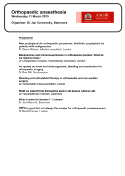

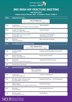

© Copyright 2026