ABC
docz
Explore
Log in
Create new account
Download
Report
health and fitness
disease
Acute Transverse Myelitis: Demyelinating, flammatory, and Infectious Myelopathies In Timothy W. West, M.D.
The following excerpt has been taken from the Christopher &... Paralysis Resource Center website.
Transverse Myelitis U.S. DEPARTMENT OF HEALTH AND HUMAN SERVICES
Evidence-based guideline: Clinical evaluation and treatment of
Current and future treatment approaches for neuromyelitis optica Review
Infectious Myelopathies Tracey A. Cho, MD; Henrikas Vaitkevicius, MD, MS
WMEDI2123 respiratory sector : from diagnosis to treatment.
Differential diagnosis in acute cardiac care Differential diagnosis
1/15/2013
Miguel Zamorano Serrano. Servicio de Urgencias Hospital Ramón y Cajal.
Document 8040
Acute General Oncological Wound Healing
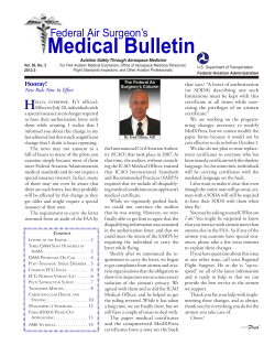
Medical Bulletin Federal Air Surgeon’s
© Copyright 2026
About abcdocz
DMCA / GDPR
Report