
Microvesicle protein levels are associated with increased risk for future... disease ☆ ⁎
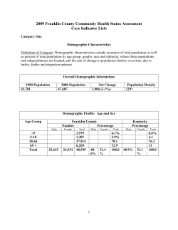
IJCA-15868; No of Pages 6
International Journal of Cardiology xxx (2013) xxx–xxx
Contents lists available at SciVerse ScienceDirect
International Journal of Cardiology
journal homepage: www.elsevier.com/locate/ijcard
Microvesicle protein levels are associated with increased risk for future vascular
events and mortality in patients with clinically manifest vascular disease☆
Danny A. Kanhai a, Frank L.J. Visseren a,⁎, Yolanda van der Graaf b, Arjan H. Schoneveld c, d,
Louise M. Catanzariti c, Leo Timmers c, L. Jaap Kappelle e, Cuno S.P.M. Uiterwaal b, Sai Kiang Lim f,
Siu Kwan Sze g, Gerard Pasterkamp c, Dominique P.V. de Kleijn c, d, h
on behalf of the SMART Study Group 1
a
Department of Vascular Medicine, University Medical Center Utrecht (UMC Utrecht), Utrecht, The Netherlands
Julius Center for Health Sciences and Primary Care, UMC Utrecht, Utrecht, The Netherlands
c
Experimental Cardiology Laboratory, UMC Utrecht, Utrecht, The Netherlands
d
ICIN, Netherlands Heart Institute, Utrecht, The Netherlands
e
Department of Neurology and Rudolf Magnus Institute of Neuroscience, UMC Utrecht, Utrecht, The Netherlands
f
Institute of Medical Biology, A-Star, Singapore
g
School of Biological Sciences, Nanyang Technological University, Singapore
h
Cardiovascular Research Institute & Surgery, NUHS, Singapore
b
a r t i c l e
i n f o
Article history:
Received 21 August 2012
Received in revised form 6 January 2013
Accepted 19 January 2013
Available online xxxx
Keywords:
Microvesicles
Proteins
Vascular events
Mortality
Epidemiology
a b s t r a c t
Background and Objectives: Microvesicles (MVs) are small membrane vesicles that are involved in atherotrombotic
processes. In the present study, we evaluated the risk of MV protein levels on the occurrence of new vascular events
in patients with clinically manifest vascular disease.
Methods: In this cohort study 1060 patients were prospectively followed for the occurrence of a new vascular event or death (median follow up 6.4 years, interquartile range 5.2–7.3 years). MVs were isolated from
plasma and MV protein levels of Cystatin C, Serpin G1, Serpin F2 and CD14 were measured. Multivariable
Cox proportional hazards models were used to estimate the risk for new vascular events, vascular mortality
and all-cause mortality. During follow up 136 vascular events occurred, 65 vascular mortality and 114
all-cause mortality.
Results: An increase in 1 standard deviation (SD) of Cystatin C MV level was related to an increased risk for
myocardial infarction (HR 1.49; 95%CI 1.20–1.86), vascular mortality (HR 1.48; 95%CI 1.17–1.86), vascular
events (HR 1.27; 1.07–1.52) and all-cause mortality (HR 1.41; 95%CI 1.18–1.69). Serpin F2 MV levels were
related to an increased risk for myocardial infarction (HR 1.22; 95%CI 1.00–1.51), vascular mortality (HR
1.25; 95%CI 1.00–1.56), and all-cause mortality (HR 1.22; 95% CI 1.03–1.45). CD14 MV levels were related
to an increased risk for myocardial infarction (HR 1.55; 95%CI 1.27–1.91), vascular mortality (HR 1.37;
95%CI 1.10–1.70), vascular events (HR 1.32; 95%CI 1.12–1.55), all-cause mortality (HR 1.36; 95%CI
1.15–1.62) and occurrence of ischemic stroke (HR 1.32; 95%CI 1.00–1.74).
Conclusions: Cystatin C, Serpin F2 and CD14 MV levels are related to an elevated risk for future vascular
events and mortality in patients with clinically manifest vascular disease.
© 2013 Elsevier Ireland Ltd. All rights reserved.
1. Introduction
Patients with manifest vascular disease are at elevated risk for successive vascular events and mortality, even after adequate treatment
of well-known vascular risk factors. This residual risk may be caused
by new, yet unrecognized, pathophysiological mechanisms.
☆ All authors take responsibility for all aspects of the reliability and freedom from bias of
the data presented and their discussed interpretation.
⁎ Corresponding author at: University Medical Center Utrecht, F02.126, Heidelberglaan
100, 3584 CX Utrecht, The Netherlands. Tel.: +31 88 7557155; fax: +31 30 2522693.
E-mail address: [email protected] (F.L.J. Visseren).
1
Listed in acknowledgments.
Microvesicles (MVs) are 50 to 1000 nm membrane shed vesicles released in the extracellular space after cell activation or apoptosis; they
include various types as microparticles and exosomes [1,2]. MVs are defined by size and antigen expression, which indicates their originating
cell type [3,4]. Release of MVs allows cells to influence (patho)physiological processes over a distance in contrast to cell-cell contact. MVs
can directly interact with ligands present on the surface of target cells
and activate cascade signaling. In addition, MVs can transfer proteins,
mRNA, miRNA, and bioactive lipids by interacting with target cells by either fusion or internalization [5]. By internalization, target cells acquire
new surface antigens and therefore new biological properties and activities [4]. Most cell types, including circulating cells and cells present in
0167-5273/$ – see front matter © 2013 Elsevier Ireland Ltd. All rights reserved.
http://dx.doi.org/10.1016/j.ijcard.2013.01.231
Please cite this article as: Kanhai DA, et al, Microvesicle protein levels are associated with increased risk for future vascular events and mortality in
patients with clinicall..., Int J Cardiol (2013), http://dx.doi.org/10.1016/j.ijcard.2013.01.231
2
D.A. Kanhai et al. / International Journal of Cardiology xxx (2013) xxx–xxx
the vessel wall, such as platelets, leukocytes, monocytes and endothelial
cells are capable of producing MVs [6].
The membrane of MVs consists of phospholipids and various
cell-specific proteins such as CD4 +, CD8 +, CD20 + for lymphocyte
shed MVs and CD14 + for monocyte shed MVs [7]. Besides membrane proteins, MVs also contain high concentrations of different
cytoplasmic proteins.
In the past, MVs were simply regarded as cellular debris [8], but now
circulating MVs are associated with different types of cancer [9–11], infectious diseases [12–15], diabetes [16] and with the presence of vascular
disease [17–22]. It is suggested that MVs play a role in the development
of vascular disease as circulating MVs contain and express procoagulant
metabolites, e.g. tissue factor, phosphatidylserine and Serpin F2 [23]. Additionally, MVs increase the synthesis and release of proinflammatory
cytokines by endothelial cells and leukocytes in vitro [24,25]. Annexin
V and CD31, both membrane-bound proteins present on circulating
MVs, are associated with increased risk for vascular events in patients
with stable coronary artery disease [26]. Plasma Cystatin C, a protease inhibitor, is related to decreased kidney function [27], and is also related to
an increased risk for vascular events and mortality [31]. Serpin G1
(C1 inhibitor) as well as Serpin F2 (α2-antiplasmin) inhibit fibrinolysis
[29,30], while monocyte derived MVs, marked by CD14 proteins are
procoagulant [31], which potentiates these three (MV-)proteins in
becoming markers of atherothrombotic processes. Additionally, soluble CD14 has recently been associated with incident vascular disease and mortality in elderly subjects [32]. No large clinical studies
have previously assessed the risk of these MV proteins on vascular
events or mortality.
In the present cohort study, we investigated the determinants of
specific MV protein levels (Cystatin C, Serpin G1, Serpin G2 and CD14)
and determined the risk of these MV protein levels on the occurrence
of new vascular events and mortality in a cohort of patients with various
clinical manifestations of vascular disease.
SMART study was approved by the Ethics Committee of the University Medical Center
Utrecht and all patients gave their written informed consent.
2.3. Follow-up
During follow up, patients were asked to fill out a standardized questionnaire biannually, to report newly diagnosed diseases and hospital admissions. When suspected
for a vascular event, patients' medical records and documentation were retrieved
from their treating specialist or general practitioner (GP). For those who died, specific
cause of death was retrieved (also through GP or specialist) in order to differentiate between vascular and non-vascular causes of death. Suspected vascular events and mortality were assessed separately by 3 physicians of the SMART endpoint committee. The
definite endpoint was scored upon majority of their evaluation. Study endpoints included ischemic stroke, myocardial infarction, vascular mortality and all-cause mortality. Complete definitions of the study endpoints are stated in Table 1. Patients were
followed up until March 2010.
2.4. Microvesicle measurements
Venous blood was drawn after an overnight fast. Tubes were instantly placed on
ice and centrifuged at 1850 ×g for 15 min at 4 °C. EDTA-plasma was aliquotted and immediately stored at −80 °C until recollection for MV measurement, which began in
December 2010 after a median freezer-stay of 7.5 years.
MVs were isolated using ExoQuick™ (SBI) according to the manufacturer's protocol (Fig. 1). Briefly, 150 μl EDTA plasma was centrifuged for 15 min at 3000 ×g. The supernatant was filtered over a 0.45 μm Spin-X filter (Corning), which was flushed with
preheated PBS (37 °C) and 38 μl ExoQuick™ solution was added to the filtrate. After
vortexing, the sample was stored overnight at 4 °C. The following day, the sample
was centrifuged at 1500 ×g for 30 min at room temperature. After removing the supernatant, the pellet was lysed in 100 μl Roche Complete Lysis-M with protease inhibitors (EDTA free). Subsequently, the sample was filtered over a 0.22 μm Spin-X filter
(Corning) and protein concentration was determined using a Pierce® BCA Protein
Assay Kit (Pierce Biotechnology, Rockford, USA) before storing the sample at −80 °C.
After thawing, the lysed sample was diluted 20× with Roche complete Lysis-M buffer.
50 μl of this diluted sample was analyzed in a multiplex immunoassay on levels of
Cystatin C, Serpin G1, Serpin F2 & CD14, using a Bio-Rad Bioplex 200 system as described before [34]. Capture antibody, biotinylated detection antibody and antigen of
all 4 proteins were purchased from R&D systems.
Of the 1062 eligible EDTA samples, 2 had insufficient material for MV measurement,
leaving a final number of 1060 patients with at least 1 valid MV marker (i.e. Cystatin C,
Serpin G1, Serpin F2 or CD14 MV) measurement.
2. Methods
2.5. Data analyses
2.1. Proteomics
A full description of preceding biomarker proteomics discovery work is provided in
supplements 1–5. Briefly, EDTA plasma proteomics was applied on 100 vascular patients,
split in a prospective 1:1 case–control design. 50 patients undergoing endarterectomy
(carotid or femoral) with incident coronary events, were age and gender-matched with
50 endarterectomy patients without future vascular events. The mean follow-up time
was 1.36 years for events and 3.08 years for controls. This resulted in 116 potential biomarker proteins for vascular events. 102 of these 116 proteins were listed in the Ingenuity
database. Ingenuity analysis revealed 3 canonical pathways that were significantly overrepresented in these 102 database proteins: Acute phase signaling, coagulation system
& atherosclerosis signaling. Within these 3 pathways, 4 proteins were selected for
which 2 antibodies and antigens were available and that were measurable within one
multiplex immunoassay: Cystatin C, Serpin G1, Serpin F2 and CD14.
2.2. Study population
The study cohort consists of patients participating in the Second Manifestations of
ARTerial disease (SMART) study, an ongoing prospective single-center cohort study at
the University Medical Center Utrecht that started in September 1996. Study rationale
and detailed description are published elsewhere [33]. The SMART cohort comprises patients with a recent history of clinically manifest vascular disease and patients with severe
vascular risk factors referred to the University Medical Center Utrecht. Exclusion criteria
were age under 18 years, malignancy, dependency in daily activities and not sufficiently
fluent in the Dutch language. After inclusion, patients underwent a standardized vascular
screening including measurements of risk factors and non-invasive measurement of subclinical atherosclerosis.
Consecutive participants in the SMART study with a recent diagnosis, i.e. within
4–8 weeks, of Cerebrovascular Disease (CVD), Coronary Artery Disease (CAD), Peripheral
Arterial Disease (PAD) and Aneurysm of the Abdominal Aorta (AAA) included between
January 2001 and December 2005 were considered eligible for the current study. CVD
was defined as a recent diagnosis of ischemic stroke, transient ischemic attack or amaurosis fugax. CAD was defined as a recent diagnosis of angina pectoris, myocardial infarction
or coronary revascularization (coronary artery bypass graft or coronary angioplasty). PAD
was defined as a recent clinical diagnosis of PAD (Fontaine stage 2, 3 or 4). AAA was defined as an abdominal aortic aneurysm of ≥3.0 cm or recent aneurysm surgery. The
Central estimators and variance measures were calculated for baseline characteristics of the included patients. In order to elucidate potential biological and clinical determinants of the MV protein levels, multivariable linear regression models were
used to analyze the relationship between various determinants and Cystatin C, Serpin
G1, Serpin F2 and CD14 MV protein levels. These analyses were adjusted for age, gender and eGFR, as plasma levels of Cystatin C are known to be strongly related to kidney
function [35]. Variables with skewed distributions were transformed to fulfill linear regression criteria.
Univariable and multivariable Cox proportional hazards models were applied to compute hazard ratios (HRs) and their 95% confidence intervals (95%CI) for subsequent
Table 1
Study endpoints.
Ischemic stroke
Myocardial infarction
Vascular mortality
Composite vascular
endpoint
All-cause mortality
Relevant clinical features causing an increase in
impairment of at least one grade on the modified
Rankin scale, without signs of hemorrhage on
repeat brain imaging.
At least two of the following criteria:
(I) Chest pain for at least 20 min, not disappearing
after administration of nitrates;
(II) ST-elevation >1 mm in two following leads or
a left bundle branch block on the electrocardiogram;
(III) Creatinine kinase (CK) elevation of at least two
times the normal value of CK and a myocardial
band-fraction >5% of the total CK.
Sudden death: unexpected cardiac death occurring
within 1 h after onset of symptoms, or within 24 h
given convincing circumstantial evidence.
Death from stroke, myocardial infarction, congestive
heart failure, or rupture of abdominal aortic aneurysm.
Vascular death from other causes
A composite of stroke, myocardial infarction, retinal
infarction, and vascular mortality.
Death from any cause
Please cite this article as: Kanhai DA, et al, Microvesicle protein levels are associated with increased risk for future vascular events and mortality in
patients with clinicall..., Int J Cardiol (2013), http://dx.doi.org/10.1016/j.ijcard.2013.01.231
D.A. Kanhai et al. / International Journal of Cardiology xxx (2013) xxx–xxx
3
Table 2
Baseline characteristics.
N = 1060
Fig. 1. Electron microscopy image of microvesicles isolated with ExoQuick™ out of
human plasma. Microvesicles are clearly visible with particles of various sizes. Magnification is 60,000×. Complete methodology is stated in supplemental 2.
vascular events or mortality. The proportional hazards assumption was confirmed by testing the correlations between scaled Schoenfeld residuals for mean baseline MV marker
and various functions of time. No significant non-proportionality (pb 0.05) was observed.
To directly compare the risk for vascular events or mortality, HRs per 1 standard deviation
(SD) increase in MV protein levels of Cystatin C, Serpin G1, Serpin F2 and CD14 were
calculated.
Single imputation methods were used to reduce missing covariate data for homocysteine (n = 17; 1.6%), eGFR (n = 14; 1.3%) and smoking (n = 7; 0.7%), since complete
case analysis leads to loss of statistical power and possible bias. To elaborate potential
effect modification, interaction terms for gender as well as type of vascular disease
were added to the most complete adjusted models. Data analyses were conducted
using SPSS version 18 (SPSS Inc. Chicago, IL).
Age (years)a
Male gender, n (%)
Prevalent type 2 diabetes, n (%)
Metabolic syndrome, n (%)c
Body mass index (kg/m2)a
Waist circumference (cm)a
HOMA-IRb
Blood pressure (mmHg)a
Systolic
Diastolic
LDL-cholesterol (mmol/L)a
HDL-cholesterol (mmol/L)b
Triglycerides (mmol/L)b
Glucose (mmol/L)a
HsCRP (mmol/L)b
eGFR (ml/min/1.73 m2)a
Albuminuria, n (%)
Micro
Macro
Homocysteine (μmol/L)a
Smoking, n (%)
Never
Ever
Current
Packyears smokinga
History of vascular disease, n (%)
Cerebrovascular disease
Coronary artery disease
Peripheral artery disease
Aneurysm of the abdominal aorta
Medication, n (%)
Platelet-aggregation inhibitors
Blood pressure-lowering agents
Lipid-lowering agents
Oral anticoagulants
a
b
3. Results
c
59 ± 10
839 (79)
170 (16)
406 (38)
26.9 ± 3.9
96 ± 11
2.54 (1.68–3.92)
143 ± 22
83 ± 12
2.84 ± 0.93
1.25 (1.02–1.50)
1.49 (1.09–2.08)
6.3 ± 1.86
1.86 (0.90–3.72)
77.5 ± 17.8
210 (20)
32 (3)
14.1 ± 6.3
177 (17)
492 (46)
391 (37)
23.3 ± 20.5
285
617
261
108
(27)
(58)
(25)
(10)
792 (75)
770 (73)
725 (68)
84 (8)
Values are expressed as: Mean ± standard deviation.
Values are expressed as: Median (interquartile range).
Defined according to the National Cholesterol Education Program ATPIII-revised
guidelines.
3.1. Patient characteristics
The baseline characteristics of the 1060 patients are displayed in
Table 2. The average age was 59 ± 10 years and 79% were males. 37%
of the patients were current smokers, 58% had a history of coronary
artery disease, 27% a history of CVD and 25% a history of PAD. 38% of
the patients had the metabolic syndrome (defined according to the
National Cholesterol Education Program Adult Treatment Panel III
revised-criteria), of which 68% had central obesity, 80% were hypertensive, 72% had dyslipidemia and 67% had an impaired fasting
glucose.
3.2. Microvesicle characteristics
After Exoquick isolation, MVs were visualized through electron microscopy (Fig. 1), which revealed large numbers of vesicles of different
sizes. Floatation experiments in a sucrose gradient revealed that Serpin
F2, Serpin G1, CD14 are located in floating MVs and that Cystatin C is
partly located in particles and partly in large protein complexes (supplement 3).
3.3. Determinants of microvesicle protein levels
MV levels of Serpin G1 were lower in females compared to male
patients (linear regression coefficient (β) − 14.45; 95%CI − 27.43 to
− 1.47), whereas MV level of Serpin F2 and CD14 were higher in female patients (β 7.37; 95%CI 2.79–11.94 and β 0.64; 95%CI 0.07–1.22
respectively) adjusted for age (Table 3). High-sensitive CRP was related to MV level of Cystatin C (β 0.76 95%CI 0.53–1.00), Serpin G1
(β 14.60; 95%CI 10.07–19.13), Serpin F2 (β 5.99; 95%CI 4.41–7.58)
and CD14 (β 1.02; 05%CI 0.83-1.21). Serpin F2 concentrations were inversely related to the use of platelet aggregation inhibitors (β −6.44;
95%CI −10.63 to −2.24), blood pressure-lowering agents (β −4.93;
95%CI–9.06 to − 0.80) and lipid-lowering agents (β − 4.56; 95%CI
− 8.50 to − 0.63).
Cystatin C and Serpin G1 were also inversely related to the use
lipid-lowering agents (β −1.25; 95%CI −1.84 to −0.64 and β −16.87;
95%CI − 27.98 to − 5.77 respectively). MV levels of Cystatin C
and CD14 were lower in patients using platelet aggregation inhibitors (β − 1.34; 95%CI − 1.97 to − 0.70 and β − 0.87;
95%CI–1.40 to − 0.35). In CAD patients the plasma MV concentrations of Serpin F2 (β − 7.39; 95%CI − 11.10 to − 3.68) and
CD14 (β − 1.88; 95%CI − 2.33 to − 1.42) were lower compared
to patients with a vascular disease at another location.
3.4. Microvesicle protein levels and risk of vascular events and mortality
During a median follow up of 6.4 years (interquartile range
5.2–7.3 years) the clinical endpoints ischemic stroke (n= 44), myocardial infarction (n= 78), vascular mortality (n= 65), composite vascular
endpoint (n=136) and all-cause mortality (n= 114) were observed.
An increase in 1 SD MV level of Cystatin C resulted in an increased
risk for myocardial infarction (HR 1.49; 95%CI 1.20–1.86), vascular
mortality (HR 1.48; 95%CI 1.17–1.86), all-cause mortality (HR 1.41;
95%CI 1.18–1.69) and the composite vascular endpoint (HR 1.27;
95%CI 1.07–1.52) (Table 4). An increase in 1 SD MV level of Serpin
F2 led to an increase in risk for myocardial infarction (HR 1.22;
95%CI 1.00–1.51), vascular mortality (HR 1.25; 95%CI 1.00–1.56)
Please cite this article as: Kanhai DA, et al, Microvesicle protein levels are associated with increased risk for future vascular events and mortality in
patients with clinicall..., Int J Cardiol (2013), http://dx.doi.org/10.1016/j.ijcard.2013.01.231
4
D.A. Kanhai et al. / International Journal of Cardiology xxx (2013) xxx–xxx
Table 3
Determinants of microvesicle protein levels.
Age (years)a
Male genderb
Type 2 diabetes
Metabolic syndrome (ATPIII)
BMI (kg/m2)
Waist circumference (cm)
HOMA-IR
Blood pressure (mmHg)
Systolic
Diastolic
LDL-cholesterol (mmol/L)
HDL-cholesterol (mmol/L)
Triglycerides (mmol/L)
Glucose (mmol/L)
log hsCRP (mmol/L)
eGFR (ml/min/1.73 m2)c
Albuminuria no vs micro or macro
Homocysteine (μmol/L)
Packyears smoking
History of vascular disease
CVD
CAD
PAD
AAA
Medication
Platelet aggregation inhibitors
Lipid-lowering agents
Blood pressure-lowering agents
Cystatin C (pg/μg)
Serpin G1(pg/μg)
Serpin F2 (pg/μg)
CD14+ (pg/μg)
n = 1054
n = 1054
n= 1051
n = 1057
β coefficients (95%CI)
β coefficients (95%CI)
β coefficients (95%CI)
β coefficients (95%CI)
0.06
−0.36
0.90
0.97
−0.03
0.02
0.06
(0.03–0.10)
(−1.06–0.34)
(0.13–1.66)
(0.41–1.54)
(−0.11–0.04)
(−0.01–0.05)
(0.01–0.11)
−0.33
−14.45
−15.75
6.30
0.25
0.37
−0.26
(−0.90–0.25)
(−27.43–-1.47)
(−29.89 to −1.61)
(−4.32–16.92)
(−1.09–1.58)
(−1.23–0.86)
(−1.22–0.70)
−0.19 (−0.40–0.01)
7.37 (2.79–11.94)
2.16 (−2.86–7.18)
0.69 (−3.07–4.46)
−0.46 (−0.93–0.02)
−0.03 (−0.20–0.15)
−0.14 (−0.51–0.23)
0.04
0.64
0.55
0.27
−0.11
−0.01
0.02
(0.02–0.07)
(0.07–1.22)
(−0.08–1.18)
(−0.21–0.74)
(−0.17 to −0.05)
(−0.03–0.01)
(−0.04–0.07)
0.00
−0.01
0.15
−1.63
0.44
0.11
0.76
−0.14
1.16
0.17
0.01
(−0.01–0.02)
(−0.04–0.01)
(−0.15–0.452)
(−2.41 to −0.86)
(0.19–0.70)
(−0.04–0.27)
(0.53–1.00)
(−0.16 to −0.12)
(0.49–1.83)
(0.13–0.22)
(−0.01–0.02)
0.23
0.23
2.84
−9.80
3.56
−1.00
14.60
−0.33
9.56
1.77
0.43
(−0.01–0.48)
(−0.22–0.67)
(−2.76–8.44)
(−24.27–4.69)
(−1.23–8.36)
(−3.82–1.82)
(10.07–19.13)
(−0.66 to −0.00)
(−2.96–22.07)
(0.90–2.63)
(0.17–0.68)
−0.08 (−0.16–0.01)
−0.06 (−0.22–0.10)
2.71 (0.72–4.71)
0.49 (−4.64–5.61)
−0.11 (−1.81–1.59)
−0.17 (−1.16–0.83)
5.99 (4.41–7.58)
−0.03 (−0.14–0.09)
3.45 (−0.97–7.87)
0.34 (0.03–0.64)
0.21 (0.12–0.30)
0.01
0.00
0.81
0.03
0.11
0.18
1.02
−0.02
1.36
0.13
0.03
(−0.01–0.02)
(−0.02–0.02)
(0.57–1.06)
(−0.62–0.67)
(−0.11–0.07)
(0.05–0.30)
(0.83–1.21)
(−0.04 to −0.01)
(0.81–1.91)
(0.09–0.17)
(0.02–0.04)
−0.05
−0.55
0.39
1.35
(−0.68–0.59)
(−1.12–0.02)
(−0.26–1.04)
(0.41–2.29)
−12.09
7.96
−9.99
8.32
(−23.79 to −0.40)
(−2.61–18.52)
(−22.02–2.04)
(−9.18–25.83)
2.76 (−1.37–6.90)
−7.39 (−11.10 to −3.68)
5.16 (0.91–9.41)
10.16 (3.98–16.34)
0.99
−1.88
1.47
2.05
(0.47–1.51)
(−2.33 to −1.42)
(0.94–2.00)
(1.32–2.85)
−7.55 (−19.47–4.37)
−16.87 (−27.98 to −5.77)
3.84 (−0.785–15.54)
−6.44 (−10.63 to −2.24)
−4.56 (−8.50 to −0.63)
−4.93 (−9.06 to −0.80)
−0.87 (−1.40 to −0.35
−1.57 (−2.05 to −1.08)
−1.32 (−1.83 to −0.80)
−1.34 (−1.97 to −0.70)
−1.25 (−1.84 to −0.65)
−0.13 (−0.76–0.50)
Bold values indicate significance (p b 0.05).
CVD: Cerebrovascular Disease; CAD: Coronary Artery Disease; PAD: Peripheral Arterial Disease; AAA: Aneurysm of the Abdominal Aorta.
a
Gender and eGFR adjusted.
b
Age and eGFR adjusted.
c
Age and gender adjusted; all other models are age, gender and eGFR adjusted.
Table 4
Microvesicle protein levels and risk of vascular events or mortality.
Study outcome
Ischemic stroke
Myocardial infarction
Vascular
Mortality
Composite vascular Endpointa
All-cause mortality
I
II
III
IV
I
II
III
IV
I
II
III
IV
I
II
III
IV
I
II
III
IV
Cystatin C (pg/μg)
Mean = 10.55
SD = 5.38
n = 1054
Serpin G1 (pg/μg)
Mean = 142.19
SD = 85.63
n = 1054
Serpin F2 (pg/μg)
Mean = 43.26
SD = 30.35
n = 1051
CD14 (pg/μg)
Mean = 12.22
SD = 3.88
n = 1057
# events
HR (95%CI)
# events
HR (95%CI)
# events
HR (95%CI)
# events
HR (95%CI)
43
1.38
1.26
1.05
1.05
1.46
1.36
1.46
1.49
1.70
1.50
1.45
1.48
1.43
1.30
1.27
1.27
1.58
1.39
1.37
1.41
44
1.16
1.13
1.11
1.21
0.98
0.97
0.96
0.95
1.03
1.00
1.00
0.99
1.10
1.07
1.06
1.08
1.05
1.01
1.00
0.99
44
1.23
1.21
1.24
1.20
1.19
1.26
1.26
1.22
1.23
1.27
1.30
1.25
1.16
1.19
1.19
1.14
1.22
1.24
1.25
1.22
44
1.57
1.50
1.37
1.32
1.51
1.50
1.52
1.55
1.70
1.59
1.56
1.37
1.48
1.43
1.41
1.32
1.58
1.44
1.45
1.36
76
62
133
111
(1.10–0.74)
(0.97–1.64)
(0.76–1.47)
(0.75–1.48)
(1.24–1.71)
(1.14–1.62)
(1.19–1.80)
(1.20–1.86)
(1.47–1.97)
(1.25–1.79)
(1.15–1.81)
(1.17–1.86)
(1.26–1.62)
(1.13–1.50)
(1.07–1.51)
(1.07–1.52)
(1.40–1.79)
(1.20–1.60)
(1.15–1.64)
(1.18–1.69)
76
64
134
113
(0.91–1.48)
(0.88–1.45)
(0.86–1.44)
(0.92–1.59)
(0.78–1.23)
(0.77–1.22)
(0.76–1.21)
(0.74–1.20)
(0.82–1.30)
(0.80–1.26)
(0.79–1.26)
(0.76–1.29)
(0.95–1.28)
(0.92–1.25)
(0.91–1.24)
(0.91–1.27)
(0.88–1.24)
(0.85–1.20)
(0.84–1.18)
(0.82–1.20)
78
64
135
113
(0.96–1.59)
(0.93–1.58)
(0.96–1.60)
(0.92–1.57)
(0.98–1.45)
(1.03–1.55)
(1.03–1.55)
(1.00–1.51)
(1.00–1.51)
(1.02–1.59)
(1.04–1.62)
(1.00–1.56)
(1.00–1.34)
(1.02–1.39)
(1.02–1.40)
(0.97–1.33)
(1.04–1.43)
(1.05–1.46)
(1.06–1.48)
(1.03–1.45)
78
65
136
114
(1.27–1.93)
(1.19–1.89)
(1.08–1.74)
(1.00–1.74)
(1.29–1.78)
(1.27–1.78)
(1.27–1.82)
(1.27–1.91)
(1.44–2.00)
(1.33–1.91)
(1.29–1.88)
(1.10–1.70)
(1.31–1.68)
(1.25–1.64)
(1.22–1.62)
(1.12–1.55)
(1.38–1.81)
(1.24–1.67)
(1.24–1.69)
(1.15–1.cp)
Bold values indicate significance (p b 0.05).
Hazard ratios represent risk per 1 SD increase in MV marker. Model I: Univariable model; Model II: adjusted for age, gender and smoking; Model III: Model II with additional adjustments for, systolic blood pressure, eGFR (MDRD), medication (blood pressure lowering medication, platelet aggregation inhibitors); Model IV: Model III with additional adjustments for prevalent type 2 diabetes, prevalent metabolic syndrome, homocysteine, history of vascular disease (CVD, CAD, PAD, AAA), albuminuria, LDL-cholesterol and hsCRP.
a
Composite vascular endpoint: composite of stroke, myocardial infarction, retinal infarction, or vascular mortality.
Please cite this article as: Kanhai DA, et al, Microvesicle protein levels are associated with increased risk for future vascular events and mortality in
patients with clinicall..., Int J Cardiol (2013), http://dx.doi.org/10.1016/j.ijcard.2013.01.231
D.A. Kanhai et al. / International Journal of Cardiology xxx (2013) xxx–xxx
and for all-cause mortality (HR 1.22; 95%CI 1.03–1.45) while 1 SD
MV level increase in CD14 accompanied an increased risk for myocardial infarction (HR1.55; 95%CI 1.27–1.91), vascular mortality (HR
1.37; 95%CI 1.10–1.70), all-cause mortality (HR 1.36; 95%CI 1.15–1.62)
and the composite vascular endpoint (HR 1.32; 95%CI 1.12–1.55).
Only MV levels of CD14 conferred an increased risk for ischemic
stroke (HR 1.32; (95%CI 1.00–1.74). MV levels of Serpin G1 were
not associated with vascular events or mortality. No significant interaction terms for gender or for type of vascular disease were
found (data not shown).
4. Discussion
Patients with clinically manifest vascular disease are at increased
residual risk for successive vascular events and mortality even after
treatment of vascular risk factors. In this prospective study consisting
of 1060 patients with various manifestations of vascular disease, we
assessed determinants of MV protein levels as well as the effects of
MV protein levels on vascular risk and mortality. Gender, lipid-lowering
medication use, hsCRP and eGFR were amongst the strongest determinants of MV level of Cystatin C, Serpin G1 Serpin F2 and CD14. However,
we chose to keep the linear regression models comprehensible by
adjusting only for verified robust confounding factors (age, gender and
eGFR), we cannot exclude that these results might be subject to residual
confounding. Cystatin C, Serpin F2 and CD14MV levels were linearly related to an increased risk for the occurrence of new vascular events, vascular mortality and all-cause mortality. The latter association remained
solid after adjustment for CRP and smoking, indicating that these MV
proteins describe a pathway to effectuate clinical vascular events
or mortality other than low-grade inflammation. To explore potential non-linearity, categorical analyses were also performed, which
essentially resulted in similar found linear associations (data not shown).
Circulating MVs isolated by Exoquick contain all MVs of various sizes,
originating from all kinds of tissue and molecules such as fibrin-bound
plasmin, since virtually every cell is capable of producing MVs. In this
study, we did not distinguish between sub-types of MVs or cellular origins. We therefore refer to the ExoQuick isolated particles as MVs.
In patients with stable CAD, the quantity of CD31 +/Annexin V +
MVs was associated with an increased risk for the composite of myocardial infarction, percutaneous coronary intervention, coronary artery bypass graft, stroke and vascular mortality [26]. In the present
study, we describe for the first time in a large clinical cohort that
the protein content of MVs is related to increased risk of vascular
events and mortality. Little is known about the content of MVs as
studies have mainly focused on the quantity of MVs by using a specific membrane-bound MV protein as a marker. The exact pathophysiological mechanism by which MVs cause vascular events is not yet
known. The 4 investigated MV proteins have been associated with
pathophysiologic processes in the vessel wall. Plasma concentrations
of Cystatin C are related to decreased kidney function [27], and are
also related to an increased risk for vascular events and mortality
[28]. Our results indicate that MV levels of Cystatin C are also related
with vascular events and mortality (Table 3). Plasma Cystatin C is related to hsCRP concentrations in patients with manifest vascular disease [29,36,37]. In the present study, Cystatin C MV levels were also
associated with plasma concentrations of hsCRP. Cystatin C antagonizes TGFβ-receptor, resulting in inhibition of TGFβ signaling. TGFβ
is an anti-inflammatory cytokine which inhibits proliferation and migration of smooth muscle cells, promotes extracellular matrix formation and inhibits expression of endothelial adhesion molecules [38].
By blocking the TGFβ receptor, Cystatin C counteracts the protective
role of TGFβ in vascular biology. Whether plasma Cystatin C concentrations are related to MV Cystatin C levels and whether plasma Cystatin C
or MV Cystatin C levels are causally related to vascular events or mortality remains to be determined.
5
Plasma Cystatin C concentrations are also associated with higher
levels of circulating adhesion molecules [39]. By expressing these
adhesion molecules on endothelial cells, circulating platelets will attach to the vessel wall; a process that potentially could be expedited
due to Cystatin C + MVs. The (activated) platelets release Serpin F2
[40], also known as α2-antiplasmin, which inactivates plasmin and
thus inhibits fibrinolysis. As activated platelets shed MVs [6,22], it
is conceivable that Serpin F2 reaches its target cells through these
MVs.
Fibrinolysis is also reduced by SerpinG1 (C1 inhibitor), which decelerates the conversion of plasminogen into plasmin by inhibiting
kalikrein [30]. Simultaneously, monocytes also release MVs, marked
by CD14 transmembrane proteins [7]. Monocyte derived MVs have
been described as procoagulant since they contain TF, which is responsible for thrombin formation [31], an important step in the coagulation pathway initiation.
Since both Serpin G1 and Serpin F2 inhibit fibrinolysis and the
subsequent degradation of a thrombus, while CD14 + MVs may stimulate thrombus formation via TF, we hypothesized that high levels of
Serpin F2, Serpin G1 and CD14 in MVs indicate that these MVs are
more procoagulant, leading to a potentially larger thrombus resulting
in an increased risk for a clinical event. However, in the present study
MV levels of Serpin G1 were not associated with clinical vascular outcomes or mortality, but were positively correlated with hsCRP. Plasma Serpin G1 is additionally thought to play an important role in
the regulation of vascular permeability, endothelial integrity and in
the suppression of inflammation [30]. As patients with manifest vascular disease have an increased inflammatory state, it is likely that
cells shed SerpinG1 + MVs as a counter mechanism. The finding of
this increased risk, may guide development in preventive strategies.
If MVs are causally related to the development of vascular diseases
and not just an indicator of risk, a distinction future research has to
produce, direct lowering of MV protein levels may reduce the risk
for successive vascular events and mortality. Future clinical studies
in the field of MV or MV-proteins and vascular risk may be focused
on other patient groups, other MV-proteins and on methods to lower
MV plasma concentrations. In vitro studies are needed to evaluate the
pathophysiologic relation between MV and MV-proteins and atherogenesis on a cellular level.
Strengths of this study include the prospective study design, a
large cohort of patients with various manifestations of vascular disease, and a large number of validated vascular endpoints and mortality. Potential study limitations should be considered. First, only MV
protein levels were measured, thus no statements concerning the
amount of MVs could be made. Secondly, as a result of our previous
work (supplemental 1), only 4 of the numerous proteins on the surface or within MVs were measured. It could be very well that besides
these biomarkers, various other MV proteins have their etiology in the
development of vascular events or mortality. Finally, it still has to be determined whether the suggested markers improve risk prediction and
could therefore be used in clinical practice for risk stratification in patients with manifest vascular disease. Unfortunately, a reliable, externally validated prediction model for patients with clinical manifest
vascular disease is not available.
In conclusion, Cystatin C, Serpin F2 and CD14 microvesicle protein
levels are associated with an increased risk for new vascular events
and mortality in patients with manifest vascular disease. Increased
levels of these MV protein levels may contribute to the residual
risk for vascular events and mortality in patients with vascular
disease.
Funding sources
This work was financially supported by the UMC Utrecht Vascular
Prevention Project.
Please cite this article as: Kanhai DA, et al, Microvesicle protein levels are associated with increased risk for future vascular events and mortality in
patients with clinicall..., Int J Cardiol (2013), http://dx.doi.org/10.1016/j.ijcard.2013.01.231
6
D.A. Kanhai et al. / International Journal of Cardiology xxx (2013) xxx–xxx
Disclosures
D.P.V.d.K. and G.P. are consultants for Cavadis; a company for the
development of biomarker kits.
All other authors have no relationships with industry that might
have a commercial interest in the submitted work.
Acknowledgments
We gratefully acknowledge the members of the SMART study group
of UMC Utrecht: P.A. Doevendans, MD, PhD, Department of Cardiology;
A. Algra, MD, PhD; Y. van der Graaf, MD, PhD; D.E. Grobbee, MD, PhD,
G.E.H.M. Rutten, MD, PhD, Julius Center for Health Sciences and Primary
Care; L.J. Kappelle, MD, PhD, Department of Neurology; W.P.T.M. Mali,
MD, PhD, Department of Radiology; F.L. Moll, MD, PhD, Department of
Vascular Surgery; F.L.J. Visseren, MD, PhD, Department of Vascular Medicine. The authors of this manuscript have certified that they comply
with the Principles of Ethical Publishing in the International Journal of
Cardiology.
Appendix A. Supplementary data
Supplementary data to this article can be found online at http://
dx.doi.org/10.1016/j.ijcard.2013.01.231.
References
[1] Cocucci E, Racchetti G, Meldolesi J. Shedding microvesicles: artefacts no more.
Trends Cell Biol 2009;19:43–51.
[2] van der Vlist EJ, Nolte-'t Hoen EN, Stoorvogel W, Arkesteijn GJ, Wauben MH. Fluorescent labeling of nano-sized vesicles released by cells and subsequent quantitative and
qualitative analysis by high-resolution flow cytometry. Nat Protoc 2012;7:1311–26.
[3] Jy W, Horstman LL, Jimenez JJ, et al. Measuring circulating cell-derived microparticles. J Thromb Haemost 2004;2:1842–51.
[4] Morel O, Morel N, Hugel B, et al. The significanceof circulating microparticles in
physiology, inflammatory and thrombotic diseases. Rev Med Interne 2005;26:
791–801.
[5] Martinez MC, Tual-Chalot S, Leonetti D, Andriantsitohaina R. Microparticles: targets and tools in cardiovascular disease. Trends Pharmacol Sci 2011;32:659–65.
[6] Rautou PE, Vion AC, Amabile N, et al. Microparticles, vascular function, and
atherothrombosis. Circ Res 2011;109:593–606.
[7] Aharon A, Tamari T, Brenner B. Monocyte-derived microparticles and exosomes
induce procoagulant and apoptotic effects on endothelial cells. Thromb Haemost
2008;100:878–85.
[8] Wolf P. The nature and significance of platelet products inhuman plasma. Br J
Haematol 1967;13:269–88.
[9] Hron G, Kollars M, Weber H, et al. Tissue factor-positive microparticles:cellular
origin and association with coagulation activation in patients with colorectal cancer. Thromb Haemost 2007;97:119–23.
[10] Taylor DD, Gercel-Taylor C. MicroRNA signatures of tumor-derived exosomes as
diagnostic biomarkers of ovarian cancer. Gynecol Oncol 2008;110:13–21.
[11] Davila M, Amirkhosravi A, Coll E, et al. Tissue factor-bearing microparticles derived from tumor cells: impact on coagulation activation. J Thromb Haemost
2008;6:1517–24.
[12] Mack M, Kleinschmidt A, Brühl H, et al. Transfer of the chemokine receptor CCR5
between cells by membrane-derived microparticles: a mechanism for cellular
human immunodeficiency virus 1 infection. Nat Med 2000;6:769–75.
[13] Rozmyslowicz T, Majka M, Kijowski J, et al. Platelet-and megakaryocyte-derived
microparticles transfer CXCR4 receptor to CXCR4-null cells and make them susceptible to infection by X4-HIV. AIDS 2003;17:33–42.
[14] Fevrier B, Vilette D, Archer F, et al. Cells release prions in association with
exosomes. Proc Natl Acad Sci U S A 2004;101:9683–8.
[15] Robertson C, Booth SA, Beniac DR, Coulthart MB, Booth TF, McNicol A. Cellular
prion protein is released on exosomes from activated platelets. Blood 2006;107:
3907–11.
[16] Sabatier F, Darmon P, Hugel B, et al. Type 1 and type 2 diabetic patients display
different patterns of cellular microparticles. Diabetes 2002;51:2840–5.
[17] Mallat Z, Benamer H, Hugel B, et al. Elevated levels of shed membrane microparticles with procoagulant potential in the peripheral circulating blood of patients
with acute coronary syndromes. Circulation 2000;101:841–3.
[18] Boulanger CM, Scoazec A, Ebrahimian T, et al. Circulating microparticles from patients
with myocardial infarction cause endothelial dysfunction. Circulation 2001;104:
2649–52.
[19] Bernal-Mizrachi L, Jy W, Jimenez JJ, et al. High levels of circulating endothelial microparticles in patients with acute coronary syndromes. Am Heart J 2003;145:
962–70.
[20] Boulanger CM, Amabile N, Tedgui A. Circulating microparticles: a potential prognostic marker for atherosclerotic vascular disease. Hypertension 2006;48:180–6.
[21] Nozaki T, Sugiyama S, Sugamura K, et al. Prognostic value of endothelial microparticles in patients with heart failure. Eur J Heart Fail 2010;12:1223–8.
[22] Azevedo LC, Janiszewski M, Pontieri V, et al. Platelet-derived exosomes from septic shock patients induce myocardial dysfunction. Crit Care 2007;11:R120.
[23] Yin W, Ghebrehiwet B, Peerschke EI. Expression of complement components and
inhibitors on platelet microparticles. Platelets 2008;19:225–33.
[24] Barry OP, Praticò D, Savani RC, FitzGerald GA. Modulation of monocyte-endothelial
cell interactions by platelet microparticles. J Clin Invest 1998;102:136–44.
[25] Mesri M, Altieri DC. Endothelial cell activation by leukocyte microparticles. J Immunol
1998;161:4382–7.
[26] Sinning JM, Losch J, Walenta K, Böhm M, Nickenig G, Werner N. Circulating
CD31+/Annexin V + microparticles correlate with cardiovascular outcomes. Eur
Heart J 2011;32:2034–41.
[27] Simons PC, Algra A, van de Laak MF, Grobbee DE, van der Graaf Y. Second manifestations of ARTerial disease (SMART) study: rationale and design. Eur J Epidemiol
1999;15:773–81.
[28] de Jager W, te Velthuis H, Prakken BJ, Kuis W, Rijkers GT. Simultaneous detection
of 15 human cytokines in a single sample of stimulated peripheral blood mononuclear cells. Clin Diagn Lab Immunol 2003;10:133–9.
[29] Dharnidharka VR, Kwon C, Stevens G. Serum cystatin C is superior to serum creatinine as a marker of kidney function: a meta-analysis. Am J Kidney Dis 2002;40:
221–6.
[30] Shlipak MG, Katz R, Sarnak MJ, et al. Cystatin C and prognosis for cardiovascular
and kidney outcomes in elderly persons without chronic kidney disease. Ann Intern Med 2006;145:237–46.
[31] Shlipak MG, Sarnak MJ, Katz R, et al. Cystatin C and the risk of death and cardiovascular events among elderly persons. N Engl J Med 2005;352:2049–60.
[32] Reiner AP, Lange EM, Jenny NS, et al. Soluble CD14: genomewide association analysis and relationship to cardiovascular risk and mortality in older adults.
Arterioscler Thromb Vasc Biol 2013;33:158–64.
[33] Koenig W, Twardella D, Brenner H, Rothenbacher D. Plasma concentrations of
cystatin C in patients with coronary heart disease and risk for secondary cardiovascular events: more than simply a marker of glomerular filtration rate. Clin
Chem 2005;51:321–7.
[34] Arpegård J, Ostergren J, de Faire U, Hansson LO, Svensson P. Cystatin C—a marker
of peripheral atherosclerotic disease? Atherosclerosis 2008;199:397–401.
[35] Blobe GC, Schiemann WP, Lodish HF. Role of transforming growth factor beta in
human disease. N Engl J Med 2000;342:1350–8.
[36] Upadhyay A, Larson MG, Guo CY, et al. Inflammation, kidney function and albuminuria in the Framingham Offspring cohort. Nephrol Dial Transplant 2011;26:920–6.
[37] Harrison P, Cramer EM. Platelet alpha-granules. Blood Rev 1993;7:52–62.
[38] Moroi M, Aoki N. Isolation and characterization of alpha2-plasmin inhibitor from
human plasma. A novel proteinase inhibitor which inhibits activator-induced clot
lysis. J Biol Chem 1976;251:5956–65.
[39] Davis III AE, Mejia P, Lu F. Biological activities of C1 inhibitor. Mol Immunol 2008;45:
4057–63.
[40] Palmerini T, Coller BS, Cervi V, et al. Monocyte-derived tissue factor contributes to
stent thrombosis in an in vitro system. J Am Coll Cardiol 2004;44:1570–7.
Please cite this article as: Kanhai DA, et al, Microvesicle protein levels are associated with increased risk for future vascular events and mortality in
patients with clinicall..., Int J Cardiol (2013), http://dx.doi.org/10.1016/j.ijcard.2013.01.231


© Copyright 2026