
GAIT PARAMETERS OF PATIENTS WITH OSTEOARTHRITIS OF THE KNEE JOINT Zoltán Bejek
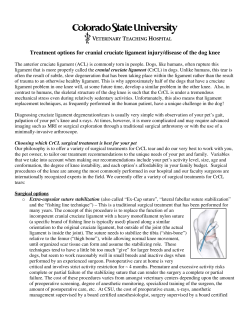
FACTA UNIVERSITATIS
Series: Physical Education and Sport Vol. 4, No 1, 2006, pp. 9 - 16
Scientific Paper
GAIT PARAMETERS OF PATIENTS WITH OSTEOARTHRITIS
OF THE KNEE JOINT
UDC 796.012 : 612.72-002
Zoltán Bejek1, Róbert Paróczai2, Árpád Illyés1,
László Kocsis2, Rita M. Kiss3
1
Semmelweis University, Department of Orthopaedics, Budapest, Hungary
2
Department of Applied Mechanics,
Budapest University of Technology and Economics, Hungary
3
Hungarian Academy of Sciences, Research Group of Structures, Budapest, Hungary
E-mail: [email protected]
Abstract. It is difficult to identify objective parameters for assessing joint function
when evaluating the outcome of orthopaedic procedures, especially endoprosthetic
replacement. Spatial and temporal parameters of gait have clinical relevance in the
assessment of motor pathologies, particularly in orthopaedics. The objective of this
study is to compare gait patterns in patients with unilateral osteoarthritis of the knee
joint to the gait pattern of healthy control subjects. A total of 20 patients with severe
unilateral osteoarthritis of the knee and 20 healthy elderly subjects without any history
of lower extremity joint pathology were investigated at constant gait speeds (two km/h).
The gait analysis equipment used consisted of an infinitely adjustable forceinstrumented treadmill and an ultrasound-based motion analyzer system with
electromyography. Our findings indicate that changes in gait parameters may occur in
patients with unilateral osteoarthritis of the knee joint as compared to the gait pattern
of healthy control subjects. Knee joint degeneration was compensated for in part by the
pelvis and other joints in the lower limb. Reduced motion of the knee joint leads to
increased pelvic motion, which should affect the natural mobility of the lumbar spine
and cause pain in the lumbar region of the spine due to kinematic interaction.
Key words: Biomechanics, gait analysis, osteoarthritis, knee
1. INTRODUCTION
Kinematics, kinetics, and electromyography are fundamental for characterizing gait
patterns and their underlying mechanisms (Frigo et al., 1996, 9). Complex gait analysis
Received May 23, 2006
10
Z. BEJEK, R. PARÓCZAI, Á. ILLYÉS, L. KOCSIS, R. M. KISS
presents itself as a possible tool for evaluating the rehabilitation process after orthopaedic
surgery (Knoll et al., 2004, 12). Patients with osteoarthritis suffer from pain and functional impairment of the knee over a long period. Although different functional scores are
widely used to assess improvements after surgery, the patients' responses are often subjective and disparities between patients' and doctors' evaluations can be significant
(Bullinger, 1996, 35; Liebermann et al., 1996, 28). Therefore, objective and quantified
data from gait analysis could be useful.
Osteoarthritis is the most prevalent form of arthritis among the elderly. It is estimated
that 10% of men and 21% of women over the age of 65 have osteoarthritis in the knee joint,
in the hip joint or in both joints (Al-Zahrani and Bakheit, 2002, 24). Patients may adapt
their gait in response to pain, deformity or laxity in the joints of the lower extremities. Patients with osteoarthritis of the knee joint often adapt an antalgic type of gait as their disease
progresses (Murray et al., 1971, 53A). It is not known if gait adaptation is mainly related to
the severity of the disease, pain, muscle weakness, or limitations in a passive range of motion (Hurwitz et al., 2000, 18; Mc Gibbon and Krebs, 2002, 29). Furthermore, adaptations
protecting the knee joint may influence the motion of the lower back and other joints of the
lower extremities (Andriacchi et al., 1982, 64; Gök et al., 2002, 73).
Gait measurement has been used for the analysis of the biomechanical parameters of the
lower extremities of patients with knee osteoarthritis. Patients with gonarthritis have demonstrated a lower walking speed, lower cadence, shorter step length, and shorter single
stance phase of the involved leg (Al-Zahrani and Bakheit, 2002, 24; Andriacchi et al., 1982,
64; Baliunas et al., 2002, 10; Chao et al., 1983, 16; Gök et al., 2002, 73; Hurwitz et al.,
2000, 18; Mattson et al., 1990, 22; Weidenhielm et al., 2001, 25), while a shorter double
stance phase of the involved leg has been observed when compared to the normal group
(Chao et al., 1983, 16; Gök et al., 2002, 73; Weidenhielm et al., 2001, 25). Several studies
have established that the range of motion of the affected joint has significantly decreased
and the range of motion of other joints of the lower extremities have increased significantly
in patients with gonarthritis (Al-Zahrani and Bakheit, 2002, 24; Gök et al., 2002, 73; Hurwitz et al., 2000, 18). The osteoarthritis of the knee joint significantly influenced the ground
reaction force (Gök et al., 2002, 73), the muscle moments (Baliunas et al., 2002, 10; Hurwitz et al., 2000, 18; Munderman et al., 2004, 50) and muscle power (McGibbon and Krebs,
2002, 29). These studies underlined a specific aspect of advanced osteoarthritis gait. It can
be assumed that the observed locomotor perturbation caused by compensatory or adaptive
mechanisms comes as a result of severe osteoarthritis.
The objective is to compare gait patterns in patients with unilateral osteoarthritis of the
knee to the gait patterns of healthy control subjects. We hypothesized that the biomechanical parameters were significantly influenced by osteoarthritis of the knee joint compared
to the healthy control group.
2. MATERIALS AND METHODS
Subjects
The population of the healthy group consisted of 12 women and 8 men. Their age was
52-84 (68.8±9.1years), body weight was 61.3-100.3 kg (73.3±11.4kg), and height was
1.50-1.96 m (1.69±0.19). The average HSS Knee Score of the healthy elderly people was
98.9 points (± 1.1); all of the subjects had excellent results (HHS~100 points). The
subjects were not limited in their normal daily or recreational activities. These subjects
Gait Parameters of Patients with Osteoarthritis of the Knee Joint
11
had no history of osteoarthritis of the knee joint and of hip joint, knee instability or major
lower extremity joint surgery. These individuals had normal strength, a full range of
motion of the lower extremities and no neurological deficiencies.
The population of patients with unilateral gonarthritis consisted of 12 women and 8
men. Their age was 45-93 (68.2±7.1), body weight was 55.3-122.1 kg (71.1±11.9), and
mean height was 1.47-1.86 m (1.69±0.11). The average HSS Knee Score of the patients
with unilateral gonarthritis was 47.9 points (± 13.6), and all of the patients had poor results
(HHSK<70 points). All of the patients were seriously limited in their activities due to pain.
The age, weight and height of the healthy subjects and patients with gonarthritis can
be compared to each other, with no significant differences (p=0, 71; p=0.87; p=0.67 respectively). The joint function of patients with gonarthritis is significantly poorer compared to the one of the healthy control group (p=0.003; p=0.001 respectively). Each subject provided informed consent before participation and signed a consent form approved
by the Hungarian Human Subjects Compliance Committee (112/2004).
The severity of the osteoarthritis of the knee joint was determined by the Kellgren and
Lawrence radiographic index (Kellgreen and Lawrence, 1957, 16), a 5-point scale (0-4)
on the basis of anteroposterior and Laustein X-ray projections. The Kellgreen and Lawrence radiographic index of 15 patients was 4, and the Kellgren and Lawrence radiographic index of 5 patients was 3. All of the patients were classified as having severe
osteoarthritis of the knee joint. It means they have large osteophytes, a marked joint space
narrowing, severe sclerosis and definite bone contour deformity.
Methods
The objective functional evaluation was based on three-dimensional gait analysis.
Evaluation in the gait laboratory lasted about one hour and included the recording of
lower extremity kinematics and kinetics.
Spatial coordinates for the determination of kinematic data were collected using an
ultrasound-based Zebris CMS-HS system (ZEBRIS, Medizintechnik GmbH, Germany) in
the Biomechanical Laboratory of the Department of Applied Mechanics at the Budapest
University of Technology and Economics. The measuring head with three sensors was
positioned behind the individual and the five ultrasound triplets with three active markers
on each were placed on the sacrum, left and right thighs, and left and right calves (Figure
1). The data obtained from the measuring system recording the active markers allowed
for the determination of coordinates of optional anatomical points of the lower limb. The
measuring method was developed by Kocsis (2002). The biomechanical model developed
by Knoll et al. (2004, 12) was chosen for our study. The spatial coordinates were recorded at a frequency of 100Hz. Simultaneously, the ground forces were measured at
1000 Hz. The patients were asked to walk at constant gait speed of 2 km/h on a motorized
and instrumented 330mm * 1430 mm treadmill with a built-in force plate (Bonte Zwolle
B.V, Austria). Walking on the treadmill can initially be an unfamiliar experience, which
in turn may influence the parameters being measured. Therefore, measurements are to
start after six minutes of familiarization time (Alton et al., 1998, 13; Matsas et al., 2000,
11). Kinematic data were collected for six gait cycles.
12
Z. BEJEK, R. PARÓCZAI, Á. ILLYÉS, L. KOCSIS, R. M. KISS
Triplet
on pelvis
Triplet
on right thigh
Triplet
on left
hi h
Measuring
head
Triplet
on left calf
Fig. 1. Arrangement of measurements
The assessed kinematic parameters are the following:
• Temporal and spatial parameters: stance, swing and double stance phase in percent of
gait cycle; step length, step width (in centimeters); cadence (steps per minute)
• Angular parameters: knee, hip and pelvic angles, presented by Kocsis and Beda (2001, 18);
• Force parameters: first peak force (F1) in the early stance phase and second peak
force (F2) in the late stance phase (in percent of body weight).
The parameters above were calculated by a software package presented first in (Jurak and Kocsis, 2002).
Statistical analysis
Data processing and statistical analyses were performed using an MS Excel-based
software of our own development. In the case of each of the subject examined, we calculated the average and the standard deviation of kinematic and kinetic parameters calculated from the measurement results of the motion cycles recorded, and these data were
processed further.
The biomechanical properties of individuals pertaining to a given group and those of
various groups were statistically analyzed using the MS Excel Analysis ToolPak software. The uniformity of standard deviations was checked by an F-test; the significance
levels of the difference between the average values of identical parameters were determined by a t-test applying a symmetrical critical range. A two-sample t-test was applied
when comparing the results for healthy people and patients with osteoarthritis. It was
assumed that the gait parameters of the healthy group and the patients with osteoarthritis
at constant gait speeds should be different, and the results present statistically significant
differences if p < 0.05.
Gait Parameters of Patients with Osteoarthritis of the Knee Joint
13
3. RESULTS
All of the results are summarized in Tables 1 and 2. Significant statistical differences
were observed for 13 parameters (cadence, step length, walking base, time of double support phase motion of the knee joint, motion of the hip joint at osteoarthritis side, motion
of the pelvis joint –Table 1) out of 22, comparing the patients with gonarthritis to the
healthy group examined at equal gait speed. There are no statistical differences in the
time of the swing phase, in unaffected hip motion, in the motion of pelvis rotation, flexion (Table 1) and in the kinetic parameters (Table 2).
Table 1. The results of spatial-temporal and angular parameters
of the patients and control subjects.
Parameters
Spatial-temporal parameters
Cadence (step/minutes)
Step length (mm)
Walking base (mm)
Double support phase (% of gait cycle)
Swing phase (percent of gait cycle)
Knee angle (degree)
ROM
maximum
minimum
Hip angle (degree)
ROM
maximum
minimum
Rotation of pelvis (degree)
ROM
Maximum
Minimum
Obliquity of pelvis (degree)
ROM
Maximum
Minimum
Flexion/extension of pelvis (degree)
ROM
Maximum
Minimum
Side
A
B
A
B
A
B
Healthy
94.5
999.8
934.7
21.7
27.9
20.9
33.7
30.9
Patients
± 20.7 97.04 ± 8.1
± 109.5 973.6 ± 80.9
± 110.7 903.1 ± 80.5
±
7.9 23.6 ± 6.3
±
8.7 27.8 ± 5.2
±
1.9 18.2 ± 1.7
±
5.6 41.5 ± 3.3
±
5.5 31.4 ± 2.8
A
B
A
B
A
B
57.5 ± 7.3
48.8 ± 6.1
63.6 ± 6.3
56.3 ± 5.7
6.1 ± 3.4
7.5 ± 3.4
41.7 ± 8.5
30.7 ± 9.1
49.8 ± 6.5
46.5 ± 12.3
8.2 ± 5.4
15.7 ± 11.4
A
B
A
B
A
B
33.6 ± 7.9
27.9 ± 7.1
84.8 ± 17.6
82.8 ± 10.1
51.2 ± 14.6
54.9 ± 17.8
28.0 ± 9.3
31.9 ± 11.3
71.9 ± 15.0
81.8 ± 11.2
43.9 ± 8.1
43.9 ± 8.1
5.5 ± 3.0
4.3 ± 2.8
−1.2 ± 1.1
10.8 ± 5.6
7.1 ± 3.4
−3.7 ± 1.5
4.6 ± 1.4
6.9 ± 1.7
2.3 ± 3.5
6.1 ± 1.7
6.7 ± 6.7
0.6 ± 2.6
5.9 ± 2.3
16.4 ± 5.8
10.5 ± 6.0
11.7 ± 2.8
14.2 ± 3.8
2.5 ± 4.9
Comments: side A: dominant side of the healthy people, contralateral (healthy) side of the patients
side B: nondominant side of the healthy people, affected side of the patients
14
Z. BEJEK, R. PARÓCZAI, Á. ILLYÉS, L. KOCSIS, R. M. KISS
Table 2. The results for the force parameters of the patients and control subjects
Parameters
F1 (percent of body weight)
F2 (percent of body weight)
Side
A
B
A
B
Healthy
120.3 ± 15.5
120.8 ± 14.9
118.5 ± 13.4
116.5 ± 14.8
Patients
110.7 ± 8.9
113.9 ± 9.0
115.9 ± 15.3
111.5 ± 13.2
4. DISCUSSION
The objective of the present study was to compare the gait patterns of patients with
unilateral osteoarthritis of the knee joint to the gait patterns of healthy control subjects.
Walking is a common functional activity of daily living. This study provides meaningful
information on gait adaptations used by patients with osteoarthritis of the knee joint
compared to healthy subjects. Assessment of gait changes should be helpful in the
occurrence of compensatory mechanisms during the course of diseases.
In our study the cadence, step length, walking base, and the time of double support
phase in patients with gonarthritis changed significantly compared to those of the healthy
subjects (Table 1). These findings are similar to those found in previous studies (AlZahrani and Bakheit, 2002, 24; Andriacchi et al., 1982, 64; Baliunas et al., 2002, 10;
Börjesson et al., 2005, 12; Gök et al., 2002, 73; Hurwitz et al., 2000, 18; Kaufmann et al.,
2001, 34; Mattson et al., 1990, 22; McGibbon and Krebs, 2002, 29; Messier et al., 1992,
57). According to Hurwitz et al. (2000, 18) the different spatial-temporal parameters were
part of the adaptive mechanism to reduce pain, which is similar to the findings of
McGibbon and Krebs (2002, 29).
The results of joint angle kinematics show that the minimal value of knee motion was
increased, which represents a decreased knee extension; the maximal value of knee motion
decreased, which means a decreased knee flexion; and the motion range of the knee joint
decreased on both sides, as compared to that of the healthy subjects (Table 1). These findings were also noted by other researchers (Gök et al., 2002, 73; Kaufmann et al., 2001, 34;
McGibbon and Krebs, 2002, 29; Messier et al., 1992, 57). Messier et al. (1992, 57) established that the abnormalities of the knee joint seem to reflect mechanical changes secondary
to osteoarthritis rather than underlying factors involved in pathogenesis. In the case of patients with gonarthrosis, asymmetry was observed in the motion of the knee joint, due to the
fact that the motion of the un-affected knee joint significantly increased compared to that of
the affected joint. However, the motion of the un-affected knee joint was significantly decreased compared to the values of the dominant side of healthy subjects. Our results support
the findings of Andriacchi et al. (1982, 64).
The next joint in the kinematic chain of the human body is the hip joint. It could be
hypothesized that the reduced motion of the knee joint would be compensated by an
increased motion of the hip joint. However, we found a decreased hip motion of the
osteoarthritis side compared to the one of the healthy subjects. These findings support
results from previous studies (Al-Zahrani and Bakheit, 2002, 24; Andriacchi et al., 1982,
64; Baliunas et al., 2002, 10; Börjesson et al., 2005, 12; Hurwitz et al., 2000, 18; McGibbon
and Krebs, 2002, 29; Messier et al., 1992, 57) and can be attributed to a compensatory
strategy which acts to prevent the knee joint with increased neuromuscular control
(Childs et al., 2004, 19).
Gait Parameters of Patients with Osteoarthritis of the Knee Joint
15
As a result of our measurement, it could be assumed that decreased knee motion was
associated with increased motion occurring in the pelvis. An increase in pelvic motion
obliquity was observed compared to the one of the healthy subjects (Table 2). The pelvis
adaptation enabled the patient to minimize the shorter step length resulting from a
reduced motion range of the knee joint (Hurwitz et al., 2000, 18) and to reduce the pain
(Hurwitz et al., 2000, 18). Similarly, the findings of Whittle (1996) - the modified pelvis
obliquity motion and interaction between the modified pelvis kinematics and the lumbar
spine - could explain in part why pains are often noticed in patients with gonarthritis in
the HSS Knee Score.
When ground reaction forces were analyzed, we found no difference in patients with
gonarthritis compared to healthy subjects (Table 2). These results are consistent with the
data reported by Gök et al. (2002, 73).
Our findings indicate that changes in gait parameters may occur in patients with
unilateral osteoarthritis of the knee joint compared to the gait patterns of healthy control
subjects. Knee joint degeneration was compensated for in part by the pelvis and other joints
in the lower limb. Reduced motion of the knee joint leads to increased pelvic motion, which
should affect the natural mobility of the lumbar spine and cause pain in the lumbar region of
the spine because of the existing kinematic interaction.
Acknowledgements. This work was supported in part by the Hungarian Scientific Fund T049471.
T46126 and the Semmelweis Foundation.
REFERENCES
1. Alton, F., Baldey, L., Caplan, S., & Morrissey, M.C. (1988). A kinematic comparison of over ground and
treadmill walking. ClinBiomech 13, 434-440
2. Al-Zahrani, K.S., & Bakheit, A.M. (2002). A study of gait characteristic of patients with chronic
osteoarthritis of the knee. Disabil Rehabil .,24, 275-280
3. Andriacchi TP., Galante J, & Fermier R. (1982). The influence of total knee replacement design on
walking and stair-climbing. JBone Joint Surg Am., 64, 1328-1335
4. Baliunas, A.J., Hurwitz, D.E., Ryals, A.B., Karrar, A., Case, J.P., Block, J.A., & Andriacchi, T.P. (2002).
Increased knee joint loads during walking are presented in subjected with knee osteoarthritis.
Osteaoarthritis Cartalidge, 10, 573-579
5. Börjesson, M., Weidenhielm, L., Atsson, E., & Olsson, E. (2005). Gait and clinical measurements in
patients with knee osteoarthritis after surgery: prospective 5-year follow-up study. The Knee, 12, 121-127
6. Bullinger, M. (1996). Assessment of health related quality of life with the SF-36 Health Survey.
Rehabilitation, 35, 17-27
7. Chao, E.Y., Laughmann, R.K., Schneider, E., & Staffer, R.N. (1983). Normative data of the knee joint
motion and ground reaction forces in adult level walking. J of Biomech, 16, 219-233
8. Childs, J.D., Sparto, P.J., Fitzgerald, K., Bizzini, M., & Irrgang, J.J. (2004). Alterations in lower
extremity movement and muscle activation patterns in individuals with knee osteoarthritis. Clin
Biomech,19, 44-49
9. Frigo, C., Bardare, M., Corona, F., Casnaghi, D., Cimaz, R., & Naj Fivono, P.L. (1996). Gait alterations
in patients with juveline chronic arthritis: a computerized analysis. J Orthop Rheumatol., 9, 82-90
10. Gök, H., Ergin, S., & Yavuzer, G. (2002). Kinetic and kinematic characteristic of gait in patients with
medial knee arthrosis. Acta Orthop Scan., 73, 647-652
11. Hurwitz, D.E., Ryals, A.R., Block, J.A., Sharma, L., Schnitzer, T.J., & Andriacchi, T.P. (2000). Knee
pain and joint loading in subjects with oasteoarthritis of the knee. J Orthop Res., 18, 572-579
12. Jurak, M., & Kocsis, L. (2002). New package for calculation of gait parameters. Proceedings of Third
Conference on Mechanical Engineering (pp. 500-504). Budapest, Hungary.
13. Kaufman, K.R., Hughes, C., Morrey, B.F., Morrey, B., & An, K.N. (2001). Gait characteristics of
patients with knee osteoarthritis. J of Biomech., 34, 907-915
16
Z. BEJEK, R. PARÓCZAI, Á. ILLYÉS, L. KOCSIS, R. M. KISS
14. Kellgreen, J.H., Lawrence, J.S. (1957). Radiological assessment of osteoarthritis. Ann Rheum Dism., 16,
494-502
15. Kocsis, L., & Béda, G. (2001). Closed formulae to determine the angular velocity of a body-segment
based on 3D measurements. Acta Physiologica Hungarica, 88. 1-13
16. Kocsis, L. (2002). More precise measurement method for 3D gait analysis. Proceedings of Third
Conference on Mechanical Engineering (pp. 848-851). Budapest, Hungary.
17. Knoll, Zs., Kocsis, L., & Kiss, R.M. (2004). Gait patterns before and after anterior cruciate ligament
reconstruction. Knee Surgery Sports Traumatology Arthroscopy, 12, 7-13
18. Liebermann, J.R., Dorey, F., Shekelle, P., Schumaker, L., Thomas, B.J., & Kilgus, D.J. (1996).
Differences between patients and physicians' evaluations of outcome after hip arthroplasty. J. Bone Joint
Surg Am., 78, 835-838
19. Matsas, A., Taylor, N., & McBurney, H. (2000). Knee joint kinematics from familiarized treadmill
walking can be generalized to over ground walking in young unimpaired subjects. Gait and Posture, 11,
46-53.
20. Mattsson, E., Olsson, E., & Brostörm, L.A. (1990). Assessment of walking before and after
unicompartmental knee arthroplasty. Scand J Rehabil Med., 22, 45-50
21. McGibbon, C.A., & Krebs, D.E. (2002). Compensatory gait mechanics in patients with unilateral knee
arthritis. J Rheumatol., 29, 2410-2419
22. Messier, S.P., Loeser, R.F., & Hoover, J.L. (1992). Osteoarthritis of the knee: effects on gait, strength
and flexibility. Arch Phys Med Rehabil., 57, 571-577
23. Mundarmann, A., Dyrby, C.O., Hurwitz, D.E., Sharma, I., & Andriacchi, T.P. (2004). Potential strategies
to reduce medial compartment loading in patients with knee osteoarthritis of varying severity: reduced
walking speed. Arthritis Rheum., 50, 1172-1178
24. Weidenhielm, L., Olsson, E., Broström, L.A., Börjesson-Hederström, M., Mattsson, E. (2001).
Improvement in gait one year after surgery for knee osteoarthritis: a comparision between high tibial
osteomy and prosthetic replacement in a prospective randomized study. Scand J Rehabil Med., 25, 25-31
25. Whittle, M.W. (1996). Gait analysis. 2nd Ed Oxford: Buttherworth Heinemann.
PARAMETRI HODA KOD PACIJENATA SA
OSTEOARTRITISOM ZGLOBA KOLENA
Zoltán Bejek, Róbert Paróczai, Árpád Illyés,
László Kocsis, Rita M. Kiss
Teško je odrediti objektivne parametre za procenu funkcije pokretljivosti zglobova kada
ocenjujemo rezultate ortopedskih procedura, naročito endoprostetičkih zamena. Spacijalni i
temporalni parametri načina hoda imaju kliničku relevantnost u proceni patologije motoričkog
aparata, naročito u ortopediji.
Cilj ovog istraživanja je da se izvrši komparacija načina hoda kod pacijenata sa unilateralnim
osteoartritisom zgloba kolena i kod zdravih ispitanika. Uzorak je činilo 20 pacijenata sa ozbiljnim
ostećenjima –unilateralnim osteoartritisom kolena i 20 zdravih starijih ispitanika bez ikakve
istorije bolesti donjih ekstremiteta, a ispitavan je hod pri brzini od 2 km/h. Aparatura koja je
korišćena sastojala se od podešive staze za šetanje sa instrumentima za merenje sile i ultrazvučnog
analizatora kretanja sa elektromiografijom.
Rezultati pokazuju da se promene u načinu hodanja mogu javiti kod pacijenata sa
unilateralnim osteortritisom zgloba kolena ako se porede sa obrascima hoda zdravih kontrolnih
ispitanika. Degeneracija zgloba kolena se kompenzira delimično većom pokretljivošću karlice i
ostalih zglobova donjeg ekstremiteta. Smanjena pokretljivost zgloba kolena uslovljava povećanu
pokretljivost pelvisa što utiče na prirodnu sposobnost kičmene moždine i što izaziva bolove u
lumbalnoj regiji kičme zbog kinematičke interakcije.
Ključne reči: biomehanika, analiza hoda, osteoartritis, koleno.


© Copyright 2026