
Evaluation of Melissa officinalis (Lemon Balm) Effects on Heart
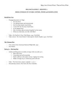
Res Cardiovasc Med. 2015 May; 4(2): e27013.
DOI: 10.5812/cardiovascmed.4(2)2015.27013
Research Article
Published online 2015 May 23.
Evaluation of Melissa officinalis (Lemon Balm) Effects on Heart Electrical
System
Siyavash Joukar
1,2,*
; Haleh Asadipour
2
1Physiology Research Center, Institute of Neuropharmacology, Kerman University of Medical Sciences, Kerman, IR Iran
2Department of Physiology and Pharmacology, School of Medicine, Kerman University of Medical Sciences, Kerman, IR Iran
*Corresponding author: Siyavash Joukar, Physiology Research Center, Institute of Neuropharmacology, Kerman University of Medical Sciences, P. O. Box: 7616914115, Kerman, IR Iran.
Tel/Fax: +98-3413220081, E-mail: [email protected], [email protected]
Received: January 14, 2015; Revised: March 5, 2015; Accepted: March 11, 2015
Background: Melissa officinalis, an herbal drug, is well known and frequently applied in traditional and modern medicine. Yet, there is
inadequate information regarding its effects on electrical properties of the heart. The present study attempted to elucidate the effects of
Melissa officinalis aqueous extract on electrocardiogram (ECG) in rat.
Objectives: ECG is an easy, fast and valuable tool to evaluate the safety of used materials and drugs on heart electrical and conductivity
properties. Many drugs with no cardiovascular indication or any overt cardiovascular effects of therapeutic dosing become cardiotoxic
when overdosed (16). On the other hand, there are numerous substances and drugs that can cause ECG changes, even in patients without a
history of cardiac disease. Therefore, this study was conducted to elucidate safety and outcome of one-week administration of M. officinalis
aqueous extract on blood pressure and ECG parameters of rats.
Materials and Methods: Four animal groups received tap water (control group), aqueous extracts of Melissa officinalis 50 (M50), 100 (M100)
and 200 (M200) mg/kg/day, respectively and orally for a week. ECG and blood pressure were recorded on the eighth day of experiment.
Results: Consumption of Melissa officinalis extract associated with prolonged QRS interval (P < 0.05 for M50 and M100 groups and P < 0.01
for M200 group versus the control group, respectively), prolonged QTc and JT intervals (P < 0.01 for different M groups versus the control
group) and prolonged TpTe interval (P < 0.001 when M groups compared with the control group) of ECG. However, different doses of the
extract had no significant effect on RR interval, PR interval, amplitudes of ECG waves, heart rate and blood pressure.
Conclusions: For the first time, this study revealed that consumption of Melissa officinalis extract is associated with significant ECG
alterations in rat. Future studies are necessary to determine potential clinical outcomes.
Keywords: Melissa officinalis; Electrocardiography; Blood Pressure
1. Background
Melissa officinalis (M. officinalis) belongs to the genus
Melissa (family Lamiaceae) (1) known as “Badrangboya”
in Persian, “Mufarrehal qhalb” and “Utrajul Raihath”
in Arabic, “Billi lotan” in Hindi, “Mountain balm” and
“Lemon balm” in English (2). It is a salient medicinal
and commercial herb popular worldwide (3). M. officinalis is used in traditional medicine for various illnesses
such as menstrual problems, hypertension, migraines,
vertigo and fever (4, 5), depression and melancholy (4),
bronchitis and asthma (6), eczema and gout (4), epilepsy, paralysis, bell's palsy and arthritis (7). Advanced
researches showed its neuroprotective (8), anxiolytic
(9, 10), antispasmodic (11), antihyperlipidemic and hepatoprotective effects (12). The beneficial effect of lemon
balm on Alzheimer’s disease (13) and heart palpitation
was also reported (4). Gazola et al. (14) indicated negative chronotropic but no inotropic effect of M. officinalis extract on isolated rat heart. In an experimental
study, we showed that acute injection of M. officinalis
aqueous extract had a mild protective effect against
reperfusion-induced lethal ventricular arrhythmias in
rats (15). Nowadays, M. officinalis is commonly used for
its therapeutic properties; however, there is a lack of information about its long intake on electrical properties
of the heart.
ECG is an easy, fast and valuable tool to evaluate the
safety of used materials and drugs on heart electrical
and conductivity properties. Many drugs with no cardiovascular indication or any overt cardiovascular effects of
therapeutic dosing, become cardiotoxic when overdosed
Copyright © 2015, Rajaie Cardiovascular Medical and Research Center, Iran University of Medical Sciences. This is an open-access article distributed under the terms
of the Creative Commons Attribution-NonCommercial 4.0 International License (http://creativecommons.org/licenses/by-nc/4.0/) which permits copy and redistribute the material just in noncommercial usages, provided the original work is properly cited.
Joukar S et al.
(16). On the other hand, there are numerous substances
and drugs that can cause ECG changes, even in patients
without a history of cardiac disease.
2. Objectives
Therefore, this study was conducted to elucidate the
safety and outcome of one-week administration of M. officinalis aqueous extract on blood pressure and ECG parameters of rats.
3. Materials and Methods
The present study was conducted in accordance with
the guidelines of animal studies provided by the Ethics
committee permission No K/90/486, Kerman University
of Medical Sciences, Kerman, Iran.
3.1. Extract Preparation
Melissa officinalis was collected in spring from Sirjan
area (Kerman, Iran). After its identification, a voucher
specimen (KF1429-1) was delivered to the Herbarium
Center, Faculty of Pharmacy, and Kerman University of
Medical Sciences. The dried aerial parts of the plants
were ground and powdered to a particle size of about
0.5 mm. Later, 10 mL boiling distilled water was added
to the powder and left for 10 minutes to brew the M. officinalis. The mixture was then filtered and its liquid
evaporated under vacuum at 45 - 50°C. The resulting material was then dried completely at 70°C (14, 15). Finally,
the prepared extract was stored in glass vials and kept
at 20°C before use. On each day of the experiment, the
required amounts of extract were weighed, dissolved in
distilled water and then orally given to the animals.
3.2. Animals Groups and Experimental Protocol
Thirty-two male Wistar rats (250 - 300 grams) were purchased and kept in appropriate condition of temperature
(21 ± 2°C), light-dark cycle (12/12 hour) and free access to rat
regular diet and water. After adaptation, they were divided
into four groups (8 in each) and were treated for seven
days as: Control (CTL) group, which was gavaged with 1mL
of distilled water/daily (equal to the volume of the extract),
Melissa officinalis 50 (M 50), Melissa officinalis100 (M 100)
and Melissa officinalis 200 (M 200) groups, which were gavaged with the aqueous extract of Melissa officinalis 50, 100
and200 mg/kg/daily, respectively. These doses of extract
were chosen based on previous in vivo studies.
On the eighth day, animals were anesthetized with
50 mg/kg sodium thiopental (i.p.) (17). Selection of
the study period, surgical preparation and recording
method of parameters were performed based on previous studies (18). Briefly, the trachea of the animal was
cannulated while spontaneous breathing was allowed
throughout the experiment. The left common carotid
artery was cannulated with a filled catheter (saline
2
with 15 IU/mL heparin). The catheter was connected to
a pressure transducer of a PowerLab system (AD Instruments, Australia) to allow arterial pressure recording.
After connection of electrodes, standard limb lead II
of ECG and arterial blood pressure (BP) were recorded
following recovery time from surgery (15 minutes). ECG
parameters (RR, PR, QRS, QTc, JT and TpTe intervals and
waves amplitude) were assessed using ECG analysis
module for PowerLab and LabChart from five minutes
continuous ECG recorded strip. PR interval embraces
the atrial depolarization (P wave) plus the duration of
the electric current conduction through AV node and
the specialized His-Purkinje system to the ventricles. It
is the interval time from the P-wave onset to QRS complex (Q- or R wave) onset (19). The QT interval includes
both depolarization and repolarization of the heart and
it is the interval time from the earliest Q or R-wave onset to the end of T wave. By using the Bazett’s formula
normalized as QTcn - B = QT/(RR/ f) 1/2, a modified and
applicable formula for QT correction in rat, corrected QT
(QTc) interval was calculated to prevent the dependence
of QT interval on heart rate. RR is R-R interval and f = 150
ms (20). JT interval an interval from junction point J to T
wave end and T peak to T end interval (TpTe), the period
from the peak of the T wave until the end of the T wave,
were also calculated.
3.3. Statistical Analysis
Data was shown as Mean ± SD. Normal distribution of
data was confirmed by Kolmogorov-Smirnov test. Oneway ANOVA and the post hoc Tukey test were used to compare data using IBM SPSS Statistics 20 for Windows (IBM
Inc., Armonk, NY, USA). The significance level was set at
0.05.
4. Results
Figure 1 demonstrates samples of the traces of the arterial blood pressure and lead II of ECG recorded simultaneously in a rat selected from each of the animal groups.
Administration of different doses of M. officinalis extract
did not induce significant alteration in the heart rate and
also RR interval, P duration, P amplitude, Q amplitude,
R amplitude, S amplitude, T amplitude and ST height of
ECG in rats. M. officinalis extract was associated with an
incremental trend in PR interval; however, this alteration
was not statistically significant (Table 1). In addition, M.
officinalis extract significantly prolonged the QRS interval
(P < 0.05 for M50 and M100 group and P < 0.01 for M200
group versus CTL group, respectively, Figure 2), the QTc
and JT intervals (P < 0.01 for different M groups than the
CTL group, Figures 3 and 4), also the TpTe interval (P <
0.001 when M groups were compared with the CTL group,
Figure 5). In the present study, different doses of M. officinalis extract had no significant effect on blood pressure
(Figure 6).
Res Cardiovasc Med. 2015;4(2):e27013
Joukar S et al.
Figure 1. The Strips of Arterial Blood Pressure and Lead II of ECG Simultaneously Recorded From an Animal in Each Group
CTL: the control group, M50: animal group which received 50 mg/kg/day of M. officinalis extract, M100: animal group which received 100 mg/kg/day of M.
officinalis extract, M200: animal group which received 200 mg/kg/day of M. officinalis extract.
Table 1. Heart Rate and Some ECG Parameters of Animal Groups a
Groups Number Heart rate, RR Interval, PR Interval, P Duration, P Amplitude, Q Amplitude, R Amplitude, S Amplitude, ST Height, T Amplitude,
Beat/Min
ms
ms
ms
mV
mV
mV
mV
mV
mV
CTL b
M50 b
M100 b
M200 b
8
393 ± 25
151 ± 13
42 ± 2
15 ± 4
0.09 ± 0.04
-0.02 ± 0.01
0.66 ± 0.24
-0.41 ± 0.15
0.03 ± 0.06
0.07 ± 0.08
8
400 ± 45
152 ± 17
44 ± 4
18 ± 2
0.11 ± 0.03
-0.02 ± 0.02
0.78 ± 0.22
-0.32 ± 0.11
0.03 ± 0.04
0.08 ± 0.02
8
395 ± 16
152 ± 6
46 ± 3
17 ± 3
0.13 ± 0.02
-0.02 ± 0.01
0.85 ± 0.19
-0.38 ± 0.09
0.04 ± 0.04
0.09 ± 0.02
8
390 ± 31
155 ± 13
45 ± 4
16 ± 3
0.13 ± 0.02
-0.02 ± 0.01
0.84 ± 0.08
-0.37 ± 0.13
0.04 ± 0.05
0.09 ± 0.03
a Values are presented as Mean ± SD.
b CTL: control, M50: animal group which received 50 mg/kg/day of M. officinalis extract, M100: animal group which received 100 mg/kg/day of M.
officinalis extract, M200: animal group which received 200 mg/kg/day of M. officinalis extract, n = 8. Differences were not significant among the groups.
Res Cardiovasc Med. 2015;4(2):e27013
3
Joukar S et al.
Figure 2. QRS Interval in All Groups
Figure 5. TpTe Interval in All Groups
35
25
30
TpT e interval (ms)
QRS interval (ms)
20
15
10
5
25
20
15
10
5
0
0
CTL
M50
M100
M200
CTL
M50
M100
M200
Values are expressed in Mean ± SD, CTL: the control group, M50: animal
group which received 50 mg/kg/day of M. officinalis extract, M100: animal
group which received 100 mg/kg/day of M. officinalis extract, M200: animal group which received 200 mg/kg/day of M. officinalis extract. n = 8, * P
< 0.05 and ** P < 0.01 vs. CTL group.
Values are expressed in Mean ± SD, CTL, the control group; M50: animal
group which received 50 mg/kg/day of M. officinalis extract, M100: animal
group which received 100 mg/kg/day of M.officinalis extract, M200: animal
group which received 200 mg/kg/day of M. officinalis extract. n = 8. ● P <
0.001 vs. CTL group.
Figure 3. Corrected QT Interval Measured Using the Bazett’s Formula
Figure 6. Arterial Blood Pressure in Different Experimental Groups
Normalized in Animals Groups
70
Blood pressure (mmHg)
Q T cB interval (ms)
60
50
40
30
20
10
140
120
100
80
60
40
0
CTL
M50
M100
M200
Values are expressed in Mean ± SD, CTL: the control group, M50: animal
group which received 50 mg/kg/day of M. officinalis extract, M100: animal
group which received 100 mg/kg/day of M. officinalis extract, M200: animal group which received 200 mg/kg/day of M. officinalis extract. n = 8, **
P < 0.01 vs. CTL group.
Figure 4. JT Interval in Animals Groups
50
45
40
JT interval (ms)
d
20
0
35
30
25
20
15
10
5
0
CTL
M50
M100
M200
Values are expressed in Mean ± SD, CTL: the control group, M50: animal
group which received 50 mg/kg/day of M. officinalis extract, M100: animal
group which received 100 mg/kg/day of M. officinalis extract, M200: animal group which received 200 mg/kg/day of M. officinalis extract. n = 8, **
P < 0.01 vs CTL group.
4
s
160
CTL
M50
M100
M200
Values are Mean ± SD. CTL, the control group; M50: animal group which received 50 mg/kg/day of M. officinalis extract, M100: animal group which received 100 mg/kg/day of M. officinalis extract, M200: animal group which
received 200 mg/kg/day of M. officinalis extract. n = 8.
5. Discussion
This study was conducted to elucidate the effect of M.
officinalis extract on BP and ECG parameters of rat heart.
The results showed that compared to the control group,
doses of 50, 100 and 200 (mg/kg/day) M. officinalis significantly increased QRS, QTc, TpTe and JT intervals in rats.
QRS complex indicates depolarization of ventricles and
its prolongation may stem from a wide variety of processes involved in the conduction system, the myocardium or both. Hypertrophic or infiltrative diseases, drugs
or focal damage from infarction may be associated with
prolonged QRS (19). Sodium channel blockers including
some class IA or 1C antiarrhythmics by blocking inward
sodium current, can slow depolarization, reduce conduction velocity, prolong the QRS interval and consequently
it may increase the risk of arrhythmia (19, 21). Inward rectifier potassium channel blockers such as chloroquine
cause a more positive resting membrane potential, parRes Cardiovasc Med. 2015;4(2):e27013
Joukar S et al.
tial inactivation of sodium channels and hence can prolong the QRS interval (22).
Based on QRS interval prolongation induced by M. officinalis extract, it is possible that consumption of this
extract can slow ventricular conductivity by blocking sodium or potassium (IK1) or both currents. The blockade
of the sodium channels, in a manner similar to the action
of Class 1 antiarrhythmic drugs, could be potentially an
antiarrhythmic effect. However, in high dosage of these
drugs, this beneficial effect may convert to an arrhythmogenic adverse effect.
QTc interval is equal to the sum of depolarization and
repolarization periods of the heart. JT interval is an interval in electrocardiogram made from the junction point
J to the T wave end and describes the duration of ventricular repolarization (23). TpTe is a mark of transmural
dissemination of repolarization in the left ventricle (24,
25). Some drugs or agents such as Class 3 antiarrhythmic
drugs may induce QTc and JT prolongation by blocking
the potassium channels (26). However, very long QT interval may trigger early after-depolarizations (EADs), a
phenomenon that may be associated with ventricular
extrasystoles and in turn increase the risk of reentry and
torsade de pointes (TdP) tachyarrhythmia (27).
There are three cell types identified in the ventricular
myocardium: the endocardial, epicardial and subendocardial M cells (Masonic mid-myocardial Moe cells) (28,
29). Likely, due to larger late sodium and sodium/calcium exchange currents and a weaker slowly activating
delayed rectifier current (IKs) (30), the action potential
of the M cells is more vulnerable to prolongation compared to the other two cell types (25). During the period
of TpTe interval in which the epicardium has been repolarized, M cells are still in the process of repolarization
and vulnerable to the occurrence of early after depolarizations (EADs) (31) and in turn it can lead to reentry
and consequently ventricular tachycardia or ventricular
fibrillation. Therefore, prolongation of QRS, JT, QTc and
TpTe intervals following Melissa officinalis pretreatment,
can be a double-edged sword, indicating antiarrhythmic
or proarrhythmic effect. If the proarrhythmic effect be
dominant, it could increase the potential vulnerability
to re-entry ventricular arrhythmias (32-34) and also elevate the risk of sudden cardiac death (35, 36). In this regard, we previously showed mild anti-arrhythmic effect
of single doses of Melissa officinalis (15). Recent studies
have also shown the electrophysiological effects of some
herbal medicine, which led to expansion of our approach
in the use of herbal medicines in the field of cardiac electrophysiology. Some examples include: (Ι) suppressing
and preventing effect of Wenxin Keli, a herbal extract, on
atrial and ventricular arrhythmias by depression of Ito
and INa-dependent electrophysiological parameters (37,
38), (ΙΙ) anti-arrhythmic effect of saffron extract (39)
and its effects on the Wenckebach block cycle length and
nodal functional refractory period in in vitro model (40),
(ΙΙΙ) anti-arrhythmic and electrophysiological efRes Cardiovasc Med. 2015;4(2):e27013
fects of European Lamiaceae Leonurus cardiaca extract as
calcium channels I (Ca.L) blockade, reduction of repolarizing current I (K.r) and prolongation of action potential
duration (41) and (IV) bidirectional tachycardia induced
by aconites present in a Chinese herbal decoction (42).
The present study demonstrated that a week consumption of Melissa officinalis, a famous well-known herbal
medicine, can cause ECG alteration in rat. Changes included prolongation of QRS, QTc and JT and TpTe intervals. These effects may result from modulation in expression or conductance properties of involving ion channels
in the heart. Further studies are necessary to elucidate
the exact mechanisms and determine potential clinical
outcomes.
Acknowledgements
We wish to thank Mrs. Mahdavi, The head of Herbarium
Center, Faculty of Pharmacy, Kerman University of Medical Sciences, Kerman, Iran for helping in extract preparation of plant. We would like to thank Nadia GhazanfariMoghaddam for proofreading of the manuscript.
Authors’ Contributions
Haleh Asadipour contributed in preparation, treatment
and training of animals. She also helped in the experimental procedure and sampling. Siyavash Joukar developed the original idea and the protocol, supervised the
study, helped in experimental procedure and data analysis and also wrote the manuscript.
References
1.
2.
3.
4.
5.
6.
7.
8.
9.
10.
11.
Moradkhani H, Sargsyan E, Bibak H, Naseri B, Sadat-Hosseini M,
Fayazi-Barjin A, et al. Melissa officinalis L., a valuable medicine
plant: A review. Med Plant Res J. 2010;4:2753–9.
Basar SN, Zaman R. An Overview of Badranjboya (Melissa officinalis). Int. Res. J Biol Sci. 2013;2:107–9.
Aharizad S, Rahimi MH, Moghadam M, Mohebalipour N. Study
of genetic diversity in lemon balm (Melissa officinalis l.) populations based on morphological traits and essential oils content.
Annals Biological Res. 2012;3(12):5748–53.
Lemon Balm: An Herb Society of America Guide. Kirtland, Ohio: The
Herb Society of America; 2007. Available from: http://www.herbsociety.org/factsheets/Lemon%20Balm%20Guide.pdf.
Duke J. Handbook of Medicinal Herbs.Florida: CRC Press; 2002.
Mamedov N, Craker LE. Medicinal Plants Used for the Treatment
of Bronchial Asthma in Russia and Central Asia. Journal of Herbs,
Spices & Medicinal Plants. 2001;8(2-3):91–117.
Kabeeruddin HM. Maghzanul Mufarradat Almaroof Khawasul Advia. 2nd edDelhi: Aijaz Publishing house; 2000.
Lopez V, Martin S, Gomez-Serranillos MP, Carretero ME, Jager AK,
Calvo MI. Neuroprotective and neurological properties of Melissa officinalis. Neurochem Res. 2009;34(11):1955–61.
Kennedy DO, Little W, Scholey AB. Attenuation of laboratoryinduced stress in humans after acute administration of Melissa
officinalis (Lemon Balm). Psychosom Med. 2004;66(4):607–13.
Cases J, Ibarra A, Feuillere N, Roller M, Sukkar SG. Pilot trial of
Melissa officinalis L. leaf extract in the treatment of volunteers
suffering from mild-to-moderate anxiety disorders and sleep
disturbances. Med J Nutrition Metab. 2011;4(3):211–8.
Sadraei H, Ghannadi A, Malekshahi K. Relaxant effect of essential
oil of Melissa officinalis and citral on rat ileum contractions. Fitoterapia. 2003;74(5):445–52.
5
Joukar S et al.
12.
13.
14.
15.
16.
17.
18.
19.
20.
21.
22.
23.
24.
25.
26.
27.
6
Bolkent S, Yanardag R, Karabulut-Bulan O, Yesilyaprak B. Protective role of Melissa officinalis L. extract on liver of hyperlipidemic rats: a morphological and biochemical study. J Ethnopharmacol. 2005;99(3):391–8.
Akhondzadeh S, Noroozian M, Mohammadi M, Ohadinia S, Jamshidi AH, Khani M. Melissa officinalis extract in the treatment of
patients with mild to moderate Alzheimer's disease: a double
blind, randomised, placebo controlled trial. J Neurol Neurosurg
Psychiatry. 2003;74(7):863–6.
Gazola R, Machado D, Ruggiero C, Singi G, Macedo Alexandre M.
Lippia alba, Melissa officinalis and Cymbopogon citratus: effects
of the aqueous extracts on the isolated hearts of rats. Pharmacol
Res. 2004;50(5):477–80.
Joukar S, Zarisfi Z, Sepehri G, Bashiri A. Efficacy of Melissa officinalis in suppressing ventricular arrhythmias following ischemia-reperfusion of the heart: a comparison with amiodarone.
Med Princ Pract. 2014;23(4):340–5.
Lionte C, Bologa C, Sorodoc L. Advances in Electrocardiograms Clinical Applications. Intech. 2014:271–97.
Joukar S, Najafipour H, Dabiri S, Sheibani V, Esmaeili-Mahani S,
Ghotbi P, et al. The effect of chronic co-administration of morphine and verapamil on isoproterenol-induced heart injury. Cardiovasc Hematol Agents Med Chem. 2011;9(4):218–24.
Joukar S. Electrocardiogram alterations following one-week consumption of Crocus sativus L.(saffron). EXCLI J. 2012;11:480–6.
Nada A, Gintant GA, Kleiman R, Gutstein DE, Gottfridsson C, Michelson EL, et al. The evaluation and management of drug effects
on cardiac conduction (PR and QRS intervals) in clinical development. Am Heart J. 2013;165(4):489–500.
Kmecova J, Klimas J. Heart rate correction of the QT duration in
rats. Eur J Pharmacol. 2010;641(2-3):187–92.
Epstein AE, Hallstrom AP, Rogers WJ, Liebson PR, Seals AA, Anderson JL, et al. Mortality following ventricular arrhythmia suppression by encainide, flecainide, and moricizine after myocardial
infarction. The original design concept of the Cardiac Arrhythmia Suppression Trial (CAST). JAMA. 1993;270(20):2451–5.
Rodriguez-Menchaca AA, Navarro-Polanco RA, Ferrer-Villada T,
Rupp J, Sachse FB, Tristani-Firouzi M, et al. The molecular basis
of chloroquine block of the inward rectifier Kir2.1 channel. Proc
Natl Acad Sci U S A. 2008;105(4):1364–8.
Zumbakyte-Sermuksniene R, Kajeniene A, Berskiene K, Daunoraviciene A, Sedereviciute-Kandrataviciene R. Assessment of the
effect of anthropometric data on the alterations of cardiovascular parameters in Lithuanian elite male basketball players during physical load. Medicina (Kaunas). 2012;48(11):566–71.
Taggart P, Sutton PM, Opthof T, Coronel R, Trimlett R, Pugsley
W, et al. Transmural repolarisation in the left ventricle in humans during normoxia and ischaemia. Cardiovasc Res. 2001;
50(3):454–62.
Antzelevitch C, Sicouri S, Litovsky SH, Lukas A, Krishnan SC, Di Diego JM, et al. Heterogeneity within the ventricular wall. Electrophysiology and pharmacology of epicardial, endocardial, and M
cells. Circ Res. 1991;69(6):1427–49.
Brunton L, Chabner B, Knollman B. Goodman & Gilmans The pharmacological basis of therapeutics. 12th edNew York: McGraw–Hill; 2011.
Gupta A, Lawrence AT, Krishnan K, Kavinsky CJ, Trohman RG.
28.
29.
30.
31.
32.
33.
34.
35.
36.
37.
38.
39.
40.
41.
42.
Current concepts in the mechanisms and management of druginduced QT prolongation and torsade de pointes. Am Heart J.
2007;153(6):891–9.
Antzelevitch C, Shimizu W, Yan G. Electrical heterogeneity and
the development of arrhythmias. Dispersion of ventricular repolarization: state of the art. Armonk (NY): Futura Publishing Company,
Inc. 2000:3–21.
Sicouri S, Antzelevitch C. A subpopulation of cells with unique
electrophysiological properties in the deep subepicardium of
the canine ventricle. The M cell. Circ Res. 1991;68(6):1729–41.
Antzelevitch C. Role of spatial dispersion of repolarization in
inherited and acquired sudden cardiac death syndromes. Am J
Physiol Heart Circ Physiol. 2007;293(4):H2024–38.
Gupta P, Patel C, Patel H, Narayanaswamy S, Malhotra B, Green JT,
et al. T(p-e)/QT ratio as an index of arrhythmogenesis. J Electrocardiol. 2008;41(6):567–74.
Watanabe N, Kobayashi Y, Tanno K, Miyoshi F, Asano T, Kawamura
M, et al. Transmural dispersion of repolarization and ventricular
tachyarrhythmias. J Electrocardiol. 2004;37(3):191–200.
Yan GX, Antzelevitch C. Cellular basis for the normal T wave and
the electrocardiographic manifestations of the long-QT syndrome. Circulation. 1998;98(18):1928–36.
Antzelevitch C, Shimizu W, Yan G, Sicouri S. Cellular basis for QT
dispersion. Journal of Electrocardiology. 1998;30:168–75.
Chugh SS, Reinier K, Singh T, Uy-Evanado A, Socoteanu C, Peters D,
et al. Determinants of prolonged QT interval and their contribution to sudden death risk in coronary artery disease: the Oregon
Sudden Unexpected Death Study. Circulation. 2009;119(5):663–70.
Straus SM, Kors JA, De Bruin ML, van der Hooft CS, Hofman A,
Heeringa J, et al. Prolonged QTc interval and risk of sudden
cardiac death in a population of older adults. J Am Coll Cardiol.
2006;47(2):362–7.
Minoura Y, Panama BK, Nesterenko VV, Betzenhauser M, BarajasMartinez H, Hu D, et al. Effect of Wenxin Keli and quinidine to
suppress arrhythmogenesis in an experimental model of Brugada syndrome. Heart Rhythm. 2013;10(7):1054–62.
Burashnikov A, Petroski A, Hu D, Barajas-Martinez H, Antzelevitch C. Atrial-selective inhibition of sodium-channel current by
Wenxin Keli is effective in suppressing atrial fibrillation. Heart
Rhythm. 2012;9(1):125–31.
Joukar S, Ghasemipour-Afshar E, Sheibani M, Naghsh N, Bashiri
A. Protective effects of saffron (Crocus sativus) against lethal
ventricular arrhythmias induced by heart reperfusion in rat: a
potential anti-arrhythmic agent. Pharm Biol. 2013;51(7):836–43.
Khori V, Alizadeh AM, Yazdi H, Rakhshan E, Mirabbasi A, Changizi
S, et al. Frequency-dependent electrophysiological remodeling
of the AV node by hydroalcohol extract of Crocus sativus L. (saffron) during experimental atrial fibrillation: the role of endogenous nitric oxide. Phytother Res. 2012;26(6):826–32.
Ritter M, Melichar K, Strahler S, Kuchta K, Schulte J, Sartiani L,
et al. Cardiac and electrophysiological effects of primary and
refined extracts from Leonurus cardiaca L. (Ph.Eur.). Planta Med.
2010;76(6):572–82.
Tai YT, Lau CP, But PP, Fong PC, Li JP. Bidirectional tachycardia
induced by herbal aconite poisoning. Pacing Clin Electrophysiol.
1992;15(5):831–9.
Res Cardiovasc Med. 2015;4(2):e27013


© Copyright 2026