
The spatial profiles and metabolic capabilities of microbial
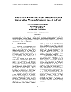
Downloaded from http://rsif.royalsocietypublishing.org/ on May 13, 2015
rsif.royalsocietypublishing.org
Research
Cite this article: Kaushik KS, Ratnayeke N,
Katira P, Gordon VD. 2015 The spatial profiles
and metabolic capabilities of microbial
populations impact the growth of
antibiotic-resistant mutants. J. R. Soc. Interface
12: 20150018.
http://dx.doi.org/10.1098/rsif.2015.0018
Received: 6 January 2015
Accepted: 22 April 2015
The spatial profiles and metabolic
capabilities of microbial populations
impact the growth of antibiotic-resistant
mutants
Karishma S. Kaushik1,2,†, Nalin Ratnayeke2,†, Parag Katira2,‡
and Vernita D. Gordon2,3
1
3
Department of Molecular Biosciences, 2Center for Nonlinear Dynamics and Department of Physics, and
Institute of Cellular and Molecular Biology, University of Texas, Austin, TX 78712, USA
Antibiotic resistance adversely affects clinical and public health on a global
scale. Using the opportunistic human pathogen Pseudomonas aeruginosa,
we show that increasing the number density of bacteria, on agar containing
aminoglycoside antibiotics, can non-monotonically impact the survival of
antibiotic-resistant mutants. Notably, at high cell densities, mutant survival
is inhibited. A wide range of bacterial species can inhibit antibiotic-resistant
mutants. Inhibition results from the metabolic breakdown of amino acids,
which results in alkaline by-products. The consequent increase in pH acts
in conjunction with aminoglycosides to mediate inhibition. Our work
raises the possibility that the manipulation of microbial population structure
and nutrient environment in conjunction with existing antibiotics could
provide therapeutic approaches to combat antibiotic resistance.
Subject Areas:
biophysics
Keywords:
bacterial cell density, spatial structure of
bacterial populations, antibiotic resistance,
aminoglycoside, Pseudomonas aeruginosa,
cystic fibrosis
Author for correspondence:
Vernita D. Gordon
e-mail: [email protected]
†
These authors contributed equally to this
study.
‡
Present address: Department of Biomedical
Engineering, Columbia University, New York,
NY 10027, USA.
Electronic supplementary material is available
at http://dx.doi.org/10.1098/rsif.2015.0018 or
via http://rsif.royalsocietypublishing.org.
1. Introduction
Understanding how populations of antibiotic-susceptible bacteria give rise to
resistant strains is essential to designing treatments that prevent or delay the
development of antibiotic resistance [1]. Recent work has shown that heterogeneity in the spatial distribution of antibiotic in the environment can accelerate the
evolution of genetically based antibiotic resistance [2–5]. However, the role of
ecological changes resulting from the spatially structured microbial population
itself remains to be explored. The spatial structure of a microbial population
includes the density of cells, the combination of species and strains making
up the population, and the spatial organization of cell types.
Most microbial communities consist of interacting, heterogeneous, multispecies populations [6,7]. The per-species spatial structure of microbial
populations is a primary determinant of inter- and intraspecies interactions.
The number density of cells is the simplest parameter characterizing spatial structure. Bacterial biofilms are high-density aggregates of bacteria that are embedded
in an extracellular matrix and are often associated with phenotypic antibiotic
resistance [8–12]. However, the impact of a microbial population’s spatial distribution on the development of genotypic antibiotic resistance is unknown. Unlike
biofilm-based phenotypic antibiotic resistance, genetically based antibiotic resistance can play a role in planktonic cells as well as in biofilms and has the potential
to be inherited and spread to descendent populations.
Here, we show that, for spatially structured populations of bacteria with
planktonic-like phenotype, increasing cell density can impair the survival of
antibiotic-resistant mutants in the presence of aminoglycoside antibiotics. We
term this negative impact ‘inhibition’. The antibiotic-resistant mutants in this
study are strains of Pseudomonas aeruginosa, a common nosocomial pathogen
that affects burned, wounded and ventilator-assisted patients, and also one of
the leading proximate causes of death in patients with the genetic disorder
cystic fibrosis (CF). We observe inhibition with two different aminoglycoside
& 2015 The Author(s) Published by the Royal Society. All rights reserved.
Downloaded from http://rsif.royalsocietypublishing.org/ on May 13, 2015
2.1. Bacterial strains and growth conditions
Pseudomonas aeruginosa PA14 was used as the wild-type (WT)
strain (gift from Marvin Whiteley, UT Austin). WT overnight cultures, grown in antibiotic-free media, were plated on tobramycin
8 mg ml21 agar. Spontaneous antibiotic-resistant mutants grew
colonies and were archived in 20–30% glycerol at 2808C. Upon
subsequent plating, samples that showed mixed colony types
were re-isolated.
To generate mutants in the presence of antibiotic, WT
were passaged in twofold increasing tobramycin concentrations,
starting at 0.3 mg ml21, for eight successive days. Samples were
archived at 2808C daily.
Transposon-insertion mutants (electronic supplementary
material, table S2) were obtained from the PA14 non-redundant
transposon library [15]. P. aeruginosa strain PAO1, 17 clinical
Pseudomonas isolates [16], Pseudomonas sandgrass isolate,
P. fluorescens, methicillin-resistant Staphylococcus aureus Mu50
[17], Escherichia coli DH5a, Burkholderia cepacia and Dphz1/2
mutant [18] (gifts from Whiteley, UT Austin), Serratia marcescens
(gift from Rasika Harshey, UT Austin) and Saccharomyces
cerevisiae S288C (gift from Edward Marcotte, UT Austin).
All bacterial strains were grown in Luria– Bertani (LB) broth
or on LB agar except where otherwise indicated [19]. Saccharomyces cerevisiae was grown in yeast– peptone – dextrose (YPD)
broth or agar except where otherwise indicated [20]. Overnight
cultures were shaken at 180 r.p.m. for 16 – 18 h at 3788 C, except
P. fluorescens and B. cepacia were grown at 308C. When indicated,
agar incorporated tobramycin (Indofine Chemical Company, NJ)
or gentamicin (Fisher Scientific, NJ). Except when indicated
otherwise, the concentrations used were 8 mg ml21.
2.2. Minimum inhibitory concentration
Minimum inhibitory concentration (MIC) was measured using
broth microdilution [21].
2.3. Whole-genome sequencing
Except when stated otherwise, one spontaneous mutant strain was
used for all experiments. This mutant strain was characterized using
whole-genome sequencing (electronic supplementary material, text
2.4. Experiments in spatially mixed systems
Cells (106 – 1011) in 3 ml of 0.6% LB agar were overlaid on tobramycin-containing agar (4 mg ml21). The number of cells was
found from the optical density (OD600) of the overnight cultures
(Nanodrop 2000c, Thermo Scientific), using the conversion factor
OD 1 is 0.8 109 cells ml21. Plates were incubated at 378C for
48 h and colonies were counted (IMAGEJ v. 1.47 [22]).
2.5. Disc diffusion assay for spatially structured systems
To form uniform lawns of antibiotic-resistant mutants on the
surface of antibiotic agar, we used an overlay agar technique,
approximately 108 mutants, unless otherwise stated, were added
to 3 ml of 0.6% (soft) LB agar and overlaid on antibiotic-containing
LB agar.
For deposition onto discs, overnight cultures (32 ml) were
centrifuged to form a pellet (4000 r.p.m., 20 min), washed three
times by removing the supernatant and adding sterile media,
and resuspended in 100 ml of fresh, sterile LB broth. From this,
10 ml (approx. 109 –1010 bacteria, approx. 1011 –1012 yeast, unless
otherwise stated) was deposited on sterile 7 mm diameter filter
discs (Whatman) on the agar. We used the conversion factor OD
1 is 0.8 109 cells ml21 for bacteria and 3 107 cells ml21 for
yeast. Plates were incubated at 378C for 24–48 h, after which the
width of inhibition (X ) was measured.
2.6. Creating structured agar plates
To create agar with spatially structured composition, agar
was poured and allowed to cool as usual. Then, cores of different
diameters (7, 10 and 15 mm) were created with a sterile metal
punch. Wells were filled with agar containing a different set of
nutrients or antibiotic, and allowed to set. As soon as the cores set
(within 10 min), filter discs were placed on the core and WT cells
were deposited.
2.7. Using pH change to probe the nature
of the released inhibitory factor
Bromthymol blue (BTB) was added to agar at a final concentration
of 0.002%. Thirty-two millilitres of overnight microbial cultures
was deposited as above. Plates were incubated at 378C and
observed every hour for an alkaline or acidic change around the
filter discs. Images were taken immediately after deposition (for
nutrient-free BTB agar) or 2 h after deposition (for LB–tobramycin
BTB agar) or after overnight incubation (for antibiotic-resistant
mutant lawns).
2.8. Testing the effect of different nutrient conditions
of overnight growth media on inhibition
In LB and YPD, WT PA14, S. aureus and E. coli grew to a density of
approximately 109 cells ml21. In YPD, S. cerevisiae grew to a density of approximately 108 cells ml21. S. cerevisiae grew poorly in
LB medium, to a density of approximately 107 cells ml21. The
pH of the cell cultures and of the filter-sterilized supernatant was
measured using pH indicator strips (Cardinal Health, IL).
Overnight cultures were centrifuged (4000 r.p.m., 20 min),
washed three times, and resuspended in fresh, sterile LB or YPD
broth (100 ml). Approximately 109 cells were deposited on filter
discs. Filter-sterilized supernatant from each culture condition
2
J. R. Soc. Interface 12: 20150018
2. Material and methods
S1). Sequences were deposited in the National Center for Biotechnology Information Short-Read Archive http://www.ncbi.nlm.
nih.gov/sra (accession no. SRP042054). Three single-nucleotide
polymorphisms unique to the antibiotic-resistant mutant strain
were identified (electronic supplementary material, table S3).
rsif.royalsocietypublishing.org
antibiotics: tobramycin, a frontline drug for treatment of
P. aeruginosa infections, and gentamicin, used in the treatment
of a wide range of Gram-negative bacterial infections [13,14].
Furthermore, we find that a wide range of microbial species can
produce inhibition. Our results indicate that this is because the
molecular agent of inhibition is a by-product of native bacterial
metabolism of amino acids and that the mechanism of inhibition
involves an alkaline change in the pH of the environment.
We use a diffusion-based analytical model to estimate the
molecular weight (MW) of the agent of inhibition, which we
find to be consistent with known alkaline by-products of
metabolism. We also develop a model of inhibition in a
well-mixed system, which considers microscopic, stochastic
variation in cell density. This model quantitatively describes
our observations and offers an explanation of the apparently
contradictory observation that high-density mutant colonies
are capable of growth in the presence of antibiotics.
Our work indicates that microbial population structure
might be a target for, or a tool in, novel therapeutic approaches
to combat antibiotic resistance, and that the lifetime of current
antibiotics might be extended with the use of natural products
as synergistic agents.
Downloaded from http://rsif.royalsocietypublishing.org/ on May 13, 2015
and sterile, fresh LB and YPD broth were also deposited onto filter
discs. Plates were incubated and pH change monitored as above.
2.10. Detection of ammonia and/or amine emission
An ion-selective electrode (Orion 920A, Thermo Scientific) was
used to detect ammonia or amine emission. LB – tobramycin
agar filled 20 ml sterile, narrow-necked glass vials almost filled
to capacity. This reduced the headspace available between the
upper meniscus of the agar and cap of the vial. Cells were deposited on the filter discs placed on the upper surface of the agar,
and the vials were immediately capped. The electrode was
calibrated using 0.1, 0.5, 1, 2.5 and 5 mg l21 concentrations of
ammonium sulfate. While measuring ammonia levels for calibration and in the experiment vials, the electrode was held at a
distance of 1 cm above the upper surface of the media (gaseous
phase). All measurements were conducted at 378C.
2.11. Modelling diffusion of the IF in a spatially
structured system
The disc diffusion assay can be modelled using diffusion laws. In
this system, a finite quantity of IF is released by cells at the disc
and diffuses into a cylindrical agar volume with radius much
larger than height. A system with this geometry has been solved
with a non-closed form solution [23]. However, this solution has
been shown numerically to be approximated by the solution of
one-dimensional diffusion from a constant source [23]:
[IF]0
x2
[IF] ¼
exp ,
(2:1)
4pDt
4Dt
where [IF] is the concentration of IF as a function of distance from
the disc x, time t, the initial concentration at the disc [IF]0 and the
diffusion coefficient D.
At the critical time Tc when zone size is set, the distance x ¼
X defines the region in which the concentration of IF exceeds the
critical concentration [IF]c. Assuming the amount of IF produced
is proportional to the number of cells on the disc gives equation
(2.1). If the mutant overlay is pre-incubated for a set time h before
the disc is placed, the zone size X is given by
X2 ¼ 4D(Tc h) ln (N0 ) þ F(Nc , D, Tc ),
where N0 is the number of cells placed on the disc and Nc is the
minimum number of cells on the disc required to produce a zone
of inhibition. The diffusion coefficient of a molecule is inversely
proportional to its radius. To first order, a molecule’s volume and
therefore its MW scales as the cube of its radius. Thus, for IF and
a molecule A
3
DA
MFIF ¼ MWA
:
DIF
2.12. Measurement of the critical incubation time Tc
and the diffusion coefficient D of the IF
Increasing numbers of WT cells (107 –1011 in 10 ml) were deposited on filter discs. A linear regression of (X 2) as a function
2.13. Modelling the effect of small fluctuations in
population density of antibiotic-resistant mutant
survival in a spatially mixed system
We model the probability of mutant survival P in spatially mixed
systems by
P ¼ 1 eVe r0
GVe rh0 1
X (Ve r )i
0
,
i!
i¼0
where r0 is the average cell density, Ve is an effective volume in
which cells reduce antibiotic quantities per cell, and G and h are
lumped parameters. The function P is defined for densities in
which GVe rh0 1 is a positive integer n, such that r0 must be
equal to (n þ 1/GVe)1/h. We linearly interpolate between values
of r0 which satisfy this condition to generate a continuous
~ Model parameters were estimated using a nonlinear
function P.
least-squares regression of the colony counts in replicate 1
(figure 1b; R v. 2.15.2 [24]). R 2 was calculated as the ratio of
explained variance to total variance.
2.14. Statistical tests
Correlation statistics and Student’s t-tests were done in MICROSOFT
EXCEL.
3. Results and discussion
3.1. In a spatially mixed system, increasing cell
densities negatively impact the survival
of antibiotic-resistant mutants
We grew P. aeruginosa PA14 cultures overnight and then plated
portions of the resulting populations onto LB agar containing
tobramycin at 4 mg ml21. This is approximately four times the
MIC for WT PA14 cells (electronic supplementary material,
table S1). The overnight cultures consisted of a mixture of WT
PA14 cells and spontaneously generated antibiotic-resistant
mutants (electronic supplementary material, table S1). We
used each overnight culture to prepare multiple agar plates,
varying the density of bacteria spread on the plate over four
orders of magnitude. The number of antibiotic-resistant colonies that grow on antibiotic agar is the product of (A) the
number of antibiotic-resistant mutants plated and (B) the probability that an antibiotic-resistant mutant will grow after
plating. The first number, (A), will linearly increase as more
of each culture is plated.
However, we find that the number of antibiotic-resistant
colonies that grow depends non-monotonically on the initial
density of the plated population. These data show a maximum, or ‘sweet spot’, for the number of plated mutants
that survive antibiotic exposure and grow into colonies
J. R. Soc. Interface 12: 20150018
Ten microlitres of ammonium hydroxide (NH4OH; 14.8, 7.4, 4.9,
3.7, 2.9, 2.4 M), sodium hydroxide (NaOH; 1, 0.5, 0.33, 0.25,
0.2 M), sodium bicarbonate (NaHCO3; 1, 0.5, 0.33, 0.25, 0.2 M)
and 1 M ammonium chloride (NH4Cl) were deposited on agar
plates. Plates were incubated and pH change monitored as
above. Ten microlitres of exogenous alkali compounds (2.1 M
NH4OH, 0.5 M NaOH and 0.5 M NaHCO3) were also deposited
on decreasing dilutions (108 –105 cells) antibiotic-resistant mutant
lawns overlaid on antibiotic-free LB agar.
3
rsif.royalsocietypublishing.org
2.9. Deposition of exogenous alkali compounds
of ln(N0), where X is the zone size and N0 is the number
of cells placed on the disc, was performed using R v2.15.2 (R
Development Core Team, Vienna, Austria) [24].
In parallel, to measure critical time Tc, antibiotic-resistant
mutants were overlaid onto antibiotic agar and then preincubated. At set increments of pre-incubation time, WT cells
(approx. 1010) were placed on filter discs. To estimate the time
after which no zone of inhibition would be formed, a linear
regression of X 2 as a function of pre-incubation time was used to
find the X 2 ¼ 0 intercept. This is Tc. Corresponding experiments
were performed for tobramycin as an MW calibrator.
Downloaded from http://rsif.royalsocietypublishing.org/ on May 13, 2015
2000
60
WT PA14 cells
4
replicates
1
X2 (mm2)
2
1000
3
rsif.royalsocietypublishing.org
no. mutant colonies
(a)
X
40
20
0
mutant colony frequency
per million cells
1.0
0.5
0
100
1000
10 000
100 000
plated cell density (cells nl–1)
Figure 1. In a spatially mixed system, high cell densities negatively impact the
survival of antibiotic-resistant mutants in the presence of antibiotic. Different cell
densities from overnight PA14 cultures, containing WT cells and spontaneously
generated antibiotic-resistant mutants, were plated on LB–tobramycin
4 mg ml21 agar. (a) The number of growing antibiotic-resistant mutant colonies varies non-monotonically with cell density. n ¼ 3; independent biological
replicates are indicated by diamonds, circles and multiplication symbols. (b) The
fraction of the plated culture that grows into antibiotic-resistant colonies
decreases monotonically with increasing cell density. n ¼ 3. In (a), the red,
dashed line shows our analytical model, derived using a Poisson distribution
to describe random fluctuations in density, that describes the number of surviving antibiotic-resistant mutants as a function of cell density. This model was
fitted to the colony number data from replicate 1. R 2 ¼ 0.95. In (b), the
red line shows the analytical model describing the frequency of mutant colonies
as a function of cell density, using parameters determined from the previous fit.
R 2 ¼ 0.78. Model parameters: Ve ¼ 0.004, h ¼ 1.164, G ¼ 0.370 nlh 21
and m ¼ 1.10 1026. (Online version in colour.)
(figure 1a). The fraction of the plated culture that gives rise
to mutant colonies, also known as the ‘mutation frequency’
[25], decreases monotonically with increasing cell density
(figure 1b). Therefore, the probability of a mutant growing
into a colony must also decrease with increasing cell density.
Because there are orders of magnitude more WT than
antibiotic-resistant mutant cells present in the initially
plated cultures, we postulate that WT cells are inhibiting
the antibiotic-resistant mutants.
3.2. In a spatially structured system, wild-type cells
inhibit the growth of antibiotic-resistant mutants
For all further experiments, we used the same spontaneously
generated mutant strain and tobramycin at 8 mg ml21, except
when stated otherwise. (See electronic supplementary material
for characterization of this mutant strain using whole-genome
sequencing.) To investigate the role of WT cells in inhibition,
we overlaid antibiotic-resistant mutants onto LB–tobramycin
agar, placed filter-paper discs onto this background, and
deposited WT PA14 cells onto the filter discs. This system
offers a simplified model to examine inhibition in
19
20
ln (N0)
21
22
Figure 2. In a spatially structured system, WT PA14 cells inhibit the growth
of antibiotic-resistant mutants. WT cells (approx. 109 – 1010) were deposited
on antibiotic-resistant mutant lawns on LB – tobramycin 8 mg ml21 agar.
Inset: WT PA14 cells produce inhibition of antibiotic-resistant mutants
(X ¼ 4 mm). The width of inhibition X is measured from the edge of the
filter disc to the edge of the inhibition zone. Scale bars, 5 mm. Increasing
numbers of WT PA14 cells were deposited on antibiotic-resistant mutant
lawns overlaid on LB – tobramycin 4 mg ml21 agar. The square of the
width of inhibition (X 2) varies linearly with the log of the number of WT
cells deposited. R 2 ¼ 0.87. n ¼ 4. (Online version in colour.)
populations exhibiting macroscopic heterogeneity in cell density, and is inspired by the disc diffusion assay often used to
evaluate antibiotic susceptibility [26]. After growth of the
antibiotic-resistant mutant lawn, we observed circular zones
of inhibition surrounding the filter discs (figure 2 and electronic supplementary material, figure S1). From these
experiments, we conclude that, in the presence of antibiotic,
WT PA14 cells inhibit antibiotic-resistant mutants.
We also find that cell-to-cell contact or proximity, nutrient
depletion, pyocins, quorum sensing and reactive oxygen
species are not involved in inhibition, and that inhibition is
caused by a diffusible factor that does not stick to glass and
therefore may not be a protein (electronic supplementary
material, figures S2 –S7).
3.3. The inhibitory factor has a low-molecular weight
We model the disc diffusion assay, informed by previous studies
modelling antibiotic diffusion [23,27]. Our model assumes
(1) the IF is released by WT cells to a concentration [IF]0 at
the disc;
(2) the IF diffuses out of the disc with a constant diffusion
coefficient D;
(3) a threshold concentration of IF, [IF]th, is required to inhibit mutant cells; and
(4) IF no longer causes inhibition after a critical time Tc of
incubation.
Assumption (4) corresponds to an increase in the number of
cells in the lawn that causes the per-cell concentration of IF
to drop to subinhibitory levels [28,29]. This agrees with our
finding that the size of the inhibition zone decreases with
increasing initial lawn density (electronic supplementary
material, figure S8a). The width of the inhibition zone is the distance between the edge of the disc and the edge of the inhibition
zone, X (figure 2, inset). Our model approximates X 2 as
X2 ¼ 4DTc ln (N0 ) þ F(D, Tc , Nc )
(3:1)
J. R. Soc. Interface 12: 20150018
0
18
(b)
Downloaded from http://rsif.royalsocietypublishing.org/ on May 13, 2015
When the agar does not contain antibiotic, inhibition does not
occur (electronic supplementary material, figure S11). This
could mean that antibiotic is required for production of the
IF, the activity of the IF or both. To dissect this, we developed
a set of complementary experiments, in which the filter disc
was centred on top of a cylindrical core of agar with a different composition than that of the surrounding agar (electronic
supplementary material, figure S12a).
When the core does not contain antibiotic, tobramycin must
diffuse into the core from the surrounding agar before reaching
the WT cells deposited on the filter disc. This introduces a delay
before WT cells are exposed to tobramycin which increases with
increasing core diameter. Because the timescale for diffusive
transport over a distance L scales as L 2/D, the core sizes we
use correspond to delays in the range of tens of hours to reach
the agar under the centre of the disc and up to 13 h to reach
the agar under the edge of the disc. If exposure to tobramycin
is required for IF production, this delay should act as a preincubation time and thereby decrease the size of the inhibition
zones. On the contrary, we find no significant decrease in the
size of the inhibition zones with increasing size of antibioticfree cores (electronic supplementary material, figure S12b).
3.5. A wide range of bacterial species and a second
aminoglycoside antibiotic, produce inhibition
We find that antibiotic-resistant cells deposited on antibioticresistant mutant lawns also produce inhibition (electronic
supplementary material, figure S13). This is true both for
the mutants we generated and for PA14 strains that have a
gentamicin resistance cassette (aacC1) inserted into their
genome [15]. These data indicate that growing cells also inhibit antibiotic-resistant mutants, and that mutants can inhibit
their clonal siblings.
To determine whether IF is produced by organisms other
than P. aeruginosa strain WT PA14, we evaluated a set of
Gram-negative and Gram-positive bacteria as well as one
strain of yeast. These organisms include known co-pathogens
with P. aeruginosa as well as 17 clinical isolates of P. aeruginosa
from CF patients [16]. All bacteria tested produce inhibition
(table 1; electronic supplementary material, figures S14– S16
and table S2). However, the yeast Saccharomyces cerevisiae
fails to produce inhibition. In the absence of antibiotic in
the agar, no inhibition is observed with the bacterial strains.
This is consistent with the idea that the inhibitory factors in
these cases are the same as that produced by WT PA14
cells. Our results suggest that IF is produced by a wide
range of bacterial species but not the yeast S. cerevisiae.
We tested five antibiotic-resistant mutant strains with
different origins for susceptibility to inhibition. Three of
these strains were spontaneously generated in antibioticfree media and two evolved in the presence of increasing
tobramycin concentrations. All show similar susceptibility to
inhibition (electronic supplementary material, figure S13).
Therefore, we infer that susceptibility to inhibition likely
does not depend sensitively on the details of the mutation(s)
conferring antibiotic resistance.
The tobramycin-resistant mutants that we generated are
also resistant to another aminoglycoside, gentamicin (MIC ¼
9.7 mg ml21; electronic supplementary material, table S1).
WT PA14 cells deposited on antibiotic-resistant mutant lawns
on gentamicin agar also produce inhibition. This result
demonstrates that inhibition occurs in the presence of two
different aminoglycoside antibiotics (electronic supplementary
material, figure S13).
3.6. Inhibition is observed when bacterial cells are
deposited on media devoid of nutrients
It is striking that antibiotic-susceptible WT PA14 cells produce
inhibition even though the antibiotic agar contains an inhibitory
concentration of antibiotic, which should kill or inhibit WT PA14
cells shortly after plating. This suggests that IF production may
5
J. R. Soc. Interface 12: 20150018
3.4. The inhibitory factor is produced without antibiotic
but causes inhibition only in the presence
of antibiotic
These results show that antibiotic is not required for the
production of IF.
To evaluate whether antibiotic is required for the activity
of IF, we made cores of LB–tobramycin agar in antibiotic-free
LB agar. With this geometry, we find no inhibition even at
low lawn densities (electronic supplementary material,
figure S12c). This confirms that antibiotic is required for the
IF to cause inhibition.
Collectively, these results indicate that IF is likely a native
product of WT PA14 cells that is produced without antibiotic but requires the presence of antibiotic to exert its
inhibitory effect.
rsif.royalsocietypublishing.org
N0 is the number of WT cells deposited on the disc, which we
assume to be linearly proportional to the concentration of IF
produced. F is a function independent of N0. Nc is the minimal
number of WT cells that produce measurable inhibition. The
slope of X 2 as a function of ln(N0) gives the diffusion coefficient
D, if critical time Tc is known.
To vary N0, we deposited increasing numbers of WT
cells on antibiotic-resistant mutant lawns on tobramycin
4 mg ml21 agar and measured X. On a semi-log plot, X 2
varies linearly with ln(N0), as predicted by the model
(figure 2). To measure Tc, we pre-incubated the mutant lawn
before depositing WT cells. X decreases with increasing preincubation times until X ¼ 0 (electronic supplementary
material, figure S8b). We measure Tc to be 110 + 30 min and
Nc to be of the order of a million cells.
By fitting the model to the data in figure 2, we estimate the
diffusion coefficient D of the IF to be 4.5 + 0.5 1026 cm2 s21.
To calibrate the relationship between D and the MW of the IF,
we performed the disc diffusion assay using varying concentrations of tobramycin (MW ¼ 467.5 Da) deposited on filter
discs on a WT lawn on antibiotic-free agar (electronic supplementary material, figure S9). We did two replicates of this
experiment. Fitting these results gives a diffusion coefficient
for tobramycin (averaged for the two replicates) of 1.2 +
0.1 1026 cm2 s21. Using the Stokes–Einstein relation, we
estimate that the IF has a MW of the order of 10 Da, indicating
it is a small molecule.
We also performed the above experiments, twice, using a
tobramycin concentration of 8 mg ml21 in the agar and find
D of the IF (averaged for the two replicates) is 2.6 + 0.3 1026 cm2 s21, so the MW of IF was again determined to be a
few tens of Daltons (electronic supplementary material,
figure S10). These results very nearly overlap with those
taken at 4 mg ml21, and suggest that the size of the IF is near
the lower limit of what we can measure using this approach.
Downloaded from http://rsif.royalsocietypublishing.org/ on May 13, 2015
Table 1. Microbial species/strains that produce inhibition.
rsif.royalsocietypublishing.org
species/strain
6
additional remarks
Pseudomonas strains
P. aeruginosa (WT PA14, WT PAO1)
clinical P. aeruginosa strains (17)
laboratory strains
cystic fibrosis isolates [16]
sandgrass isolate
P. fluorescens
uncharacterized environmental isolate
environmental isolate
laboratory strains
S. aureus (Mu50)
Gram-positive, methicillin- and vancomycin-resistant clinical isolate [17], co-pathogen with P. aeruginosa [30]
Serratia marcescens
Burkholderia cepacia
Gram-negative, nosocomial human pathogen [31]
Gram-negative, nosocomial co-pathogen with P. aeruginosa [32]
not depend on utilization of nutrients from the agar. To test this,
WT PA14 cells were deposited on ‘cores’ of nutrient-free, tobramycin-containing agar (electronic supplementary material,
figure S17a). To test for effects of diffusion from the surrounding
LB agar into the cores, increasing core diameters were used. WT
PA14 cells produce inhibition even when deposited on nutrientfree cores that is comparable to those seen with the ‘no core’ and
‘LB–tobramycin core’ controls (electronic supplementary
material, figures S17b,c). These data indicate that the production
of IF does not depend on the utilization of nutrients in the
agar plate.
figure S19). Yeast produces a slightly acidic change or
no change, with an intensity ratio greater than or equal
to 1. The width of inhibition X and the width of the alkaline
colour change Xc are correlated (figure 3b). These results
agree with the idea that inhibition is caused by the alkaline
bacterial by-products. Alkaline change on nutrient-free agar
(electronic supplementary material, figure S20) is consistent
with our finding that inhibition does not result from
utilization of nutrients from the agar plate. Rather, metabolic
by-products from cells in the overnight cultures continue to
be released following deposition on the agar.
3.7. Alkaline metabolic products may play a role
in inhibition
3.8. The mechanism of inhibition is related
to the alkaline pH change
Because S. cerevisiae does not produce inhibition but all tested
bacterial strains do, we infer that the nutritional substrate for
microbial growth may be linked to the production of IF. All
bacterial strains were grown in LB medium [19] but S. cerevisiae
was grown in YPD medium. In LB medium, amino acids are
the only significant source of carbon and bacteria grow by
catabolism, which produces alkaline by-products [33,34]; overnight culture pH approximately 8. In contrast, S. cerevisiae
grows in YPD medium using the sugar dextrose as a source
of carbon [20]. Yeast’s fermentation of sugar produces acidic
by-products [35]; overnight culture pH approximately 6. To
evaluate the relationship between pH and inhibition, we
used agar plates that incorporated the pH indicator BTB. BTB
has a pH range of 6–7.6, which permits detection of slight
perturbations from neutral pH.
WT PA14 cells, WT PAO1 cells, S. aureus, E. coli, Serratia
marcescens, Burkholderia cepacia, Pseudomonas sandgrass isolate,
Pseudomonas PAO-JP2, Pseudomonas Dphz1/2 and S. cerevisiae
were deposited on nutrient-free tobramycin agar and
nutrient-containing LB–tobramycin agar containing BTB.
To quantify changes in colour, images were split into red–
green–blue channels and the red intensity profiles were
analysed (electronic supplementary material, figure S18). We
define intensity ratio as the intensity 2 mm from the disc edge
divided by the intensity at a distance greater than 8 mm from
the disc edge. With no colour change, the intensity ratio is
expected to be approximately 1.
When deposited on nutrient-containing BTB agar, bacteria produce an alkaline change, with an intensity ratio
less than 1 (figure 3a and electronic supplementary material,
To investigate a causative link between alkalinity and inhibition, we test exogenous alkaline solutions and find they
produce both an alkaline change on BTB agar and inhibition of antibiotic-resistant mutants (figure 4 and electronic
supplementary material, figures S21 and S22). We vary the concentration deposited and use linear regression to determine the
relationship between concentration and X. From these, we
extrapolate that deposition of 10 ml of 2.04 M NH4OH, 1 M
NaOH or 1 M NaHCO3 produces inhibition of antibioticresistant mutants comparable to that seen with WT PA14
cells (electronic supplementary material, figures S21–S23).
On antibiotic-free LB agar, zones of inhibition are either
absent (with NH4OH and NaHCO3) or markedly reduced
(with NaOH; electronic supplementary material, figure S24).
Collectively, these results indicate that alkaline compounds
can recreate the characteristics we have found for the IF.
Other researchers have shown that alkalinity significantly
enhances the bactericidal activity of aminoglycosides [35,36]. At
least two possible mechanisms have been suggested (i) enhanced
active uptake of aminoglycosides [36–39]; (ii) more aminoglycosides in their non-ionized, more-bactericidal form [36].
Thus, we infer that the IF inhibits mutants by increasing pH.
We modify equation (3.1) to use a threshold pH rather
than a threshold IF concentration as the determinant of cell
survival, thus:
X2 ¼ 4DTc ln (N0 ) þ F(D, Tc , pHc , k0 , a, b)
where pHc is the pH above which cells die, k0 is the IF production by one cell, and a and b are constants which
account for the acid–base properties of the IF (electronic
J. R. Soc. Interface 12: 20150018
Other microbial species
E. coli (DH5a, SM10)
Downloaded from http://rsif.royalsocietypublishing.org/ on May 13, 2015
(a)
PAO1
E.
coli
5
X (mm)
0
X = 0 mm
BTB
intensity
ratios
0
X
6
PA14
PAO1
X (mm)
S. aureus
E. coli
S. marcescens
B. cepacia
R2 = 0.82
4
5
6
Xc (mm)
7
Figure 3. Alkaline metabolic by-products from a wide range of bacterial species correlate with inhibition. (a) Different microbial strains were deposited on nutrientfree tobramycin BTB agar, on nutrient-containing LB – tobramycin BTB agar, and on antibiotic-resistant mutant lawns overlaid on LB – tobramycin agar. Images were
taken immediately after deposition (for nutrient-free BTB agar), 2 h after deposition (for nutrient-containing LB BTB agar) or after overnight incubation
(for antibiotic-resistant lawns on LB – tobramycin 8 mg ml21 agar). The width of inhibition (X ) is shown as hatched bars. All bacterial strains produce inhibition
of antibiotic-resistant mutant lawns, however, the yeast strain fails to produce inhibition. Error bars (in red) represent standard error of the mean; n ¼ 3. (b) For
different inhibiting strains, when the width of alkaline change (Xc, see electronic supplementary material, figure S18) is plotted as a function of the width of
inhibition (X ), a correlation is observed (R 2 ¼ 0.82 from a linear fit, Pearson’s correlation coefficient r ¼ 0.9). Intensity profiles were used to measure the
width of alkaline colour change (Xc), representing the distance from the edge of the disc to the edge of the alkaline colour change. For each replicate, Xc
and X represent the average of four measurements made along two perpendicular axes. Error bars represent standard error of the mean; n ¼ 2. (Online version
in colour.)
supplemental material). The N0-dependent portion of this
function is unchanged from equation (3.1). Therefore,
pH-mediated inhibition does not change our MW measurements. Further, this model predicts a linear relationship
between X 2 and the squared size of alkaline colour change
(Xc2 ), consistent with figure 3b.
3.9. Biogenic bases are plausible candidates
for the inhibitory factor
The biogenic bases ammonia and amines are widespread byproducts of bacterial catabolism of amino acids [33,34,40].
Ammonia has an MW of 17 Da, consistent with that of
the IF. Microbially produced ammonia and amines can mediate long-range intercellular interactions and influence
antibiotic resistance [41 –46]. Gaseous ammonia, from
bacterial supernatant, and trimethylamine, can decrease
resistance to the aminoglycoside antibiotics kanamycin and
spectinomycin [45,46].
We used an ion-selective electrode to measure ammonia
and amine emission when bacteria and yeast were deposited
onto filter discs on LB–tobramycin agar. WT PA14 produce
an emission nearly five times that of S. cerevisiae (figure 5).
In the disc diffusion assay, deposition of 1 M ammonium
chloride (NH4Cl) produces a slightly acidic change and no
inhibition (figure 4). Thus, we find a correlation between
alkaline change and inhibition, but no inhibitory effect by
the ammonia molecule itself when the pH is acidic. Therefore,
we attribute inhibition to an alkaline pH change that is
mediated, in part or in whole, by biogenic ammonia and/
or low-molecular-weight amines.
3.10. Nutrient conditions provide a ‘switch’ to control
inhibition
Microbes capable of using either amino acids or sugars as
carbon sources should switch between producing inhibition
and not producing inhibition depending on the carbon
J. R. Soc. Interface 12: 20150018
nutrient-free agar
LB agar
intensity ratio
1
(b)
7
S.
B.
S.
marcescens cepacia cerevisiae
rsif.royalsocietypublishing.org
PA14
S.
aureus
Downloaded from http://rsif.royalsocietypublishing.org/ on May 13, 2015
(a)
8
**
LB–
tobramycin
BTB agar
antibioticresistant mutant
lawn
ammonium
hydroxide
sodium
bicarbonate
sodium
hydroxide
WT PA14
cells
ammonium
chloride
Figure 4. Exogenous alkaline solutions can recapitulate the phenomenon of
inhibition. (a,b) Ten microlitres of exogenous compounds—2.4 M ammonium
hydroxide (NH4OH), 1 M sodium bicarbonate (NaHCO3), 1 M sodium hydroxide
(NaOH), WT PA14 cells and 1 M ammonium chloride (NH4Cl)—were deposited
on LB–tobramycin BTB agar and on antibiotic-resistant mutant lawns on
LB–tobramycin agar. Images were taken 2 h after deposition (for LB–
tobramycin BTB agar) or after overnight incubation (for LB–tobramycin agar
with antibiotic-resistant mutant lawns). (a) BTB colour intensity ratios were
determined as for figure 3, with the baseline grey value measured at a distance
of more than 10 mm for NaOH, NaHCO3 and NH4Cl and WT PA14 cells, and more
than 40 mm for NH4OH. Intensity ratios less than 1 indicate an alkaline change,
as produced by ammonium hydroxide, sodium hydroxide, sodium bicarbonate
and WT PA14 cells. Ammonium chloride produces an intensity ratio approximately 1. Error bars (red) represent standard error of the mean; n ¼ 2.
(b) Representative images showing BTB colour change and presence or absence
of inhibition produced by 2.4 M ammonium hydroxide (NH4OH; X ¼ 10 mm,
Xc ¼ 15 mm), 1 M sodium bicarbonate (NaHCO3; X ¼ 5 mm, Xc ¼
5.5 mm), 1 M sodium hydroxide (NaOH; X ¼ 5 mm, Xc ¼ 7.5 mm), WT
PA14 cells (X ¼ 5 mm, Xc ¼ 6 mm) and 1 M ammonium chloride (NH4Cl;
X ¼ 0 mm, Xc ¼ 0 mm). Scale bar, 5 mm. (Online version in colour.)
source available. S. aureus and E. coli can either catabolize
amino acids or ferment glucose. P. aeruginosa is incapable of
fermentation. S. cerevisiae ferments glucose. We grew each
sterile
medium
S. cerevisiae
PA14
ammonium chloride
WT PA14 cells
sodium
hydroxide
sodium
bicarbonate
ammonium
hydroxide
intensity ratio
(b)
0
Figure 5. Production of ammonia or amines is associated with inhibition.
Using an ion-selective electrode, ammonia or amine emission was measured
following deposition of WT PA14 and S. cerevisiae cells on filter discs on LB –
tobramycin agar in vials. Emission levels are normalized to the PA14 average.
Significantly higher levels of ammonia or amine emission are detected following deposition of WT PA14 cells than following deposition of S. cerevisiae.
Error bars represent standard error of the mean; n ¼ 3. *p , 0.005 by
two-tailed Student’s t-test. **p , 0.02 by two-tailed Student’s t-test.
organism in two culture media—LB, which contains almost
entirely amino acids as a carbon source, and YPD, which likewise contains copious amino acids but is also supplemented
with the sugar dextrose. The pH of overnight cultures grown in
LB was alkaline (pH ¼ 9) for all bacterial strains and neutral
( pH ¼ 7) for the yeast strain. S. cerevisiae grew poorly in LB
broth, so we pool cells from multiple cultures so that approximately the same number of cells were deposited onto filter
discs in all cases. The pH of overnight cultures grown in YPD
was alkaline ( pH ¼ 8) for WT PA14, and acidic for S. aureus,
E. coli and S. cerevisiae ( pH ¼ 4, 4 and 5, respectively).
WT PA14, S. aureus and E. coli cultures grown in LB produce an alkaline colour change and inhibit antibiotic-resistant
mutants (figure 6a; electronic supplementary material, figures
S25a and S26). S. cerevisiae produces neither a pH colour change
nor inhibition. When grown in YPD, WT PA14 cells produce an
alkaline change and inhibition. However, S. aureus, E. coli and
S. cerevisiae grown in YPD produce an acidic change and no
inhibition (figure 6b; electronic supplementary material,
figure S25b and S26). Culture supernatants and sterile LB
and YPD media produce no pH colour change on nutrientcontaining media and produce no inhibition (electronic
supplementary material, figures S27–S29).
From this, we conclude that microbial metabolism and the
nutrient environment can provide a switch to control inhibition.
3.11. Spatial fluctuations in population density
influence the survival of antibiotic-resistant
mutants in spatially mixed populations
In the spatially mixed system, mutant survival probability
approaches zero with increasing cell density. Similarly, in
disc diffusion assays, inhibition zone size increases with
increasing density of deposited cells. Therefore, it seems paradoxical that mutant cells are able to grow into high-density
colonies on the plate without inhibiting themselves. To resolve
this paradox, we examine the effect of spatial fluctuations in
initial population density on the survival of resistant mutants.
It is often found that the molar amount of an inhibitory
substance per cell is a more accurate determinant of cell survival during exposure than the concentration, and therefore,
higher-density populations of bacteria can survive higher
J. R. Soc. Interface 12: 20150018
0
100
rsif.royalsocietypublishing.org
1
relative ammonia or
amine emission
*
Downloaded from http://rsif.royalsocietypublishing.org/ on May 13, 2015
(a)
9
grown with amino acids
5
rsif.royalsocietypublishing.org
X (mm)
no inhibition
intensity ratio
intensity ratio
1
0
0
(b)
grown with amino acids and sugar
J. R. Soc. Interface 12: 20150018
X
5
0
PA14
S.
aureus
E. coli
S.
cerevisiae
X (mm)
no inhibition
no inhibition
no inhibition
intensity ratio
1
0
Figure 6. The carbon source used for growth can switch inhibition on or off. WT PA14, S. aureus, E. coli and S. cerevisiae were each grown in (a) LB medium, for
which amino acids are the major carbon sources, and (b) YPD medium, which contains both amino acids and sugar as carbon sources. Following growth, cultures
were deposited onto LB – tobramycin BTB agar and onto antibiotic-resistant mutant lawns on LB – tobramycin agar. (a) When grown on amino acids, the first three
organisms produce both a BTB intensity ratio less than 1 and a zone of inhibition. The yeast S. cerevisiae produces no change in pH and no inhibition. (b) When
grown in the presence of sugars, S. aureus, E. coli and S. cerevisiae ferment sugars and thereby produce an acidic change and no inhibition. WT PA14 catabolizes
amino acids, causing an alkaline change and inhibition. Sterile LB and YPD media, and filter-sterilized culture supernatants, produce no BTB colour change and no
inhibition (electronic supplementary material, figures S28 and S29). (Online version in colour.)
concentrations of antibiotics than lower-density populations
[8,9]. In a well-mixed, homogeneous system with a constant
antibiotic concentration [AB], increasing cell density
decreases the moles of antibiotic per cell, fABg, thereby
enhancing the resistance of cells to a given [AB]. However,
we have postulated that the IF released by cells modulates
antibiotic resistance by changing pH. Moreover, physically
realistic spatial distributions of cells will have random fluctuations in cell density. Together, these two observations may
lead to non-trivial probabilities of cell survival as a function
of the average cell density.
We assume a uniform [AB] throughout the plate. fABg
in a small effective volume Ve containing N cells is thus
[AB]Ve/N. If fABg is less than a critical value fABgth,
mutant cells in Ve will grow. Because the number of WT
cells is much greater than the number of antibiotic-resistant
mutants and the volumes in question are small, we assume
that no more than one mutant will be in Ve and that it will
survive if NWT þ 1 . ([AB]Ve)/(fABgth), where NWT is the
number of WT in Ve.
We model random deposition of bacteria as a Poisson
process. The Poisson cumulative distribution function gives
the probability that the number of WT in Ve will be greater
P WT
i
than NWT as P ¼ 1 eVe rWT N
i¼0 ((Ve rWT ) )=ði!Þ rWT is the
average density of WT on the plate. Because rWT r0,
where r0 is the total cell density on the plate, we approximate
P WT
i
P ¼ 1 eVe r0 N
i¼0 ((Ve r0 ) )=(i!):
Previous studies at constant cell density have found an
approximately exponential relationship between the MIC to
aminoglycoside antibiotics and the pH [47]. We assume a
similar exponential form for fABgth ¼ A 102bpH þ
fABgmin, where A and b are scaling constants, and fABgmin
is the minimum value of fABgth. We approximate the pH
resulting from the release of IF as pH ¼ a þ blog10[IF], where
a and b are constants describing the acid–base properties of
the IF, and [IF] is the concentration of IF on the plate (electronic
supplementary material). Assuming fABgmin is small compared to fABgth, the condition for mutant survival can be
approximated as N . ([AB][IF]bb Ve)/(A 102ab). If [IF] ¼
k0r0, where k0 is a production constant and r0 is the initial
cell density, the probability of mutant survival becomes
Ve r0
P¼1e
GVe rh0 1
X (Ve r )i
0
,
i!
i¼0
(3:2)
where G ¼ [AB]k0bb =A 10ab and h ¼ bb. G is a lumped parameter which accounts for the alkalinity of the IF and a
strain’s production of IF and degree of antibiotic resistance; h
accounts for the sensitivity of the antibiotic resistance to
changes in pH; Ve accounts for the spatial extent over which
surrounding cells can lower the fABg to which a mutant is
exposed. P is discrete with respect to r0, and a continuous func~ can be approximated from it using linear interpolation.
tion P
~ gives the fraction of plated antibioticThe function P
resistant mutants that will grow to form colonies and as a
function of the initial density of cells. The fraction of the
plated culture that grows into antibiotic-resistant colonies,
~ where m is the true proM, is thus described by M ¼ mP,
portion of mutants in the population. Similarly, the number
Downloaded from http://rsif.royalsocietypublishing.org/ on May 13, 2015
We have found that bacteria, regardless of their antibiotic
susceptibility, can inhibit the survival and growth of antibiotic-resistant mutants in the presence of aminoglycosides.
An alkaline by-product of amino acid catabolism, possibly
ammonia or amines, mediates this inhibition. Further, these
alkaline by-products likely mediate this effect by an increase
in pH, which enhances the bactericidal effect of aminoglycosides. We find that microbial population structure, including
cell density, spatial distribution and interspecies interactions,
can impact the growth of pre-existing antibiotic-resistant
mutations through this mechanism.
For a favourable mutation (i.e. one that confers a fitness
advantage in the environment), the probability of fixation
in a population increases with decreasing population size,
which is a measure of the number of competitors [48,49].
Although our liquid cultures are grown without antibiotics,
it is possible that the resistance-conferring mutation(s) conveys some other growth advantage in antibiotic-free liquid
culture. In that case, the probability of the mutation fixing
would depend on whether the mutation occurred early or
late in culture growth. However, this possibility does not confound the results we present here, because in figure 1 the ratio
of mutants to WT is the same within each experiment, because
10
J. R. Soc. Interface 12: 20150018
4. Conclusion
all the data points for each experiment are done for the same
overnight culture suspension. Therefore, figure 1 measures
the probability that a pre-existing mutant will grow into a
colony, not the fixation of the mutants in a population.
Our finding, that increasing bacterial density can increase
antibiotic susceptibility, is superficially in contrast to previous observations that in compact bacterial populations,
such as in biofilms, the susceptibility against antibiotics is
in fact decreased. However, it should be noted that bacteria
in biofilms are in a different phenotypic state than that of
the bacteria we plated from suspension onto agar. Most notably, biofilms are structured by embedding extracellular
polymers, which can provide protection against aminoglycoside antibiotics. The bacteria in biofilm interiors also have
limited access to growth substrates, which results in slowed
growth and altered metabolic pathways that can protect
against antibiotics.
Thus, the structure and the phenotypic state of the bacterial population needs to be considered when designing
treatment strategies.
This can easily be seen in the case of CF, which is a genetic
condition that renders the lung unusually susceptible to infection by P. aeruginosa and other pathogens. The CF lung
contains an abundance of free amino acids [34,50,51]. In
microbes sampled from CF lungs, the genes and pathways
involved in amino acid catabolism are significantly upregulated
[34,50]. In accordance, elevated ammonia levels have been
found in CF sputum [34]. These observations would seem to
suggest that the microbial population in the CF lung is well
poised to inhibit each other in the presence of aerosolized
tobramycin, which is commonly used in the treatment of CF.
However, CF results from a genetic defect in the CF transmembrane receptor, which causes decreased bicarbonate ion
transport and acidification of CF airway fluids [52,53]. This
would prevent inhibition. Inhaled bicarbonate therapy is a
possible remediation for this and is already being studied
as a therapy for CF because it may help restore the innate
antimicrobial activity of airway surface fluids as well as facilitate the thinning and clearing of airway mucus [53,54]. Our
results suggest a potential additional benefit for bicarbonate
therapy in CF, where it could augment the activity of aminoglycosides and help curtail antibiotic resistance. Thus,
optimizing therapeutic benefits of combining aminoglycosides with bicarbonate would require further work
determining how the physiological environment of the host
impacts microbial metabolism, pH changes and pharmacokinetics of base and aminoglycoside in the lung. Moreover,
while early infections of the CF lung are transient and associated with a planktonic bacterial phenotype, decades-long
chronic infections are associated with a biofilm phenotype.
Because biofilms have phenotypic resistance to aminoglycosides, full realization of the potential benefits of the work
we present here would require determination of the degree
to which pH-mediated inhibition is active against clinical
biofilm infections.
More broadly, our findings indicate that manipulating
the nutritional and metabolic environment of the CF lung,
chronic wounds or other sites of infection could potentially
provide a set of management strategies that are complementary to and synergistic with conventional antibacterial
therapies. Developing approaches that limiting the availability
of sugars, and/or using anti-fungal therapy would promote amino acid catabolism and prevent microbe-caused
rsif.royalsocietypublishing.org
~ where Vtot is
of colonies formed, C, is given by C ¼ mr0 Vtot P,
the total volume of the plated cell suspension.
We fit this model to our experimental data. A wide range
of values of G, Ve, h and m result in trends consistent with our
measurements in figure 1 (electronic supplementary material,
figure S30). We can estimate physically reasonable values of
Ve by examining how an individual cell might impact its surrounding antibiotic concentration. By modelling a cell as an
antibiotic sink, we estimate the lowest reasonable Ve value
to be 0.004 nl, corresponding to 1000 times the volume of a
cell, or a sphere of radius 10 mm (electronic supplementary
material). With a Ve of 0.004 nl, values of h ranging from
0.90 to 2.5, G ranging from 8 1026 to 4 nlh 21 and m ranging
from 4 1027 to 4 1026, produce reasonable fits to our
data, and the least-squares fit has an h of 1.186, G of
0.307 nlh21 and m of 1.02 1026 (R 2 ¼ 0.92; figure 1).
Additionally, Ve of 0.04, 0.4 and 4 nl, produce similar
trends and fits (electronic supplementary material, figure S30
and S31). Overall, the wide range of parameter values that
produce reasonable fits reflects a robustness of this model to
changes in specific values, and indicates that fine-tuning of
the parameters is not necessary to match the trends which
we observe.
Guided by this model, we postulate that antibioticresistant mutants are able to grow into high-density colonies
on antibiotic-containing agar because they are shielded from
antibiotics by their WT neighbours. A mutant will grow into
a colony if it is in a region with high local cell density that
reduces the fABg to which it is exposed below a threshold
value. However, as the average cell density deposited on
the plate increases, the resulting increase in [IF] on the plate
lowers this threshold value by causing an increase in pH,
so there is a net decrease in mutant survival probability
with increasing average density. This interpretation implies
that small initial fluctuations in density will be magnified
as they control the subsequent growth of bacteria.
Downloaded from http://rsif.royalsocietypublishing.org/ on May 13, 2015
Acknowledgements. We thank Andreas Matouschek for use of the
spectrophotometer and ultracentrifuge, Mary Jo Kirisits for use of
the ammonia-selective electrode, and Jeffrey Barrick and Daniel
Deatheridge for guidance with the BRESEQ pipeline. We are grateful
to Marvin Whiteley, Andy Ellington and Rasika Harshey for their
experimental suggestions.
Funding statement. This work was supported by start-up funds to Dr
Vernita Gordon at the Center for NonLinear Dynamics, Department
of Physics, UT Austin, and by a gift to Dr Gordon from ExxonMobile.
K.S.K. received a Graduate School Continuing Fellowship from UT
Austin. N.R. received an undergraduate and summer research fellowship from UT Austin.
1.
Davey P, Sneddon J, Nathwani D. 2010 Overview of
strategies for overcoming the challenge of
antimicrobial resistance. Expert Rev. Clin. Phamacol.
3, 667–686. (doi:10.1586/ecp.10.46)
2. Zhang Q, Lambert G, Liao D, Kim H, Robin K, Tung
C-k, Pourmand N, Austin RH. 2011 Acceleration of
emergence of bacterial antibiotic resistance in
connected microenvironments. Science 333,
1764–1767. (doi:10.1126/science.1208747)
3. Perron GG, Gonzalez A, Buckling A. 2007 Source–
sink dynamics shape the evolution of antibiotic
resistance and its pleiotropic fitness cost.
Proc. R. Soc. B 274, 2351 –2356. (doi:10.1098/rspb.
2007.0640)
4. Hermsen R, Barrett Deris J, Hwa T. 2012 On the
rapidity of antibiotic resistance gradient facilitated
by a concentration gradient. Proc. Natl Acad. Sci.
USA 109, 10 775–10 780. (doi:10.1073/pnas.
1117716109)
5. Greulich P, Waclaw B, Allen RJ. 2012 Mutational
pathway determines whether drug gradients
accelerate evolution of drug-resistant cells. Phys.
Rev. Lett. 109, 088101. (doi:10.1103/PhysRevLett.
109.088101)
6. Li K, Bihan M, Yooseph S, Methe´ BA. 2012 Analyses
of the microbial diversity across the human
microbiome. PLoS ONE 7, e32118. (doi:10.1371/
journal.pone.0032118)
7. Delhaes L et al. 2012 The airway microbiota in cystic
fibrosis: a complex fungal and bacterial
community—implications for therapeutic
management. PLoS ONE 7, e36313. (doi:10.1371/
journal.pone.0036313)
8. Eng RH, Smith SM, Cherubin C. 1984 Inoculum
effect of new beta-lactam antibiotics on
Pseudomonas aeruginosa. J. Antimicrob. Chemother.
26, 42 –47. (doi:10.1128/AAC.26.1.42)
9. Udekwu KI, Parrish N, Ankomah P, Baquero F, Levin
BR. 2009 Functional relationship between bacterial
cell density and the efficacy of antibiotics.
J. Antimicrob. Chemother. 63, 745–757. (doi:10.
1093/jac/dkn554)
10. Connell JL, Wessel AK, Parsek MR, Ellington AD,
Whiteley M, Shear JB. 2010 Probing prokaryotic
social behaviors with bacterial ‘lobster traps’. mBio
1, e00202-210. (doi:10.1128/mBio.00202-10)
11. Connell JL, Ritschdorff ET, Whiteley M, Shear JB.
2013 3D printing of microscopic bacterial
12.
13.
14.
15.
16.
17.
18.
19.
20.
21.
22.
23.
communities. Proc. Natl Acad. Sci. USA 110,
18 380– 18 385. (doi:10.1073/pnas.1309729110)
Costerton JW, Lewandowski Z, Caldwell DE, Korber
DR, Lappin-Scott HM. 1995 Microbial biofilms.
Annu. Rev. Microbiol. 49, 711–745. (doi:10.1146/
annurev.mi.49.100195.003431)
Geller DE, Pitlick WH, Nardella PA, Tracewell WG,
Ramsey BW. 2002 Phamacokinetic and bioavailability of
aerosolized tobramycin in cystic fibrosis. Chest 122,
219–226. (doi:10.1378/chest.122.1.219)
Gonzalez III US, Spencer JP. 1998 Aminoglycosides: a
practical review. Am. Fam. Physician 58, 1811–1820.
Liberati NT, Urbach JM, Miyata S, Lee DG, Drenkard
E, Wu G, Villanueva J, Wei T, Ausubel FM. 2006 An
ordered, nonredundant library of Pseudomonas
aeruginosa strain PA14 transposon insertion
mutants. Proc. Natl Acad. Sci. USA 103,
2833 –2838. (doi:10.1073/pnas.0511100103)
Huse H, Kwon T, Zlosnik JEA, Speert DP, Marcotte
EM, Whiteley M. 2010 Parallel evolution in
Pseudomonas aeruginosa over 39,000 generations
in vivo. mBio 1, e00199-10. (doi:10.1128/mBio.
00199-10)
Cui L, Murakami H, Kuwahara-Aria K, Hanaki H,
Hiramatsu K. 2000 Contribution of a thickened cell
wall and its glutamine nonamidated component to
the vancomycin resistance expressed by
Staphylococcus aureus Mu50. Antimicrob. Agents
Chemother. 44, 2276–2285. (doi:10.1128/AAC.44.9.
2276-2285.2000)
Dietrich LEP, Price-Whelan A, Peterson A, Whiteley M,
Newman DK. 2006 The phenazine pyocyanin is a
terminal signaling factor in the quorum sensing network
of Pseudomonas aeruginosa. Mol. Microbiol. 61,
1308–1321. (doi:10.1111/j.1365-2958.2006.05306.x)
Miller JH. 1972 Experiments in molecular genetics.
Plainville, NY: Cold Spring Harbor Laboratory, Cold
Spring Harbor Laboratory Press.
Treco DA, Winston F. 2008 Growth and manipulation
of yeast. Curr. Protoc. Mol. Biol. 82, 13.2.1–13.2.12.
(doi:10.1002/0471142727.mb1302s19)
CLSI. 2010 Performance standards for antimicrobial
susceptibility testing. M100-S20 Wayne, PA: CLSI.
Abramoff MD, Magalhaes PJ, Ram SJ. 2004 Image
processing with ImageJ. Biophoton. Int. 11, 36 –42.
Wu X, Guan Y, Wei G. 1990 Theoretical equations
for agar-diffusion bioassay. Ind. Eng. Chem. Res 29,
1731 –1734. (doi:10.1021/ie00104a025)
24. R Development Core Team. 2008 R: a language and
environment for statistical computing. Vienna,
Austria: R Foundation for Statistical Computing.
See http://www.R-project.org.
25. Pope CF, O’Sullivan DM, McHugh TD, Gillespie SH.
2008 A practical guide to measuring mutation rates
in antibiotic resistance. Antimicrob. Agents
Chemother. 52, 1209 –1214. (doi:10.1128/AAC.
01152-07)
26. Kirby WMM, Yoshihara GM, Sundsted KS, Warren JH.
1957 Clinical usefulness of a single disc method for
antibiotic sensitivity testing. Antibiotics. Annu.
1956–1957, 892–897.
27. Cooper KE. 1955 Theory of antibiotic inhibition
zones in agar media. Nature 176, 510–511.
(doi:10.1038/176510b0)
28. Finn RK. 1959 Theory of agar diffusion methods for
bioassay. Anal. Chem. 31, 975–977. (doi:10.1021/
ac60150a040)
29. Linton AH. 1958 Influence of inoculum size on
antibiotic assays by the agar diffusion technique
with Klebsiella pneumoniae and streptomycin.
J. Bacteriol. 76, 94 –103.
30. Baldan R, Cigana C, Testa F, Bianconi I, De Simone
M, Pellin D, Di Serio C, Bragonzi A. 2014
Adaptation of Pseudomonas aeruginosa in cystic
fibrosis airways influences virulence of
Staphylococcus aureus in vitro and murine models
of co-infection. PLoS ONE 9, e89614. (doi:10.1371/
journal.pone.0089614)
31. Kurz L et al. 2003 Virulence factors of the human
opportunistic pathogen Serratia marcescens
identified by in vivo screening. EMBO J. 22,
1451– 1460. (doi:10.1093/emboj/cdg159)
32. Eberl L, Tu¨mmler B. 2004 Pseudomonas aeruginosa
and Burkholderia cepacia in cystic fibrosis: genome
evolution, interactions and adaptation. Int. J. Med.
Microbiol. 294, 123– 131. (doi:10.1016/j.ijmm.
2004.06.022)
33. Sezonov G, Joseleau-Petit D, D’Ari R. 2007
Escherichia coli physiology in Luria–Bertani broth.
J. Bacteriol. 189, 8746–8749. (doi:10.1128/JB.
01368-07)
34. Quinn RA, Lim YW, Maughan H, Conrad D, Rohwer
F, Whiteson KL. 2014 Biogeochemical forces shape
the composition and physiology of polymicrobial
communities in the cystic fibrosis lung. mBio 5,
e00956-13. (doi:10.1128/mBio.00956-13)
J. R. Soc. Interface 12: 20150018
References
11
rsif.royalsocietypublishing.org
acidification. Because the inhibition we characterize here
results from highly conserved bacterial metabolic pathways,
evolutionary escape is likely to be difficult. In addition,
because the IF is produced by bacteria and will therefore be
localized to infection sites, approaches to treatment that exploit
the structure of microbial populations may reduce systemwide high concentrations of antibiotics and resultant toxicity
to patients. This approach could both help to curtail the rise
of antibiotic-resistant strains and also extend the usage of
currently available antibiotics.
Downloaded from http://rsif.royalsocietypublishing.org/ on May 13, 2015
48. Wright S. 1931 Evolution in Mendelian populations.
Genetics 16, 97 –159.
49. Wright S. 1938 Size of population and breeding
structure in relation to evolution. Science
87, 430 – 431. (doi::10.1126/science.87.2263.
425-a)
50. Palmer KL, Aye LM, Whiteley M. 2007 Nutritional
cues control Pseudomonas aeruginosa multicellular
behavior in cystic fibrosis sputum. J. Bacteriol. 189,
8079– 8087. (doi:10.1128/JB.01138-07)
51. Thomas SR, Ray A, Hodson ME, Pitt TL. 2000
Increased sputum amino acid concentrations and
auxotrophy of Pseudomonas aeruginosa in severe
cystic fibrosis lung disease. Thorax 55, 795–797.
(doi:10.1136/thorax.55.9.795)
52. Tate S, McGregor G, Davis M, Innes JA, Greening AP.
2002 Airways in cystic fibrosis are acidified:
detection by exhaled breath condensate. Thorax 57,
926–929. (doi:10.1136/thorax.57.11.926)
53. Pezzulo AA et al. 2012 Reduced airway surface pH
impairs bacterial killing in the porcine cystic
fibrosis lung. Nature 487, 109–113. (doi:10.1038/
nature11130)
54. Pier GB. 2012 The challenges and promises of new
therapies for cystic fibrosis. J. Exp. Med. 209,
1235– 1239. (doi:10.1084/jem.20121248)
12
J. R. Soc. Interface 12: 20150018
42. Weise T, Kai M, Piechulla B. 2013 Bacterial ammonia
causes significant plant growth inhibition. PLoS ONE
8, e63538. (doi:10.1371/journal.pone.0063538)
43. Kwon DH, Lu C-D. 2007 Polyamines effects on antibiotic
susceptibility in bacteria. Antimicrob. Agents Chemother.
51, 2070–2077. (doi:10.1128/AAC.01472-06)
44. Cepl J, Blahu˚ˇskova´ A, Cvrcˇkova´ F, Markosˇ A. 2014
Ammonia produced by bacterial colonies promotes
growth of ampicillin-sensitive Serratia sp. By means
of antibiotic inactivation. FEMS. Micorbiol. Lett. 354,
126 –132. (doi:10.1111/1574-6968.12442)
45. Bernier SP, Le´toffe´ S, Delepierre M, Ghigo J-M. 2011
Biogenic ammonia modifies antibiotic resistance at
a distance in physically-separated bacteria. Mol.
Microbiol. 81, 705–716. (doi:10.1111/j.1365-2958.
2011.07724.x)
46. Le´toffe´ S, Audrain B, Bernier SP, Delepierre M,
Ghigo J-M. 2014 Aerial exposure to the bacterial
volatile compound trimethylamine modifies
antibiotic resistance of physically separated bacteria
by raising the culture medium pH. mBio 5,
e00944-13. (doi:10.1128/mBio.00944-13)
47. Damper PD, Epstein W. 1981 Role of the membrane
potential in bacterial resistance to aminoglycoside
antibiotics. Antimicrob. Agents Chemother. 20,
803 –808. (doi:10.1128/AAC.20.6.803)
rsif.royalsocietypublishing.org
35. Pronk JT, Steensma HY, Van Dijken JP. 1996
Pyruvate metabolism in Saccharomyces cerevisiae.
Yeast 12, 1607–1633. (doi:10.1002/(SICI)10970061(199612)12:16<1607)
36. Moriarty TF, Elborn JS, Tunney MM. 2007 Effect of
pH on the antimicrobial susceptibility of planktonic
and biofilm-grown clinical Pseudomonas aeruginosa
isolates. Br. J. Biomed. Sci. 64, 101–104.
37. Lebeaux D, Chauhan A, Letoffe S, Fischer F, de
Reuse H, Beloin C, Ghigo JM. 2014 pH-mediated
potentiation of aminoglycosides kills bacterial
persisters and eradicates in vivo biofilms. J. Infect.
Dis. 210, 1357–1366. (doi:10.1093/infdis/jiu286)
38. Taber H, Mueller JP, Miller PF, Arrow AS. 1987
Bacterial uptake of aminoglycoside antibiotics.
Microbiol. Rev. 51, 439 –457.
39. Frainmow HS, Greenman JB, Leviton IM, Dougherty
TJ, Miller MH. 1991 Tobramycin uptake in
Escherichia coli is driven by either electrical potential
or ATP. J. Bacteriol. 173, 2800 –2808.
40. Gale EF. 1940 The production of amines by bacteria.
Biochem. J. 34, 853 –857.
41. Palkova Z, Janderova´ B, Gabriel J, Zika´nova´ B,
Pospı´sek M, Forstova´ J. 1997 Ammonia mediates
communication between yeast colonies. Nature 390,
532–536. (doi:10.1038/37398)


© Copyright 2026