
Impact of Physical Activity during Pregnancy and Postpartum on Chronic Disease Risk
SPECIAL COMMUNICATIONS
Roundtable Consensus Statement
Impact of Physical Activity
during Pregnancy and
Postpartum on Chronic
Disease Risk
century, the number participating in school-sponsored
sports exceeded 2.6 million (121). Many high school
athletes wish to continue their physical activity programs
during childbearing years, and seek information regarding
the safety of remaining physically active during pregnancy
and postpartum. Unfortunately, in the early years following
Title IX, little information was available.
In 1985, the American College of Obstetricians and
Gynecologists (ACOG) provided recommendations for
exercise during pregnancy that were conservative, based
on limited evidence available at that time (4). For example,
women were told not to perform intense activity continuously for more than 15 minutes per session, and limit their
heart rate to 140 beatsIminj1. Following these original
ACOG guidelines, research over the past 20 years has
focused on the safety, as well as the potential benefit of
physical activity performed during the peripartum period.
Results from most studies have shown clearly that any
effects of physical activity on the maternal-fetal unit are
likely to be beneficial (37,83,110,176). Most recently
updated ACOG Guidelines, and those jointly published
by the Society of Obstetricians and Gynaecologists of
Canada and Canadian Society of Exercise Physiologists are
proactive, and suggest that virtually all women having a
normal pregnancy can benefit from a physical activity
program (6,52).
Recently, researchers have begun to consider the role of
physical activity in a more traditional chronic disease
prevention light, for both mother and offspring. This
roundtable was undertaken to discuss current findings, as
well as future directions that should be considered when
studying the role of physical activity during pregnancy/
postpartum on long-term health. Specific topic areas
included the role of physical activity on pregnancy induced
hypertension, gestational diabetes, musculoskeletal disorders, breastfeeding and weight loss, mental health, and
offspring health and development. Our educational objectives for the roundtable were to summarize current research
on the role of physical activity during pregnancy and
ABSTRACT
Research over the past 20 years has focused on the safety of physical
activity during pregnancy. Guidelines for health care providers and
pregnant/postpartum women have been developed from the results of these
studies. The overwhelming results of most studies have shown few negative
effects on the pregnancy of a healthy gravida, but rather, be beneficial to the
maternal-fetal unit. Recently, researchers have begun to consider the role of
maternal physical activity in a more traditional chronic disease prevention
model, for both mother and offspring. To address the key issues related to
the role of physical activity during pregnancy and postpartum on chronic
disease risk, the American College of Sports Medicine convened a
Scientific Roundtable at Michigan State University in East Lansing, MI.
Topics included preeclampsia, gestational diabetes, breastfeeding and
weight loss, musculoskeletal disorders, mental health, and offspring
health and development. Key Words: DIABETES, EXERCISE,
HYPERTENSION, OBESITY, WOMEN
EXPERT PANEL
James M. Pivarnik (Chair)
Heather O. Chambliss
James F. Clapp
Sheila A. Dugan
Maureen C. Hatch
Cheryl A. Lovelady
Michelle F. Mottola
Michelle A. Williams
INTRODUCTION
Prior to 1970, less than 300,000 girls participated in high
school sports in the U.S. Following the Title IX Education
Act of 1972, there was a dramatic increase in sports
opportunities for girls, and by the end of the 20th
0195-9131/06/3805-0989/0
MEDICINE & SCIENCE IN SPORTS & EXERCISEÒ
Copyright Ó 2006 by the American College of Sports Medicine
DOI: 10.1249/01.mss.0000218147.51025.8a
989
Copyright @ 2006 by the American College of Sports Medicine. Unauthorized reproduction of this article is prohibited.
postpartum on a woman’s health and chronic disease risk,
summarize current research on the role of maternal
physical activity on growth, development, and chronic
disease risk of the offspring, and establish directions for
future research on the role of physical activity during
pregnancy/postpartum on chronic disease risk.
PHYSICAL ACTIVITY DURING PREGNANCY
MAY REDUCE THE OCCURRENCE
OF PREECLAMPSIA
In this communication we provide a brief overview of the
clinical and public health significance of preeclampsia, and
highlight evidence regarding whether physical activity may
play a role in its prevention. We will also discuss available
data regarding specific details (e.g., duration, type and
intensity) of physical activity associated with clinically
relevant reductions in the preeclampsia occurrence.
What is Preeclampsia and Why is it Important?
Hypertensive disorders during pregnancy are the second
leading cause, after embolism, of maternal mortality in the
United States, accounting for approximately 15% of such
deaths (165). Hypertension in pregnancy is associated with
potentially lethal complications including abruptio placentae, disseminated intravascular coagulation, cerebral
hemorrhage, hepatic failure, and acute renal failure (122).
Preeclampsia, one of the hypertensive disorders of pregnancy, has an incidence of 3–7%.
Preeclampsia is characterized clinically by maternal high
blood pressure, proteinuria and edema. Preeclamptics are
more likely than normotensive pregnant women to experience metabolic disturbances similar to those seen in
nonpregnant patients with coronary heart disease. Metabolic disturbances consistently associated with preeclampsia include hypertriglyceridemia (65,87), excessive lipid
peroxidation (167), antioxidant deficiency (182), insulin
resistance (87), sympathetic nervous system overreactivity
(147), plasma elevations of proinflammatory cytokines
and C-reactive protein (134,170), hyperhomocystinemia
(153), hyperleptinemia (124,170), and a thromboxaneprostacyclin imbalance that favors vasoconstriction (168).
Moreover, pathologic lesions found in preeclamptic placentas are similar to those seen in atherosclerosis (137).
There is considerable overlap between the epidemiology of
preeclampsia, essential hypertension and coronary heart
disease. Risk factors for the respective disorders include
obesity, sedentary lifestyle, history of diabetes, familial
history of essential hypertension, depression and anxiety.
Women with a history of preeclampsia have increased risk of
developing essential hypertension in the years following such
pregnancies (97,125,150). For example, Nisell et al. (125)
reported that after seven years of follow-up, women with a
history of gestational hypertension or preeclampsia experienced an increased risk of developing essential hypertension compared to normotensive controls (37% and 20% vs.
2%, respectively).
990
Official Journal of the American College of Sports Medicine
Overall, available evidence suggests that the pathophysiological and epidemiological characteristics of preeclamptics
are remarkably similar to those seen among men and
nonpregnant women with essential hypertension (165,172).
However, relatively little research has been conducted to
assess the occurrence of the former condition in relation
to modifiable risk factors such as physical activity. This
is an important area that should be explored further in
future studies.
What is the Evidence of Reduced Risk of
Preeclampsia with Physical Activity?
To date there are only three published studies that have
assessed the occurrence of preeclampsia in relation to
maternal recreational physical activity. Over a decade ago,
Marcoux et al. performed a case-control study of previously pregnant Canadian women (106). Women who
participated regularly in recreational physical activity
during the first 20 weeks of pregnancy experienced a
43% reduction in risk of preeclampsia as compared to
sedentary women. The investigators also noted that the
relative risk of preeclampsia decreased as average time
spent performing physical activities increased. Sorensen
et al. found that regular participation in recreational
physical activity during the first 20 weeks of pregnancy
was associated with a 35% reduced risk of preeclampsia
(154). The risk of preeclampsia decreased in proportion to exercise intensity and total energy expended during
the activities. The authors also documented a possible
reduction in risk of preeclampsia in relation to walking and
stair climbing, activities that pregnant women perform routinely. Women who climbed stairs regularly experienced a
44% to 69% reduction in risk of preeclampsia, and the
reduction was evident among all women, irrespective of
their participation in recreational physical activities. In
2004, Saftlas et al. (143) reported results from a nested
case-control study of New Haven, CT women that corroborated the previous reports from Montreal and Seattle
(105,154).
What Mechanisms are Likely to Account for the
Observed Reductions in Preeclampsia Risk in
Relation to Maternal Participation in Recreational
Physical Activity during Pregnancy?
Evidence from epidemiological, controlled clinical metabolic, and animal studies suggests that physical activity
may impact preeclampsia occurrence through a number of
biological pathways. Potential intermediate effects include
reduced blood pressure, decreased concentrations of proinflammatory cytokines and leptin in peripheral circulation,
reduced oxidative stress, and improved plasma lipid and
lipoprotein concentrations (21,27,61,81,108,123,180). The
following paragraphs include a brief review of the
literature, which documents metabolic and other physiologic changes associated with habitual physical activity.
Where possible, specific indication is provided regarding
whether studies included pregnant women.
http://www.acsm-msse.org
Copyright @ 2006 by the American College of Sports Medicine. Unauthorized reproduction of this article is prohibited.
Results from observational epidemiological studies and
randomized trials have consistently demonstrated an
inverse relation between recreational physical activity and
blood pressure in nonpregnant (89,108) and pregnant
women (180). In a meta-analysis of randomized controlled
trials, investigators observed that aerobic exercise performed by nonpregnant women significantly reduced
resting systolic and diastolic blood pressures (89). Exercise
therapy has also been shown to result in reduced diastolic
blood pressures in pregnant women with a history of mild
hypertension, gestational hypertension or family history of
hypertensive disorders (180).
Recreational physical activity is also associated with
improvements in lipid concentrations in men and nonpregnant women. Specific improvements include reduced plasma
triglycerides and increased high-density lipoproteins (61).
Every aspect of lipid metabolism is dramatically altered
during pregnancy. Maternal serum or plasma cholesterol and
triglyceride concentrations increase 1.5- and 3-fold, respectively, above nonpregnant levels by the mid-third trimester
(132). Pregnancy-associated hyperlipidemia is further exaggerated in women with preeclampsia. Recently, Butler et al.
(27) reported that mean triglyceride concentrations were
lower (j23.6 mgIdLj1) among women in the highest tertiles
of time performing physical activity (>2 hIwkj1) compared
to inactive women. Similar reductions were seen in total
cholesterol. Linear relationships were observed across levels
of physical activity measures for triglyceride and total
cholesterol. There was no association between physical
activity and HDL-cholesterol (27). These data suggest that
habitual physical activity performed during pregnancy may
mitigate the pregnancy-associated dyslipidemia commonly
noted in preeclampsia.
Moderate intensity physical activity results in decreased
concentrations of proinflammatory cytokines and Creactive protein in peripheral circulation. Clapp and Kiess
reported that regular weight-bearing exercise during
pregnancy influences alterations in plasma tumor necrosis
factor-> during pregnancy (40). Women randomized to the
physical activity group experienced a greater attenuation of
the proinflammatory cytokine concentrations during pregnancy compared to sedentary women. As late pregnancy is
characterized by both an increase in insulin resistance and
elevated circulating TNF-> levels, regular weight-bearing
exercise during pregnancy may moderate insulin resistance, which is characteristic of preeclampsia (87).
Results from studies of the relationship between physical
activity and increased risk of oxidative stress are equivocal
(2,141). Evidence suggests that physical activity also results in
increased antioxidant enzyme activity. This increase compensates for greater oxidative stress that may be attributable to
vigorous physical activity (87). Given the central role of
oxidative stress in the pathophysiology of preeclampsia (167),
more studies are needed to clarify the relationship between
maternal and fetal enzymatic and non-enzymatic antioxidant
response to physical activity during pregnancy.
Hyperleptinemia has emerged as a promising clinical
risk factor for preeclampsia (123,171). As noted previROUNDTABLE CONSENSUS STATEMENT
ously, preeclampsia risk is inversely associated with
maternal physical activity before and/or during pregnancy
(105,143,154). In a study of 879 normotensive, nondiabetic pregnant women, Ning et al. reported that
maternal leptin concentration in early pregnancy was
inversely related with multiple measures of maternal
physical activity (e.g., duration, intensity and total energy
expenditure) (123). The authors reported that mean leptin
was 5.8 ngImLj1 lower among active vs. inactive women
(P = 0.001). Mean leptin was approximately 8 ngImLj1
lower (P G 0.001) among women in the highest levels
(>12.8 hIwkj1) of time performing physical activity and
energy expenditure (>70.4 METIhIwkj1), compared with
inactive women. These findings are consistent with
previously published observational studies that have evaluated the impact of habitual physical activity on leptin
concentrations in men and nonpregnant women (57,68,140).
Physical activity may influence plasma leptin concentrations via several postulated biological mechanisms. Some
investigators (92,148,161) have noted that exerciseinduced sympathetic nervous system modifications result
in increased catecholamine concentration, which may attenuate leptin synthesis and release. Alternatively, physical
activity may influence plasma leptin concentrations directly through its impact on synthesis. Results from animal and
human studies (69,135) support this thesis. Lastly, some
investigators postulate that improved insulin sensitivity,
secondary to physical activity, may influence leptin
synthesis and concentrations, independent of adipose tissue
mass (48). Whatever the mechanism, results from animal
and human studies using diverse methodologies suggest
that physical activity is an independent determinant of
leptin concentration in the peripheral circulation.
Finally, recreational physical activity has been linked to
improvements in emotional well-being and reductions in
stress and anxiety (107). Investigators have shown that
pregnant women experiencing anxiety and/or depression
are at a threefold increased risk of preeclampsia (95). It is
reasonable to speculate that the risk of preeclampsia,
particularly those cases attributable to maternal psychosocial stress, may be impacted by the psychophysiological
benefits of regular physical activity.
Should PA be Evaluated as a
Nonpharmacological Therapy for Women with
Hypertension in Pregnancy?
Questions remain unanswered concerning contraindications for recreational physical activity during pregnancy.
For instance, although physical activity is often prescribed
as a nonpharmacological therapy for mild hypertension in
nonpregnant individuals, pregnant women presenting with
mild hypertension may be directed to go on bed-rest. The
question of whether women with high-normal or slightly
elevated blood pressures in early pregnancy should be
encouraged to begin a supervised moderate-intensity
physical activity regimen, is a good one. Prospective
Medicine & Science in Sports & Exercised
Copyright @ 2006 by the American College of Sports Medicine. Unauthorized reproduction of this article is prohibited.
991
studies, conducted in diverse populations, are needed to
address this question.
Summary
Physical activity may reduce the risk of preeclampsia
through several relevant physiological and metabolic pathways. Emerging results from small clinical and epidemiological studies in pregnant women offer compelling
evidence of both short and long-term benefits of physical
activity in pregnancy. These data provide evidence to
support the biological plausibility of using recreational
physical activity to help prevent preeclampsia in pregnant
women. Future studies in this area should also explore the
link between physical activity and risk of developing
chronic hypertension.
THE ROLE OF MATERNAL PHYSICAL
ACTIVITY TO TREAT OR PREVENT
GESTATIONAL DIABETES
Introduction
Physical inactivity is a risk factor for obesity and type 2
diabetes, both of which are reaching epidemic proportions
(14,99). Obesity and gestational diabetes mellitus (GDM)
are closely linked (63,71). The prevalence of GDM in
obese women is 17%, and overweight women have a
1.8–6.5 times greater risk of developing GDM than normal
weight women (99). In addition, women with GDM are at
increased risk for type 2 diabetes later in life (99).
Sedentary lifestyle is a risk factor for developing
GDM (28) and thus, the common link between obesity
and GDM is physical inactivity, with both obesity and
GDM as risk factors for type 2 diabetes.
Definition of GDM
GDM is defined as a form of diabetes first diagnosed
during pregnancy (7,28). During a normal pregnancy, a
series of hormonal events contributes to insulin resistance
at the muscle level. An increase in insulin resistance
decreases maternal blood glucose uptake by the muscles,
ensuring an adequate glucose supply for fetal growth and
development (9). If risk factors for GDM are present,
pregnancy may tip the balance and abnormally high
maternal blood glucose and insulin concentrations may
result (58). The cause of GDM is unknown, but may be a
combination of genetics and lifestyle, perhaps the cumulative effects of unmodifiable and modifiable risk factors.
Risk Factors for Developing GDM
developing type 2 diabetes is a function of the number of
GDM pregnancies a woman has (181). In addition, there is
increased health risk for offspring born to GDM women.
These babies are more likely to develop type 1 or 2
diabetes later in life (28). Large babies are at greater risk
for obesity, which is a risk factor for diabetes (30).
Effects of GDM on the Fetus
High blood glucose concentration in women diagnosed
with GDM is available to the fetus for excessive growth
and birth weight, which may result in difficult labor and
delivery. The increase in body size usually consists of
excessive body fat and the baby is born lethargic. In
addition, baby growth is disproportional in that the
shoulders grow bigger than the head, leading to difficult
delivery through the birth canal, often resulting in a
cesarean section delivery. Babies born to women with
GDM may also be delivered with very low blood sugars
due to high fetal insulin concentrations produced in
response to excess maternal glucose diffusing into fetal
blood. Once the umbilical cord is cut, the high maternal
blood glucose supply is cut off, while the fetal pancreas
continues to deliver high concentrations of insulin into the
fetal circulation. This results in hypoglycemia at birth,
requiring intravenous glucose supply (28).
Diagnosis of GDM
Most pregnant women in the US and Canada are
screened for GDM between 24 and 28 weeks gestation
(7,28). Screening is a random test in which the woman is
given a 50 g glucose load to drink. A blood sample is
analyzed 1 h after glucose ingestion. If blood glucose concentration is Q 7.8 mmolILj1, a fasted oral glucose tolerance
test is warranted. If the concentration is Q 10.3 mmolILj1,
GDM is diagnosed. In Canada, the oral glucose tolerance
test (OGTT) is two hours and utilizes a drink containing
75 g of glucose. In the United States, the glucose drink is
100 g and the test is 3 h in length. If two of the blood
glucose values are met or exceeded, GDM is diagnosed
((7,28), Table 1A).
GDM—Conventional Management
GDM is managed primarily through energy intake control.
The goals are to achieve normal blood glucose levels through
TABLE 1. Comparative tables of glucose concentrations (mmolILj1) for diagnoses of
glucose intolerance and gestational diabetes in Canadian and United States, oral
glucose tolerance test (1A) and self-capillary glucose monitoring concentrations to
determine course of action for women with gestational diabetes mellitus (1B).
Canada (75 g)
Unmodifiable risk factors include a history of diabetes in
the immediate family, ethnicity, and maternal age
(58,78,138,151). Modifiable risk factors for GDM include
physical inactivity, obesity/overweight, previous GDM,
and a history of macrosomic (> 4000 g) babies (14,85).
Up to 60% of women with GDM will develop type 2
diabetes within 4 years after delivery (88). Risk of
992
Official Journal of the American College of Sports Medicine
1A
Fasting
One hour
Two hours
Three hours
1B
Fasting
One hour
Two hours
United States (100 g)
5.3
10.6
8.9
—
5.3
10.0
8.6
7.8
5.3
7.8
6.7
5.8
8.6
7.2
http://www.acsm-msse.org
Copyright @ 2006 by the American College of Sports Medicine. Unauthorized reproduction of this article is prohibited.
good food choices, and to provide sufficient energy and
nutrients to meet pregnancy needs (75). Dietary plans
usually include smaller, more frequent meals, reducing
simple and increasing more complex carbohydrates (28).
Management of women with GDM encompasses selfglucose monitoring up to seven times per day. After two
weeks of dietary intervention, failure to control capillary
glucose concentrations may result in a need for blood glucose management through insulin injections. It is imperative that maternal blood glucose be maintained below target
values (Table 1B), either through dietary intervention or
diet plus insulin injections. The course of insulin injections
depends on how well maternal blood glucose is controlled
throughout the day (119). The type and amount of insulin
injected is beyond the scope of this paper but depends on
medical intervention and management.
rests), three times per week. Because no differences were
found between groups, the authors suggested that exercise
might be sufficient therapy for many patients due to an
increase in insulin sensitivity (25). Garcia-Patterson et al.
also found a positive effect of physical activity on GDM
women (70). Those who performed light postprandial
walking at 2.25 kmIhj1 showed decreased glucose excursion. More recently, Brankston et al. randomized GDM
women to either diet alone or diet plus circuit-type
resistance training groups (20). The number of women
requiring insulin did not differ between groups. However,
overweight women who exercised were less likely to need
insulin compared to overweight women who had diet
only interventions. Finally, Avery and Walker showed
that, compared to rest, a single 30 min bout of cycle
exercise at 35% or 55% of V̇O2max improved glucose
excursion (11).
Exercise as an Adjunctive Therapy for GDM
Many health professionals who counsel women with
GDM recommend clients increase their physical activity.
The Canadian Diabetes Association suggests that, ‘‘physical activity should be encouraged, with the frequency, type,
duration and intensity tailored to individual obstetric risk’’
(28). The American Diabetes Association suggests, ‘‘women without medical or obstetrical contraindications be
encouraged to start or continue a program of moderate
exercise as part of treatment for GDM’’ (7). ACOG
suggests that, ‘‘women with GDM who lead an active
lifestyle should be encouraged to continue a program of
exercise approved for pregnancy’’ (5). In fact, research has
shown that the most physically active women have the
lowest prevalence of GDM (62). However, frequency,
intensity, time and type of activity leading to the best
possible outcomes for women with GDM have yet to be
determined. Thus, exercise remains an adjunctive therapy.
When exercise has been evaluated for controlling blood
glucose concentrations, or, for delaying or preventing insulin
therapy, results have been inconclusive (10,25,86,96). Mixed
results could be due to nonrandomization of the subject
pool, small sample sizes, not well-controlled or reported
exercise intensity, no evaluation of chronic physical activity,
difference in exercise modalities, or exercise program compliance issues.
Studies Showing Improvement in
Glucose Excursion
Jovanovic-Peterson et al. found a 6-wk arm ergometry
exercise program to be successful in normalizing fasted
plasma glucose concentrations and glycosylated hemoglobin (A1c) in GDM women randomized to diet therapy
plus exercise, compared to diet therapy alone (86). The
exercise program consisted of 20 min of arm ergometry,
three times per week, at an intensity e 50% V̇O2max.
Bung et al. randomized GDM women into a group with
diet and insulin therapy or diet and exercise (25). The
exercise program consisted of stationary cycle ergometry
(50% V̇O2max) for 45 min (three 15-min bouts with two
ROUNDTABLE CONSENSUS STATEMENT
Studies Showing No improvement in
Glucose Excursion
Lesser et al. determined the effects of a single bout of
stationary cycling for 30 min at 60% V̇O2max, on six
normal and five GDM gravidas (96). Eating a mixed meal
14 h postexercise did not result in improved glucose
excursion in the GDM women (96). In another study,
GDM women assigned to a partial home-based exercise
program (70% of estimated heart rate max) were compared
to GDM women with no structured exercise program (10).
Although cardiorespiratory fitness improved in GDM
women who exercised, glucose excursion did not differ
between groups (10).
Preliminary Work
Since 2004, Davenport et al. have assessed conventional
management of women diagnosed with GDM using retrospective chart review of patients from London,
Canada (51). After conventional management performed
for two weeks after diagnoses, 62% of these women
required insulin therapy by 30 weeks gestation. Overweight
women were 2.6 times more likely to require insulin
therapy than normal weight women. Average prepregnancy
BMI of women requiring insulin therapy was 30.6 T
6.4 kgImj2 (51). This high incidence of insulin use in
overweight women demonstrates the need for an alternative
therapy. In a pilot study, only 50% of women diagnosed
with GDM at 16–20 weeks gestation required insulin
therapy if they followed a structured walking program
(30% V̇O2peak, 3–4 times per week) in addition to
conventional management (51). Although these data are
preliminary, it appears that early diagnoses in women at risk
for GDM (especially overweight women) and the addition
of structured physical activity, may have promising results.
Maternal Physical Activity to Prevent GDM
In high-risk groups, GDM is considered a significant
initiating factor for type 2 diabetes (62), so prevention is
Medicine & Science in Sports & Exercised
Copyright @ 2006 by the American College of Sports Medicine. Unauthorized reproduction of this article is prohibited.
993
crucial. Unfortunately, few prospective studies have been
done in this area. Dyck et al. examined the feasibility of
using first trimester exercise to prevent GDM in Aboriginal
women (62). Despite incentives, compliance was minimal
as the investigators recruited only eight women over two
years. The exercise program may have been too strenuous
as the women were required to exercise at 70% of agepredicted heart rate maximum. Due to small subject
number, the authors were not able to determine whether
the program truly prevented GDM, but it was nevertheless
important to assess the feasibility of early GDM diagnosis
and intervention.
Dempsey et al. studied women who participated in
recreational activities using a self-reported questionnaire (53). The authors found that women who were most
active within the first 20 weeks of pregnancy had a 48%
reduction in GDM. In addition, women who were most
active one year prior to pregnancy had a 51% reduction in
GDM risk. When both variables were combined, overall
GDM risk was reduced by 60%. The authors concluded that
even though their physical activity measurement tool was
imprecise, the relationship between physical activity and
GDM risk reduction was robust (53). These data were
confirmed later by the same authors using a prospective
cohort design (54).
Preliminary Work
Mottola et al. (117) investigated low-risk pregnant
women and showed that mild (30% V̇O2peak) cycling
performed in late gestation was better than more intense
(70% V̇O2peak) exercise at promoting glucose tolerance in
response to an oral glucose load. Vastus lateralis biopsies
showed that GLUT4 (glucose transporters sensitive to
insulin) was elevated in the mild compared to more intense
exercisers, starting at 16–20 weeks gestation until
delivery (116). In a subsequent study, nutritional intake
was controlled during pregnancy to approximately
8350 kJIdayj1 and included 200 g of carbohydrate per
day (113). This combination of nutritional control and mild
exercise (30% V̇O2peak on stair climber) was better than
mild exercise alone in controlling blood glucose concentrations and preventing excessive weight gain during
pregnancy. The blood glucose control remained at two
months postpartum. These preliminary studies led to the
development of a Nutrition, Exercise and Lifestyle Intervention Program (NELIP) which included mild walking
(30% V̇O 2peak ) combined with nutritional control
(8350 kJIdj1; 200 g carbohydrate per day) for women at
risk for GDM (152). Preliminary results are encouraging,
as women at risk for GDM did not develop this disease
while on NELIP (N = 23), excessive weight gain was
prevented, and normal glucose tolerance remained at two
months postpartum (13). In addition, high-risk women on
NELIP maintained an insulin sensitivity index similar to
those at low risk for GDM, and none developed the disease
(115). Preliminary results suggest that overweight women
at risk for GDM can enter a NELIP at 16 weeks gestation
994
Official Journal of the American College of Sports Medicine
to help maintain their insulin sensitivity and glucose
excursion, as well as prevent excessive weight gain and
GDM. Assessment of glycosylated hemoglobin in these
women also showed values well below the diabetic range
(114). It may be that mild exercise, regardless of modality
(e.g., bike, stair climber or walking), in combination with
nutritional control, is key in helping women at risk for
GDM regulate blood glucose concentrations and prevent
excessive weight gain during pregnancy.
Summary
Researchers have not been able to determine a costeffective, easily accessible evidence based program with
guidelines for frequency, intensity, duration of exercise and
type of activity that will produce optimal outcomes for
women who are at risk for, or who develop GDM. Exercise
is considered a valuable adjunctive therapy, and preliminary results are encouraging. However, until guidelines are
available from well-controlled studies, the true effectiveness of a specific structured exercise program remains unknown. If modifiable GDM risk factors such as excessive
weight gain and glucose intolerance can be improved by
incorporating appropriate nutrition and physical activity
intervention, we may help reduce diabetes and obesity
epidemics in subsequent generations. Future studies should
include the role of physical activity on subsequent type II
diabetes incidence in women who were at risk for GDM
during pregnancy.
MATERNAL PHYSICAL ACTIVITY AND
POSTPARTUM MUSCULOSKELETAL ISSUES
Introduction
Virtually all women experience some degree of musculoskeletal discomfort during pregnancy. Twenty-five percent have symptoms that are at least temporarily disabling.
Common pregnancy-related musculoskeletal conditions
have been identified. Knowledge of musculoskeletal and
neuroanatomy and physiology allows for correct diagnosis,
prognosis, and treatment of these disorders. Musculoskeletal adaptations of pregnant women should be considered
to insure safe and effective exercise prescriptions and
avoid provocation of symptoms or injury. There have been
few randomized controlled studies designed to evaluate
either acute or chronic maternal musculoskeletal effects of
exercise. Much available literature is based on case series
and expert opinion derived from clinical practice experience. Further research is needed to establish true evidencebased practice and appropriate interventions.
Low Back Pain
Studies have shown that 50–90% of pregnant women
experience some type of back pain during pregnancy (66,
93,127,129). Several investigators have followed women
with back pain during pregnancy for months to years after
http://www.acsm-msse.org
Copyright @ 2006 by the American College of Sports Medicine. Unauthorized reproduction of this article is prohibited.
delivery. Factors associated have included severity of pain
during pregnancy, history of low back pain prior to
pregnancy and advanced maternal age (24,127,129,
130,155,160,162). Back pain is also the most commonly
reported reason for missed days of work during pregnancy
(128). It is not clear how frequently pain extends postpartum
work leave.
Specific causes of back pain during pregnancy are
unclear but have been attributed to increased biomechanical strain and altered hormonal influences (93). The
sacroiliac joints, located under the dimples on both sides
of the upper buttock, are often involved. Increased biomechanical strain should be considered in exercise prescription, especially in individuals performing high impact
exercise. Obesity, in relation to maintained maternal
weight gain, may also influence chronic musculoskeletal
disease risk related to low back pain, and hip and knee
osteoarthritis.
Spondylolisthesis, a condition where there is forward
slipping of one vertebra on an adjacent vertebra, is secondary to a defect in the posterior elements of the spine.
Degenerative spondylolisthesis is most common at the
L(lumbar)4-L5 level and more common in women than
men. Studies have shown a higher incidence of L4-L5
spondylolisthesis in women who have had children compared to those who have not (142). Therefore, pregnancy
may play a role in the development of degenerative spondylolisthesis in susceptible women.
Kihlstrand et al. investigated the effects of water exercise
on the incidence and severity of low back pain experienced
during pregnancy (90). The authors found that water
exercise during the second half of pregnancy resulted in a
significant reduction in low back pain intensity. Since
severity is correlated with symptom chronicity, exercise
interventions that reduce pain intensity may be useful in
limiting chronic disability from low back pain. A recently
completed randomized control trial showed physical therapy
with guided stabilization exercises demonstrated greater
positive effect on postpartum pelvic girdle pain compared to
a program without stabilization exercises (157).
More research is needed to determine if specific
exercise programs that decrease low back and pelvic pain
during pregnancy and the postpartum period also reduce
chronic pain. In addition, pregnancy and postpartum
exercises leading to weight management and diabetes
prevention may have the secondary effect of reducing the
incidence of chronic neuropathic pain. Further, shortening
the active stage of labor may be beneficial in reducing
labor and delivery related nerve injuries that can lead to
chronic impairments.
Pelvic Floor Muscle Dysfunction and
Pregnancy-Related Urinary Incontinence
Review of pregnancy-related urinary incontinence (UI)
indicates multifactorial causes (23). Pregnancy-related UI
is a debilitating, chronic issue for many women. Issues of
labor and delivery are most frequently cited, in particular
ROUNDTABLE CONSENSUS STATEMENT
nerve injury (including lumbar nerve root, lumbosacral
plexus, and peripheral nerves, in particular pudendal) and
soft tissue injury related to rapid fetal descent and
episiotomy. While exercise and physical activity may have
no impact on features of labor and delivery related to UI,
physical fitness level may have some impact on maternal
pushing ability. Prolonged pushing has been associated
with nerve injury (178).
Many women are counseled to perform Kegel exercises
(voluntarily squeezing the muscles of the pelvic floor)
during pregnancy and the postpartum period to restore
neuromuscular control to the pelvic floor musculature.
Studies have demonstrated reduced postpartum UI in
women involved in specific postpartum pelvic floor
exercises performed with a vaginal device providing
resistance or feedback, likely by increasing strength (79).
Kegel exercises alone have not been shown to be helpful in
reducing pregnancy-related UI. In another study of risk
factors, Burgio et al. showed that nonspecific pelvic floor
exercises were not related to reduced UI at 12 months
postpartum (26). Pelvic floor muscle training during
pregnancy has been shown to prevent urinary incontinence
during pregnancy and after delivery when physical therapists provided once per week group training for 12 weeks
(112). Therapists instructed patients on pelvic floor muscle
exercises and encouraged intensive contractions. Participants also performed the training at home.
Pregnancy Related Abdominopelvic
Musculoskeletal Conditions that may be
Impacted by Physical Activity/Exercise in Pregnancy
Separation or diastasis of the rectus abdominus muscle
from the linea alba, the midline anchor point of the
stomach muscles, can occur in pregnancy with an incidence as high as 67% (18). Rectus abdominus muscles
originate from the rib cage and diaphragm, and attach on
each pubic bone. Although spontaneous reduction usually
occurs, separation may continue chronically in some
women. Long-term sequelae may include abdominal wall
pain, pelvic dysfunction given the distal attachment of the
rectus abdominus to the pubic bones, or low back pain
given the key role the abdominal muscles play in
stabilization. It is not known whether specific physical
activity or exercise performed in pregnancy or postpartum
can prevent this abdominal muscular separation.
The pubic symphysis is the fibrocartilagenous joint that
connects the pelvic ring in the front by joining the right and
left pubic tubercles. Pubic symphysis abnormalities can be
pregnancy or labor and delivery related. Pubic symphysis
dysfunction and regional pain occur as a result of increased
motion related to ligamentous laxity. In a recent European
study, the authors estimated the prevalence of this condition as
1 in 36 women (131). Mild cases of symphysis inflammation
generally respond to rest and ice. Once a woman becomes
symptomatic, careful attention to maintaining short arc or
symmetric movements of her lower limbs during exercise is
critical in limiting symptoms and mechanical stress.
Medicine & Science in Sports & Exercised
Copyright @ 2006 by the American College of Sports Medicine. Unauthorized reproduction of this article is prohibited.
995
Osteitis pubis is characterized by bony resorption about
the symphysis followed by spontaneous reossification of the
pubic tubercles (136). The pregnant or postpartum woman
has a gradual onset of pubic symphysis pain followed by
rapid progression over the course of a few days. Eventually, excruciating pain may radiate down the inside of both
thighs and is exacerbated by any movement of the
extremities. Prognosis for recovery is invariably good with
a self-limited course that lasts from several days to weeks,
before gradually subsiding (72,94). Long-term sequelae
have not been studied.
Rupture of the symphysis pubis is rare, and refers to a true
rupture of the ligaments supporting the symphysis pubis.
This is believed to occur as a result of the wedge effect of the
forceful descent of the fetal head against the pelvic ring,
usually during delivery, creating a separation of more than
one centimeter (82). In a case series performed by
Cappiello et al., the authors suggested that symphyseal
rupture can occur as a result of forceful and excessive
abduction of the thighs with labor epidural anesthesia (29).
Characteristically, there is a sudden pain in the region of
the symphysis pubis, possibly an audible crack, followed
by radiation of pain to the back or thighs. A gap may be
palpable with associated soft tissue swelling. No studies
have addressed whether a woman’s physical activity or
fitness level is associated with pubis symphysis diastasis.
Musculoskeletal Conditions of the
Lower Extremity
The literature on joint and muscle injury is related
primarily to injury concomitant with pregnancy. There are
no studies correlating acute injury in pregnancy with chronic
symptoms. However, women with pregnancy-related ligamentous injuries, such as anterior cruciate ligament tears,
are at higher risk of degenerative joint disease of the
involved knee, as is any individual with this condition. In
addition, transient bone density abnormalities of pregnancy
resulting in stress fractures may predispose a woman to
chronic bone disease. In a case-control study, approximately
100 postpartum and matched nulliparous controls were
surveyed regarding lower extremity pain complaints (164).
Postpartum subjects were twice as likely as nulliparas to
have symptoms of leg and foot pain, with the majority of
pain beginning in the second and third trimesters. The
study authors did not find an association between regular
exercise and pregnancy-related complaints.
Ligamentous laxity may be associated with lower
extremity injury. Blecher et al. documented transient
ACL laxity in a pregnant woman in her third trimester
and postpartum period after reconstruction performed two
months before conception (16). Relaxin-related dissociation of large collagen fibrils was thought to be causative.
Differential diagnosis in pregnant and postpartum women
with musculoskeletal pain should include other bone, joint
and soft tissue structures in addition to ligaments. For
instance, the labrum of the hip or meniscus of the knee may
be at greater risk of injury in the setting of ligamentous
996
Official Journal of the American College of Sports Medicine
laxity. Flik et al. reviewed two cases of pregnant women
presenting with acute locking of the knee; one patient
required urgent arthroscopic repair of a torn meniscus (67).
Their findings showed history of previous injury in the
area, current injury in adjacent areas, or systemic metabolic
conditions such as pregnancy related osteoporosis could be
associated with an acute musculoskeletal injury in pregnant
women. These injuries could then lead to chronic musculoskeletal sequelae, such as joint replacement. Case studies
also document sacral and tibial stress fractures in pregnant
women related to osteoporosis in addition to rib and
vertebral fractures (3,47,144,159). In a case study of a
pregnant woman with normal lumbar and femoral bone
density, bilateral sacral stress fractures were related to
fatigue fracture due to unaccustomed loading in the last
trimester (145). This finding might provide insight into
whether previously inactive women should avoid third
trimester high impact exercise.
Musculoskeletal Conditions of the
Upper Extremity
Peripheral nerves are susceptible to injury in the pregnant,
laboring and postpartum woman by several mechanisms
including compression, traction, ischemia and less commonly, laceration. From a biomechanical perspective, labor
and delivery are more likely to compromise the lumbosacral
plexus, the web of nerves that forms in the pelvis after the
nerve roots exit the spine, and lower extremity peripheral
nerves. Activities of daily living and childcare, especially
those requiring repetitive or prolonged positioning of the
upper limb, are associated with upper extremity peripheral
nerve injury. Upper extremity neuropathies (such as median
neuropathy at the wrist or carpal tunnel syndrome) can also
occur during pregnancy due to peripheral edema.
Hand pain (primarily from carpal tunnel syndrome
[CTS]) is the second most frequent musculoskeletal symptom of pregnancy (82). The median nerve can be entrapped
at the wrist in the enclosed space formed by the carpal
bones and the overlying transverse carpal ligament. CTS
typically presents with pain and numbness in the first three
digits of the hand, often bilaterally, and is most frequently
diagnosed during the third trimester (156). The incidence
of CTS varies from 2 to 25% of pregnant women, and is
most common in older primiparas (64,163,169). The pain
can worsen with repetitive wrist flexion or extension.
Peripheral edema has been implicated in pregnancy related
CTS. We found no studies addressing the exercise habits of
pregnant women with carpal tunnel syndrome. Exercises
that require repetitive or prolonged wrist flexion or extension may be causally implicated in CTS while water
activities that reduce peripheral edema via hydrostatic pressure may reduce CTS risk. Exercise in normal temperature
pool water is advantageous to the pregnant woman. Buoyancy helps unload the joints to ease painful movements.
DeQuervain’s tenosynovitis is an inflammatory condition of the thumb tendons that travel on the posterolateral
aspect of the wrist. It can develop in pregnancy or during
http://www.acsm-msse.org
Copyright @ 2006 by the American College of Sports Medicine. Unauthorized reproduction of this article is prohibited.
the postpartum period with localized pain along the radial
aspect of the wrist. Fluid retention related to hormonal
status of pregnancy and lactation is suspected in the
pathophysiology. Overuse during childcare activities is
also implicated (146,149). Symptoms may persist until
nursing is discontinued (84). Exercise programs have not
been studied when evaluating the epidemiology of this
overuse injury in pregnancy.
Summary
Research is limited in the area of chronic maternal
musculoskeletal conditions related to physical activity and
exercise in pregnancy. Regular exercise in pregnancy has
been shown to provide medical and psychological benefits
to both mother and child (6). It may also be useful in
preventing chronic musculoskeletal conditions, but this
deserves further study. The impact of biomechanical and
hormonal changes of pregnancy on the musculoskeletal
system should be factored into a woman’s exercise
program during pregnancy to reduce her risk of injuries
and their sequelae.
MATERNAL PHYSICAL ACTIVITY-EFFECTS
ON BREASTFEEDING AND POSTPARTUM
WEIGHT LOSS
Excess weight gain during pregnancy lifestyle changes
during the postpartum period are likely important contributors to obesity among women (43,76,173,177). While many
epidemiological studies report that average pregnancy
related weight retention (PRWR) ranges from only 0.5–3.0
kg, 15–20% of women are at least 5 kg heavier at 6–18
months postpartum than they were before pregnancy (76).
Rooney and Schauberger (139) measured weight change in
540 women during pregnancy and then 5–10 yr after
delivery. They reported that women who lost their pregnancy weight by six months postpartum gained 2.4 kg in
the following 8.5 yr, compared to an 8.3-kg gain in women
who did not. Women with the smallest weight gain breastfed
their infants for longer than 12 wk or participated in aerobic
exercise. These data support the hypothesis that the
postpartum period may be an ideal time to implement an
exercise and diet program to prevent obesity. However,
while exercise and caloric restriction may improve maternal
health during lactation, their effect on milk volume and
composition and consequently, infant growth and health, must
be considered.
Lovelady et al. (102) investigated the impact of postpartum exercise on lactation performance in a crosssectional study of exclusively breastfeeding women with
infants aged 9–24 wk. Exercising women had significantly higher V̇O2max (46 T 2 vs. 30 T 5 mLIkgj1Iminj1),
lower percent body fat (21.7 T 3.5 vs. 27.9 T 4.7%) and
consumed significantly more calories (2739 T 309 vs. 2051 T
335 kcalIdj1) than the sedentary women. The women
reported exercising (mainly swimming and running)
~88 minIdj1 during lactation, expending significantly
ROUNDTABLE CONSENSUS STATEMENT
more energy than the sedentary women (3169 T 273 vs.
2398 T 214 kcalIdj1, including the calories produced in
breast milk). However, breast milk did not differ in
volume, energy, protein, lactose, or lipid composition
between exercising and sedentary women. These results
suggest that aerobically fit and physically active women
can exercise for long periods of time without affecting
lactation performance if caloric intake is adequate to meet
increased energy needs.
Dewey et al. (55) examined the effect of exercise on
lactation performance in sedentary lactating women who
had not been physically active during late pregnancy and
early postpartum. At six to eight weeks postpartum, subjects
were randomly assigned to either an exercise or control
group. The exercise program consisted of walking, jogging
or cycling at 60–70% of heart rate maximum, 5 dIwkj1
for 12 wk. Initial sessions lasted 20 min, with 5-min
increases every 3 d until 45 minIdj1 was achieved. Women
in the control group did not exercise more than once per
week during the study. All participants were instructed to
maintain their normal dietary intake.
Both groups in the Dewey et al. study lost an average of
1.6 kg over 12 wk (55). However, women in the exercise
group showed greater increases in aerobic fitness (25% vs.
5%), and greater decreases in insulin response to a test
meal, compared to controls (103). Plasma triglycerides,
total cholesterol and LDL-cholesterol decreased significantly in both groups over time. There was a trend for
HDL-cholesterol to increase in the exercise group and
decrease in the control group. There were no differences
between groups in breast milk volume or composition.
Infant weight gain was also similar between groups. These
results demonstrated that sedentary women can begin an
exercise program without affecting milk composition or
volume. However, exercise without energy (calorie) restriction did not promote weight loss.
While some researchers have investigated the effect of
exercise on breast milk macronutrients (protein, lipid and
lactose) or volume, others have examined the effect of
exercise on other biological compounds in breast milk.
Investigators comparing sedentary women with women
who exercised aerobically for at least 30 minIdj1, 3 dIwkj1,
reported no significant difference in breast milk concentrations of vitamin B6 (104) and long-chained polyunsaturated fatty acids (19). Wright et al. (179) measured lactic
acid levels in milk before and after either a 30-min bout of
moderate exercise or rest, and reported no difference in
lactic acid concentrations after either session. However,
there was an increase in milk lactic acid after a maximum
exercise test. Using the same protocol, Lovelady et al.
(101) reported no differences in concentrations of S-IgA,
lactoferrin or lysozyme in breast milk after a 30-min
exercise session compared with seated rest. These results
are in contrast to those reported by Gregory et al. (74) who
reported S-IgA concentrations were lower 10 and 30 min
after maximal exercise, compared to sitting. By 60 min
postexercise, sIgA concentrations had returned to baseline.
Difference in exercise intensity between the Lovelady et al.
Medicine & Science in Sports & Exercised
Copyright @ 2006 by the American College of Sports Medicine. Unauthorized reproduction of this article is prohibited.
997
(101) and Gregory et al. (74) studies may have caused the
conflicting results. Thus, while moderate exercise may
have no effect on immunological components in breast
milk, exhaustive exercise may be detrimental.
McCrory et al. (109) examined the effects of exercise
and 11 days of calorie restriction on milk volume and
composition. Exclusively breastfeeding women were randomly assigned to one of three groups at approximately
12 wk postpartum: 1) diet group, which restricted energy
intake by approximately 1000 kcalIdj1, 2) diet plus
exercise group, which decreased energy intake by approximately 720 kcalIdj1 and increased exercise to 86 minIdj1
for 9 of 11 days; or 3) control group, which had no energy
deficit. Women in the diet group lost 1.9 T 0.7 kg, the diet
and exercise group lost 1.6 T 0.5 kg, and the control group
lost 0.2 T 0.6 kg. This short-term energy deficit did not
affect infant growth, nor breast milk volume and macronutrient composition.
Lovelady et al. (100) investigated the effects of a 10-wk
calorie restriction and exercise program on infant growth in
overweight (BMI = 25–30 kgImj2), sedentary, exclusively
breastfeeding women. Beginning at 4 wk postpartum, the
diet and exercise group restricted energy intake by
500 kcalIdj1 and exercised for 4 dIwkj1 at 65–80% of
maximum heart rate. Exercise sessions were initially
15 min, and were increased gradually until women were
walking, jogging or aerobic dancing for 45 minIdj1.
Women in the control group were instructed not to restrict
calorie intake or exercise vigorously more than once per
week. Women in the diet and exercise group lost
significantly more weight (4.8 T 1.7 kg vs. 0.8 T 2.3 kg)
and increased their aerobic fitness significantly more
(13 vs. 2%) than mothers in the control group. Infant
weight and length gains were not different between groups.
Summary
Results of studies performed on breastfeeding women
suggest that exercise improves aerobic fitness, plasma
lipids and insulin response. However, exercise alone
without calorie restriction does not promote weight loss.
Once lactation is established, overweight women may
restrict energy intake by 500 kcalIdj1 to promote a weight
loss of 0.5 kgIwkj1 without affecting infant growth. In
addition, moderate exercise does not affect immunological
component concentrations (sIgA, lactoferrin and lysozyme), vitamin B6, or essential fatty acids in breast milk.
While these studies have determined the safety and
efficacy of moderate exercise and calorie restriction during
lactation, further research is needed to determine the most
effective way to implement these lifestyle changes during
the postpartum period.
MATERNAL PHYSICAL ACTIVITY AND
MENTAL HEALTH
Mood fluctuations are common during pregnancy.
During the first trimester, negative mood symptoms such
as fatigue, anxiety, and somatic complaints are often re998
Official Journal of the American College of Sports Medicine
ported. Symptoms generally improve during the second
trimester and worsen during the third. During the first few
weeks postpartum, most women exhibit negative mood
symptoms or emotional reactivity (i.e., baby blues), which
resolves without treatment (111,174). In addition to normal
experiences of negative mood or mood lability during the
perinatal period, women also have greater susceptibility
to clinically defined mood disorders, namely depression
and anxiety.
Major Depressive Disorder (MDD) is the second leading
global disease burden in industrialized nations (118) and
occurs twice as often in women as in men. Most studies
performed during the perinatal period suggest that MDD
prevalence is similar during and after pregnancy, with an
estimated prevalence rate of 10–20% (111). Anxiety disorders are also common in the perinatal period and often
occur comorbidly with depression.
Several factors may increase the risk for mood disturbances during the perinatal period. Reproductive hormones influence limbic and stress response system
function in the brain (31). Psychosocial factors—such as
weight gain and body image, maternal stress and worry,
sleep difficulties, change in routine, perceived lack of
control, and changing role functions—can also contribute
to stress vulnerability. Recognized risk factors for perinatal
depression include positive family history of depression,
previous depressive episodes, social isolation, chronic
health conditions, and life stress (174).
Potential consequences of untreated perinatal mood
disorders are substantial. Depression can contribute to
substance use, lack of compliance with medical care, fewer
positive health practices, reduced role functioning, and loss
of income. Disruptions may occur in family relationships,
including infant attachment, and vulnerability to subsequent depression (98,111). Thus, recognition and treatment
of perinatal mood disorders are important to the physical
and psychosocial well-being of both mother and child.
Antidepressant treatment recommendations during the
perinatal period generally follow standard guidelines
(111,174). Antidepressant medications are commonly administered during postpartum as well as pregnancy. However, little is known about their effects on the developing
fetus, and low medication concentrations have been found in
breast milk. Thus, use of medications during the perinatal
period requires careful consideration, with many women
being reluctant to take them. Psychotherapy, including cognitive behavioral therapy and interpersonal therapy, has also
shown benefit during the perinatal period. However, treatment for mood disorders is often inadequate; thus, additional
treatment modalities, such as exercise, warrant investigation.
Exercise and Mental Health
There is consistent evidence for improved mood with
both acute and chronic physical activity. Improvements
include increased vigor, reduced fatigue, reduced stress and
anxiety, decreased symptoms of negative mood and
depression, and improved self-concept (56). Evidence
suggests that exercise can also be effective in preventing
http://www.acsm-msse.org
Copyright @ 2006 by the American College of Sports Medicine. Unauthorized reproduction of this article is prohibited.
and treating mild to moderate depression and anxiety (60).
For example, Blumenthal et al. (17) examined the effectiveness of exercise compared to antidepressant medication
in older adults diagnosed with mild to moderate MDD.
After 16 wk of treatment, all groups showed improved
depression scores. After six months of follow-up, reported
exercise participation, independent of original group
assignment, was associated with a decreased likelihood of
depression diagnosis. More recently, authors of the
Depression Outcomes Study of Exercise (DOSE) examined
the dose-response relation of exercise, in terms of total
weekly energy expenditure and frequency, on symptoms of
depression in adults diagnosed with mild to moderate
MDD (59). After 12 wk of treatment, the energy
expenditure dose of exercise that approximated public
guidelines was associated with lower depression scores
relative to the control and low dose groups. There was no
effect of exercise frequency (59). Although most research
has focused on depression in clinical populations, there is
also evidence that exercise can reduce anxiety symptoms
among patients with conditions such as panic disorder (22).
Exercise and Mental Health during Pregnancy
Despite increased attention to perinatal mood disturbances and heightened recognition of the mental health
benefits of exercise, few studies have examined maternal
physical activity and mental health. Within available
research, there is consistent evidence for a beneficial effect
of physical activity on body image, self-esteem, and general
well-being during pregnancy. For example, Wallace
et al. (166) examined pregnant women participating in an
aerobic exercise program and found that exercisers had
higher self-esteem and lower fatigue compared to sedentary controls. Similarly, Hall and Kaufmann showed that
pregnant women with high attendance in exercise classes
had better self-image and less tension compared to women
who had low attendance (77). Studies have also shown a
positive association between physical activity and mental
health during pregnancy (133). For example, Goodwin
et al. (73) examined different aspects of mood and wellbeing during pregnancy by self-reported exercise categories. The researchers found that exercisers reported better
well-being in the dimensions of total, somatic, and anxiety/
insomnia. Symptoms of depression and anxiety have also
been found to be lower in pregnant women who reported
being physically active (50).
Although few studies have examined the effects of a
physical activity intervention on mental health during
pregnancy, available evidence supports a beneficial effect.
For example, Koniak-Griffin (91) compared pregnant
adolescents with high levels of depressive symptoms who
elected to participate in a 6-wk, supervised exercise
program to nonexercise controls. Participants in the
exercise group reported reductions in depression symptoms
whereas there were no changes in the control group. In a
randomized study of exercise during pregnancy, women in
an exercise group reported positive changes in energy level
ROUNDTABLE CONSENSUS STATEMENT
as well as improvements in perceptions of health and physical self-concept compared to sedentary controls (107).
Exercise and Postpartum Mental Health
The influence of physical activity during pregnancy on
postpartum depressive symptoms could have significant
clinical implications in terms of preventing postpartum
negative mood or depression. For example, moderate physical activity during the third trimester has been associated
with lower depression scores 6 wk postpartum (126).
Similarly, Abraham et al. (1) examined physical activity
in the context of eating disordered behaviors during
pregnancy and observed that women who exercised during
early pregnancy also reported lower postnatal distress.
Recent evidence also suggests that a postpartum
exercise intervention may be effective in reducing
symptoms of depression (8). Women at risk for postpartum depression were identified using the Edinburgh
Postpartum Depression Scale and randomized to exercise
or control groups. Exercise consisted of 3 dIwkj1 of group
stroller walking at moderate intensity. Once per week,
informal social time followed the walking. The exercise
group exhibited significantly greater reductions in depressive symptoms at 6 and 12 wk (8). Study limitations
included confounding of the exercise stimulus with social
support, small sample size, and use of a no-treatment
control. However, results support the potential for an
exercise intervention to treat postpartum depression effectively, and demonstrate that new mothers reporting
symptoms of depression are willing to engage in structured
physical activity.
Motivational Considerations
The perinatal period may represent a prime intervention
point for promoting physical activity for mental, as well
as physical, health. Well-being and mood are top
motivators for physical activity reported by women
during and after pregnancy (158). Women are more aware
of mood vulnerability during this life stage, and the
psychosocial mechanisms associated with physical activity
(e.g., social support, body image, distraction, control)
provide reasonable explanations for mental health benefits.
Many women are reluctant to use psychotropic medications
during pregnancy or breastfeeding and look for alternative
treatments to manage mood, particularly depressive symptoms. In a large survey of Australian mothers, an overwhelming majority endorsed walking as a way to reduce
stress and depression (49).
There are also challenges to prescribing physical activity
for mental health. Negative mood symptoms common to
pregnancy such as fatigue, helplessness and lack of
motivation, may pose barriers to physical activity. With
new role responsibilities, many women may have problems
planning realistic strategies to be active, particularly postpartum. Planning for exercise is likely to be even more
difficult for women experiencing depression symptoms. In
addition, women often receive mixed advice on physical
Medicine & Science in Sports & Exercised
Copyright @ 2006 by the American College of Sports Medicine. Unauthorized reproduction of this article is prohibited.
999
activity from health professionals, which may be more
problematic during pregnancy when fears of harming the
baby may escalate. Also, many women are advised to
delay activity until their first postpartum exam, usually
4 – 6 wk after delivery. Thus, they are not likely to be
active during the time when most women experience baby
blues or mood lability.
Recommendations for Future Research
Key limitations of previous research on maternal
physical activity and mental health include small sample
size, self-selection and observational design, and positive
mental health at baseline. Research is needed in several
areas to expand our current knowledge base. First, it is
important to study at-risk populations, including women
with a history of major depression or perinatal mood
disturbances. In terms of postpartum depression, women
who exhibit elevated depressive symptoms during pregnancy or severe baby blues are at risk for future depression
and would be good candidates for inclusion in intervention
studies. Second, it is imperative that randomized intervention studies be conducted to both treat and prevent
depression. Currently, there is no evidence suggesting that
exercise would not be at least as beneficial for mental
health during the perinatal period as during other life
stages. Given psychosocial mechanisms, physiological
stress associated with pregnancy, and problems with
traditional treatment modalities, exercise may be even
more effective during this time. However, much evidence
for a positive effect of physical activity on mental health
during the perinatal period has involved self-selected
samples. Randomized studies are needed to establish a
causal relationship, as individuals with higher self-esteem
and better mental health may be more likely to be active.
Third, valid measures should be used to assess both mental
health and physical activity. To understand the totality of
mental health benefits, multiple mood variables are
important, particularly anxiety, depression, fatigue, and
self-esteem. When studying depression and anxiety, clinical rating scales should be used to assess both frequency
and severity of symptoms. Lastly, more frequent monitoring of symptoms, during pregnancy and postpartum, may
give important insight to mood changes and potential
effects of physical activity.
Summary
Most women experience negative mood sometime
during the perinatal period. The prevalence of clinically
defined postpartum depression is approximately 10–15%.
Depression screening and treatment are often not adequate
during pregnancy and postpartum. Stress, anxiety, and
fatigue are also common in the perinatal period. These
symptoms are problematic in isolation and may contribute
further to the development of mood disorders. In nonpregnant populations, exercise prescriptions consistent with
public health recommendations generally demonstrate a
reduction in depression and anxiety symptoms comparable
1000
Official Journal of the American College of Sports Medicine
to standard pharmacological and psychotherapeutic treatments. Available evidence suggests that physical activity
during pregnancy is associated with improved mental
health throughout gestation and the postpartum period.
Additional research is needed to evaluate more thoroughly,
the role of physical activity in promoting mental health
during the perinatal period.
MATERNAL RECREATIONAL EXERCISE AND
OFFSPRING HEALTH AND DEVELOPMENT
Introduction
Regular sustained exercise during pregnancy has been a
cause for concern because exercise-associated changes in
visceral blood flow, body temperature, carbohydrate
utilization and shear stress could adversely affect the
course and outcome of pregnancy (15,32,33,36). During
pregnancy, potential adverse effects include: abortion,
malformation, growth restriction, premature labor, hypoxia,
brain damage, death and premature separation of the
placenta. Potential adverse neonatal outcomes include:
low Apgar score, acidosis, hypo or hypertonia, poor suck,
seizures, and failure to thrive. Potential problems during
infancy and childhood are: poor growth and neurodevelopmental delay manifested by poor coordination, short
attention span and poor academic performance. Through
adolescence and young adulthood, long-term negative
effects could include: poor athletic performance, dropping
out of school, obesity, hypertension and insulin resistance
(12). This review of actual outcomes observed suggests
that concerns regarding maternal physical activity are not
warranted, and that most forms of exercise during
pregnancy appears to provide some benefit rather than risk
to the offspring (34).
However, although current data is extremely positive, all
exercise is not the same and many forms of physical
activity have not yet received adequate attention. Furthermore, the women studied to date are probably not
representative of society at large; study designs and the
quality of data obtained are often marginal; and sample
sizes for specific outcomes (congenital malformation for
example) are inadequate to detect a significant statistical
(2 cases/1000) as opposed to a significant clinical (2 cases/
100) difference in outcome. Clearly, more work is needed,
especially in the area of physical activity interventions for
sedentary women and those at the two extremes of
reproductive age.
Observed Fetal Outcomes
Detailed, prospective, observational data are available in
over 250 healthy, physically fit, middle and upper
socioeconomic class women who continued regular weight
bearing aerobic exercise regimens during pregnancy. In
this populace, the clinical incidence of abortion and
malformation is not increased, fetuses exhibit normal stress
responses to exercise and have normal non-stress tests and
biophysical profiles (36,80). Furthermore, fetuses exhibit
http://www.acsm-msse.org
Copyright @ 2006 by the American College of Sports Medicine. Unauthorized reproduction of this article is prohibited.
clinically normal growth and the incidences of premature
labor and premature separation of the placenta are not
increased (33). Results appear to be similar when previously sedentary women begin a variety of exercise
regimens at various time points during pregnancy (37)
but sample sizes are relatively small (20–30 per group).
Finally, fetuses of women who continue vigorous weightbearing exercise until onset of labor have normal amniotic
fluid erythropoietin levels, fewer worrisome fetal heart
patterns during labor, and decreased rates of both meconium staining and cord entanglement at birth (33,44).
While these reports are encouraging, detailed observational
and interventional studies in large, more diverse populaces
are needed.
Observed Neonatal Outcomes
Although the problems of sample size and selective
study populaces persist, more information is available for
multiple types of exercise. At birth, offspring of women
who engage in a wide variety of exercise regimens have
normal 1- and 5-min Apgar scores. Newborns of women
who continued a moderate to high volume weight-bearing
exercise regimen throughout pregnancy demonstrate advanced neurodevelopment at 5 days of age (45). However,
similar data is not available for other exercise regimens.
Neonatal morphometrics vary with exercise performance
and diet (38,39,41,42). Birth weights of offspring of
women who engage in stationary cycling, spinning,
swimming, weight training and/or yoga are unchanged or
increased (37,120). If women perform weight-bearing
exercise, birth weight appears to be dependent on the
pattern of exercise performance during pregnancy
(37,41,42). Mean (TSEM) values from one large prospective observational and two small prospective randomized
studies of weight-bearing exercise are shown in Table 2.
Note that low physical activity level (beginning walking,
water or low impact aerobics) throughout pregnancy
produces an effect similar to that seen in physically active
women who do not exercise regularly. Decreasing exercise
volume in late pregnancy results in higher birth weight,
while offspring born to women who increase exercise
volume in late pregnancy are born lighter. Furthermore, the
type of carbohydrate in the maternal diet modifies the
effect of both low and moderate to high levels of weightbearing exercise on birth weight. For example, 10 women
who performed low level exercise and were randomized to
a diet containing 55–60% high-glycemic index carbohydrates (processed foods and root vegetables) delivered
infants who weighed and average of 4.17 kg. A similar
number of women randomized to a diet containing 55–60%
low-glycemic index carbohydrates (fruits, vegetables, nuts
and whole grains) delivered infants who weighed an
average of 3.33 kg (38).
Observed Outcomes at 1 and 5 Years of Age
Information on outcomes at 1 and 5 years of age is
currently limited to small numbers of offspring from
women who either began or continued weight-bearing
exercises during pregnancy (46). In an unmatched urban
populace from Cyahoga County, OH, 52 offspring born of
women who exercised were compared to those of 50 physically active controls. Their weights (9.7 versus 9.8 kg),
lengths (75 versus 75 cm) abdominal (47 versus 48 cm)
and head (47 versus 47 cm) circumferences were virtually
identical at 1 year of age. Preliminary unpublished data
indicate that their anthropometric characteristics are still
similar at 5 years of age. In the same populace, the exercise
offspring have demonstrated normal to advanced neurodevelopment (Bayley scales of infant development) and
preliminary data at age 5 indicate normal neurodevelopment and normal blood pressure. Additional data are
available from a smaller but carefully matched rural
populace from northern Vermont (35). At age 5, the 20
offspring from the exercising women are 2 kg lighter, have
less subcutaneous fat, but have similar axial growth
compared to 20 matched controls. They also scored significantly higher on tests of oral language skills and general
intelligence (Wechsler Preschool and Primary Scale of
Intelligence) with no differences noted in academic
readiness, perceptual motor or motor development.
Observed Outcomes at Ages 8 to 12 Years
Preliminary questionnaire data from over 75 urban
exercisers in the greater Cleveland area indicate that the
growth and general health of their offspring continues to be
normal (unpublished observations). To date, their academic
performance is significantly better than that reported in
control offspring, as is their performance in nonathletic
extracurricular activities. Sports performance also indicates
equal or advanced hand–eye coordination, balance,
strength, speed and endurance in offspring of mothers
who exercised during pregnancy.
Observed Outcomes at Ages 17 to 20 Years
TABLE 2. Mean (T SD) birth weights according to maternal exercise history.
Exercise History
Sedentary Control (N = 24)
Physically Active Control (N > 250)
Low Level Throughout (N = 22)
Decrease in Late Pregnancy (N = 25)
Increase in Late Pregnancy (N = 26)
Data are taken from references (37), (41), and (42).
ROUNDTABLE CONSENSUS STATEMENT
Birth Weight (kg)
3.47
3.69
3.75
3.82
3.34
T
T
T
T
T
0.07
0.04
0.08
0.06
0.07
Detailed physiological evaluations of the rural college
age offspring of Vermont women who continued vigorous
weight-bearing exercise regimens are currently underway.
Limited information currently available suggests continued
superior academic and sports performance with no evidence of compromised health, specifically in the areas of
obesity, cardiovascular.
Medicine & Science in Sports & Exercised
1001
Copyright @ 2006 by the American College of Sports Medicine. Unauthorized reproduction of this article is prohibited.
Summary
Many types of maternal non–weight-bearing and weightbearing exercise have been studied during pregnancy and
none appear to increase the risk of an abnormal short-term
outcome. Long-term follow-up data are quite limited, but it
appears that beginning or continuing weight-bearing types
of exercise during pregnancy has no adverse effects on
postnatal growth, health or neurodevelopment. Beginning
or continuing recreational weight-bearing exercise during
pregnancy appears to have some positive short- and longterm effects on offspring outcome. However, the volume
and quality of much of the information is not ideal and
there is a great need for prospective, randomized, exerciseintervention studies in more diverse populaces, which carefully monitor compliance and examine both short- and
long-term outcomes of both mothers and their offspring
obesity, cardiovascular function, and insulin resistance.
CONCLUDING THOUGHTS
In the past few decades, research on the role of physical
activity during pregnancy has come a long way. Research
questions no longer focus on cautious concern for the
health and well-being of mother and offspring, but rather,
how maternal physical activity might affect future chronic
disease risk. Although one must remember that each pregnancy is different, and a woman’s physical activity experience should be considered in concert with guidance from
her health care providers, positive results from studies
examining exercise during pregnancy are very encouraging.
Recent research on the Leading Health Indicators from
Healthy People 2010 indicate that increasing physical activity
and reducing obesity are the greatest priorities for enhancing
women’s health (105). Future randomized clinical trials will
help sort out the appropriate physical activity regimens that
are optimal to help prevent chronic disease and obesity in
women, and be most beneficial for offspring growth and
development. Results from this Scientific Roundtable have
helped provide direction for these future research efforts.
Support for the Roundtable from the following is gratefully
acknowledged:
Life Measurement, Inc.; Centers for Disease Control and
Prevention, Physical Activity Branch; Michigan State University—
Center for Physical Activity and Health; Michigan State University—
Department of Radiology; and Sparrow Health Systems.
This manuscript represents a consensus document from an
official ACSM Roundtable held April 26–27, 2005.
REFERENCES
1. ABRAHAM, S., A. TAYLOR, and J. CONTI. Postnatal depression,
eating, exercise, and vomiting before and during pregnancy.
Int. J. Eat. Disord. 29:482–487, 2001.
2. ALESSIO, H. M., and E. R. BLASI. Physical activity as a natural
antioxidant booster and its effects on a healthy life span. Res. Q.
Exerc. Sport. 68:292–302, 1997.
3. AMAGADA, J. O., L. JOELS, and S. CATLING. Stress fracture of rib
in pregnancy: what analgesic? J. Obstet. Gynaecol. 22:559, 2002.
4. A MERICAN C OLLEGE OF OBSTETRICIANS AND G YNECOLOGISTS
(ACOG). Technical Bulletin: Exercise During Pregnancy and
the Postnatal Period. Washington DC: ACOG; 1985.
5. AMERICAN COLLEGE OF OBSTETRICIANS AND GYNECOLOGISTS Gestational Diabetes. ACOG Practice Bulletin 30:525–538, 2001.
6. AMERICAN COLLEGE OF OBSTETRICIANS AND GYNECOLOGISTS Exercise during pregnancy and the postpartum period. ACOG
Committee Opinion 267. Obstet. Gynecol. 99:171–173, 2002.
7. AMERICAN DIABETES ASSOCIATION. 2004 Gestational diabetes
mellitus. Diabetes Care 27:S88–90, 2004.
8. ARMSTRONG, K., and H. EDWARDS. The effects of exercise and
social support on mothers reporting depressive symptoms: a
pilot randomized controlled trial. Int. J. Mental Health Nurs.
12:130–138, 2003.
9. ARTAL, R. Exercise: the alternative therapeutic intervention for
gestational diabetes. Clin. Obstet. Gynecol. 46:479–487, 2003.
10. AVERY, M. D., A. S. LEON, and R. A. KOPHER. Effects of a
partially home-based exercise program for females with
gestational diabetes. Obstet. Gynecol. 89:10–15, 1997.
11. AVERY, M. D., and A. J. WALKER. Acute effect of exercise on
blood glucose and insulin levels in women with gestational
diabetes. J. Maternal-Fetal Med. 10:52–58, 2001.
12. BARKER, D. J. P. Fetal Origins of Cardiovascular and Lung
Disease, New York: National Institutes of Health; 2000.
13. BATADA, A., M. F. MOTTOLA, C. BRUN, et al. Effects of a
Nutrition, Exercise and Lifestyle Intervention Program (NELIP)
1002
Official Journal of the American College of Sports Medicine
14.
15.
16.
17.
18.
19.
20.
21.
22.
23.
on women at risk for gestational diabetes (GDM). Can. J. Appl.
Physiol. 28(Suppl):S29, 2003.
BEN-HAROUSH, A., Y. YOGEV, and M. HOD. Epidemiology of
gestational diabetes and its association with Type 2 diabetes.
Diabetic Med. 21:103–113, 2003.
BERGMANN, A., M. ZYGMUNT, and J. F. CLAPP. Running throughout
pregnancy: effect on placental villous vascular volume and cell
proliferation. Placenta 25:694–698, 2004.
BLECHER, A. M., and J. C. RICHMOND. Transient laxity of an
anterior cruciate ligament-reconstructed knee related to pregnancy. Arthroscopy 14:77–79, 1998.
BLUMENTHAL, J. A., M. A. BABYAK, K. A. MOORE, et al. Effects
of exercise training on older patients with major depression.
Arch. Intern. Med. 159:2349–2356, 1999.
BOISSONNAULT, J. S., and M. J. BLASCHAK. Incidence of diastasis
recti abdominis during the childbearing year. Phys. Ther.
68:1082–1086, 1988.
BOPP, M. J., C. A. LOVELADY, C. P. HUNTER, and T. C. KINSELLA.
Maternal diet and exercise: effects on long-chain polyunsaturated fatty acid concentrations in breast milk. J. Amer. Diet.
Assoc. 105:1098–1103, 2005.
BRANKSTON, G., B. MITCHELL, E. RYAN, and N. OKUN. Resistance
exercise decreases the need for insulin in overweight women
with gestational diabetes mellitus. Am J. Obstet. Gynecol.
190:188–193, 2004.
BRITES, F. D., P. A. EVELSON, M. G. CHRISTIANSEN, et al. Soccer
players under regular training show stress by an improved
plasma antioxidant status. Clin. Sci. 96:381–385, 1999.
BROOCKS, A., B. BANDELOW, G. PEKRUN, et al. Comparison of
aerobic exercise, clomipramine, and placebo in the treatment of
panic disorder. Am. J. Psychiatry 155:603–639, 1998.
BROWN, J. S., L. M. NYBERG, J. W. KUSEK, et al. National
Institute of Diabetes and Digestive Kidney Diseases International Research Working Group on Bladder Dysfunction.
http://www.acsm-msse.org
Copyright @ 2006 by the American College of Sports Medicine. Unauthorized reproduction of this article is prohibited.
24.
25.
26.
27.
28.
29.
30.
31.
32.
33.
34.
35.
36.
37.
38.
39.
40.
41.
42.
43.
44.
Proceedings of the National Institute of Diabetes and Digestive
and Kidney Diseases International Symposium on Epidemiologic Issues in Urinary Incontinence in Women. Am. J. Obstet.
Gynecol. 188:S77–S88, 2003.
BRYNHILDSEN, J., A. HANSSON, A. PERSSON, and M. HAMMAR.
Follow-up of patients with low back pain during pregnancy.
Obstet. Gynecol. 91:182–186, 1998.
BUNG, P., C. BUNG, R. ARTAL, N. KHODOGIUAN, F. FALLENSTEIN,
and L. SPATLING. Therapeutic exercise for insulin-requiring
GDM. Results from a randomized prospective longitudinal
study. J. Perinat. Med. 21:125–137, 1993.
B URGIO , K. L., H. Z YCZYNSKI , J. L. L OCHER , H. E. R ICHTER ,
D. T. REDDEN, and K. C. WRIGHT. Urinary incontinence in
the 12-month postpartum period. Obstet. Gynecol. 102:
1291–1298, 2003.
BUTLER, C. L., M. A. WILLIAMS, T. K. SORENSEN, I. O. FREDERICK,
and W. M. LEISENRING. Relationship between maternal recreational physical activity and plasma lipids in early pregnancy.
Am. J. Epidemiol. 160:350–359, 2004.
CANADIAN DIABETES ASSOCIATION CLINICAL PRACTICE GUIDELINES
EXPERT COMMITTEE. Clinical practice guidelines for the prevention and management of diabetes in Canada. Can. J. Diabetes
27 (suppl. 2):S99–S105, 2003.
CAPPIELLO, G. A., and B. C. OLIVER. Rupture of the symphysis
pubis caused by forceful and excessive abduction of the thighs with
labor epidural anesthesia. J. Fla. Med. Assoc. 82:261–263, 1995.
CATALANO, P., A. THOMAS, L. HUSTON-PRESLEY, and S. AMINI.
Increased fetal adiposity: a very sensitive marker of abnormal
in utero development. Am. J. Obstet. Gynecol. 189:1689–1704,
2003.
CHROUSOS, G. P., D. J. TORPY, and P. W. GOLD. Interactions
between the hypothalamic-pituitary-adrenal axis and the female
reproductive system: clinical implications. Ann. Int. Med.
129:229–240, 1998.
CLAPP, J. F. III. The course of labor after endurance exercise
during pregnancy. Am. J. Obstet. Gynecol. 163:1799–1805, 1990.
CLAPP, J. F. A clinical approach to exercise during pregnancy.
Clin. Sports Med. 13:443–458, 1994.
CLAPP, J. F. Exercise during pregnancy. In: Perspectives in
Exercise Science and Sports Medicine: Exercise and the Femalea Life Span Approach, O. Bar-Or, D. Lamb, and P. Clarkson.
Carmel, IN: Cooper Publishing Group, 1996, pp. 413–451.
CLAPP, J. F. Morphometric and neurodevelopmental outcome at
five years of the offspring of women who continued to exercise
throughout pregnancy. J. Pediatr. 129:856–863, 1996.
CLAPP, J. F. Exercise during pregnancy-a clinical update.
Clin. Sportsmed. 19:273–286, 2000.
CLAPP, J. F. Exercising Through Your Pregnancy, Omaha, NE:
Addicus Books; 2002.
CLAPP, J. F. Maternal carbohydrate intake and pregnancy
outcome. Proc. Nutr. Soc. 61:45–50, 2002.
C LAPP , J. F. The effects of maternal exercise on fetal
oxygenation and feto-placental growth. Eur. J. Obstet. Gynecol.
Reprod. Biol. 110:S80–S85, 2003.
CLAPP, J. F. III, and W. KIESS. Effects of pregnancy and exercise
on concentrations of the metabolic markers tumor necrosis
factor > and leptin. Am. J. Obstet. Gynecol. 182:300–306, 2000.
CLAPP, J. F., H. KIM, B. BURCIU, and B. LOPEZ. Beginning
regular exercise in early pregnancy: effect on feto-placental
growth. Am. J. Obstet. Gynecol. 183:1484–1488, 2000.
CLAPP, J. F., H. KIM, B. BURCIU, S. SCHMIDT, K. PETRY, and
B. LOPEZ. Continuing regular exercise during pregnancy: effect
of exercise volume on feto-placental growth. Am. J. Obstet.
Gynecol. 189:142–147, 2002.
CLAPP, J. F. III, and K. D. LITTLE. Effect of recreational exercise
on pregnancy weight gain and subcutaneous fat deposition.
Med. Sci. Sports Exerc. 27:170–177, 1995.
C LAPP , J. F., K. D. L ITTLE , S. A. A PPLEBY -W INEBERG , and
J. A. WIDNESS. The effect of regular exercise in late pregnancy
ROUNDTABLE CONSENSUS STATEMENT
45.
46.
47.
48.
49.
50.
51.
52.
53.
54.
55.
56.
57.
58.
59.
60.
61.
62.
63.
on erythropoietin levels in amniotic fluid and cord blood. Am. J.
Obstet. Gynecol. 172:1445–1451, 1995.
CLAPP, J. F., B. LOPEZ, and R. HARCAR-SEVCIK. The neonatal
behavioral profile of the offspring of women who continued to
exercise regularly throughout pregnancy. Am. J. Obstet.
Gynecol. 180:91–94, 1999.
CLAPP, J. F., S. SIMONEAN, B. LOPEZ, S. APPLEBY-WINEBERG, and
R. HARCAR-SECIK. One year morphometric and neurodevelopmental outcome of the offspring of women who continued to
exercise regularly throughout pregnancy. Am. J. Obstet.
Gynecol. 178:594–599, 1998.
CLEMETSON, I. A., A. POPP, K. LIPPUNER, et al. Postpartum
osteoporosis associated with proximal tibial stress fracture.
Skeletal Radiol. 33:96–98, 2004.
CONSIDINE, R. V. Invited editorial on ‘‘Acute and chronic effects of
exercise on leptin levels in humans’’. J. Appl. Physiol. 83:3–4, 1997.
CURRIE, J. L., and E. DEVELIN. Stroll your way to well-being: a
survey of the perceived benefits, barriers, community support,
and stigma associated with pram walking groups. Health Care
Women Int. 23(8):882–893, 2002.
DACOSTA, D., N. RIPPEN, M. DRITSA, and A. RING. Self-reported
leisure-time physical activity during pregnancy and relationship
to psychological well-being. J. Psychosom. Obstet. Gynaecol.
24:111–119, 2003.
DAVENPORT, M. H., H. YAKUBCHUK, R. MCMANUS, and M. F.
MOTTOLA. The need for an alternative lifestyle treatment for
Gestational Diabetes. Can. J. Appl. Physiol. In Press. (Abstract).
DAVIES, G. A., L. A. WOLFE, M. F. MOTTOLA, et al. Joint SOGC/
CSEP clinical practice guideline: exercise in pregnancy and the
postpartum period. Can. J. Appl. Physiol. 28:330–341, 2003.
DEMPSEY, J., C. BUTLER, T. SORENSEN, et al. A case-control
study of maternal recreational physical activity and risk of
gestational diabetes mellitus. Diabetes Res. Clin. Pract. 66:
203–215, 2004.
DEMPSEY, J. C., T. K. SORENSEN, M. A. WILLIAMS, et al. Prospective study of gestational diabetes mellitus risk in relation to
maternal recreational physical activity before and during
pregnancy. Am. J. Epidemiol. 159:663–670, 2004.
DEWEY, K. G., C. A. LOVELADY, L. A. NOMMSEN-RIVERS, M. A.
MCCRORY, and B. LONNERDAL. A randomized study of the effects
of aerobic exercise by lactating women on breast-milk volume
and composition. N. Engl. J. Med. 330:449–453, 1994.
DISHMAN, R. K. Physical activity and mental health. In:
Encyclopedia of Mental Health, Vol. 3, H. S. Friedman. San
Diego, CA: Academic Press, 1998, pp. 171–188.
DONAHUE, R. P., P. ZIMMET, J. A. BEAN, et al. Cigarette smoking,
alcohol use, and physical activity in relation to serum leptin
levels in a multiethnic population: the Miami Community
Health Study. Ann. Epidemiol. 9:108–113, 1999.
DORNHORST, A., and M. ROSSI. Risk and prevention of type 2
diabetes in women with gestational diabetes. Diabetes Care
21:B43–B49, 1998.
DUNN, A. L., M. H. TRIVEDI, J. B. KAMPERT, C. G. CLARK, and
H. O. CHAMBLISS. Exercise treatment for depression: efficacy
and dose response. Am. J. Prev. Med. 28:1–8, 2005.
DUNN, A. L., M. TRIVEDI, and H. A. O’NEAL. Physical activity
dose-response effects on outcomes of depression and anxiety.
Med. Sci. Sports Exerc. 33:S587–S597, 2001.
DURSTINE, J. L., P. W. GRANDJEAN, P. G. DAVIS, M. A. FERGUSON,
N. L. ALDERSON, and K. D. DUBOSE. Blood lipid and lipoprotein
adaptations to exercise: a quantitative analysis. Sports Med.
31:1033–1062, 2001.
DYCK, R., H. KLOMP, L. TAN, R. TURNELL, and M. BOCTOR. A
comparison of rates, risk factors, and outcomes of gestational
diabetes between aboriginal and non-aboriginal women in the
Saskatoon health district. Diabetes Care 25:487–493, 2002.
DYE, T. D., K. L. KNOX, R. ARTAL, R. H. AUBRY, and M. A.
WOJTOWYCZ. Physical activity, obesity, and diabetes in pregnancy. Am. J. Epidemiol. 146:961–965, 1997.
Medicine & Science in Sports & Exercised
1003
Copyright @ 2006 by the American College of Sports Medicine. Unauthorized reproduction of this article is prohibited.
64. EKMAN-ORDEBERG, G., S. SALGEBACK, and G. ORDEBERG. Carpal
tunnel syndrome in pregnancy. A prospective study. Acta
Obstet. Gynec. Scand. 66:233–235, 1987.
65. ENQUOBAHRIE, D. A., M. A. WILLIAMS, C. BUTLER, I. O.
FREDERICK, R. S. MILLER, and D. A. LUTHY. Maternal plasma
lipid concentrations in early pregnancy and risk of preeclampsia.
Am. J. Hypertens. 17:574–558, 2004.
66. FAST, A., D. SHAPIRO, E. J. DUCOMMUN, L. W. FRIEDMANN,
T. BOUKLAS, and Y. FLOMAN. Low-back pain in pregnancy.
Spine 2:368–371, 1987.
67. FLIK, K., D. ANDERSON, W. URNEY, et al. Locked knee during
pregnancy. Arthroscopy 20:191–195, 2004.
68. FRANKS, P. W., S. FAROOQI, J. LUAN, et al. Does physical activity
energy expenditure explain the between-individual variation
in plasma concentrations after adjusting for differences in
body composition? J. Clin. Endocrinol. Metab. 88:3258–3263,
2003.
69. FRIEDMAN, J. E., C. M. FERRARA, K. S. AULAK, et al. Exercise
training down-regulates ob gene expression in the genetically
obese SHHF/Mcc-facp rat. Horm. Metab. Res. 29:214–219, 1997.
70. G ARCIA -P ATTERSON , A., E. M ARTIN , J. U BEDA , M. M ARIA ,
A. DELEIVA, and R. CORCOY. 2001. Evaluation of light exercise
in the treatment of Gestational Diabetes. Diabetes Care 24:
2006–2007, 2001.
71. GOLDMAN, M., J. KITXMILLER, B. ABRAMS, R. COWAN, and R. LAROS.
Obstetric complications with GDM: effects of maternal weight.
Diabetes 40(S2):79–82, 1991.
72. GONIK, B., and C. A. STRINGER. Postpartum osteitis pubis. South
Med. J. 78:213–214, 1985.
73. GOODWIN, A., J. ASTBURY, and J. MCMEEKEN. Body image and
psychological well-being in pregnancy. A comparison of
exercisers and non-exercisers. Aust. N. Z. J. Obstet. Gynaecol.
40:442–447, 2000.
74. GREGORY, R. L., J. P. WALLACE, L. E. GFELL, J. MARKS, and
B. A. KING. Effect of exercise on milk immunoglobulin A. Med.
Sci. Sports. Exerc. 29:1596–1601, 1997.
75. GUNDERSON, E. P. Intensive nutrition therapy for GDM. Diabetes
Care 20:221–226, 1997.
76. GUNDERSON, E. P., B. ABRAMS, and S. SELVIN. The relative
importance of gestational gain and maternal characteristics
associated with the risk of becoming overweight after pregnancy.
Int. J. Obes. 24:1660–1668, 2000.
77. HALL, D. C., and D. A. KAUFMANN. Effects of aerobic and
strength conditioning on pregnancy outcomes. Am. J. Obstet.
Gynecol. 157:1199–1203, 1987.
78. HARRIS, S. B., L. CAULFIELD, M. SUGAMORI, E. WHELAN, and
B. HENNING. The epidemiology of diabetes in pregnant Native
Canadians. A risk profile. Diabetes Care 20:1422–1425, 1997.
79. HARVEY, M. A. Pelvic floor exercises during and after pregnancy:
a systematic review of their role in preventing pelvic floor
dysfunction. J. Obstet Gynaecol. Canada 25:487–498, 2003.
80. HATOUM, N., J. F. CLAPP, M. R. NEUMAN, N. DEJANI, and S. B.
AMINI. Effects of maternal exercise on fetal activity late in
gestation. J. Matern.-Fetal Med. 6:134–139, 1997.
81. HE, J., and L. A. BAZZANO. Effects of lifestyle modification on
treatment and prevention of hypertension. Curr. Opinion
Nephrol. Hypertens. 9:267–271, 2000.
82. HECKMAN, J. D., and R. SASSARD. Current concepts review.
Musculoskeletal considerations in pregnancy. J. Bone Joint Surg.
Am. 76-A:1720–1730, 1994.
83. HORNS, P. N., L. P. RATCLIFFE, J. C. LEGGETT, and M. S. SWANSON.
Pregnancy outcomes among active and sedentary primiparous
women. J. Obstet. Gynecol. Neonatal Nurs. 25:49–54, 1996.
84. JOHNSON, C. A. Occurrence of DeQuervain’s disease in postpartum women. J. Fam. Prac. 32:325–327, 1991.
85. JOVANOVIC, L. What is so bad about a big baby? Diabetes Care
24:317–318, 2001.
86. JOVANOVIC-PETERSON, L., E. P. DURAK, and C. M. PETERSON.
Randomized trial of diet vs. diet plus cardiovascular condition-
1004
Official Journal of the American College of Sports Medicine
87.
88.
89.
90.
91.
92.
93.
94.
95.
96.
97.
98.
99.
100.
101.
102.
103.
104.
105.
106.
107.
108.
ing on glucose levels in gestational diabetes. Am. J. Obstet.
Gynecol. 161:415–419, 1989.
KAAJA, R., M. J. TIKKANEN, L. VINNIKKA, and O. YLIKORKALA.
Serum lipoproteins, insulin, and urinary prostanoid metabolites
in normal and hypertensive pregnant women. Obstet. Gynecol.
85:353–356, 1995.
KELLY, C., and G. BOOTH. Diabetes in Canadian Women. BMC
Women’s Health 4:S16–S24, 2004.
KELLY, G. A. Aerobic exercise and resting blood pressure
among women: a meta-analysis. Preven. Med. 28:264–275, 1999.
KIHLSTRAND, M., B. STENMAN, S. NILSSON, and O. AXELSSON.
Water-gymnastics reduced the intensity of back/low back
pain in pregnant women. Acta Obstet. Gynecol. Scand. 78:
180–185, 1999.
KONIAK-GRIFFIN, D. Aerobic exercise, psychological well-being,
and physical discomforts during adolescent pregnancy. Res.
Nurs. Health 17:253–263, 1994.
KOSAKI, A., K. YAMADA, and H. KUZUYA. Reduced expression of
the leptin gene (ob) by catecholamine through a G(S) protein
pathway in 3T3-L1 adipocytes. Diabetes 45:1744–1749, 1996.
KRISTIANSSON, P., K. SVARDSUDD, and B. VON SCHOULTZ. Back
pain during pregnancy: a prospective study. Spine 21:
702–709, 1996.
KUBITZ, R. L., and R. C. GOODLIN. Symptomatic separation of
the pubic symphysis. South Med. J. 79:578–580, 1986.
K URKI , T., V. H ILESMAA , R. R AITASALO , H. M ATTILA ,
and O. YLIKORKALA. Depression and anxiety in early pregnancy
and risk of preeclampsia. Obstet. Gynecol. 95:487–490, 2000.
LESSER, K. B., P. A. GRUPPUSO, R. B. TERRY, and M. W. CARPENTER.
Exercise fails to improve postprandial glycemic excursion in
females with Gestational Diabetes. J. Matern. Fetal. Med. 5:
211–217, 1996.
LINDEBERG, S., O. AXELSSON, U. JORNER, L. MALMBERG, and
B. SANSTROM. A prospective controlled five-year follow-up
study of primiparas with gestational hypertension. Acta
Obstet. Gynecol. Scand. 67:605–609, 1988.
LINDGREN, K. Relationships among maternal-fetal attachment,
prenatal depression, and health practices in pregnancy. Res.
Nurs. Health 24:203–217, 2001.
LINNE, Y. Effects of obesity on women’s reproduction and
complications during pregnancy. Obesity Rev. 5:137–143, 2004.
LOVELADY, C. A., K. E. GARNER, K. L. MORENO, and J. P.
WILLIAMS. The effect of weight loss in overweight, lactating
women on the growth of their infants. N. Engl. J. Med. 342:
449–453, 2000.
LOVELADY, C. A., C. P. HUNTER, and C. GEIGERMAN. Effect of
exercise on immunological factors in breast milk. Pediatrics
111:e148–e152, 2003.
LOVELADY, C. A., B. LONNERDAL, and K. G. DEWEY. Lactation
performance of exercising women. Am. J. Clin. Nutr. 52:
103–109, 1990.
LOVELADY, C. A., L. A. NOMMSEN-RIVERS, M. A. MCCRORY, and
K. G. DEWEY. Effects of exercise on plasma lipids and metabolism of lactating women. Med. Sci. Sports Exerc. 27:22–28, 1995.
LOVELADY, C. A., J. P. WILLIAMS, K. E. GARNER, K. L. MORENO,
M. L. TAYLOR, and J. E. LEKLEM. Effect of energy restriction and
exercise on vitamin B-6 status of women during lactation. Med.
Sci. Sports Exerc. 33:512–518, 2001.
MAIESE, D. R. Healthy people 2010-leading health indicators for
women. Womens Health Issues 12:155–164, 2002.
MARCOUX, S., J. BRISSON, and J. FABIA. The effect of leisure time
physical activity on the risk of preeclampsia and gestational
hypertension. J. Epidemiol. Community Health 43:147–152, 1989.
MARQUEZ-STERLING, S., A. C. PERRY, T. A. KAPLAN, R. A.
HALBERSTEIN, and J. F. SIGNORILE. Physical and psychological
changes with vigorous exercise in sedentary primigravidae.
Med. Sci. Sports Exerc. 32:58–62, 2000.
MAYER-DAVIS, E. J., R. D’AGOSTINO Jr., A. J. KARTER, et al.
Intensity and amount of physical activity in relation to insulin
http://www.acsm-msse.org
Copyright @ 2006 by the American College of Sports Medicine. Unauthorized reproduction of this article is prohibited.
109.
110.
111.
112.
113.
114.
115.
116.
117.
118.
119.
120.
121.
122.
123.
124.
125.
126.
sensitivity: the Insulin Resistance Atherosclerosis Study. JAMA
279:669–674, 1998.
M C C RORY , M. A., L. A. N OMMSEN -R IVERS , P. A. M OLE ,
B. LONNERDAL, and K.G. DEWEY. A randomized trial of the
short-term effects of dieting vs dieting with aerobic exercise on
lactation performance. Am. J. Clin. Nutr. 69:959–967, 1999.
MCMURRAY, R. G., M. F. MOTTOLA, L. A. WOLFE, R. ARTAL,
L. MILLAR, and J. M. PIVARNIK. Recent advances in understanding maternal and fetal responses to exercise. Med. Sci. Sports
Exerc. 25:1305–1321, 1993.
MILLER, L. J. Postpartum depression. JAMA 287:762–765, 2002.
MORKVED, S., K. BO, B. SCHEI, and K. A. SALVESEN. Pelvic floor
muscle training during pregnancy to prevent urinary incontinence: a single blind randomized controlled trial. Obstet.
Gynecol. 101:313–319, 2003.
MOTTOLA, M. F., J. M. S. HAMMOND, R. MCMANUS, C. LEBRUN,
and N. LEWIS. Effects of a controlled nutrition and mild exercise
program on glucose metabolism in healthy pregnant women.
Can. J. Appl. Physiol. 24:468, 1999.
MOTTOLA, M. F., S. LANDER, I. GIROUX, et al. Glucose and insulin
responses in women at risk for GDM before and after a Nutrition,
Exercise and Lifestyle Intervention Program (NELIP). Med. Sci.
Sports Exerc. 37(Suppl):S309–S310, 2005, (abstract).
MOTTOLA, M. F., M. M. SOPPER, D. VANDERSPANK, S. CHARLESWORTH, and A. HANLEY. Insulin sensitivity is maintained in late
pregnancy among overweight women at risk for gestational
diabetes participating in a Nutrition and Exercise Lifestyle
Intervention Program (NELIP). Can. Federation Biological
Societies Proceedings: 62. 2005
MOTTOLA, M. F., C. A. WEIS, J. M. S. HAMMOND, et al. Effects of
mild vs moderate exercise training on GLUT4. Can. J. Appl.
Physiol. 23:496, 1998.
MOTTOLA, M. F., C. WEIS, N. LEWIS, et al. Effects of mild vs.
moderate maternal exercise on glucose metabolism. Med. Sci.
Sports Exerc. 30(Suppl.):S259, 1998, (abstract).
MURRAY, C. J., and A. D. LOPEZ. Regional patterns of disabilityfree life expectancy and disability-adjusted life expectancy:
global burden of disease study. Lancet 349:1347–1352, 1997.
NACHUM, Z., I. BEN-SHOLOMOK, E. WEINER, and E. SHALEV. Twice
daily versus four times daily insulin dose regimens for diabetes in
pregnancy: randomised controlled trial. Brit. Med. J. 319:
1223–1227, 1999.
NARENDRAN, S., R. NAGARATHNA, V. NARENDRAN, S. GUNASHEELA,
and H. R. R. NAGENDRA. Efficacy of yoga on pregnancy
outcome. J. Alt. Comp. Med. 11:237–244, 2005.
NATIONAL FEDERATION OF STATE HIGH SCHOOL ASSOCIATIONS.
Athletics Participation Totals. Available at http://www.nfhs.
org/Participation/SportsPart01.htm, 2001.
National High Blood Pressure Education Program Working
Group on High Blood Pressure in Pregnancy: Report of the
National High Blood Pressure Education Program Working
Group on High Blood Pressure in Pregnancy. Am. J. Obstet.
Gynecol. 183:S1–S22, 2000.
NING, Y., M. A. WILLIAMS, J. C. DEMPSEY, T. K. SORENSEN, I. O.
FREDERICK, and D. A. LUTHY. Correlates of recreational physical
activity in early pregnancy. J. Matern. Fetal Neonatal Med.
13:385–393, 2003.
NING, Y., M. A. WILLIAMS, M. MUY-RIVERA, W. M. LEISENRING,
and D. A. LUTHY. Relationship of maternal plasma leptin and
risk of preeclampsia: a prospective study. J. Matern. Fetal
Neonatal Med. 15:186–192, 2004.
N ISELL , H., H. L INTU , N. O. L UNELL , G. M OLLERSTROM , and
E. PETTERSSON. Blood pressure and renal function seven years
after pregnancy complicated by hypertension. Br. J. Obstet.
Gynaecol. 102:876–881, 1995.
NORDHAGEN, I. H., and J. SUNDGOT-BORGEN. Physical activity
among pregnant women in relation to pregnancy-related
complaints and score of depression (Norwegian). Tidsskr. Nor
Laegeforen 122:470–474, 2002.
ROUNDTABLE CONSENSUS STATEMENT
127. NOREN, L., S. OSTGAARD, G. JOHANSSON, and H. C. OSTGAARD.
Lumbar back and posterior pelvic pain during pregnancy: a 3year follow-up. Eur. Spine J. 11:267–271, 2002.
128. OSTGAARD, H. C., and G. B. ANDERSSON. Previous back pain and
risk of developing back pain in a future pregnancy. Spine
16:432–436, 1991.
129. OSTGAARD, H. C., G. B. ANDERSSON, and K. KARLSSON. Prevalence of back pain in pregnancy. Spine 16:549–552, 1991.
130. OSTGAARD, H. C., G. ZETHERSTROM, and E. ROOS-HANSSON. Back
pain in relation to pregnancy: a 6-year follow-up. Spine
22:2945–2950, 1997.
131. OWENS, K., A. PEARSON, and G. MASON. Symphysis pubis
dysfunction-a cause of significant obstetric morbidity. Eur. J.
Obstet. Gynecol. Reprod. Biol. 105:143–146, 2002.
132. POTTER, J. M., and P. J. NESTEL. The hyperlipidemia of
pregnancy in normal and complicated pregnancies. Am. J.
Obstet. Gynecol. 133:165–171, 1979.
133. POUDEVIGNE, M. S., and P. J. O’CONNOR. Physical activity and mood
during pregnancy. Med. Sci. Sports Exerc. 37:1374–1380, 2005.
134. QIU, C., D. A.LUTHY, C. ZHANG, S. W. WALSH, W. M. LEISENRING,
and M. A. WILLIAMS. A prospective study of maternal serum Creactive protein concentrations and risk of preeclampsia. Am. J.
Hypertens. 17:154–160, 2004.
135. RACETTE, S. B., S. W. COPPACK, M. LANDT, and S. KLEIN. Leptin
production during moderate-intensity aerobic exercise. J. Clin.
Endocrinol. Metab. 82:2275–2277, 1997.
136. RITCHIE, J. R. Orthopedic considerations during pregnancy. Clin.
Obstet. Gynecol. 46:456–466, 2003.
137. ROBERTSON, W. B., T. Y. KHONG, I. BROSENS, F. D. WOLF, B. L.
SHEPPARD, and J. BONNER. The placental bed biopsy: review from
three European centers. Am. J. Obstet. Gynecol. 155:401–412,
1986.
138. RODRIGUES, S., E. ROBINSON, and K. GRAY-DONALD. Prevalence of
gestational diabetes mellitus among James Bay Cree women in
northern Quebec. Can. Med. Assoc. J. 160:1293–1297, 1999.
139. ROONEY, B. L., and C. W. SCHAUBERGER. Excess pregnancy
weight gain and long-term obesity: one decade later. Obstet.
Gynecol. 100:245–252, 2002.
140. RUIGE, J. B., J. M. DEKKER, W. F. BLUM, et al. Leptin and
variables of body adiposity, energy balance, and insulin
resistance in a population-based study. The Hoorn Study.
Diabetes Care 22:1097–1104, 1999.
141. SACHECK, J. M., E. A. DECKER, and P. M. CLARKSON. The effect
of diet on vitamin E intake and oxidative stress in response to
acute exercise in female athletes. Eur. J. Appl. Physiol. 83:
40–46, 2000.
142. SANDERSON, P. L., and R. D. FRASER. The influence of pregnancy
on the development of degenerative spondylolisthesis. J. Bone
Joint Surg. (Br) 78:951–954, 1996.
143. SAFTLAS, A. F., N. LOGSDEN-SACKETT, W. WANG, R. WOOLSON,
and M. B. BRACKEN. Work, leisure-time physical activity, and
risk of preeclampsia and gestational hypertension. Am. J.
Epidemiol. 160:758–765, 2004.
144. SARIKAYA, S., S. OZDOLAP, G. ACIKGOZ, et al. Pregnancy-associated osteoporosis with vertebral fractures and scoliosis. Joint,
Bone, Spine: Revue du Rhumatisme 71:84–85, 2004.
145. SCHMID, L., C. PFIRRMANN, T. HESS, et al. Bilateral fracture of the
sacrum associated with pregnancy: a case report. Osteoporos.
Int. 10:91–93, 1999.
146. SCHNED, E. S. DeQuervain’s tenosynovitis in pregnant and
postpartum women. Obstet. Gynecol. 68:411–414, 1986.
147. SCHOBEL, H. P., T. FISCHER, K. HEUSZER, H. GEIGER, and R. E.
SCHMIEDER. Preeclampsia-a state of sympathetic overactivity. N.
Engl. J. Med. 335:1480–1485, 1996.
148. SCRIBA, D., I. APRATH-HUSMANN, W. F. BLUM, and H. HAUNER.
Catecholamines suppress leptin release from in vitro differentiated subcutaneous human adipocytes in primary culture via
A1-and A2-adrenergic receptors. Eur. J. Endocrinol. 143:439–
445, 2000.
Medicine & Science in Sports & Exercised
1005
Copyright @ 2006 by the American College of Sports Medicine. Unauthorized reproduction of this article is prohibited.
149. SHUMACHER, H. R. Jr., B. B. DORWART, and O. M. KORZENIOWSKI.
Occurrence of DeQuervain’s tendonitis during pregnancy. Arch.
Intern. Med. 145:2083–2084, 1985.
150. SIBAI, B. M., A. EL-NAZER, and A. GONZALEZ-RUIZ. Severe
preeclampsia-eclampsia in young primigravid women: subsequent pregnancy outcome and remote prognosis. Am. J. Obstet.
Gynecol. 155:1011–1016, 1986.
151. SOLOMON, C. G., W. C. WILLETT, V. J. CAREY, et al. A
prospective study of pregravid determinants of gestational
diabetes mellitus. JAMA 278:1078–1083, 1997.
152. SOPPER, M. M, J. HAMMOND, I. GIROUX, R. MCMANUS, and M. F.
MOTTOLA. Genesis of NELIP: A Nutrition, Exercise and Lifestyle
Intervention Program to help prevent excess weight gain and
GDM in high-risk women. Can. J. Diabetes 28:296, 2004.
153. SORENSEN, T. K., M. R. MALINOW, M. A. WILLIAMS, I. B. KING,
and D. A. LUTHY. Elevated second trimester serum homocyst(e)ine levels and subsequent risk of preeclampsia. Gynecol. Obstet.
Invest. 48:98–103, 1999.
154. SORENSEN, T. K., M. A WILLIAMS, I.-M. LEE, E. E. DASHOW, M. L.
THOMPSON, and D. A. LUTHY. Recreational physical activity
during pregnancy and risk of preeclampsia. Hypertension
41:1273–1280, 2003.
155. STAPLETON, D. B., A. H. MACLENNAN, and P. KRISTIANSSON. The
prevalence of recalled low back pain during and after pregnancy: a South Australian population survey. Aust. N. Z. J.
Obstet. Gynaecol. 42:482–485, 2002.
156. STOLP-SMITH, K. A., M. K. PASCOE, and P. L. OGBURN. Carpal
tunnel syndrome in pregnancy: frequency, severity and prognosis. Arch. Phys. Med. Rehabil. 79:1285–1287, 1998.
157. STUGE, B., E. LAERUM, G. KIRKESOLA, and N. VOLLESTAD. The
efficacy of a treatment program focusing on specific stabilizing
exercises for pelvic girdle pain after pregnancy. A randomized
controlled trial. Spine 29:351–359, 2004.
158. SYMONS-DOWNS, D., and H. A. HAUSENBLAS. Women’s exercise
beliefs and behaviors during their pregnancy and postpartum.
J. Midwifery Womens Health 49:138–144, 2004.
159. THIENPONT, E., J. P. SIMON, and G. FABRY. Sacral stress fracture during pregnancy-a case report. Acta Orthop. Scand.
70:525–526, 1999.
160. TO, W. W., and M. W. WONG. Factors associated with back
pain symptoms in pregnancy and the persistence of pain 2
years after pregnancy. Acta Obstet. Gynecol. Scand. 82:
1086–1091, 2003.
161. TRAYHURN, P., J. S. DUNCAN, N. HOGGARD, and D. V. RAYNER.
Regulation of leptin production: a dominant role for the sympathetic nervous system? Proc. Nutr. Soc. 57:413–419, 1998.
162. TURGUT, F., M. TURGUT, and M. CETINSAHIN. A prospective study
of persistent back pain after pregnancy. Eur. J. Obstet. Gynecol.
Reprod. Biol. 80:45–48, 1998.
163. VOITK, A. J., J. C. MUELLER, D. E. FARLINGER, et al. Carpal tunnel
syndrome in pregnancy. Canadian Med. Assn. J. 128:277–281, 1983.
164. VULLO, V. J., J. K. RICHARDSON, and E. A. HURVITZ. Hip, knee
and foot pain during pregnancy and the postpartum period.
J. Fam. Pract. 43:63–68, 1996.
1006
Official Journal of the American College of Sports Medicine
165. WALKER, J. J. Preeclampsia. Lancet 356:1260–1265, 2000.
166. WALLACE, A. M., D. B. BOYER, A. DAN, and K. HOLM. Aerobic
exercise, maternal self-esteem, and physical discomforts during
pregnancy. J. Nurse Midwifery 31:255–262, 1986.
167. WALSH, S. W. Lipid peroxidation in pregnancy. Hypertens in
Pregnancy 13:32–41, 199.
168. WALSH, S. W., J. B. MICHAEL, and N. H. ALLEN. Placental
prostacyclin production in normal and toxemic pregnancies. Am.
J. Obstet. Gynecol. 151:110–115, 1985.
169. WAND, J. S. Carpal tunnel syndrome in pregnancy and lactation.
J. Hand Surg. Br. 15:93–95, 1990.
170. WILLIAMS, M. A., A. FARRAND, R. MITTENDORF, et al. Maternal
second-trimester serum tumor necrosis factor-> soluble receptor
p55 (sTNFp55) and subsequent risk of preeclampsia. Am. J.
Epidemiol. 149:323–329, 1999.
171. WILLIAMS, M. A., P. J. HAVEL, M. W. SCHWARTZ, et al.
Preeclampsia disrupts the normal relationship between serum
leptin concentrations and adiposity in pregnant women. Paediatr. and Perinatal Epidemiol. 13:190–204, 1999.
172. WILLIAMS, M. A., and R. MITTENDORF. Maternal morbidity. In:
Women and Health, M. B. Goldman and M. Hatch. Academic
Press Inc, 2000, pp. 172–181.
173. WILLIAMSON, D. F., J. MADANS, E. PAMUK, K. M. FLEGAL, J. S.
KENDRICK, and M. K. SERDULA. A prospective study of childbearing
and 10-year weight gain in US white women 25 to 45 years of age.
Int. J. Obes. Relat. Metab. Disord. 18:561–569, 1994.
174. WISNER, K. L., B. L. PARRY, and C. M. PIONTEK. Postpartum
depression. N. Engl. J. Med. 347:194–199, 2002.
175. WOLFE, L. A., and M. F. MOTTOLA. PARmed-X for pregnancy,
Ottawa, Ont. Canada: Canadian Society for Exercise Physiology, 2002, pp. 1–4.
176. WOLFE, L. A., P. J. OHTAKE, M. F. MOTTOLA, and M. J.
MCGRATH. Physiological interactions between pregnancy and
aerobic exercise. Exerc. Sport Sci. Rev. 17:295–351, 1989.
177. WOLFE, W. S., J. SOBAL, C. M. OLSON, E. A. FRONGILLO, and
D. F. WILLIAMSON. Parity-associated weight gain and its modification by sociodemographic and behavioral factors: a prospective analysis in US women. Int. J. Obes. 21:802–810, 1997.
178. WONG, C. A., B. M. SCAVONE, S. DUGAN, et al. Incidence of
postpartum lumbosacral spine and lower extremity nerve
injuries. Obstet. Gynecol. 101:279–288, 2003.
179. WRIGHT, K. S., T. J. QUINN, and G. B. CAREY. Infant acceptance of
breast milk after maternal exercise. Pediatrics 109:585–589, 2002.
180. YEO, S., N. M. STEELE, M. C. CHANG, S. M. LECLAIRE, D. L. RONIS,
and R. HAYASHI. Effect of exercise on blood pressure in pregnant
women with a high risk of gestational hypertensive disorders.
J. Reprod. Med. 45:293–298, 2000.
181. YOGEV, Y., E. XENAKIS, and O. LANGER. The association between
preeclampsia and the severity of gestational diabetes: the impact
of glycemic control. Amer. J. Obstet. Gynecol. 191:1655–1660,
2004.
182. ZHANG, C., M. A. WILLIAMS, I. B. KING, et al. Dietary intake and
plasma vitamin C status in relation to preeclampsia risk.
Epidemiology 13:409–416, 2002.
http://www.acsm-msse.org
Copyright @ 2006 by the American College of Sports Medicine. Unauthorized reproduction of this article is prohibited.


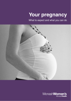


© Copyright 2026