
(Sheldon)Isolated From Common Bean (Phaseolus Vulgaris .L)Seeds
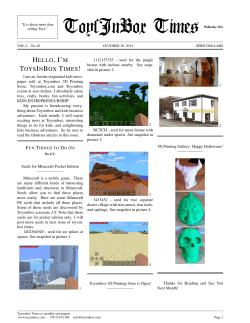
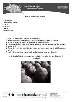
SOME STUDIES ON THE FUNGUS Fusarium moniliforme (SHELDON)ISOLATED FROM COMMON BEAN (Phaseolus vulgaris .L)SEEDS. BY Mutasim Mohamed Osman B.Sc (Agric) University of Manofiya (Egypt) 1981 A thesis presented in partial fulfillment of the requirements for the Degree of M.Sc (Agric) (Plant Pathology) Supervisor Prof. Ahmed Mohamed Baghdadi From the Department of Crop Protection Faculty of Agriculture, University of Khartoum. 2005 DEDICATION To the memory of my father…. to my mother, to my wife Inshirah, and my sons, Awwab and Mohamed Osman, with great love and respect i ACKNOWLEDGEMENTS First of all I am greatly indebted to the merciful “ALLAH” who offered me everything to accomplish this study. I would like to express my sincere gratitude and thanks to my supervisor Prof. Ahmed Mohamed Baghdadi for his close and careful supervision , guidance, patience , encouragement and help throughout this study. Deepest thanks are extended to: - My brother …Dr. El-Dirdiri Mohammed Osman for his assistances, encouragement, and financial support. - Staff of Plant Pathology Labratory, Plant Protection Directory, Khartoum North- Sudan , Mrs; Awatif Mohamed Ibrahim , Dr. Soheir Ahmed Abd El-wahab ,Miss Mashair Ahmed Abd El-Hafiz for their cooperation assistances and help in identification of the isolated fungi. - Plant Protection Directory. Ministry of Agriculture & Forestry for financing this study. - All my friends and colleagues for cooperation and encouragement. ii Contents page Dedication ………………………………………………………… i Acknowledgment …………………………………………………. ii Contents…………………………………………………………… iii List of tables ………………………………………………………. iv List of figures……………………………………………….……… v List of plates…………………………………………………….….. vi Abstract………………………………………………….………….. vii CHAPTER ONE: INTRODUCTION………….......…………..…… 1 CHAPTER TWO REVIEW OF LITERATURE…..…………..…… 3 2-1: Seed-borne diseases associated with common bean……. 3 2-2: Seed –borne fungi associated with common bean……... 3 2-3: Common bean diseases caused by Fusarium spp…….... 5 2-4: The fungus Fusarium moniliforme (Sheldon)……….… 6 2-5: Diseases caused by Fusarium moniliforme………….… 7 2-6: Control………………………………………….………… .7 CHAPTER THREE: MATERIALS AND METHODS:…………….. 9 3-1: Common been seed sample supply…………..………… 9 3-2: Detection of seed- borne fungi……………………..…….. 9 3-2-1: Dry seed inspection…………………………………….. 9 3-2-2: Blotter test………………………………………..…… 9 3-2-3: Agar plate test…………………………………….….. 10 3-3: Identification……………………………………….…… 10 3-4: Effect of different media on linear growth of Fusarium moniliforme………………………………………………..11 3-5: Effect of fungicides on growth of Fusarium moniliforme………………………………………...….…11 CHAPTER FOUR: RESULTS: …………………………………….....13 4-1: Detection of seed-borne fungi……………………...… …13 4-2: Media test…………………………………………… …..14 4-3: The effect of fungicides on the growth of Fusarium moniliforme……………………………………..…………14 CHAPTER FIVE: DISCUSSION ………………………………… ..28 REFERENCES ……………………………………………………..…30 APPENDICES………………………………………………………....35 ARABIC ABSTRACT. iii LIST OF TABLES Table page Table (1): Common bean seed samples tested………….………15 Table (2): Dry inspection test…………………………………....15 Table (3): Percentage incidence of fungi recovered by blotter method………………………………………………………..….16 Table (4): Percentage incidence of fungi on agar plate method…17 Table (5): Effect of media type on the linear growth of Fusarium moniliforme………………………………………..…21 Table (6): Effect of Tilt on the linear growth of Fusarium moniliforme……………………………………….…..23 Table (7): Effect of Bayleton on the linear growth of Fusarium moniliforme…………………………………………...25 iv LIST OF FIGURES Figure Fig: (1): Effect of media type on the linear growth of page Fusarium moniliforme………………...……………………………..…….23 Fig: (2): Effect of fungicides on the linear growth of Fusarium moniliforme………………………………………… ….27 v LIST OF PLATES Plate (1) Chain of microconidia of the fungus Fusarium moniliforme ………………..……………..18 Plate (2) Macroconidia of the fungus Fusarium moniliforme……………………………………..………………..18 Plate (3): Culture of Fusarium moniliforme (Upper side)…...….19 Plate (4): Culture of Fusarium moniliforme (under side)………..20 Plate (5): Effect of media type on growth of Fusarium moniliforme……….………………………………………22 Plate (6): Effect of Tilt on growth of Fusariu monmiliforme.…..24 Plate(7): Effect of Bayleton on growth of Fusarium moniliforme………….…………………… …26 vi ABSTRACT Four samples of common bean (PhasIeolus vulgaris ) were used in this study , two samples from Hudieba Research Station, one sample from Berber area and one sample from Shendi area. Samples were tested by dry inspection, blotter test and agar plate method. The dry inspection revealed malformation, discoloration and some impurities. In blotter method Fusarium moniliforme isolated might be for the first time in Sudan. Also the following fungi were detected : Fusarium oxysporum, Macrophomina phaseolina, Alternaria alternata, Drechslera specifer, Cladosporium sp, Curvulavria lunata, Aspergillus niger and Rhizopus sp. Disinfection of seeds with sodium hypochlorite before plating showed that Fusarium moniliforme was a seed-borne fungus Czapek's Dox agar was found to be the best medium for the growth of the fungus Fusarium moniliforme. In vitro the fungicides Tilt and Bayleton were found effective in controlling the growth of the fungus Fusarium moniliforme. vii CHAPTER ONE INTRODUCTION One of the most important food legumes in the tropical countries is common bean. It is a major supplement of diet protein in tropical countries (Owera,1990). There are different names beside the name common bean such as kidney, haricot, garden, dwarf, snap and dry bean (Mmbaga et al. 1990). Common bean is consumed in dry and green forms and the straw can be used as fodder. Like other legume crops it has the ability of nitrogen fixation in the soil. Common bean (Phaseolus vulgaris L.) belongs to the family Leguminosae and to the sub- family Papilionidae. The origin of common bean is Latin America and Mexico (Kaplan et al, 1973). The cool tropical regions are the best area's for cultivating beans, but they can be grown in temperate areas for their immature pods (Allen 1983). There are about 26837 thousand hectars cultivated with common bean all over the world. The annual world production of bean dry seeds is about 18.3 million tons. About one third of this production is in the center of origin beans (Latin America and North America). Brazil and Mexico grow about 82% of the total production (FAO, 2002). In Sudan common bean is cultivated mainly in the northern part of the country. The major production area in Sudan is Shendi – Berber area .(Mohamed & Salih,1990). It is also cultivated in the southern region around Katari, Juba, Loka West, Yambio, Tonj and Wau (Tarr, 1955). In Sudan common bean is considered as the second important food legume used by the majority of families. It provide them with a large portion of their protein needs. In Sudan common bean has been reported to be infected by several important fungi, such as Alternaria spp, Ascochyta phaseolorum, Auerobasidium pullulans , Curvularia spp,Drechslera spp, Fusarium spp, Macrophomina phaseolina , Myrothecium sp, Periconia byssoides , Phyllosticta sp , Pythium sp, Ulocladium atrum and Uromyces appendiculatus . Many serious diseases were found to be caused by most of these fungi in the country (Boughey,1946; Tarr, 1955: Abdelwahab,1987,Salih et al., 1990 ). Several of these fungi are seed-borne and the Fusarium spp. represent the most important fungi among them. Many species of Fusarium had been isolated from bean seeds in Sudan. In this study the isolation of Fusarium moniliforme from common bean seeds might be the first record in Sudan according to the available literature. 2 Therefore this study was carried out to provide some information about this fungus. 3 CHAPTER TWO LITERATURE REVIEW 2-1 Seed-borne diseases associated with common bean: According to Richardson (1968) the following diseases were reported on common bean as seed- borne diseases: A. Fungal diseases: Ascochyta leaf spots, grey mould, pod rot, ashy stem blight, charcoal rot , angular leaf spot, damping off, stem canker, white mould, downy mildew, Cladosporium spot , anthracnose , wilt and root rot. B. Bacterial diseases : Bacterial wilt, brown stem, grease spot, bacterial brown spot, common bacterial blight, fuscous blight. C. Viral diseases: Bean mosaic virus , cowpea mild mottle virus, 2-2 Seed- borne fungi associated with common bean: Several fungi had been found to be carried on or in the seeds of common bean. Many investigators discussed the diseases caused by seed- borne fungi in common bean for example, Ellis et al (1976) showed that many 4 fungi can be borne internally or as surface contaminants on common bean seeds, Ellis et al .(1976) and Winter et al (1974) suggested that the following fungi : Fusarium solani , F. oxysporum , F equiseti , F. semitectum, F. moniliforme, Macrophomin phaseolina Phyllosticta phaseolina , Aspergillus flavus , Drechslera tetramera, Phoma sp. Colletotrichum Lindemuthianum and Botrytis cinerea were seed-borne fungi associated with common bean seeds in various countries . Booth (1971) isolated F.equisti from bean plants . Fulco et al. (1977) found that Fusarium spp, Phytophthora sp, Rhizoctonia sp. Colletotrichum sp. and Diporthe sp were the predominant seed- borne fungi in the seeds of three bean cultivars tested by the blotter method. Gomes and Dhingra (1983) found that Rhizoctonia solani and Alternaris alternata were seed-borne fungi on common bean. In Sudan, AbdElwahab (1987) isolated the following fungi from the seeds of common bean : Alternaria alternata , A. tenusissima, A. dianthi ,Auerobasidium pullulans, Cladosporium sp., Curvularia brachyspora , Fusarium solani, F. semitectum, Fusariella intermedia , Fuariella aegyptiaca, Myrothecium sp , Cephalosporium sp, Rhizopus sp and Ulodadium atrum . Eltayeb (1993) isolated the following fungi: Fusarium scripi , F. proliferatum , F. oxysporum, F. equiseti, F. brachygibbosurm, Alternaria alternata , Macrophomina 5 phaseolina, Drechslera specifer, Cladosporium herberum,Stemphylium sp. , Curvularia lunata, Chaetomium globosum, Aspergillus flavus, A.niger, Rhizopus sp., and Penicillium sp. In Kenya Buruchara (1989) recorded the following fungi from 26 samples of bean seeds using the standard blotter method : Fusarium moniliforme ,F. semitectum , F.equiseti , F. solani, F. oxysporum, Colletotrichum lindemuthianam, C. dematium, Phoma sp , Cephalosporium sp, Verticillum sp , Cercospra sp, Rhizoctonia solani, Macrophomina phaseolina , Botryodiplodia theobromae , Rhizopus sp, Penicillium sp Aspergillus sp, and Cladosprium spp. Also other investigators reported about seed-borne fungi in other hosts not including common bean. For example, Khan et al. (1974) in Pakistan isolated Aspergillus spp, F. moniliforme and F. semitectum from the seeds of chick pea (Cicer aritinum). Mebalds (1987) reported that Phoma medicaginis , Fusarium acuminatum and Stemphylium sp, were found to be transmitted from Medicago truncatula seeds to the plants. 2-3 Common bean diseases caused by Fusarium spp. In various developmental stages common beans are exposed to many pathogenic fungi Fusarium spp are considered as the most important pathogenic fungi of common bean plants. However, Schwartz et al 6 .(1989) demonstrated that the infection with Fusarium spp may occur on seedlings and mature plants. Kendrich and Snyder (1942) reported that bean yellow disease caused by Fusarium oxysporum F,sp phaseoli was first reported in California in 1929. Schwartz et al (1989) showed that F.oxysporum caused wilt disease in commercial bean fields . Gupta and Saharan (1973) asserted that F.solani caused pre- emergence rot when soil inoculation was used before sowing and caused seedling blight when seedling inoculation was performed. Also Ykema and Stutz (1991) stated that the isolates of Fusarium equiseti, F. oxysporum and F. solani might cause root rot symptoms. 2-4 The fungus Fusarium moniliforme (Sheldon) : This fungus is well known and has been worldwide in its distribution. It is a seed- borne fungus which is encountered in many host plants such as : Allium cepa, Avena sativa , Capsicum spp, Corchorus capsularis, Glysine max, Gossypium spp., Hibiscus esculentus, Hordeum vulgaris, Lycopersicon esculentum, Oryza sativa. Phaseolus spp , Pisum sativum, Ricinus communis, Solanum melongena, Sorghum bicolor , Trifolium hybridum, Triticum aestivum and Zea mays (Ram et al,.1970). 7 2-5 Diseases caused by Fusarium moniliforme: Many diseases have been known to be caused by Fusarium moniliforme in graminae, leguminosae, malvaceae solanaceae, musaceae and tiliaceae. These diseases are seedling blight scorch foot rot, stunting and hypertrophy (Booth 1971), This fungus was found to be the most prevalent pathogen in maize causing ear rot disease (Koehler,1942). However, there are now many reports which indicate that Fusarium moniliforme is a serious pathogen in cereal crops; Segall et al, (1955) reported that Fusarium moniliforme causes bud rot in Zea mays in Puerto Rico;Tarr (1962) reported that this fungus causes root and stalk rot in Africa; Futrell and Webster,(1967) showed that it causes seedling blight, stalk rot, head blight and scabbed seed in Nigeria. In other crops the fungus was found to be associated with Vicia fabae (Boughey, 1946), Where as in other crops the fungus was found to be associated with symptoms in Vicia Fabae (Boughey, 1946),where it causes stem rot and wilt in Saccharum officinarum (Singh et al, 1975). 2-6 Control: 8 Much work in literature was done for the control of Fusarium moniliforme in various crops such as rice, maize cotton , wheat, sorghum and other crops and vegetables. In the control of seed – borne pathogens certain measures are used, these include use of resistant cultivars, enforcement of quarantine regulations, seed treatment and production of disease free seeds. Use of seed health testing in the control of seed-borne pathogens has been documented and has recently been showed that treating rice seed with Thiram reduced F. moniliforme effect on germination. According to Neergaard (1977) seed treatment is a benefit practice in seed protection to avoid seed loss. Dey et al.(1989) showed that maize seeds treated with Bavistin gave good control against F. moniliforme, resulting in best improvement and seedling survival. 9 CHAPTER THREE MATERIALS & METHODS 3-1 Common bean seed sample supply : Four seed samples of common bean were collected from different production areas in River Nile State (North Sudan). Two samples from Hudeiba Research Station, one sample from Shendi area and the fourth sample from Berber area (Table 1). 3-2 Detection of seed- borne fungi: 3-2-1 Dry seed inspection: Four hundred seeds from each sample were taken at random and were examined under the stereoscopic binocular microscope for impurities such as plant debris, sclerotia, malformation and seed discolouration. 3-2-2 Blotter test : (a) Un- treated seeds: Four hundred seeds from each sample were tested by the standard blotter method (ISTA, 1966). Each seed sample was plated onto three layers of moistened blotters in sterilized Petri- dishes (9cm- diameter) at a rate of 10 seeds per dish equidistantly placed. The plates were then incubated at 28 10 o C + 3 oC under alternating cycles of 12 hours near ultraviolet light and 12 hours darkness for 7 days (Sohieb, 1983). On the 8th day the seeds were examined under the stereoscopic binocular microscope and the incidence of seed –borne fungi was recorded. The compound microscope was also used to confirm identification of different fungi. (b). Chlorine pre- treated seeds: Four hundred seeds from each sample were first soaked for 5 minutes in sodium hypochlorite (1% available chlorine ) and then washed repeatedly in several changes of sterile distilled water, then the seeds were plated and incubated for 7 days, examined under the stereoscopic binocular microscope and the incidence of the seed- borne fungi were recorded. 3-2-3 Agar plate test: Two hundred seeds of each sample were disinfected with sodium hypochlorite as mentioned before and then plated on sterilized Petri-dishes containing sterilized Czapek's Dox agar medium at a rate of 5 seeds per dish. Seeds were then incubated for 7 day at 28 oC and on the 8th day were examined using the stereoscopic binocular microscope and the isolated seedborne fungi were recorded. 11 3-3 Identification : The identification of the isolated fungi was based on: Booth, (1971), Ellis (1971) and the help of staff of plant Pathology Laboratory –Plant Protection Directory- Khartoum North- Sudan. 3-4 Effect of different media on linear growth of Fusarium moniliforme. Three types of media were tested in this study. These were potato dextrose agar (PDA), Czapek's Dox agar and malt extract agar (MEA). From each medium four plates (9cm.diameter) were prepared. Two diameters were drawn on the back of each plate for centering the inoculum. The plates were then inoculated with 5 mm. disc from 7 days old culture of the fungus Fusarium moniliforme . the plates were then incubated at 28oC and the rate of the fungus growth was estimated daily by measuring the colony size along the two diameters on the back of each plate , then the mean of colony diameter was calculated for each medium. 3-5 Effect of fungicides on linear growth of Fusarium moniliforme: - 12 Two fungicides were tested in this experiment ,Bayleton and Tilt. Four dilutions for each chemical were used. The dilutions were prepared by taking one gram (or one ml.) from the chemical and then dissolved in one liter of sterilized distilled water to give 1000 ppm. From this dilution 1,3,5 and 7 ml. were added to four flasks containing 99. 97, 95 and 93ml of sterilized Czapek's medium, respectively. This gave a final concentrations of 10,30,50 and 70 ppm. for each chemical, respectively. Then the content of each flask was poured in sterilized 4 Petri-dishes and two diameters were drawn on the back of each plate for centering the inoculum. Each plate was inoculated with 5mm disc from a 7 days old culture of Fusarium moniliforme. The plates were then incubated at 28oC and the rate of the fungus growth was estimated daily as mentioned before. Other eight Petridishes (four dishes for each replicate) containing sterilized Czapek's medium were inoculated with the fungus to serve as control. 13 CHAPTER FOUR RESULTS 4-1 Detection of seed – borne fungi (a) In dry seeds: Four hundred seeds from each sample were tested for dry inspection test. The test showed that 20% of these samples were lighter in weight and smaller in size with yellow and brown to black discoloration. Malformation of seeds was also observed ( Table 2). (b) Blotter test (untreated seeds): The following fungi were isolated on the 8th day after plating : Alternaria alternata , Aspergillus niger, Cladosporium sp, Curvularia lunata, Derchslera specifer, Fusarium moniliforme, Fusarium oxysporum , Macrophomina phaseolina and Rhizopus sp.( Table 3). (c) Blotter test (treated seeds): Seeds were treated with sodium hypochlorite (1% chlorine) before germination on the blotter. The following fungi were observed: Alternaria alternata , Cladosprium sp ,Curvularia lunata , Drechslera 14 specifer , Fusarium moniliforme, Fusarium oxysporum, M. phaseolina (Table 3). (d) Agar test (treated seeds) : The following seed-borne fungi were detected in the agar plates: Alternaria alternata , Curvularia lunata Drechslera specifer, Fusarium moniliforme, Fusarium oxysporum and M. phaseolina. (Table 4) 4-2 Media test: The fungus Fusarium moniliforme was grown on three types of media Czapek's Dox agar was found to be the best medium for the growth of the fungus . In Table 5 the effect of media type on the growth of the fungus can be seen. 4-3 The effect of fungicides on the growth of the fungus Fusarium moniliforme: Two fungicides were examined in this test, Tilt and Bayleton. Different concentrations were tested, 10ppm, 30ppm, 50ppm and 70 ppm from each fungicides. 15 Tilt was found inhibiting the growth of the fungus at the low conc. (10ppm) and was found completely inhibiting the growth at the conc, 70 ppm (Table 6) Bayleton was found reducing the growth even at the lowest concentration (10ppm) but the fungal growth was not inhibited even at the highest concentration (70ppm) (Table 7) Table (1) Common bean seed samples tested Variety Locality 1. Ibaria cv Hudeiba Station Hudeiba Station Berber area Shendi area 2. Mutwakil cv 3. local cv 4. local cv Year of Harvest Research 2003-2004 Research 2003-2004 2003-2004 2003-2004 Table (2) Dry inspection test of four samples of common bean tested Sample 1. Ibaria cv 2. Mutwakil cv Healthy seeds 86.5% 77.25% Seeds with sclerotia 4% 6.5% 16 Malformed seeds 3.5% 7.25% Discoloured seeds 6% 9% 3. local cv (Berber area) 4. local cv. (Shendi area ) 77.5% 12.5% 4.5% 5.5% 78.75% 9% 5.75% 6.5% Table (3) Mean percentage incidence of fungi recovered by blotter method (treated & untreated seeds) for four CV. tested Fungi % incidence in untreated seeds % incidence in treated seeds 1. Alternaria alternata 8 5 2.Aspergillus spp 10 0 3.Cladosporium sp 12 4 4.Curvularia lunata 6 3 5.Drechslera specifer 7 3 6. Fusarium moniliforme* 12 3 17 7. Fusarium oxysporum 24 9 8. Macrophomina phaseolina 11 5 9.Rhizopus sp 6 0 * from Ibaria cv. only 18 Table (4) Mean percentage incidence of fungi on agar plate method (treated seeds) by 1% sodium hypochlorite for four cv. tested Fungus % incidence . Alternaria alternate 5 2.Curvularia lunata 2 3.Drechslera specifer 3 4. Fusarium moniliforme 2 5. Fusarium oxysporum 8 6. Macrophomina phaseolina 4 19 Plate .1 chain of microconidia of the fungus Fusarium moniliforme 20 Plate .2 macroconidia of the fungus Fusarium moniliforme 21 22 23 Table (5) The effect of 3 types of media on the linear growth of F. moniliforme (c.m) 2nd 3rd 4th 5th 6th Czapek’s 0.6 1.5 2.2 6.2 7.2 8.2 PDA 0.4 1.1 1.8 4.8 5.6 6.4 MEA 0.3 1.0 1.5 4.2 5.2 5.8 media 1st days 24 25 10 1st 8 2nd 3rd 6 4th 4 5th 2 6th 0 MEA PDA Czapek's Dox Agar fig. 3 Effect of media type on linear growth of the fungus Fusarium moniliforme Table (6) The effect of Tilt on the linear growth of F. moniliforme (cm) in vitro 1st 2nd 3rd 4th 5th 6th Control 0.6 1.4 2.2 6.2 7.2 8.4 10 ppm 0.2 0.5 0.7 1.0 1.2 1.5 30ppm 0.1 0.3 0.4 0.6 0.8 1.0 50ppm 0.1 0.2 0.3 0.4 0.6 0.8 Dose Days 26 70ppm 0 0 0 0 27 0 0 Table (7) The effect of Bayleton on the linear growth of F. moniliforme(cm) 1st 2nd 3rd 4th 5th 6th control 0.6 1.4 2.2 6.2 7.2 8.4 10 ppm 0.5 1.0 1.5 4.0 4.8 6.7 30 ppm 0.1 0.3 0.7 2.3 2.7 3.0 50 ppm 0.1 0.2 0.5 1.7 2.1 2.4 70 ppm 0.1 0.2 0.5 1.6 2.0 2.2 Dosage Days 28 29 30 9 8 7 1st 6 2nd 5 3rd 4 4th 3 5th 2 6th 1 0 Control 10 ppm 30ppm 50ppm 70ppm Fig.4 Effect of Tilt on linear growth of the fungus Fusarium moniliforme growth (cm) effect of Bayleton on linear growth of F.momiliforme 9 8 7 6 5 4 3 2 1 0 1st 2nd 3rd 4th 5th 6th control 10 ppm 30 ppm 50 ppm 70 ppm Fig.4 Effect of Bayleton on linear growth of the fungus Fusarium moniliforme 31 CHAPTER FIVE DISCUSSION In this study seed health testing was carried out for four samples of common bean according to ISTA (1966) rules. The standard methods used were dry inspection test, blotter method and agar plate method. In dry inspection a percentage from 12.5-20% of the seeds were found associated with sclerotia, discolouration and malformation. In the blotter and agar methods, several seed-borne fungi were isolated from untreated or chlorine pre- treated seeds. This result coincides with Neergaard (1977) who recorded that blotter method was superior to the agar plate method. Fusarium moniliforme was isolated from the variety Ibaria of Hudeiba Research Station. The isolation of Fusarium moniliforme from common bean seeds is considered as the first record in Sudan as far as the literature is available (Osman, 2005). The investigation and emphasis on this fungus Fusarium moniliforme was due to the fact that it is the first record. Fusarium oxysporum was encountered in all samples in high percentages (9-24%) compared to the other fungal species and this result coincides with Neergaard (1977) who reported that the blotter method is ideal for detection 32 of different fusaria. Some other fungi such as Alternaria alternata, Macrophomina phaseolina , Drechslera specifer , Cladosporium sp., Curvularia lunata, Aspergillus sp, and Rhizopus sp. were also detected in rather low percentages. Several investigators had reported similar seed-borne fungi associated with common bean(Winter et al 1974; Ellis et al 1976; Abdel Wahab,1987; Eltayeb. 1993). The physiological study conducted was for the suitable medium for the growth of the fungus Fusarium moniliforme. Three types of media were tested in this study, PDA, MEA and Czapek’s Dox agar. The later medium was found to be the suitable medium for the growth of the fungus. This finding agreed with Agarwal and Singh (1974), who found that Czapek’s medium was highly selective for the isolation of the fungus Fusarium moniliforme. The characteristic features of Fusarium moniliforme were identical to those described by Booth (1971) who reported that Fusarium moniliforme has a culture pigmentation of peach , salmon, vinaceous or purple to violet colour. Microconidia fusoid to clavate 5-12× 1.5-2.5 µ. Occasionally becoming 1 septate and produced in chains. Macroconidia when present in some strains are in equilaterally fusoid, thin walled,3-7 septate, 25-60× 2.4-4 µ. Chlamydospores absent. 33 The control of Fusarium moniliforme by the fungicides Tilt and Bayleton in vitro was found effective by both fungicides. This result coincides with the finding of Sohieb (1983) who found that Benlate had a toxic effect to the linear growth of Fusarium moniliforme. REFERENCES Abdel Wahab, S.A.(1987). A survey of seed –borne fungi of some legumonus crops of the Sudan M.Sc. thesis, Univ. Of Khartoum. Agarwal, V.K and Singh, O.V(1974), Relative percentage incidence of seed-borne fungi associated with different varieties of rice seeds . Seed Research, 2: 23-25 Allen, D.J.(1983). The Pathology of Tropical Food Legumes: Diseases Resistance in Crop Improvement. Wiley, Chichester, England, 413p . Booth,C.(1971). The genus Fusarium. CMI,Kew surrey, England,237p. Boughey,A.S.(1946). Apreliminary list of plant diseases in the AngloEgyptian Sudan.Mycological papers No.14,C.M.I Kew, Surrey England. Buruchara.R,A (1989). Preliminary information on seed- borne fungi of bean (Phaseolus vulgaris) in Kenya.CIAT(Intertional Center for Tropical Agriculture) African Workshop, series No.7 Dey, S.K,Gill,G.S.Aulakh,K.S,Soki,S.S. KHera,A.S and Grewal,P.K (1989) effect of seed dressing chemicals on germination and seedling vigour of maize. Plant Disease Reptr. 4:3642. Ellis,M.A., Galves,G.E.and Simclair, J.B.(1976). Effect of pod contact 34 with soil on fungal infection of dry bean seeds .Plant Disease Reptr.60: 974-976. Ellis,M.A. and Paschal, E.H.(1979). Effect of fungicides seed treatment on internally seed- borne fungi affecting germination and field emergence of pigeon pea. Seed Sci, and Technol. 7:75-81. Ellis,M.B. (1971). Demataceous Hyphomycetes. C.M.I., Kew,Surrey, England. Eltayeb, E.M.,(1993) Detection of seed borne fungi associated with common bean ( Phesolus vulgaris ) in the Northern region of Sudan. M.Sc Thesis, University of Khartoum. FAO (Food & Agriculture Organization of United Nations) (2002).FAO Production Year book. Fulco,W.,Das, S.,Gamergo,M.R, DE,O.,Eltmyer,M.B.and Brusamoum, R.(1977). Health testing of seeds of three bean cultivars from two regions in Rio Grande. Rev. PL.Path.58- 2025. Futrell,M.C. and Webster,O.J.(1967). Fusarium scab of sorghum in Nigeria ,PL.DIS.Rertr. 51:174 -178. Gomes,J.L. and Dhingra,O.D.(1983) Alternaria alternata aserious pathogen of white coloured snap bean (Phaseolus vulgaris L.) seeds, Rev. PL.Path.63:2588 Gupta,V.K.and Saharan,C.S.(1973) seed rot , root rot complex of beans (Phaseolus vulgaris L.) seeds, Rev. PL.Path.52:3883. ISTA (International Seed Testing Association )(1966) International rules of Seed Testing .Proc.Int.Seed Test Ass/ 31:1-15. Kaplan, L.Lynch, T.F.and Smith,C.E.(1973).Early cultivated beans from an Intermontane Peruvian Valley. Science 179:76-77. Kendrich,J.B.and Snyder,W.C.(1942).Fusarium yellows of beans 35 .Phytopathology 32:1010-1014. Khan.S.A.J.Mathur, S.B.and Neergaard,P.(1974) Survey on seed –borne organisms of Pakistan. Seed Sci.and Technol. 2: 427-429. Koehler, B.(1942) Natural mode of enterance of fungi into corn ears and some symptoms that indicate infection.JAgric,Res 14: 421-442. Mathur, S.B(1983).testing seed of tropical species for seed-borne diseases. Seed Sci and Technol, 11:113-128 Mebalds,M.I.(1987) . Effect of seed mycoflora on the development of disease in stands of Medicago truncatula. Seed Sci. and Technol. 15: 811-820. Mmbaga , M.E.T,Koiange, E.M.K ,Gondive,B.Mbuye,O.S., Mushi,C.S and Kamala,R.(1990). Bean production and research highlights in the northern Tanzania,CIAT African workshop series No.7. Mohamed ,A.K. and Salih,H.S. (1990) Evaluation of dry bean Phaseolus vulgaris L.cultivars in Sudan . CIAT African workshop series No.7. Neergaard,P.(1977).Seed pathology.VOl.1. the Greshman press Old wording Surrey ,England. Osman,M.M. (2005). Some studies on the fungus Fusarium moniliforme (Sheldon.) isolated from common bean (Phaseolus vulgaris.L) seeds. M.Sc Thesis, University of Khartoum. Owera,E.(1990) .Analysis serotypes and strains of bean common mosaic virus (BCMV) in countries with CIAT's regional programe on beans in Eastern Africa . CIAT African workshop series No.7. Ram,N.,Mathur,S.B.and Neergaard,P.(1970) Identification of Fusarium spp. as they occur in blotter test .Proceeding of 36 International Seed Testing Association ,Seed Pathology, 35:121-144. Richardson,N.M.(1968).An Annotated List of Seed –borne Diseases. Proceeding of International Seed testing Association.33:64-65. Salih,H.S.Bushara,A,G.and Khalid, A.M.E.(1990).common bean (Phaseolus vulgaris L.) Production and research in the Sudan CIAT African workshop series No.7. Schwartz, H.F.Mc Millan, M.S. and Silbernagel,M.J.(1989). Occurance of Fusarium wilt in clorado. PL.Dis.Reptr. 73:518. Segall,R.H. and Veles, F.J.(1955).Gibberella Fujikuroi, the cause of pod rot of corn in Puerto Rico. PL.Dis.Reptr. 39:238. Singh,R.S.and Singh,N.(1975). An observation on the association of Fusarium moniliforme with sugar cane wilt Indian Phytopathology,28:271-272. Sohieb.A.Y.(1983). Detection of Fusarium moniliforme (Sheldon) in okra seeds (Hibiscus esculentus) its significance and control. M.Sc. thesis, Unive, of Khartoum. Tarr,S.A.J.(1955). The Fungi and Plant Diseases of the Sudan. C.M.I . Kew, Surrey, England . Tarr,S.A.J(1962). Diseases of sorghum, Sudan grass and Broom corn .C.M.I. Kew. Surrey. England. Winter,W.E.,Mathur,S.B. and Neergaard,P.(1974). Seed-borne organisms of Argentina. Plant disease Reptr. 58:507-511 Ykema, R.E. and Stutz, J.C.(1991). Isolation, identification and Pathogenicity of Fusarium spp from guayule in Arizona. Plant Disease Reptr. 75:736-738. 37 APPENDICES 38 CULTURE MEDIA 1- Potato Dextrose Agar (PDA) was made from the following constituents: Potato Extract 250 gm. Dextrose 20 gm. Agar 15 gm. Distilled water 1000ml. 2- Czapek’s Dox Agar: Dipotasium Phosphate Sodium nitrate Magnesium sulphate Potassium chloride Ferrous sulphate Sucrose Distilled water 1.0 gm. 0.5 gm. 0.5 gm. 0.5 gm. 0.1 gm. 20.0 gm. 1000ml. 3- Malt Extract Agar (MEA): Malt Extract Agar Distilled water 30 gm. 20 gm. 1000ml. 39 ﻣﻠﺨﺺ اﻻﻃﺮوﺣﺔ ﺍﺠﺭﻱ ﻫﺫﺍ ﺍﻟﺒﺤﺙ ﻋﻠﻲ ﺍﺭﺒﻌﺔ ﻋﻴﻨﺎﺕ ﻤﻥ ﺒﺫﻭﺭ ﺍﻟﻔﺎﺼﻭﻟﻴﺎ ﺍﻟﺠﺎﻓﺔ ﻟﻤﻌﺭﻓـﺔ ﺍﻟﻔﻁﺭﻴـﺎﺕ ﺍﻟﻤﺤﻤﻭﻟﺔ ﻋﻠﻲ ﺍﻭ ﺩﺍﺨل ﺍﻟﺒﺫﻭﺭ ﺍﻟﻌﻴﻨﺎﺕ ﺍﻟﻤﺴﺘﺨﺩﻤﺔ ﻓﻲ ﺍﻟﺒﺤﺙ ﺍﺜﻨﺘﺎﻥ ﻤﻨﻬﺎ ﻤﻥ ﻤﺤﻁـﺔ ﺍﺒﺤﺎﺙ ﺍﻟﺤﺩﻴﺒﺔ ﻭﻋﻴﻨﺔ ﻤﻥ ﻤﻨﻁﻘﺔ ﺒﺭﺒﺭ ﻭﺍﻟﻌﻴﻨﺔ ﺍﻻﺨﻴﺭﺓ ﻤﻥ ﻤﻨﻁﻘﺔ ﺸﻨﺩﻱ ﺒﻭﻻﻴﺔ ﻨﻬـﺭ ﺍﻟﻨﻴل ﺍﺠﺭﻴﺕ ﺍﻻﺨﺘﺒﺎﺭﺍﺕ ﺍﻟﻤﻭﺼﻲ ﺒﻬﺎ ﻟﻔﺤﺹ ﺍﻟﺒﺫﻭﺭ ) (ISTA 1966ﻭﻫﻲ ﺍﺨﺘﺒـﺎﺭ ﻭﺭﻕ ﺍﻟﺘﺭﺸﻴﺢ ﻭﺍﺨﺘﺒﺎﺭ ﻁﺒﻕ ﺍﻻﺠﺎﺭ ﺍﻀﺎﻓﺔ ﺍﻟﻲ ﺍﻟﻔﺤﺹ ﺍﻟﺠﺎﻑ ﻟﻠﺒﺫﻭﺭ . ﺍﻟﻔﺤﺹ ﺍﻟﺠﺎﻑ ﻟﻠﺒﺫﻭﺭ ﺍﻭﻀﺢ ﺍﻥ ﻫﺫﻩ ﺍﻟﻌﻴﻨﺎﺕ ﺘﺤﺘﻭﻱ ﻋﻠﻲ ﺒـﺫﻭﺭ ﻤﻠﻭﻨـﺔ ﻭﺍﺨـﺭﻱ ﻤﺸﻭﻫﺔ ﻤﻊ ﻭﺠﻭﺩ ﺒﺫﻭﺭ ﻤﻜﺴﻭﺭﺓ ﻁﺭﻴﻘﺔ ﻭﺭﻕ ﺍﻟﺘﺭﺸﻴﺢ ﻭﺒﻌﺩ ﻤﻌﺎﻤﻠﺔ ﺍﻟﺒﺫﻭﺭ ﺒﻬﻴﻜﻠﻭﺭﻴﺩ ﺍﻟﺼﻭﺩﻴﻭﻡ (Sodium hypochlorite) %1ﺍﻅﻬﺭﺕ ﻭﺠﻭﺩ ﺍﻟﻔﻁﺭﻴﺎﺕ ﺍﻟﺘﺎﻟﻴﺔ . Alternaria alternata , Curvlaria lunata,Drechslera specifer. Fusarium moniliforme F.oxysporium and Macrophomina phaseolina ﻋﺯل ﺍﻟﻔﻁﺭ Fusarium moniliformeﺭﺒﻤﺎ ﻴﻌﺘﺒﺭ ﺍﻟﻤﺭﺓ ﺍﻻﻭﻟﻲ ﺍﻟﺘﻲ ﻴﺘﻡ ﻓﻴﻬﺎ ﻋﺯﻟﻪ ﻤﻥ ﺒﺫﻭﺭ ﺍﻟﻔﺎﺼﻭﻟﻴﺎ ﻓﻲ ﺍﻟﺴﻭﺩﺍﻥ . ﺍﻭﻀﺤﺕ ﺍﻟﺩﺭﺍﺴﺔ ﺍﻥ ﺒﻴﺌـﺔ Czapek's Dox Agarﻫـﻲ ﺍﻟﻤﺜﻠـﻲ ﻟﻨﻤـﻭ ﺍﻟﻔﻁـﺭ Fusarium moniliforme ﺍﻭﻀﺤﺕ ﺍﻟﺩﺭﺍﺴﺔ ﺍﻥ ﻤﻌﺎﻤﻠﺔ ﺍﻟﻔﻁﺭ Fusarium moniliformeﻓﻲ ﺍﻟﻤﻌﻤل ﺒﻤﺒﻴـﺩﻱ ﺍﻟﺒﺎﻴﻠﺘﻭﻥ ﻭﺍﻟﺘﻠﺕ ﻗﺩ ﺍﺩﻱ ﺍﻟﺤﺩ ﻤﻥ ﻨﻤﻭ ﺍﻟﻔﻁﺭ ﺤﺘﻲ ﻓﻲ ﺍﻟﺘﺭﻜﻴﺯ ﺍﻻﺩﻨﻲ ﻤـﻥ ﺍﻟﻤﺒﻴـﺩﻴﻥ ) ( 10ppmﺒﻴﻨﻤﺎ ﺘﻭﻗﻑ ﻨﻤﻭ ﺍﻟﻔﻁﺭ ﺘﻤﺎﻤﺎ ﻋﻨﺩ ﺘﺭﻜﻴﺭ 70ppmﻤﻥ ﻤﺒﻴﺩ ﺍﻟﺘﻠﺕ. 40 ﺑﻌﺾ اﻟﺪراﺳﺎت ﻋﻠﻲ اﻟﻔﻄﺮFusarium moniliforme. اﻟﻤﻌﺰول ﻣﻦ ﺑﺬور اﻟﻔﺎﺻﻮﻟﻴﺎ اﻟﺠﺎﻓﺔ. ﺗﻘـــــﺪﻳﻢ: ﻣﻌﺘﺼﻢ ﻣﺤﻤﺪ ﻋﺜﻤﺎن ﺑﻜﻼرﻳﻮس اﻟﻌﻠﻮم ﻓﻲ اﻟﺰراﻋﺔ ﺟﺎﻣﻌﺔ اﻟﻤﻨﻮﻓﻴﺔ )ﻣﺼﺮ ( 1981م أﻃﺮوﺣﺔ أﻋﺪت ﻟﻨﻴﻞ درﺟﺔ اﻟﻤﺎﺟﺴﺘﻴﺮ ﻓﻲ اﻟﻌﻠﻮم اﻟﺰراﻋﻴﺔ ) أﻣﺮاض اﻟﻨﺒﺎت( اﻟﻤﺸﺮف :ب ـ أﺣﻤﺪ ﻣﺤﻤﺪ ﺑﻐﺪادي ﻣﻦ ﻗﺴﻢ وﻗﺎﻳﺔ اﻟﻤﺤﺎﺻﻴﻞ ) أﻣﺮاض اﻟﻨﺒﺎت( آﻠﻴﺔ اﻟﺰراﻋﺔ ﺟﺎﻣﻌﺔ اﻟﺨﺮﻃﻮم ﻳﻮﻟﻴﻮ 2005م 41 42


© Copyright 2026