
Participation of Medial Pontine Reticular Formation in Eye
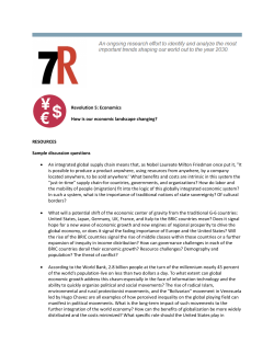
Participation
of Medial
Eye Movement
Pontine
Generation
Reticular
Formation
in
in Monkey
E. L. KELLER
Department
Research
of Electrical
Laboratory,
Engineering
University
and
Computer
of California,
THE
DEEP
CORE
Of the brain stem, and in
particular
the med ial pontine
reticu lar formation
(PRF),
has long been considered
to be directlv
involved
in the immediate
supranuclear
control
of eye movements.
This belief is based on the demonstration
of direct
anatomic
connections
from this
area to all the oculomotor
nuclei
(ZZ), on
the severe and permanent
disruption
of eye
movement
that results from focal lesions
placed in this area (3), and on the shortlatency eye movements
that are evoked by
electrical
stimulation
in this
area
(6).
Single-neuron
recordings
in this or closely
adjacent
brain stem areas (5, 7, 8, 15, 24)
have revealed
several categories
of neural
activity
that is closely and differentially
correlated
with various
types of eye movemen ts.
These previous
studies have either been
conducted
on animals without
the primate’s
repertory
of eye movements
(7, S), have
been brief,
qualitative
reports
concerned
only with segregating
the units by type (5,
24), or have not considered
unit behavior
during
smooth pursuit
or vestibularly
driven movements
(15). It has been previously
shown
that each oculomotor
neuron
participates
in all the types of primate
eye
movement
(21). Therefore,
in working
out
the details of the supranuclear
organization
of the oculomotor
system the question
that
remains
to be clarified
is how
and at
what
level signals
from
the various
separate eye-movement-generating
subsystems
are brought
together.
In this respect it is
important
to have .more precise
and detailed recordings
on each type of reticular
formation
whose activity is correlated
with
Received for publication
316
July 19, 1973.
Sciences
Berkeley,
California
and
the
Electronics
94720
all the types of eye movement
in the primate repertory.
This line of attack is initiated by this report on the PRF.
Following
the detailed
recording
of a
unit’s behavior,
the recordin .g site was el ecthe microel ectricall v stimulated
through
trode. The eye movements
resulting
from
these focal
stimulations
were
compared
with
the type of unit activity
previously
recorded
at the site. Previous
stimulation
studies of this area carried
out with gross,
implanted
electrodes
at much higher levels
only
smooth
eve
of current
(6) evoked
movements.
While
t was no t expected
that
the small currents
utilized
in this studv
would
stimulate
just the unit
being
recorded,
the technique
of microstimulation
can eff ectivelv
limit
the area and, hence,
number
of neurons
excited
to very small,
discrete colonies
in cortical
areas (1). In a
structure
like the reticular
formation
the
affected area might be expected to be larger
due to the extensive
array and spread of
connecting
fibers throughout
the area (22).
Nevertheless,
since the types of units recorded in this area respond very differently
for various
types of eye movements,
some
reflection
of unit characteristics
in the type
induced
bv stimulation
of eye movement
was expected.
To a limited
extent this expectation
was fulfilled.
METHODS
Six young monkeys (Macaca
mulatta)
were
prepared
for these studies by chronically
impentobarbital
planting
three devices under
sodium anesthesia and aseptic conditions.
A
coil of Teflon-covered,
stainless steel wire was
implanted
on one globe. This coil, placed on
an animal put in alternating
magnetic fields
kept in spatial and temporal quadrature,
pro-
RETICULAR
FORMATION
vided
a signal
which
is a function
of vertical
and horizontal
eye position
with
a sensitivity
of 15’ of arc. Details
of this method
of measuring eye movements
are the same as those previously
given
(11) except
that fields alternating
at a frequency
of 20 kHz
were
utilized.
Steep
attenuation
of the gain of the microelectrode
recording
electronics
above
a frequency
of 10
kHz
prevented
contamination
of the
neural
recordings
with
signals
from
the
magnetic
fields.
During
the
preliminary
stages
of the
experiments,
eye movements
in the first
two
monkeys
were
measured
with
direct-coupled
electrooculograms.
Data from
these two animals
are pooled
with
that from
the four
later
monkeys.
A stainless
steel
chamber
was
implanted
stereotaxically
on the skull
above
the pontine
reticular
formation.
Tungsten
microelectrodes
were
driven
through
the
chamber
using
an
eccentric
hydraulic-drive
system.
The
dura
was
left intact
and electrodes
were
passed
through
it within
a sterile
guard
tube. A light
aluminum
crown
was bolted
to the skull to provide
a platform
for restraining
the monkey’s
head during
recording
sessions.
Following
recovery
from
these surgical
procedures
the monkeys
were
placed
in a primate
chair
with
heads
immobilized
for
the
daily
recording
sessions.
The
animals
had been
previously
trained
to attend
to a visual
display
and
to fixate
a variable-position
target
lamp.
With
this
simple
target
display
a controlled
and
reproducible
sequence
of both
small
and large
saccades
and steady
fixations
at selected
positions could
be quickly
elicited
whenever
a unit
correlated
with
eye movement
was located.
Smooth
movements
were
produced
by rotating a large
mirror
in front
of the monkey.
Vestibular
movements
were
obtained
by manual rotations
of a turntable
on which
the
primate
chair
and
magnetic
field
coils
were
rigidly
mounted.
In the last three
monkeys,
after
unit
activity
had
been
recorded,
the
microelectrode
was
switched
from
the recording
preamplifier
to the
output
of a constant-current
stimulator.
The
site was stimulated
with
brief
pulse
trains
of
variable-intensity
cathodal
current
and the resulting
eye movements
were recorded.
The stimulus
parameters
could be varied
from
currents
of 5-40
PA, train
lengths
of 10 ms to
several
seconds,
and intratrain
pulse frequencies
of 100-1,000/s.
Pulse
width
was constant
at
0.3 ms.
Neural
activity
was coupled
through
an FET
preamplifier
to a conventional
amplifier
with
a
band
pass of 300-10
kHz.
Unit
activity
and
voice annotations
were recorded
on direct
chan-
AND
EYE
MOVEMENT
317
nels of a magnetic
tape recorder
with
a band
pass of 300-11
kHz,
while
simultaneous
eye
positions
and
a signal
proportional
to chair
position
were
recorded
on separate
FM
channels (band
pass DC to 1,250 Hz)
of the same
recorder.
For
analysis
the recorded
data
were
transcribed
on photosensitive
paper
by an ultraviolet
mirror
galvanometer
recorder.
The
overall system
bandwidth
for eye movements
was
250 Hz and for neural
data was 5,000 Hz. The
recorder
was run at paper
speeds of 125 or 250
mm/s
for the analysis
of unit
discharge
rates
during
fixation
and 500 or 1,000 mm/s
for saccadic activity
analysis.
The accuracy
of the time
measurements,
determined
by recording
the
output
of a precision
oscillator
through
the
entire
system,
was within
t 37&
Electrolytic
lesions
were
made
through
the
tip of the recording
electrode
at the site of units
At least two such lesions
of particular
interest.
were
made
in each animal,
but generally
not
more
than
three.
Following
the final
track
the
animals
were
heavily
anesthetized
with
pentobarbital
and then
perfused
with
normal
saline
and formalin.
Subsequent
histological
examination
(25-p
sections
stained
with
cresyl
violet)
verified
the
location
of the
lesions
and
the
recording
sites on other
electrode
tracks
were
reconstructed
from
the marked
locations.
RESULTS
in this study were run
that portion
of the brain
stem reticular
formation
from about 2 mm
anterior
to 2 mm posterior
of the rostra1
and caudal poles of the abducens
nucleus.
The
lateral
limit
of the region
explored
extended
out to about 3 mm from the midline. The ventral border of the area studied
was generally
established
by the olivocerebellar
tract. Some of the more posterior
tracks passed through
the vestibular
nuclei,
but eye-movement-related
units isolated
in
this complex
will not be described
in this
report. The tracks, run at a 25” angle from
the sagi t tal plane, usually
passed through
the brachium
conjunctivum.
Soon
after
leaving this dense fiber track units related
to eye movements
began to be encountered.
Most of the isolated
units were judged
to
be cell bodies on the basis of their spike
shape and duration
(15).
The behavior
of 233 eye-movement-correlated
units were recorded
and analyzed
in this study. Since the area of the brain
stem studied
and the qualitative
response
Recording
uniformly
tracks
into
318
E. L. KELLER
of the recorded
units
correspond
very
closely
to that
previously
described
by
Luschei
and Fuchs (15), the units will be
divided
for descriptive
purposes
into the
same four main categories
established
by
these investigators.
Using
their
nomenclature
we isolated
120 burst units, which
exhibited
high-frequency
bursts of activity
in association
with rapid
eye movements
in a single or several directions
(called the
on-direction)
but no sustained
tonic
activity,
and 37 tonic units whose activity
was related
to steady eye position
during
fixation
and, in some cases, eye velocity
during
pursuit
or ves tibularly
driven
m9vements.
Twenty
units were isolated
that paused
during
all rapid eye movements
or for rapid
movements
in a particular
direction.
During periods
of fixation
between
saccades
these units exhibited
a tonic discharge
that
was only poorly or not at all correlated
with
eye position
or velocity.
We also isolated
41 burst-tonic
units
which
exhibited
bursts
of firing
during
rapid eye movements
in a particular
direction and tonic activity
related
to fixation
positions
in the same direction.
In their
behavior
such units resemble
oculomotor
neurons
(21) but were located outside
recognized
motor
nuclei.
In addition,
track
reconstructions
always located
these units
outside the PRF, either in the several small
nuclei
located
at this level in the central
gray, in the vestibular
nuclei,
or in the
medial
longitudinal
fasciculus.
Therefore
these responses were not considered
as representative
of activitv in the reticular
formation
and are not further
described.
Finally,
15 additional
units
were
isolated in the reticular
formation
that displayed
eye-movement-correlated
behavior
that could not be categorized
under
these
four main headings
and will not be discussed further.
Single-unit
recording
UNITS.
Bursting
units could be
further
subdivided
according
to the preferred directions
of the movements
associated with the burst and according
to the
temporal
relationship
of the burst to the
onset of the rapid
eye movement
(15).
Medium-lead
burst
units.
Sixty-eight
BURSTING
units were isolated
in the current
study
which exhibited
medium-lead
bursts. These
units invariably
discharge
a high-frequency
burst of activity
prior to the initiation
of
any rapid eye movement
either visually
or
vestibularly
evoked
with a component
of
the movement
in the proper direction.
The
defining
criteria
of proper
direction
was
variable
among the units, but the required
component
of the movement
was strictly
ipsilateral
for only 16 units or about 25y0
of the medium-lead
units observed.
Figure IA illustrates
the behavior
of this
type of burst-lead
unit for saccadic movements,
while
Fig. 1B shows similar
behavior for the same unit during
the quick
phase of rotatory
nystagmus.
It should
be
particularly
noted
that these u .ni ts never
discharge
during
pure vertical
or medial
rapid movements
whi ch ser ves to differentiate them from the larger
population
of
medium-lead
burst units to be described
later.
The lengths
of the burst lead in milliseconds, the duration
of the burst, and the
intraburst
firing
frequencies
were closely
examined
to determine
if any quantitative
differences
existed
for visually
evoked
or
vestibulary
driven
rapid
eye movements.
No detectable
difference
could
be determined
in the burst lead. The mean burst
lead-determined
from
at last 25 rapid
movements
for each unit-was
the same
for both saccades and quick phases for all
16 units, and varied from 7 to 12 ms with
an overall
mean of 9 ms for the group
as
a whole.
This is very similar
to the burst
leads found by Luschei
and Fuchs (15) for
this type of unit for saccadic
eye movements.
In comparison
the average
burst
lead for abducens
motoneurons
is 6 ms
(14). Likewise
there was no detectable
difference in burst length for rapid eye movements
of either
type for movements
of
similar
duration.
In confirmation
of the
findings of Luschei and Fuchs (15) the burst
length
and rapid
eye movement
duration
were almost
identical
for movements
of
duration
greater than 15 ms (corresponding
to a movement
of about 5”).
A comparison
of the intraburst
discharge
rate during
ipsilateral
saccadic and quick
phase eye movements
was obtained
by separately pooling
the data for 10 saccades and
RETICULAR
FORMATION
AND
EYE MOVEMENT
319
/-------
LAT.
3
W
‘--\----=
MED.
r
I
I
I
0
TIME
(MSEC)
FIG. 1. Activity
of an ipsilateral
medium-lead
burst unit during
saccadic eye movements
(A) and the
movements
of rotationally
induced
vestibular
nystagmus
(23). In A and B upper
trace is
quick-phase
vertical
and middle
trace is horizontal
eye position.
The time calibration
for both shown below B. Insets
in A and B are high-speed
records of one horizontal
rapid
eye movement
and associated
unit
activity.
Time calibration
shown below B the same for both insets. Eye-movement
calibration
the same as in lowspeed records.
for 10 quick
phases of 40°, 20°, and 5”
deviations
for each of the ipsilateral
medium-lead
burst units.
Reproducible
saccades of these sizes (within
-F- 0.5’) were
readily
available
because the animals
were
trained
to fixate the target positions.
As a
general
rule,
quick-phase
movements
of
reproducible
size were not obtained
and
therefore
the averaged
data were obtained
from 10 movements
of the above sizes -F- 2”.
The onset of the burst was abrupt
and the
discharge
rate reached levels as high as 950
spikes/s
which
were sustained
until
the
final 20-30 ms of the burst, during
which
time a decreasing
and more variable
discharge obtained.
For rapid eye movements
320
E. L. KELLER
greater than loo an identical
high level of
rather
steady discharge
was reached.
For
movements
less than 5O the discharge
rate
was significantly
lowered.
Statistical
analysis of these averaged
burst rate profiles
indicated
that there was no significant
difference in intraburst
firing
frequency
for
visually
evoked
and vestibularly
induced
rapid movements.
Finally
it was found that
the burst rate was not dependent
on the
initial
eye position
within
arange
of -t- 20”
from the primary
position.
Although
burst rate increased
very little
for saccades larger than lo”, it was related
nonlinearly
to saccadic velocity.
Measurements of maximum
saccadic velocities
for a
range of movement
amplitudes
were pooled
for all the animals utilized
in these experiments. The curve drawn
through
the plotted means of these measurements
shown in
Fig. 2A illustrates
the well-known
dependence of saccadic
velocity
on movement
amplitude
(9). On this same figure
are
plotted
the overall mean burst rates (and
ranges of the means) for the 16 ipsilateral
bursting
units during
saccades of the same
amplitude
for which
maximum
velocities
of eye movement
are plotted.
It is apparent
that burst frequencies
show
a much more complete
and earlier
saturation for larger saccades than does saccaclic
velocity. To clarify the relationship
between
burst frequency
and saccadic velocity
the
means from Fig. 24 are replotted
as the
filled circles in 2e, which
shows that saccadic velocity
depends
mono tonically
on
burst frequency.
The amplitude
of the saccade associated
with each datum
point
is
shown in parentheses.
The data plotted in Fig. 2A were obtained
with the monkey
training
and attempting
to fixate
the randomly
appearing
target
lights.
Both
maximum
saccadic
velocity
and movement
duration
showed
the expected
relationship-for
fully
motivated
monkeys-to
saccade amplitude.
If the target display
was turned
off, the monkey
would
continue
to make spontaneous
saccades to various
objects of interest around
the experimental
room. Under these conditions it was always possible to find saccades
in the records with abnormally
long durations and lower maximum
velocities
when
compared
to movements
of corresponding
1000
1000
0
0
I
I
I
IO
20
30
AMPLITUDE
L
1
40
( DEG 1
6
.(40)
800.(30)
l (20)
C
8
co
\
--28'
ii!
Y
-33
4 (15)
.
A
::CJ
. (IO)
4W
-
>
A
8.
IO'
A
-I
o(6)
014)
so -A
l (2)
0
f
0
I
1
200
400
BURST
DISCHARGE
I
I
600
RATE
800
( SPIKES
I
1000
/ SEC)
2. A: relationship
between
saccadic
amplitude and maximum
saccadic velocity
(filled
circles).
Each point
represents
the pooled
mean
from
at
least 10 saccadic movements
of each size for each of
the six monkeys.
Also shown
is the relationship
between
saccade size and the associated
burst
frequency
for medium-lead
burst
units (open circles).
Each point represents
the grand mean of the burst
rates associated
with
the 10 saccades of each size
for all 16 ipsilateral
burst units (bars are ranges of
means for the 16 units).
B: means (burst
rate and
velocity)
from A replotted
to show the relationship
between
intraburst
firing
frequency
and saccadic
velocity
(filled
circles).
Numbers
in parentheses
are
amplitudes
of saccades associated
with each point.
Preceding
data all obtained
with
animals
executing changes in fixation
angle in response
to target
movement.
Triangles
show
maximum
saccadic
velocity
and associated
burst
rate for several
abnormally
slow
spontaneous
saccades
of the size
shown on the interrupted
horizontal
lines.
FIG.
amplitude
in Fig. 2A. Nevertheless,
all 16
units continued
to burst for every saccadic
movement
with
an ipsilateral
component.
Moreover,
the burst continued
to be of almost the same duration
as the ipsilateral
RETICULAR
FORMATION
AND
Values
Mean
1.9
1.7
1.4
1.5
in parentheses
321
number
of spikes per degree of eye movement
(mean
burst units during
horizontal,
ipsilateral
saccades
Saccade
Size, O
5
10
20
30
MOVEMENT
units also discharged
only in association
with rapid eye movements
but with a more
complicated
determinate
for the on-direction of movement.
Each unit in this group
displayed
a burst
for every rapid
movement with an ipsilateral
component,
but
the ipsilateral
component
was not a necessary condition
for a burst. No units were
found that were exclusively
active for pure
vertical
saccades. Specifically,
10 units were
isolated
that burst for both ipsilateral
and
contralateral
rapid
movements.
In every
case the burst frequency
was greater
for
movements
with an ipsilateral
component.
Twenty-one
additional
units were located
that burst for all rapid
movements,
but
nine of these showed the greatest discharge
frequency
for movements
with an ipsilateral
component,
five units the greatest frequency
for movements
that contained
a down component,
and six the greatest frequency
for
movements
that
contained
an up component.
Ten units burst for saccades in all
directions
except pure contralateral
movements. For each of these 10 units the greatest discharge
frequency
was associated with
rapid movements
having an ipsilateral
component.
Finally,
11 units produced
a burst
with every ipsilateral
or vertical
saccade or
combination
of the two, however,
almost
complete
inhibition
of the burst,
which
normally
accompanied
the verticle
component, was a concomitant
of every saccade
that contained
even the slightest
contralateral component.
Long-lead
burst units. Forty units were
isolated
that began to discharge
substantially before rapid
eye movements
with a
variable
interval
of uneven
or ragged firing. The length
of this interval
was extremely
variable
from saccade to saccade
for a given unit and among different
units.
Figure
3A shows a unit whose behavior
is
component
even for saccades of very low
velocity that occurred
as the animal became
less alert. Such movements
are probably
the result of a continuous
scale of central
nervous
system alertness
from fully alert
and motivated
to drowsy,
and would
not
normally
be of interest
for study. However, in this case, the characteristic
behavior of the burst units is clarified
by the
depressed
state of alertness.
Not only do
such units continue
to discharge with bursts
of the same duration
as the movement,
but
the intraburst
discharge
rate remains
correlated with velocity and not amplitude
of
the movement.
This is shown on Fig. ZB
for a few selected “slow saccades.” The triangles show the paired
velocity
and burst
frequency
associated
with saccades of the
size shown
on the interrupted
horizontal
lines. When this number
is compared
with
the number
in parentheses
for the nearest
filled circle (fully motivated
saccade) it is
apparent
that these movements
have much
lower velocities
than “normal
saccades” for
the given amplitudes
and yet the associated
burst frequencies
continue
to plot in correspondence
with the velocity of the movement.
Since the mean burst rate increased
very
little with saccade size and movement
duration increased
almost linearly,
the number
of spikes per degree of movement
calculated
by dividing
the total number
of spikes in a
burst by the size of the associated
movement, might be expected
to be rather
constant over a wide range of movements.
In
fact this relationship
(gain factor) was surprisingly
constant,
as shown
in Table
1,
for a sample of the ipsilateral
medium-lead
burst units recorded.
Spot checks on the
remaining
10 ipsilateral
bursting
units
showed that they behaved similarly.
The
remaining
52 medium-lead
burst
TABLE
1.
Average
for six medium-lead
EYE
(.3)
(.2)
(.3)
(.5)
are SD. Data
2.5
2.4
2.7
2.4
(.4)
(2)
(.5)
(.4)
are taken
Gain
2.2
2.3
2.5
2.2
from
(.4)
(.3)
(.6)
(.4)
at least
Factor,
Spikes/
1.8
1.9
2.1
1.9
five saccades
gain
factor)
O
(.4)
(.3)
(.4)
(.7)
2.2
2.3
2.3
2.1
of each size.
(.2)
(.4)
(.2)
(.5)
c.1
3.4
2.9
3.2
(.3)
(.6)
(.5)
(.7)
E.L. KELLER
322
typical
of this group.
The prelude
of uneven discharge,
which
was not correlated
in frequency
or duration
with saccade size,
began between
30-300 ms before the onset
of a saccade and terminated
with a more
intense burst of activity which also preceded
the movement.
The high-speed
record
in
Fig. 3A illustrates
the large variability
in
interspike
interval
during
the
low-frequency
prelude
and during
the more intense burst. This variability
makes definition of the burst duration
and initiation
time somewhat
arbitrary.
For 22 units in
this group
the duration
of the period
of
more
intense
activity
corresponded
very
roughly
with the duration
of the saccade.
For the remaining
18 units the period
of
intense activity was of much shorter
duration than the saccade, so that the burst
ended
almost
synchronously
or slightly
after the onset of the movement.
The burst lead was more variable
among
these units than had been the case for the
medium-lead
group,
but was fairly
consistent for any one unit. The distribution
of the burst lead ranged
from 28 to 8 ms
with the group
mean at 14 ms. Thus,
in
this group
the period
of more intense
activity tends to be initiated
before the burst
in the medium-lead
group.
The on-direction
of the units in the longlead group included
27 ipsilateral,
5 ipsilatera1 and down, 3 ipsilateral
and up, and 2
omnidirectional.
z
LAT.
A
All the units
of this group
continue
to display their characteristic
uneven interval of discharge
and period of more intense
activity prior
to every rapid movement
in
the on-direction
during
vestibular
nystagmus. The extreme
variability
in unit discharge even for similar
saccades made any
quantitative
comparison
of the possible differences occurring
during
quick phases and
saccades very difficult.
Figure 3B illustrates
that the pattern
of discharge
was essentially the same with respect to the length
of the uneven
discharge
interval
and the
burst lead.
Following-lead
burst units.
The consistent characteristic
of this group of 12 units
was that they never initiated
their burst
prior to the onset of rapid eye movements,
but instead began several milliseconds
after
the movement.
The group
continued
to
show similar
behavior
during
rotatory
nystagmus. That
is they still fired bursts of
irregular
activity at variable
short intervals
after contralateral
quick-phase
movements.
UNITS.
Thirty-seven
units were isolated
which
discharged
steadily
during
fixation
at rates that increased
monotonically with ipsilateral
eye position.
Figure 4
shows the behavior
of a unit representative
of this group. The majority
of units in this
group
(26 units) had thresholds-the
eye
position
at which the neuron
began to discharge tonically-in
the nasal field of movement or were never totally inhibited
even
TONIC
B
FIG. 3. Activity
of a long-lead
burst
unit during
saccadic
eye movements
(A) and the quick-phase
movements
of vestibular
nystagmus
(B). Eye movement
and time calibration
the same in A and B. Only
the horizontal
component
of eye position
shown. Insets below A and B are high-speed
records of one horizontal
rapid eye movement
of each type and associated
unit activity.
RETICULAR
-
FORMATION
EYE
MOVEMENT
323
A
UP
20
I
g
-
AND
/
01
2OJ
k
DOWN
C
1
0
FIG.
tional
Time
1
TIME
I
(MSEC)
1
1
400
4. Activity
of tonic units during
gaze fixation
(A), optokinetic
nystagmus
(B), and vestibular
rotanystagmus-in
total darkness
(C). Only horizontal-component
of eye movement
shown in B and C.
and eye movement
calibration
the same for all traces.
for extreme
medial
gaze fixations.
The remainder
of the cells in this group (11 units)
had thresholds
in the lateral field of movement. The distribution
of units into these
two groups was far from discrete however,
as demonstrated
in Fig. 5B which shows a
distribution
of tonic unit thresholds
from
the most medial
fixation
positions
and beyond (cross-hatched
area) to about 25” into
the lateral field. There was tendency for the
units with lateral
thresholds
to have more
linear
frequency-eye
position
curves
of
greater slope. About 75% of the units with
nonlinear
curves showed a saturation
effect
(decreased
slope at more lateral
positions)
and the other quarter
had increased
slopes
at more lateral
eye positions.
Figure
5A
shows three rate-position
plots for units
from this group
that are representative
of
the range
of behavior
displayed
during
fixation
by tonic units.
Whenever
possible tonic units were tested
carefully
for response during
pursuit
movements. A consistent
dichotomy
emerged for
the 12 units that were held long enough
to
ascertain
their
behavior
during
pursuit
movements.
The relationship
of firing rate
to eye velocity was determined
by plotting
the instantaneous
firing rate at a selected
position
(usually for a position
on the most
linear portion
of the rate-position
curve for
the unit, but in any case always the same
position
for each rate-velocity
analysis for
any one unit)
for a number
of different
324
E.L.KELLER
velocities
through
the chosen position.
Instantaneous
firing
rate was estimated
by
averaging
about
six interspike
intervals
which most closely symmetrically
bracketed
that point in time at which the eye passed
through
the selected position.
An example
of the data from which these measurements
were made is shown in Fig. 4B where firing
rates were determined
during
a pursuit
movement
directed
in one case through
a
position
in the lateral
direction
(upward
arrow) and then through
the same position
in the medial
direction
(downward
arrow).
When a number
of such firing rates were
plotted
for different
velocities,
curves similar to those shown in Fig. 6 resulted. Three
units,
all of which
had extreme
medial
thresholds
during
fixation,
were similar
to
unit 1 with essentially
no change in firing
rate due to velocity
for either laterally
or
medially
directed
pursuit
movements.
The
other nine units, all of which
had lateral
thresholds,
discharged
at rates greater
by
an amount
proportional
to eye velocity
in
the lateral
direction
but a constant
rate
for various
velocities
in the medial
direc-
0
/
f
I
I
I
I
I
I
0
HORIZONTAL
B
EYE
POSITION
40
( DEG)
-6
I
40
30
20
IO
0
IO
MEDIAL
I
1
!
30
40
LATERAL
TONIC
FIG. 5.
20
FIRING
THRESHOLD
(DEG)
A : relationship
of unit
discharge
rate
during
fixation
to horizontal
eye position
for three
Data points
(filled
circles)
obtypical
tonic units.
tained
during
visual
fixations
are shown
for only
one unit (unit 1). Curves
that indicate
the trend of
data are drawn
by hand. Instantaneous
firing
rates
measured
for a number
of eye positions
during
the
slow-phase
of rotatory
nystagmus
in the dark (filled
circles) are shown for unit 1 for comparison.
8: distribution
of thresholds
of 37 tonic units recorded
in
this study. Gaze deviation
required
to initiate
tonic
discharge
is divided
into 50 bins on the absissca.
Cross-hatched
bin on the extreme
left contains
all
units that were still active at the limit
of contralateral
gaze.
300,
UNIT
I
UNIT
2
4
-20
EYE
0
0
VELOCITY
(DEG
/ SEC1
FIG. 6. Relationship
between
unit discharge
rate
and eye velocity
for two tonic units. Discharge
rates
for visual
pursuit
velocities
(open)
and vestibular
slow-phase
velocities
(filled)
in both the ipsilateral
(+)
and contralateral
(-)
directions
at instants
when the eye passed through
a given position
for
each unit.
tion. The slope of the rate-velocity
relationship for lateral
velocities
was correlated
with
threshold
in that units
with
more
lateral
threshold
also had the largest
increase in rate for lateral
following
movements.
Nine of the twelve tonic units studied
during
pursuit
movements
were also tested
for their respon.se during
rotatory
nystagmus in total darkness.
All the units participated
by smoothly
increasing
or decreasing their firing rate during
lateral or medial
slow-phase
movements,
respectively,
as
shown in Fig. 4C. The instantaneous
firing
rates at a number
of positions
were calculated
by averaging
about
six interspike
intervals
about the point in time at which
the eye crossed each position.
For three of
these units,
when
their
firing
rates were
plotted on rate-position
graphs for the same
unit
(one example
in Fig. 5A), they fell
with some slight scatter on the same estimated curve for the unit obtained
during
visual fixation.
These were the same three
units which had not shown any change in
discharge
rate related
specifically
to the
velocity
of pursuit
movements.
The
remaining
six units, which
had displayed
a
velocity
proportional
increment
in firing
rate for lateral
pursuit
movements,
also
showed a similar
relationship
during
slowphase movements
(unit 2, Fig. 6). However,
when the discharge
rate-slow
phase velocity
relationship
was plotted
on the same coordinates
as the rate-velocity
relationship
RETICULAR
FORMATION
for pursuit movements,
the rate data points
for slow-phase
velocities
always plotted
below the estimated
curve for rate-pursuit
velocity relationship
as shown in Fig. 6.
PAUSING
UNITS.
These 20 units fired rather
steadily during
fixation
but stopped
firing
completely
slightly before and during
rapid
An example
of the beeye movements.
havior of this type of unit is shown in Fig.
7. The discharge
rate during
fixation
was
not at all correlated
with eye position
for 12
of these units, but for the other 8 there was
a consistent
trend toward
higher
discharge
rates for more ipsilateral
gaze deviation.
The typical standard
deviation
of the discharge rates for repetitive
fixations
at the
same angle was ZO-257& of the mean rate.
This wider range of scatter was in marked
contrast
to similar
measurements
on the
tonic units in this study and on abducens
motoneurons
(14) where
typical
standard
deviations
of discharge
rate of 10 and 57&
were found, respectively.
For the pausing
group
discharge
rates
ranged from 180-250 spikes/s for the units
not correlated
with eye position,
while those
that displayed
maximum
rates for ipsilateral positions
never increased to more than
150 spikes/s. Thus discharge
rate served to
further
differentiate
these two subtypes.
The occurrence
of a pause was directionally sensitive
in that the 8 position-correlated units paused only for saccades with
ipsilateral
components,
while the remaining
12 units paused
for all rapid
eye movemen ts.
AND
EYE
325
MOVEMENT
The onset of the pause for both subtypes
was abrupt
and preceded
the initiation
of
the rapid
movement
by an interval
that
ranged
from 12 to 25 ms for the 20 units
with no systematic
difference
between
the
sub types.
The length of the pause was almost equal
to the duration
of the rapid
movement,
which confirms
the findings of Luschei
and
Fuchs (15) in opposition
to the power-law
relationship
found
by Cohen
and Henn
(5). There
was no consistent
difference
in
either pause lead or duration
for saccades
or quick-phase
movements
in any of these
pausing
units.
None
of the high-frequency,
non-position-related
units
showed
any detectable
difference
in discharge
rate correlated
with
slow-phase
eye movements
during
rotatory
nystagmus.
The eight other pausing
units
changed rate smoothly
during
rotation
(and
the concomitant
compensatory
slow-phase
eye movements)
in addition
to pausing
for
ipsilateral
quick-phase
movements.
Since
the discharge
rate-position
relationship
was
so inconsistent
for these units it was difficult
to make a quantitative
measure of discharge
rate during
rotation.
However
the discharge
rate during
one direction
of rotation
could
be compared
to the immediately
succeeding
rotation
in the opposite
direction
during
sinusoidal
rotation.
When this was done it
was found that the maximum
instantaneous
discharge
rate always occurred
in phase or
slightly
lagging
maximum
contralateral
rotational
velocity
and, hence, maximum
ipsilateral
slow-phase
eye movement.
UP
20-l
DOWN
LAT.
MED.
FIG. 7. Activity
of pause units during
a sequence
of eye movements.
A: unit that
Unit
firing
rate
only for saccades with an ipsilateral
component
of eye movement.
fixation
is also correlated
with
ipsilateral
position.
Time
calibration
in milliseconds.
pauses
during
completely
periods
of
326
E. L. KELLER
Microstimulation
Luschei
and Fuchs (15) in their singleunit study of a wider area of the alert monkey brain
stem that included
the pontine
reticular
formation
attempted
without
clear success to anatomically
segregate
the
anterior-posterior
locations
of units
of
similar
discharge
characteristics
on the
basis of several coronal
slices of the brain
stem through
the area bracketing
the abducens
nucleus.
This
question
has been
reexamined
here on the basis of the units’
mediolateral
location
instead.
Since
the
electrode
tracks all penetrated
the reticular
formation
at a 25” angle from the vertical
and the point at which the electrode
crossed
the midline
was usually
easy to determine
from
the background
activity,
we could
calculate
the approximate
lateral
location
with respect to the midline
of each unit
that we recorded.
Sagittal
sections of the
brain stem of 0.5 mm width were graphically
constructed
and the responses recorded
in
each section were grouped
according
to unit
type as shown in the histogram
of Fig. 8.
While
considerable
overlap
exists, it does
appear that medium-lead
burst units with
complexly
conditioned
on-directions,
are
grouped
closer
to the
midline
than
medium-lead
burst
types that discharge
only for saccades with an ipsilateral
component.
On the other
hand,
tonic types
were almost evenly distributed
throughout
this 3 mm extent of the PRF.
Long-lead
burst types tended
to be distributed
similarly
to medium-lead
typesthat is, ipsilateral
on-direction
units were
located more lateral than bilateral,
vertical,
and omnidirectional
types-but
they are
not included
in the histogram
for clarity.
The location
of the subtype
of pausing
cell that interrupted
its high
frequency,
steady discharge
for all rapid
eye movements was more discrete.
All these units
were recorded
in a small circumscribed
area
just anterior
to the rostra1 pole of the abducens nucleus. Once a pausing unit of this
type was found, others were always located
nearby,
in contrast
to the situation
elsewhere
in the reticular
formation
where
medium-lead
and long-lead
burst
units
were found closely intermingled
with each
other and tonic units.
NUMBER
OF UNITS
1
,.
0
UNIT
LOCATION
(MM)
FIG. 8. Schematic
drawing
of a coronal
section
through
the brain stem at the level of the abducens
nucleus.
Electrode
tracts were run into the pontine
reticular
formation
within
the approximate
area
shown by the dashed
lines throughout
5-mm-thick
sections. The histogram
below the section was constructed
by summing
the number
of omnidirectional
medium-lead
burst units (solid bars), the ipsilateral
medium-burst
units (open bars), and the tonic units
(crosshatched
bars),
respectively,
encountered
in
each 0.5-mm
sagittal
section of brain stem throughout the entire 5-mm-thick
area. BC, brachium
conj unctivum;
Gn VII, genu of the facial
nerve;
VI,
abducens
nucleus;
IV, 4th ventricle;
IO, inferior
olive.
The other type of pausing
unit, which
discharges
at lower tonic rates interrupted
only
for ipsilateral
saccades, was found
scattered
throughout
the PRF. This may
explain
the difference
in our results from
that of Luschei
and Fuchs (15) who found
no clustering
tendency
for pausing
units,
since thev did not differentiate
between
the
locations
of the two subtypes.
When the sites of units located about 1
mm or more from the midline
were stimulated, one stereotyped
form of eye movement resulted regardless
of the type of unit
-medium-lead,
long-lead,
tonic, or ipsilateral
pausing
type-which
had been recorded at the site. These movements
were
qualitatively
similar to those reported
to be
evoked by order-of-magnitude
higher
currents
from
gross electrodes
permanently
RETICULAR
FORMATION
implanted
in the PRF (6). An example
of
the eye movement
resulting
from
these
stimulations
is shown in Fig. 9. The movement began about 10 ms after the onset of
the stimulus,
which
was a typical
latency
that did not depend
on the type of unit
recorded
at the site. The eye accelerates
smoothly
through
a transient
phase to reach
a constant
velocitv
of rotation
which
is
maintained
continuously
for as long as the
stimulus
is presented.
The transient
phase
of the evoked
movement
may be closely
compared
to that of a normal
pursuit
movement
(upper trace) evoked in the same
animal by rotating
a mirror
in front of its
eyes. The eyes are accelerated
in both cases
to constant velocity in 50 ms, which is about
one-half
the time constant
of a first-order
approximation
of the eye muscle to orbital
mechanical
system (13). This
observation
indicates
that an excess rate of change of
force above the constantly
increasing
force
required
to maintain
the steady-state
rotation must be present
during
the initial
stage of the stimulated
movement
in
analogy
to that present during
the initiation of a pursuit
movement
(16). Thus the
hypothesis
that the eye movements
evoked
by stimulation
in this area are the result
of a neural
integration
of the stimulation
train (6) is an oversimplification.
The
velocity
of the smooth,
stimulusevoked movement
increased
linearly
with
stimulation
frequencies
up to 500/s, then
gently saturated for frequencies
up to 800/s,
and finally decreased sharply
at 1,000/s-a
frequency
above that previously
investigated (6) but of importance
since PRF units
fire at rates above 800/s. The movement
1
I
STIMULUS
TIME
(MSEC)
9. Stimulus-evoked
eye movement
(middle
trace) compared
to smooth
pursuit
movement
(upper trace) elicited
in the same animal
by mirror
rotation.
The lower trace shows the envelope
of the
stimulus
which was a lOO-ms train of 500/s, 20 PA,
0.3 ms pulse width,
current
impulses.
FIG.
AND
EYE
MOVEMENT
327
velocity
was also dependent
on stimulus
current
strength,
a parameter
not previously
investigated.
Evoked-eye-movement
velocity
increased
linearly
with
currents
from 5 to 40 PA. At this higher
level of
current
strength
other
facial
movements
began to be evoked, so the effect of currents
above this level were not explored.
Finally
it was noted that the velocity
of
the evoked
smooth
movements
were not
position
dependent
for a range of initial
positions
-+ 20” on either
side of the
primary
position,
which
confirms
previous
observations
(6).
When
sites were stimulated
in the most
medial
area of the reticular
formation
extending
out about 1 mm from the midline,
some differences
in the evoked eye movement were noted. In all cases (12 sites) when
the unit recorded
at the site had a vertical
component
in its on-direction
the stimulated movement,
although
still a smooth
movement,
also had a vertical
component
in the same direction.
Eye movements
with
vertical
components
were never obtained
with gross stimulation
of the PRF (6). The
ipsilateral,
horizontal
component
always
predominated,
so that the stimulated
movements were never at an angle of more than
20” from the horizontal
except as noted
below.
The second difference
that was noted on
stimulating
medial
locations,
and this was
true regardless
of the type of unit recorded
at the site, was that the evoked eye movements had become initial-position
sensitive.
The horizontal
component
of the movement
was most noticeably
affected. With the eye
positioned
initially
at extreme
medial
deviations
the highest
ipsilateral,
constant
velocities
were obtained.
When
the initial
position
was at smaller
medial
deviations
the horizontal
velocity
was lower
and,
hence, for a constant-duration
stimulus
the
amplitude
of the evoked
movement
was
smaller.
The
effect became
more
pronounced
as the initial
eye position
moved
to lateral deviations,
until at some angle of
lateral
gaze
(typically
about
20”),
no
further
ipsilateral
component
could
be
evoked
and the stimulated
movement
became purely vertical.
When
the stimulated
site was located in
the area just rostra1 to the abducens nucleus
E.L.
328
that contained
the high-frequency
omnilateral pause units (10 sites), the results were
quite dramatic.
Even at very low current
levels the outcome was the almost complete
inhibition
of further
voluntary
saccadic
movements
for the duration
of the stimulation. Figure
1OA shows an example
of one
such stimulation.
Prior to the stimulus
onset the monkey
was looking
spontaneously
at objects around
the laboratory
with saccades occurring
on the average about every
0.5 s. When the stimulus
was applied
and
maintained
almost
all
saccades
were
eliminated
although
the viewing
conditions
had not changed.
In fact, all attempts
to
evoke saccadic movements,
including
moving threatening
objects near the monkey’s
B ^h
-
c -
II
0
TIME
(SEC)
5
Results
of high-frequency
stimulation
(200/s) in the area containing
saccade pause units.
In each case the upper trace is vertical
eye position,
the middle
trace horizontal
eye position,
and the
lower
trace the stimulus
envelope.
Eye-movement
calibrations
are the same for all traces. In B the
lowest trace is primate-chair
(animal-head)
position
-calibration
given on the left is 500. Time calibration is the same for all traces. A: stimulation
delivered with the animal
making - spontaneous
saccades
about
the experimental
room. -Usually
no further
saccades would
occur for the duration
of the stimulation.
B: stimulation
delivered
during
rotational
vestibular
nystagmus.
Only
the quick-phase
movements were affected by being completely
eliminated.
C: stimulation
delivered
during
optokinetic
nystagmus driven
by rotating
a mirror
in front
of the
monkey’s
face. Quick-phase
movements
are eliminated
and the gain of the smooth-phase
system
reduced.
FIG.
10.
KELLER
face and making
loud noises at eccentric
positions,
failed. The quick phases of vestibular and optokinetic
nystagmus
were also
totally
eliminated
by stimulation
of the
pausing
area as shown in Fig. 1OB and C.
The
slow phase of vestibular
nystagmus
was not effected by the stimulation,
but
the velocity
of the smooth
phase of the
optokinetic
nystagmus
was also greatly
reduced during
the period
of stimulation,
even though
the mirror
rotation
inducing
the optokinetic
nystagmus
continued
at the
same rotational
velocities.
DISCUSSION
Saccadic pulse generation
Since
the transmittance
characteristics
between
presynaptic
terminals
and oculomotor neurons
is unknown,
the form of the
input signal required
to produce
the pulselike increase
or decrease
in motoneuron
firing rate during
saccadic movements
(10,
17, 23) is uncertain.
Nevertheless,
it has
been demonstrated
that oculomotor
neurons respond
to 55-ms steady depolarizing
current
pulses injected
through
an intracellular
electrode
with a steady discharge
of spikes of up to 400/s or higher
for the
same duration
as the current
pulse (2). This
activity
resembles
very closely the activity
observed
in typical
oculomotor
neurons
during
saccades. Thus,
it seems highly
likely that the input required
by the motoneuron
to produce
its saccade-associated
pulse of activity is a pulse of presynaptic
activity of the same duration.
Luschei
and Fuchs (15) have hypothesized
that
the unilateral,
medium-lead
burst units provide
the input
which
generates this high-frequency
burst of activity
in motoneurons
during
saccades.
This
hypothesis
was based on the relative
time
of onset of burst unit discharge
and their
burst duration.
The results
presented
in
this study on the burst lead and duration
of
similar
units supports
this hypothesis.
In
addition,
the detailed
analysis of intraburst
frequency
during
saccades
of different
velocities
adds additional
weight
to the
argument.
The consistent
relationship
between saccadic velocity and burst frequency
for a variety of levels of alertness also argues
in favor of the directness of the connections
to oculomotor
neurons.
RETICULAR
FORMATION
The question
now arises concerning
the
nature
and location
of the neural
mechanism that generates
the precise,
saccadelinked burst of activity in the medium-lead
burst units themselves.
It is clear that a
spatial-temporal
translation
of information
must occur between
cortical
or collicular
sites where saccadic movements
are coded
in terms of anatomical
location
(19, 20) and
the PRF where they seem to be coded by
the duration
of the burst of medium-lead
burst units.
The activity
of the pausing
units,
also
recorded
in this area, sheds some light on
intermediate
form
of this
the possible
mechanism.
On the basis of the single-unit
behavior
and on the results obtained
by
stimulation,
we propose that the pulse-gating mechanism
includes
an input from the
high-frequency
pausing
units.
The input
from this group
of neurons
could exert a
powerful
inhibitory
influence
on all medium-lead
burst units and prevent
them
from discharging
except during
rapid
eye
movements
when input
from the pausing
group is interrupted
for the brief interval
of the movement.
This hypothesis
is consistent with the observation
that the average onset of the pause in this type of unit
occurs before the average initiation
of the
burst
in medium-lead
burst
units.
Also
stimulation
of the pausing
group,
which
presumably
prevents
the group from pausin<g for the duration
of the stimulus,
almost
completely
disables
saccadic
and quickphase generation
for the same period.
Fixation
The step change in steady discharge
rate
of oculomotor
neurons
that occurs for each
saccade (10, 17, 23) could also be generated
from
the activity
of medium-lead
burst
units. Under
this hypothesis,
in addition
to direct connections
to oculomotor
neurons, the medium-lead
bursting
cells would
provide the input to a neural integrator-a
would
progrouP of cells whose discharge
vide and hold a central
representation
of
current
eye position.
The presence of such
a steady signal of rather high fidelity seems
necessary on. the basis of human
psychophysical
experiments.
Such a signal could
also be used to provide
the steady drive
to motoneurons
during
fixation.
The fact
AND
EYE
MOVEMENT
329
that even on an individual-unit
basis the
ipsilateral
bursting
units provide
a rather
constant
number
of discharges
per degree
of eye movement
(Table
1) makes them an
ideal candidate
for the input to this integrator. The results of the focal stimulations
also support
this concept,
if one assumes
that medium-lead
burst units were the neurons activated
by the stimulus
currents,
which seems likely in view of the fact that
they were the only units which responded
naturally
at rates in excess of 500,/s. Then
their direct projections
to oculomotor
neurons could provide
the additional
excitation required
for the rapid acceleration
of
the constant
velocity-evoked
movements,
while the parallel
input into the tonic pool
after
integration
would
provide
the required
steadily
increasing
drive
to the
motoneuron.
The behavior
of the tonic cells recorded
in this study fits that expected
of neural
elements in such an integrator
in that they
changed
their discharge
rates in a stepwise
manner
for each saccade and maintained
steady discharge
during
fixation.
Luschei
and Fuchs (15) suggested
and the current
study supports
the idea that parallel
inputs
to motoneurons
from
the low-threshold,
saturating-type
tonic cell and high-threshold, high-gain
type could help provide
the
linear
rate-position
relationship
observed
during
fixation
in oculomotor
neurons.
However
it should
be emphasized
that no
conclusion
may be drawn
about the location of the integrator
on the basis of these
studies. The tonic cells might
only represent neurons at the output of a neural structure located elsewhere
in the brain that was
providing
the neural
circuitry
carrying
out
the integration.
Carpenter
has presented
evidence in cat that this function
may involve pathways
through
the cerebellum
(4).
Smooth
movements
We have found
evidence
in this study
that the tonic pool of reticular
units may
provide
additional
drive
to oculomotor
neurons
resulting
in their
increased
discharge during
pursuit
or vestibular
smooth
movements
(21). In contrast to the situation
found in motoneurons
where every unit discharged
at a higher
or lower rate in proportion
to velocity of the movement
in the
E.L.
330
on- or off-direction,
the PRF tonic units of
medial
threshold
showed
no eye-velocitydependent
increase or decrease of discharge
rate. Only
those units
of more
lateral
threshold-which,
if the monkey’s
head
had not been restrained,
would
not have
been
active
often
during
fixation-exhibited
a marked
increased
in discharge
rate proportional
to ipsilateral
eye velocity.
Also in contrast to motoneurons,
tonic units
did not show any decrement
in rate during
contralateral
smooth movements.
Oculomotor
subsystem
combination
Although
it is well accepted
that at more
central levels there is a neurological
separation of the oculomotor
system into saccadic,
pursuit,
vergence, and vestibular
portions,
oculomotor
neuron
studies have shown that
at the level of the final common
path these
separate
inputs
are represented
in each
motoneuron.
The
most
straightforward
hypothesis
would
be that the motoneuron
itself serves as the summation
site for these
various control
signals. That this does not
always occur has been found recently
(12)
for the case of version and vergence movements.
In the current
study we have found
a
similar
case with respect to visual saccades
and vestibular
quick-phase
movements.
The
characteristics
of those burst cells probably
directly
responsible
for the activity
of
motoneurons
during
rapid eye movements
was identical
during
both types of movement. Moreover
no difference
in the onset,
duration,
or frequency
of activity
was
found
in any of the types of burst cells
studied
throughout
this whole area. These
findings
lend considerable
support
to the
concept that much of the same lower brain
stem neural
machinery
is shared
by the
phylogenetically
ancient
quick-phase
generator
and the newer
voluntary
saccadic
generator.
The study of the tonic-cell
types provided
preliminary
evidence that some summation
of visual and vestibular
control
signals may
also occur prior to the motoneuron.
On the
other hand,
at least some summation
at
the level of the final common
path was to
be expected
for these two inputs
since the
well-known
three-neuron
arc from the vesti-
KELLER
bular end organ projects directly
on oculomotor neurons.
Since vestibular
nucleus
units discharge
nearly
in phase with
head velocity
and
oculomotor
neurons
discharge
approximately
in phase with
head position,
the
signal
from
vestibular
neurons
must
be
integrated
to provide
the additional
90”
of phase lag. Robinson
(18) has hypothesized that this integration
occurs through
parallel
neurological
pathways-the
direct
input to motoneurons
from vestibular
units
and another
more
diffuse,
multisynaptic
pathway
whose transfer
function
resembles
that of an imperfect
integrator
(an integrator that forgets its input with an exponential time course). Those
tonic cells which
showed a rate-velocity
relationship
in addition to a rate-position
relationship
would
not correspond
to the expected behavior
of
either
units in the direct vestibuloocular
path nor in the parallel
integrator.
Instead
they resemble
motoneurons
in responding
as linear
combinations
of these two pathways. The fact that the slope of the ratevelocity
relationship
was always found
to
be lower for vestibular
input in comparison
to that for visual pursuit
movements
would
indicate
that these two subsystems
share
this pathway
but with different
gains. The
reduced
gain for vestibular
input
might
be accounted
for by the additional
direct
velocity proportional
input reaching
mo toneurons
from vestibular
units through
the
medial
longitudinal
fasciculus.
SUMMARY
The activity of 233 neurons in the medial
pontine
reticular
formation
of alert, behaving monkeys
was studied
during
saccadic
and smooth
pursuit
eye movements,
rotatory
vestibular
nystagmus,
and
during
steady gaze fixation.
Three
main categories
of discharge
pattern
were observed:
burst
units which exhibited
high-frequency
bursts
of activity
in association
with
rapid
eye
movements,
tonic units whose activity was
continuous
and related
to gaze position
during
fixation,
and pause units that fired
at steady rates during
fixation
but stopped
firing
completely
during
rapid
eye movements. After recording
each unit’s discharge
characteristics,
the recording
sites were
RETICULAR
FORMATION
stimulated
with
focal electrical
currents
passed through
the recording
microelectrode.
No difference
in the onset, duration,
or
intraburst
firing frequency
was found during saccades or vestibular
quick-phase
eye
movements
for the group
of burst units.
Burst rate increased
monotonically
with
rapid eye movement
velocity.
In addition,
the number
of discharges
in each burst was
a rather accurate determinate
of the amplitude of gaze shift for each rapid eye movement.
The tonic units were most directly
related to the eye position-coded
discharge
of oculomotor
neurons
during
fixation.
In
addition,
a fraction
of these units,
those
with low thresholds,
also fire at rates related
and slowto eve position
du ring pursuit
phase movemen Is, jwh ile the remainder
ihow an additional
increment
in discharge
rate proportional
to eye velocity. This latter
group may be responsible
for the additional
activity
generated
in oculomotor
neurons
during
pursuit
movements.
Burst units with ipsilateral
directions
of
activation
were segregated
in more lateral
portions
of the medial
reticular
formation.
Stimulation
of sites located here resulted
in
constant-velocity,
ipsilateral,
conjugate
rotations of the eyes. Similar movements
were
AND
EYE
MOVEMENT
331
evoked at sites containing
either
burst or
tonic
units
confirming
the observation
gained from recording
unit types that burst
and tonic units are closely linked
anatomically and functionally
throughout
this portion of the reticular
formation.
When
the stimulated
site was located
near the midline
of the brain
stem, the
constant-velocity
movements
became more
eye position.
The
dependent
on initial
evoked movement
also contained
a vertical
component
if the recorded
unit at the site
had a vertical
preferred
direction.
Pause units were divided
into one subtype that demonstrated
a discharge
hiatus
for all rapid
eye movements
and another
group that paused only for ipsilateral
movements. There
was no difference
in pause
duration
or time of initiation
for either
type of rapid movement.
The onset of the
inhibition
in pause units preceded the initation of discharge
in burst
units.
Microtimulation
near the sites of multidirectional
pause units, which were clustered
near the
rostra1 pole of the abducens
nucleus,
resulted
in abrupt
disabling
inhibition
of
all voluntary
saccadic or quick-phase
eye
movements.
ACKNOWLEDGMENT
This
Institute
research
Grant
was sponsored
00955- .02.
by
National
Eye
REFERENCES
1.
H.
AND
SAKATA,
H. Functional
organization
of a cortical
efferent
system
examined
with
focal depth
stimulation
in cats.
J. Neurophysiol.
30: 35-54,
1967.
2. BAKER,
R. AND PRECHT, Mr.
Electrophysiological
as reproperties
of trochlear
motoneurons
vealed by IVth nerve stimulation.
Exf~tZ. Brain
Res. 14: 127-157,
1972.
3. BENDER,
M. B. AND SHANZER,
S. Oculomotor
pathways
defined
by electrical
stimulation
and
lesions in the brainstem
of monkey.
In: The
Oculomotor
System, edited by M. Bender.
New
York:
Harper
and Row, 1964.
4. CARPENTER,
R. H. S. Cerebellectomy
and the
transfer
function
of the vestibulo-ocular
reflex
in the decerebrate
cat. Proc. Roy. Sot., London,
Ser. B 181: 353-374,
1972.
5. COHEN,
B. AND HENN,
V. Unit activity
in the
pontine
reticular
formation
associated
with eye
movements.
Brain
Res. 46: 403-410,
19’72.
6. COHEN,
B. ‘AND KOMATSUZAKI,
A. Eye movements induced
by stimulation
of the pontine
reticular
formation:
evidence
for integration
ASANUMA,
7.
in oculomotor
117, 1972.
pathways.
Exptl.
Neural.
36: lOl-
DUENSING,
F. AND SCHAEFER,
K.-P.
Die Neuronenaktivitat
in der Formatio
reticularis
des
Rhombencephalons
beim
vestibularen
Nystagmus. Arch.
Psychiat.
Nervenkrankh.
196: 265290, 1957.
8. DUENSING,
F. AND SCHAEFER,
K.-P.
Die Aktivitgt
einzelner
Neurone
der Formatio
reticularis des nicht gefesselten
Kaninchens
bei Kopfwendungen
un vestibularen
Reizen.
Arch.
Psychiat.
Nervenkrankh.
201: 97-122,
1960.
9. FUCHS, A. F. Saccadic and smooth
pursuit
eye
movements
in the monkey.
J. Physiol.,
London
191: 609-631,
1967.
10. FUCHS, A. F. AND LUSCHEI,
E. S. Firing
patterns
of abducens
neurons
of alert monkeys
in relationship
to horizontal
eye movement.
J. Neurophysiol.
33: 382-392,
1970.
11. FUCHS,
A. F. AND ROBINSON,
D. A. A method
for measuring
horizontal
and vertical
eye movements
chronically
in the monkey.
J. A@Z.
Physiol.
21: 1068-1070,
1966.
332
12.
13.
14.
15.
16.
17.
18.
19.
E. L. KELLER
KELLER, E. L. Accommodation
vergence
in the
alert monkey.
Motor
unit analysis.
Vision Res.
13: 1565-1575,
1973.
KELLER,
E. L. AND ROBINSON,
D. A. Absence of
a stretch
reflex
in extraocular
muscles
of the
monkey.
J. Neurophysiol.
34: 908-919,
1971.
KELLER,
E. L. AND ROBINSON,
D. A. Abducens
unit behavior
in the monkey
during
vergence
movements.
Vision
Res. 12: 369-382,
1972.
LUSCHEI,
E. S. AND FUCHS,
A. F. Activity
of
brain
stem neurons
during
eye movements
of
alert
monkeys.
J. Neurophysiol.
35: 455-461,
1972.
ROBINSON,
D. A. The
mechanics
of human
smooth
pursuit
eye movement.
J. Physiol.,
London
180: 569-591,
1965.
ROBINSON,
D. A. Oculomotor
unit behavior
in
the monkey.
J. NeurophysioZ.
33: 393-404,
1970.
ROBINSON,
D. A. Models
of oculomotor
neural
organization.
In: The
Control
of Eye Movements, edited by P. Bach-y-Rita
and C. C. Collins. New York:
Academic,
1971.
ROBINSON,
D. A. Eye movements
evoked
by
20.
21.
22.
23.
24.
collicular
stimulation
in the alert
monkey.
Vision Res. 12: 1795-1808,
1972.
ROBINSON,
D. A. AND FUCHS, A. F. Eye movements evoked
by stimulation
of the frontal
eye
fields. J. Neurophysiol.
32: 637-649,
1969.
ROBINSON,
D. A. AND KELLER,
E. L. The behavior
of eye movement
motoneurons
in the
alert
monkey.
In:
Cerebral
Control
of Eye
Mor/ements
and Motion
Perception,
edited
by
J. Dichgans
and E. Bizzi. Basel: Karger,
1972.
SCHEIBEL,
M. E. AND SCHEIBEL,
A. B. Structural
substrates
for integrative
patterns
in the brain
stem reticular
core. In: Reticular
Formation
of
the Brain,
edited by H. H. Jasper. Boston:
Little, Brown,
1958.
SCHILLER,
P. H. The discharge
characteristics
of single units in the oculomotor
and abducens
nuclei
of the unanesthetized
monkey.
Exptl.
Brain Res. 10: 347-362,
1970.
SPARKS, D. L. AND TRAVIS,
R. P. Firing
patterns
of reticular
formation
neurons
during
horizontal eye movements.
Brain
Res. 33: 477-481,
1971.


© Copyright 2026