
Stop the Ringing - Eye and Ear Foundation of Pittsburgh
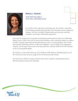
2015
SPRING EDITION
Stop the Ringing
by Zack Butovich
“We are at a point in our research where I feel I can say that we are one
million dollars away from a cure for tinnitus,” states Dr. Jonas Johnson,
Chairman of our Department of Otolaryngology, where some of world’s
leading research on the condition known as tinnitus takes place.
Making this bold statement in a room full of University of Pittsburgh alumni and
friends in Naples, Florida, it was met with audible surprise. For anyone suffering
tinnitus (over 10% of Americans), the prospect of a cure for the permanent ringing
sound, which can sometimes be as severe as a loud crashing or exploding, is a
dream come true.
In This Issue
Re-Imagining Sight Seeing with our Brain
and Not With Our Eyes
2
In Search of a New Business
Model to Improve Patient Care 3
Optic Nerve Regeneration
for Transplant and Repair
4
Personalized Medicine for
Head and Neck Cancer
5
Exciting Development in
Treating Damaged Corneas
6
Focus Turned to Otitis Media
7
Setting Expectations and
Overcoming Obstacles
8
Pictured above: Thanos Tzounopoulos,
PhD, Assistant Professor,
Department of Otolaryngology,
University of Pittsburgh
Photo by Joshua Franzos.
In previous issues of Sight + Sound, we have mentioned the incredible progress
Thanos Tzounopoulos, PhD, has made in his tinnitus research since arriving at the
University of Pittsburgh in 2008 as part of the then brand new Auditory Sciences
Center in our Department of Otolaryngology. He has helped to locate the source
of tinnitus (the brain, not the ear); actually see it in action on a circuit and cellular
level; identified possible causes of the “phantom sound” (a decrease of a specific
potassium channel associated with the auditory control center within the brain);
and recently, and most significantly, Dr. Tzounopoulos has hypothesized a way to
prevent the development of tinnitus.
Dr. Tzounopoulos, in collaboration with the Drug Discovery Institute at the
University of Pittsburgh, has identified at least a dozen molecules or combinations
of molecules (essentially the chemicals that makes medicine and drug therapies),
with the potential to restore the activity of the deficient potassium channel in the
auditory center of the brain, thereby relieving the tinnitus effect.
This is the closest any research has ever gotten to finding an explanation
and a potential cure for tinnitus.
Dr. Tzounopoulos is excited to begin working towards clinical trials with many of
the molecules he has identified. Collaboration with Catherine Palmer, PhD, in the
Department of Audiology, and Yael Raz, MD, in the Department of Otolaryngology,
has begun, in an effort to identify how to test these potential treatments for tinnitus.
“My studies have elucidated the mechanisms underlying the induction of tinnitus;
the next crucial step in my research is the determination of the mechanisms
underlying the maintenance of long-term tinnitus,” says Dr. Tzounopoulos. This
is an important distinction, because sometimes new onset tinnitus may have
different origins or characteristics than tinnitus that has been maintained for
a significant period of time. Furthermore, Dr. Tzounopoulos believes that the
continued on page 6
Re-Imagining Sight Seeing with our Brain and
Not With Our Eyes
by Carrie Fogel
B
lindness, be it lifelong
or gradual, represents a
significant,
life
altering
impairment that can result in
increased
risk
of
accidents,
depression, co-morbid disability,
and even premature death. For any
demographic, acquired blindness is a
significant impediment to maintaining
independence, yet, for children,
absence or loss of sight is especially
heartbreaking. In the United States,
the National Federation for the Blind
reports that there are currently more
than 60,000 blind children.
Dr. Ken Nischal and the Department
of Pediatric Ophthalmology at the
University of Pittsburgh are currently
working on near-term solutions to
restore a sense of sight to the blind.
Already, the first stage of this project
has begun with the development of a
research study that allows for children
to experiment with a device that lets
them ‘see’, and has, until recently, only
been tested with adults. The BrainPort
project is a promising research endeavor
in the Louis J. Fox Center for Vision
Restoration. Preliminary support has
been provided by one of Pittsburgh’s
Dr. Ellen Mitchell’s patient using the BrainPort device
to “see.”
2
most charitable organizations, the Jack
Buncher Foundation. Bernita Buncher,
Chair of the Jack Buncher Foundation,
states, “as the daughter of a father
who suffered with retinitis pigmentosa,
a degenerative eye disease causing
severe vision impairment, I am keenly
aware of the profound effect the loss of
sight has on one’s life.”
The BrainPort is a visual assistive device
that provides some environmental
awareness for those who have severe
visual impairment or total blindness.
The device operates by enabling
perception of visual information using
the sensitivity of the tongue. Generally,
the device works by receiving visual
input from a small camera attached
to a pair of sunglasses, which then
transmits the signal to a small handheld
computer (about the size of a cell phone)
and translates the visual information
into an electrical impulse, sending it
to a small tactile array that is placed
on the tongue. The array, often called
the “lollipop,” emits gentle electrical
signals, similar to soda or champagne
bubbles, in specific patterns based
on the camera’s focus. The tongue is
one of the most sensitive parts of the
human body, with 10,000 points of
discrimination per square inch. With
appropriate training and motivation,
users can learn to perceive shapes,
sizes of objects, and even some large
letters or basic words. Overall, the
device can be utilized as a functional,
non-surgical device, developed as an
aid to the visually impaired.
Ellen Mitchell, MD is continuing
the BrainPort research begun by
the Department of Ophthalmology
approximately five years ago. Initially,
Dr. Amy Nau’s research involved the
BrainPort’s use in adults. Dr. Mitchell
plans to continue
this research in
children; hoping
that their ability
to uptake new
concepts
and
skills will help
them
to
use
the BrainPort in
exciting,
new
Ellen Mitchell, MD
ways. Once this
information is gathered, Dr. Mitchell
hopes that parents and practitioners
can determine whether the BrainPort
would be a good fit for their child. The
support for the BrainPort project comes
at an especially exciting time for the
Department of Pediatric Ophthalmology.
This year, the Department, along with
several other community organizations
(including the Western Pennsylvania
School for the Blind), is developing a
central hub of information, access, and
rehabilitative care, called the Vision
Enhancement Center (VEC). This center
is a community-wide effort to alleviate
some of the difficulties that families
face in accessing information and
the resources on caring for a visually
impaired child.
This Vision Enhancement Center will
provide crucial information on a broad
range of services, resources, and
visual assistive equipment for children
and their parents, as well as host a
mechanism to communicate with
clinicians, therapists, and rehabilitation
specialists, and manage educational
spaces to help families learn how to
live with their impairment. Dr. Nischal
hopes that the center will allow children
and parents to access the world in
a way they never thought possible.
Ms. Buncher is also hopeful at what
possibilities lie in store, “I was shocked
to discover that the World Health
Organization estimates every minute
one child in the world goes blind; I feel
privileged to be able to help the Eye &
Ear Foundation of Pittsburgh in their
multi-faceted efforts to care for and cure
people with diseases and disorders of
the eye. As Helen Keller had said, ‘The
only thing worse than being blind is
having sight but no vision’”.
Please continue to refer to eyeandear.org
for updates on the BrainPort research and
the Vision Enhancement Center. Once the
VEC’s website is available, we will list the
address on our website.
In Search of a New Business
Model to Improve Patient Care
By Zack Butovich
T
he
Endoscopic
Endonasal
Approach (EEA), a type of skull
base surgery, involves removing
tumors at the base of the skull using
the nasal passages as a corridor,
thereby eliminating the need for external
incisions. The EEA is a very complex
technique, and due to its proximity to the
brain, requires the combined expertise of
a head/neck surgeon and neurosurgeon.
For patients, however, the surgery is not
always the most difficult part of their
cancer treatment—the coordination of
care among the various clinicians and
specialists is often burdensome, costly,
and ineffective.
From a business standpoint, the process
of healthcare delivery used to be
relatively uncomplicated. You became ill,
saw a doctor, and paid for the treatment.
But as our understanding of illness
increased, and the vastness of medical
knowledge grew, doctors became
specialists, and the responsibility for
healthcare gradually became centered in
hospitals, clinics, and universities. Over
time, as complex conditions presented
in patients, treatments correspondingly
evolved into multi-specialty efforts.
For instance, a patient with a pituitary
tumor may be evaluated by a head/neck
surgeon, neurosurgeon, endocrinologist,
ophthalmologist, and a number of other
specialists, depending on the severity of
their case.
Nonetheless, the business of healthcare
delivery has been slow to catch up to
new treatment necessities. Currently,
the patient is responsible for managing
the entire complex process of finding
the proper specialist, coordinating
appointments and sharing test results,
often resulting in a frustrated patient and
a less than optimal patient experience.
What is needed is a new business model
to improve patient care. Fortunately, Dr
Carl Snyderman and Dr. Paul Gardner,
Co-Directors of the Center for Skull Base
Surgery, the leading center for such
surgery and treatment in the world, have
a plan to help alleviate some of these
issues.
To help create more synergies and
streamlined processes within healthcare
delivery systems, the co-directors have
come up with a simple solution: bring
in a clinical coordinator whose primary
responsibility will be to follow patients
through their evaluation, treatment, and
care process while diligently assessing
any redundancies, prominent points of
inefficiencies, and less than optimal uses
of resources.
With a one-year timeframe, Drs.
Snyderman and Gardner plan to evaluate
Paul A. Gardner, MD and Carl H. Snyderman, MD
the coordinator’s findings, and bring
on part-time students from diverse
disciplines, such as business, healthcare
administration, and medicine to create
new protocols and standards of practice
aimed at increasing positive outcomes
and experiences for patients. Finally,
they aim to test these protocols in a
pilot program to determine efficacy
and quantify changes in the patients’
complete experience. If successful,
Drs. Snyderman and Gardner will begin a
process of working to integrate changes
into larger hospital systems, with the
hope of maximizing clinical outcomes,
while also reducing costs and increasing
patient satisfaction. While the process
of change is never perfect, they are
confident that improved patient flow
from the perspective of the patient
will drive better outcomes and create
value for patients and the healthcare
system.
To find out more about this pilot program,
its progress, or the related efforts of the
Center for Skull Base Surgery, please
contact the Eye & Ear Foundation or find us
online at www.eyeandear.org.
The Endoscopic Endonasal Approach to skull base tumors.
3
Optic Nerve Regeneration
for Transplant and Repair
by Zack Butovich
W
hy fight biology when
we are on the same
team?Regenerative
medicine approaches are shifting how
we approach previously unsolvable
problems like central nervous system
regeneration. The
University
of
Pittsburgh, the Louis J. Fox Center for
Vision Restoration, and the McGowan
Institute for Regenerative Medicine
have one of the most comprehensive
and successful regenerative medicine
collaborations ever established. Within
this collaboration, enthusiasm is now
growing in developing functional optic
nerve repair and regeneration strategies
to prevent vision loss after injury or
disease, as well as enabling a whole
eye transplant (WET) procedure, an idea
more fitting for science fiction just a
decade ago.
Regenerating
this
optic
nerve
functionality is the goal of Michael
Steketee, PhD and his lab, as part of the
Fox Center for Vision Restoration.
The standard model for any regeneration
tests typically involves severing the
tissue and observing the scar response.
While a scar on your arm or leg may
have a permanent appearance, but
otherwise makes no difference to
your ability to use your arm or leg, a
scar on your optic nerve completely
interrupts blood flow and restricts the
neural communication between the
eye and the brain. Therefore, the key
to rebuilding healthy, functional tissue,
is to keep the occurrence of natural
scarring at a minimal level. By utilizing
extra-cellular matrix (ECM) from the labs
of Dr. Steven Badylak, and bio-hybrid
wraps designed by Dr. William Wagner
(both of the McGowan Institute), Dr.
Steketee has found remarkable success
in reducing scarring and increasing
regeneration in optic nerve models.
4
This success implies that repairing
the optic nerve after trauma (which
can be the result of physical force, but
also degenerative diseases such as
glaucoma), is possible. Furthermore
it suggests ECM technology may be
beneficial when used in conjunction
with the WET project, as a way to replace
lost ocular and optic nerve structures,
potentially restoring communication
between the eye and the brain, and
restoring vision.
Success such as this has helped lend to
the growing optimism for the previously
unthinkable goal of whole eye transplant.
Adding to that, is Kia M. Washington,
MD, a physician-scientist with UPMC
and the VA, and highly skilled plastic
surgeon. Dr. Washington is the first
surgeon in the world to successfully
develop a vascularized ophthalmic
transplant model in mammals – in other
words, a full transplant of an entire eye
from a donor to a recipient in a reliable,
repeatable, and sustainable way that
maintains tissue health, structural
integrity, and connects the optic nerve
from donor to recipient.
However, due to the eye’s status of
being one of the human body’s most
complicated systems, succeeding in
the vascular transplant of the eye is
not enough to restore a patient’s sight
(how can we be sure, that following
transplant, the eye will actually see?).
This embodies the next crucial step of
the project: ensuring the transplanted
eye can connect and then communicate
visual information to the brain, which
can only be achieved with a healthy optic
nerve that transmits neural stimuli from
the retina in the eye to the visual cortex
in the brain, enabling usable perception.
Luckily, determining the functionality
of ophthalmic structures, especially
following degeneration, treatment, and
potentially, transplant, is the expertise
An OCT image: A. Fetal urinary bladder extracellular
matrix nerve wrap. B. Retinal ganglion cell axons
projecting into to the optic nerve head. C. Intact cornea
D. and an intact retina showing restored blood flow
after whole eye transplant ion.
of Dr. Kevin C. Chan, PhD, whose lab
in our Department of Ophthalmology,
is pioneering new and innovative ways
at imaging the structural integrity of the
eye. Dr. Chan has designed ways to
test vision in animal models, potentially
creating a way to test the success of
Dr. Steketee’s optic nerve regeneration
techniques
following
trauma
or
transplant, as well as examine the
viability of internal tissue integrity.
Yolandi Van Der Merwe, a master’s level
student, is acting as a bridge between
the labs of Dr. Steketee and Dr. Chan.
Ms. Van de Merwe, whose background
is in bioengineering, has developed
an important model for evaluating the
healing properties of ECM on retinal
blood flow and structural integrity after
injury in glaucoma and stroke models.
Her efforts bring together the expertise
of both robust research labs, allowing
the necessary advances in regenerative
medicine approaches to ocular repair
to accurately tracked and evaluated in
ways previously unimagined. This is an
example of the power of the Louis J.
Fox Center for Vision Restoration and
the McGowan Institute for Regenerative
Medicine.
These types
collaborations,
of interdisciplinary
highly encouraged,
continued on page 7
Personalized Medicine for
Head and Neck Cancer
by Carrie Fogel
O
ver time, modern medicine
has developed the capability
to truly ‘flip the model’ of
how patients consider their
health. No longer is medical treatment
only a reactionary measure to making a
patient better. With the advancements in
the understanding of how our individual
genetic makeups influence our health
over the course of our lives, doctors
are able to engineer highly specific
treatments that are not only reactionary,
once a condition has presented itself,
but are also preventative. Using one’s
particular genetic sequence, doctors have
the ability to track genetic markers that
signify the risk or presence of disease
before symptoms appear. Aside from
the many overall benefits of personalized
medicine, this method of treatment can
be especially effective in the treatment
of various types of cancer; arguably the
deadliest and most pervasive disease to
affect humankind.
In the Department of Otolaryngology at
the University of Pittsburgh, physicians
see patients who develop cancer of the
head and neck. Cancer of the head and
neck includes sites such as the mouth,
throat, or voice box affects more than
50,000 people in the United States every
year. Cancer of the head and neck can
be attributed to environmental causes,
such as tobacco use, or infections of
the mouth caused by the proliferation of
human papillomavirus (HPV). It can affect
the form and function of the face, mouth
and throat and is truly a terrible and lifealtering disease.
As a former patient of Dr. Eugene N.
Myers, Marian Mosites knew firsthand
how disruptive head and neck cancer
could be. In July of 1997, Marian noticed
a lump in her neck, and while preliminary
tests did not indicate a spread of cancer,
she visited Dr. Myers in the UPMC Ear,
Nose, and Throat offices to be sure.
Initially not convinced that it was cancer,
Dr. Myers performed a biopsy and
discovered it was in fact cancer, which
had spread from her tonsil into the lymph
nodes of her neck. Surgical removal of
the cancer, followed by radiation and
chemotherapy proved to be successful
for Marian, and she has been cancer free
and enjoying a normal life for nearly 18
years.
“Dr. Myers was my knight in shining
armor”, she says, “throughout the
diagnoses, the surgery, the postoperative treatment and the follow-up
care, Dr. Myers and the department
were so very skilled, knowledgeable and
on the cutting edge of [their] field, yet so
very kind, caring and humble.” Marian
and her family were so impressed with
the treatment she received and grateful
for saving her life that making a gift to the
Eye & Ear Foundation of Pittsburgh was,
as she calls it “a sure bet.” Marian and
her husband, Steven, after hearing about
the ways in which personalized medicine
was the future of innovative treatment
for head and neck cancer, decided to
establish the Marian Mosites Initiative
for Personalized Head and Neck Cancer
Research.
Robert L. Ferris, MD, PhD, FACS, Chief, Division of
Head and Neck Surgery
samples and studying how drugs that are
already FDA approved can be used to
change the way that HPV cells in humans
with different genetic profiles proliferate
as non-cancer conditions.
Steven Mosites, Sr, Cindy Sunseri (daughter and EEF
Board Member), and Marian Mosites
With enhanced screening opportunities
in personalized medicine, the perceived
inevitability of an invasive surgical
removal, chemotherapy and radiation is
becoming less and less. One specific goal
of these personalized forms of therapies
is the reduction of toxic substances
and lowered appearance of negative
side effects that often come hand in
hand with these traditional treatments.
This approach has already proven to be
effective in clinical trials, and as enhanced
screening practices are adopted, the
world will see truly groundbreaking new
options for curing cancers of the head
and neck.
Leading the Mosites Initiative will be
Robert Ferris, MD, PhD, a member
of the faculty in the Department of
Otolaryngology at the University of
Pittsburgh, who is not only treating
cancers of the head and neck aggressively,
but also working to understand why
one’s immune system failed to reject this
particular cancer. He strives to answer
why the immune system was weakened,
what weakened it, and how he can turn it
back on to work for the patients he sees.
Dr. Ferris is spearheading the effort to
screen adults with HPV, a leading cause
of cancer of the head and neck, by taking
Marian and her husband, along with
their family, are hopeful about this new
venture into personalized medicine, and
she states that, “it is incredibly exciting
for someone who has experienced
cancer treatment first hand. The idea
that doctors and researchers can look to
the genetics of the cancer to categorize
it and to determine how best to prevent
and treat it is awe-inspiring. With this
commitment of funds over a five year
period, we are hoping for a breakthrough
in the research involving the genetic
testing and categorizing of cancers to
advance the treatment of head and neck
cancer for patients everywhere.”
5
laboratories to learn how to prepare
corneal stem cells. Working with a
team of Pittsburgh scientists, Dr. Basu
was able to show that the stem cells
could reverse corneal scarring in an
animal model. The procedure that these
researchers developed was very simple
and could readily be adaptable to human
use on an outpatient basis. Dr. Basu
also found that the stem cells could
be obtained from small tissue samples
transplants. World-wide, the situation is taken from a patient’s eye. Such a
not so simple. As many as 6–8 million procedure will allow an individual to be
individuals endure corneal blindness treated with their own stem cells. The
with little hope of obtaining a transplant. results of this exciting research were
published in December 2014 in the
At the University of Pittsburgh’s Louis prestigious journal, Science Translational
J. Fox Center for Vision Restoration, Medicine, with Dr. Basu and Pittsburgh
scientists have been working for graduate student Andrew Hertsenberg
over 10 years to find a treatment for as co-first authors.
corneal scarring, which is simpler and
cheaper than corneal transplantation. Since his return to India, Dr. Basu
As a Professor of Ophthalmology at has received approval to conduct the
the University of Pittsburgh and the pilot study through India’s Institutional
Associate Director of the Louis J. Fox Review Board and he is performing
Center for Vision Restoration, I am stem-cell transplantation procedures
leading a research group to pursue the on individuals with corneal blindness at
idea that stem cell-therapy may be able the L V Prasad Eye Institute. In the U.S.,
to reverse corneal blindness without the translating the stem-cell procedure to
need for surgery. As part of this research, human treatment faces some obstacles.
my associates and I have isolated stem It must be proven successful in human
cells from human corneas and shown clinical trials, receive U.S. Food and Drug
that they can produce transparent tissue Administration approval, and be cost
effective. It is our hope that success of
in the lab.
the clinical trials in India will pave the
Two years ago, a donation from the way soon for corneal stem cell therapy
Western Pennsylvania Medical Eye Bank in the United States.
Foundation was used by the Fox Center
to support a collaborative research The team behind the study includes
project between University of Pittsburgh co-lead author Andrew J. Hertsenberg,
and the LV Prasad Eye Institute in B.S., Martha L. Funderburgh, M.S.P.H,
Hyderabad, India. This study was Michael K. Burrow, B.S., Mary M.
Mann, M.S., Yiqin Du, M.D., Ph.D.,
designed to bring laboratory research
Kira L. Lathrop, M.A.M.S., and Fatima
on corneal stem cells to a point where
N. Syed-Picard, Ph.D., all of Pitt; and
the cells could be used for therapy.
Sheila M. Adams, Ph.D., and David E.
The funding brought Dr. Sayan Basu,
Birk, Ph.D., both of the University of
a corneal surgeon to the Fox Center South Florida.
Exciting Development in
Treating Damaged Corneas
By James Funderburgh, PhD
C
orneal
scarring
affects
millions of individuals around
the world. The most common
treatment for this blinding condition is a
corneal transplantation. For more than
a century, corneal transplants have been
used to repair damaged and diseased
corneas. Eye banks in the United States
provide donor corneas to meet national
demand, with over 48,000 corneal
transplants performed in 2013. When
a donor cornea is available, surgery
is necessary to remove the damaged
cornea and replace it with a healthy
one. The patient who receives a cornea
transplant then faces post-surgical
follow-up care, a regimen of life-long
medications, and a chance of rejection.
Even with these complications, corneal
grafting is the most widely carried
out and most successful of organs
Four of the corneal scarring study authors: (back row)
James Funderburgh, PhD; Sayan Basu, MD; Jian Wu,
Otero Fellow; (front row) Martha Funderburgh;
Andrew Hertsenberg.
Stop the Ringing
continued from front page
maintenance of long-term tinnitus
moves away from the auditory
brainstem over time, and locates closer
to the thalamus and cortex.
This implies that treatment for longterm tinnitus may be slightly different
than recently onset tinnitus. While
unfortunate for longtime sufferers of
6
the persistent “sound,” this does not
mean that there is still no hope for
relief. The next phase of any project
to cure tinnitus would be to continue
delving deeper into the brain’s auditory
control channels, and to locate
the source of the maintenance of
tinnitus. Fortunately, Dr. Tzounopoulos
already has a several ideas as to
where that might be. As the leading
researcher on the tinnitus sound and
the malfunctioning brain response to
sound inhibition, Dr. Tzounopoulos
is the best positioned scientist to
continue searching for a cure.
However, help is needed to further
this project. Resources have already
been allocated for early tinnitus drug
discovery and testing, but as Dr.
Johnson has said, “We need a million
dollars to find a cure” for the remainder
of the aspects of tinnitus that remain
difficult to grasp.
Focus Turned to Otitis Media
by Zack Butovich
most significantly, in some cases, death
(in 2013, complications of otitis media
resulted in approximately 2,400 deaths
primarily in developing countries).
Charles D. Bluestone, MD,
founding member of the
Society for Middle Ear Disease
D
r. Charles D. Bluestone
officially retired in June
2014 from the University
of
Pittsburgh’s
Department
of
Otolaryngology
as
Distinguished
Professor Emeritus of Otolaryngology
in the School of Medicine, the highest
possible honor that can be accorded to a
faculty member at the University. A giant
in the field of pediatric otolaryngology
and former Chairman of the Department
of Pediatric Otolaryngology at Children’s
Hospital of Pittsburgh, Dr. Bluestone
is
an
internationally
respected
otolaryngologist and specialist in
middle-ear disease. Even though now
retired from direct patient care, he
remains active in the Society for MiddleEar Disease (SMED), which he founded
in 2012.
Middle ear disease occurs between
the ear drum at the end of ear canal,
and the inner ear, where the cochlea
and other important auditory tissues
are located. The most frequently
encountered middle ear disease is otitis
media, which is an infection that usually
causes severe pain in young children,
and is often related to dysfunction of
the Eustachian tube and inflammation
of the mucous membranes in the back
of the throat.
While relatively common throughout
the US (approximately 11% of the
population is affected each year, of
whom half are under the age of 5), otitis
media is very treatable with a variety
of options, ranging from medication
to simple surgery. However, if left
untreated, otitis media can cause longterm or permanent hearing damage, or
Despite its treatability with simple
interventions, otitis media is very often
under-treated in developing nations and
in impoverished regions, such as SouthCentral Asia and Aboriginal Australia.
Adding to the lack of available care,
access to it, or awareness of the disease,
is the fact that many of these populations
are highly genetically susceptible to
otitis media. Australian Aborigines, for
example, have a nearly 50% chance of
developing otitis media in their childhood.
Inuit populations in Alaska have a similar
rate of infection. Populations in Eastern
Africa, Thailand, Malaysia, and the Asian
Sub-Continent all exhibit high risk factors
for otitis media.
Identifying this prevalent issue around
the world, Dr. Bluestone, in the years
leading up to his retirement, worked
to create SMED to help provide
information on, bring awareness to,
and ultimately unite physicians around
the world on the issues of otitis media,
middle-ear disease, and the need for
greater education and care worldwide.
SMED is a not for profit international
advocacy society with leading experts
in middle-ear disease from around the
world as its advisors. Relying upon
support from the Eye & Ear Foundation,
Children’s Hospital Foundation, the
DePaul School for Hearing and Speech,
the University of Pittsburgh, and a vast
consortium of clinical specialists from
all over the world, SMED is dedicated
Optic Nerve Regeneration
continued from page 4
and more importantly, supported at
the University of Pittsburgh, not only
advance the capability of Dr. Steketee
and Dr. Washington’s labs, but advance
the field closer toward clinical translation.
As Dr. Joel S. Schuman, Chairman of
the Department of Ophthalmology,
has quoted time and again, “The idea
to helping provide resources for those
suffering from middle-ear disease, their
families, and their care providers.
The Society for Middle-Ear Disease
is now working directly with the Eye
& Ear Foundation of Pittsburgh to
establish funding for an International
Visiting Scholars Program. This program
would grant physicians from developing
nations the chance to learn from experts
in middle ear disease at the University
of Pittsburgh, thereby enabling them
to provide the best quality care and
information for the patients in their
country suffering from middle-ear
diseases, especially otitis media. This
Visiting Scholars program may be a
crucial first step in delivering important
treatment and prevention of middle ear
disease to developing areas around the
world in a way that is sustainable and
self-perpetuating.
More information on the Society
for Middle-Ear Disease, including
information on advances in middle
ear evaluation and treatment, can be
found at societyformiddleeardisease.
org. Donations in support of the
Society can be made at eyeandear.
org.
of the lone scientist working in his
lab no longer applies. It’s not how
you can succeed in science anymore.
Collaboration is the key to everything.” It
is also the cornerstone to the optimism
surrounding optic nerve repair and the
WET project. With so many brilliant
researchers, working together in so
many open and collaborative ways,
optimism prevails in that we may just
have the keys to success.
7
NONPROFIT ORG
U.S. Postage
PAID
Pittsburgh, PA
Permit No. 612
UPMC Eye Center
(412) 647-2200
University Ear, Nose &
Throat Specialists of UPMC
(412) 647-2100
If you no longer wish to receive our
newsletter, please submit requests
to our mailing address, or email
[email protected]
The official registration and financial
information of the Eye & Ear Foundation
may be obtained from the Pennsylvania
Department of State by calling toll free,
within Pennsylvania, 1-800-732-0999.
Registration does not imply endorsement.
eyeandear.org
203 Lothrop Street
Suite 251 EEI
Pittsburgh, PA 15213
412.864.1300 O
412.864.1305 F
Setting Expectations and
Overcoming Obstacles
O
Lawton Snyder
Executive Director
The Eye & Ear Foundation
of Pittsburgh
ur good friend and Ambassador to the Louis J. Fox Center for Vision
Restoration, Melody Goodspeed, sent me a podcast that has inspired me
to share some thoughts with you. The podcast is from an NPR program and,
if any of you are interested in listening to it, the URL is www.thisamericanlife.org/
radio-archives/episode/544/batman#play. The topic is about setting expectations and
overcoming obstacles. The context of the program refers to seeing without your eyes,
as the subject of the editorial, Mr. Daniel Kish, the real life Batman, a blinded individual
who has learned to see by using echo location. The overarching message is about
never lowering expectations for ourselves and others. Mr. Kish states the reason he
was able to develop echo location, to the point where he can ride a bike and hike in
the woods without the use of his eyes, is because nobody ever set limits on him
and, therefore, he has never set limits on himself. This is an amazing true story and
it does remind us that we can truly accomplish anything, even if it others believe it to
be impossible.
The clinicians and scientists we are working with in the Departments of Ophthalmology
and Otolaryngology are not limiting themselves. They are actively expanding their
ideas to find better treatments or cures for disorders such as hearing loss, macular
degeneration, glaucoma, poor balance, tinnitus (ringing in the ears), corneal blindness,
or cancers of the head and neck. What is often the only limiting factor in their research
is resources. Your support is what allows our scientists to set their sights on
doing what no one else thought possible. Why not be the first to transplant an
eye? Why can’t surgeries of the head and neck all be closed procedures? Daniel Kish’s
story reminds me that we cannot limit ourselves. We need to continue to find a way
to see the world beyond our limitations. Thank you for helping us make all things
possible with your donations to the Eye & Ear Foundation of Pittsburgh.


© Copyright 2026