
Full-term abdominal extrauterine pregnancy complicated by post-operative ascites with
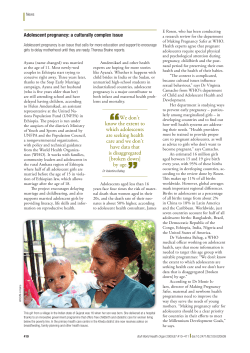
Masukume et al. Journal of Medical Case Reports 2013, 7:10
http://www.jmedicalcasereports.com/content/7/1/10
JOURNAL OF MEDICAL
CASE REPORTS
CASE REPORT
Open Access
Full-term abdominal extrauterine pregnancy
complicated by post-operative ascites with
successful outcome: a case report
Gwinyai Masukume1*, Elton Sengurayi1, Alfred Muchara1, Emmanuel Mucheni2, Wedu Ndebele3
and Solwayo Ngwenya1
Abstract
Introduction: Advanced abdominal (extrauterine) pregnancy is a rare condition with high maternal and fetal
morbidity and mortality. Because the placentation in advanced abdominal pregnancy is presumed to be
inadequate, advanced abdominal pregnancy can be complicated by pre-eclampsia, which is another condition with
high maternal and perinatal morbidity and mortality. Diagnosis and management of advanced abdominal
pregnancy is difficult.
Case presentation: We present the case of a 33-year-old African woman in her first pregnancy who had a full-term
advanced abdominal pregnancy and developed gross ascites post-operatively. The patient was successfully
managed; both the patient and her baby are apparently doing well.
Conclusion: Because most diagnoses of advanced abdominal pregnancy are missed pre-operatively, even with the
use of sonography, the cornerstones of successful management seem to be quick intra-operative recognition, surgical
skill, ready access to blood products, meticulous post-operative care and thorough assessment of the newborn.
Introduction
Advanced abdominal (extrauterine) pregnancy (AAP) is
defined as a fetus living or showing signs of having once
lived and developed in the mother’s abdominal cavity
after 20 weeks of gestation [1].
A hospital-based study from Zimbabwe showed an
AAP incidence of one in 9500 total deliveries and one in
60 ectopic pregnancies [2].
AAP can be classified as being primary or secondary
[3]. Primary AAP occurs when the fertilized ovum
implants directly into the peritoneal cavity; primary AAP
is the less common type. Secondary AAP occurs when
the fertilized ovum first implants in the fallopian tube or
uterus and then due to fimbrial abortion or rupture [4,5]
of the fallopian tube or uterus the fetus comes to live
and develop in the mother’s abdominal cavity. Ruptured
tubal ectopic pregnancies account for the majority
of AAP.
Ovarian, tubal and intraligamentary pregnancies are
excluded from the definition of AAP.
Maternal and fetal morbidity and mortality are high
with AAP [1-5].
Making the diagnosis of AAP is difficult even with the
ubiquitous use of sonography during pregnancy [6].
Most cases of AAP are missed and are only diagnosed
during surgery [1]. Operative and post-operative management of AAP is also difficult [7].
In addition, all degrees of pre-eclampsia may occur
more frequently with AAP [7-13] presumably due to inadequate placentation during early pregnancy [14] as a
result of the commonly abnormally sited placenta found
in AAP. Pre-eclampsia and its serious complications are
leading causes of maternal and perinatal morbidity and
mortality.
We report a case of full-term AAP without maternal
or fetal death.
* Correspondence: [email protected]
1
Department of Obstetrics and Gynaecology, Mpilo Central Hospital,
Bulawayo, Zimbabwe
Full list of author information is available at the end of the article
Case presentation
A 33-year-old primigravid African woman who had been
pregnant for 40 weeks and three days dated from the
© 2013 Masukume et al.; licensee BioMed Central Ltd. This is an Open Access article distributed under the terms of the
Creative Commons Attribution License (http://creativecommons.org/licenses/by/2.0), which permits unrestricted use,
distribution, and reproduction in any medium, provided the original work is properly cited.
Masukume et al. Journal of Medical Case Reports 2013, 7:10
http://www.jmedicalcasereports.com/content/7/1/10
first day of her last normal menstrual period presented
to our unit with a three-day history of continuous pain
around the umbilical area. She did not have any other
complaints.
A review of her antenatal records revealed that she had
sought medical attention at her local clinic for lower abdominal pain at eight weeks and four days and once again
at 14 weeks and two days of gestation. Both episodes of
lower abdominal pain were managed conservatively.
Throughout the pregnancy the patient continued to
experience on and off abdominal pain which became
better as the pregnancy progressed.
She denied any per vaginal bleeding during her
pregnancy.
Of note, the patient said that her mother had developed hypertension when she was pregnant.
The results of her serologic tests for human immunodeficiency virus as well as rapid plasma reagin were negative at the antenatal clinic. Her hemoglobin concentration
at 22 weeks of pregnancy was 10.7g/dL. Methyldopa and
nifedipine were commenced for pregnancy-induced hypertension from 22 weeks of gestation. During antenatal care
visits at 26 weeks and three days, 30 weeks and three days,
34 weeks and four days, and 37 weeks and three days she
was normotensive. At 38 weeks and four days her blood
pressure was 150/100mmHg.
The patient had an ultrasound scan done at 33 weeks
and three days at a local clinic which showed a single
viable breech intrauterine pregnancy with a left lateral
upper segment placenta, slightly reduced liquor volume
and some small free fluid in the hepatorenal area. The
estimated gestational age by scan was 34 weeks and two
days. From the time of this ultrasound scan, the presentation by clinical examination which had previously been
documented as cephalic was then documented as breech.
Besides the on and off abdominal pain, her pregnancy
had been uneventful until she developed periumbilical
pain prompting her to present to our unit.
On examination her blood pressure was 140/90mmHg
and other vital signs were within normal limits. Her
body mass index (her weight in kilograms divided by the
square of her height in meters) was 27. A breech presentation was the remarkable finding on obstetric examination. Admission and urgent delivery by cesarean section
was advised because of the breech presentation and suspected pre-eclampsia; the patient agreed and she gave
her informed consent.
She continued taking antihypertensives. During her
admission, several of her blood pressure readings were
in the severe range (≥160/110mmHg). At 40 weeks and
five days of pregnancy our patient had a cesarean section
performed via a Pfannenstiel incision by a junior member of the obstetric team. On entering the ‘parietal peritoneum’ a sudden gush of meconium-stained liquor
Page 2 of 5
issued forth. The fetal back was visualized. Delivery of
the baby was done, the umbilical cord was clamped and
cut and part of the placenta was removed. On recognition of the unusual circumstances, artery forceps were
used to arrest bleeding from the remaining placenta and
then senior help was sought. The cesarean section which
was being done under spinal anesthesia was continued
under general anesthesia (the patient was intubated) because severe hemorrhage was anticipated and also to
minimize patient anxiety.
AAP was diagnosed by the next most senior doctor
who in turn sought assistance from the consultant.
When the consultant arrived, the patient’s placenta
was noted to be predominantly attached to her small
bowel (Figures 1 and 2), large bowel and the superior
surface of her urinary bladder. After assessment of the
placental attachment site, a decision was made to remove the remaining placenta. There were large bleeders
on the placental bed which was thick enough to allow
figure of eight sutures to be used without compromising
nearby organs.
The meconium-stained bowel was cleaned with normal saline; no obvious bowel defects were noted. Part of
the membranes attached to the small and large bowel
and bladder were left in situ. The uterus was palpable
and about the size of a uterus at 10 weeks of pregnancy
Figure 1 Placenta and membranes adherent to loop of bowel
(anterior view). Note linea nigra.
Masukume et al. Journal of Medical Case Reports 2013, 7:10
http://www.jmedicalcasereports.com/content/7/1/10
Figure 2 Placenta and membranes adherent to loop of bowel
(posterior view).
beneath the sac. Estimated blood loss was 1.5liters. Our
patient received three units of packed red blood cells.
The baby girl delivered weighed 2850g and had Apgar
scores of eight and 10 at one minute and five minutes
respectively. Besides slight cranial asymmetry the examination of the newborn baby was unremarkable. The
baby’s head circumference was 32cm (5th centile). By
day 5 post-laparotomy the mother could mobilize fully,
eat, drink and had normal bowel motions and micturition. The baby was breast feeding and eliminating well.
Of note, the mother’s blood pressure normalized postdelivery; however, the results of a urine dipstick test
showed that she had proteinuria (3+ = 3g/L).
On day 5 post-delivery, an ultrasound scan showed a
slightly bulky uterus measuring nine cm in length not consistent with a post-delivery uterus. Notably, the scan
showed ascites with fibrinous strands and internal echoes.
The patient requested discharge and she was discharged
on day 5 after delivery on iron and folate tablets. During
her stay, she was counseled on AAP and future care.
On day 9 after her laparotomy our patient was readmitted with a three-day history of vomiting after meals,
abdominal pain and distension; she was passing stool and
flatus normally.
A physical examination revealed a grossly distended abdomen. A repeat ultrasound scan showed gross ascites.
Functional intestinal obstruction secondary to increased
intra-abdominal pressure from the gross ascites was diagnosed. Serial abdominal ascitic tapping for symptomatic
relief, intravenous crystalloid and antibiotics as well as low
molecular weight heparin were commenced.
On day 15 post-surgery, 5700mL of a dark brown fluid
was drained at one go from the urinary catheter and her
Page 3 of 5
abdominal distension subsided. Over the following days,
her condition was monitored.
The patient and her baby were discharged from hospital on hematinics with no complaints 26 days after the
birth of her baby.
Five weeks following surgery, the patient and her baby
were apparently doing well.
At about six months of age, the infant’s growth and
development were seemingly normal. The infant’s head
circumference was 40cm (5th centile); the cranial asymmetry was barely noticeable.
At about six months after delivery, the mother had no
complaints and her physical examination was unremarkable.
Her blood pressure was 160/93mmHg; she had hypokalemia (3.3mmol/L, reference range (RR) 3.6 to 5.0mmol/L)
and hyponatremia (124mmol/L, RR 135 to 145mmol/L).
Her chloride was 101mmol/L, RR 101 to 111mmol/L and
bicarbonate, 15mmol/L, RR 21 to 31mmol/L.
The mother’s laboratory blood investigations are
shown in Table 1.
Discussion
In retrospect, this patient’s history, physical examination
and radiologic findings were in line with a diagnosis of
AAP. Lower abdominal pain at eight weeks and four
days of gestation coincides with the peak period of ruptured ectopic pregnancies [15].
It is probable that the suspected tubal ectopic pregnancy ruptured and then subsequently re-implanted in
the peritoneal cavity at this time.
Most cases of AAP have an abnormal lie [1]; our patient had a breech presentation.
Free maternal intraperitoneal fluid as was noted on our
patient’s ultrasound scan at 33 weeks and three days has
been noted to be a feature of AAP [3,4,7]. In addition,
reduced amniotic fluid [16] is another feature of AAP
which was also noted on our patient’s ultrasound scan.
As mentioned earlier, pre-eclampsia is not uncommon
with AAP and this could have further led one to suspect
AAP. However, a lack of resources to confirm the diagnosis, a family history suggestive of the disease and nulliparity which are both risk factors for developing preeclampsia, as well as an ultrasound scan which apparently
showed an intrauterine pregnancy all confounded issues.
Taken together, the history of lower abdominal pain in
early pregnancy, continued abdominal pain during pregnancy, an abnormal lie, free maternal intraperitoneal
fluid and reduced amniotic fluid on sonography could
have all helped clinch the diagnosis of AAP. However,
the diagnosis of AAP remains difficult.
We attribute the patient’s continuous periumbilical
pain at presentation to irritation of the visceral peritoneum somewhere between the jejunum and transverse
colon, originating from the placental attachment site.
Masukume et al. Journal of Medical Case Reports 2013, 7:10
http://www.jmedicalcasereports.com/content/7/1/10
Page 4 of 5
Table 1 Laboratory data
Variable
Reference
range
Day 1*
Day 9
Day 12
Day 16
White cell count (× 109/L)
6–14
10.89
16.72
13.2
11.59
Day 19
Approximately
6 months**
2.82
Hemoglobin (g/dL)
11.5–15.5
8.7
9.4
8.6
10.4
12.1
Platelets (× 109/L)
150–400
118
642
725
576
214
Mean cell volume (fl)
80–95
87.5
89.6
93.8
90.3
85.0
Mean cell hemoglobin (pg)
27–34
29.5
28.8
26.6
29
28.9
Mean cell hemoglobin concentration (g/dL)
30–35
33.7
32.2
28.4
32.1
Urea (mmol/L)
3.2–6.7
7
16.8
21
14.9
3.8
347
377
316
40
Creatinine (micromol/L)
53–115
75
Aspartate aminotransferase (IU/L)
10–42
50
Alanine aminotransferase (IU/L)
6–28
20
12
Direct bilirubin (micromol/L)
0–3
18
3
Albumin (g/L)
23–38
18
22
Total protein (g/L)
67–82
49
77
34.0
26
There is a normochromic normocytic anemia consistent with acute blood loss with a possibly reactive thrombocytosis. The initial thrombocytopenia could have
been caused by pre-eclampsia. Raised urea and creatinine are probably secondary to hypovolemia due to loss of fluid into the peritoneal cavity leading to prerenal acute kidney injury. Aspartate aminotransferase is usually raised after cesarean section. Conjugated hyperbilirubinemia is attributed to intra-operative
hemorrhagic shock. Because albumin is a negative acute phase reactant, the trauma of surgery may have contributed to the hypoalbuminemia which improved
over time post-operatively. Hypoproteinemia was probably due to proteinuria and also possibly resulted from the period of starvation before and after surgery.
Leucopenia at six months did not escape our attention. The patient was rhesus D positive.
* Days post-surgery.
** Different reference ranges may apply; there was no proteinuria.
Pain from the jejunum to transverse colon is felt around
the umbilicus (referred pain) [17].
The central controversy in managing AAP regards
management of the placenta [1].
Removing the placenta can result in catastrophic
hemorrhage [3] and damage to adjacent structures,
whereas leaving it in situ may lead to secondary
hemorrhage, abscess formation, adhesions, coagulopathy,
continued pre-eclampsia, failure of lactogenesis [18], a
need for second surgery and a need for longer follow-up
among other complications.
In our case, we had ready access to immediate blood
transfusion which most patients with AAP receive [1].
Where blood transfusion and intensive care are not readily
available, leaving the placenta in situ has been recommended [19]. Placental removal is done after assessing for
safety and feasibility of removal.
The finding of slight cranial asymmetry in the newborn was not unusual because about 20% of AAP infants
have malformations or deformations [20]. We ascribe
this cranial asymmetry to skull compression due to the
lack of the protective myometrial wall and perhaps the
reduced amniotic fluid noted on sonography. Although
perinatal morbidity and mortality are high with AAP,
normal development of the infant at follow-up as in our
case is possible although very rare [19].
We speculate that the ascites was due to probable
transudation or exudation of fluid from the site at which
the placenta was attached. We are of the opinion that the
fluid drained from the urinary catheter on day 15 postsurgery came from the peritoneal cavity through an abnormal communication which developed due to increased
intra-abdominal pressure where the placenta was adherent
to the urinary bladder (a point of weakness). We lacked
the financial resources to do the necessary tests to ascertain the origin and nature of this fluid.
We are aware of what may seem like deficiencies in management; we work in a low-income country. Pathologic
examination of the placenta would have been useful [21].
This patient is probably at higher risk of having a future ectopic pregnancy [22], cardiovascular and renal
disease [14]. In order to optimize the patient’s next pregnancy, she was advised to attend pre-conception counseling. She was also advised of the need to have close
follow-up of her current baby.
The mother’s constellation of hypertension, hypokalemia and hyponatremia at about six months after delivery raises the possibility of a hyperreninemic state which
requires further investigation to clarify and manage [23].
The significance of a hyperreninemic state in relation to
AAP is uncertain.
It is very rare to have a live AAP proceeding to term
and being successfully managed [4]. It is because of this
we report this case.
Conclusion
Because most diagnoses of AAP are missed pre-operatively
even with the use of sonography, the cornerstones of
Masukume et al. Journal of Medical Case Reports 2013, 7:10
http://www.jmedicalcasereports.com/content/7/1/10
successful management seem to be quick intra-operative
recognition, surgical skill, ready access to blood products,
meticulous post-operative care and thorough assessment of
the newborn.
Consent
Written informed consent was obtained from the patient
for publication of this case report and any accompanying
images. A copy of the written consent is available for review by the Editor-in-Chief of this journal.
Competing interests
The authors declare that they have no competing interests.
Authors’ contributions
SN was the patient’s primary physician. GM wrote the first draft of the article.
ES, AM, EM, WN and SN revised the manuscript making important
intellectual contributions. All authors read and approved the final version of
the manuscript.
Acknowledgements
We thank the patient for giving us permission to share her case. We express
our gratitude to Vonai Chaoma and Similo Nkiwane for taking photographs.
Doctors S Ndlovu and R Sarai are acknowledged for helping in the
management of this patient. We thank Mr M Makonese for doing the postdelivery ultrasound scan. We also record our sincere appreciation of the
Mpilo Central Hospital staff.
This case report was presented at the combined Mpilo Central Hospital and
United Bulawayo Hospitals Obstetrics and Gynaecology grand rounds on
11th May 2012.
Author details
1
Department of Obstetrics and Gynaecology, Mpilo Central Hospital,
Bulawayo, Zimbabwe. 2Department of Anaesthesia, Mpilo Central Hospital,
Bulawayo, Zimbabwe. 3Department of Paediatrics, Mpilo Central Hospital,
Bulawayo, Zimbabwe.
Page 5 of 5
10. Anderton KJ, Duncan SL, Lint RL: Advanced abdominal pregnancy with
severe pre-eclampsia. Br J Obstet Gynaecol 1976, 83(1):90–93.
11. Moodley J, Subrayen KT, Sankar D, Pitsoe SB: Advanced extra-uterine
pregnancy associated with eclampsia. A report of 2 cases. S Afr Med J
1987, 71(7):460–461.
12. Piering WF, Garancis JG, Becker CG, Beres JA, Lemann J Jr: Preeclampsia
related to a functioning extrauterine placenta: report of a case and 25year follow-up. Am J Kidney Dis 1993, 21(3):310–313.
13. Ekele BA, Adamu AN, Ladan H, Abitare H: Eclampsia in advanced
abdominal pregnancy. Niger J Clin Pract 2007, 10(4):343–345.
14. Steegers EA, von Dadelszen P, Duvekot JJ, Pijnenborg R: Pre-eclampsia.
Lancet 2010, 376(9741):631–644.
15. Masukume G: Nausea, vomiting, and deaths from ectopic pregnancy. BMJ
2011, 343:d4389.
16. Mittal SK, Singh N, Verma AK, Agarwal H, Kulkarni CD, Kanaujia R: Fetal MRI
in the preoperative diagnosis and assessment of secondary abdominal
pregnancy: a rare sequela of a previous caesarean section. Diagn Interv
Radiol 2012, 18(5):496–502.
17. Rogers AW: The digestive system. In Textbook of Anatomy. Edinburgh:
Churchill Livingstone; 1992:456.
18. Pieh-Holder KL, Scardo JA, Costello DH: Lactogenesis failure following
successful delivery of advanced abdominal pregnancy. Breastfeed Med
2012, 7:543–546.
19. Oneko O, Petru E, Masenga G, Ulrich D, Obure J, Zeck W: Management of
the placenta in advanced abdominal pregnancies at an East African
tertiary referral center. J Womens Health (Larchmt) 2010, 19(7):1369–1375.
20. Stevens CA: Malformations and deformations in abdominal pregnancy.
Am J Med Genet 1993, 47(8):1189–1195.
21. Chang KT: Pathological examination of the placenta: raison d'être, clinical
relevance and medicolegal utility. Singapore Med J 2009, 50(12):1123–1133.
22. Barnhart KT: Clinical practice. Ectopic pregnancy. N Engl J Med 2009,
361(4):379–387.
23. Pillai U, Mandakapala C, Mehta K: Resistant hyponatremia and hypokalemia
treated successfully with nephrectomy. Clin Kidney J 2011, 5(1):68–69.
doi:10.1186/1752-1947-7-10
Cite this article as: Masukume et al.: Full-term abdominal extrauterine
pregnancy complicated by post-operative ascites with successful
outcome: a case report. Journal of Medical Case Reports 2013 7:10.
Received: 23 June 2012 Accepted: 5 December 2012
Published: 9 January 2013
References
1. Nkusu Nunyalulendho D, Einterz EM: Advanced abdominal pregnancy:
case report and review of 163 cases reported since 1946. Rural Remote
Health 2008, 8(4):1087.
2. White RG: Advanced abdominal pregnancy–a review of 23 cases. Ir J Med
Sci 1989, 158(4):77–78.
3. Dahab AA, Aburass R, Shawkat W, Babgi R, Essa O, Mujallid RH: Full-term
extrauterine abdominal pregnancy: a case report. J Med Case Reports
2011, 31(5):531.
4. Amritha B, Sumangali T, Priya B, Deepak S, Sharadha R: A rare case of term
viable secondary abdominal pregnancy following rupture of a
rudimentary horn: a case report. J Med Case Reports 2009, 29(3):38.
5. Naim NM, Ahmad S, Siraj HH, Ng P, Mahdy ZA, Razi ZR: Advanced
abdominal pregnancy resulting from late uterine rupture. Obstet Gynecol
2008, 111(2 Pt 2):502–504.
6. Roberts RV, Dickinson JE, Leung Y, Charles AK: Advanced abdominal
pregnancy: still an occurrence in modern medicine. Aust N Z J Obstet
Gynaecol 2005, 45(6):518–521.
7. Worley KC, Hnat MD, Cunningham FG: Advanced extrauterine pregnancy:
diagnostic and therapeutic challenges. Am J Obstet Gynecol 2008,
198(3):297. e1-e7.
8. Clark JF, Niles JH: Abdominal pregnancy associated with toxemia of
pregnancy. Report of two cases and review of the literature. J Natl Med
Assoc 1967, 59(1):22–24.
9. Baehler RW, Copeland WE, Stein JH, Ferris TF: Plasma renin and
aldosterone in an abdominal pregnancy with toxemia. Am J Obstet
Gynecol 1975, 122(5):545–548.
Submit your next manuscript to BioMed Central
and take full advantage of:
• Convenient online submission
• Thorough peer review
• No space constraints or color figure charges
• Immediate publication on acceptance
• Inclusion in PubMed, CAS, Scopus and Google Scholar
• Research which is freely available for redistribution
Submit your manuscript at
www.biomedcentral.com/submit


© Copyright 2026