
Aloe vera leaf gel: a review update T. Reynolds *, A.C. Dweck
Journal of Ethnopharmacology 68 (1999) 3 – 37 www.elsevier.com/locate/jethpharm Review article Aloe vera leaf gel: a review update T. Reynolds a,*, A.C. Dweck b a Jodrell Laboratory, Royal Botanic Gardens, Kew, Richmond, Surrey, UK b Dweck Data, 8 Merrifield Road, Ford, Salisbury, Wiltshire, UK Received 20 April 1999; accepted 20 May 1999 Abstract Research since the 1986 review has largely upheld the therapeutic claims made in the earlier papers and indeed extended them into other areas. Treatment of inflammation is still the key effect for most types of healing but it is now realized that this is a complex process and that many of its constituent processes may be addressed in different ways by different gel components. A common theme running though much recent research is the immunomodulatory properties of the gel polysaccharides, especially the acetylated mannans from Aloe 6era, which are now a proprietary substance covered by many patents. There have also been, however, persistent reports of active glycoprotein fractions from both Aloe 6era and Aloe arborescens. There are also cautionary investigations warning of possible allergic effects on some patients. Reports also describe antidiabetic, anticancer and antibiotic activities, so we may expect to see a widening use of aloe gel. Several reputable suppliers produce a stabilized aloe gel for use as itself or in formulations and there may be moves towards isolating and eventually providing verified active ingredients in dosable quantities © 1999 Elsevier Science Ireland Ltd. All rights reserved. Keywords: Aloe 6era gel; Active polysaccharides; Therapeutic properties 1. Introduction Aloes have been used therapeutically, certainly since Roman times and perhaps long before (Morton, 1961; Crosswhite and Crosswhite, 1984), different properties being ascribed to the inner, colourless, leaf gel and to the exudate from the outer layers. During the 12 years since the last major review of Aloe 6era (L.) Burm.f. gel (Grind* Corresponding author. lay and Reynolds, 1986) popular interest and use of the gel have increased dramatically. In this country it is now a familiar ingredient in a range of healthcare and cosmetic products widely available and advertised in shops. The preserved but otherwise untreated gel is also sold as a therapeutic agent in its own right as are various concentrated, diluted and otherwise modified products. This need has been met by a number of wholesalers who get their supplies from plantations in Texas, Florida and Venezuela while new ones are 0378-8741/99/$ - see front matter © 1999 Elsevier Science Ireland Ltd. All rights reserved. PII: S 0 3 7 8 - 8 7 4 1 ( 9 9 ) 0 0 0 8 5 - 9 4 T. Reynolds, A.C. Dweck / Journal of Ethnopharmacology 68 (1999) 3–37 being proposed for Israel, Queensland and East Africa (Jamieson, 1984). This commercial activity has been accompanied by an upsurge of both clinical and chemical research which is reaching more closely towards the active ingredients and their biological activity. There is now less said about doubts as to the efficacy of the material, although there are some warnings of allergic side effects (Klein and Penneys, 1988; Briggs, 1995). Harmful reactions to aloe gel treatment are recorded infrequently (Hunter and Frumkin, 1991; Schmidt and Greenspoon, 1993) but need to be taken seriously. There is still confusion between the leaf exudate and the gel, Morsy and Ovanoviski (1983), Natow (1986) and Duke (1985) where a great number of folk medicine uses are described and Ahmad et al. (1993). However many commentators clearly distinguish between the two parts (Watson, 1983; McKeown, 1987; Capasso et al., 1998) and describe in some detail how the gel is prepared (McAnalley, 1988, 1990; Agarwala, 1997). At one time there was much discussion about the relative efficiency of ‘decolorized’ and ‘colorized’, i.e. with exudate components, gels (Danof, 1987; Agarwala, 1997). There is also a feeling that some of the variable results reported in the literature may be due to treatment of the gel subsequent to harvest (Fox, 1990; Marshall, 1990; Briggs, 1995; Agarwala, 1997). A number of reviews have appeared in recent years covering various aspects of aloe gel use, as well as much commercial literature. Exaggerated claims are still being made and although doubts as to the substance’s efficacy are more muted, there is still room for the caution which has been voiced (Hecht, 1981; Marshall, 1990). The emphasis is changing towards definition of the active constituent or constituents so that they can be used accurately in formulations (Reynolds, 1998). There has been a greater willingness to investigate reasons for the recorded variability in curative properties. Reasons presented for aloe gel efficacy are still varied perhaps because there are in fact several different healing activities operating (Capasso et al., 1998). The action of aloe gel as a moisturizing agent is still a popular concept (Meadows, 1980; Watson, 1983; Natow, 1986; Danof, 1987; McKe- own, 1987; Fox, 1990; Marshall, 1990; Briggs, 1995) and may account for much of its effect. More speculative is the presence of salicylates, by implication having an aspirin-like effect (Robson et al., 1982; Klein and Penneys, 1988; Marshall, 1990; Shelton, 1991; Canigueral and Vila, 1993), although the differences between natural salicylates and aspirin, a synthetic product, were pointed out (Frumkin, 1989). Another simple substance, magnesium lactate, is said to inhibit the production of histamine by histidine decarboxylase and is claimed as a gel constituent (Rubel, 1983; Natow, 1986; Marshall, 1990; Shelton, 1991; Canigueral and Vila, 1993). Inhibition of pain-producing substances such as bradykinin or thromboxane is often claimed (Rubel, 1983; Natow, 1986; Danof, 1987; Fox, 1990; Marshall, 1990; Shelton, 1991; Canigueral and Vila, 1993). On a more sophisticated level, action on the immune system has been postulated and to some extent tested (Rubel, 1983; Schechter, 1994; Griggs, 1996). A recent, very interesting book (Davis, 1997), dwells at some length on the immunomodulatory properties of the gel poysaccharides and presents a viewpoint complementary to the present review. Polysaccharides are another group of gel constituents to which activity has been ascribed, particularly in immunomodulatory reactions and one, acemannan, has reached proprietary status (Schechter, 1994; McAnalley, 1988, 1990; Agarwala, 1997). There has been much interest recently in the biological activity of polysaccharides, which is greater and more diverse than previously realized. Although the substances are varied and widespread in plants some are well known as entities, albeit often with uncertain structures (Franz, 1989; Tizard et al., 1989; McAuliffe and Hindsgaul, 1997). Also often mentioned are the antibacterial, antifungal and even antiviral properties demonstrated by the gel (Klein and Penneys, 1988; Marshall, 1990; Ahmad et al., 1993), while anti-oxidant effects are becoming of interest. Although A. 6era gel is the only one being used commercially, there is the possibility of discovering useful properties among the other 300 or more species (Newton, 1987). In this update the trend to be emphasized is away from the more naive arbitrary use of the leaf T. Reynolds, A.C. Dweck / Journal of Ethnopharmacology 68 (1999) 3–37 gel (Reynolds 1996) and towards a quest for deeper, more precise understanding of its constituents and the varied biological activities which they may or may not display (Reynolds 1996). 2. Test systems and clinical trials1 2.1. Burns and incisions For testing the efficacy of aloe gel or its various components on inflammation a number of tests have been used, usually in relation to some sort of deliberate wounding. These need to be distinguished from clinical trials where the injuries already exist and are treated more or less systematically by a number of putative therapeutic agents. The earliest experimentation related to skin burns and arose in relation to clinical observations, going back to the 1930s (Grindlay and Reynolds, 1986). It was often inconclusive due to inadequate controls and replication and an imprecise correlation of cause and effect. One of the most detailed and accurate of these clinical trials took place in 1957 with use of aloe gel against controlled thermal and radiation burns on rats and rabbits compared with clinical studies on human patients (Ashley et al., 1957). This failed to demonstrate any healing properties of the gel. In contrast another careful study but with far fewer replicates gave a positive result (Rovatti and Brennan, 1959). Another approach a little later, was to measure the tensile strength of the healing of a precise incision wound, post mortem (Goff and Levenstein, 1964). An undescribed aloe ‘extract’ speeded healing but had no effect on the final result. A similar wound tensile strength test was used later to compare the effects of steroids and aloe gel on inflammation and healing (Davis et al., 1994b), and later of antibacterial agents (Heggers et al., 1995). Then in the early 1980s both precise experimental scald burns as well as previous injuries were again compared by a vari1 The authors wish to make it clear that neither they nor anyone else at RBG, Kew is in any way envolved in vertebrate experimentation, which is only described here as part of the review. Ethical approval is not implied. 5 ety of criteria (Cera et al., 1980, 1982; Robson et al., 1982) and therapeutic benefits were recorded. This type of test system was returned to later (Heggers et al., 1993) with similar positive results. A study, with good replication, of healing after a precise skin hole punch demonstrated the antiinflammatory properties of the gel leading to more rapid healing (Davis et al., 1987a) A different approach was taken whereby the subjects (mice) were fed aloe gel for some time before hole punch wounding and compared with those treated topically after wounding (Davis et al., 1989c). Both methods produced healing. A further variation was the treatment of punch wounds on mice or rats made diabetic by streptozotocin and therefore more slow to heal. Again healing by aloe gel was demonstrated (Davis et al., 1988; Davis and Maro, 1989). A return to wounding by precision burns using a hot metal plate with adequate replication again demonstrated positive healing activity (Rodriguez-Bigas et al., 1988). This technique was further elaborated to produce first, second or third degree burns by precisely timed exposures to the hot metal plate (Bunyapraphatsara et al., 1996a). Two special examples of burns are sunburn and frostbite and these have been used experimentally. Thus, precise UVB burns were produced with a light pen but were unaffected by aloe gel (Crowell et al., 1989). In a later trial to test the effect of the gel on UV-induced immune suppression a bank of UV lights were used (Strickland et al., 1994). Frostbite was produced by exposing rabbit ears to ethanol and solid carbon dioxide (Heggers et al., 1993; Miller and Koltai, 1995) and was relieved by application of aloe gel. 2.2. Irritating compounds producing oedema Experimental production of swelling, caused by fluid accumulation in a tissue (Oedema) initiated by irritating compounds has been used as an inflammatory model with the mouse ear or rat hind paw as subjects. Croton oil, a powerful irritant, was applied to the right ear with the left remaining as control. Inflammation was measured by weighing a tissue punch sample and was shown to decrease after topical application of aloe gel 6 T. Reynolds, A.C. Dweck / Journal of Ethnopharmacology 68 (1999) 3–37 (Davis et al., 1987b; 1989a,b). A subsequent trial demonstrated an even greater decrease when the gel was combined with a corticosteroid (Davis et al., 1991, 1994b). This trial was accompanied by a similar one where mustard as the initiating agent was injected into a rat paw, subsequent swelling being measured volumetrically and fluid withdrawn to determine leucocyte infiltration. In this case aloe gel, with or without steroids was injected previously rather than being applied topically. This study had followed some more or less similar ones, where a variety of irritants, gelatine, albumin, dextran, carrageenan and kaolin had been used (Davis et al., 1989a) and the inflammation successfully treated with aloe gel, orally or topically. 2.3. The air pouch A modification of these models involving oedema was developed in which air was injected under the skin to form a cavity, a simulated synovium, to which irritants and therapeutic agents could be added. This was taken to be analogous to the joint cavity, containing synovial fluids which becomes inflamed during arthritis. Such an air pouch was produced on the backs of anaesthetized mice, irritated with carrageenan and treated with A. 6era gel solution (Davis et al., 1992). Healing effect was measured histologically by counting the number of mast cells in the cavity fluid, decreased by aloe gel treatment and by examining pouch wall vascularity, also decreased by the gel. 2.4. Adju6ant arthritis An important extension of experiments on lesions caused by applied irritants is the deliberate production of a condition resembling arthritis in an animal model, usually rat. This can then be followed by treatment with putative therapeutic agents to suppress either the inflammation or immunologic consequences. The irritating agent used was a suspension of heat-killed Mycobacterium butyricum in mineral oil which produced inflammation directly in the injected paw and also in the other paw by an immunologic pathway (Saito et al., 1982). 2.5. In 6itro studies A number of experiments have been carried out in which the effects of components of Aloe leaf on various biochemical or microbiological systems, relevant to the four inflammation responses and to wound healing, have been studied. Enzymes in aloe gel destroying the nonapeptide bradykinin which causes vasodilatation and pain production, were the subject of one such investigation, although angiotensin-converting-enzyme received less attention. Prostaglandins were another obvious subject and their presence in Aloe was claimed, as well as a complex lipid inhibiting arachidonic acid oxidation. On the other hand production of the vasodilator histamine from histidine was said to be inhibited by the gel. Effects of aloe gel on separate components of wound healing in tissue culture have been made, notably fibroblast proliferation (Brasher et al., 1969; Danof and McAnalley, 1983) and growth of new blood capillaries (Lee et al., 1995). The involvement of immunological effects has been recognized by examining stimulus of phagocyte formation and activity (Imanishi and Suzuki, 1984). Alongside these studies of in vitro systems there were also many attempts to analyse the plant material and to identify the active fraction or fractions. 3. Treatment of inflammation Inflammation is a tissue reaction by the body to injury and typically follows burns or other skin insults. It is classically characterized by swelling (tumor), pain (dolor), redness (rubor) and heat (calor) as well as loss of function (Macpherson, 1992). It is thus a complex process and investigations into the therapeutic properties of the gel should take account of its effects on these various symptoms. In addition, the gel may have more than one active constituent, which may be addressing different parts of the healing process. Failure to take all this into account may be responsible for ambiguities which may have arisen in the past about the efficacy of the gel. Although inflammatory processes are a natural response to T. Reynolds, A.C. Dweck / Journal of Ethnopharmacology 68 (1999) 3–37 injury and may hinder healing it may also be undesirable to suppress them in an unstructured way before their purpose is accomplished. Leucocytes accompanied by fluid accumulate in the damaged tissues producing the swelling, these movements being the result of increased capillary permeability. Pain is a complex reaction following the release of short peptides and prostaglandins. The redness and heat are caused by vasodilatation which reduces blood pressure and increases circulation, although this gradually slows. Inflammation can be either caused, or intensified by invasion with micro-organisms. As well as in wounds, inflammation is involved in conditions such as arthritis. Continuing research into inflammation has shown that it is a complex process involving many biochemical pathways and a variety of agents and mediators (summarized in Davis et al., 1989a). In particular these authors distinguish three components, 1. Vasoactive substances; agents causing dilation of blood vessels and opening of junctions between cells of the ultimate capillaries, produced by altering contractile elements in endothelial cells. These factors include vasoactive amines, bradykinin and also prostaglandins. 2. Chemotactic factors; these agents cause increased cell motility, especially of white blood cells (leucocytes) into stressed areas. These include several proteins and peptides. 3. Degradative enzymes; these are hydrolytic enzymes breaking down tissue components. Proteases in particular participate in inflammatory states causing chemotactic factors to be released. It was also shown that aloe gel contained both an inhibitory system and a stimulatory system that influenced both inflammatory and immune responses (Davis et al., 1991a,b). The healing effects of A. 6era gel are therefore now being seen as more complex than previously realized (Ho¨rmann and Korting, 1994). It now appears that several activities are operating each with its own part to play in the overall therapy. These activities may well reflect the presence of several different active agents in the gel. There seems to be a need for two types of investigation. 7 The first, the academic, analytical approach, seeks to dissect the processes and reveal the individual biochemical and physiological reactions, while the second, the clinical approach, puts the various processes back together and studies their interactions. The second can only be ultimately successful when the first is well known. In the best, recent, precise experimentation, care has been taken to separate the inner parenchyma of the aloe leaf completely from the outer layers rich in phenolics, both experimentally and conceptually. However in some trials this separation has not been complete and two preparations were deliberately made and tested, one decolorized and the other containing anthraquinones from the outer layers (Davis et al., 1989). These substances were to some degree toxic and reduced for instance suppression of polymorphonuclear lymphocyte infiltration (Davis et al., 1986b). They also greatly reduced the healing of croton oil-induced inflammation (Davis et al., 1989b, 1991). A component extracted from whole Aloe barbadensis (sic) leaves and probably originating from the exudate rather than the gel was characterized as a cinnamic acid ester of aloesin and shown to reduce croton oil-induced inflammation (Hutter et al., 1996). A low molecular weight component claimed to be extracted from the gel but probably also of exudate origin, was shown to have cytotoxic effects similar to barbaloin (Avila et al., 1997). This raises the possibility of both irritating and healing agents in the exudate as well as the gel. Even this comparison is not strictly accurate as it is not clear how the anthraquinone-free gel is made and if other substances are removed, or not, at the same time. Mannose-6-phosphate was shown to have antiinflammatory activity and this was said to resemble the known activity of acetylated mannan, a gel component (Davis et al., 1994a). Two aspects of inflammation reduction following aloe gel treatment by injection in rats were observed (Davis et al., 1986a,b, 1987c). Mustard induced oedema of the paw was reduced by between 445 and 70% while infiltration of polymorphonuclear lymphocytes into a skin blister was reduced by 58%. Two other substances, RNA and vitamin C synergized with the gel in inhibiting oedema. Similarly mouse ear inflammation induced with 8 T. Reynolds, A.C. Dweck / Journal of Ethnopharmacology 68 (1999) 3–37 croton oil was reduced by up to 67% following topically applied aloe gel (Davis et al., 1987b). In another study these workers tested the action of topical or injected aloe gel against inflammation produced by a variety of agents which were considered to induce different types of inflammation (Davis et al., 1989a). Thus the gel relieved the inflammatory effects of kaolin, carrageenan (an algal polysaccharide), albumin, gelatin, mustard and croton oil which were said to act either by promoting prostaglandin synthesis or by increasing infiltration of leucocytes. Elsewhere, aqueous or chloroform extracts of the gel reduced a carrageenan-induced inflammation and migration of neutrophils (Vazquez et al., 1996). Aloe gel was less effective against inflammatory agents which produced allergic reactions through the action of bioactive amines such as histamine, even if their synthesis might be inhibited by magnesium lactate. In other ways aloe gel was found to show immunomodulatory properties (t’Hart et al., 1988) so the full picture is still not clear. Another aspect is the use of the gel as a vehicle for application of other active substances to which it may additionally impart its own activity (Davis et al., 1989c). 3.1. Wound healing If inflammation is a complex process, then wound healing is much more so and the intervention of aloe gel is likely to be multifaceted. A wound to the skin may pierce two layers, the epidermis and dermis as well as damaging appendages. A temporary repair is effected by fibrin clot which is then invaded by a variety of cells, some of which produce the inflammatory response and which eventually carry out a permanent repair (Martin, 1997). It may well be that the repair is not perfect in that scar tissue is produced and appendages do not regenerate. The epidermis is repaired in three phases, migration of cells, proliferation and maturation, while new connective tissue is found in the dermis (Davis et al., 1987a). As well as repair of structure there is an urgency to avoid microbiological entry which can retard wound contraction (Hayward et al., 1992). Increased speed of repair was seen in an early detailed study of healing of a surgical cut which showed that ‘A. 6era extract in an ointment base’ speeded repair but did not alter the ultimate result (Goff and Levenstein, 1964). Perhaps aloe gel removes delaying effects rather than accelerating healing as such. The use of aloe gel to heal wounds is the classic use of the material and one of the first explanations of its efficacy was its high water content which kept the wound moist and increased epithelial cell migration (Morton, 1961; Erazo et al., 1985), although even this has been questioned (Roberts and Travis, 1995). Indeed the beneficial effects on oral wounds where moisture is abundant, indicates that other factors operate (Sudworth, 1997). A report of effective aloe gel healing of pressure sores recorded rapid granulation (Cuzzell, 1986), an effect also noted with various incision wounds in rats and attributed to more rapid maturation of collagen (Udupa et al., 1994). In a more detailed study, a skin punch wound healed more rapidly when treated with ‘decolorized’ gel than with ‘colorized’ gel (Davis et al., 1986). These observations were made on the 7th day after wounding a mouse or rat skin which was said to be optimal for recording healing. Treatment by daily injection of the gel reduced wound diameter, increased skin circulation and seemed to reduce scarring (Davis et al., 1987a, 1989a). Acute inflammation was also inhibited. The colour mentioned is due to anthraquinones from the aloe leaf exudate but details of their removal (‘decolorized’ gel) were not given. Elsewhere, trials using cultures of human endothelial cells or fibroblasts demonstrated cytotoxicity in gel samples contaminated with leaf exudate (Danof and McAnalley, 1983). In contrast, cytotoxicity was shown to be reduced in neutrophils treated witha low molecular weight fraction of gel, probably exudate-derived, although this was not stated, following inhibition of release of reactive oxygen species (t’Hart et al., 1990). In a further study, ‘A. 6era’ (sic), presumably the gel, was administered orally over two months or applied to the wounded skin in a cream. Both treatments improved wound healing. It was suggested that one of the factors, out of several, enhanced by aloe gel was increased oxygen access T. Reynolds, A.C. Dweck / Journal of Ethnopharmacology 68 (1999) 3–37 as a result of increased blood supply. (Davis et al., 1989b). In another trial using topical application, stimulation of fibroblast activity and collagen proliferation was demonstrated (Thompson 1991). Angiogenesis, the growth of new blood capillaries, is a necessary part of tissue regeneration and vascularity of burn tissue of a guinea pig was shown to be reestablished by topical application of aloe gel (Heggers et al., 1992). A low molecular weight component of freeze-dried aloe gel was shown to stimulate blood vessel formation in a chick chorioallantoic membrane, while a methanol-soluble fraction of the gel was shown to stimulate proliferation of artery endothelial cells in an in vitro assay and to induce them to invade a collagen substrate (Lee et al., 1998). Activation of matrix proteinases, which allow penetration, was thought to be involved. Tissue survival following arterial damage in a rabbit ear, which mimicked drug abuse damage, was maintained by topical aloe gel application (Heggers et al., 1993). Healing of an experimental excision wound was promoted by topically applied aloe preparation and this was enhanced when the gel was combined with a nitric oxide inhibitor (Heggers et al., 1997). Subsequent work showed that another important impediment to wound healing, microbiological activity, infection, was also addressed by aloe gel treatment (Heggers et al., 1995). Here, cuts to rat skin were more rapidly healed by topical applications of aloe gel compared with an untreated control or by applications of potential anti-microbials. Many antibiotic agents are more toxic to fibroblasts than to bacteria and seem to retard the healing process (Lineaweaver et al., 1985). The gel was thought to contain a growth factor which enhanced the breaking strength of wounds. Only buffered sodium hypochlorite solution (0.025%) had therapeutic effects similar to aloe gel (Heggers et al., 1996). Macrophages play a considerable part in controlling microorganisms and it was shown that young active macrophages accelerated the rate of wound healing in aged rats, compared with rates where senescent cells in these animals were left alone to act. Activation of macrophages by acemannan, an aloe gel polysaccharide, was claimed (Maxwell et al., 1996). This followed previous observations on the healing 9 powers of acemannan on wounds in elderly or obese rats also attributed to macrophage stimulation (Tizard et al., 1994). Total regeneration of the skin also requires that ‘difficult’ cells such as neurons are replaced. Proliferation of neuron-like cells and also perhaps cell adhesion, in a culture of rat adrenal cells was stimulated by gel preparations (Bouthet et al., 1995). Following the idea that there were factors with different types of activity in the gel an attempt was made to separate anti-inflammatory and wound healing components (Davis et al., 1991c). A precipitate formed by treatment with 50% aqueous ethanol seemed to have most of the wound healing activity observed in the raw gel when used against punch wounds in mouse skin. The supernatant contained anti-inflammatory activity which was attributed to glycoprotein. Elsewhere, significant wound healing was produced by mannose-6-phosphate (Davis et al., 1994a) Healing of an incision wound by aloe gel was found to be accompanied by higher levels of hyaluronic acid and dermatan sulphate produced more rapidly. This was suggested to stimulate collagen synthesis and fibroblast activity (Chithra et al., 1998a). There was also increased activity of b-glucuronidase and N-acetyl glucosaminidase which was said to increase carbohydrate turnover in the wound matrix. Fibroblast proliferation in vitro and in vivo was observed after treatment with the acetylated mannan fraction carrisyn™ (McAnalley, 1988). Increased collagen formation in wounded diabetic rats treated orally and topically with aloe gel was later demonstrated (Chithra et al., 1998b). The collagen formed had a higher degree of crosslinking indicating enhanced levels of type III (Chithra et al., 1998c). There were also higher levels of protein and DNA. In another test on surgical cuts in mice, hydrocortisone given by injection, while reducing inflammation, hindered wound healing but when ‘A. 6era’ (presumably the gel) was included then wound suppression was reversed and inflammation further reduced (Davis, et al., 1994b). Healing and control of acute inflammation, distinct from chronic inflammation, was observed following gel treatment of excision and incision wounds in rats (Udupa et al., 1994). 10 T. Reynolds, A.C. Dweck / Journal of Ethnopharmacology 68 (1999) 3–37 Widespread acclamation of the healing powers of aloe gel is not however universal. Some older studies were unable to demonstrate any curative properties (Ashley et al., 1957; Gjerstad, 1969; Ship, 1977; Spoerke and Elkins, 1980; Kaufman et al., 1988) and more recently a trial with surgical wounds in human patients even suggested that healing was delayed (Schmidt and Greenspoon, 1993). No effects of aloe gel on re-epithelization or wound contraction of excision wounds in pigs was observed (Watcher and Wheeland, 1989). No healing properties at all were observed with corneal punch wounds (Green et al., 1996), in contrast to earlier positive observations with corneal flash burns (Lawrence, 1984). Elsewhere it was found that acemannan had an equal, not better, healing and bactericidal effect on shave biopsy wounds as the antibiotic bacitracin® (Phillips et al., 1995). The two lines of conflicting evidence may be explained by the fragility of the active ingredients as it appears from several accounts that the treatment of the gel after harvesting is crucial for activity. Effects may also vary with the type and location of wound. Using a proprietary aloe dressing on pad wounds of dogs it was concluded that healing processes during the first 7 days were speeded, an advantage to a wound exposed to weight bearing, although the end result was the same as that with antibiotic treatment alone (Swaim et al., 1992). 3.2. Burn healing Burn healing can be regarded as a special type of wound healing and most of the skin reactions are the same. It has been pointed out however that conditions for healing would differ according to the depth of the burn wound and that several factors can interfere with the healing process (Kaufman et al., 1989) Thus three zones have been recognized in a burn, an inner zone (coagulation zone) where cell damage is irreversible, a middle zone (statis zone) where damage is severe and an outer zone (hyperemic zone) where recovery is likely. In addition there are three degrees of burns, the first in which the epidermis only is damaged, the second where some dermal damage also occurs but where epithelial regeneration is possible and the third where both epidermis and dermis are irreversibly damaged (Bunyapraphatsara et al., 1996a). Like wound healing it is one of the classic subjects for aloe gel treatment (Ashley et al., 1957; Rovatti and Brennan, 1959; Cera et al., 1982), although as for wound healing some studies demonstrate little benefit (Heck et al., 1981). In a large, double-blind trial using 194 patients with radiation burns, no difference to a placebo was observed (Williams et al., 1996). However other samples of the preparation used in this trial (Fruit of the Earth) were found elsewhere to have no mucopolysaccharide content (Ross et al., 1997). The existence of diverse components of a burn and the diverse components of aloe gel which might be healing the burn, were soon recognized (Robson et al., 1982). Here the gel was said to possess an anaesthetic effect, a bactericidal action and an anti-thromboxane effect. Recognizing the possible multifarious activities of Aloe constituents, a series of tests of aloe gel on heat burns, electrical burns and frostbite in guinea pigs, rabbits and in clinical studies with humans demonstrated a therapeutic potential across the wide variety of soft tissue injuries (Heggers et al., 1993). The gel was shown to penetrate tissue, relieve pain, reduce inflammation and increase blood supply by inhibiting the synthesis of thromboxane A2, a potent vasoconstrictor. Hotplate burns to guinea pig skin healed more quickly after topical aloe gel application and interestingly, the bacterial count was reduced by 60% (Rodriguez-Bigas et al., 1988; Kivett, 1989). A recent study demonstrated healing activity towards gamma-radiation burns but only if applied quickly, when it produced more rapid healing than controls but only because peak reaction levels were reduced (Roberts and Travis, 1995). Here it was speculated that aloe gel affected the induction of the skin reaction but not the later healing phases. In a similar trial using mice, differences were seen in the effect on first, second and third degree burns. Gel preparations delayed the inflammatory response and speeded the recovery time for first and second degree burns and epithelialization was rapid. Third degree burns proved more intractable (Bunyapraphatsara et al., 1996a). A synergism was noted between the gel T. Reynolds, A.C. Dweck / Journal of Ethnopharmacology 68 (1999) 3–37 and the cream base used. Elsewhere, partial thickness burns were observed to heal more rapidly when treated with aloe gel, compared with vaseline, both growth of epithelial cells and organization of fibro-vascular and collagen tissue being stimulated (Visuthikosol et al., 1995). 3.3. Frostbite Direct and indirect cellular injury arising from frostbite can be regarded as a type of burn (Heggers et al., 1990), although the stages described differ. One classification distinguished four degrees, the first with numbness and erythema, the second where oedema and blisters occur and thromboxane is released, the third where damage extends to the subdermis and the fourth with full tissue thickness damage. (McCauley et al., 1990). Another classification recognized four phases, the first (pre-freeze phase) with chill but no ice crystal formation, the second (freeze– thaw phase) with ice formation, The third (vascular stasis phase) with plasma leakage and the fourth (ischemic phase) with thrombosis, blood loss and even gangrene (Miller and Koltai, 1995). Thromboxane is a powerful vasoconstrictor and pain producer (McCauley et al., 1983; Miller and Koltai, 1995) and has been implicated in frostbite injuries (Heggers et al., 1987) It was suggested that the main function of aloe gel in healing frostbite is the reduction of thromboxane levels (Raine et al., 1980) and has been used clinically on this assumption to treat the more severe blisters where there was structural damage (McCauley et al., 1983). Topical application of A. 6era cream (sic) enhanced tissue survival of frost-bitten rabbit ear. In an accompanying clinical trial with humans, 68% of the aloe-treated patients achieved full healing, while only 33% of those receiving other treatments were fully healed. In the first group 7% required amputation, compared with 33% in the second group (Heggers et al., 1990). In another trial with rabbit ears, 24% survived from those treated with A. 6era cream while only 6% of the untreated ears survived (Miller and Koltai, 1995). 11 3.4. Adju6ant arthritis One very troublesome instance of inflammation is rheumatoid arthritis where the joints become inflamed and a complex syndrome of pathological effects appears. An experimental model set up to probe this disorder is the so called adjuvant arthritis produced by injection of a substance which unspecifically intensifies the immune response without itself being antigenic. In one experimental design the adjuvant is injected into the right hand paw of a rat where it soon produces inflammation, whereas later inflammation in the left hand paw is held to be an immunologic phenomenon. A whole leaf extract from A. africana (sic), strictly A. ferox Mill. or a hybrid, but probably in fact A. 6era, was injected and decreased inflammation (48%) in the right paw also inhibiting the immunological response (72%) in the left paw (Hanley et al., 1982). It was speculated without experimental evidence that these effects resulted from inhibition of prostaglandin synthesis. In another test A. 6era extract, described as a 5% leaf homogenate, (also called A. africana in parts of the text) together with ascorbic acid and RNA was applied topically in a hydrophilic cream base, again produced reduction of both immediate inflammation (39%) and subsequent arthritis (45%) (Davis et al., 1985). The gel itself, included in this mixture produced 45% regession (Davis, 1988). A further study attempting to pinpoint active ingredients found that injected aqueous suspensions of anthraquinone, anthracene, cinnamic acid or anthranilic acid inhibited inflammation to various extents (Davis et al., 1986), while anthraquinone and cinnamic acid had some effect on the immune response. Another experimental model attempting to simulate the synovial cavity in a joint, where inflammatory reactions occur and produce arthritis is the ‘synovial pouch’ where air is injected under the skin to form a cavity. The walls of the cavity are said to resemble the synovial membrane and the action of carrageenan on this is said to resemble arthritic inflammation (Davis et al., 1992). Subsequent injection of aloe gel reduces this inflammation rapidly and then induces fibroblast 12 T. Reynolds, A.C. Dweck / Journal of Ethnopharmacology 68 (1999) 3–37 growth. The number of mast cells migrating from surrounding connective tissue was also reduced. 3.5. Bradykinin In studying the effect of aloe gel on skin lesions, one line of research that has been pursued follows the part played by the nonapeptide bradykinin in the inflammatory process. A peptidase, bradykinase, active in breaking down bradykinin to inactive units was isolated from A. arborescens Mill. leaves (Fujita et al., 1976) and shown subsequently to be a carboxypeptidase (Fujita et al., 1979) and then a serine carboxypeptidase (Ito et al., 1993). In a separate study with the same species, a glycoprotein with carboxypeptidase activity was isolated (Yagi et al., 1987a). Aloe arborescens is much used in Japan in a way similar to A. 6era, although in these studies it appears that the whole leaf was used in the preparation rather than the isolated gel. A high molecular weight, water-soluble fraction was separated so it could be assumed that this probably came from the gel. However, in yet another study a carboxypeptidase was prepared from the ‘leaf skin’ and partially purified (Obata et al., 1993). This latter preparation alleviated pain after a burn and inhibited the acceleration of vascular permeability. These effects were attributed to the hydrolysis of bradykinin and angiotensin I. It was also shown that intravenous dosing before the burn was more effective than dosing after the burn. Some of these latter authors had previously isolated a bradykinase from yet another species, A. saponaria (Aiton) Haw., this time from the gel alone (Yagi et al., 1982). 4. Steroids and prostaglandins Because of the effect of various prostaglandins in either stimulating or inhibiting aggregation of platelets in relation to wound healing it would clearly be of interest to seek interactions between these compounds and aloe gel components or even presence of the compounds themselves. Steroids are another obvious group of active compounds of interest. The triterpenoid lupeol and the steroids cholesterol, campestrol and b-sitosterol were all found in whole leaf extracts of A. 6era (Waller et al., 1978, Ando and Yamaguchi, 1990) while b-sitosterol was isolated from A. arborescens leaves (Yamamoto et al., 1986) and found to have anti-inflammatory prpoerties in common with some of the exudate compounds (Yamamoto et al., 1991). Lupeol and b-sitosterol were again isolated from A. 6era leaves, accompanied by b-sitosterol-3-glucoside and its 6%-palmitate (Kinoshita et al., 1996). Lupeol, campestrol and b-sitosterol were found to be significantly anti-inflammatory in wounded mice (Davis et al., 1994b). Other, unknown factors in the gel promoted healing which would have been hindered by the sterols alone. Another analysis of the lipid fraction of A. 6era leaves revealed various common substances, including cholesterol and also a range of more complex polar lipids (Afzal et al., 1991). Among the fatty acids, of which the chief was g-linolenic acid (42% of total fatty acids), they determined arachidonic acid (3% of total fatty acids), a significant precursor of prostaglandins. This compound in turn is produced in reactions involving phosphatidyl choline and phosphatidyl ethanolamine and these were each found in A. 6era at levels around 12% of the polar lipids. Although they did not detect prostaglandins as such, they demonstrated the presence of cyclo-oxygenase by the production of prostanoids from radioactive arachidonic acid added to homogenized leaf tissue. The possible presence of prostaglandins and their effects on platelet activity in wounded tissue is complex. It depends on the molecular species present and other biochemical factors in the tissue (Venton et al., 1991). Some prostaglandins are essential for normal processes in the skin such as cell function and integrity, while others, notably thromboxane A2 and B2 can have devastating effects on the cells (Heggers and Robson 1983, 1985). The level of thromboxane B2 in guinea pig burns was reduced by topical application of aloe gel (Robson et al., 1982). Other studies have suggested that unspecified substances in aloe gel inhibited arachidonic acid oxidation (Penneys, 1982) and thereby reduced inflammation. Against this, another study claimed that aloin, a com- T. Reynolds, A.C. Dweck / Journal of Ethnopharmacology 68 (1999) 3–37 pound from Aloe leaf exudate, and a common gel contaminant, stimulated prostaglandin synthesis (Capasso et al., 1983). The presence or absence of this compound perhaps explains the apparent contradiction between the results of Afzal et al., (1991) and Penneys, (1982). From a different aspect it was suggested that aloe exudate anthraquinones might act as false substrate inhibitors to enzymes involved in the synthesis of thromboxane A2, a potent vasoconstrictor (Heggers and Robson, 1985). An aqueous extract from aloe gel inhibited the production of prostaglandin E2 from arachidonic acid in vitro and sterols were detected in the extract as well as ‘anthraglycosides’ (sic) (Vazquez et al., 1996). Inhibition of thromboxane production and consequent vasoconstriction, was said to be useful in frostbite treatment where restriction of circulation is a problem (Raine et al., 1980; McCauley et al., 1990). A glycoprotein component of the gel, Aloctin A, was shown to inhibit prostaglandin E2 production but over a relatively long incubation time (Ohuchi et al., 1984), in contrast to drugs such as aspirin, so perhaps the anti-inflammatory factor needs to be sought elsewhere. It is evident that there is much complexity both in the damaged tissues and the plant extracts and that precise mechanisms and pathways have yet to be determined in the field of prostaglandins and their interaction with platelets. 5. Interaction with macromolecules: the immune system The interactions of large molecules in biological systems play an important part in many life processes. Both polysaccharides and glycoproteins are involved in such activities, especially in connection with the immune system. Some glycoproteins of non-immune origin, termed lectins, specifically bind to cells causing agglutination, or precipitate macromolecules with specific sugar structures. The immune system itself is very much more complex, having at its centre the reaction of a host’s antibodies with invasive antigens. The many reactions surrounding this and contributing to it are susceptible to interference by certain 13 outside agents, both harmful and beneficial and it is among the latter that aloe constituents may play a part. High molecular weight substances prepared from A. arborescens extracts were shown to precipitate serum proteins from a range of animals (Fujita et al., 1978a) and described as lectins. Two fractions were purified and both characterized as glycoproteins but with differing biological activities. One, P2, with a molecular weight of approximately 18 000 Da, had some haemagglutinating activity, precipitated serum proteins and also showed mitogenic activity against human lymphocytes. The other, S1, with a molecular weight of approximately 24 000 Da showed much stronger haemagglutinating activity against erythrocytes but no other biological properties (Suzuki et al., 1979a). The S1 fraction was named Aloctin B but its properties remain relatively unknown. The P2 fraction, named Aloctin A, was shown to agglutinate tumour cells but to show no cytotoxicity (Suzuki et al., 1979b) and to inhibit the growth of fibrosarcoma in vivo but not in vitro (Imanishi et al., 1981). It was also shown to inhibit chemically induced arthritis (Saito et al., 1982) and to inhibit uptake of foreign erythrocytes by activated rat macrophages (Ohuchi et al., 1984). It was then shown that aloctin A injected intravenously into mice, stimulated the cytotoxicity of harvested spleen cells and peritoneal exudate cells towards tumour cells in vitro, under somewhat limited conditions (Imanishi and Suzuki, 1984). Evidence points to an apparent stimulation of host activity against tumour cells and also elevated levels of plasma proteins (Imanishi and Suzuki, 1986). Lymphokine production as a result of T cell stimulation by aloctin A was increased (Imanishi, 1993). It was then confirmed that the aloctin A-induced killer cells were indeed lymphokine activated and that there was the promise that these cells would be sensitized to tumour antigens (Imanishi et al., 1986). Aloctin A had no direct cytotoxic effect on tumour cells themselves. Later, a range of pharmacological activities was described for aloctin A (Saito, 1993). These included suppression of tumour growth, not apparently directly but by stimulating 14 T. Reynolds, A.C. Dweck / Journal of Ethnopharmacology 68 (1999) 3–37 host response, increase in macrophage levels and activity and also reduction of gastric lesions and ulcers. A third glycoprotein from A. arborescens, molecular weight 40 000 Da, was shown to agglutinate sheep erythrocytes and to stimulate DNA synthesis in cell cultures (Yagi et al., 1985). Yet another lectin fraction from A. arborescens, designated ATF1011,distinct from the Aloctins, was shown to activate helper T cells by binding to the cell surface (Yoshimoto et al., 1987). On the other hand, phagocytosis of yeast cells by neutrophils from human asthmatics was stimulated by both glycoprotein and polysaccharide fractions of whole leaves of A. arborescens (Shida et al., 1985), while activity was also shown by a mixture of proline and cysteine from the low molecular weight fraction (Yagi et al., 1987c). This in turn contrasts with yet other findings which strongly ascribe enhancement of phagocytosis to a polysaccharide gel component, acemannan, described below (McDaniel et al., 1987). With another species, A. 6ahombe(sic), it was shown that mice inoculated with a leaf preparation were protected from infection by Klebsiella pneumoniae apparently by stimulation of the immune system (Solar et al., 1979). However, they appear to have obtained their active fraction from the leaf exudate which with other workers occurs as a contaminant to the gel as ‘colorized’ gel. A fraction separated on Sephadex G50 was shown to protect mice against infection by a range of bacteria and fungi (Brossat et al., 1981). The same fraction, now characterized as containing a glucomannan of molecular weight above 30 000 Da suppressed growth, in vivo, of one type of tumour in mice, but not of others (Ralamboranto et al., 1982). Later, cell division in lymphocytes in culture was shown to be stimulated (Ralamboranto et al., 1987). A commercial aloe product (ALVA) was presented as an immunostimulant, augmenting the production of tumour necrosis factor (Michel et al., 1989). The therapeutic properties of A. 6era are of course of prime interest and studies similar to those on A. arborescens have been made on that plant. Using a membrane filter a high molecular weight compound of aloe gel above 10 000 Da was separated from a low molecular weight com- ponent. The high molecular weight fraction, perhaps polysaccharide, was shown to deplete complement components while the low molecular weight fraction interfered with processes in activated polymorphonuclear leukocytes which led to the production of oxygen free radicals (t’Hart et al., 1988) and subsequent cytotoxicity (t’Hart et al., 1990). The high molecular weight fraction was further separated by gel filtration into two polysaccharide components, B1 (320 000 Da) and B2 (200 000 Da), largely composed of mannose (t’Hart et al., 1989). Both substances show anticomplement activity at the C3 activation step. Elsewhere a proprietary substance isolated from the gel and called acemannan (Carrisyn™) by Carrington Laboratories, Texas was described as an acetylated polymannan at the 1987 meeting of the American Society of Clinical Pathologists and reported to stimulate the immune system (McDaniel and McAnalley, 1987). It may be that the whole range of activities described for this substance (Table 3) relate to immunological properties. Acemannan was shown to stimulate antigenic responses of human lymphocytes as well as the mitogenic response (Womble and Helderman, 1988) but not to be mitogenic itself. The reaction seemed to be specific for acemannan compared with other polysaccharides and the effect specific for the stimulated generation of T cells (Womble and Helderman 1992). Acemannan injected subcutaneously into irradiated mice (myelosuppressed) stimulated the formation of all types of leucocytes (Egger et al., 1996b), from both spleen and bone marrow, although responses in the two locations were different and depended on the dose rate (Egger et al., 1996a). Mouse macrophages in monolayer culture were stimulated by acemannan to show an enhanced respiratory burst and increased phagocytosis (Stuart et al., 1997). This was reflected clinically by an observed increase in leucocyte count in horses suffering from lethargy syndrome, associated with persistant leucopaenia (Green, 1996). Acemannan injected into mice or added to murine macrophage cultures stimulated the synthesis of a variety of immunologicaly active interleukins (Merriam et al., 1996). Involvement with interferons was suggested by observations of the induction of programmed cell T. Reynolds, A.C. Dweck / Journal of Ethnopharmacology 68 (1999) 3–37 death (apoptosis) of macrophages in culture in the presence of IFNg (Ramamoorthy and Tizard, 1998). One feature of acemannan-induced macrophages in a chicken bone marrow cell culture was an increase in nitric oxide production said to contribute to cytotoxicity (Karaca et al., 1995). In mouse macrophage cultures, nitric oxide synthase levels were increased by transcriptional activation of the appropriate gene caused by acemannan (Ramamoorthy et al., 1996). In contrast it was shown in vivo that nitric oxide inhibitors enhanced wound healing by limiting generation of oxygen radicals, while addition of aloe gel also enhanced the process (Heggers et al., 1997). The active gel constituents just described were found to be polysaccharides free of any polypeptide chains. Other work with A. 6era went back to the idea of active glycoproteins, lectins. Partially purified fractions prepared by differential stepwise centrifugation from whole leaves of A. 6era and A. saponaria were shown to agglutinate human or canine erythrocytes and to stimulate immune reactions against human, canine and baboon sera (Winters et al., 1981) These fractions were also shown to stimulate cell division of lymphocytes (blastogenesis) and that lectin-like haemagglutination was associated with terminal D-mannose (Winters, 1991). Subsequently other gel fractions were found to suppress cell growth (Winters, 1992). Separation by gel filtration enabled these activities to be measured more accurately and suggested that activity resided at the glucose and mannose sites of the glycoprotein (Winters, 1993). They resembled the A. arborescens lectins in many of their properties. Further separation by polyacrylamide gel electrophoresis revealed the presence of 23 distinct polypeptides in whole leaf preparations, of which 13 occurred in the gel (Winters and Bouthet, 1995). Of these, 19 showed lectin activity towards specific antibodies against five known lectins in an immuno-blot assay. Most showed the presence of mannose and glucose in the polysaccharide moiety and a few the presence of galactose and N-acetylgalactosamine. In the same study five major polypeptides were found in whole leaf preparations from A. saponaria. A commercial sample of lyophilized 15 A. 6era gel was found to contain 12 polypeptides by the same method (Bouthet et al., 1996). Lectin activity was determined only for the unseparated material and shown as haemagglutination which was glucosamine specific. 6. Effects on gastrointestinal function and ulcers Aloe gel is offered commercially for oral consumption and many claims are made for benefits in various internal inflammatory conditions. A series of trials on human patients indicated a tonic effect on the intestinal tract with a reduced transit time. Also the bacterial flora appeared to benefit, with a reduction in the presence of yeasts and a reduction in pH. Bowel putrefaction was reduced and protein digestion/absorption improved (Bland, 1985). Preadministration of a water extract of whole A. 6era leaves to rats, reversed the inhibition by blood ethanol of alcohol dehydrogenase and aldehyde dehydrogenase activities. It also reversed the increase of lactate/pyruvate ratio which could decrease NAD supply. Thus ethanol levels in the blood were decreased but not uptake (Sakai et al., 1989). An early trial with human patients found oral administration of aloe gel effective in the treatment of peptic ulcers (Blitz et al., 1963) although the mode of action could not be determined. However, these observations were contradicted later using experimentally induced gastric and duodenal ulcers in rats where both the exudate and the gel were found to be ineffective (Parmar et al., 1986). However, other workers claimed that a component from Cape Aloe exudate named aloe ulcin, suppressed ulcer growth and L-histidine decarboxylase in rats (Yamamoto, 1970, 1973), while another, cruel, experiment where ulcers were induced in rats by severe stress, showed that aloe gel, administered in advance, had a prophylactic effect and was also curative if given as a treatment (Galal et al., 1975) A lectin fraction (glycoprotein) from A. arborescens, Aloctin A, was active against gastric lesions in rats (Saito et al., 1989),while another high molecular weight fraction, not containing glycoprotein, was very effective in healing mechanically and chemically induced ulcers but 16 T. Reynolds, A.C. Dweck / Journal of Ethnopharmacology 68 (1999) 3–37 not those induced by stress (Teradaira et al., 1993). This fraction contained substances with molecular weights between 5000 and 50 000 Da, which were considered to both suppress peptic ulcers and heal chronic gastric ulcers. Oral ulcers (aphthous stomatitis) are troublesome because of the difficulty of applying and retaining a therapeutic agent. A clinical trial with the polysaccharide Acemannan accelerated healing time and reduced pain without the side effects attributed to other agents (Plemons et al., 1994). 7. Anti-diabetic activity Diabetes mellitus is a disorder of carbohydrate metabolism characterized by lowered insulin secretion. It is a syndrome with both hereditary and environmental factors and has been classified into a number of types or groups, among which are the insulin-dependent and non-insulin dependent types. It is evident that causes, symptoms and treatments are varied and need to be carefully distinguished. An early clinical trial in India where over 3000 ‘mildly’ diabetic patients were fed with bread incorporating aloe gel, demonstrated a reduction in blood sugar levels in over 90% of the cases (Agarwal, 1985). A survey of patients in Texas showed that 17% of those of Mexican origin used A. 6era in an unspecified way, presumably with satisfaction (Noel et al., 1997). Dried aloe exudate has been used in Arabia in diabetes treatment. Administration to non-insulin dependent human patients in a small trial resulted in a sustained lowering of blood sugar levels (Ghannam et al., 1986). A similar effect was achieved on mice, made diabetic with alloxan treatment (Ajabnoor, 1990). Again, a number of diabetic patients in Thailand were treated orally with ‘A. 6era juice’, to their benefit. Blood sugar and triglyceride levels fell during the treatment period (Yongchaiyudha et al., 1996). In parallel trials, patients that failed to respond to other anti-diabetic medication responded to the aloe treatment in a similar way (Bunyapraphatsara et al., 1996b). On the other hand an A. 6era gel preparation was found to be ineffective in lowering blood glucose levels of alloxan-treated rats (Koo, 1994) and in fact seemed to cause an increase. Elsewhere, no effect was found using normal rats (Herlihy et al., 1998b). The question with all these studies is what Aloe leaf constituents are being tested. It is sometimes not clear how rigorous is the separation of the mucilaginous gel and the exudate anthraquinones. Polysaccharide fractions from water extracts of whole leaves of A. 6era, A ferox Mill., A. perryi Baker, A. africana Mill. and A. arborescens were found to lower blood glucose levels in normal mice (Hikino et al., 1986). Two polysaccharides were separated from A. arborescens extract and described as Arboran A (molecular weight 12 000 Da, 17% O-acetyl groups, 2.5% peptides) and Arboran B (molecular weight 57 000 Da, 5% O-acetyl groups, 10% peptides). Both lowered blood glucose levels in alloxan-induced diabetic mice. On the other hand a ‘bitter principle’ separated from crystalline (sic) aloes, presumed to be from A. 6era, produced significant lowering of fasting blood glucose levels when injected into alloxan-treated mice, both in a few hours and after several days (Ajabnoor, 1990). A more detailed study using leaf skin and pulp preparations as well as those from the whole leaves of A. arborescens, showed that the effects were more complex than previously thought (Beppu et al., 1993). By using acetone precipitation to prepare the active fraction they aimed to eliminate anthraquinone material which previous workers might have included. They found that preparations from both the outer regions of the leaf and the inner gel caused a decrease in blood glucose level in mice. With gel components, perhaps glycoproteins, this rapid fall in glucose was followed by a rise when treatment was discontinued. There was also a significant rise in insulin level. The leaf skin preparation also lowered blood glucose and in artificially induced diabetic animals normal insulin production was resumed. High levels of carboxypeptidases were found in this fraction. Decreased wound healing associated with diabetes is a likely subject for aloe gel treatment. It was demonstrated that in rats an A. 6era gel preparation injected subcutaneously promoted diabetic wound healing, reduced abnormal sensitivity to pain and reduced oedema induced by T. Reynolds, A.C. Dweck / Journal of Ethnopharmacology 68 (1999) 3–37 mustard (Davis et al., 1988). In a following study, both A. 6era gel and surprisingly, gibberellic acid, were reported as having almost equal inflammation-reducing properties in chemically induced diabetic mice (Davis and Maro, 1989). In a later trial both excision and incision wounds in chemically induced diabetic rats healed more rapidly after both oral or topical applications of aloe gel. Collagen and hexosamine levels were higher during the early part of healing (Chithra et al., 1998b). It would seem that at least two processes are being described in these reports. The first results in lowering of blood glucose levels and involves either a leaf exudate component or a glycoprotein and the second results in wound healing, recalling the classic effects of gel polysaccharides. 8. Anti-cancer activity Agents active against neoplasms are much sought after and aloe preparations are of course obvious candidates. An early report claimed antitumour activity in an ethanol-precipitated fraction, alomicin, from ‘Cape Aloe’ here described as A. ferox, A. 6era and A. africana (sic) (Soeda, 1969). A large epidemiologic survey of lung cancer and smoking in Japan suggested that ingestion of aloe ‘juice’, presumably the gel, prevented (sic) pulmonary carcinogenesis and was said to prevent (sic) stomach and colon cancer. Whether it was claimed to also suppress cancers already established was not clear (Sakai, 1989). Whole freeze-dried leaves of A. arborescens were fed to rats subsequently challenged with either of two carcinogens, an undefined pyrolysis product (the initiative stage) or diethyl nitrosamine (the promotion stage), acting on the liver. The initiation stage was somewhat depressed, while there was a significant reduction in tumour promotion (Tsuda et al., 1993). In 1995, a Japanese patent was filed claiming mutagenesis inhibition by aloe-emodin from A. arborescens (Inahata and Nakasugi, 1995). A much earlier paper had described antileukemic activity by aloe emodin from Rhamnus frangula L. (Kupchan and Karim, 1976) and later cytotoxicity against hu- 17 man leukemia cells in culture was observed (Grimaudo et al., 1997). Activity has been claimed for two fractions from aloes, glycoproteins (lectins) and polysaccharides. Lectin-like substances (sic) from leaves of A. 6era and A. saponaria and a commercial aloe gel were shown to have haemoagglutinating properties and fresh preparations also promoted growth of normal human cells in culture but inhibited tumour cell growth (Winters et al., 1981). The commercial aloe gel showed an unspecific cytotoxicity. Interestingly another commercial aloe gel had previously showed cytotoxicity (Brasher et al., 1969). Two glycoproteins, aloctin A and aloctin B were separated from A. arborescens. The first one which is smaller (molecular weight 7500) is water soluble. Thus growth of an induced fibrosarcoma in mice was inhibited by aloctin A perhaps by an immunologic route, as cytotoxicity was not observed (Imanishi et al., 1981). The level of a mouse serum protein named hemopexin was shown to increase during development of some tumours, implying a defensive response. Injection of aloctin A also produced a serum protein increase for a short period and this was correlated with anti-tumour activity (Ishiguro et al., 1984). Another glycoprotein, ATF1011, from A. arborescens, distinct from the aloctins was shown to augment anti-tumour immunity in mice by T-cell activation but not by direct cytotoxicity (Yoshimoto et al., 1987). A polysaccharide, ‘aloe mannan’, molecular weight 15 000 Da, isolated from A. arborescens inhibited growth of an implanted sarcoma in mice (Yagi et al., 1977). Later another polysaccharide fraction, molecular weight above 30 000 Da from A. 6ahombe (sic) was shown to reduce the growth of an induced fibrosarcoma in mice, perhaps by stimulation of phagocyte activity (Ralamboranto et al., 1982). Similar effects of the commercial polysaccharide fraction Acemannan™, an acetylated mannan from A. 6era, against tumour growth were later noted. Growth of a murine sarcoma implanted in mice, showed regression after acemannan treatment (Peng et al., 1991), probably through an immune attack. Injection of mice with acemannan inhibited the growth of murine sarcoma cells implanted subsequently and 18 T. Reynolds, A.C. Dweck / Journal of Ethnopharmacology 68 (1999) 3–37 decreased mortality by about 40% (Merriam et al., 1996). Elsewhere, activation of macrophages was again reported (Zhang and Tizard, 1996). Clinical observations on acemannan-treated animals suggested that soft tissue sarcomas initially increased in size but that this was followed by fibrous encapsulation, invasion by lymphocytes and necrosis (Harris et al., 1991). In another clinical survey initial tumour (fibrosarcoma) growth was again observed, followed by necrosis (King et al., 1995). In a very recent study, carcinogenesis by DNA adduct formation was shown to be inhibited by a polysaccharide-rich aloe gel fraction in an in vitro rat hepatocyte model (Kim and Lee, 1997). 9. Microbiological effects There has been interest over the years in the effects of aloes on microorganisms, with conflicting results. Infection hinders wound healing and it may be that part of the efficacy of aloe gel lies in its antibiotic properties (Cera et al., 1980). Reports of positive antibacterial effects are shown in Table 1. An early test on the action of a large number of plant extracts against growth of Mycobacterium tuberculosis in tube cultures, showed positive activity for water and ethanol extracts of A. chinensis (sic) but not A. barbadensis (synonym for A. 6era) (Gottshall et al., 1949) which is odd as the former is held to be merely a synonym, at best a variety, of the latter (Reynolds, 1966). Similar results were found with leaf ‘sap’ from Table 1 A selection of microorganisms inhibited by aloe gel preparations Organism Material Procedure Referencea Streptococcus pyogenes Gel Exudate Tube dilution Agar diffusion Ethanol extract Agar diffusion Exudate Agar diffusion Escherichia coli Whole leaf Whole leaf (A., littoralis) Exudate Liquid culture Agar diffusion Agar diffusion Candida albicans Mycobacterium tuberculosis acemannan Exudate (4 species) phagocyte culture Tube dilution Corynebacterium xerose Salmonella paratyphi Pseudomonas aeruginosa Exudate Exudate Exudate Agar diffusion Agar diffusion Proteus 6ulgaris Streptococcus faecalis Exudate Gel H. L. H. H. H. Hk. H. H. L. Hk. H. L. H. G. GP. L. H. G. GP. H., S. B. G. L. L. S. Hk. S. R. S. agalactiae Citrobacter sp. Serratia marcescens Enterobacter aerogenes Enterobacter sp. Bacillus subtilis Klebsiella pneumoniae Klebsiella sp. Staphylococcus aureus Agar diffusion a References: GP., George and Pandalai, 1949; G., Gottshall et al., 1949; L., Lorenzetti et al., 1964; S., Soeda et al., 1966; B., Bruce, 1967; H., Heggers et al., 1979; Hk., Heck et al., 1981; R., Robson et al., 1982; L., Levin et al., 1988; S., Stuart et al., 1997. T. Reynolds, A.C. Dweck / Journal of Ethnopharmacology 68 (1999) 3–37 several Aloe species where barbaloin, however, was said to be the most active component (Dopp, 1953). A later, very careful study, with Staphylococcus aureus and Escherichia coli in both agar plate and liquid broth cultures failed to show any activity in either the gel or the outer leaf layers (Fly and Kiem, 1963). A vehement rebuttal of this was made the following year, reporting inhibition of bacterial growth by very fresh or freeze-dried Aloe ‘juice’, presumably the exudate (Lorenzetti et al., 1964). Various anthraquinones were found to be inactive but the main such compound in the exudate, barbaloin, an anthrone-C-glucoside, was not tested. Shortly afterwards other workers reported antibacterial activity in various Aloe preparations (Soeda et al., 1966; Bruce, 1967; Heggers et al., 1979; Robson et al., 1982). In a clinical trial, aloe gel used to treat burns, controlled bacterial growth which was otherwise present in the untreated controls (Heck et al., 1981) and similar results were achieved in experimental trials (Rodriguez-Bigas et al., 1988; Kivett, 1989), although the relevance of casual microorganisms was challenged (Kaufman et al., 1989). There remained a divergence of opinion as to the active agent, on the one hand said to be in the ‘anthraquinonic’ fraction (Bruce, 1967, 1975; Anton and Haag-Berrurier, 1980), on the other to reside in the gel (Heggers et al., 1979). The former view was supported by the demonstration of activity against Bacillus subtilis in ethanol extracts of the leaf (Levin et al., 1988). It was suggested here that controversial data could be due to beneficial nutrients present in some aloe preparations. There are two useful related outcomes if antibacterial activity of Aloe can be confirmed. Firstly there is the obvious general antibiotic activity against pathogens exemplified in the very first paper quoted (Gottshall et al., 1949) and secondly there is activity against bacteria which may be hindering the wound healing process and contributing to inflammation (Heggers et al., 1995). A report of clinical cases suggested that the gel was bactericidal towards Pseudomonas aeruginosa (Cera et al., 1980). In a much later study, acemannan prevented adhesion of P. aeruginosa to human lung epithelial cells in monolayer culture (Azghani et al., 1995). However, some studies 19 failed to demonstrate antibacterial activity, especially in deep wounds which became so heavily infected that death eventually ensued (Bunyapraphatsara et al., 1996b). In a trial with incision wounds in rats, aloe gel was compared with standard antimicrobials and was found to speed wound healing, while the antimicrobials had an initial retardant effect (Heggers et al., 1995). It may be that antibiotic factors are released by the healing tissues in response to aloe treatment. Antifungal activity has received less attention. Unspecified inhibitory activity was reported against Trichophyton spp. (Soeda et al., 1966) by A. ferox ‘juice’. More detailed work demonstrated weak inhibitory activity against spore germination and hyphae growth of T. mentagrophytes by high molecular weight components of A. arborescens leaves (Fujita et al., 1978b). In subsequent work an antifungal low molecular weight fraction was described, which almost certainly contained barbaloin (Kawai et al., 1998). At the same time inflammation of guinea pig paws infected with T. mentagraphytes was reduced by treatment with a whole leaf homogenate. Growth of the yeast Candida albicans was also somewhat inhibited by A. ferox ‘juice’ (Soeda et al., 1966) or by a processed A. 6era gel preparation (Heggers et al., 1979). Later, extracellular killing of Candida by acemannan-stimulated macrophages was demonstrated (Stuart et al., 1997). Anti-viral activity would be more remarkable and especially activity against human immunodeficient virus type 1 (HIV-1), which would arouse topical interest. Indeed, aloe gel was included in nutritional supplements used in a clinical trial with acquired immunodeficient syndrome (AIDS) patients, where it was said to be beneficial, without specific effects being recorded, rather, nutritional supplementation was emphasized as being very important (Pulse and Uhlig, 1990). A polysaccharide fraction from aloe gel, the acetylated mannan acemannan, had previously been used to treat AIDS patients. A 71% reduction in symptoms was recorded, perhaps due to stimulation of the immune system (McDaniel et al., 1987), although some patients seemed to show no response (McDaniel et al., 1988). A further clinical study using cats infected with fe- 20 T. Reynolds, A.C. Dweck / Journal of Ethnopharmacology 68 (1999) 3–37 line leukemia, a normally fatal disease showed, again, a 71% survival rate over 12 weeks (Sheets et al., 1991) probably through immunostimulation. Acemannan used as an adjuvant to Newcastle disease virus and infectious bursal disease virus antigens in chick, increased virus titre with no toxic side effects (Chinnah et al., 1992). A similar effect was shown with acemannan as an adjuvant for turkey herpes virus vaccine to control Marek’s disease in both laboratory and field trials with chickens (Nordgren et al., 1992). Injection with acemannan reduced immunosupression following reovirus challenge, perhaps by macrophage stimulation (Sharma et al., 1994). In a similar trial acemannan was successfully used as an antigen adjuvant against polyomavirus in a variety of birds again with the mildest of side effects (Ritchie et al., 1994). Acemannan also showed antiviral activity in vitro against measles, herpes, feline rhinotracheitis and HIV in monolayer culture (McAnalley et al., 1988). In a trial using cats suffering from feline immunodeficiency virus, sepsis was decreased and lymphocyte count increased together with an extension of survival rates, following injection with acemannan (Yates et al., 1992). Infectivity of HIV-1, herpes simplex and Newcastle disease viruses and virus-induced cell fusion in two cultured target cell lines was reduced in the presence of acemannan (Kemp et al., 1990). Again, in human lymphocyte cultures infected with HIV-1, acemannan increased cell viability and reduced viral load, perhaps by inhibiting glycosylation of viral glycoproteins (Kahlon et al., 1991a). Other effects included inhibition of virus-induced cell fusion and suppression of virus release. Acemannan acted synergistically with azidothymidine, enabling lower doses of this agent to be used effectively (Kahlon et al., 1991b). It may well be that many of the antibiotic and also indeed antitumour effects are brought about by stimulation of natural killer cell activity (Marshall and Druck, 1993), an effect also observed with the lectin, Aloctin A (Imanishi and Suzuki, 1984). In another trial with advanced HIV patients treated with acemannan, no increase in CD4 cells or viral burden could be demonstrated (Montaner et al., 1996). Another aloe component, aloe emodin, was shown to disrupt the coating of enveloped viruses such as herpes and influenza virus A, while showing no cytotoxicity to the host cells (Sydiskis et al., 1991). In a clinical trial genital herpes was treated by either an ethanolic extract of freeze dried leaves made up as a cream or the raw gel, both applied topically. Significant healing was achieved but it was not clear if this was a direct action on the virus or some host mediated response (Syed et al., 1996a). Fractions from aloe gel containing lectins were also shown to directly inhibit proliferation of cytomegalovirus in cell culture, perhaps by interfering with protein synthesis (Saoo et al., 1996). 10. Various activities 10.1. Radiation effects on skin The modern awakening of interest in aloe gel resulted from treatment of X-ray burns, so it was natural to extend these observations to other forms of radiation. Topical applications of gel were found not to alter the development of either erythema or increased blood flow in human skin exposed to UVB radiation (Crowell et al., 1989). A detailed study of the interactions of UVB and aloe gel on mouse skin demonstrated that the gel prevents immune suppression by UV. This was shown where UV suppressed the immune reaction to either fluorescein or Candida infection but the effect was reversed by gel application. No sunscreen activity was found but the effects of exposure were less deleterious following gel application up to 48 h after exposure. The gel restored the activity of various epidermal cells reduced by UV exposure (Strickland et al., 1994). A much briefer study on photo-ageing of skin indicated that treatment of skin with aloe extracts increased the soluble collagen level (Danof, 1993 quoting Stachow et al., 1984). A later study demonstrated that acetylated mannan from Aloe increased collagen biosynthesis perhaps through macrophage stimulation (Lindblad and Thul, 1994). Elsewhere, a gel component of between 500 and 1000 Da recovered the supression of Langerhans cell acces- T. Reynolds, A.C. Dweck / Journal of Ethnopharmacology 68 (1999) 3–37 sory cell function induced by UVB radiation (Lee et al., 1997). Damage by free radicals has often been invoked to explain radiation effects and it is interesting that both glutathione peroxidase (Sabeh et al., 1993) and superoxidase dismutase (Sabeh et al., 1996) activities have been reported from A. 6era gel. 10.2. Cholesterol le6els In a small trial with monkeys it was found that orally administered aloe gel lowered total cholesterol by 61% and also that proportion in the high density lipoprotein (HDL) increased (Dixit and Joshi, 1983). 10.3. Hormone le6els In a trial with rats, ingestion of aloe gel lowered plasma levels of calcitonin and parathyroid hormone (Herlihy et al., 1998b). 10.4. Psoriasis In a large clinical trial, an A. 6era extract, compared with a placebo, significantly cured a large number of patients (Syed et al., 1996b). 11. Deleterious effects In contrast to clinical reports of no useful activity with aloe gel, there were also a few cautionary accounts of harmful effects. An early report of a single case of an eczema appearing after topical and internal application of A. 6era gel (Morrow et al., 1980) was followed by another on A. arborescens gel with a hypersensitive patient (Shoji, 1982) and then with a young child (Nakamura and Kotajima, 1984). On the other hand a patch test trial on 20 human subjects exposed to UV radiation showed only a persistent skin pigmentation (Dominguez-Soto, 1992). The effect of an allergic dermatitis arising in regions remote from the area of application was again described, in some detail (Hogan, 1988), where it hindered the treatment of chronic leg ulcers. In another study of this intransigent lesion, healing with the gel was successful, 21 although local pain was experienced at first, attributed to improved circulation (El-Zawahry et al., 1973). In a study of burns it was suggested that the nature of the healing process depended on the type of damage, which in turn depended on the depth of the wound and that aloe gel could impair some wound healing by not fulfilling all the healing requirements (Kaufman et al., 1988). This multiplicity of factors in the healing of wounds was emphasized in clinical trials where facial skin was deliberately abraded (Fulton, 1990). Here aloe gel-treated zones healed more rapidly and completely than untreated zones although again burning sensations were sometimes noted. In a quite separate case application of aloe gel resulted in a severe burning sensation, followed by long term erythema (Hunter and Frumkin, 1991). With a different type of wound, those following Caesarean delivery, treatment with a proprietary gel fraction delayed healing and was discontinued (Schmidt and Greenspoon, 1993). A controlled toxicological evaluation of acemannan administered by injection into mice, rats and dogs failed to identify any adverse effects but there was an increase in circulating leucocyte count probably as a result of stimulation of the immune system. There was also a concentration of macrophages in lungs, liver and spleen (Fogleman et al., 1992b). Elsewhere macrophages in culture were shown to be stimulated by acemannan (Zhang and Tizard, 1996). A study of ingestion by rats of a diet containing up to 1% aloe gel showed no adverse effects on growth or pathological effects. (Herlihy et al., 1998a). A factor which may or may not be relevent to the preceding remarks is the presence of exudate phenolic substances, notably anthrone C-glycosides, in the gel as contaminants. It has been shown that the yellow leaf exudate killed fibroblasts in cell culture, whereas the clear gel stimulated cell growth (Danof, 1987). Elsewhere we have the concept of ‘colorized’ and ‘decolorized’gels (Davis et al., 1986a), where the former had much less healing capacity. The decolorized gel reduced wound swelling caused by infiltration of polymorphonuclear leucocytes to a greater extent than colorized gel (Davis et al., 1986b), as 22 T. Reynolds, A.C. Dweck / Journal of Ethnopharmacology 68 (1999) 3–37 well as reducing wound diameter more quickly (Davis et al., 1987a). Similar studies had compared a gel fresh from the plant and dialysed to remove low molecular weight components with a ‘commercial stabilized’ gel. Cytotoxic effects of the ‘commercial sample’ were observed but ascribed to substances introduced during processing (Winters et al., 1981). Some commercial samples were found to contain ‘yellow sap’ and were cytotoxic in fibroblast cell cultures (Danof and McAnalley, 1983). Later studies on a low molecular weight fraction (B10 000 Da) from whole A. 6era leaves showed that this had a disruptive effect on monolayer cell cultures and inhibited neutrophils from releasing bactericidal reactive oxygen species (Avila et al., 1997). Aloe emodin and aloin (barbaloin) had a similar effect. 12. Aloe gel constituents During all these discussions on the pharmaceutical properties of aloes a clear distinction should be made between substances in the colourless, tasteless parenchyma cells, the aloe gel and substances in the bitter exudate from cells associated with vascular bundles in the outer green rind of the leaf (Agarwala, 1997). As mentioned above, this distinction has sometimes been clouded by using extracts of the whole leaf or allowing, during preparation of the gel, exudate compounds to infiltrate. The concept of colorized and decolorized gels described above, leads to confusion in ascribing activities to individual components. There may indeed be synergies which would not appear if the fractions were kept separate. In view of the complexities inherent in aloe pharmacology it might be better to be as rigorous as possible in separation, at least initially, and only combine factors at later stages of the investigation. 12.1. Exudate compounds These are largely phenolic in nature and were reviewed some time ago (Reynolds, 1985). Many of the exudates from around 300 species have been examined chromatographically and about 80 main constituents distinguished. Of these many remain unidentified. 12.2. Gel compounds Few Aloe species have been examined for gel constituents. Up to 1986 polysaccharides had been extracted and described from A. arborescens, A. 6ahombe (sic), A. plicatilis (L.)Mill. and A. 6era (Grindlay and Reynolds, 1986) and A. saponaria and A. 6anballenii Pillans (Gowda, 1980). Later, A. ferox was added to the list (Mabusela et al., 1990). In this species, arabinogalactans and rhamnogalacturonans were conspicuous whereas glucomannans, common in other aloes, were less so. Work on the components of A. 6era gel has of course continued. A study of the rheology of the gel suggested that glucomannans in aloes were rarely found in most other plants and had plastic properties akin to those of human body fluids (Yaron 1991). In order to maintain the viscosity on storage, addition of other natural polysaccharides was beneficial (Yaron et al., 1992; Yaron, 1993). Table 2 summarises the types of polysaccharide so far reported from the seven species examined. They are largely glucomannans of various compositions, some acetylated and some not, although polymers of other hexoses occur, notably those in A. ferox. Galactose and galacturonic acid polymers are frequently found. It should be noted that observations by different investigators reveal differing polysaccharide structures, especially with A. 6era on which most work was done. An acetylated mannan became available commercially and was known as acemannan or Carrisyn™ (McDaniel et al., 1987). Its existence was announced at a conference in 1987 but minimal details of extraction, purification and characterization given, although much more information was released in subsequent patents (McAnalley, 1988, 1990). It is an acetylated mannan prepared from A. 6era gel with a range of interesting biological activities (Table 3) and appears to consist of three chemical entities. Recently NMR studies have been reported and used as a means of quality control of the gel (Diehl and Teichmu¨ller, 1998). It may be chemically related to the ‘aloe mannan’ isolated somewhat earlier from A. arborescens (Yagi et al., 1977). It was followed in 1988 by another only partially described mannan species (OS2) also only described at a conference Table 2 Polysaccharides from aloe leaf gel Species 3) 4) 5) Type Molecular weight (Da) Hexose composition Glucomannan Glucomannan Glucomannan Acetylated glucomannan Acetylated glucomannan Galactogalacturan Galactogalacturan Galactogalacturan 450 000 \2×10⁵ \2×10⁵ Glc:Man:GlcA =19:19:1 A1a A1b A2 B A(C1) A(C2) A(C5) A3 Glucomannan Glc:Man= 1:22 B2(f2) Galactogalacturan Gal:GalA= 1:5 Frn 1 Glucogalactomannan Galactoglucoarabinoman- 320 000 nan Galactoglucoarabinoman- 200 000 nan Acetylated mannan 80 000 Frn 2 Frn 3 10 000 1 000 A B A B Mannan Acetylated mannan Glucan Arabinogalactan 15 000 30 000 Arab:Gal =1.5:1 C Acetylated mannan 40 000 Man:Ac=9:1 B1 B2 6) Acemannan (Carrisyn™) Aloe arborescens 1) 2) Glc:Man= 1:13.5 Glc:Man =1:19 Gal:GalA:Rha = 1:20:1 Gal:GalA = 1:1 Gal:GalA = 25:1 Glc:Gal:Man=2:1:2 Gal:Glc:Arab:Man= 4:3:1:89 Linkage 1 4, 1 4, 14, 14, Reference Farkas (1967) Gowda et al. (1979) Mandal and Das (1980a) 1 4, 1 6 1 4, 1 6 1 4,1 3 14 Mandal and Das (1980b) Mandal et al. (1983) Haq and Hannan (1981) t’Hart et al. (1989) Gal:Glc:Arab:Man= 2:1:1:22 Man:Ac=16:5 (approx.) 15 000 1 4 McAnalley (1988) Manna and McAnalley (1993) 14 Yagi et al. (1977) 1 6 12, 16 14 Yagi et al. (1986) T. Reynolds, A.C. Dweck / Journal of Ethnopharmacology 68 (1999) 3–37 Aloe 6era 1) 2) Fraction 23 Table 2 (Continued) Fraction Type 3) Arboran A Acetylated glucorhamnogalactan Mannoglucan 1 1.2×10⁴ Arboran B 4) 2 Hexose composition Linkage Reference Hikino et al. (1986) Glc:Rha:Gal= 0.3:0.3:1 1×10⁶ Man:Glc= 0.3:1 Glc:Man=5:95 14 12 000 Glc:Man =5:95 14 50 000 Arab:Gal:Rha:Glc Wozniewski et al. (1990) = 43:43:7:7 Aloe plicatilis Acetylated glucomannan Aloe 6ahombe(sic) 1) A 2) I II III IV Acetylated glucomannan Glucomannan Glucomannan Acetylated glucomannan Glucomannan 1.2×10⁶ Glc:Man= 1:2.8 14 Paulsen et al. (1978) Glc:Man= 1:3 14 Radjabi et al. (1983) \105 Glc:Man= 1:2 14 100 000 20 000 Glc:Man= 7:3 Glc:Man =2:7 1 4 1 4 Vilkas and Radjabi Nassab (1986) Radjabi-Nassab et al. (1984) Vilkas and Radjabi Nassab (1986) 2500 Glc:Man =1:4 1 4 Arab:Rha:Gal= 1.2:0.6:1 1 4, 15 Mabusela et al. (1990) Aloe ferox B1 B2 B3 B4 B5 Aloe saponaria 1) 2) Arabinorhamnogalactan Arabinorhamnogalacturan Xylorhamnogalacturan Xyloglucan Xyloglucan AS1a AS1b AS2 AS1 AS2 Glucan Glucogalactomannan Acetylated mannan Acetylated mannan 15 000 Acetylated gluco66 000 mannan AV1a AV1b AV2 Glucan Glucogalactomannan Acetylated mannan Arab:Rha:GalA =0.7:0.6:1 1 4 Xyl:Rha:GalA= 1:6:1 1 4, 1 5 Xyl:Glc= 2:1 Xyl:Glc= 1.3:1 1 4 1 4 Gowda (1980) Glc:Gal:Man= 1:0.25:1.25 Glc:Man = 5:95 1 4 1 4 1 4,1 2 Yagi et al. (1984) Aloe 6anbalenii Gowda (1980) Glu:Gal:Mann = 1:0.5:1 1 4 T. Reynolds, A.C. Dweck / Journal of Ethnopharmacology 68 (1999) 3–37 3 5.7×10⁴ Acetylated glucomannan Acetylated glucomannan Arabinogalactan Molecular weight (Da) 24 Species T. Reynolds, A.C. Dweck / Journal of Ethnopharmacology 68 (1999) 3–37 Table 3 A selection of references to biological activity of Acemannan (Carrisyn™) Inhibition of bacterial adhesion to human lung cells Adjuvant to virus Stimulation of macrophage formation Lack of toxic reactions Lack of oral toxicity Stimulation of leucocyte production Necrosis of canine and feline tumours Supression of virus replication in vitro AIDS therapy Modifdication of glycosylation of viral glycoproteins Regression of fibrosarcomas Stimulation of collagen synthesis Anti-viral activity in cell cultures AIDS therapy AIDS therapy Stimulation of natural killer cell activity Induction of cytokines Adjuvant to herpes vaccine Regression of murine sarcoma Healing of oral ulcers Adjuvant to virus antigen Healing of radiation burns Clinical stabilization of feline leukemia Stimulation of phagocytosis Wound healing Induction of cytokines Stimulation of lymphocyte response to alloantigen Relief of feline AIDS Stimulation of macrophages Azghani et al. (1995) Chinnah et al. (1992) Egger et al. (1996a) Fogleman et al. (1992a) Fogleman et al. (1992b) Green (1996) Harris et al. (1991) Kahlon et al. (1991a) Kahlon et al. (1991b) Kemp et al. (1990) King et al. (1995) Lindblad and Thul (1994) McAnalley et al. (1988) McDaniel et al. (1988) McDaniel (1987) Marshall and Druck (1993) Marshall et al. (1993) Nordgren et al. (1992) Peng et al. (1991) Plemons et al. (1994) Ritchie et al. (1994) Roberts and Travis (1995) Sheets et al. (1991) Stuart et al. (1997) Tizard et al. (1994) Tizard et al. (1991) Womble and Helderman (1988) Yates et al. (1992) Zhang and Tizard (1996) 25 (Eberendu et al., 1988). Apart from technical inconsistencies it appears that the range of carbohydrate types may relate to plants of different geographical origin or possible varieties or even subspecies. Most of the polysaccharide preparations mentioned above contain very little or no nitrogen. However a fraction from A. arborescens gel was shown to be a glycoprotein, appearing as a single electrophoretic band (Yagi et al., 1986), while two glycoprotein fractions were separated by differential precipitation (Kodym, 1991). Haemagglutinating activity typical of lectins was found in fractions from the gels of A. 6era, A. saponaria and A. chinensis (sic) (Winters, 1993). More recently the polypeptide composition of gel proteins from Aloe species has been determined by sodium dodecyl sulphate polyacrylamide gel electrophoresis (SDS-PAGE) which disrupts oligomeric proteins and sorts the resultant polypeptides according to molecular size (Winters and Yang, 1996). It was shown that A. saponaria, A. 6era and A. arborescens had five major polypeptides in common with molecular weights 15 000, 46 000, 65–66 000, 71 000 and 76– 77 000 Da. The species had totals of 11, 12 and nine major polypeptides, respectively. Various biological activities have been ascribed to Aloe proteins, mentioned elsewhere in this review. Lectin activity is prominent among the glycoproteins and two entities, Aloctin A and Aloctin B were isolated from A. arborescens (Suzuki et al., 1979a; Saito, 1993). Aloctin A had a molecular weight of 18 000 Da and was made up of two subunits of 7500 and 10 500 Da with a carbohydrate content of 18%. Aloctin B was 24 000 Da with two subunits of 12 000 Da each and a carbohydrate content of 50%. A different glycoprotein, designated ATF191, was subsequently prepared from the same species (Yoshimoto et al., 1987), while later another lectin, molecular weight 35 000 Da, was prepared from the outer layers of the leaf (Koike et al., 1995). An aloe preparation which healed exicsional wounds in rats was shown to contain a high molecular weight polypeptide (Heggers et al., 1996). Recently a glycoprotein (Pg21-2b) with cell proliferation-promoting activity has been reported 26 T. Reynolds, A.C. Dweck / Journal of Ethnopharmacology 68 (1999) 3–37 from A. 6era gel (Yagi et al., 1997). It has a molecular weight of 29 000 Da and consisted of two subunits. Other protein fractions showed growth inhibiting activity which seemed however to be associated with phenolic contaminants. A variety of simple substances have been found from time to time in aloe gel (Grindlay and Reynolds, 1986) although there is always the problem of complete separation from leaf exudate components (Agarwala, 1997). A recent, seemingly careful, study reported the presence of aluminium, boron, barium, calcium, iron, magnesium, manganese, sodium, phosphorus, silicon and strontium (Yamaguchi et al., 1993). Among the organic material was b-sitosterol, reported frequently before and large number of long chain hydrocarbons and esters which are more typical of industrial contaminants. chosen (Yamoto, 1983). It may be that irrigation affects gel composition so that leaves from well irrigated plants have less polysaccharide than drier plants (Yaron, 1993). It was also claimed however that plant grown hydroponically had a higher carbohydrate content (Pierce, 1983). There are still other factors operating because a careful analysis of plants from many origins showed great variation in leaf size, pH, fibre content, calcium and magnesium contents and certain HPLC peaks (Wang and Strong, 1993). Finally a continuing problem is the presence of anthraquinone derivatives (‘aloin’) derived from the mesophyll exudate. Methods for addressing all these problems are set out in detail in two US Patents (McAnalley 1988, 1990). 14. Commercial production 13. Gel preparation It would seem that many of the inconsistent clinical results obtained for therapeutic efficacy of aloe gel result from the history of the sample after removal from the leaf, or even growing conditions of the plant (Yaron, 1993). In the commercial literature there are claims and counter-claims as to the superiority of one or another process. This was supposedly finalised in a report from the United Aloe Technologists Association (Morsy et al., 1983) and was reviewed more recently (Agarwala, 1997). Heat during pasteurization is one of the stresses imposed on the gel and there are advantages in using high temperatures for short times preferably with the addition of an antioxidant such as ascorbic acid (Ashleye, 1983). Mucopolysaccharide integrity during storage was found to be preserved by the addition of other natural polysaccharides which act synergistically (Yaron, 1991, 1993; Yaron et al., 1992). An HPLC analysis using size exclusion chromatography, of a number of commercial ‘aloe’ products revealed widely differing levels of mucopolysaccharides (Ross et al., 1997). These processes are also important when the gel is intended for internal use where organoleptic properties are important (Gorloff, 1983) and additives must be carefully Use of aloe gel and preparations containing it has become widespread and consequently a large industry has developed, mostly in Texas and Florida. One of the earliest producers was Carrington Laboratories (http://www.carringtonlabs.com/ about.html) which used the expertise of staff from Texas A. and M. University and grows its plants in Costa Rica. Among a range of products the preparation named Acemannan or Carrisyn™ was much studied. An associated firm, Mannatech™ Incorporated (http://www.mannatechinc.com/) produced a similar mannose-based mucopolysaccharide from A. 6era, marketed as Manapol® by a Carrington subsidiary, Caraloe (http://www.aloevera.com/) backed by HPLC validation. Dr Madis Laboratories of New Jersey is another firm that was early in the field, supplying both the fresh gel and derived products. In view of the many claims made by aloe producers and the variable results achieved, the International Aloe Science Council (http://www2.iasc.org/iasc/ articles.html) was set up in 1981 by the Trade to try to establish standards. One major supporter of the Council is Aloecorp (http:// www.aloecorp.com/aloecorp.htm) with estates in Texas and Mexico. They support a wide range of research activities and supply products in the form of the gel either in the raw form, concen- T. Reynolds, A.C. Dweck / Journal of Ethnopharmacology 68 (1999) 3–37 trated or freeze or spray-dried. Another well established (1973) firm is Terry Laboratories (http:// www terrylabs.com/index.htm) who are major suppliers of gel to many multinational companies and are major supporters of aloe research and quality control. Dr Madis Laboratories Inc.offers the gel either as a purified extract or in a number of formulations. AloeVera Company UK (Forever Living Products) (http:// www.aloevera.co.uk/home.htm) are active in selling the gel and derived products by franchise, using aloes grown in Texas. Many firms concentrate the gel by either mild air drying or freeze drying. Examples are Concentrated Aloe Corporation (http://www.geocities.com/Heartland/Ridge/1396/concentrated – aloe/lisa.html), CRH International Inc (http://www.aloealoe.com/ raw.html) and Valley Aloe Vera Inc (http:// www.quikpage.com/valleyaloe). This is by no means a complete list, there are many other producers, large and small, some of which have pages on World Wide Web. An information site ‘The Aloe 6era studies organization’ (http://www.aloe-vera.org/) gives some interesting hints, although its botany is a little quaint. A similar site has been set up by ‘Miracle of Aloe’ (http://www.miracleofaloe.com/ internal.htm) and another by ‘Triputic Laboratories’ (http://www.primenet.comp hidden/ hayward.html). Although these informative sites make very positive, often triumphalist statements in favour of the efficacy of aloe gel for a variety of ills, they do not make the extravagant claims which are a feature of some promotional literature, even if their scientific descriptions are sometimes a little garbled. 15. Conclusion The literature covered by the previous review (Grindlay and Reynolds, 1986) contained many case reports and more or less anecdotal accounts of the healing powers of A. 6era gel, especially for skin lesions but extended by some to a host of other complaints (Bloomfield, 1985). Laboratory studies indicated that there was indeed in vitro activity present but the relevance to in vivo activ- 27 ity was not always clear. Since then much more experimental work has been carried out and a picture of biological activity properties is emerging. One feature that is becoming clear is that the systems undergoing healing contain several interacting factors, each of which may be affected by more than one component of the raw gel. It may be that some of the inconsistencies reported are caused by unknown variation in any of these factors. It certainly seems that one feature, immunostimulation, is frequently appearing as a major contributory factor. This is associated with the presence in the gel of polysaccharides. These substances occur in all plants, often as storage carbohydrates such as starch or inulin or structural carbohydrates such as cellulose while others have a more limited distribution. Many of these specialized polysaccharides of unknown function in the plant have been found to be physiologically active in animals and subjects for new therapies (Franz, 1989; Tizard et al., 1989; McAuliffe and Hindsgaul, 1997). Mucopolysaccharides also occur in saliva and it is fascinating to speculate if the supposed therapeutic powers of dogs, licking wounds, is due to these substances. In Aloe an acetylated glucomannan was found the be biologically active, so much so that it was named acemannan (Carrysin™). If the presence of acetyl groups is necessary for activity, one wonders if this is because they cover a number of hydrophilic hydroxyl groups and thus make the molecule more able to cross hydrophobic barriers in the cell. It may also be that some of these ester bonds are particularly labile, accounting for differences of reported efficacy of different preparations. No investigations appear to have been made into this or into the possibility of using other residues to cover hydroxyl groups, except for methylation, described in an American patent (Farkas, 1967) and this was to confer stability to the polymer chain. It should also be noted however that active glycoproteins have also been demonstrated in aloe gel and may well play some part in therapeutic activity, either immunologically as lectins or as proteases such as anti-bradykinins. There seems to be ever-decreasing doubt that aloe gel has genuine therapeutic properties, cer- 28 T. Reynolds, A.C. Dweck / Journal of Ethnopharmacology 68 (1999) 3–37 tainly for healing of skin lesions and perhaps for many other conditions. It is also clear that the subject is by no means closed and much needs to be discovered, both as to the active ingredients and their biological effects. These ingredients, acting alone or in concert, include at least polysaccharides, glycoproteins, perhaps prostaglandins, small molecules such as magnesium lactate, infiltrating exudate phenolics and even, simplest of all, water. Acknowledgements The authors would like to thank Professors M.D. Bennett and Monique Simmonds for their encouragement and support and Dr N.C. Veitch for critically reviewing the manuscript. References Afzal, M., Ali, M., Hassan, R.A.H., Sweedan, N., Dhami, M.S.I., 1991. Identification of some prostanoids in Aloe vera extracts. Planta Medica 57, 38–40. Agarwal, O.P., 1985. Prevention of atheromatous heart disease. Angiology 36, 485–492. Agarwala, O.P., 1997. Whole leaf aloe gel vs.standard aloe gel. Drug and Cosmetics Industry February, 22–28. Ahmad, S., Kalhoro, M.A., Kapadia, Z., Badar, Y., 1993. Aloe’ a biologically active and potential medicinal plant. Hamdard Medicus 36, 108–115. Ajabnoor, M.A., 1990. Effect of aloes on blood glucose levels in normal and alloxan diabetic mice. Journal of Ethnopharmacology 28, 215–220. Ando, N., Yamaguchi, I., 1990. Sitosterol from Aloe vera (Aloe 6era(L.) Burm. f.) gel. Kenkyu Kiyo-Tokyo Kasei Daigaku 30, 15 – 20. Anton, R., Haag-Berrurier, M., 1980. Therapeutic use of natural anthraquinone for other than laxative actions. Pharmacology 20, 104–112. Ashley, F.L, O’Loughlin, B.J., Peterson, R., Fernandez, L., Stein, H., Schwartz, A.N., 1957. The use of Aloe vera in the treatment of thermal and irradiation burns in laboratory animals and humans. Plastic and Reconstructive Surgery 20, 383 – 396. Ashleye, A.D., 1983. Applying heat during processing the commercial Aloe vera gel. Erde International 1, 40–44. Avila, H., Rivero, J., Herrera, F., Fraile, G., 1997. Cytotoxicity of a low molecular weight fraction from Aloe 6era (Aloe barbadensis Miller) gel. Toxicon 35, 1423–1430. Azghani, A.O., Williams, I., Holiday, D.B., Johnson, A.R., 1995. A beta-linked mannan inhibits adherence of Pseu- domonas aeruginosa to human lung epithelial cells. Glycobiology 5, 39 – 44. Beppu, H., Nagamura, Y., Fujita, K., 1993. Hypoglycemic and anti-diabetic effects of Aloe arborescens Miller var. natalensis Berger. Phytotherapy Research 7, S37 – S42. Bland, J., 1985. Effect of orally consumed Aloe vera juice on gastrointestinal function in normal humans. Preventive Medicine 14, 152 – 154. Blitz, J., Smith, J.W., Gerard, J.R., 1963. Aloe vera gel in peptic ulcer therapy: preliminary report. Journal of the American Osteopathic Association 62, 731 – 735. Bloomfield, F., 1985. Miracle Plants: Aloe Vera. Century, London. Bouthet, C.F., Schirf, V.R., Winters, W.D., 1995. Stimulation of neuron like cell growth by aloe substances. Phytotherapy Research 9, 185 – 188. Bouthet, C.F., Schirf, V.R., Winters, W.D., 1996. Semi-purification and characterization of haemagglutinin substance from Aloe barbadensis Miller. Phytotherapy Research 10, 54 – 57. Brasher, W.J., Zimmermann, E.R., Collings, C.K., 1969. The effects of prednisolone, indomethacin, and Aloe 6era gel on tissue culture cells. Oral Surgery, Oral Medicine and Oral Pathology 27, 122 – 128. Briggs, C., 1995. Herbal medicine: Aloe. Canadian Pharmaceutical Journal 128, 48 – 50. Brossat, J.Y., Ledeaut, J.Y., Ralamboranto, L., Rakotovao, L.H., Solar, S., Gueguen, A., Coulanges, P., 1981. Immunostimulating properties of an extract isolated from Aloe vahombe. 2. Archives Institut Pasteur Madagascar 48, 11 – 34. Bruce, W.G.G., 1967. Investigations of antibacterial activity in the Aloe. South African Medical Journal 41, 984. Bruce, W.G.G., 1975. Medicinal properties in the Aloe. Excelsa 57 – 68. Bunyapraphatsara, N., Jirakulcaiwong, S., Thirawarapan, S., Manonukul, J., 1996a. The efficacy of Aloe vera cream in the treatment of first, second and third degree burns in mice. Phytomedicine 2, 247 – 251. Bunyapraphatsara, N., Yongchaiyudha, S., Rungpitarangsi, V., Chokechaijaroenporn, O., 1996b. Antidiabetic activity of Aloe 6era L.juice.II.Clinical trial in diabetes mellitus patients in combination with glibenclamide. Phytomedicine 3, 245 – 248. Canigueral, S., Vila, R., 1993. Aloe. British Journal of Phytotherapy 3, 67 – 75. Capasso, F., Mascolo, N., Autore, G., Duraccio, M.R., 1983. Effect of indomethacin on aloin and 1,8 dioxianthraquinone-induced production of prostaglandins in rat isolated colon. Prostaglandins 26, 557 – 562. Capasso, F., Borrelli, F., Capasso, R., DiCarlo, G., Izzo, A.A., Pinto, L., Mascolo, N., Castaldo, S., Longo, R., 1998. Aloe and its therapeutic use. Phytotherapy Research 12, S124 – S127. Cera, L.M., Heggers, J.P., Robson, M.C., Hagstrom, W.J., 1980. The therapeutic efficacy of Aloe vera cream (Dermaide Aloe(TM)) in thermal injuries. Two case reports. T. Reynolds, A.C. Dweck / Journal of Ethnopharmacology 68 (1999) 3–37 Journal of the American Animal Hospital Association 16, 768 – 772. Cera, L.M., Heggers, J.P., Hagstrom, W.J., Robson, M.C., 1982. Therapeutic protocol for thermally injured animals and its successful use in anextensively burned Rhesus monkey. Journal of the American Animal Hospital Association 18, 633 – 638. Chinnah, A.D., Baig, M., Tizard, I.R., Kemp, M.C., 1992. Antigen-dependent adjuvant activity of a polydispersed b-(1,4)-linked acetylated mannan. Vaccine 10, 551–558. Chithra, P., Sajithial, G.B., Chandrakasan, G., 1998a. Influence of Aloe vera on the glycoaminoglycans in the matrix of healing dermal wounds in rats. Journal of Ethnopharmacology 59, 179 – 186. Chithra, P., Sajithial, G.B., Chandrakasan, G., 1998b. Influence of Aloe vera on the healing of dermal wounds in diabetic rats. Journal of Ethnopharmacology 59, 195–201. Chithra, P., Sajithial, G.B., Chandrakasan, G., 1998c. Influence of Aloe vera on collagen characteristics in healing dermal wounds in rats. Molecular and Cellular Biochemistry 181, 71 – 76. Crosswhite, F.S., Crosswhite, C.D, 1984. Aloe 6era, plant symbolism and the threshing floor. Desert Plants 6, 43–50. Crowell, J., Hilsenbeck, S., Penneys, N., 1989. Aloe vera does not affect cutaneous erythema and blood flow following ultraviolet B exposure. Photodermatology 6, 237–239. Cuzzell, J.Z., 1986. Readers’ remedies for pressure sores. Aloe vera. American Journal of Nursing, 923. Danof, I.E., 1987. Aloe in cosmetics—does it do anything? Cosmetics and Toiletries 102, 62–63. Danof, I.E., 1993. Potential reversal of chronological and photo-aging of the skin by topical application of natural substances. Phytotherapy Research 7, S53–S56. Danof, I.E., McAnalley, W., 1983. Stabilised Aloe vera: effect on human skin cells. Drug and Cosmetic Industry 133, 52, 54, 105 – 106. Davis, R.H., 1988. Topical influence of Aloe vera on adjuvant arthritis, inflammation and wound healing. Physiologist 31, 206. Davis, R.H., 1997. Aloe Vera: A Scientific Approach. Vantage Press, New York. Davis, R.H., Maro, N.P., 1989. Aloe vera and gibberellin. Anti inflammatory activity in diabetes. Journal of the American Podiatric Medical Association 79, 24–26. Davis, R.H., Shapiro, E., Agnew, P.S., 1985. Topical effect of Aloe with ribonucleic acid and vitamin C on adjuvant arthritis. Journal of the American Podiatric Medical Association 75, 229 – 237. Davis, R.H., Agnew, P.S., Shapiro, E., 1986. Antiarthritic activity of anthraquinones found in Aloe for podiatric medicine. Journal of the American Podiatric Medical Association 76, 61 – 66. Davis, R.H., Kabbani, J.M., Maro, N.P., 1986a. Wound healing and antiinflammatory activity of Aloe 6era. Proceedings of the Pennsylvania Academy of Science 60, 79. Davis, R.H., Kabbani, J.M., Maro, N.P., 1986b. Aloe 6era and inflammation. Proceedings of the Pennsylvania Academy of Science 60, 67–70. 29 Davis, R.H., Kabbani, J.M., Maro, N.P., 1987a. Aloe vera and wound healing. Journal of the American Podiatric Medical Association 77, 165 – 169. Davis, R.H., Leitner, M.G., Russo, J., 1987b. Topical anti inflammatory activity of Aloe vera as measured by ear swelling. Journal of the American Podiatric Medical Association 77, 610 – 612. Davis, R.H., Leitner, M.G., Russo, J., Maro, N.P., 1987c. Biological activity of Aloe 6era. Medical Science Research 15, 235. Davis, R.H., Leitner, M.G., Russo, J., 1988. Aloe vera. A natural approach for treating wounds, edema, and pain in diabetes. Journal of the American Podiatric Medical Association 78, 60 – 68. Davis, R.H., Leitner, M.G., Russo, J.M., Byrne, M.E., 1989a. Anti inflammatory activity of Aloe vera against a spectrum of irritants. Journal ofthe American Podiatric Medical Association 79, 263 – 276. Davis, R.H., Rosenthal, K.Y., Cesario, L.R., Rouw, G.A., 1989b. Processed Aloe 6era administered topically inhibits inflammation. Journal of the American Pediatric Association 79, 395 – 397. Davis, R.H., Leitner, M.G., Russo, J.M., Byrne, M.E., 1989c. Wound healing. Oral and topical activity of Aloe vera. Journal of the American Podiatric Medical Association 79, 559 – 562. Davis, R.H., Parker, W.L., Murdoch, D.P., 1991. Aloe vera as a biologically active vehicle for hydrocortisone acetate. Journal of the American Podiatric Medical Association 81, 1 – 9. Davis, R.H., Parker, W.L., Samson, R.T., Murdoch, D.P., 1991a. Isolation of a stimulatory system in an Aloe extract. Journal of the American Podiatric Medical Association 81, 473 – 478. Davis, R.H., Parker, W.L., Samson, R.T., Murdoch, D.P., 1991b. The isolation of an active inhibitory system from an extract of Aloe vera. Journal of the American Podiatric Medical Association 81, 258 – 261. Davis, R.H., Parker, W.L., Samson, R.T., Murdoch, D.P., 1991c. Isolation of a stimulatory system in an Aloe extract. Journal of the American Podiatric Medical Association 81, 473 – 478. Davis, R.H., Stewart, G.J., Bregman, P.J., 1992. Aloe vera and the inflamed synovial pouch model. Journal of the American Podiatric Medical Association 82, 140 – 148. Davis, R.H., DiDonato, J.J., Hartman, G.M., Haas, R.C., 1994a. Anti-inflammatory and wound healing activity of a growth substance in Aloe 6era. Journal of the American Podiatric Medical Association 84, 77 – 81. Davis, R.H., DiDonato, J.J., Johnson, R.W., Stewart, C.B., 1994b. Aloe vera, hydrocortisone, and sterol influence on wound tensile strength and antiinflammation. Journal of the American Podiatric Medical Association 84, 614 – 621. Diehl, B., Teichmu¨ller, E.E., 1998. Aloe vera,quality inspection and identification. Agro-food-Industry Hi-tech 9 (1), 14 – 16. 30 T. Reynolds, A.C. Dweck / Journal of Ethnopharmacology 68 (1999) 3–37 Dixit, V.P., Joshi, S., 1983. Effect of Aloe barbedensis and clofibrate on serum lipids in triton induced hyperlipidaemia in Presbytis monkeys. Indian Journal of Medical Research 78, 417 – 421. Dominguez-Soto, L., 1992. Photodermatitis to Aloe vera. International Journal of Dermatology 31, 372. Dopp, W., 1953. Die tuberkulostatische wirkung der Aloe und ihrer wichtigsten inhaltsstoffe in vitro. ArzneimittelForschung 3, 627 – 630. Duke, J.A., 1985. Aloe barbadensis Mill. (Liliaceae). CRC Handbook of Medicinal Herbs. CRC Press, Boca Raton, FL, pp. 31 – 32. Eberendu, A., McAnalley, B.H., Carpenter, R.H., 1988. Isolation and characterization of a new viral polysaccharide extracted from Aloe barbadensis Miller. Proceedings of the Texas Society of Pathologists. Egger, S., Brown, G.S., Kelsey, L.S., Yates, K.M., Rosenberg, L.J., Talmadge, J.E., 1996a. Studies on optimal dose and administration schedule of a hematopoietic stimulatory b-(1,4)-linked mannan. International Journal of Immunopharmacology 18, 113–126. Egger, S., Brown, G.S., Kelsey, L.S., Yates, K.M., Rosenberg, L.J., Talmadge, J.E., 1996b. Hematopoietic augmentation by a beta-(1,4)-linked mannan. Cancer Immunology Immunotherapy 43, 195–205. Erazo, S., Lemus, I., Garcia, R., 1985. Evaluation of the humectant properties of Aloe perryi Baker. Plantes Medicinales et Phytotherapie 19, 240–247. Farkas, A., 1967. Methylated polysaccharide and method of making. US Patent 3,360,510. Fly, L.B., Kiem, I., 1963. Tests of Aloe 6era for antibiotic activity. Economic Botany 14, 46–49. Fogleman, R.W., Shellenberger, T.E., Balmer, M.F., Carpenter, R.H., McAnalley, B.H., 1992a. Subchronic oral administration of acemannan in the rat and dog. Veterinary and Human Toxicology 34, 144–147. Fogleman, R.W., Chapdelaine, J.M., Carpenter, R.H., McAnalley, B.H., 1992b. Toxicologic evaluation of injectable acemannan in the mouse, rat and dog. Veterinary and Human Toxicology 34, 201–205. Fox, T.R., 1990. Aloe vera Revered, mysterious healer. Health Foods Business, 45 – 46. Franz, G., 1989. Polysaccharides in pharmacy: current applications and future concepts. Planta Medica 55, 493–497. Frumkin, A., 1989. Aloe 6era, salicylic acid and aspirin for burns. Plastic and Reconstructive Surgery 83, 196. Fujita, K., Teradaira, R., Nagatsu, T., 1976. Bradykinase activity of aloe extract. Biochemical Pharmacology 25, 205. Fujita, K., Suzuki, I., Ochiai, J., Shinpo, K., Inoue, S., Saito, H., 1978a. Specific reaction of aloe extract with serum proteins of various animals. Experientia 34, 523–524. Fujita, K., Yamada, Y., Azuma, K., Hirozawa, S., 1978b. Effect of leaf extracts of Aloe arborescens Mill subsp. natalensis Berger on growth of Trichophyton entagrophytes. Antimicrobial Agents and Chemotherapy 35, 132–136. Fujita, K., Ito, S., Teradaira, R., Beppu, H., 1979. Properties of a carboxypeptidase from aloe. Biochemical Pharmacology 28, 1261 – 1262. Fulton, J.E. Jr., 1990. The stimulation of postdermabrasion wound healing with stabilized Aloe 6era gel-polyethylene oxide dressing. Journal of Dermatology and Surgical Oncology 16, 460 – 467. Galal, E.E., Kandil, A., Hegazy, R., 1975. Aloe vera and gastrogenic ulceration. Journal of Drug Research 7, 73. George, M., Pandalai, K.M., 1949. Investigations on plant antibiotics, Part IV. Further search for antibiotic substances in Indian medicinal plants. Indian Journal of Medical Research 37, 169. Ghannam, N., Kingston, M., Al Meshaal, I.A., Tariq, M., Parman, N.S., Woodhouse, N., 1986. The antidiabetic activity of aloes: preliminary clinical and experimental observations. Hormone Research 24, 288 – 294. Gjerstad, G., 1969. An appraisal of the Aloe vera juice. American Perfumer and Cosmetics 84, 43 – 46. Goff, S., Levenstein, I., 1964. Measuring the effects of topical preparations upom the healing of skin wounds. Journal of the Society of Cosmetic Chemists 15, 509 – 518. Gorloff, D.R., 1983. Study of the organoleptic properties of the exuded mucilage from the Aloe barbadensis leaves. Erde International 1, 46 – 59. Gottshall, R.Y., Lucas, E.H., Lickfeldt, A., Roberts, J.M., 1949. The occurrence of antibacterial substances active against Mycobacterium tuberculosis in seed plants. Journal of Clinical Investigation 28, 920 – 923. Gowda, D.C., 1980. Structural studies of polysaccharides from Aloe saponaria and Aloe 6anbalenii. Carbohydrate Research 83, 402 – 405. Gowda, D.C., Neelisiddaiah, B., Anjaneyalu, Y.V., 1979. Structural studies of polysaccharides from Aloe 6era. Carbohydrate Research 72, 201 – 205. Green, P., 1996. Aloe vera extracts in equine clinical practice. Veterinary Times 26, 16 – 18. Green, K., Tsai, J., Luxenberg, M.N., 1996. Effect of Aloe vera on corneal epithelial wound-healing. Journal of Toxicology 15, 301 – 304. Griggs, B., 1996. Aloe, aloe. Country Living, 122 – 124. Grimaudo, S., Tolomeo, M., Gancitano, R.A., D’Allessandro, N., Aiello, E., 1997. Effects of highly purified anthraquinoid compounds from Aloe 6era on sensitive and multidrug resistant leukemia cells. Oncology Reports 4, 341 – 343. Grindlay, D., Reynolds, T., 1986. The Aloe vera phenomenon: a review of the properties and modern uses of the leaf parenchyma gel. Journal of Ethnopharmacolgy 16, 117 – 151. Hanley, D.C., Solomon, W.A.B., Saffran, B., Davis, R.H., 1982. The evaluation of natural substances in the treatment of adjuvant arthrtis. Journal of the American Podiatry Association 72, 275 – 284. Haq, Q.N., Hannan, A., 1981. Studies on glucogalactomannan from the leaves of Aloe 6era, Tourn.(ex Linn.). Bangladesh Journal of Scientific and Industrial Research 16, 68 – 72. Harris, C., Pierce, K., King, G., Yates, K.M., Hall, J., Tizzard, I., 1991. Efficacy of acemannan in treatment of canine and feline spontaneous neoplasms. Molecular Biotherapy 3, 207 – 213. T. Reynolds, A.C. Dweck / Journal of Ethnopharmacology 68 (1999) 3–37 t’Hart, L.A., van Enckevort, P.H., van Dijk, H., Zaat, R., de Silva, K.T.D., Labadie, R.P., 1988. Two functionally and chemically distinct immunomodulatory compounds in the gel of Aloe vera. Journal of Ethnopharmacology 23, 61– 71. t’Hart, L.A., van den Berg, A.J.J., Kuis, L., van Dijk, H., Labadie, R.P., 1989. An anti-complementary polysaccharide with immunological adjuvant activity from the leaf parenchyma gel of Aloe vera. Planta Medica 55, 509–512. t’Hart, L.A., Nibbering, P.H., van den Barselaar, M.T., van Dijk, H., van den Berg, A.J., Labadie, R.P., 1990. Effects of low molecular constituents from Aloe 6era gel on oxidative metabolism and cytotoxic and bactericidal activities of human neutrophils. International Journal for Immunopharmacology 12, 427–434. Hayward, P., Hokanson, J., Heggers, J., Fiddes, J., Klingbeil, C., Goeger, M., Robson, M., 1992. Fibroblast growth factor reverses the bacterial retardation of wound contraction. The American Journal of Surgery 163, 288–293. Hecht, A., 1981. The overselling of Aloe vera. FDA Consumer 15, 26 – 29. Heck, E., Head, M., Nowak, D., Helm, P., Baxter, C., 1981. Aloe vera (gel) cream as a topical treatment for outpatient burns. Burns 7, 291 –294. Heggers, J.P., Robson, M.C., 1983. Prostaglandins and thromboxanes. In: Ninnemann, J.L. (Ed.), Traumatic Injury Infection and other Immunologic Sequelae. University Park Press, Baltimore. Heggers, J.P., Robson, M.C., 1985. Prostaglandins and thromboxane. Critical Care Clinics 1, 59–77. Heggers, J.P., Pineless, G.R., Robson, M.C., 1979. Dermaide Aloe/Aloe 6era Gel®: comparison of the antimicrobial effects. Journal of American Medical Technologists 41, 293– 294. Heggers, J.P., Robson, M.C., Manavalan, K., Weingarten, M.D., Carethers, J.M., Boertman, J.A., Sachs, R.J., 1987. Clinical and experimental observations on frostbite. Annals of Emergency Medicine 16, 126. Heggers, J.P., Phillips, L.G., McCauley, R.L., Robson, M.C., 1990. Frostbite: experimental and clinical evaluations of treatment. Journal of Wilderness Medicine 1, 27–32. Heggers, J.P., Pelley, R.P., Hill, D.P., Stabenau, C.J., Winters, W.D., 1992. Wound healing with aloe substances. Academic/Industry Joint Conference 1992, 41. Heggers, J.P., Pelley, R.P., Robson, M.C., 1993. Beneficial effects of Aloe in wound healing. Phytotherapy Research 7, S48 – S52. Heggers, J.P., Kucukcelibi, A., Stabenau, C.J., Ko, F., Broemeling, L.D., Robson, M.C., Winters, W.D., 1995. Wound healing effects of Aloe gel and other topical antibacterial agents on rat skin. Phytotherapy Research 9, 455 – 457. Heggers, J.P., Kucukcelibi, A., Listengarten, D., Stabenau, C.J., Ko, F., Broemeling, L.D., Robson, M.C., Winters, W.D., 1996. Beneficial effect of Aloe on wound healing in an excisional wound model. Journal of Alternative and Complementary Medicine 2, 271–277. 31 Heggers, J.P., Elzaim, H., Garfield, R., Goodheart, R., Listengarten, D., Zhao, J., Phillips, L.G., 1997. Effect of the combination of Aloe vera, nitroglycerin and L-NAME on wound healing in the rat excisional model. Journal of Alternative and Complementary Medicine 3, 149 – 153. Herlihy, J.T., Bertrand, H.A., Kim, J.D., Ikeno, J., Yu, B.P., 1998a. Effects of Aloe 6era ingestion in the rat. I. Growth, food and fluid intake and serum chemistry. Phytotherapy Research 12, 183 – 188. Herlihy, J.T., Kim, J.D., Kalu, D.N., Nelson, J.F., Ward, W.F., Ikeno, Y., Yu, B.P., 1998b. Effects of Aloe 6era ingestion in the rat. II. Hormonal and metabolic characteristics. Phytotherapy Research 12, 355 – 360. Hikino, H., Takahashi, M., Murakami, M., Konno, C., Mirin, Y., Karikura, M., Hayashi, T., 1986. Isolation and hypoglycemic activity of arborans A and B, glycans of Aloe arborescens var. natalensis leaves. International Journal of Crude Drug Research 24, 183 – 186. Hogan, D.J., 1988. Widespread dermatitis after topical treatment of chronic leg ulcers and stasis dermatitis. Canadian Medical Association Journal 138, 336 – 338. Ho¨rmann, H.P., Korting, H.C., 1994. Evidence for the efficacy and safety of topical herbal drugs in dermatology: part 1: anti-inflammatory agents. Phytomedicine 1, 161 – 171. Hunter, D., Frumkin, A., 1991. Adverse reactions to vitamin E and Aloe vera preparations after dermabrasion and chemical peel. Cutis 47, 193 – 196. Hutter, J.A., Salman, M., Stavinoha, W.B., Satsangi, N., Williams, R.F., Streeper, R.T., Weintraub, S.T., 1996. Antiinflammatory C-glucosyl chromone from Aloe barbadensis. Journal of Natural Products 59, 541 – 543. Imanishi, K., 1993. Aloctin A, an active substance of Aloe arborescens Miller as an immunomodulator. Phytotherapy Research 7, S20 – S22. Imanishi, K., Suzuki, I., 1984. Augmentation of natural cellmediated cytotoxic reactivity of mouse lymphoid cells by Aloctin A. International Journal of Immunopharmacology 6, 539 – 543. Imanishi, K., Suzuki, I., 1986. Induction of nonspecific cellmediated cytotoxic reactivity from non-immune spleen cells treated with Aloctin A. International Journal of Immunopharmacology 8, 781 – 787. Imanishi, K., Ishiguro, T., Saito, H., Suzuki, I., 1981. Pharmacological studies on a plant lectin, Aloctin A. I. Growth inhibition of mouse methylcholanthrene induced fibrosarcoma (Meth A) in ascites form by Aloctin A. Experientia 37, 1186 – 1187. Imanishi, K., Tsukuda, K., Suzuki, I., 1986. Augmentation of lymphokine-activated killer cell activity in 6itro by Aloctin A. International Journal of Immunopharmacology 8, 855 – 858. Inahata, K., Nakasugi, T., 1995. Mutagenesis inhibitors. Japanese Patent JP 7053397. Ishiguro, T., Imanishi, K., Suzuki, T., 1984. Hemopexin levels in mice. International Journal of Immunopharmacology 6, 241 – 244. 32 T. Reynolds, A.C. Dweck / Journal of Ethnopharmacology 68 (1999) 3–37 Ito, S., Teradaira, R., Beppu, H., Obata, M., Nagatsu, T., Fujita, K., 1993. Properties and pharmacological activity of carboxypeptidase in Aloe arborescens Mill. var. natalensis Berger. Phytotherapy Research 7, S26–S29. Jamieson, G.I., 1984. Aloe vera (Aloe barbadensis Mill.). Queensland Agricultural Journal 110, 220. Kahlon, J., Kemp, M.C.X., Carpenter, R.H., McAnalley, B.H., McDaniel, H.R., Shannon, W.M., 1991a. Inhibition of AIDS virus replication by Acemannan in vitro. Molecular Biotherapy 3, 127–135. Kahlon, J., Kemp, M.C.X., Yawei, N., Carpenter, R.H., Shannon, W.M., McAnalley, B.H., 1991b. In vitro evaluation of the synergistic antiviral effects of acemannan in combination with azidothymidine and acylovir. Molecular Biotherapy 3, 214 – 223. Karaca, K., Sharma, J.M., Nordgren, R., 1995. Nitric oxide production by chicken macrophages activated by acemannan, a complex carbohydrate extracted from Aloe 6era. International Journal of Immunopharmacology 17, 183– 188. Kaufman, T., Kalderon, N., Ullmann, Y., Berger, J., 1988. Aloe vera gel hindered wound healing of experimental second degree burns: A quantitative controlled study. Journal of Burn Care and Rehabilitation 9, 156–159. Kaufman, T., Newman, A.R., Wexler, M.R., 1989. Aloe vera and burn wound healing. Plastic and Reconstructive Surgery 83, 1075 – 1076. Kawai, K., Beppu, H., Shimpo, K., Chihara, T., Yamamoto, N., Nagatsu, T., Ueda, H., Yamada, Y., 1998. In 6i6o effects of Aloe arborescens Miller var.natalensis Berger (Kidachi aloe) on experimental Tinea Pedis in guinea-pig feet. Phytotherapy Research 12, 178–182. Kemp, M.C., Kahlon, J.B., Chinnah, A.D., Carpenter, R.H., McAnalley, B.H., McDaniel, H.R., Shannon, W.M., 1990. In 6itro evaluation of the antiviral effects of acemannan on the replication and pathogenesis of HIV-1 and other enveloped viruses: modification of the processing of glycoprotein precursors. Antiviral Research 13, 83. Kim, H.S., Lee, B.M., 1997. Inhibition of benzo[a]pyreneDNA adduct formation by Aloe barbadensis Miller. Carcinogenesis 18, 771 – 776. King, G.K., Yates, K.M., Greenlee, P.G., Pierce, K.R., Ford, C.R., McAnnalley, B.H., Tizard, I.R., 1995. The effect of acemannan immunostimulant in combination with surgery and radiation therapy on spotaneous canine and feline fibrosarcomas. Journal of the American Animal Hospital Association 31, 439 – 447. Kinoshita, K., Koyama, K., Takahashi, K., Noguchi, Y., Amano, M., 1996. Steroid glucosides from Aloe barbadensis. Journal of Japanese Botany 71, 83–86. Kivett, W.F., 1989. Aloe 6era for burns. Plastic and Reconstructive Surgery 83, 195. Klein, A.D., Penneys, N.S., 1988. Aloe vera. Journal of the American Academy of Dermatology 18, 714–720. Kodym, A., 1991. The main chemical components contained in fresh leaves and in a dry extract from three years old Aloe arborescens Mill.grown in hothouses. Pharmazie 46, 217 – 219. Koike, T., Beppu, H., Kuzuya, H., Maruta, K., Shimpo, K., Suzuki, M., Titani, K., Fujita, K., 1995. A 35 kDa mannose-binding lectin with hemagglutinating and mitogenic activities from ‘Kidachi Aloe’ (Aloe arborescens Miller var. natalensis Berger). Journal of Biochemistry 118, 1205 – 1210. Koo, M.W.L., 1994. Aloe vera: antiulcer and antidiabetic effects. Phytotherapy Research 8, 461 – 464. Kupchan, S.M., Karim, A., 1976. Tumor inhibitors. 114. Aloe emodin:antileukemic principle isolated from Rhamnus frangula L. Lloydia 39, 223 – 224. Lawrence, D., 1984. Treatment of flash burns of the conjunctiva. The New England Journal of Medicine 311, 413. Lee, C.K., Han, S.S., Mo, Y.K., Kim, R.S., Chung, M.H., Park, Y.I., Lee, S.K., Kim, Y.S., 1997. Prevention of ultraviolet radiation-induced suppression of accessory cell function of Langerhans cells by Aloe 6era gel components. Immunopharmacology 37, 153 – 162. Lee, M.J., Yoon, S.H., Lee, S.K., Chung, M.H., Park, Y.I., Sung, C.K., Choi, J.S., Kim, K.W., 1995. In vivo angiogenic activity of dichloromethane extracts of Aloe 6era gel. Achives of Pharmacological Research. 18, 332 – 335. Lee, M.J., Lee, O.H., Yoon, S.H., Lee, S.K., Chung, M.H., Park, Y.I., Sung, C.K., Choi, J.S., Kim, K.W., 1998. In 6itro angiogenic activity of Aloe 6era on calf pulmonary artery endothelial (CPAE) cells. Archives of Pharmacological Research 21, 260 – 265. Levin, H., Hazenfratz, R., Friedman, J., Palevitch, D., Perl, M., 1988. Partial purification and some properties of an antibacterial compound from Aloe 6era. Phytotherapy Research 2, 67 – 69. Lineaweaver, W., McMorris, S., Soucy, D., Howard, R., 1985. Cellular and bacterial toxicities of topical antimicrobials. Plastic and Reconstructive Surgery 75, 394 – 396. Lindblad, W.J., Thul, J., 1994. Sustained increase in collagen biosynthesis in acemannan impregnated PVA implants in the rat. Wound Repair and Regeneration 2, 84. Lorenzetti, L.J., Salisbury, R., Beal, J.L., Baldwin, J.N., 1964. Bacteriostatic propert of Aloe 6era. Journal of Pharmaceutical Science 53, 1287. Mabusela, W.T., Stephen, A.M., Botha, M.C., 1990. Carbohydrate polymers from Aloe ferox leaves. Phytochemistry 29, 3555 – 3558. Macpherson, G. (Ed.), 1992. Inflammation, Black’s Medical Dictionary, A&C Black, London, p. 296. McAnalley, B.H., 1988. Process for preparation of aloe products products, produced thereby and composition thereof. US 4,735,935. McAnalley, B.H., 1990. Processes for preparation of aloe products products, produced thereby and composition thereof. US 4,917 890. McAnalley, B.H., McDaniel, H.R., Carpenter, R.H., 1988. Demonstration of in vitro antiviral action of acemannan (ACE-M) against multiple viruses including the HIV virus. Proceedings of the IV International Conference on AIDS 1988, 158. T. Reynolds, A.C. Dweck / Journal of Ethnopharmacology 68 (1999) 3–37 McAuliffe, J.C., Hindsgaul, O., 1997. Carbohydrate drugs-an ongoing challenge. Chemistry and Industry, 170–174. McCauley, R.L., Hing, D.N., Robson, M.C., Heggers, J.P., 1983. Frostbite injuries: a rational approach based on the pathophysiology. Journal of Trauma 23, 143–147. McCauley, R.L., Heggers, J.P., Robson, M.C., 1990. Frostbite. Methods to minimize tissue loss. Postgraduate Medicine 88, 73 – 77. McDaniel, H.R., 1987. A clinical pilot study using Carnsyn (Aloe vera concentrate) treatment of AIDS patients. American Journal of Clinical Pathology 88, 68. McDaniel, H.R., McAnalley, B.H., 1987. Evaluation of poymannoacetate (Carrisyn) in the treatment of AIDS. Clinical Research 35, 483A. McDaniel, H.R., Perkins, S., McAnalley, B.H., 1987. A clinical pilot study using Carrsyn in the treatment of aquired immunodeficiency syndrome (AIDS). American Journal of Clinical Pathology 88, 534. McDaniel, H.R., Pulse, T., Watson, T., McAnalley, B.H., 1988. Prediction and results obtained using Acemannan (ACE-M) in 41 symptomatic HIV patients. Proceedings of the IV International Conference on AIDS 1988, 158. McKeown, E., 1987. Aloe vera. Cosmetics and Toiletries 102, 64– 65. Mandal, G., Das, A., 1980a. Structure of the glucomannan isolated from the leaves of Aloe barbadensis Miller. Carbohydrate Research 87, 249–256. Mandal, G., Das, A., 1980b. Structure of the D-galactan isolated from Aloe barbadensis Miller. Carbohydrate Research 86, 247 – 257. Mandal, G., Ghosh, R., Das, A., 1983. Characterisation of polysaccharides of Aloe barbadensis Miller: Part III— structure of an acidic oligosaccharide. Indian Journal of Chemistry 22B, 890 – 893. Manna, S., McAnalley, B.H., 1993. Determination of the position of the O acetyl group in a b (14) mannan (acemannan) from Aloe barbardensis Miller. Carbohydrate Research 241, 317 – 319. Marshall, J.M., 1990. Aloe vera gel: what is the evidence? Pharmaceutical Journal 360–362. Marshall, G.D., Druck, J.P., 1993. In 6itro stimulation of NK activity by acemannan. Journal of Immunology 150, 241A. Marshall, G.D., Gibbons, A.S., Parnell, L.S., 1993. Human cytokines induced by acemannan. Journal of Allergy and Clinical Immunology 91, 295. Martin, P., 1997. Wound healing-aiming for perfect skin regeneration. Science 276, 75–81. Maxwell, B., Chinnah, H., Tizard, I., 1996. Activated macrophages accelerate wound healing in aged rats. Wound Repair and Regeneration 4, 165. Meadows, T.P., 1980. Aloe as a humectant in new skin preparations. Cosmetics and Toiletries 95, 51–56. Merriam, E.A., Campbell, B.D., Flood, L.P., Welsh, C.J.R., McDaniel, H.R., Busbee, D.L., 1996. Enhancement of immune function in rodents using a complex plant carbohydrate which stimulates macrophage secretion of immunoreactive cytokines. Advances in Anti-Aging Medicine 1, 181 – 203. 33 Michel, P., Pignon, T., Ralamboranto, L., Randrianandraina, S., Ratovonarivo, A., Coulanges, P., 1989. Prospective study of the immunomodulator properties of i.m. administered ‘ALVA’ extract in patients with solid tumors under a course of chemical immunosuppressive therapy. Archives Institut Pasteur Madagascar 56, 253 – 259. Miller, M.B., Koltai, P.J., 1995. Treatment of experimental frostbite with pentoxifylline and aloe vera cream. Archives of Otolaryngology-Head & Neck Surgery 121, 678 – 680. Montaner, J.K., Gill, J., Singer, J., Raboud, J., Arseneau, R., McLean, B.D., Schechter, M.T., Ruedy, J., 1996. Doubleblind placebo-controlled pilot trial of acemannan in advanced human immunodeficiency virus disease. Journal of Aquired Immune Deficiency Syndromes and Human Retrovirology 12, 153 – 157. Morrow, D.M., Rapaport, M.J., Strick, R.A., 1980. Hypersensitivity to aloe. Archives of Dermatology 116, 1064 – 1065. Morsy, E.M., Ovanoviski, H., 1983. Evaluating the healing characteristics of the exuded mucilage from Aloe barbadensis Miller. Erde International 1, 26 – 32. Morsy, E.M., Gorloff, D.R., Yamoto, W.W., Ovanoviski, H., 1983. The Final Technical Report On: Aloe vera. United Aloe Technologists Association, Phoenix, Arizona. Morton, J.F., 1961. Folk uses and commercial exploitation of Aloe leaf pulp. Economic Botany 15, 311 – 319. Nakamura, T., Kotajima, S., 1984. Contact dermatitis from aloe arborescens. Contact Dermatitis 11, 51. Natow, A.J., 1986. Aloe vera, fiction or fact. Cutis 37, 106 – 108. Newton, L.E., 1987. On the suitability of Kenyan aloes for commercial cultivation. East Africa Natural History Society Bulletin 17, 5 – 8. Noel, P.H., Pugh, J.A., Larme, A.C., Marsh, G., 1997. The use of traditional plant medicines for non-insulin dependent diabetes mellitus in South Texas. Phytotherapy Research 11, 512 – 517. Nordgren, R.M., Stewart-Brown, B., Rodeberg, J.H., 1992. The role of acemannan as an adjuvant for Marek’s disease vaccine. Proceedings of the XIX World’s Poultry Congress, 165 – 169. Obata, M., Ito, S., Beppu, H., Fujita, K., Nagatsu, T., 1993. Mechanism of anti-inflammatory and anti-thermal burn action of Aloe arborescens Miller var. natalensis Berger. Phytotherapy Research 7, S30 – S33. Ohuchi, K., Watanabe, M., Takahashi, E., Tsurufuji, S., Imanishi, K., Suzuki, I., Levine, L., 1984. Lectins modulate prostaglandin E2 production by rat peritoneal macrophages. Agents and Actions 15, 419 – 423. Parmar, N.S., Tariq, M., Al-Yahya, M.A., Ageel, A.M., AlSaid, M.S., 1986. Evaluation of Aloe 6era leaf exudate and gel for gastric and duodenal anti-ulcer activity. Fitoterapia 57, 380 – 383. Paulsen, B.S., Fagerheim, E., Overbye, E., 1978. Structural studies of the polysaccharide from Aloe plicatilis Miller. Carbohydrate research 60, 345 – 351. Peng, S.Y., Norman, J., Curtin, G., Corrier, D., McDaniel, H.R., Busbee, D., 1991. Decreased mortality of Norman 34 T. Reynolds, A.C. Dweck / Journal of Ethnopharmacology 68 (1999) 3–37 murine sarcoma in mice treated with the immunomodulator, acemannan. Molecular Biotherapy 3, 79–87. Penneys, N.S., 1982. Inhibition of arachidonic acid oxidation in vitro by vehicle components. Acta Dermato-Venereologica 62, 59 – 61. Phillips, T., Ongenae, K., Kanj, L., Slaterfreedberg, J., 1995. A randomized study of an aloe vera derivative gel dressing versus conventional treatment after shave biopsy excisions. Wounds 7, 200 – 202. Pierce, R.F., 1983. Comparison between the nutritional contents of the aloe gel from conventionally and hydroponically grown plants. Erde International 1, 37–38. Plemons, J.N., Rees, T.D., Binnie, W.H., Wright, J.M., Guo, I., Hall, J.E., 1994. Evaluation of acemannan in the treatment of recurrent aphthous stomatitis. Wounds 6, 40–45. Pulse, T.L., Uhlig, E., 1990. A significant improvement in a clinical pilot study utilizing nutritional supplements, essential fatty acids and stabilized Aloe vera juice in 29 HIV seropositive, ARC and AIDS patients. Journal of Advancement in Medicine 3, 209–230. Radjabi, F., Amar, C., Vilkas, E., 1983. Structural studies of the glucomannan from Aloe 6ahombe. Carbohydrate Research 116, 166 – 170. Radjabi-Nassab, F., Ramiliarison, C., Monneret, C., Vilkas, E., 1984. Further studies of the glucomannan from Aloe 6ahombe (liliaceae). II. Partial hydrolyses and NMR 13C studies. Biochimie 66, 563–567. Raine, T.J., London, M.D., Goluch, L., Heggers, J.P., Robson, M.C., 1980. Antiprostaglandins and antithromboxanes for treatment of frostbite. American College of Surgeons 1980 Surgical Forum 31, 557–559. Ralamboranto, L., Rakotovao, L.H., Coulanges, P., Corby, G., Janot, C., 1987. Induction of lymphoblastic transformation by a polysaccharide extract of a native Madagascar plant Aloe 6ahombe: ALVA. Archives Institut Pasteur Madagascar 53, 227 –231. Ralamboranto, L. et al., (9 others), 1982. Immunomodulating properties of an extract isolated and partially purified from Aloe 6ahombe. Study of antitumoral properties and contribution to the chemical nature and active principle. Archives Institut Pasteur Madagascar 50, 227–256. Ramamoorthy, L., Tizard, I.R., 1998. Induction of apoptosis in a macrophage cell line RAW 264.7 by acemannan, a-(1,4)-acetylated mannan. Molecular Pharmacology 53, 415 – 421. Ramamoorthy, L., Kemp, M.C., Tizard, I.R., 1996. Acemannan, a-(1,4)-acylated mannan, induces nitric oxide production in macrophage cell line RAW 264.7. Molecular Pharmacology 50, 878–884. Reynolds, G.W., 1966. The Aloes of Tropical Africa and Madagascar. The Aloes Book Fund, Mbabne, Swaziland, p. 144. Reynolds, T., 1985. The compounds in Aloe leaf exudates: a review. Botanical Journal of the Linnean Society 90, 157– 177. Reynolds, T., 1996. Aloe vera: modern urban ethnopharmacology. Proceedings of the Active Ingredients Conference, Paris, 1996, pp. 131 –141. Reynolds, T., 1998. Aloe vera:what are the active ingredients? Proceedings of the Active Ingredients Conference, Paris, 1998, pp. 85 – 93. Ritchie, B.W., Niagro, F.D., Latimer, K.S., Pritchard, N., Greenlacre, C., Campagnoli, R.P., Lukert, P.D., 1994. Antibody response and local reactions to adjuvant avian polyomavirus vaccines in psittacine birds. Journal of the Association of Avian Veterinarians 8, 21 – 26. Roberts, D.B., Travis, E.L., 1995. Acemannan containing wound dressing gel reduces radiation induced skin reactions in C3H mice. International Journal of Radiation Oncology, Biology, Physics 32, 1047 – 1052. Robson, M.C., Heggers, J.P., Hagstrom, W.J., 1982. Myth, magic, withchcraft or fact? Aloe vera revisited. Journal of Burn Care and Rehabilitation 3, 157 – 163. Rodriguez-Bigas, M., Cruz, N.I., Sua´rez, A., 1988. Comparative evaluation of Aloe vera in the management of burn wounds in guinea pigs. Plastic and Reconstructive Surgery 81, 386 – 389. Ross, S.A., ElSohly, M.A., Wilkins, S.P., 1997. Quantitative analysis of Aloe vera mucilagenous polysaccharides in commercial Aloe vera products. Journal of AOAC International 80, 455 – 457. Rovatti, B., Brennan, R.J., 1959. Experimental thermal burns. Industrial Medicine and Surgery 28, 364 – 368. Rubel, B.L., 1983. Possible mechanisms of the healing actions of aloe gel. Cosmetics and Toiletries 98, 109 – 114. Sabeh, F., Wright, T., Norton, S.J., 1993. Purification and characterization of a glutathione peroxidase from the Aloe 6era plant. Enzyme Protein 47, 92 – 98. Sabeh, F., Wright, T., Norton, S.J., 1996. Isozymes of superoxide dismutase from Aloe 6era. Enzyme Protein 49, 212 – 221. Saito, H., 1993. Purification of active substances of Aloe arborescens Miller. and their biological and pharmacological activity. Phytotherapy Research 7, S14 – S19. Saito, H., Ishiguro, T., Imanishi, K., Suzuki, I., 1982. Pharmacological studies on a plant lectin aloctin A II. Inhibitory effect of aloctin A on experimental models of inflammation in rats. Japanese Journal of Pharmacology 32, 139 – 142. Saito, H., Imanishi, K., Okabe, S., 1989. Effects of aloe extract, Aloctin A on gastric secretion and on experimental gastric lesions in rats. Yakugaku Zasshi 109, 335 – 339. Sakai, R., 1989. Epidemiologic survey on lung cancer with respect to cigarette smoking and plant diet. Japanese Journal of Cancer Research 80, 513 – 520. Sakai, K., Saitoh, Y., Ikawa, C., Nishihata, T., 1989. Effect of water extracts of aloe and some herbs in decreasing blood ethanol concentration in rats. Chemical and Pharmaceutical Bulletin 37, 155 – 159. Saoo, K., Miki, H., Ohmori, M., Winters, W.D., 1996. Antiviral activity of aloe extracts against cytomegalovirus. Phytotherapy Research 10, 348 – 350. Schechter, S.R., 1994. Aloe vera:the healing plant. Health Foods Business, 23 – 24. Schmidt, J.M., Greenspoon, J.S., 1993. Aloe vera dermal wound gel is associated with a delay in wound healing. Obstetrics and Gynaecology 78, 115 – 117. T. Reynolds, A.C. Dweck / Journal of Ethnopharmacology 68 (1999) 3–37 Sharma, J.M., Karaca, K., Pertile, T., 1994. Virus-induced immunosuppression in chickens. Poultry Science 73, 1082– 1086. Sheets, M.A., Unger, B.A., Giggleman, G.F., Tizzard, I.R., 1991. Studies of the effect of acemannan on retrovirus infections:clinical stabilization of feline leukemia virus-infected cats. Molecular Biotherapy 3, 41–45. Shelton, R.M., 1991. Aloe vera Its chemical and therapeutic properties. International Journal of Dermatology 30, 679– 683. Shida, T., Yagi, A., Nishimura, H., Nishioka, I., 1985. Effect of Aloe extract on peripheral phagocytosis in adult bronchial asthma. Planta medica 51, 273–275. Ship, A.G., 1977. Is topical Aloe vera plant mucus helpful in burn treatment? Journal of the American Medical Association 238, 1770. Shoji, A., 1982. Contact dermatitis to Aloe arborescens. Contact Dermatitis 8, 164–167. Soeda, M., 1969. Studies on the anti-tumor activity of Cape Aloe. Journal of The Medical Society of Toho University 16, 365 – 369. Soeda, M., Otomo, M., Ome, M., Kawashima, K., 1966. Studies on anti-bacterial and anti–fungal activity of Cape Aloe. Nippon Saikingaku Zasshi 21, 609–614. Solar, S., Zeller, H., Rasolofonirina, N., Coulanges, P., Ralamboranto, L., Andriatsimahavandy, A.A., Rakotovao, L.H., Le Deaut, J.Y., 1979. Immunostimulant properties of an extract isolated and partially purified from Aloe 6ahombe. Archives Institut Pasteur Madagascar 47, 9–39. Spoerke, D.G., Elkins, B.R., 1980. Aloe vera-fact or quackery. Veterinary and Human Toxicology 22, 418–424. Stachow, A., Sawicka, B., Kleniewska, D., 1984. Hydroxyproline determinations for evaluation of the effect of cosmetic creams on guinea pig skin collagen. Artzliche Kosmetisch 14, 376 – 380. Strickland, F.M., Pelley, R.P., Kripke, M.L., 1994. Prevention of ultraviolet radiation-induced suppression of contact and delayed hypersensitivity byAloe barbadensis gel extract. Journal of Investigative Dermatolgy 102, 197–204. Stuart, R.W., Lefkowitz, D.L., Lincoln, J.A., Howard, K., Gelderman, M.P., Lefkowitz, S.S., 1997. Upregulation of phagocytosis and candicidal activity of macrophages exposed to the immunostimulant, acemannan. International Journal of Immunopharmacology 19, 75–82. Sudworth, R., 1997. The use of Aloe vera in dentistry. Positive Health, 49. Suzuki, I., Saito, H., Inoue, S., 1979a. A study of cell agglutination and cap formation on various cells with Aloctin A. Cell Structure and Function 3, 379. Suzuki, I., Saito, H., Inoue, S., Migita, S., Takahashi, T., 1979b. Purification and characterization of two lectins from Aloe arborescens. Journal of Biochemistry 85, 163– 171. Swaim, S.F., Riddell, K.P., McGuire, J.A., 1992. Effects of topical medications on the healing of open pad wounds in dogs. Journal of the American Animal Hospital Association 28, 499 – 502. 35 Sydiskis, R.J., Owen, D.G., Lohr, J.L., Rosler, K.H., Blomster, R.N., 1991. Inactivation of enveloped viruses by anthraquinones extracted from plants. Antimicrobial Agents and Chemotherapy 35, 2463 – 2466. Syed, T.A., Ahmad, A., Holt, A.H., Ahmad, S.A., Ahmad, S.H., Afzal, M., 1996a. Management of psoriasis with Aloe vera extract in a hydrophilic cream:a placebo-controlled,double-blind study. Tropical Medicine and International Health 1, 505 – 509. Syed, T.A., Cheema, K.M., Ahmad, S.A., Holt, A.H., 1996b. Aloe vera extract 0.5% in hydrophilic cream versus Aloe vera gel for the management of genital herpes in males.A placebo-controlled, double-blind,comparative study. Journal of the European Academy of Dermatology and Venerology 7, 294 – 295. Teradaira, R., Shinzato, M., Beppu, H., Fujita, K., 1993. Anti-gastric ulcer effects of Aloe arborescens Mill. var. natalensis Berger. Phytotherapy Research 7, S34 – S36. Thompson, J.E., 1991. Topical use of aloe vera derived allantoin gel in otolaryngology. Ear, Nose and Throat Journal 70, 56, 119. Tizard, I., Busbee, D., Maxwell, B., Kemp, M.C., 1994. Effects of Acemannan, a complex carbohydrate, on wound healing in young and aged rats. Wounds 6, 201 – 209. Tizard, I., Carpenter, R.H., Kemp, M., 1991. Immunoregulatory effects of a cytokine release enhancer (Acemannan). International Congress of Phytotherapy, 1991, Seoul, Korea, 68. Tizard, I., Carpenter, R.H., McAnalley, B.H., Kemp, M., 1989. The biological activity of mannans andrelated complex carbohydrates. Molecular Biotherapy 1, 290 – 296. Tsuda, H., Ito, M., Hirono, I., Kawai, K., Beppu, H., Fujita, K., Nagao, M., 1993. Inhibitory effect of Aloe arborescens Miller. var. natalensis Berger(Kidachi aloe) on induction of preneoplastic focal lesions in the rat liver. Phytotherapy Research 7, S43 – S47. Udupa, S.L., Udupa, A.L., Kulkarni, D.R., 1994. Anti inflammatory and wound healing properties of Aloe 6era. Fitoterapia 65, 141 – 145. Vazquez, B., Avila, G., Segura, D., Escalante, B., 1996. Anti inflammatory activity of extracts from Aloe 6era gel. Journal of Ethnopharmacology 55, 69 – 75. Venton, D.L., Kim, S.O., Le Breton, G.C., 1991. Anti platelet activity from plants. Economic and Medicinal Plant Research 5, 323 – 351. Vilkas, E., Radjabi Nassab, F., 1986. The glucomannan system from Aloe 6ahombe (liliaceae). III. Comparative studies on the glucomannan components isolated from the leaves. Biochimie 68, 1123 – 1127. Visuthikosol, V., Chowchuen, B., Sukwanarat, Y., Sriurairatana, S., Boonpucknavig, V., 1995. Effect of aloe vera gel to healing of burn wound a clinical and histologic study. Journal of the Medical Association of Thailand 78, 403 – 409. Waller, G.R., Mangiafico, S., Ritchey, C.R., 1978. A. Chemical investigation of Aloe barbadensis Miller. Proceedings of the Oklahoma Academy of Science 58, 69 – 76. 36 T. Reynolds, A.C. Dweck / Journal of Ethnopharmacology 68 (1999) 3–37 Wang, Y.T., Strong, K.J., 1993. Monitoring physical and chemical properties of freshy harvested field-grown Aloe 6era leaves. A preliminary report. Phytotherapy Research 7, S1 – S4. Watcher, M.A., Wheeland, R.G., 1989. The role of topical agents in the healing of full-thickness wounds. Journal of Dermatology and Surgical Oncology 15, 1188– 1195. Watson, J.B., 1983. Aloe. Fairchild Tropical Garden Bulletin 38, 24 – 28. Williams, M.S., Burk, M., Loprinzi, C.L., Hill, M., Shomberg, P.J., Nearhood, K., O’Fallon, J.R., Laurie, J.A., Shanahan, T.G., Moore, R.L., Urias, R.E., Kuske, R.R., Engel, R.E., Eggleston, W.D., 1996. Phase III double-blind evaluation of an aloe vera gel as a prophylactic agent for radiation-induced skin toxicity. International Journal of Radiation Oncology, Biology, Physics 36, 345– 349. Winters,W.D., 1991. Lymphocyte stimulation by newly identified Aloe lectin-mitogens. Advances in New Drug Development, 391 – 397. Winters, W.D., 1992. Aloe substances:effects on cell growth and immune function. Academic/Industry Joint Conference 1992, 40. Winters, W.D., 1993. Immunoreactive lectins in leaf gel from Aloe barbadensis Miller. Phytotherapy Research 7, S23– S25. Winters, W.D., Bouthet, C.F., 1995. Polypeptides of Aloe barbadensis Miller. Phytotherapy Research 9, 395–400. Winters, W.D., Yang, P.B., 1996. Polypeptides of the three major medicinal aloes. Phytotherapy Research 10, 573– 576. Winters, W.D., Benavides, R., Clouse, W.J., 1981. Effects of aloe extracts on human normal and tumor cells in vitro. Economic Botany 35, 89–95. Womble, D., Helderman, J.H., 1988. Enhancement of allo-responsiveness of human lymphocytes by Acemannan (Carrisyn TM). International Journal of Immunopharmacology 10, 967 – 974. Womble, D., Helderman, J.H., 1992. The impact of acemannan on the generation and function of cytotoxic Tlymphocytes. Immunopharmacology and Immunotoxicology 14, 63 – 77. Wozniewski, T., Blaschek, W., Franz, G., 1990. Isolation and structure analysis of a glucomannan from the leaves of Aloe arborescens var. Miller. Carbohydrate Research 198, 387 – 391. Yagi, A., Makino, K., Nishioka, I., Kuchino, Y., 1977. Aloe mannan,polysaccharide from Aloe arborescens var. natalensis. Planta medica 31, 17–20. Yagi, A., Harada, N., Yamada, H., Iwadare, S., Nishioka, I., 1982. Antibradykinin active material in Aloe saponaria. Journal of Pharmaceutical Sciences 71, 1172–1174. Yagi, A., Hamada, K., Mihashi, K., Harada, N., Nishioka, I., 1984. Structure determination of polysaccharides in Aloe saponaria (Hill.) Haw. (Liliaceae). Journal of Pharmaceutical Sciences 73, 62 – 65. Yagi, A., Machii, K., Nishimura, H., Shida, T., Nishioka, I., 1985. Effect of aloe lectin on deoxyribonucleic acid synthesis in baby hamster kidney cells. Experientia 41, 669 – 671. Yagi, A., Nishimura, H., Shida, T., Nishioka, I., 1986. Structure determination of polysaccharides in Aloe arborescens var. natalensis. Planta Medica 52, 213 – 217. Yagi, A., Harada, N., Shimomura, K., Nishioka, I., 1987a. Bradykinin-degrading glycoprotein in Aloe arborescens var. natalensis. Planta Medica 53, 19 – 21. Yagi, A., Shida, T., Nishimura, H., 1987c. Effect of amino acids in Aloe extract on phagocytosis by peripheral neutrophil in adult bronchial asthma. Japanese Journal of Allergology 36, 1094 – 1101. Yagi, A., Egusa, T., Arase, M., Tanabe, M., Tsuji, H., 1997. Isolation and characterization of the glycoprotein fraction with a proliferation-promoting activity on human and hamster cells in 6itro from Aloe 6era gel. Planta Medica 63, 18 – 21. Yamaguchi, I., Mega, N., Sanada, H., 1993. Components of the gel of Aloe 6era (L.) Burm.f. Bioscience, Biotechology, Biochemistry 57, 1350 – 1352. Yamamoto, I., 1970. A new substance, aloe ulcin, its chemical properties and inhibition on histamine synthetic enzyme. Journal of The Medical Society of Toho University 17, 361 – 364. Yamamoto, I., 1973. Aloe ulcin, a new principle of Cape Aloe and gastrointestinal function, especially experimental ulcer in rats. Journal of The Medical Society of Toho University 20, 342 – 347. Yamamoto, M., Akimoto, N., Masui, T., 1986. Studies on determination of b-sitosterol in aloe. Shizuoka-ken Eisei Kankyo Senta Hokoku 29, 47 – 51. Yamamoto, M., Masui, T., Sugiyama, K., Yokota, M., Nakagomi, K., Nakazawa, H., 1991. Anti-inflammatory active constituents of Aloe arborescens Miller. Agricultural and Biological Chemistry 55, 1627 – 1629. Yamoto, W.W., 1983. Formulating beverage products from the stabilised aloe gel. Erde International 1, 60 – 67. Yaron, A., 1991. Aloe vera: chemical and physical properties and stabilization. Israel Journal of Botany 40, 270. Yaron, A., 1993. Characterisation of Aloe 6era gel composition and autodegradation, and stabilisation of the natural fresh gel. Phytotherapy Research 7, S11 – S13. Yaron, A., Cohen, E., Arad, S.M., 1992. Stabilization of aloe vera gel by interaction with sulfated polysaccharides from red microalgae and with xanthan gum. Journal of Agricultural and Food Chemistry 40, 1316 – 1320. Yates, K.M., Rosenberg, L.J., Harris, C.K., Bronstad, D.C., King, G.K., Biehle, G.A., Walker, B., Ford, C.R., Hall, J.E., Tizard, I.R., 1992. Pilot study of the effect of acemannan in cats infected with feline immunodeficiency virus. Veterinary Immunology and Immunopathology 35, 177 – 189. Yongchaiyudha, S., Rungpitarangsi, V., Bunyapraphatsara, N., Chokechaijaroenporn, O., 1996. Antidiabetic activity of Aloe 6era L. juice. I. Clinical trial in new cases of diabetes mellitus. Phytomedicine 3, 241 – 243. T. Reynolds, A.C. Dweck / Journal of Ethnopharmacology 68 (1999) 3–37 37 aloe in treating leg ulcers and dermatoses. International Journal of Dermatology 12, 68 – 73. Zhang, L., Tizard, I.R., 1996. Activation of a mouse macrophage line by acemannan:the major carbohydrate fraction from Aloe 6era gel. Immunopharmacology 35, 119 – 128. Yoshimoto, R., Kondoh, N., Isawa, M., Hamuro, J., 1987. Plant lectin,ATF1011,on the tumor cell surface augments tumor-specific immunity through activation of T cells specific for the lectin. Cancer Immunology Immunotherapy 25, 25 – 30. El-Zawahry, M.E., Hegazy, M.R., Helal, M., 1973. Use of .
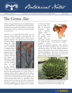

© Copyright 2026